Embed Size (px)
DESCRIPTION
kulit
Citation preview

JOURNAL READINGICHTYOSIS ASSOCIATED WITH WIDESPREAD TINEA
CORPORIS : REPORT OF THREE CASES
Camila Fernanda Novak Pinheiro de Freitas1Hanae Rafaela Fontana3
Mariana Hammerschmidt1Fabiane Mulinari-Brenner2
Arthur Conelian Gentili4DOI: http://dx.doi.org/10.1590/abd1806-4841.20131854
Pembimbing :Dr. Bowo Wahyudi, Sp.KK
Disusun Oleh :Vicca Selsiana 2010730109
INTRODUCTION
Ichthyoses are a common group of keratinization disorders
INTRODUCTION
Several forms of ichthyosis are classified according to inheritance, clinical
appearance, pathological features and systemic disorders
INTRODUCTION
Patients with ichthyosis demonstrate increased cell turnover, stratum corneum
thickening and sebaceous and sweat glands functional changes, which seem to
favor dermatophyte infectio
INTRODUCTION
One of the most prevalent dermatophytes is Trichophyton rubrum, with universal
occurrence.
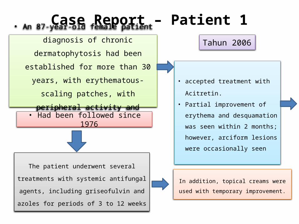
Case Report – Patient 1
• An 87-year-old female patient diagnosis of
chronic dermatophytosis had been
established for more than 30 years, with
erythematous-scaling patches, with
peripheral activity and central clearing.
The patient underwent several treatments
with systemic antifungal agents, including
griseofulvin and azoles for periods of 3 to 12
weeks
Tahun 2006
• accepted treatment with
Acitretin.
• Partial improvement of
erythema and desquamation
was seen within 2 months;
however, arciform lesions
were occasionally seen
• Had been followed since 1976
In addition, topical creams were
used with temporary improvement.

Case Report – Patient 1
Sept 2011
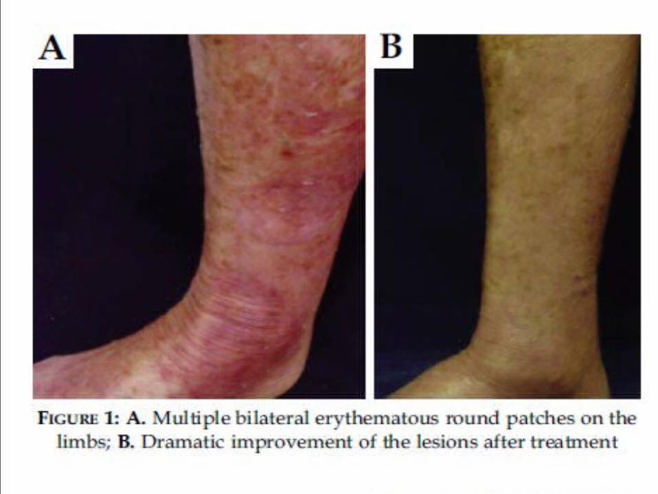
she showed signs of clinical deterioration with
widespread lamellar scaling on the limbs and trunk,
ectropion and multiple bilateral erythematous round
patches on the lower limbs. (Figure 1A)
Direct examination demonstrated hyaline septate hyphae with
a positive culture for T. rubrum. Association with
dermatophytosis was diagnosed.
Thirty days after prescription of terbinafine 250 mg qd the
patient showed dramatic improvement of the lesions. (Figure
1B)
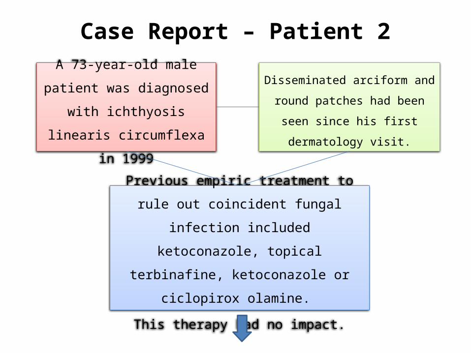
Case Report – Patient 2
A 73-year-old male patient was
diagnosed with ichthyosis
linearis circumflexa in 1999
Previous empiric treatment to rule out
coincident fungal infection included
ketoconazole, topical terbinafine,
ketoconazole or ciclopirox olamine.
This therapy had no impact.
Disseminated arciform and
round patches had been seen
since his first dermatology visit.

Case Report – Patient 2
During one of his regular visits in
July 2011, he presented ill-defined
erythematous scaly papules and
plaques on the trunk and upper
limbs (Figure 2A).
A mycological examination
demonstrated septate hyaline hyphae,
and a positive culture for T. rubrum.
Onycholysis, distal leukonychia,
subungual hyperkeratosis and
onychorrexis on the toenails were also
evident
Case Report – Patient 2
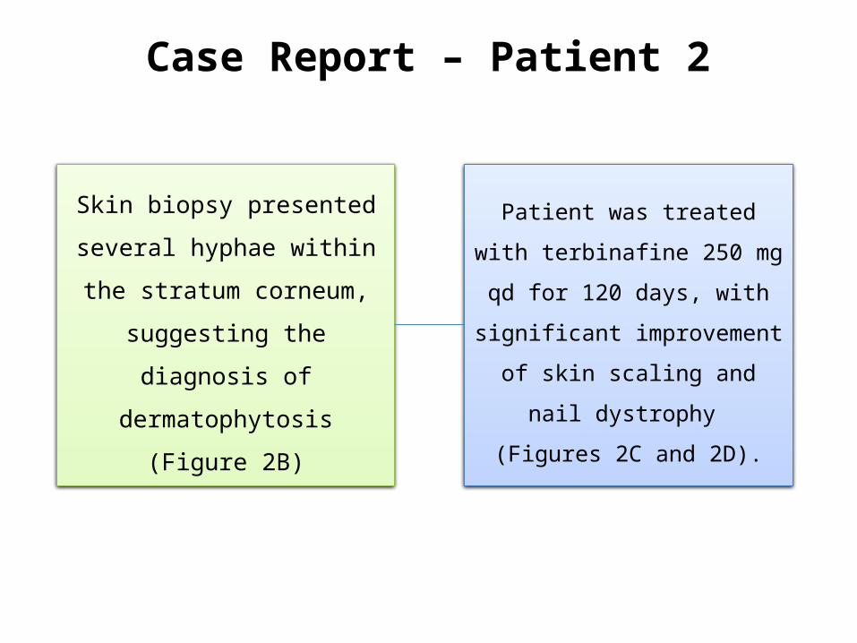
Patient was treated with
terbinafine 250 mg qd for 120
days, with significant
improvement of skin scaling
and nail dystrophy
(Figures 2C and 2D).
Skin biopsy presented several
hyphae within the stratum
corneum, suggesting the
diagnosis of dermatophytosis
(Figure 2B)

Case Report – Patient 3
A 27-year-old female patient, with
Sjogren Larsson Syndrome
(congenital ichthyosis associated to
spasticity), had been followed since
2003.
Tahun 2008
she had onycholysis and
subungual hyperkeratosis
on toenails.
She was on Acitretin since 2004
and presented with sudden
worsening.
She was on ciclopirox olamine
nail lacquer and ketoconazole
cream for more than 2 years
with mild improvement.
Case Report – Patient 3
Tahun 2011
her mother reported severe worsening of
ichthyosis, despite regular use of Acitretin.
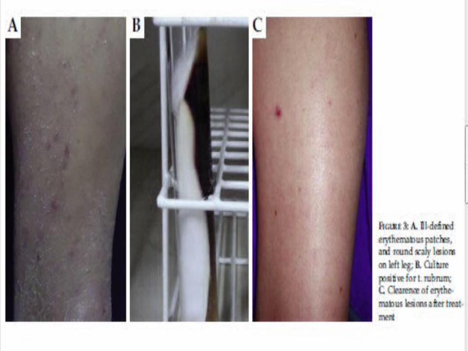
Clinical examination demonstrated lamellar
desquamation of the lower limbs, ill-defined
erythematous patches, and round scaly lesions
on trunk and left leg. (Figure 3A).
Direct mycological examination showed hyaline
septate hyphae and the culture was positive for T.
rubrum (Figure 3B).
Case Report – Patient 3
Tahun 2011
Terbinafine 250 mg qd for 30 days led to
clearance of the erythematous lesions, slight
scaling persisted due to baseline ichthyosis
(Figure 3C).
Skin biopsy demonstrated several hyphae within
the stratum corneum.
Discussion
Several factors may lead to chronic dermatophytosis
in patients with ichthyosis: defects of the skin barrier;
defective cell-mediated immunity, primarily
responsible for immunity against T. rubrum, in some
cases with atopic background; and finally, delayed
keratin scaling, facilitating persistence of fungal
infection.

Lanjutan…
The three cases investigated had positive
cultures for T. rubrum, which is the
mostprevalent pathogen in fungal cultures in
Brazil, accounting for 37.4% to 58.3% of
superficial fungal infections in several studies in
the Brazilian population.

Lanjutan…
Typically, the clinical appearance of
dermatophyte infections is characterized by
round patches with erythematous scaling
circinate edges, due to the combination of
keratin destruction and host inflammatory
response.
Lanjutan…
Fungicidal activity of terbinafine is higher
and initiates faster than that of azoles.
Lanjutan…
Dermatologists should be aware that associated
dermatophytosis in patients with ichthyosis may
present as clinical worsening of baseline scaling,
sometimes resistant to usual therapies, such as
acitretin.

Lanjutan…
Allylamines seem to elicit a better clinical
response from these patients


![Journal Reading[1]](https://img.pdfslide.net/doc/110x75/5695d3251a28ab9b029cfc6d/journal-reading1-56dbaa4d014b7.jpg)