Embed Size (px)
Citation preview
King’s Research Portal
DOI:10.1111/bph.13388
Document VersionPublisher's PDF, also known as Version of record
Link to publication record in King's Research Portal
Citation for published version (APA):Xiao, F., Ma, L., Zhao, M., Smith, R. A., Huang, G., Jones, P. M., ... Lombardi, G. (2016). APT070 (Mirococept),a membrane-localizing C3 convertase inhibitor, attenuates early human islet allograft damage in vitro and in vivoin a humanized mouse model. British Journal of Pharmacology, 173(3), 575-587. 10.1111/bph.13388
Citing this paperPlease note that where the full-text provided on King's Research Portal is the Author Accepted Manuscript or Post-Print version this maydiffer from the final Published version. If citing, it is advised that you check and use the publisher's definitive version for pagination,volume/issue, and date of publication details. And where the final published version is provided on the Research Portal, if citing you areagain advised to check the publisher's website for any subsequent corrections.
General rightsCopyright and moral rights for the publications made accessible in the Research Portal are retained by the authors and/or other copyrightowners and it is a condition of accessing publications that users recognize and abide by the legal requirements associated with these rights.
•Users may download and print one copy of any publication from the Research Portal for the purpose of private study or research.•You may not further distribute the material or use it for any profit-making activity or commercial gain•You may freely distribute the URL identifying the publication in the Research Portal
Take down policyIf you believe that this document breaches copyright please contact [email protected] providing details, and we will remove access tothe work immediately and investigate your claim.
Download date: 18. Feb. 2017
RESEARCH PAPER
APT070 (mirococept), amembrane-localizing C3convertase inhibitor,attenuates early human isletallograft damage in vitro andin vivo in a humanized mousemodelFang Xiao1, Liang Ma1, Min Zhao2, Richard A Smith1, Guocai Huang2,Peter M Jones3, Shanta Persaud3, Attilio Pingitore3, Anthony Dorling1,Robert Lechler1 and Giovanna Lombardi1
1Division of Transplantation Immunology and Mucosal Biology, MRC Centre for Transplantation,
King's College London, 5th Floor Tower Wing, Guy's Hospital, Great Maze Pond, London SE1 9RT,
UK, 2Department of Diabetes & Endocrinology, King's College London, 2nd Floor, The Rayne
Institute, 123 Coldharbour Lane, London SE5 9NU, UK, and 3Division of Diabetes & Nutritional
Sciences, School of Medicine, King's College London, Guy's Hospital, Great Maze Pond, London SE1
9RT, UK
CorrespondenceDr. Fang Xiao or Professor GiovannaLombardi, Division ofTransplantation Immunology andMucosal Biology, MRC Centre forTransplantation, King's CollegeLondon, 5th Floor Tower Wing, Guy'sHospital, Great Maze Pond, LondonSE1 9RT, UK.E-mail: [email protected];giovanna.lombardi@kcl.ac.uk---------------------------------------------------------
Received17 June 2015Revised3 October 2015Accepted3 November 2015
BACKGROUND AND PURPOSEA major obstacle to islet cell transplantation is the early loss of transplanted islets resulting from the instant blood-mediated in-flammation reaction (IBMIR). The activation of complement pathways plays a central role in IBMIR. The aim of this study was totest the inhibitory effect of “painting” human islets with APT070, a membrane-localizing C3 convertase inhibitor, on inflamma-tion evoked by exposure to human serum in vitro and by transplantation in vivo in a humanized diabetic mouse model.
EXPERIMENTAL APPROACHIn vitro, human islets pre-incubated with APT070 were exposed to allogeneic whole blood. In vivo, similarly treated islets weretransplanted underneath the kidney capsule of streptozotocin-induced diabetic NOD-SCID IL2rγ�/� mice that had beenreconstituted with human CD34+ stem cells. Complement activation and islet hormone content were assayed using enzyme-linked immunosorbent assays. Supernatants and sera were assayed for cytokines using cytometric beads array. Morphology of theislets incubated with human serum in vitro and in graft-bearing kidney were evaluated using immunofluorescence staining.
KEY RESULTSPre-incubation with APT070 decreased C-peptide release and iC3b production in vitro, with diminished deposition of C4d andC5b-9 in islets embedded in blood clots. In vivo, the APT070-treated islets maintained intact structure and showed less infiltrationof inflammatory cells than untreated islets. The pretreatments also significantly reduced pro-inflammatory cytokines in superna-tants and sera.
CONCLUSIONS AND IMPLICATIONSPre-treatment of islets with APT070 could reduce intra-islet inflammation with accompanying preservation of insulin secretion bybeta cells. APT070 could be as a potential therapeutic tool in islet transplantation.
AbbreviationsCR1, complement receptor 1; hu-NSG, humanized NSG; IBMIR, instant blood-mediated inflammation reaction; iC3b,inactivated C3b; IEQs, islet equivalents; LDH, lactate dehydrogenase; NSG, non-obese diabetic/severe combinedimmunodeficiency/interleukin-2 receptor γ-chain knockout; sCR1, soluble complement receptor 1 (sCR1); UCB, umbilicalcord blood
BJP British Journal ofPharmacology
DOI:10.1111/bph.13388www.brjpharmacol.org
© 2015 The Authors. British Journal of Pharmacologypublished by John Wiley & Sons Ltd on behalf of British Pharmacological Society.
British Journal of Pharmacology (2016) 173 575–587 575
This is an open access article under the terms of the Creative Commons Attribution License, which permits use, distribution and reproduction in any medium,provided the original work is properly cited.
Introduction
Type 1 diabetes (T1D) is characterized by permanent destruc-tion of insulin-producing beta cells in the pancreatic islets.Transplantation of islets of Langerhans via intra-portal injec-tion is a promising treatment procedure for patients with se-vere T1D (Robertson, 2010). However, despite recentprogress, the procedure is associated with massive tissue losscaused by an inflammatory reaction, referred to as the instantblood-mediated inflammatory reaction (IBMIR) (Nilssonet al., 2011). It is believed that complement attack is a majorcause of the damage to the islets that occurs during the IBMIR(Tjernberg et al., 2008) and the reaction involves rapid activa-tion of the coagulation and complement system, activationand binding of platelets to the islet surface, and infiltrationof leukocytes into the islets (Nilsson et al., 2011). The comple-ment cascade therefore makes a logical therapeutic target forthe reduction of graft rejection (Farrar and Sacks, 2014).
The central step in the complement cascade is the cleav-age of C3 to C3b by C3 convertase. This leads ultimately tothe assembly of the membrane attack complex (MAC) andthe release of soluble C3a and C5a, which in turn activateand recruit inflammatory cells (Sacks et al., 2009). Once C3is deposited covalently as C3b on a membrane, it has twofates. The first is an amplification step, in which more C3 iscleaved and bound to the membrane. This process involvesa further complement protein, factor B. When factor B bindsto C3b, it is activated by the complement enzyme factor Dand forms the C3 convertase enzyme C3bBb. This enzymecleaves more C3, causing manymore C3b molecules to be de-posited on the membrane. The second possible fate of boundC3b is catabolism to iC3b (inactivated C3b) and C3dg, creat-ing adhesive contact with corresponding receptors onleucocytes through the complement receptors, CR1-4, partic-ularly CR3 and CRIg (Walport, 2001a; Walport, 2001b). Thisstep is mediated by regulators that inactivate complementbinding to C3b thus promoting its removal and degradationfrom the enzyme complexes that convert C3 and C5 intotheir active forms. The regulators exist as membrane bound(e.g. CD35, CD46 and CD55) and soluble (e.g. Factor H) pro-teins. APT070, also known as Mirococept, is a modified frag-ment of the complement receptor 1 (CR1, CD35)(Mossakowska et al., 1999; Smith, 2002). It consists of the first3 consensus domains of the human CR1 and a membrane-interacting synthetic peptide, which mediates binding tophospholipids on the cell surface and therefore protects thecell against complement activation (Smith and Smith, 2001;Smith, 2002). APT070 showed inhibition of complementand neutrophil activation during cardiopulmonary bypass
in man in vitro (De Silva et al., 2006), and of the inflammatoryresponses that follow intestinal (Souza et al., 2005) and renal(Patel et al., 2006) ischaemia and reperfusion injury in ratmodels. These studies have prompted clinical investigationof the utility of APT070 in man for ischaemia-reperfusion in-jury during renal transplantation (Smith, 2007). These prop-erties suggested the possible use of APT070 in protectinghuman islet transplantation.
The development ofmice that are ‘humanised’ by engraft-ment of human tissues, haematopoietic stem cells orperipheral-blood mononuclear cells (PBMC), provides an op-portunity to study human biological processes in vivo thatwould otherwise not be possible (Shultz et al., 2007). We haverecently described human islet allograft rejection in theCD34+ stem cell-reconstituted humanized mouse model(Xiao et al., 2014). Although islets were placed underneaththe kidney capsule, rejection was associated with C3b deposi-tion and we suggested that complement may be involved inthe recruitment and activation of innate inflammatory cells.We therefore hypothesised that inhibition of complement ac-tivation might enhance islet survival. The present studyaimed to investigate whether APT070 could protect humanislets from complement-mediated destruction in vitro, and at-tenuate the immune response to human islet allograft in vivoin the humanized mouse model (Xiao et al., 2014).
Methods
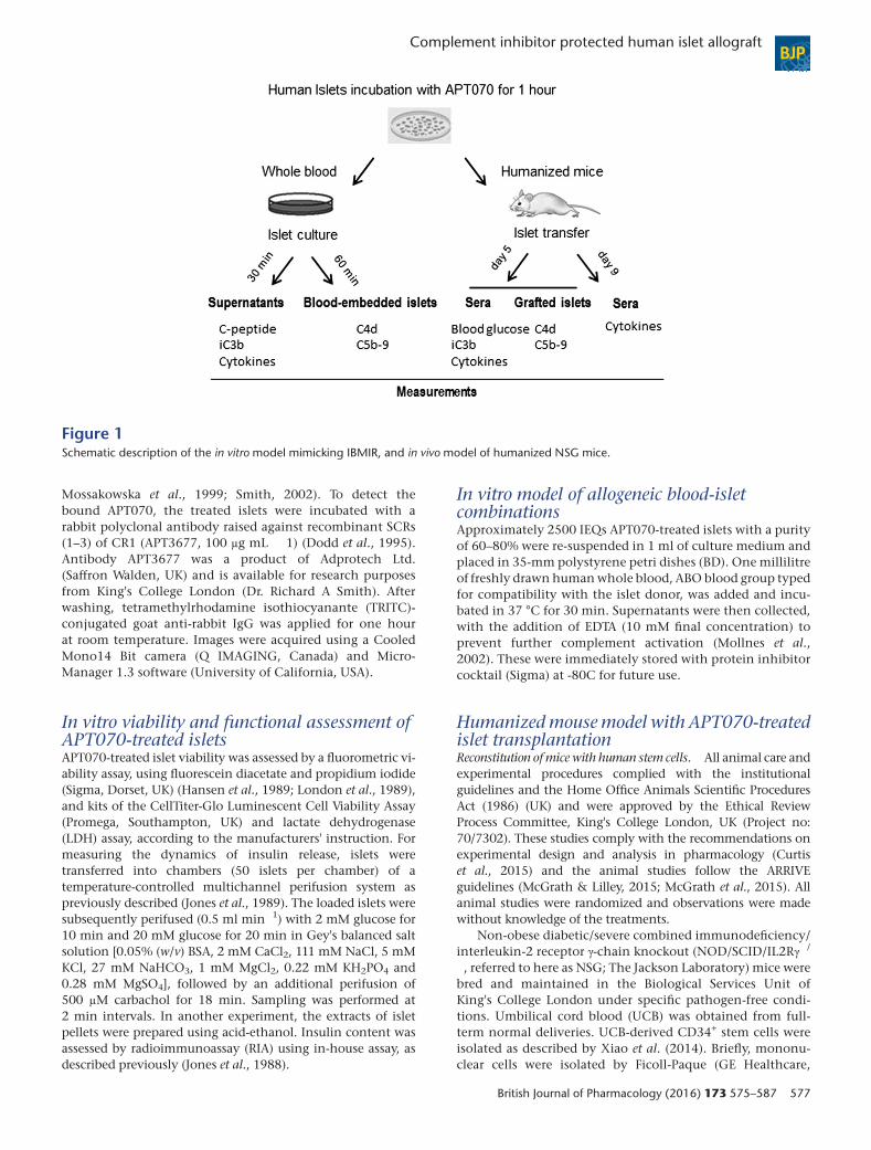
Preparation of human isletsThe procurement and use of all human tissues were withsigned informed consent, in accordance with the Declarationof Helsinki and approval from the Institutional Review Boardof King's College London. Human islets were obtained fromKing's Cell Isolation Unit, London, UK. Freshly isolated hu-man islets were incubated with serial dilutions ofmembrane-localizing complement regulator APT070 [0.4–1.6 μM; 3000–4000 human islet equivalents (IEQ) mL � 1],or its control molecule APT154 at the equivalent molar con-centration, in CMRL 1066 culture medium (Invitrogen) con-taining 2.5% human albumin at 37 °C in an atmosphere of95% air 5% CO2 for one hour (Figure 1). The recombinantnon-tailed CR1 fragment APT154 possesses chemical andcomplement inhibitory properties identical to those ofAPT070, but is untagged and therefore does not contain themembrane-binding moiety (Banz et al., 2007). This controlmolecule was prepared using the cloning and expressionstrategy as previously described (Dodd et al., 1995;
TARGETS
GPCRs
C3a receptor
LIGANDS
C3a, complement component C3a
Carbachol
Tables of Links
These Tables list key protein targets and ligands in this article which are hyperlinked to corresponding entries in http://www.guidetopharmacology.org, thecommon portal for data from the IUPHAR/BPS Guide to PHARMACOLOGY (Pawson et al., 2014) and are permanently archived in the Concise Guide toPHARMACOLOGY 2013/14 (Alexander et al., 2013).
BJP F Xiao et al.
576 British Journal of Pharmacology (2016) 173 575–587
Mossakowska et al., 1999; Smith, 2002). To detect thebound APT070, the treated islets were incubated with arabbit polyclonal antibody raised against recombinant SCRs(1–3) of CR1 (APT3677, 100 μg mL � 1) (Dodd et al., 1995).Antibody APT3677 was a product of Adprotech Ltd.(Saffron Walden, UK) and is available for research purposesfrom King's College London (Dr. Richard A Smith). Afterwashing, tetramethylrhodamine isothiocyanante (TRITC)-conjugated goat anti-rabbit IgG was applied for one hourat room temperature. Images were acquired using a CooledMono14 Bit camera (Q IMAGING, Canada) and Micro-Manager 1.3 software (University of California, USA).
In vitro viability and functional assessment ofAPT070-treated isletsAPT070-treated islet viability was assessed by a fluorometric vi-ability assay, using fluorescein diacetate and propidium iodide(Sigma, Dorset, UK) (Hansen et al., 1989; London et al., 1989),and kits of the CellTiter-Glo Luminescent Cell Viability Assay(Promega, Southampton, UK) and lactate dehydrogenase(LDH) assay, according to the manufacturers' instruction. Formeasuring the dynamics of insulin release, islets weretransferred into chambers (50 islets per chamber) of atemperature-controlled multichannel perifusion system aspreviously described (Jones et al., 1989). The loaded islets weresubsequently perifused (0.5 ml min�1) with 2 mM glucose for10 min and 20 mM glucose for 20 min in Gey's balanced saltsolution [0.05% (w/v) BSA, 2 mM CaCl2, 111 mM NaCl, 5 mMKCl, 27 mM NaHCO3, 1 mM MgCl2, 0.22 mM KH2PO4 and0.28 mM MgSO4], followed by an additional perifusion of500 μM carbachol for 18 min. Sampling was performed at2 min intervals. In another experiment, the extracts of isletpellets were prepared using acid-ethanol. Insulin content wasassessed by radioimmunoassay (RIA) using in-house assay, asdescribed previously (Jones et al., 1988).
In vitro model of allogeneic blood-isletcombinationsApproximately 2500 IEQs APT070-treated islets with a purityof 60–80% were re-suspended in 1 ml of culture medium andplaced in 35-mm polystyrene petri dishes (BD). One millilitreof freshly drawn humanwhole blood, ABO blood group typedfor compatibility with the islet donor, was added and incu-bated in 37 °C for 30 min. Supernatants were then collected,with the addition of EDTA (10 mM final concentration) toprevent further complement activation (Mollnes et al.,2002). These were immediately stored with protein inhibitorcocktail (Sigma) at -80C for future use.
Humanized mouse model with APT070-treatedislet transplantationReconstitution ofmice with human stem cells. All animal care andexperimental procedures complied with the institutionalguidelines and the Home Office Animals Scientific ProceduresAct (1986) (UK) and were approved by the Ethical ReviewProcess Committee, King's College London, UK (Project no:70/7302). These studies comply with the recommendations onexperimental design and analysis in pharmacology (Curtiset al., 2015) and the animal studies follow the ARRIVEguidelines (McGrath & Lilley, 2015; McGrath et al., 2015). Allanimal studies were randomized and observations were madewithout knowledge of the treatments.
Non-obese diabetic/severe combined immunodeficiency/interleukin-2 receptor γ-chain knockout (NOD/SCID/IL2Rγ�/
�, referred to here as NSG; The Jackson Laboratory) mice werebred and maintained in the Biological Services Unit ofKing's College London under specific pathogen-free condi-tions. Umbilical cord blood (UCB) was obtained from full-term normal deliveries. UCB-derived CD34+ stem cells wereisolated as described by Xiao et al. (2014). Briefly, mononu-clear cells were isolated by Ficoll-Paque (GE Healthcare,
Figure 1Schematic description of the in vitro model mimicking IBMIR, and in vivo model of humanized NSG mice.
Complement inhibitor protected human islet allograft BJP
British Journal of Pharmacology (2016) 173 575–587 577
Hatfield, UK) gradient separation and enriched for CD34+
cells using positive isolation kit according to the manufacturer'sinstructions (Miltenyi Biotech, Surrey, UK).Mice (6–8weeks old)were gamma-irradiated (240 cGy) and then intravenouslyinjected with 2 x 10⁵ CD34+ stem cells (referred to ashu-NSG mice).
Flow cytometric analysis of human haematopoietic engraftment. Tailbleeding was performed in hu-NSG mice 12–16 weeks afterinjection of CD34+ cells. Blood cells were prepared for flowcytometry as previously described (Xiao et al., 2014).Fluorochrome-coupled antibodies specific for human CD45were used for detection of human leukocytes. Flow cytometricdata were acquired using a FACS Calibur (BD Biosciences, SanJose, CA, USA) and analysed using FlowJo 7.5 software(TreeStar Inc.).
Islet transplantation into diabetic ‘humanized' NSG mice. NSGand hu-NSG mice were rendered diabetic (blood glucoselevel ≥ 20 mM) by a single i.p. injection of streptozotocin(180 mg kg�1) and transplanted with human APT070-treated islets (IEQs, 3000–4000) under the left kidneycapsule as previously published (Xiao et al., 2014). Duringthe surgery, the recipient animals were anaesthetized using3% isoflurane in a stream of 100% O2 (2 l min�1), andwere maintained using 1.5–2.0% isoflurane in a stream of100% O2 (2 l min�1). The animals were placed on a heatpad to keep body temperature at 37 °C throughout theprocedure. Blood glucose concentration of recipient micewas monitored every 24 hours at the first week and twice aweek thereafter using a blood glucose sensor (AbbottDiabetes Care Ltd., Witney, Oxon, UK). Reversal ofhyperglycaemia was defined as non-fasting blood glucoseconcentration ≤ 13.8 mM (King et al., 2008). NSG mice withsuccessful transplants were subjected to unilateral leftnephrectomy to evaluate a return to hyperglycemic state.For islet-transplanted diabetic hu-NSG mice, sera werecollected by tail sampling at day 5 and day 9 aftertransplantation, and kept at �80 °C prior to use for furthertest. The animals were sedated with CO2 and killed bydecapitation at the end of experiments.
Enzyme-linked immunosorbent assay (ELISA) for human insulin,C-peptide and iC3b. Supernatant insulin and C-peptideconcentrations were assayed using ELISA kits (Millipore,Watford, UK). ELISA kit for human iC3b (PathwayDiagnostics Ltd., Surrey, UK) was used to determine levels ofsoluble complement activation product iC3b in supernatantand sera, according to the manufacturer's instructions. Allsamples were tested in triplicate in the assays.
Cytokine detection (beads array)Human cytokines in supernatants and sera were assayedusing a human Th1/Th2 11plex kit (eBiosciences) accordingto the manufacturer's protocol. Data acquisition was per-formed on a FACS Calibur (BD Biosciences) and analysedusing The FlowCytomix Pro 3.0 Software from eBioscience(Xiao et al., 2014).
Histology analysisDeposition of activated complements was examined in theislets embedded in blood clots after 60 min incubationwith blood, and the harvested graft-bearing kidney at day5 after transplantation. The samples were fixed in 10%buffered formalin, and embedded in paraffin. Sections(5 μm) were processed with double immunofluorescencestaining. After antigen retrieval by microwaving for5 min in 0.01 M citrate buffer (pH 6.0), sections wereblocked with 10% goat normal serum for 30 min and thenincubated overnight at 4 °C with primary antibodies: rab-bit anti-human insulin (1:300; Bioss, polyclonal), mouseanti-human C4d (1:25; Abcam, clone LP69), mouse anti-human C5b-9 (1:100; Abcam, clone aE11), mouse anti-human CD11b (1:50; eBiosciences, clone ICRF44) andmouse anti-human CD66b (1:50; BioLegend, cloneG10F5). Secondary antibodies FITC-conjugated goat anti-mouse IgG and TRITC-conjugated goat anti-rabbit IgG(both Sigma) were applied for 2 hours at room temperature(Xiao et al., 2014). Negative controls with non-specific IgGwere processed in parallel. Images were acquired using aNikon Eclipse Ti-E Inverted microscopy with 4 x GaAsPDetectors and A1R Si Confocal system with a 60x objectivelens and analysed by NIS-Elements Advanced Researchimaging analysis software (Nikon, Surrey, UK). Where theinfiltrated cells were quantified, each sample from an indi-vidual animal (n = 5 animals) was assessed and analysedby using ImageJ software. The deposition of complementwas quantified by measuring the mean fluorescence inten-sity (MFI) in the region of interest area.
Data analysisData are presented as means ± SD. All statistical analysis wasperformed using the two-tailed Student's t-test or two-wayANOVA analysis, followed by Bonferroni post hoc tests. Pvalues <0.05 were considered significant.
MaterialsAPT070 and APT 154 were supplied by Adprotech Ltd.(Saffron Walden, UK); carbachol and streptozotocinwere sup-plied by Sigma (Dorset, UK).
Results
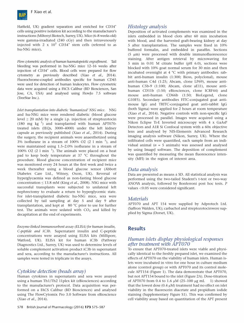
Human islets display physiological responsesafter treatment with APT070To ensure that APT070-treated islets were viable and physi-cally identical to the freshly prepared islet, we examined theeffects of APT070 on the viability of human islets. Human is-lets were incubated in vitro for one hour in culture mediumalone (control group) or with APT070 and its control mole-cule APT154 (Figure 1). The data demonstrate that APT070,but not APT154 bound to the islet (Figure 2A). Dose-titrationof APT070 from 0.4 to 1.6 μM (25–100 μg mL � 1) showedthat the lowest dose (0.4 μM) treatment had no effect on isletviability in the fluorescein diacetate and propidium iodidestaining (Supplementary Figure S1). This was confirmed bycell viability assay based on quantitation of the APT present
BJP F Xiao et al.
578 British Journal of Pharmacology (2016) 173 575–587
(Figure 2B) and the measurement of intracellular content ofinsulin (Figure 2C). Therefore APT070 was used at a concen-tration of 0.4 μM (25 μg mL � 1) in further studies. It shouldbe noted that a concentration of 10 μg mL � 1 of APT070has been used in perfusion experiments in human renaltransplantation without apparent adverse effects (Smithet al., 2007).
To evaluate islet function after incubation with APT070,measurements of the rate and pattern of insulin secretionfrom perifused islets (Figure 2D) confirmed that APT070 pre-treatment did not inhibit insulin secretory responses to glu-cose, nor to the receptor-mediated agonist, carbachol.Expressing the cumulative total insulin secretion during theperifusion as area under the curve (AUC) showed that
Figure 2Assessment of islet viability and secretion function. (A) Binding of APT070 (0.4 μM) to human islets one hour after incubation. Positive bind-ing is shown in red. Amplifier power: × 60. (B) Islet viability assay. Luminescent outputs, which correlated with islet cell numbers, were mea-sured in APT070 treated and control islets. Values represent the mean ± SD, n = 6 (wells of 96-well plate). (C) Intracellular insulin content inAPT070-treated and control islets. The data are expressed as means ± SD, n = 6. (D, E) The dynamics of insulin release. Data are shown asmeans ± SD, n = 8 perifusion channels. RLU: relative luminescence units. AUC: area under curve. Control islets received no treatment. NS: nosignificant difference.
Complement inhibitor protected human islet allograft BJP
British Journal of Pharmacology (2016) 173 575–587 579
APT070 pre-treatment had no significant effects on insulinsecretion (Figure 2E), confirming that the β-cells remainedfunctionally viable after treatment.
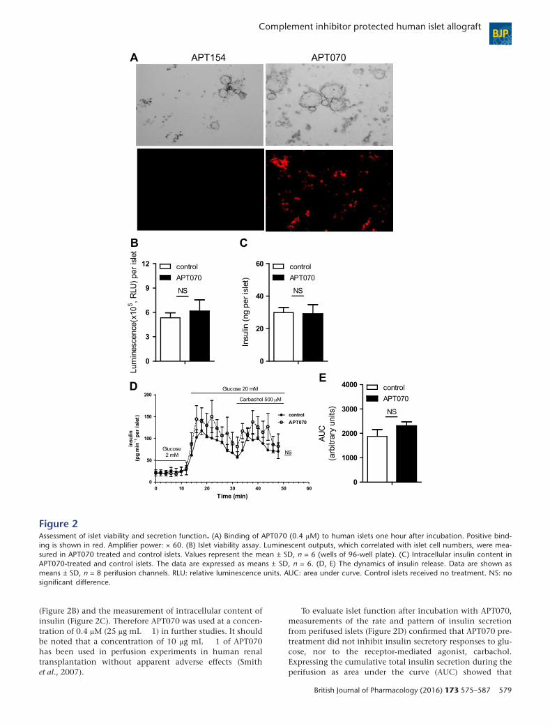
Pre-incubation of human islet with APT070prevents the production of activatedcomplement in vitro and in vivoTo test whether APT070 has an inhibitory effect on comple-ment activation, islets were exposed to whole blood for 30mi-nutes. The level of iC3b was significantly decreased in thesupernatant of APT070 treated-islets, compared with controlgroups (n = 6 for each group) (Figure 3A).
To further assess the in vitro inhibitory effects of APT070on complement activation, C4d deposition, as an indicatorof complement activation, and C5b-9, which may causecell lysis in the blood-exposed islets, were examined. Pre-treatment of islets with APT070 almost abolished deposi-tion of both C4d and C5b-9 in the islet exposed to bloodfor 60 minutes (Figure 3B, C).
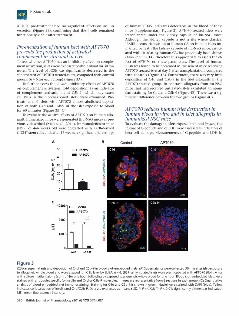
To evaluate the in vivo effects of APT070 on human allo-graft, humanized mice were generated (hu-NSG mice) as pre-viously described (Xiao et al., 2014). Immunodeficient mice(NSG) of 4–6 weeks old were engrafted with UCB-derivedCD34+ stem cells and, after 16 weeks, a significant percentage
of human CD45+ cells was detectable in the blood of thesemice (Supplementary Figure 2). APT070-treated islets weretransplanted under the kidney capsule of hu-NSG mice.Although the kidney capsule is not a site where classicalIBMIR occurs, deposition of human C3 on human islets im-planted beneath the kidney capsule of hu-NSG mice, associ-ated with circulating human C3, has previously been shown(Xiao et al., 2014), therefore it is appropriate to assess the ef-fect of APT070 on these parameters. The level of humaniC3b was found to be decreased in the sera of mice receivingAPT070 treated-islet at day 5 after transplantation, comparedwith controls (Figure 4A). Furthermore, there was very littledeposition of C4d and C5b-9 in the islet allografts in theAPT070 treated group. In contrast, allografts from hu-NSGmice that had received untreated-islets exhibited an abun-dant staining for C4d and C5b-9 (Figure 4B). There was a sig-nificant difference between the two groups (Figure 4C).
APT070 reduces human islet destruction inhuman blood in vitro and in islet allografts inhumanized NSG miceTo evaluate the damage in islets exposed to blood in vitro, therelease of C-peptide and of LDH were assessed as indicators ofbeta cell damage. Measurements of C-peptide and LDH in
Figure 3iC3b in supernatants and deposition of C4d and C5b-9 in blood clot-embedded islets. (A) Supernatants were collected 30 min after islet exposureto allogeneic whole blood and were assayed for iC3b level by ELISA, n = 6. (B) Freshly isolated islets were pre-incubated with APT070 (0.4 μM) orwith culture medium alone (control) for one hour, following by exposed to allogeneic whole blood for one hour. Blood clot-embedded islets werestained with antibodies specific for insulin and C4d or C5b-9 molecules. Images are representative from 6 sections in each group. (C) Quantitativeanalysis of blood-embedded islet immunostaining. Staining for C4d and C5b-9 is shown in green. Nuclei were stained with DAPI (blue). Yellowindicates co-localization of insulin and C4d/C5b-9. Data are expressed as means ± SD. *: P< 0.05; **: P< 0.01; significantly different as indicated.MFI: mean fluorescence intensity.
BJP F Xiao et al.
580 British Journal of Pharmacology (2016) 173 575–587
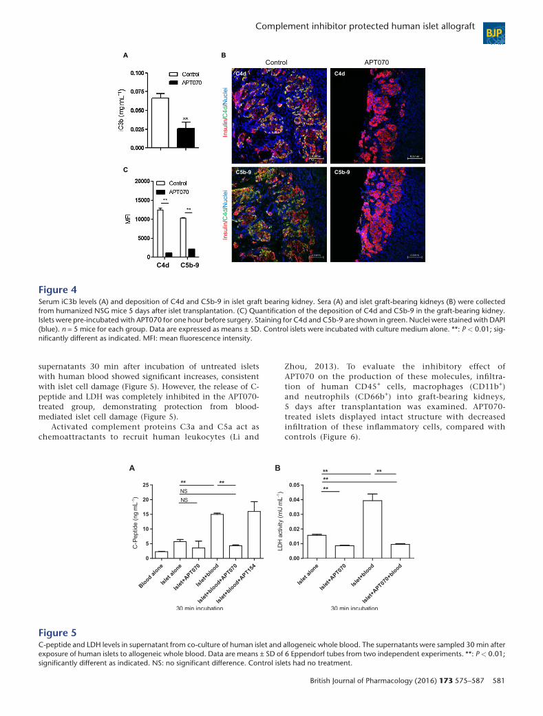
supernatants 30 min after incubation of untreated isletswith human blood showed significant increases, consistentwith islet cell damage (Figure 5). However, the release of C-peptide and LDH was completely inhibited in the APT070-treated group, demonstrating protection from blood-mediated islet cell damage (Figure 5).
Activated complement proteins C3a and C5a act aschemoattractants to recruit human leukocytes (Li and
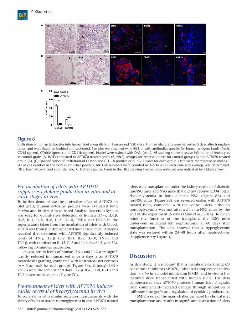
Zhou, 2013). To evaluate the inhibitory effect ofAPT070 on the production of these molecules, infiltra-tion of human CD45+ cells, macrophages (CD11b+)and neutrophils (CD66b+) into graft-bearing kidneys,5 days after transplantation was examined. APT070-treated islets displayed intact structure with decreasedinfiltration of these inflammatory cells, compared withcontrols (Figure 6).
Figure 4Serum iC3b levels (A) and deposition of C4d and C5b-9 in islet graft bearing kidney. Sera (A) and islet graft-bearing kidneys (B) were collectedfrom humanized NSG mice 5 days after islet transplantation. (C) Quantification of the deposition of C4d and C5b-9 in the graft-bearing kidney.Islets were pre-incubated with APT070 for one hour before surgery. Staining for C4d and C5b-9 are shown in green. Nuclei were stained with DAPI(blue). n = 5 mice for each group. Data are expressed as means ± SD. Control islets were incubated with culture medium alone. **: P < 0.01; sig-nificantly different as indicated. MFI: mean fluorescence intensity.
Figure 5C-peptide and LDH levels in supernatant from co-culture of human islet and allogeneic whole blood. The supernatants were sampled 30 min afterexposure of human islets to allogeneic whole blood. Data are means ± SD of 6 Eppendorf tubes from two independent experiments. **: P < 0.01;significantly different as indicated. NS: no significant difference. Control islets had no treatment.
Complement inhibitor protected human islet allograft BJP
British Journal of Pharmacology (2016) 173 575–587 581
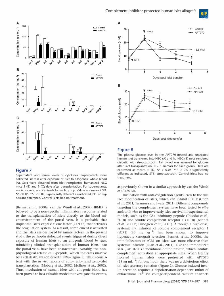
Pre-incubation of islets with APT070suppresses cytokine production in vitro and atearly stages in vivoTo further demonstrate the protective effect of APT070 onislet graft, human cytokine profiles were evaluated bothin vitro and in vivo. A bead based Analyte Detection Systemwas used for quantitative detection of human IFN-γ, IL-1β,IL-2, IL-4, IL-5, IL-6, IL-8, IL-10, TNF-α and TNF-β in thesupernatants taken from the incubation of islets with blood,and in sera from islet-transplanted humanized mice. Analysisrevealed that treatment with APT070 significantly reducedlevels of IFN-γ, IL-1β, IL-2, IL-4, IL-5, IL-10, TNF-α andTNF-β, with no effect on IL-12, IL-8 and IL-6 (n = 6) (Figure 7A),following 30 minutes incubation.
In vivo, serum levels of human IFN-γ and IL-2 were signif-icantly reduced in humanized mice 5 days after APT070treated-islet grafting, compared with untreated-islet controls(n = 5 animals for each group) (Figure 7B), although IFN-γvalues were the same after 9 days. IL-1β, IL-6, IL-8, IL-10 andTNF-α were undetectable (Figure 7C).
Pre-treatment of islets with APT070 inducesearlier reversal of hyperglycaemia in vivoTo correlate in vitro insulin secretion measurements with theability of islets to restore normoglycemia in vivo, APT070-treated
islets were transplanted under the kidney capsule of diabetichu-NSG mice and NSG mice that did not receive CD34+ cells.Hyperglycaemia in both diabetic NSG (Figure 8A) andhu-NSG mice (Figure 8B) was reversed earlier with APT070treated islets, compared with the control islets, althoughnormoglycaemia was not attained in hu-NSG mice by theend of the experiment (5 days) (Xiao et al., 2014). To deter-mine the function of the transplant, the NSG miceunderwent unilateral left nephrectomy at 60 days aftertransplantation. The data showed that a hyperglycemicstate was restored within 24–48 hours after nephrectomy(Supplementary Figure 3).
DiscussionIn this study, it was found that a membrane-localizing C3convertase inhibitor (APT070) inhibited complement activa-tion in vitro in a model mimicking IBMIR, and in vivo in hu-manized mice transplanted with human islets. The datademonstrated that APT070 protects human islet allograftsfrom complement-mediated damage through inhibition ofinfiltrates into grafts and regulation of cytokine production.
IBMIR is one of the main challenges faced by clinical islettransplantation and results in significant destruction of islets
Figure 6Infiltration of human leukocytes into human islet allografts from humanized NSG mice. Human islet grafts were harvested 5 days after transplan-tation and were fixed, embedded and sectioned. Samples were stained with H&E or with antibodies specific for human antigen: insulin (red),CD45 (green), CD66b (green), and CD11b (green). Nuclei were stained with DAPI (blue). HE staining shows massive infiltration of leukocytesin control grafts (A, H&E) compared to APT070 treated grafts (B, H&E). Images are representatives for control group (A) and APT070-treatedgroup (B). (C) Quantification of infiltration of CD66b and CD11b positive cells. n = 5 slides for each group. Data were represented as means ±SD of cell number in the field in amplifier power × 60. Cell numbers were counted in 3–5 fields in each slide and average was determined.H&E: haematoxylin and eosin staining. C: kidney capsule. Insets in the H&E staining images show enlarged area indicated by a black arrow.
BJP F Xiao et al.
582 British Journal of Pharmacology (2016) 173 575–587
(Bennet et al., 2000a; van der Windt et al., 2007). IBMIR isbelieved to be a non-specific inflammatory response relatedto the transplantation of islets directly to the blood mi-croenvironment of the portal vein. It is probable thatimplanted islets express tissue factor (CD142) that activatesthe coagulation system. As a result, complement is activatedand the islets are destroyed by innate factors. In the presentstudy, the pathophysiological events triggered during directexposure of human islets to an allogenic blood in vitro,mimicking clinical transplantation of human islets intothe portal vein, have been characterised. Notably, the non-physiological release of C-peptide, which indicates massivebeta cell death, was observed in vitro (Figure 5). This is consis-tent with the in vivo reports of auto-, allo-, and xeno-islettransplantation (Moberg et al., 2002; Mollnes et al., 2002).Thus, incubation of human islets with allogenic blood hasbeen proved to be a valuable model to investigate the events,
as previously shown in a similar approach by van der Windtet al. (2012).
Incubation with anti-coagulation agents leads to the sur-face modification of islets, which can inhibit IBMIR (Chenet al., 2011; Teramura and Iwata, 2011). Different compoundstargeting the complement system have been tested in vitroand/or in vivo to improve early islet survival in experimentalmodels, such as the C5a inhibitory peptide (Tokodai et al.,2010) and soluble complement receptor 1 (TP10) (Bennetet al., 2000b; Lundgren et al., 2001). Although a high-dose,systemic i.v. infusion of soluble complement receptor 1(sCR1) (40 mg kg�1) has been shown to improvehyperacute xenograft rejection (Bennet, et al., 2000b), theimmobilization of sCR1 on islets was more effective thansystemic infusion (Luan et al., 2011). Like the immobilizedsCR1, APT070 is a membrane-bound protein, which inhibitscomplement activation at appropriate sites. When freshlyisolated human islets were pretreated with APT070(25 μg mL�1) for one hour, there was no a deleterious effecton islet secretory function (Figure 2). Glucose-induced insu-lin secretion requires a depolarisation-dependent influx ofextracellular Ca2+ via voltage-dependent calcium channels
Figure 7Supernatant and serum levels of cytokines. Supernatants werecollected 30 min after exposure of islet to allogeneic whole blood(A). Sera were obtained from islet-transplanted humanized NSGmice 5 (B) and 9 (C) days after transplantation. For supernatants,n = 6; for sera, n = 5 animals for each group. Values are mean ± SD.*P< 0.05. ** P< 0.01; significantly different as indicated. NS: no sig-nificant difference. Control islets had no treatment.
Figure 8The plasma glucose level in the APT070-treated and untreatedhuman islet transferred into NSG (A) and hu-NSG (B) mice rendereddiabetic with streptozotocin. Tail blood was assessed for glucoseafter islet transplantation. n = 5 animals for each group. Data areexpressed as means ± SD. *P < 0.05. **P < 0.01; significantlydifferent as indicated. STZ: streptozotocin. Control islets had notreatment.
Complement inhibitor protected human islet allograft BJP
British Journal of Pharmacology (2016) 173 575–587 583

(VDCC) so the maintenance of normal secretory responses toglucose demonstrates that using the membrane boundAPT070 did not adversely affect the beta cell membrane po-tential, nor the normal operation of VDCC. Similarly, theability of pre-treated islet to respond to the muscariniccholineric agonist carbachol confirms that coating the isletswith APT70 did not interfere with receptor expression or in-tracellular signalling pathways.
The present data also show that the pretreatment withAPT070 resulted in a significant decrease in the release ofC-peptide and LDH from islets after incubation withhuman blood. Consistent with this, a peak serum levelof C-peptide can be associated with early islet cell deathand/or islet destruction in patients with allo-trans-plantation (Moberg et al., 2002). The strategy of ‘painting’APT070 on islets was also effective in preventing com-plement activation, as shown by the decreased produc-tion of soluble iC3b in vitro (Figure 3). Analysis of isletmorphology revealed damaged islets embedded in bloodclots with C4d and C5b-9 deposited on the islet surfacein untreated islets, but not in the APT070 pre-treatedgroup. This finding reflects clinical studies that suggestthat C4d-positive interacinar capillaries correlate withdonor-specific antibody-mediated rejection in pancreasallografts (Torrealba et al., 2008). Similar results havebeen obtained showing that APT070 completelyprevented complement membrane attack complex for-mation at the nerve terminals in Miller FisherSyndrome (Halstead et al., 2005).
Expression of inflammatory mediators in isolated isletswith immune modulating capacity could markedly influencethe outcome of clinical islet transplantation (Johansson et al.,2003). Use of APT070 has the potential to attenuatehyperinflammatory responses through reduction of localdamage caused by the membrane attack complex (MAC) aswell as by reduction of neutrophil activation directly throughinhibition of C3a/C5a release and indirectly through inhibi-tion of the complement-mediated release of cytokines (Souzaet al., 2005). Our data showed that inhibition of the comple-ment cascade by APT070 resulted in significantly decreasedconcentration of a number of pro-inflammatory cytokines,including IFN-γ, IL-2, IL-10, IL-4, IL-5, IL-1β, TNF-α andTNF-β (Figure 7), consistent with a protective effect ofAPT070 against cytokine-mediated damage to islet cells inthe immediate post-transplantation period. IL-10 is a majorprotective endogenous cytokine in an islet xenograft rejec-tion model (Yi et al., 2012) and during intestinal reperfusioninjury in rats (Souza et al., 2003). Thus, it is clear thatAPT070 inhibits complement activation and islet damageby a mechanism other than by IL-10 production. Interest-ingly, complement inhibition did not have an effect on IL-8,which is the most relevant pro-inflammatory cytokine foundboth in transplanted patients and in the murine counterpartafter experimental islet transplant (Citro et al., 2013). An ex-planation for this effect may lie in the time of sampling. Poly-morphonuclear leukocytes (PMNs) and monocytes are thedominant cell types infiltrating the islet and producing cyto-kines in vitro, appearing in large numbers, 2 hours after incu-bation with ABO-compatible blood (Moberg et al., 2005),which is much later than our sampling time at 30 min.Equally beneficially, as IL-6 protects pancreatic islet beta cells
from pro-inflammatory cytokine-induced cell death andfunctional impairment in vitro and in vivo (Choi et al.,2004), it is remarkable that APT070 had no effect on the pro-duction of IL-6.
Immunodeficient mice that are engrafted with humanfunctional cells (termed as humanized mice) are ideal pre-clinical models to investigate human immune responses inan in vivo setting (Brehm, et al., 2010a; Shultz et al., 2007;King et al., 2008). Non-obese diabetic/severe combined im-munodeficient mice harbouring a complete null mutationof IL-2 receptor γ chain, NOD/SCID IL2r γnull (NSG), arecharacterised by impairment in murine T-, B- and NK-celldevelopment and function(Cao et al., 1995; DiSantoet al., 1995; Ohbo et al., 1996), and can efficiently supportthe development of a functional human hemato-lymphopoiesis (Brehm, et al., 2010b; Ishikawa et al.,2005; Watanabe et al., 2009). Using the diabetic human-ized NSG mouse model (Xiao et al., 2014), we foundthat inhibition of the complement cascade by APT070significantly decreased concentration of at least twopro-inflammatory cytokines, IL-2 and IFN-γ (Figure 7).Whether APT070 had the same effects on cytokine profilein vivo are not known, because some human cytokinesare undetectable in the humanized mice system (Xiaoet al., 2014). Nevertheless, in support of this concept,there was, at an early stage in the APT070-treated animals,a marked inhibition of other cytokines, including TNF-α,TNF-b and IL-6 (Souza et al., 2005). The mechanisms un-derlying the inhibitory effects of APT070 on cytokinesneed to be further investigated. There are a number of lim-itations that prevent full use of our model system. In NSGmice, the human T cells are restricted to mouse major his-tocompatibility complex (MHC) and fail to interact pro-ductively with human antigen presenting cell within thehost, leading to lack of class switching and immunoglobu-lin G (IgG) antibody production (Watanabe et al., 2009;Xiao et al., 2014). The generation of memory T cells inthe model can also be problematic (Brehm et al., 2013).This insufficient development of antigen-specific immu-nity might explain why some of human cytokines couldnot be identified in our model system. To overcome theselimitations, investigators have developed HLA-transgenicNSG mice (Shultz et al., 2010; Covassin et al., 2011;Serra-Hassoun et al., 2014).
Using the hu-NSG mice, we have recently shown a closerelationship between complement C3d deposition and thedamage of human islet allografts (Xiao et al., 2014). The com-plement detected in this model system was human. The re-sults suggested that complement might be involved in thedestruction of islets even in the context of being placed underthe kidney capsule, a site at which classical IBMIR does notoccur (Xiao et al., 2014). Now, using the same mouse model(Supplementary Figure S2), we have found that ATP070 mark-edly decreased complement deposition and infiltration of in-flammatory cells at the early stage (Figure 4, 6), consistentwith the in vitro data presented here. Consistently, APT070treatment decreased serum levels of iC3b. These data indicatean inhibitory effect of APT070 on local production of comple-ment. Indeed, within the graft, local production of comple-ment contributes to non-specific inflammation andparenchymal destruction (Brown et al., 2006). It has been
BJP F Xiao et al.
584 British Journal of Pharmacology (2016) 173 575–587
known for several years that innate immune cells, includingmacrophages and dendritic cells, can express and secrete a va-riety of complement components (Sacks et al., 2009). Theseappear to be sufficient for the cleavage of C3 in the local in-flammatory environment (Zhou et al., 2006; Peng et al.,2008). The data also suggested that pre-incubation of humanislet with APT070 may have partly affected reversal ofhyperglycaemia in vivo in humanized NSG mice (Figure 8).Complete restoration of normoglycemia in APT070-treatedgroup was not observed, within the experimental period(5 days), whereas other studies (King et al., 2008; Xiao et al.,2014) showed that normoglycemia (blood glucose<13.8 mmol/l) was achieved after 5 days. Furthermore, thereturn of hyperglycemic state following removal of graftsuggested the functionality of APT070-treated islet in vivo(Supplementary Figure S3).
Given that the majority of the membrane-localizing com-plement regulator was internalized by 40 h after intragraft de-livery of APT070 (Patel et al., 2006), the stability of APT070 inislets is a major obstacle that remains to be overcome. Thismay explain why the inhibitory action of APT070 on IFN-γproduction was lost after 9 days of transplantation (Figure 7).Ideally, APT070 should persist and control complementactivation indefinitely (Patel et al., 2006). Consequently, thepossibility that combination therapy with APT070 cancontribute to the permanent survival of human islet grafts isbeing explored.
In conclusion, this study demonstrates that APT070prevented early islet damage involved in membrane leakage,activation of complement and deposition of the membraneattack complex in vitro and in vivo. Although the benefit didnot translate into a statistically significant improvement ofhuman islet allograft survival, the fact that both cytokinelevels and tissue damage were considerably improved indi-cates the importance of APT070 as a potential therapeutictool. These data might provide a rationale for consideringclinical trials of APT070 in human islet transplantation, andother complement dependent disorders.
Acknowledgements
We thank Professor Gavin P Vinson (The School of Biologicaland Chemical Sciences, Queen Mary University of London,UK) for help in the preparation of the manuscript. This workwas supported by the Medical Research Council, UK (MR/J006742/1).
Conflict of interestThe authors declare no conflicts-of-interest.
Authorship contribution statementF. X. and L. M. performed the research; M. Z. and A. P. helped inin vitro viability and functional assessment of the islets; F. X.,L. M., R. L. and G. L. designed the research study; R. A. S., G.C. H., P. M. J. and S. P. contributed essential reagents and tools;
F. X. and L. M. analysed the data; L. M., A. D., R. A. S., P. M. J, R. L.and G. L. edited the manuscript; F. X. wrote the paper.
References
Alexander SPH, Benson HE, Faccenda E, Pawson AJ, Sharman JL,Spedding M, et al. (2013). The Concise Guide to PHARMACOLOGY2013/14: Enzymes. Br J Pharmacol 170: 1797–1867.
Banz Y, Hess OM, Robson SC, Csizmadia E, Mettler D, Meier P, et al.(2007). Attenuation of myocardial reperfusion injury in pigs byMirococept, a membrane-targeted complement inhibitor derivedfrom human CR1. Cardiovasc Res 76: 482–493.
Bennet W, Groth CG, Larsson R, Nilsson B, Korsgren O (2000a).Isolated human islets trigger an instant blood mediatedinflammatory reaction: implications for intraportal islettransplantation as a treatment for patients with type 1 diabetes. Ups JMed Sci 105: 125–133.
Bennet W, Sundberg B, Lundgren T, Tibell A, Groth CG, Richards A,et al. (2000b). Damage to porcine islets of Langerhans after exposureto human blood in vitro, or after intraportal transplantation tocynomologus monkeys: protective effects of sCR1 and heparin.Transplantation 69: 711–719.
Brehm MA, Cuthbert A, Yang C, Miller DM, DiIorio P, Laning J, et al.(2010a). Parameters for establishing humanized mouse models tostudy human immunity: analysis of human hematopoietic stem cellengraftment in three immunodeficient strains of mice bearing theIL2rgamma(null) mutation. Clin Immunol 135: 84–98.
BrehmMA, Shultz LD, Greiner DL (2010b) Humanizedmousemodelsto study human diseases. Curr Opin Endocrinol Diabetes Obes 17:120–125.
Brehm MA, Shultz LD, Luban J, Greiner DL (2013). Overcomingcurrent limitations in humanized mouse research. J Infect Dis 208(Suppl 2): S125–S130.
Brown KM, Kondeatis E, Vaughan RW, Kon SP, Farmer CK, Taylor JD,et al. (2006). Influence of donor C3 allotype on late renal-transplantation outcome. N Engl J Med 354: 2014–2023.
Cao X, Shores EW, Hu-Li J, Anver MR, Kelsall BL, Russell SM, et al.(1995). Defective lymphoid development in mice lacking expressionof the common cytokine receptor gamma chain. Immunity 2:223–238.
Chen H, Teramura Y, Iwata H (2011). Co-immobilization of urokinaseand thrombomodulin on islet surfaces by poly(ethylene glycol)-conjugated phospholipid. Journal of controlled release: officialjournal of the Controlled Release Society 150: 229–234.
Choi SE, Choi KM, Yoon IH, Shin JY, Kim JS, ParkWY, et al. (2004). IL-6protects pancreatic islet beta cells from pro-inflammatory cytokines-induced cell death and functional impairment in vitro and in vivo.Transpl Immunol 13: 43–53.
Citro A, Cantarelli E, Piemonti L (2013). Anti-inflammatory strategiesto enhance islet engraftment and survival. Curr Diab Rep 13:733–744.
Covassin L, Laning J, Abdi R, Langevin DL, Phillips NE, Shultz LD,et al. (2011). Human peripheral blood CD4 Tcell-engrafted non-obesediabetic-scid IL2rgamma(null) H2-Ab1 (tm1Gru) Tg (humanleucocyte antigen D-related 4) mice: a mouse model of humanallogeneic graft-versus-host disease. Clin Exp Immunol 166:269–280.
Complement inhibitor protected human islet allograft BJP
British Journal of Pharmacology (2016) 173 575–587 585
Curtis, M. J., Bond, R. A., Spina, D., Ahluwalia, A., Alexander, S. P. A.,Giembycz, M. A et al., (2015), Experimental design and analysis andtheir reporting: new guidance for publication in BJP. Br J Pharmacol172: 3461–3471.
De Silva RJ, Vuylsteke A, Fritchley SJ, Trull AK, Dunning JJ, Wallwork J(2006). APT070 inhibits complement activation during in vitrocardiopulmonary bypass. European journal of cardio-thoracicsurgery: official journal of the European Association for Cardio-thoracic Surgery 30: 72–76.
DiSanto JP, Muller W, Guy-Grand D, Fischer A, Rajewsky K (1995).Lymphoid development in mice with a targeted deletion of theinterleukin 2 receptor gamma chain. Proc Natl Acad Sci U S A 92:377–381.
Dodd I, Mossakowska DE, Camilleri P, Haran M, Hensley P, Lawlor EJ,et al. (1995). Overexpression in Escherichia coli, folding, purification,and characterization of the first three short consensus repeat modulesof human complement receptor type 1. Protein Expr Purif 6:727–736.
Farrar CA, Sacks SH (2014). Mechanisms of rejection: role ofcomplement. Curr Opin Organ Transplant 19: 8–13.
Halstead SK, Humphreys PD, Goodfellow JA, Wagner ER, Smith RA,Willison HJ (2005). Complement inhibition abrogates nerve terminalinjury in Miller Fisher syndrome. Ann Neurol 58: 203–210.
Hansen WA, Christie MR, Kahn R, Norgaard A, Abel I, Petersen AM,et al. (1989). Supravital dithizone staining in the isolation of humanand rat pancreatic islets. Diabetes Res 10: 53–57.
Ishikawa F, Yasukawa M, Lyons B, Yoshida S, Miyamoto T, YoshimotoG, et al. (2005). Development of functional human blood andimmune systems in NOD/SCID/IL2 receptor {gamma} chain(null)mice. Blood 106: 1565–1573.
Johansson U, Olsson A, Gabrielsson S, Nilsson B, Korsgren O (2003).Inflammatory mediators expressed in human islets of Langerhans:implications for islet transplantation. Biochem Biophys ResCommun 308: 474–479.
Jones PM, Persaud SJ, Howell SL (1989). Time-course ofCa2 + �induced insulin secretion from perifused, electricallypermeabilised islets of Langerhans: effects of cAMP and a phorbolester. Biochem Biophys Res Commun 162: 998–1003.
Jones PM, Salmon DM, Howell SL (1988). Protein phosphorylation inelectrically permeabilized islets of Langerhans. Effects of Ca2+, cyclicAMP, a phorbol ester and noradrenaline. Biochem J 254: 397–403.
King M, Pearson T, Shultz LD, Leif J, Bottino R, Trucco M, et al. (2008).A new Hu-PBL model for the study of human islet alloreactivity basedon NOD-scid mice bearing a targeted mutation in the IL-2 receptorgamma chain gene. Clin Immunol 126: 303–314.
Li K, Zhou W (2013). Anaphylatoxins in organ transplantation.Semin Immunol 25: 20–28.
London NJ, Contractor H, Lake SP, Aucott GC, Bell PR, James RF(1989). A microfluorometric viability assay for isolated human andrat islets of Langerhans. Diabetes Res 12: 141–149.
Luan NM, Teramura Y, Iwata H (2011). Immobilization of solublecomplement receptor 1 on islets. Biomaterials 32: 4539–4545.
Lundgren T, BennetW, Tibell A, Soderlund J, Sundberg B, Song Z, et al.(2001). Soluble complement receptor 1 (TP10) preserves adultporcine islet morphology after intraportal transplantation intocynomolgus monkeys. Transplant Proc 33 (1–2): 725.
McGrath JC, Lilley E (2015). Implementing guidelines on reportingresearch using animals (ARRIVE etc.): new requirements forpublication in BJP. Br J Pharmacol 172: 3189–3193.
McGrath JC, McLachlan EM, Zeller R (2015). Transparency inResearch involving Animals: The Basel Declaration and newprinciples for reporting research in BJP manuscripts. Br J Pharmacol172: 2427–2432.
Moberg L, Johansson H, Lukinius A, Berne C, Foss A, Kallen R, et al.(2002). Production of tissue factor by pancreatic islet cells as a triggerof detrimental thrombotic reactions in clinical islet transplantation.Lancet 360: 2039–2045.
Moberg L, Korsgren O, Nilsson B (2005). Neutrophilic granulocytesare the predominant cell type infiltrating pancreatic islets in contactwith ABO-compatible blood. Clin Exp Immunol 142: 125–131.
Mollnes TE, Brekke OL, Fung M, Fure H, Christiansen D, Bergseth G,et al. (2002). Essential role of the C5a receptor in E coli-inducedoxidative burst and phagocytosis revealed by a novel lepirudin-basedhuman whole blood model of inflammation. Blood 100: 1869–1877.
Mossakowska D, Dodd I, Pindar W, Smith RA (1999). Structure-activity relationships within the N-terminal short consensus repeats(SCR) of human CR1 (C3b/C4b receptor, CD35): SCR 3 plays a criticalrole in inhibition of the classical and alternative pathways ofcomplement activation. Eur J Immunol 29: 1955–1965.
Nilsson B, Ekdahl KN, Korsgren O (2011). Control of instant blood-mediated inflammatory reaction to improve islets of Langerhansengraftment. Curr Opin Organ Transplant 16: 620–626.
Ohbo K, Suda T, HashiyamaM,Mantani A, IkebeM,Miyakawa K, et al.(1996). Modulation of hematopoiesis in mice with a truncatedmutant of the interleukin-2 receptor gamma chain. Blood 87:956–967.
Patel H, Smith RA, Sacks SH, Zhou W (2006). Therapeutic strategywith a membrane-localizing complement regulator to increase thenumber of usable donor organs after prolonged cold storage. Journalof the American Society of Nephrology: JASN 17: 1102–1111.
Pawson AJ, Sharman JL, Benson HE, Faccenda E, Alexander SP,Buneman OP, Davenport AP, McGrath JC, Peters JA, Southan C,Spedding M, Yu W, Harmar AJ; NC-IUPHAR. (2014) The IUPHAR/BPSGuide to PHARMACOLOGY: an expert-driven knowledge base of drugtargets and their ligands. Nucl. Acids Res. 42 (Database Issue):D1098-1106.
Peng Q, Li K, Anderson K, Farrar CA, Lu B, Smith RA, et al. (2008).Local production and activation of complement up-regulates theallostimulatory function of dendritic cells through C3a-C3aRinteraction. Blood 111: 2452–2461.
Robertson RP (2010). Islet transplantation a decade later andstrategies for filling a half-full glass. Diabetes 59: 1285–1291.
Sacks S, Lee Q, Wong W, Zhou W (2009). The role of complement inregulating the alloresponse. Curr Opin Organ Transplant 14: 10–15.
Serra-Hassoun M, Bourgine M, Boniotto M, Berges J, Langa F, MichelML, et al. (2014). Human hematopoietic reconstitution and HLA-restricted responses in nonpermissive alymphoid mice. J Immunol193: 1504–1511.
Shultz LD, Ishikawa F, Greiner DL (2007). Humanized mice intranslational biomedical research. Nature reviews. Immunology 7:118–130.
Shultz LD, Saito Y, Najima Y, Tanaka S, Ochi T, Tomizawa M, et al.(2010). Generation of functional human T-cell subsets with HLA-restricted immune responses in HLA class I expressing NOD/SCID/IL2r gamma(null) humanized mice. Proc Natl Acad Sci U S A 107:13022–13027.
Smith GP, Smith RA (2001). Membrane-targeted complementinhibitors. Mol Immunol 38 (2–3): 249–255.
BJP F Xiao et al.
586 British Journal of Pharmacology (2016) 173 575–587
Smith RA (2002). Targeting anticomplement agents. Biochem SocTrans 30 (Pt 6): 1037–1041.
Smith RAGKG, Chowdhury P, Smith KKC, Watson CJ, Nicholson ML,Zhou WD, et al. (2007). Membrane-localising complementinhibitors-clinical progress. Mol Immunol 44: 1.
Souza DG, Esser D, Bradford R, Vieira AT, Teixeira MM (2005). APT070(Mirococept), a membrane-localised complement inhibitor, inhibitsinflammatory responses that follow intestinal ischaemia andreperfusion injury. Br J Pharmacol 145: 1027–1034.
Souza DG, Guabiraba R, Pinho V, Bristow A, Poole S, Teixeira MM(2003). IL-1-driven endogenous IL-10 production protects against thesystemic and local acute inflammatory response following intestinalreperfusion injury. J Immunol 170: 4759–4766.
Teramura Y, Iwata H (2011). Improvement of graft survival by surfacemodification with poly(ethylene glycol)-lipid and urokinase inintraportal islet transplantation. Transplantation 91: 271–278.
Tjernberg J, Ekdahl KN, Lambris JD, Korsgren O, Nilsson B (2008).Acute antibody-mediated complement activation mediates lysis ofpancreatic islets cells and may cause tissue loss in clinical islettransplantation. Transplantation 85: 1193–1199.
Tokodai K, Goto M, Inagaki A, Nakanishi W, Ogawa N, Satoh K, et al.(2010). Attenuation of cross-talk between the complement andcoagulation cascades by C5a blockade improves early outcomes afterintraportal islet transplantation. Transplantation 90: 1358–1365.
Torrealba JR, Samaniego M, Pascual J, Becker Y, Pirsch J, Sollinger H,et al. (2008). C4d-positive interacinar capillaries correlates withdonor-specific antibody-mediated rejection in pancreas allografts.Transplantation 86: 1849–1856.
van der Windt DJ, Bottino R, Casu A, Campanile N, Cooper DK(2007). Rapid loss of intraportally transplanted islets: an overview ofpathophysiology and preventive strategies. Xenotransplantation 14:288–297.
van der Windt DJ, Marigliano M, He J, Votyakova TV, Echeverri GJ,Ekser B, et al. (2012). Early islet damage after direct exposure of pigislets to blood: has humoral immunity been underestimated? CellTransplant 21: 1791–1802.
Walport MJ (2001a). Complement. First of two parts. N Engl J Med344: 1058–1066.
Walport MJ (2001b). Complement. Second of two parts. N Engl J Med344: 1140–1144.
Watanabe Y, Takahashi T, Okajima A, Shiokawa M, Ishii N, Katano I,et al. (2009). The analysis of the functions of human B and Tcells in
humanized NOD/shi-scid/gammac(null) (NOG) mice (hu-HSC NOGmice). Int Immunol 21: 843–858.
Xiao F, Ma L, Zhao M, Huang G, Mirenda V, Dorling A, et al. (2014). Exvivo expanded human regulatory Tcells delay islet allograft rejectionvia inhibiting islet-derived monocyte chemoattractant protein-1production in CD34+ stem cells-reconstituted NOD-scidIL2rgammanull mice. PLoS One 9: e90387.
Yi S, Ji M,Wu J,Ma X, Phillips P, HawthorneWJ, et al. (2012). Adoptivetransfer with in vitro expanded human regulatory Tcells protectsagainst porcine islet xenograft rejection via interleukin-10 inhumanized mice. Diabetes 61: 1180–1191.
Zhou W, Patel H, Li K, Peng Q, Villiers MB, Sacks SH (2006).Macrophages from C3-deficient mice have impaired potency tostimulate alloreactive Tcells. Blood 107: 2461–2469.
Supporting Information
Additional Supporting Information may be found in theonline version of this article at the publisher’s web-site:
http://dx.doi.org/10.1111/bph.13388
Figure 1 Assessment of APT070-treated islet viability. Hu-man islets were treated with APT070 of serial dilutions of0.4 μM to 0.16 μM, and then stained with fluoresceindiacetate (green) and propidium iodide (red). Control isletsreceived no treatment. Inset images show enlarged area indi-cated by a white arrow. Green and red colours indicate liveand dead cells respectively.Figure 2 Flow cytometry analysis of human cell grafts threemonths after CD34+ stem cell reconstitution in NSG mice.Each plot represents one mouse. (A) The percentage of hu-man cell engraftment in the peripheral blood 16 weeks afterCD34+ stem cell injection. (B) Representative plot of flow cy-tometry analysis.Figure 3 Non-fasted plasma glucose level over time inislet transplanted NSG mice rendered diabetic withstreptozotocin. Values are shown as means ± SD. N = 3 ani-mals for each group. Nephrectomy: graft-bearing kidney wasremoved. Normoglycemia was defined as ≤ 3.8 mM glucosein plasma.
Complement inhibitor protected human islet allograft BJP
British Journal of Pharmacology (2016) 173 575–587 587