Embed Size (px)
Citation preview

Klebsiella pneumoniae infection biology living to counteract hostdefences
Bengoechea, J. A., & Pessoa, J. S. (2018). Klebsiella pneumoniae infection biology living to counteract hostdefences. FEMS microbiology reviews. https://doi.org/10.1093/femsre/fuy043
Published in:FEMS microbiology reviews
Document Version:Publisher's PDF, also known as Version of record
Queen's University Belfast - Research Portal:Link to publication record in Queen's University Belfast Research Portal
Publisher rightsCopyright 2018 the authors.This is an open access article published under a Creative Commons Attribution License (https://creativecommons.org/licenses/by/4.0/),which permits unrestricted use, distribution and reproduction in any medium, provided the author and source are cited.
General rightsCopyright for the publications made accessible via the Queen's University Belfast Research Portal is retained by the author(s) and / or othercopyright owners and it is a condition of accessing these publications that users recognise and abide by the legal requirements associatedwith these rights.
Take down policyThe Research Portal is Queen's institutional repository that provides access to Queen's research output. Every effort has been made toensure that content in the Research Portal does not infringe any person's rights, or applicable UK laws. If you discover content in theResearch Portal that you believe breaches copyright or violates any law, please contact [email protected].
Download date:26. Feb. 2022

FEMS Microbiology Reviews, fuy043
doi: 10.1093/femsre/fuy043Advance Access Publication Date: 18 November 2018Review Article
REVIEW ARTICLE
Klebsiella pneumoniae infection biology: living tocounteract host defencesJose A. Bengoechea∗,† and Joana Sa Pessoa
Wellcome-Wolfson Institute for Experimental Medicine, Queen’s University Belfast, Belfast BT9 7BL, UK∗Corresponding author: Wellcome-Wolfson Institute for Experimental Medicine, Queen’s University Belfast, 97 Lisburn Road, Belfast, BT9 7BL, UK.Tel: +44-(0)-2890976357; E-mail: [email protected] sentence summary: The human pathogen Klebsiella pneumoniae: a master puppeteer of manipulation of our body to counteract our defences.Editor: Chris Whitfield†Jose A. Bengoechea, http://orcid.org/0000-0002-9677-8751
ABSTRACT
Klebsiella species cause a wide range of diseases including pneumonia, urinary tract infections (UTIs), bloodstreaminfections and sepsis. These infections are particularly a problem among neonates, elderly and immunocompromisedindividuals. Klebsiella is also responsible for a significant number of community-acquired infections. A defining feature ofthese infections is their morbidity and mortality, and the Klebsiella strains associated with them are consideredhypervirulent. The increasing isolation of multidrug-resistant strains has significantly narrowed, or in some settingscompletely removed, the therapeutic options for the treatment of Klebsiella infections. Not surprisingly, this pathogen hasthen been singled out as an ‘urgent threat to human health’ by several organisations. This review summarises thetremendous progress that has been made to uncover the sophisticated immune evasion strategies of K. pneumoniae. Theco-evolution of Klebsiella in response to the challenge of an activated immune has made Klebsiella a formidable pathogenexploiting stealth strategies and actively suppressing innate immune defences to overcome host responses to survive in thetissues. A better understanding of Klebsiella immune evasion strategies in the context of the host–pathogen interactions ispivotal to develop new therapeutics, which can be based on antagonising the anti-immune strategies of this pathogen.
Keywords: Klebsiella; innate immunity; virulence
INTRODUCTION
Klebsiella pneumoniae was first described by Carl Friedlander in1882 as a bacterium isolated from the lungs of patients whohad died from pneumonia (Friedlander 1882). Klebsiella speciesare ubiquitously found in nature including water, soil and an-imals, and they can colonise medical devices and the health-care environment (Podschun and Ullmann 1998; Podschun et al.2001). Klebsiella species are considered opportunistic pathogenscolonising mucosal surfaces without causing pathology; how-ever, from mucosae Klebsiella may disseminate to other tis-sues causing life-threatening infections including pneumonia,UTIs, bloodstream infections and sepsis (Paczosa and Mecsas2016). K. pneumoniae infections are particularly a problem among
neonates, elderly and immunocompromised individuals withinthe healthcare setting (Magill et al. 2014). This organism is alsoresponsible for a significant number of community-acquired in-fections worldwide (Ko et al. 2002). Defining features of theseinfections are the ability to metastatically spread and their sig-nificant morbidity and mortality (Paczosa and Mecsas 2016).Klebsiella strains associatedwith these infections are regarded ashypervirulent, and recent epidemiological studies indicate thatthese strains share specific genetic characteristics (Holt et al.2015).
K. pneumoniae is gaining attention due to the rise in the num-ber of infections and the increasing number of strains resistantto antibiotics. More than a third of the K. pneumoniae isolates
Received: 10 September 2018; Accepted: 16 November 2018C© FEMS 2018. This is an Open Access article distributed under the terms of the Creative Commons Attribution License(http://creativecommons.org/licenses/by/4.0/), which permits unrestricted reuse, distribution, and reproduction in any medium, provided the orig-inal work is properly cited.
1
Dow
nloaded from https://academ
ic.oup.com/fem
sre/advance-article-abstract/doi/10.1093/femsre/fuy043/5188677 by Q
ueen's University Belfast user on 11 D
ecember 2018

2 FEMS Microbiology Reviews
reported to the European Centre for Disease Prevention andControl were resistant to at least one antimicrobial group,the combined resistance to fluoroquinolones, third-generationcephalosporins and aminoglycosides being the most commonresistance phenotype (European Centre for Disease Preventionand Control Antimicrobial resistance (EARS-Net) 2018). Further-more, Klebsiella species are a known reservoir for antibiotic-resistant genes, which can spread to other Gram-negativebacteria. In fact, many of the antibiotic-resistant genes nowcommonly found in multidrug-resistant organisms were firstlydescribed in Klebsiella. Very few therapeutic options are left forpatients infected with multidrug-resistant K. pneumoniae withadditional resistance to carbapenems, and are often limited tocombination therapy and to colistin. Alarmingly, recent stud-ies have recognised that several K. pneumoniae virulent andmultidrug-resistant clones have access to a mobile pool of vir-ulence and antimicrobial resistance genes (Holt et al. 2015; Lamet al. 2018; MC Lam et al. 2018), making then possible the emer-gence of a multidrug, hypervirulent K. pneumoniae clone capa-ble of causing untreatable infections in healthy individuals. Un-fortunately, there are already reports describing the isolation ofsuch strains (Zhang et al. 2015, 2016; Gu et al. 2018; Yao et al. 2018).Despite its clinical relevance, our understanding of K. pneumo-niae pathogenesis contains considerable gaps, thereby makinga compelling case to better understand its infection biology todesign new strategies to treat Klebsiella infections.
Recent excellent reviews have covered the epidemiology ofKlebsiella-triggered infections, the mechanisms of resistance toantibiotics and the description of some of the virulence fac-tors of this pathogen (Paczosa and Mecsas 2016; Navon-Venezia,Kondratyeva and Carattoli 2017;Martin and Bachman 2018). Thisreview focuses on the complex interaction between Klebsiellaspecies and the innate immune system, and summarises ourunderstanding of Klebsiella anti-immune strategies. Althoughcentral to the infection biology ofmultidrug-resistant pathogenssuch as Klebsiella, the repertoire of their adaptations to the hu-man immune system are generally overlooked. However, the co-evolution of these bacteria in response to the challenge of an ac-tivated immune system has made them formidable pathogens.As will be apparent in this review, Klebsiella can no longer beconsidered only as a stealth pathogen. Klebsiella has developedan array of systems that ‘surgically strike’ key regulators andeffectors of the host immune system, placing this pathogenas a master puppeteer controlling several anti-immune eva-sion systems to overcome host responses to survive in thetissues.
INNATE IMMUNE DEFENCES AGAINSTBACTERIAL INFECTIONS
Infection can be viewed as a consequence of specific interactionsbetween pathogens and the host, involving the early interactionwith the innate system, which includes mechanical, chemicaland cellular barriers. Mucociliary clearance is one of the firstmechanical defences faced by any pathogen in the respiratorytract. Pathogens may be trapped in a blanket of mucus whichcovers the airways and is constantly propelled by cilia from thedistal to proximal lung airways. The flow of urine in conjunctionwith its low pH prevents colonisation of the genitourinary tract,whereas peristalsis and the mucus lining of the gastrointesti-nal tract limit the attachment of bacteria to the gut epithelium.The presence of digestive enzymes, bile and the acid pH in thestomach further prevents pathogen colonisation of the gastroin-testinal tract.
Once pathogens overcome these mechanical barriers, theyface the challenge of chemical defences, chiefly the complementsystem, collectins and antimicrobial peptides. In the classicalpathway of activation of the complement cascade, C1q recog-nises pathogen- or damage-associated patterns (such as IgG, IgMor CRP) on foreign or apoptotic cells, inducing the formationof the C3 convertase (C4b2b) (Holers 2014). In the lectin path-way,mannose-binding lectins and ficolins bind to carbohydratesleading to the activation of C4b2b, which subsequently activatesC3 in its active fragments C3a and C3b (Holers 2014). The de-position of the latter on surfaces leads to the binding of fac-tor B and conversion into the alternative pathway C3 conver-tase (C3bBb), which cleaves more C3 into C3b, thereby ampli-fying the complement response (Holers 2014). Opsonisation byC1q, C3b and its degradation products induces phagocytosis viaa panel of complement receptors (Ricklin et al. 2016). In addition,complement factors such as C3a and C5a are powerful chemoat-tractants guiding neutrophils, macrophages and monocytes tothe sites of infection (Ricklin et al. 2016). Complement alsoshapes inflammatory responses activated via pathogen recog-nition receptors (PRRs), and even dictates the differentiation ofT cells, thereby acting as player andmediator in immune surveil-lance (Ricklin et al. 2010).
Collectins are a family of proteins that include mannan-binding lectin (mannose-binding protein) and lung surfac-tant proteins (SPs), SP-A to D (Holmskov, Thiel and Jensenius2003). These proteins share a common structure made of a C-terminal-located C-type lectin domain which is attached to acollagen-like region via an alpha-helical coiled-coil neck region(Holmskov, Thiel and Jensenius 2003). Collectins bind surfacecarbohydrates in pathogens leading to the opsonisation, ag-glutination or killing of the pathogen. Interestingly, lung SPshave also immunomodulatory roles by governing phagocy-tosis and controlling inflammation (Sano and Kuroki 2005;Kuroki, Takahashi and Nishitani 2007). Additional defencesagainst bacterial infections include antimicrobial peptides andproteins, and cathelicidins produced by epithelial cells, neu-trophils and macrophages in response to infection. The levelsof these antimicrobials in the site of infection may reach hun-dreds of micrograms creating a harsh environment. Defensinsand lysozyme have potent antibacterial activity against Gram-positive and -negative bacteria. The antibacterial action is basedon electrostatic interaction with the anionic bacterial surfaceleading to membrane perturbations. LL-37/hCAP-18, the onlycathelicidin found in humans, is also antimicrobial. Interest-ingly, defensins and cathelicidins have additional multiple rolesinfluencing diverse processes such as cell proliferation and mi-gration, immune modulation, wound healing, angiogenesis andthe release of cytokines (Ganz 2003; Bowdish, Davidson andHan-cock 2006).
Upon infection, the host activates a sophisticated programdedicated to clear the pathogen by activation of germ-lineencoded receptors referred to as pathogen recognition recep-tors (PRRs). Data support the notion that there is a com-mon host response associated to infection, the so-called ‘alarmsignal’, mainly controlled by PRRs (Jenner and Young 2005;Lachmandas et al. 2016; Li et al. 2016; Martinez et al. 2017).Elements of this antimicrobial programme are antimicrobialmolecules, cytokines, chemokines and IFNs. Early productionof type I IFN is required to limit initial viral replication.However, type I IFN-dependent responses can no longer be con-sidered virus specific since a body of mainly recent data indi-cates that type I IFNs are also produced in response to bac-teria. However, depending on the bacterial infection, type I
Dow
nloaded from https://academ
ic.oup.com/fem
sre/advance-article-abstract/doi/10.1093/femsre/fuy043/5188677 by Q
ueen's University Belfast user on 11 D
ecember 2018
Bengoechea and Sa Pessoa 3
IFNs exert seemingly opposing functions (Boxx and Cheng 2016;Kovarik et al. 2016).
The most extensively studied mammalian (human andmouse) PRRs belong to the ‘Toll-like’ receptors (TLRs), thenucleotide-binding and oligomerisation domain-like receptors(NLRs) and the retinoic acid inducible gene I (RIG-I)-like receptor(RLR) families (Takeuchi and Akira 2010). Activation of all thesePRRs converges on the activation of mitogen-activated proteinkinases (MAPKs), and a limited set of transcriptional factors,mainly IRF3, IRF7 and NF-κB. These factors act cooperatively toactivate the transcription of genes. Several members of the NLRprotein family, NLRP1, NLRP3, NLRC4, may assemble into a mul-tiprotein platform, known as inflammasome, to induce caspase-1 activation (Latz, Xiao and Stutz 2013; Guo, Callaway and Ting2015). This protease is responsible for the cytosolic processing ofpro-IL-1β and pro-IL-18 and for the secretion of their mature ac-tive forms. IL-1β and IL-18 exert crucial roles orchestrating im-mune responses to control infections. Activation of caspase-1also triggers a form of cell death called ‘pyroptosis’. The roleof pyroptosis as a bona fide cell-autonomous defence mecha-nism is still poorly understood, although recent evidence indi-cates that pyroptosis triggers pore-induced intracellular trapsthat capture bacteria and lead to their clearance by efferocytosis(Miao et al. 2010; Jorgensen et al. 2016). Other inflammatory cas-pases, caspase-11 in mouse and caspases4/5 in humans, detectcytosolic lipopolysaccharide (LPS) and trigger the activation ofthe so-called non-canonical inflammasome to produce IL1β andinduce cell death (Hagar et al. 2013; Shi et al. 2014).
Several PRRs detect RNA (Schlee and Hartmann 2016).TLR3 and TLR7 detect double-stranded RNA in the endosome,whereas TLR7 and TLR8 sense single-stranded RNA. The he-licases RIG-I and melanoma differentiation associated gene 5(MDA5) also detect double-stranded RNA in the cytosol. Stim-ulation of these receptors results in the production of type I IFN,as well as the expression of IFN-stimulated genes. There is stilllimited knowledge on the possible contribution of these RNA-sensing receptors to bacterial defence.
Only in the past years, the molecular basis of cytosolic DNAsensing by the innate immune system has begun to be revealed(Paludan and Bowie 2013). Several DNA sensors were identified,
and a new family of DNA sensors called AIM2-like receptorsformed by, at least, IFI16, AIM2 and p202 (a negative regulator ofAIM2), which are all PYHIN proteins, has been proposed. STINGis the central adaptor for cytosolic DNA sensing directing TBK1to activate IRF3 for DNA sensing pathways. The role of STINGduring bacterial diseases is controversial, ranging from protec-tive to detrimental effects for the host (Marinho et al. 2017).
MODELS TO ASSESS KLEBSIELLA SPECIESINFECTION BIOLOGY
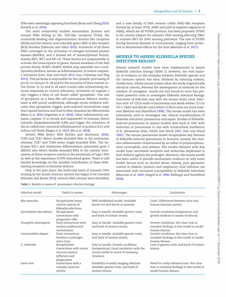
Several research models have been implemented to assessKlebsiella infection biology (Table 1). However, the vast major-ity of evidence on the interplay between Klebsiella species andthe immune system has been obtained by infecting rodents,chieflymice. Inbredmouse strains allow the study of geneticallyidentical cohorts, whereas the development of methods for thecreation of transgenic, knock-out and knock-in mice has pro-vided powerful tools to investigate Klebsiella infection biology.Outcomes of infection vary with the mouse strain used, infec-tion with 102 CFUs leads to bacteremia and death within 72 h inCD-I, CBA/J and BALB/c mice while C57BL/6 mice are more resis-tant (Mehrad and Standiford 1999). The mouse model has beenextensively used to investigate two clinical manifestations ofKlebsiella infections: pneumonia and sepsis. Studies of Klebsiella-induced pneumonia in animal models date back to 1947 withinduction of pneumonia in rats with intratracheal instillationof K. pneumoniae (Sale, Smith and Wood 1947; Sale and Wood1947). The mouse pneumonia model recapitulates key featuresof Klebsiella-induced pneumonia in humans, namely the mas-sive inflammation characterised by an influx of polymorphonu-clear neutrophils, and oedema. The results obtained with thismodel have uncovered receptors and molecules implicated inhost defence against the pathogen. Moreover, the mouse modelhas been useful to provide mechanistic evidence on why somehealth factors such as alcohol abuse, obesity, poor glycaemiccontrol in diabetic patients and respiratory viral infection areassociated with increased susceptibility to Klebsiella infections(Mancuso et al. 2002; Happel et al. 2006; Ballinger and Standiford2010).
Table 1. Models to assess K. pneumoniae infection biology.
Infection model Useful to assess Advantages Limitations
Mus musculus Recapitulates manyclinical aspects ofKlebsiella infections.
Well-established model. Availableknock-out and knock-in animals.
Costs. Differences between mice andhuman immune system.
Dyctiostelium discoideium Recapitulatesinteractions withphagocytic cells.
Easy to handle. Available genetic tools,and bank of mutant strains.
Growth conditions (temperature andgrowth medium to assess virulence).
Drosophila melanogaster Early interactions withancient antibacterialmechanisms.
Easy to handle. Available genetic tools,and bank of mutant strains.
Growth conditions. Not clear how totranslate findings in this model to modelhuman disease.
Caenorhabditis elegans Early interactionsbetween a pathogenand a host.
Easy to handle. Available genetic tools,and bank of mutant strains.
Growth conditions. Not clear how totranslate findings in this model to modelhuman disease.
G. mellonella Recapitulatesinteractions with innateimmune system(effectors andphagocytes).
Easy to handle. Growth conditions(temperature). Good correlation with themouse model in terms of assessingvirulence.
Lack of genetic tools, and bank of mutantstrains.
Danio rerio Interactions with acomplex immunesystem
Possibility of easily imaging infection.Available genetic tools, and bank ofmutant strains.
Need for costly infrastructure. Not clearhow to translate findings in this model tomodel human disease.
Dow
nloaded from https://academ
ic.oup.com/fem
sre/advance-article-abstract/doi/10.1093/femsre/fuy043/5188677 by Q
ueen's University Belfast user on 11 D
ecember 2018
4 FEMS Microbiology Reviews
In the mouse model, Klebsiella-induced pneumonia isachieved either via intratracheal/endobronchial instillation orvia intranasal inoculation, each of which has its limitations. In-tratracheal/endobronchial instillation delivers the inoculum tothe lower respiratory track bypassing the host barriers of theupper airways but modelling oropharyngeal aspiration. The in-tratracheal method of infection often results in a higher ratioof infecting organisms to local defences at the site of infectionthan those achieved with other inoculation routes. This resultsin an exuberant inflammatory response and tissue destructionalready few hours after infection. We suggest that the intratra-cheal/endobronchial inoculation route should be the one choiceto investigate the biology of Klebsiella-induced lung injury. How-ever, to gain insights into Klebsiella-triggered respiratory infec-tions, we favour the intranasal inoculation route because it cap-tures the interaction between the pathogen and the defences ofupper and lower respiratory track. Furthermore, it is a simplemethod of infection. Its major limitation is the variable deposi-tion of microorganisms in the lungs which may lead to signifi-cant differences between infected mice.
Intratracheal, intraperitoneal and intravenous routes of in-fection are used to model Klebsiella-triggered sepsis, whereas in-traperitoneal and orogastric routes of infection are used to in-vestigate the virulence of Klebsiella strains inducing pyogenicliver abscess (Siu et al. 2012). This syndrome was anecdotallyreported in Taiwan in the 1980s, although now it seems to bespreading to countries outside Asia. Clinical evidence suggeststhat healthy adults carry the virulent strains in their intestines,and liver abscess occur when bacteria translocate across the in-testinal epithelium (Siu et al. 2012). Experiments done in miceprovide initial evidence supporting this notion (Tu et al. 2009);however, it should be noted that this infection model onlydemonstrates the ability of these strains to spread from the gutto other organs. Of note, there are no specific Klebsiella geneticfeatures associated with these infections, suggesting that per-haps host factors might play a critical role in the outcome ofthis Klebsiella-triggered pyogenic liver abscess. Currently, thereis no well-developed model to investigate the gut colonisationby Klebsiella species. Recently, Krapp and colleagues (Krapp et al.2017) have developed a subcutaneous model of infection tomodel Klebsiella-induced skin and soft tissue infections. Theseare rare clinical manifestations also associated with hyperviru-lent strains.
Although the mouse model has proven useful to illuminateK. pneumoniae infection biology, it is important to acknowledgeits limitations. There exist significant differences between miceand humans in immune system development, activation andresponse to infection (Mizgerd and Skerrett 2008). For exam-ple, circulating neutrophil counts are lower in mice than inhumans (Haley 2003), and mouse neutrophils lack defensins(Eisenhauer and Lehrer 1992). There are no murine homologs ofseveral chemokines including IL8, although mice express otherchemoattractant cytokines (Strieter et al. 1996). Species differ-ences also exist in the receptors sensing infections. There are 10known functional TLRs in humans and 12 inmice (Takeuchi andAkira 2010); TLRs 1–9 are conserved in both species, althoughthe tissue expression and transcriptional regulation also dif-fer (Rehli 2002). There is general conservation between mouseand human TLR-controlled signalling pathways; however, thereare notable differences in the use of signalling adaptors (Sunet al. 2016). In addition, the ligand specificities and affinities ofTLRs also differ in humans and mice. For example, human TLR4exquisitely discriminates between lipid A structures, whereasmurine TLR4 does not and, as result, there are differences in
the inflammatory responses induced by lipid As with differentstructures (Hajjar et al. 2002; Montminy et al. 2006). These dis-crepancies and others (Mizgerd and Skerrett 2008) should becarefully considered in interpreting experiments to translate thefindings to human disease.
More recently, other non-mammalian infection models havebeen tested to investigate Klebsiella pathogenesis to circumventethical and costs issues associated with animal research, andto potentially facilitate large-scale analysis of virulence. The so-cial amoeba Dictyostelium discoideum is a phagocytic cell that canbe used to screen potential roles of phagocytic immune cellssuch as neutrophils and macrophages (Dunn et al. 2018). Thereis evidence demonstrating that D. discoideum is a valuable sys-tem for studying how pathogens evade fundamental processesof phagocytic cells (Dunn et al. 2018). Benghezal and colleagues(Benghezal et al. 2006) carried out a two-dimensional virulencearray to identify D. discoideum genes implicated in host defenceagainst Klebsiella, and Klebsiella genes require to survive in the at-tenuatedD. discoideumhost. phg1 and kil1, theD. discoideum genesidentified as essential to kill intracellular Klebsiella, have homo-logues in mammalian cells, although their potential contribu-tion to the physiology of phagocytic cells has not been studiedyet. The screen of bacterial mutants revealed that the surfacepolysaccharides expressed by Klebsiella, the capsule polysaccha-ride (CPS) and the LPS play a crucial role in the interactionwithD.discoideum (Benghezal et al. 2006). Additional studies have shownthat, in addition to the CPS and the LPS O-polysaccharide andcore, the outer membrane proteins (OMP) OmpA and OmpK36,and the lipid A decorations with aminoarabinose and palmitateare also necessary to avoid predation by D. discoideum (Marchet al. 2013). Interestingly, these factors are also required to limitphagocytosis bymouse alveolarmacrophages (March et al. 2013),suggesting that K. pneumoniae exploits the same factors to in-teract with social amoeba and macrophages. Dictyostelium dis-coideum has also proven to be useful to model the interac-tion between Klebsiella and human neutrophils (Pan et al. 2011),where CPS and LPS O-polysaccharide being also the importantfactors governing the interaction of Klebsiella with neutrophils.Further reinforcing the importance of Klebsiella surface polysac-charide on the interaction with phagocytes, D. discoideum specif-ically senses Klebsiella CPS to activate a predation programme(Lima et al. 2014).
The nematode Caenorhabditis elegans and Drosopilamelanogaster have also been used to identify host path-ways implicated in host defence against Klebsiella. In C. elegans,PI3K-AKT/mTOR and the MAPK p38 are required for host protec-tion against K. pneumonia (Kamaladevi and Balamurugan 2015,2017), whereas Phg1, important in the Klebsiella-D. discoideuminterplay, is also essential to resist K. pneumoniae infection byD. melanogaster (Benghezal et al. 2006). Whether these pathwaysplay any role in mammalian defence against K. pneumoniae iscurrently unknown.
A common limitation of these models is that the optimaltemperature for maintaining them is 28◦C, whereas the opti-mum temperature for K. pneumoniae is 37◦C. Since virulencegene expression is frequently regulated by temperature, it islikely that temperature requirements may affect the interac-tion of K. pneumonaie strains causing human infections withthese hosts. Nonetheless, it is important to note that the im-pact of environmental cues on the regulation of Klebsiella viru-lence factors is poorly understood. The larvae of the wax mothGalleria mellonella is emerging as a suitable model to study thevirulence of many human pathogens because, among other ad-vantages, it grows at 37◦C (Table 1) (Glavis-Bloom, Muhammed
Dow
nloaded from https://academ
ic.oup.com/fem
sre/advance-article-abstract/doi/10.1093/femsre/fuy043/5188677 by Q
ueen's University Belfast user on 11 D
ecember 2018

Bengoechea and Sa Pessoa 5
and Mylonakis 2012; Tsai, Loh and Proft 2016). G. mel-lonella defence against bacterial infections consists of cellu-lar and humoral immunity (Glavis-Bloom, Muhammed andMylonakis 2012; Browne, Heelan and Kavanagh 2013; Wojda2017). The cellular response is mediated by phagocytic cells,termed hemocytes, found within the haemolymph. Hemocytesgovern the clotting reaction to trap pathogens, and the melani-sation response consisting of the deposition of melanin to en-capsulate pathogens at the site of infection followed by thecoagulation of hemolymph. Melanisation can be consideredanalogous to abscess formation in mammalian infections. Thehumoral response is orchestrated by soluble effector moleculesthat immobilise or kill the pathogen and includes complement-like proteins, and antimicrobial peptides. The suitability of G.mellonella as a model to investigate K. pneumoniae pathogenesishas only been recently demonstrated (Insua et al. 2013). This in-fection model discriminates the pathogenic potential of K. pneu-moniae strains (Insua et al. 2013; Wand et al. 2013), and there is astrong correlation with the virulence previously determined inthe mouse pneumonia model (Insua et al. 2013). Furthermore,K. pneumoniae infection of G. mellonella results in responses sim-ilar to those reported in the mouse pneumonia model includ-ing cell death-associated with bacterial replication, inhibition ofphagocytosis and attenuation of host defence responses, chieflythe production of antimicrobial peptides (Insua et al. 2013). In-terestingly, virulence factors necessary in the mouse pneumo-nia model, CPS, LPS and OMPs, are also required for K. pneumo-niae survival in G. mellonella (Insua et al. 2013). The fact that allattenuated Klebsiella mutants activate G. mellonella defensive re-sponses (Insua et al. 2013) supports the notion that prevention ofhost responses is an important feature of K. pneumoniae patho-genesis. Despite the clear utility of G. mellonella as a surrogatehost to assess infections with K. pneumoniae, it is important tonote that the model only reflects early features of the interac-tion between the pathogen and ancient innate immune mech-anisms of defence. These mechanisms are indeed conserved inevolution; however, the evolutionary distance between insects,mice and humans makes that many host-specific phenomenaare likely to exist.
The larvae of Danio rerio (zebrafish) is another non-mammalian infection model receiving increasing attention be-cause it is genetically tractable, optically accessible and presenta fully functional innate immune system with macrophagesand neutrophils that mimic their mammalian counterparts(Torraca and Mostowy 2018). Although a wide variety ofpathogenic bacteria have been already investigated using ze-brafish, only recently it has been assessedwhether zebrafish lar-vae are a suitable model to investigate K. pneumoniae virulence(Cheepurupalli et al. 2017;Marcoleta et al. 2018). These studies re-port the optimisation of the model to investigate K. pneumoniaepathogenesis. Injection of larvae seems to be the most reliableinoculationmethod to ensure consistent colonisation of the gas-trointestinal tract (Cheepurupalli et al. 2017). Further studies arewarranted to validate whether the model is useful to identifyKlebsiella virulence factors and to uncover features of the inter-action between K. pneumoniae and the immune system.
CONTRIBUTION OF HOST SIGNALLINGIN DEFENCE AGAINST K. PNEUMONIAEINFECTIONS
The published evidence during more than 20 years clearly es-tablishes that pro-inflammatory signalling is crucial to K. pneu-moniae clearance (Fig. 1). One of the first conclusive piece of evi-
dence showed that mice deficient in the TNFα receptors (TNFR1)are susceptible to K. pneumoniae pneumonia (Laichalk et al. 1996,1998; Moore et al. 2005). Subsequent studies reported that micelacking the chemokines CXCL15 (Chen et al. 2001) and CCL3(Lindell et al. 2001), and unable to synthesise leukotrienes (Bailieet al. 1996; Mancuso et al. 1998) and nitric oxide (Tsai et al.1997) are also exquisitely susceptible to Klebsiella pneumonia.All these are markers associated with human pneumonia andare part of the common host response to infections (Jenner andYoung 2005; Lachmandas et al. 2016; Li et al. 2016; Martinez et al.2017). In turn, strategies to boost pro-inflammatory signallingin the airways have proven to be successful to limit K. pneumo-niae infections. Intrapulmonary expression of CCL3 (Zeng et al.2003), and KC (Tsai et al. 1998; Cai et al. 2010) leads to improvedclearance of K. pneumoniae. Intratracheal instillation of CpG in-creases the production of TNFα, and Th1 cytokines, includingIL-12, IFNγ and the IFNγ -dependent ELR-CXC chemokines (Denget al. 2004a) in a TLR9-dependent manner (Bhan et al. 2007), en-hancing bacterial clearance. Cyclic di-GMP, amolecule sensed bySTING (Burdette et al. 2011), also triggers a vigorous expression ofchemokines and Th1 cytokines (Karaolis et al. 2007). These treat-ments also result in the increased recruitment of neutrophils,αβ and γ δ T cells, and activated NK cells to the site of infection,suggesting that these cells are crucial in host defence againstK. pneumoniae. Indeed, γ δ T cells and NK cells play a pivotal rolein the resolution of Klebsiella infections by controlling early pro-duction of pro-inflammatory cytokines (Moore et al. 2000; Xuet al. 2014).
The role of different cytokines in host defence against K.pneumoniae has been also investigated (Fig. 1). Early studiesdemonstrated the importance of IFNγ and IFNγ -dependent cy-tokines to control the progression of Klebsiella-induced pneumo-nia (Yoshida et al. 2001; Moore et al. 2002; Zeng et al. 2005a,b). IL23drives the production of IFNγ and IL17 (Happel et al. 2003, 2005);however, the fact that IL17 administration restores bacterial con-trol in mice deficient on IL23 production indicates an indepen-dent role for the IL17-governed axis on host defence against K.pneumoniae (Happel et al. 2005). Adding further evidence to thisnotion, IL17 signalling is critical for the induction of Th1 re-sponses, neutrophil recruitment and local control of pulmonaryinfection (Ye et al. 2001a,b). IL17 signalling is also augmented viaIL12 production through IFNγ (Happel et al. 2005). The contribu-tion of IL12 signalling to control Klebsiella pneumonia is exem-plified by the fact that STAT4−/− knock-out mice displayed im-paired production of IFNγ and Th1 cytokines and greater bac-terial burden compared to wild-type infected mice (Deng et al.2004b). STAT4 is a critical transcriptional factor in the IL12 sig-nalling pathway (Bacon et al. 1995). IL22, produced in an IL23-dependent manner, is also important in host defence againstK. pneumoniae (Aujla et al. 2008; Zheng et al. 2016). Administra-tion of an anti-IL22 blocking antibody results in higher bacterialloads in the lungs and dissemination of bacteria to spleen (Aujlaet al. 2008), whereas therapeutic administration of IL22 attenu-ates Klebsiella-triggered peritonitis (Zheng et al. 2016). IL22 reg-ulates the levels of IL6 and CCL3 upon Klebsiella infection, andits role is predominant over IL17 in regulating these cytokines(Aujla et al. 2008). The synergistic effect of both cytokines gov-erning host defences against Klebsiella is marked by the fact thatneutralisation of IL22 in Il17a−/− mice results in greater bacte-rial growth in the lung and significantly more bacterial dissem-ination to the spleen than in those observed in infected Il17a−/−
(Aujla et al. 2008).Collectively, the summarised evidence strongly suggests
that the IL23/IL17 and IL12/IFNγ axes are essential for the
Dow
nloaded from https://academ
ic.oup.com/fem
sre/advance-article-abstract/doi/10.1093/femsre/fuy043/5188677 by Q
ueen's University Belfast user on 11 D
ecember 2018

6 FEMS Microbiology Reviews
Figure 1.Mechanisms of innate immunity to K. pneumoniae infections. The figure depicts the cells implicated in containing K. pneumoniae infection. There is conclusiveevidence demonstrating the interaction of K. pneumoniae with neutrophils, macrophages (and monocytes [not shown]), dendritic cells and epithelial cells. Theseinteractions are marked with black arrows. The interaction with different subset of T cells, NK cells and other lymphocytes has not been investigated yet, although
these cells participate in bacterial clearance. The network of connections between cells, and the role played by different cytokines activating host responses aredepicted with blue arrows.
generation of an effective innate immune response in the lungsagainst K. pneumoniae. However, two questions require addi-tional investigations: Which are the cell(s) responsible for theproduction of these cytokines? and Which are the main innatedefence mechanisms (humoral and cellular) activated by thesecytokine networks responsible for the clearance of K. pneumo-niae? To set the framework for future studies, we next discussthe available evidence. Initial data suggest that γ δ T and NKcells could be the major source of IL17 and IL22, respectively,in Klebsiella-infected mice (Xu et al. 2014; Murakami et al. 2016),whereas alveolarmacrophages could be the initial source of IL23(Happel et al. 2005). Only recently, it has been also suggested thattype 3 innate lymphocytes could be another source of IL17 invivo (Xiong et al. 2016). Altogether, there is need to dissect thesource of IL17 during K. pneumoniae infections. Dendritic cellshave been shown to orchestrate the production of IL12, IL23 andIL17 in vivo (Bhan et al. 2007), although the specific singular roleof dendritic cells in Klebsiella infections and the connection withother immune cells have not yet been fully defined. There is ev-idence showing that NK cells are the source of IFNγ in Klebsiella-infected mice (Van Elssen et al. 2010; Ivin et al. 2017), although itcannot be ruled out that other cell types such as CD4 and CD8T cells might contribute as well. Alveolar macrophages and re-cruited inflammatory monocytes are considered themain cellu-lar target for IFNγ and IL17, respectively (Xiong et al. 2015, 2016;Ivin et al. 2017). These cytokines enhance the microbicidal ac-tivity of alveolar macrophages and inflammatory monocytes byincreasing phagocytosis and facilitating bacterial killing (Xiong
et al. 2015, 2016; Ivin et al. 2017). Whether IFNγ and IL17 trig-ger other antimicrobial activity on these cells remains to be in-vestigated. IL17 and IL22 also aid in the clearance of Klebsiellaby regulating the antimicrobial activity of the lung epithelium(Aujla et al. 2008; Chen et al. 2016). Both cytokines activate an-timicrobial programmes in epithelial cells having in commonthe production of CXCL5 and lipocalin 2 (Aujla et al. 2008; Chenet al. 2016). Ablation of this programme results in higher bacte-rial loads in the lungs of infected animals. CXCL5 participates inthe recruitment of neutrophils to the site of infection (Chen et al.2016), whereas lipocalin 2 inhibits the growth of some strainsof Klebsiella in vitro and in vivo by preventing bacterial iron ac-quisition (Bachman, Miller and Weiser 2009; Chan et al. 2009;Bachman et al. 2012). Additionally, lipocalin 2 may also pro-mote the induction of pro-inflammatory responses, whichfacilitates the recruitment of neutrophils like CXCL5 does.However, it is important to be aware that there are conflictingreports on the role of neutrophils in vivo to clear Klebsiella in-fections (Greenberger et al. 1996; Broug-Holub et al. 1997; Xionget al. 2015). Therefore, the recruitment of neutrophils to the lungsof Klebsiella-infected mice cannot be rigorously taken as conclu-sive evidence of these cells being a major player in host defenceagainst the pathogen. In vivo mechanistic studies involving se-lective depletion of neutrophils together with ex vivo experi-ments testing isolated mouse neutrophils should be the normin these type of studies.
Only recently, the role of type I IFN and type I IFN-governedsignalling in host defence against Klebsiella infections has been
Dow
nloaded from https://academ
ic.oup.com/fem
sre/advance-article-abstract/doi/10.1093/femsre/fuy043/5188677 by Q
ueen's University Belfast user on 11 D
ecember 2018

Bengoechea and Sa Pessoa 7
investigated (Fig. 1). Alveolar macrophages are one of thesources of type I IFN in vivo, and in vitro experiments revealedthat K. pneumoniae activates a TLR4-TRAM-TRIF-IRF3 signallingpathway to induce type I IFN- and type I IFN-dependent genes(Ivin et al. 2017). To assess the importance of type I IFN signallingin host defence, researchers infected mice lacking type I IFN1 receptor-deficient (Ifnar1−/−) mice (Ivin et al. 2017). Ifnar1 isone of the subunits of the type I IFN receptor which mediatestype I IFN responses in innate and acquired immunity to in-fection. Ifnar1−/− are exquisitely sensitive to Klebsiella infec-tion exhibiting a markedly decreased survival, higher bacte-rial lung burden, increased dissemination to spleen and liverand severe bronchopneumonia (Ivin et al. 2017). The lack oftype I IFN signalling results in defect in the production ofIFNγ , IL-12 and CXCL10 in K. pneumoniae-infected lungs, butit has no impact on the number of alveolar macrophages, in-flammatory monocytes and neutrophils recruited to the siteof infection (Ivin et al. 2017). In contrast, the number of NKcells was lower in the lungs of Ifnar1−/−-infected mice thanin the wild-type ones (Ivin et al. 2017). This is consistentwith the reduced levels of the NK chemoattractant chemokineCXCL10 in the lungs of Ifnar1−/−-infected mice (Ivin et al. 2017).Type I IFN signalling is crucial for NK cells to produce IFNγ ,which is required for enhancing the bactericidal action andthe production of the NK cell response-amplifying IL-12 andCXCL10 by alveolar macrophages (Ivin et al. 2017). Remark-ably, type I IFN signalling is dispensable in myeloid cells in-cluding alveolar macrophages, monocytes and neutrophils forhost defence and IFNγ activation (Ivin et al. 2017), uncoveringa hitherto unknown crosstalk between alveolar macrophagesand NK cells based on type I IFN and IFNγ in Klebsiellainfection.
Few studies have addressed the contribution of PRR-governed signalling to control Klebsiella infections. As with otherbacterial infections, TLR4 signalling plays a prominent role inantibacterial defence against Klebsiella infection (Branger et al.2004; Wieland et al. 2011; Standiford et al. 2012). TLR4−/− miceshow reduced survival upon infection with increased bacte-rial loads in lungs and bronchopneumonia (Branger et al. 2004;Wieland et al. 2011; Standiford et al. 2012). The lack of TLR4 sig-nalling is associated with a decrease in the levels of IL17 andIL23 in the lungs of infected TLR4−/− mice (Happel et al. 2003),which may explain the susceptibility of these mice to Klebsiellainfection. It remains an open question which cells are more af-fected by the lack of TLR4 signalling. Initial observations suggestthat TLR4 signalling is indispensable in cells of myeloid originfor the clearance of Klebsiella (Wieland et al. 2011); however, itcannot be ruled out that other cell types may require TLR4 sig-nalling to aid in the elimination of Klebsiella infections. Support-ing this notion, TLR4 signalling is required to protect the lungepithelium from Klebsiella-induced pathophysiology (Standifordet al. 2012). TLR9-controlled signalling is also required for pro-tective immunity against Klebsiella-induced pneumonia (Bhanet al. 2007, 2010). Mice deficient in TLR9 fail to generate an ef-fective Th1 cytokine response, resulting in increased bacterialloads in the lungs and dissemination to other organs (Bhan et al.2007). TLR9−/−-infected mice present no major defects on theaccumulation of immune cells except on the influx and matu-ration of conventional dendritic cells (Bhan et al. 2007). This re-duced accumulation and activation of dendritic cells explainsthe impaired bacterial clearance because adaptive transfer ofdendritic cells from wild-type mice reconstitutes the protectiveimmunity in TLR9−/− mice (Bhan et al. 2007). Since TLR9 is lo-cated in endosomes and it recognises DNAoligonucleotideswith
unmethylated CpG base pairs (Hemmi et al. 2000), it is intriguingto consider how Klebsiella infection may lead to the activationof this intracellular receptor. Data obtained in other infectionmodels indicate that TLR9 can be activated by bacterial and hostDNA released into the airways during pneumonia (van der Meeret al. 2016), as well as by intracellular bacteria and DNA of mi-tochondrial origin released to the cytosol upon infection (Zhanget al. 2010; Arpaia et al. 2011). Future studies should address thisknowledge gap. TLR2 signalling has a dual role in host defenceagainst Klebsiella (Wieland et al. 2011). In the early phase of in-fection, TLR2 signalling has an anti-inflammatory role (Wielandet al. 2011), perhaps to prevent a detrimental overwhelming in-flammation as a result of the activation of other PRRs. Similarobservation has been done in Acinetobacter baumannii-triggeredpneumonia (Knapp et al. 2006), suggesting that the dampen-ing function of TLR2 during pneumonia is not bacterial species-specific. In the later stage of infection, TLR2 contributes toantibacterial defence (Wieland et al. 2011). Interestingly, cooper-ative roles of TLR4 and TLR2 signalling are involved in control-ling Klebsiella infection because TLR4−/−xTLR2−/− mice are moresusceptible to the infection than each of the single knock-outmice (Wieland et al. 2011).
The role of TLR signalling during K. pneumoniae infectionhas been further probed by demonstrating the contribution ofTLR adaptors in host defence. MyD88 is the universal adaptorfor all TLRs except TLR3 (O’Neill and Bowie et al. 2007), and ithas been shown to be important for pulmonary host defenceagainst several respiratory pathogens (Baral et al. 2014). TRIFis the sole adaptor for TLR3 and also contributes to TLR4 sig-nalling (O’Neill and Bowie et al. 2007). Infections of MyD88−/−
and TRIF−/− mice demonstrated that both adaptors are requiredto restrict K. pneumoniae growth in the lungs (Cai et al. 2009; vanLieshout et al. 2012). MyD88-dependent protection during Kleb-siella pneumonia is mediated by both hematopoietic and resi-dent cells excluding endothelial cells, whereas TRIF-mediatedprotection is driven by hematopoietic cells (van Lieshout et al.2012, 2014; Anas et al. 2017). MyD88 and TRIF deficiencies limitthe production of Th1 cytokines and the activation of signallingpathways controlling host defence mechanisms (Cai et al. 2009).Interestingly, the characteristic bronchopneumonia of Klebsiellainfections is virtually absent in infectedMyD88−/− mice and sig-nificantly reduced in TRIF−/− mice despite high bacterial loadsin both mice (Cai et al. 2009). This evidence indicates that thehistopathological changes associated with Klebsiella infectionare dependent on the host inflammatory response to the infec-tion. TIRAP/MAL is another adaptor linking MyD88 to the acti-vated TLR2 and TLR4 receptors (O’Neill and Bowie et al. 2007). Inthis context, it is not surprising thatMAL−/− mice have substan-tial mortality, higher bacterial burden in the lungs, enhancedbacterial dissemination, attenuated production of Th1 cytokinesand no lung histopathology following K. pneumoniae infection(Jeyaseelan et al. 2006). At present, the mechanisms underlyingMyD88-MAL-mediated defence against Klebsiella are ill-defined.However, it is important to note here that MyD88-MAL are alsorequired for the activation of other signalling pathways such asthose governed by IL1β and IFNγ (Cohen 2014; Ni Cheallaigh et al.2016). Therefore, MyD88-MAL-dependent protective immunityis most likely also mediated by IL1β- and IFNγ -governed hostantibacterial responses. Likewise IFNγ -deficient mice, IL1R−/−
mice are exquisitely susceptible to Klebsiella infection demon-strating the importance of IL1β-controlled responses for hostsurvival and bacterial clearance (Cai et al. 2012). On the otherhand, the impaired antibacterial defence of TRIF−/- mice is asso-ciated with the lack of IFNγ in the lungs of infected mice (van
Dow
nloaded from https://academ
ic.oup.com/fem
sre/advance-article-abstract/doi/10.1093/femsre/fuy043/5188677 by Q
ueen's University Belfast user on 11 D
ecember 2018

8 FEMS Microbiology Reviews
Lieshout et al. 2015). The fact that TRIF is required for type I IFNproduction following Klebsiella infection (Ivin et al. 2017) suggeststhat the impairment of IFNγ production in TRIF−/– mice is sec-ondary to the deficient type I IFN production in these mice.
A small number of studies have investigated the contribu-tion of NLR signalling to defence against Klebsiella. NLRP3−/−
mice demonstrate an increase in mortality following Klebsiellainfection albeit the protective role of NLRP3 is not as impor-tant as those of TLR4 and TLR2, and any of the TLR adaptors(Willingham et al. 2009). In vitro experiments confirmed thecontribution of NLRP3 to caspase-1 activation and IL1β releasefollowing Klebsiella infection (Willingham et al. 2009). In goodagreement, Klebsiella-induced IL1β is reduced in NLRP3−/− mice(Willingham et al. 2009). Recent evidence supports that the CPSand the LPS are the Klebsiella components responsible for prim-ing NLRP3, whereas Klebsiella-triggered ROS may be responsi-ble for the activation (Hua et al. 2015). The NLRC4 inflamma-some also contributes to Klebsiella-triggered IL1β production invitro and in vivo (Cai et al. 2012). However, there are contradic-tory results on the importance of NLRC4 to confer protectionagainst Klebsiella infection (Willingham et al. 2009; Cai et al. 2012).The main apparent difference between studies is the differentbacterial inoculum, with the study using an inoculum closer tothe LD50 showing a contribution of NLRC4 on host immunityagainst Klebsiella (Cai et al. 2012). Nonetheless, an open questionis the identification of the Klebsiella component(s) inducing theactivation of NLRC4. This receptor senses bacterial flagellin andthe type III secretion system apparatus (Zhao et al. 2011; Dun-can and Canna 2018). Notably, Klebsiella is not flagellated andin silico analysis of more than 700 genomes confirms that thispathogen does not encode any type III secretion system. It isthen tempting to speculate that any of the secretion systemsencoded by Klebsiella, including the type II and type VI secre-tion systems, might be sensed by NLRC4. Intriguingly, Klebsiella-induced pyroptosis requires NLRP3 but not NLRC4 (Willinghamet al. 2009; Cai et al. 2012). Whether pyroptosis is one of thebona fide host defence mechanisms against Klebsiella infectionis yet unknown. In fact, the specific importance of pyroptosis inhost immunity remains a challenging question. Initial evidenceshows thatNLRC4-dependent pyroptosismediates the clearanceof the intracellular pathogen Salmonella typhimurium by generat-ing a structure that entraps the previously intracellular bacte-ria and drives their elimination by containing the bacteria andelaborating signals that promote efferocytosis (Miao et al. 2010;Jorgensen et al. 2016). However, it is unlikely that this mecha-nism operates in the context of Klebsiella infections because, asdiscussed before, the lack of NLRC4 does not affect Klebsiella-triggered pyroptosis.
MICROBIOME PROTECTION AGAINSTKLEBSIELLA INFECTIONS
There is a wealth of evidence demonstrating the role of the in-testinal microbiota to prevent infection by pathogenic bacte-ria. This is achieved by interactions within the microbial com-munity and by shaping the tissue immune responses to limitinfection (McKenney and Pamer 2015). Surprisingly, there isvirtually no data on the impact of the gut microbiome on Kleb-siella gut colonisation and/or orogastric infection. This knowl-edge gap is particularly relevant considering the recent clinicalevidence demonstrating that gastrointestinal carriage is a ma-jor reservoir of K. pneumoniae infections in the healthcare en-vironment (Martin et al. 2016; Gorrie et al. 2017). On the other
hand, the gut microbiota has been shown to protect againstKlebsiella pneumonia. Experiments infecting germfree mice re-vealed that these animals are susceptible to Klebsiella in an IL-10–dependent manner (Fagundes et al. 2012). In germfree mice,IL-10 in the lungs restrains pro-inflammatory mediator produc-tion and favours Klebsiella growth and dissemination (Fagundeset al. 2012). Neutralisation of IL10, or transient TLR4 activationwith LPS, restores germfree mice resistance to K. pneumoniae in-fection (Fagundes et al. 2012). Subsequent studies provided com-pelling evidence on themembers of the gutmicrobiota that driveprotection against Klebsiella infection and the signalling path-ways responsible for this microbiota-controlled immune pro-tection. A consortium of bacterial species common to the ro-dent and human intestinal microbiota formed by Lactobacillusreuteri, Enterococcus faecalis, Lactobacillus crispatus and Clostrid-ium orbiscindens induces potent NOD2 activation to trigger IL17-GM-CSF in the lung which, in turn, stimulates pathogen killingand clearance by alveolar macrophages through MAPK extra-cellular signal-regulated kinase signalling (Clarke 2014; Brown,Sequeira and Clarke 2017). The source of IL17 in the lung andhow the microbiota governs the production of this cytokine re-main open questions. Nevertheless, these findings further high-light the critical role of IL17 in host defence against Klebsiella,and the crucial role played by alveolar macrophages promotingantibacterial defence in the lung. Although the contribution ofthe upper airway microbiota, either permanent resident or as-pirated from the oropharynx, to limit respiratory infections hasnot been demonstrated yet, it is notable that intranasal inocu-lation of bacteria colonising the upper airway of humans andmice (Lactobacillus crispatus, Staphylococcus aureus, S. epidermidis)enhances lung immunity against Klebsiella by the same IL17-GM-CSF-alveolar macrophage axis (Brown, Sequeira and Clarke2017). Collectively, this evidence demonstrates the facility of thelung immune system to integrate microbial signals from differ-ent mucosal sites to launch antibacterial defence mechanisms.
KLEBSIELLA EVASION STRATEGIESOF HOST DEFENCES
Thewidely held belief is that K. pneumoniae is a stealth pathogen,which fails to stimulate innate immune responses (Paczosaand Mecsas 2016). Essentially, Klebsiella shields its pathogen-associated molecular patterns from detection by PRRs and solu-ble effectors of the immune system, and avoids the interactionwith hematopoietic and non-hematopoietic cells to prevent theactivation of host antimicrobial responses (Table 2). However,there is now enough evidence demonstrating that Klebsiella alsoactively subverts host defences (Table 2). We will review bothimmune evasion strategies in the context of the interaction ofKlebsiella with different effectors of the immune system.
Counteracting soluble effectors of the immune system
Early research focused on investigating the interplay betweencomplement and K. pneumoniae. The OMPs and LPS of K. pneu-moniae are known to activate the classical pathway (Albertiet al. 1996a,b). OmpK36 and OmpK35, homologues to OmpF andOmpC, respectively, and two of the most abundant porins inthe outer membrane of K. pneumoniae, bind Cq1 in an antibody-independent manner triggering complement activation (Albertiet al. 1993, 1996a). K. pneumoniae LPS without O-polysaccharidealso activates the classical pathway, although less efficientlythan the OMPs (Alberti et al. 1996b). C3b deposition on the
Dow
nloaded from https://academ
ic.oup.com/fem
sre/advance-article-abstract/doi/10.1093/femsre/fuy043/5188677 by Q
ueen's University Belfast user on 11 D
ecember 2018

Bengoechea and Sa Pessoa 9
Table 2. Immune evasion strategies of K. pneumoniae.
Immune evasion strategies Mechanism Bacterial factor References
(i) Stealth pathogenPreventing the antimicrobialaction of soluble innateimmune effectors
Preventing complementbactericidal effect,and opsonisation
Limiting C3b deposition CPS, LPS O-polysaccharide Merino et al. 1992; Alvarez et al.2000; de Astorza et al. 2004
Limiting antimicrobialactivity of collectins
Blunting interaction with SP-Aand SP-D
CPS Kabha et al. 1997; Ofek et al.2001; Kostina et al. 2005
Counteracting bactericidalaction CAMPs andpolymyxins
Limiting the interaction withthe bacterial surface. Efllux ofCAMPs.
CPS, LPS lipid Adecorations, AcrAB
Campos et al. 2004; Llobet et al.2011; Kidd et al. 2017; Mills et al.2017; Padilla et al. 2010
Attenuating the interactionwith immune cells
Attenuating engulfmentby epithelial cells
CPS Cortes et al. 2002; Regueiro et al.2006
Avoiding phagocytosisby neutrophils
CPS, OmpK36 Regueiro et al. 2006; Pan et al.2011
Avoiding phagocytosisby macrophages
CPS, LPS lipid Adecorations, OmpA,OmpK36
March et al. 2013
Limiting the activation of PPRs Limiting the recognition of LPSby TLR4
LPS lipid A 2-hydroxylation Llobet et al. 2015
(ii) Subversion host defencesAttenuating cell-intrinsic immunity
Controlling maturationdendritic cells
CPS, LPS O-polysaccharide Evrard et al. 2010
Manipulation phagosomematuration
Activation PI3K-AKT-Rab14axis
Unknown Cano et al. 2015
Controlling cell death Cytotoxicity in epithelial cells.Triggering apoptosis inmacrophages.
CPS Unknown Cano et al. 2009; Leone et al.2016
Abrogating TLR-controlledinflammatory responses:
Abolishing TLR signalling CPS, LPS O-polysaccharide,OmpA, T2SS
March et al. 2011; Frank et al.2013; Tomas et al. 2015
Blunting NF-κB signalling Upregulation deubiquitinaseCYLD by targeting NOD1 andEGFR.
CPS, and other unknownfactor(s)
Regueiro et al. 2011; Frank et al.2013
Blunting MAPKs Upregulation MAPKsphosphatase MKP-1 via NOD1activation.
Unknown Regueiro et al. 2011
Manipulating mucosalimmunity
Induction of IL10. Unknown Greenberger et al. 1995;Yoshida et al. 2001
Counteracting nutritionalimmunity
Secretion of severalsiderophores
Yersiniabactin,salmochelin, aerobactin
Lawlor, O’connor and Miller2007; Bachman et al. 2011;Russo et al. 2011
bacterial surface upon complement activation results in in-creased internalisation of Klebsiella by human lung epithelialcells promoting bacterial clearance (de Astorza et al. 2004), aswell as opsonophagocytosis by neutrophils and macrophages(Domenico et al. 1994; Salo et al. 1995; Regueiro et al. 2006).Not surprisingly, the main complement evasion strategy of Kleb-siella is based on preventing C3b deposition by exploiting Kleb-siella surface polysaccharides. Whether K. pneumoniae may ex-ploit other complement evasion strategies, such as targetingfactor H, has not been described yet. The CPS is the main fac-tor protecting Klebsiella from complement; cps mutants are sus-ceptible to the bactericidal action of complement and show in-
creased deposition of C3b on the surface (Merino et al. 1992;Alvarez et al. 2000). The protection conferred by CPS is moredependent on the thickness of the polysaccharide than thechemical composition of the polysaccharide (de Astorza et al.2004), although CPS containing manno(rhamno)biose may ac-tivate the lectin complement pathway (Sahly, Keisari and Ofek2009). The LPS O-polysaccharide also protects Klebsiella fromcomplement by limiting the deposition of C3b on the bacterialsurface (Merino et al. 1992). In those strains lacking the LPS O-polysaccharide, the CPS is then the main factor protecting Kleb-siella from the bactericidal action of complement (Alvarez et al.2000).
Dow
nloaded from https://academ
ic.oup.com/fem
sre/advance-article-abstract/doi/10.1093/femsre/fuy043/5188677 by Q
ueen's University Belfast user on 11 D
ecember 2018

10 FEMS Microbiology Reviews
The CPS also confers protection against lung collectins SP-A and SP-D, components of the lung surfactant, by preventingthe binding of the collectins to the LPS (Kabha et al. 1997; Ofeket al. 2001; Kostina et al. 2005). The binding of both collectins tothe bacterial surface triggers bacterial agglutination and facili-tates phagocytosis by macrophages (Kabha et al. 1997; Ofek et al.2001; McCormack and Whitsett 2002). Interestingly, pulmonarysurfactant challenge shapes the transcriptome of K. pneumoniae,inducing a programme strongly associated with virulence in thepneumonia mouse model (Willsey et al. 2018). The CPS is one ofthe systems induced by pulmonary surfactant, further empha-sising the importance of this polysaccharide to protect Klebsiellaagainst collectins.
Like many other bacterial pathogens, K. pneumoniae has de-veloped strategies to counteract host cationic antimicrobial pep-tides (CAMPs), chiefly defensins. Importantly, CAMPs and an-tibiotics such as quinolones and polymyxins share the sameinitial target in the outer membrane of Gram-negative bacteria(Nikaido 2003). Therefore, there is a relationship between resis-tance to CAMPs and polymyxins (Campos et al. 2004; Campos,Morey and Bengoechea 2006; Nizet 2006; Llobet et al. 2011; Kiddet al. 2017). To counteract the bactericidal action of CAMPs andpolymyxins, K. pneumoniae exploits the versatility of the CPS andthe LPS. CPS limits the interaction of CAMPs and polymyxinswith Klebsiella surface, and, in fact, there is a correlation betweenthe amount of CPS expressed by a given strain and the resis-tance to polymyxin B (Campos et al. 2004). Furthermore, free CPS,which may be released from the bacterial surface by CAMPs andpolymyxins, binds CAMPs, neutralising their bactericidal effect(Llobet, Tomas and Bengoechea 2008). Therefore, the CPS acts asa bacterial decoy for CAMPs. Notably, this trait is shared by an-ionic CPS expressed by Pseudomonas aeruginosa and Streptococcuspneumoniae (Llobet, Tomas and Bengoechea 2008), strongly sug-gesting that trapping CAMPs is a general feature of anionic CPS.
K. pneumoniae also remodels its LPS lipid A domain to coun-teract CAMPs and polymyxins (Llobet et al. 2011, 2015; Kidd et al.2017; Mills et al. 2017). Klebsiella pneumoniae lipid A can be dec-orated with palmitate, 4-amino-4-deoxy-L-arabinose, phospho-ethanolamine and 2-hydroxymyristate (Llobet et al. 2011, 2015;Kidd et al. 2017). These decorations provide resistance to CAMPs,and K. pneumoniaemutants lacking these lipid A decorations areattenuated for virulence in the mouse pneumonia model (Llo-bet et al. 2011, 2015; Kidd et al. 2017). There are reports show-ing that the lipid A acylation also mediates resistance to CAMPs(Clements et al. 2007; Mills et al. 2017). However, this role couldbe indirect since mutants deficient in the late acyltransferaseslpxM and lpxL display changes in the CPS levels and the 2-hydroxylation of the lipid A, respectively (Clements et al. 2007;Mills et al. 2017).
The lipid A of K. pneumoniae shows a remarkable plasticity.Just a brief incubation with CAMPs upregulates the expressionof the loci required to modify the lipid A with a concomitantincrease in the lipid A species containing such modifications(Llobet et al. 2011). The regulatory network controlling thesetranscriptional changes is complex and involves, at least, thePhoPQ, PmrAB and the Rcs systems (Llobet et al. 2011). Of note,and in contrast to Salmonella, PhoPQ and PmrAB control inde-pendently the regulatory architecture governing these CAMPs-induced changes (Llobet et al. 2011). Intriguingly, OmpA is alsopart of the regulatory network controlling systems requiredto ameliorate CAMPs bactericidal action (Llobet et al. 2009).Whether there is any connection between the OmpA and thePhoPQ and PmrAB-regulated networks is currently unknown.
Counteracting immune cells
Although it is well appreciated that the airway epitheliumplays a central role orchestrating pulmonary inflammatory andimmune responses against infections (Whitsett and Alenghat2015), few studies detail the interaction of Klebsiella with thesecells. Initial research argued that Klebsiella entry into epithe-lial cells may protect the pathogen from the actions of an-tibiotics and the immune system (Sahly et al. 2000). Thesestudies clearly demonstrated the role of the CPS limiting the at-tachment and internalisation to epithelial cells. However, sub-sequent studies have shown that epithelial cells engulfment ofKlebsiella is most likely a host defence mechanism (Cortes et al.2002; Cano et al. 2009). This interpretation is consistent with theobservations that bacterial internalisation triggers an inflamma-tory response due to the activation of NF-κB signalling (Regueiroet al. 2006), and that K. pneumoniae triggers a cytotoxic effecton epithelial cells (Cano et al. 2009; Leone et al. 2016). Klebsiella-induced cytotoxicity requires the presence of live bacteria andof CPS since it is observed with isolates expressing differentamounts of CPS and/or different serotypes but not with non-capsulated bacteria (Cano et al. 2009). K. pneumoniae infectionalso increases the levels of TLR4 and TLR2 in human airway ep-ithelial cells which results in enhanced inflammatory responseupon stimulation with TLR agonists (Regueiro et al. 2009). TLRupregulation upon infection is dependent on the activation ofNF-κB-governed signalling pathway by the CPS (Regueiro et al.2009). Evidence demonstrates that Klebsiella CPS is sensed byTLR4 (Regueiro et al. 2009; Yang et al. 2011; Frank et al. 2013).
There is scarce data on the interaction between Klebsiellaand neutrophils, although the recruitment of neutrophils to thelung is one of the hallmarks of Klebsiella-triggered pneumonia.In vitro experiments indicate that neutrophil-dependent clear-ance of Klebsiella occurs after phagocytosis. In turn, CPS abro-gates killing by neutrophils due to its anti-phagocytic activity(Regueiro et al. 2006; Pan et al. 2011). Not surprisingly, antibodiesagainst the CPS empower neutrophil-mediated killing (Diago-Navarro et al. 2018), showing promise as new therapeutics totreat Klebsiella infections. Interestingly, K. pneumoniae does notinduce NETosis (Branzk et al. 2014), although anti-CPS antibod-ies enhance the release of neutrophil extracellular traps (NETs),whichmay contribute to extracellular killing of Klebsiella (Diago-Navarro et al. 2017, 2018). NETs are large, extracellular, web-likestructures composed of decondensed chromatin and neutrophilantimicrobial factors. NETs trap and kill a variety of microbes(Amulic et al. 2012). NETs are released primarily via a cell deathprogram that requires ROS, and the granule proteins myeloper-oxidase and neutrophil elastase (Amulic et al. 2012). Recently,it has been shown that K. pneumoniae interferes with clearanceof neutrophils by efferocytosis (Jondle et al. 2018). Efferocyto-sis is a regulated process which facilitates the elimination ofapoptotic neutrophils by phagocytic cells, mainly macrophages,which prevents heightened inflammation triggered by dead cells(Ariel and Serhan 2012; Poon et al. 2014; Angsana et al. 2016).Mechanistically, Klebsiella infection of neutrophils results in adrastic decrease in the exposure of phosphatidylserine, whichis recognised as ‘eat-me’ signal by macrophages to initiatethe engulfment of neutrophils (Jondle et al. 2018). The Kleb-siella factor(s) responsible for this phenotype is currently un-known. Interestingly, evidence suggests that Klebsiella not onlylimits the efferocytosis of neutrophils, but it may also pro-gramme their cell death towards necroptosis (Jondle et al. 2018).In contrast to apoptosis, necroptosis is a cell death triggeringinflammation (Pasparakis and Vandenabeele 2015). Therefore,
Dow
nloaded from https://academ
ic.oup.com/fem
sre/advance-article-abstract/doi/10.1093/femsre/fuy043/5188677 by Q
ueen's University Belfast user on 11 D
ecember 2018

Bengoechea and Sa Pessoa 11
Klebsiella-triggered necroptosis may also contribute to the exu-berant inflammation observed in this infection. It is then tempt-ing to speculate that inhibition of necroptosis may improveKlebsiella-induced immunopathology. Preliminary observationsindicate that this might be the case (Jondle et al. 2018).
The specific role of dendritic cells in the clearance of K.pneumoniae has been poorly characterised, although there aredata indicating that Klebsiella may activate different subsetsof dendritic cells (Hackstein et al. 2013). An outer membranefraction of K. pneumoniae, containing CPS, LPS and porins, in-duces dendritic cell maturation (Van Elssen et al. 2010). How-ever, K. pneumoniae CPS and LPS attenuate dendritic maturationby hampering bacterial binding and internalisation (Evrard et al.2010). It should be mentioned that the latter results were ob-tained testing UV-killed bacteria. This technical approach is use-ful to assess the impact of an intact bacterial surface on cellactivation/maturation but hampers any investigation on anti-immune systems deployed by live bacteria such as the injectionof effector proteins by secretion systems. Nonetheless, futureinvestigations are warranted to deconstruct the interaction be-tween dendritic cells and K. pneumoniae to address whether thepathogen is able to survive intracellularly, the impact ofKlebsiellainfection on signalling governing cell maturation and whetherKlebsiellamay interfere with the processing of antigens and pre-sentation of antigen-derived peptides to T cells, among otherquestions.
Historically, K. pneumoniae is considered an extracellularpathogen, yet there are reports suggesting that K. pneumoniaecan survive in macrophages (Willingham et al. 2009; Greco et al.2012; Fevre et al. 2013; Fodah et al. 2014). Providing compellingevidence for this hypothesis, Cano and co-workers (Cano et al.2015) have demonstrated that K. pneumoniae survives within hu-man and mouse macrophages in a unique vacuolar intracellu-lar compartment which deviates from the canonical endocyticpathway and it does not fuse with lysosomes. Furthermore, datasuggest that K. pneumoniae triggers a programmed cell death inmacrophages displaying features of apoptosis 10 h post infec-tion (Cano et al. 2015). One of the hallmarks of the Klebsiella con-taining vacuole (KCV) is its acidic pH, and, likewise for otherpathogens (Yu et al. 2010; Martinez, Siadous and Bonazzi 2018),phagosome acidification is essential for the intracellular sur-vival of K. pneumoniae (Cano et al. 2015). Klebsiella pneumoniaetargets the PI3K–AKT–Rab14 axis to control phagosome matu-ration to survive inside macrophages (Cano et al. 2015), strategyshared with S. typhimurium and Mycobacterium tuberculosis (Kuijlet al. 2007), two classical intracellular pathogens. Interestingly,this axis is suitable for therapeutic manipulation to develop newanti-infective drugs. AKT inhibitors have already proven use-ful to eliminate intracellular Salmonella and M. tuberculosis (Kuijlet al. 2007; Lo et al. 2014), suggesting these inhibitors might pro-vide selective alternatives to manage K. pneumoniae infections.Providing initial support to this hypothesis, in vitro experimentsshowed that AKT inhibition abrogates Klebsiella intracellular sur-vival (Cano et al. 2015). At present, the Klebsiella factors inter-fering with the phagosomal maturation pathway are unknown.Unexpectedly, the CPS does not play a large role, if any, in in-tracellular survival of Klebsiella as a cps mutant does not displayany loss of viability upon phagocytosis (Cano et al. 2015). In fact,Klebsiella downregulates the expression of the cps once insidethe KCV (Cano et al. 2015). It is tempting to speculate that Kleb-siella may downregulate capsule expression to better survive inthe intracellular environment which is poor in nutrients or be-cause CPS may interfere with other factors required to manipu-late phagosome maturation, among other possibilities.
Despite the crucial role of macrophages to clear Klebsiellainfections in vivo, the macrophage responses following Kleb-siella infection are poorly characterised. This is particularlyrelevant considering the plasticity of these cells, which can ad-just their phenotype and physiology in response to environ-mental cues (Lavin et al. 2014; Davies and Taylor 2015; Liddiardand Taylor 2015). These functional phenotypes led to classifymacrophages as either classically activated M1 macrophagesor alternatively activated M2 macrophages (Mills et al. 2000;Murray et al. 2014). M1 phenotype is characterised by the expres-sion of high levels of pro-inflammatory cytokines, high produc-tion of reactive oxygen intermediates and iNOS-dependent re-active nitrogen intermediates, and promotion of Th1 responseby IL12 production (Mills et al. 2000; Murray et al. 2014). Thereleased cytokines and chemokines play a crucial role dictat-ing cell-to-cell communications, hence regulating global tissueresponses to infection. M2 macrophages are characterised bylittle to no secretion of pro-inflammatory cytokines, increasedsecretion of anti-inflammatory cytokines, enhanced scaveng-ing of cellular debris, and promotion of tissue remodelling andrepair (Mills et al. 2000; Murray et al. 2014). M1 macrophagesare generally considered responsible for resistance against in-tracellular pathogens. However, uncontrolled M1 responses as-sociated with acute infections may lead to immunopathology(Benoit, Desnues and Mege 2008). The M1-M2 transition mayprovide protection against overwhelming inflammation; how-ever, a phenotypic switchmay also favour pathogen survival. In-deed, a growing number of studies show that some pathogenshave evolved different strategies to interfere with M1 polarisa-tion, whereas chronic evolution of infectious diseases is thoughtto be associated with macrophage reprogramming toward a M2profile (Benoit, Desnues and Mege 2008). Of note, reports existshowing the presence of M2 macrophages in Klebsiella-infectedmousemodels recapitulating human diseases, and the improve-ment in bacterial clearance when this macrophage populationis eliminated in vivo (Dolgachev et al. 2014; Ohama et al. 2015;Tsuchimoto et al. 2015). These observations suggest that K. pneu-moniae might induce the polarisation of macrophages towardsan M2-like phenotype. This hypothesis is initially supportedby the fact that macrophages expressing high levels of IL10,a classical marker of M2 macrophages, are required to estab-lish a macrophage/monocyte polarising tissue microenviron-ment (Fevre et al. 2013). Conversely, treating mice with GM-CSFto stimulate M1 polarisation enhances K. pneumoniae clearancein vivo (Standiford et al. 2012).
Ablating host defence signalling
Early studies showed that Klebsiella-triggered pneumonia ischaracterised by high levels of the anti-inflammatory cytokineIL10 (Yoshida et al. 2000, 2001). This cytokine is expressed bymany cells of the immune system, and impacts on many celltypes controlling the activation of innate immune responses(Gabrysova et al. 2014). The induction of the anti-inflammatoryresponse is mediated through the IL-10 receptor (IL-10R) andactivation of signal transducer and activator of transcription 3(STAT3) (Gabrysova et al. 2014). The facts that neutralisation ofIL10 in vivo results in prolonged survival of Klebsiella-infectedmice with an enhancement of bacterial clearance in the lungsand blood, and an upregulation of inflammation (Greenbergeret al. 1995) suggests that Klebsiella exploits IL10 to attenuate im-mune responses. In agreement with this idea, IL10 overexpres-sion causes a more pronounced bacteraemia and acceleratedmortality in intratracheally infectedmice (Dolgachev et al. 2014).
Dow
nloaded from https://academ
ic.oup.com/fem
sre/advance-article-abstract/doi/10.1093/femsre/fuy043/5188677 by Q
ueen's University Belfast user on 11 D
ecember 2018

12 FEMS Microbiology Reviews
In turn, IFNγ plays an important role counteracting Klebsiella-induced IL10-dependent immune evasion because production ofIL10 is significantly upregulated in infected IFNγ −/− with a con-comitant increase in bacterial burden and decreased in inflam-matory mediators (Moore et al. 2002). Collectively, this evidencestrongly supports the notion that induction of IL10 is part ofthe arsenal of K. pneumoniae immune evasion strategies. Addingadditional weight to this notion, IL10 production is associatedwith K. pneumoniae pathogenicity because high levels of IL10 areonly detected in mice infected with the wild-type strain but notin those mice infected with a cps mutant (Yoshida et al. 2001;Lawlor, Handley andMiller 2006). These resultsmay suggest thatthe CPS is necessary for induction of IL10, although this has notbeen rigorously addressed yet. The identification of the cellularsource of IL10 in vivo, the signalling pathway controlling Kleb-siella induction of IL10 and the bacterial factor(s) needed for in-duction of IL10 are questions warranting future investigations.
In vitro and in vivo evidence demonstrates that a signifi-cant number of anti-Klebsiella responses are controlled by thetranscriptional factor NF-κB upon activation of a TLR4/2-MyD88signalling pathway (Regueiro et al. 2006; Moranta et al. 2010;Wieland et al. 2011; Frank et al. 2013; Tomas et al. 2015). By lim-iting Klebsiella internalisation by epithelial cells, the CPS lim-its the activation of NF-κB and, hence, the production of IL8,ICAM1 and human defensins (Regueiro et al. 2006; Moranta et al.2010). The reduced production of defensins by epithelial cellsfollowing Klebsiella infection can be considered another mecha-nism of resistance against these antimicrobial agents. However,Klebsiella also actively supresses NF-κB signalling by hijackingthe host deubiquitinase cylindromatosis (CYLD) (Regueiro et al.2011; Frank et al. 2013). CYLD deconjugates K63-linked ubiquitinchains to factors of the NF-κB signalling pathway, thereby abro-gating the activation of the NF-κB signalling pathway (Sun 2008).In cyld knock-down cells, Klebsiella is no longer able to blunt theactivation of TLR4/2-MyD88, leading to the production of IL8 fol-lowing Klebsiella infection (Frank et al. 2013). To upregulate thelevels of CYLD, K. pneumoniae activates aNOD1 and an EGF recep-tor (EGFR)-phosphatidylinositol 3-OH kinase (PI3K)-AKT-PAK4-ERK-GSK3β signalling pathways (Regueiro et al. 2011; Frank et al.2013). To the best of our knowledge, K. pneumoniae is the firstpathogen to date activating a NLR receptor to blunt inflamma-tion. The activation of NOD1 is mediated by the inhibition of theRho GTPase Rac1, although the Klebsiella factor(s) responsible isunknown (Regueiro et al. 2011). Whilst other bacterial pathogensare known to activate EGFR (Zhang et al. 2004; Keates et al. 2007;Choi et al. 2011; Xu et al. 2011), only Klebsiella manipulates thisreceptor to ablate the production of inflammatory cytokines.Klebsiella-dependent activation of the EGFR pathway ismediatedby the CPS and, therefore, a cps mutant does not activate EGFR(Frank et al. 2013). Interestingly, CPS-mediated EGFR activation isindirect and requires the cSRC kinase, which is activated uponrecognition of the CPS by TLR4 (Frank et al. 2013). NOD1 and EGFRdo not play redundant roles in Klebsiella-triggered block of NF-κB activation because in cells in which EGFR and NOD1 expres-sions are downregulated by siRNA the anti-inflammatory effectis completely abolished in contrast to what happens in the sin-gle knockdown cells (Regueiro et al. 2011; Frank et al. 2013).
The production of inflammatory mediators and defensinsby epithelial cells following Klebsiella infection is also governedby the MAPKs p38, ERK and JNK (Wu et al. 2006; Moranta et al.2010; Regueiro et al. 2011). Klebsiella inhibits MAPKs activationvia the MAPK phosphatase-1 (MKP-1) (Regueiro et al. 2011). Kleb-siella upregulates the levels of MKP-1 by activating NOD1, andboth MKP-1 and CYLD play synergistic roles to abrogate the pro-duction of IL8 by infected epithelial cells (Regueiro et al. 2011).
It is remarkable that Klebsiella hijacks two host proteins, CYLDand MKP-1, involved in immune homeostasis after inflamma-tion to protect the host from an overwhelming inflammatoryresponse (Liu, Shepherd and Nelin 2007; Sun 2008). This im-mune evasion strategy is radically different to that deployed byother pathogens based on exploiting bacterial effectors to ablateNF-κB and MAPKs activation.
As a result of a high-throughput screen interrogating a li-brary of 5320 K. pneumoniae transposonmutants using as a read-out a gain of NF-κB activation (Tomas et al. 2015), 114 mutantsno longer blunting NF-κB signalling were identified. Metabolismand envelope-related genes are the gene ontology categories ac-counting for half of the loci identified in the screening (Tomaset al. 2015). Follow-up characterisation conclusively establishedthat the LPS O-polysaccharide and the pullulanase (PulA) type 2secretion system (T2SS) are required for full effectiveness of theimmune evasion (Tomas et al. 2015). Importantly, the CPS, theLPS O-polysaccharide and the PulA T2SS do not play a redun-dant role attenuating inflammation (Tomas et al. 2015). In goodagreementwith the in vitro results, the LPSO-polysaccharide andpulAmutants induce higher inflammation in vivo than the wild-type strain (Tomas et al. 2015). Furthermore, they are attenuatedin the pneumoniamousemodel (Tomas et al. 2015). The fact thatLPS O-polysaccharide and T2SS mutant-induced responses aredependent on TLR2-TLR4-MyD88 activation suggests that LPS O-polysaccharide and PulA perturb TLR-dependent recognition ofK. pneumoniae.
OmpA has also been shown to be implicated in prevent-ing TLR activation to limit inflammatory responses in vitro andin vivo (March et al. 2011). The contribution of OmpA to Kleb-siella immune evasion is independent of CPS because a doublemutant lacking cps and ompA induces higher inflammatory re-sponse than any of the single mutants (March et al. 2011). AnompAmutant is also attenuated in the pneumoniamousemodel(March et al. 2011), further highlighting the importance of im-mune evasion for Klebsiella virulence. However, studies testingOmpA purified from K. pneumoniae have yielded opposite results.Purified OmpA activates TLR2 signalling leading to enhancedcytokine production (Jeannin et al. 2000; Soulas et al. 2000;Pichavant et al. 2003), and instillation of the purified proteinin vivo also results in upregulation of inflammation (Jeanninet al. 2000; Soulas et al. 2000; Pichavant et al. 2003). Of note,an E. coli ompA mutant strain induces higher expression ofpro-inflammatory mediators (Selvaraj and Prasadarao 2005),strongly suggesting that indeed OmpA plays a role in immuneevasion. The contradiction between the results observed assess-ing the role of OmpA in the biological context of the bacterialmembrane and those testing the protein as ligand reflects theimportance of investigating the interplay between pathogensand the immune system interrogating whole live bacteria in-stead of purified ligands.
Subversion of nutritional immunity
To limit infections, humans and other mammals restrict ac-cess to essential metals in a process termed ‘nutritional immu-nity’. Originally referring to restriction of iron availability, theterm now also applies to mechanisms for withholding othermetals in addition to Fe such as Zn, Mn and Cu, or direct-ing the toxicity of these metals against pathogens (Palmer andSkaar 2016). In the case of Klebsiella infections, most of the re-search has focused on understanding how Klebsiella subvertsiron nutritional immunity. Like many other Enterobacteriaceae,Klebsiella secretes the siderophore enterobactin to compete ironoff of iron-loaded host proteins (Wooldridge andWilliams 1993).
Dow
nloaded from https://academ
ic.oup.com/fem
sre/advance-article-abstract/doi/10.1093/femsre/fuy043/5188677 by Q
ueen's University Belfast user on 11 D
ecember 2018
Bengoechea and Sa Pessoa 13
Notably, and as a perfect example of a host–pathogen arms race,the host employs lipocalin 2 to compete out bacteria from bind-ing the siderophore (Bachman,Miller andWeiser 2009; Bachmanet al. 2011, 2012). It is then not surprising that Klebsiella strainssecrete additional siderophores, namely aerobactin, salmoche-lin and yersiniabactin (Bachman et al. 2011; Russo et al. 2011,2014). Yersiniabactin promotes Klebsiella pneumonia by evadinglipocalin 2 (Lawlor, O’connor and Miller 2007; Bachman et al.2011). Furthermore, epidemiological studies demonstrate thatthe acquisition of yersiniabactin is one of the traits of Klebsiellastrains causing invasive infections (Bachman et al. 2011; Holtet al. 2015), highlighting the importance of overcoming nutri-tional immunity in Klebsiella infection biology. It is interesting toconsider the regulation of the expression of these siderophoresduring infection but also in the context of survival in the envi-ronment. Initial findings suggest that enterobactin plays amajorrole under more iron-restricted conditions than any of the othersiderophores (Lawlor, O’connor and Miller 2007).
Intriguingly, recent evidence argues that in Klebsiella-triggered pneumonia siderophores contribute to bacteria dis-semination by stabilising the transcriptional factor HIF-1α
(Holden et al. 2016). This transcriptional factor governs mucosalimmunity and cellular intrinsic immunity (Palazon et al. 2014).However, in the case of Klebsiella infections the data are consis-tent with the hypothesis that the pathogen exploits HIF-1α topromote infection (Holden et al. 2016). Further studies are war-ranted to validate this hypothesis, and to identify the HIF-1α-governed responses facilitating Klebsiella dissemination.
ANTIBIOTIC RESISTANCE AND KLEBSIELLAIMMUNE EVASION
Whilst the impact of antibiotic resistance in bacterial physiologyis well appreciated, it is still poorly understood the relationshipbetween virulence and antibiotic resistance in the absence ofantibiotic challenge, and whether antibiotic resistance mecha-nisms affect the interaction of pathogens with innate host de-fence mechanisms. In the case of Klebsiella infections, recentstudies have investigated these questions by comparing infec-tion outcomes between strains with different patterns of an-tibiotic resistance. For example, the inflammatory responses in-duced by one strain of each of the two clades of the globallydisseminated K. pneumoniae ST258 clonal group have been com-pared. Preliminary results might support that one of the cladestriggers more inflammation than the other (Castronovo et al.2017; Clemente et al. 2017). The ability of few clinical isolatesof the same clonal group to avoid phagocytosis by neutrophilsand to survive in serum has been also investigated (Kobayashiet al. 2016; DeLeo et al. 2017). However, the number of strainstested is small and it is not evident how representative thesestrains are. Furthermore, there is limited information on theKlebsiella factor(s) responsible for the phenotypes described. Onepossible explanation is the challenge to construct mutants inmultidrug-resistant clinical isolates. This is exemplified in therecent work from the Prince laboratory in which authors inves-tigated the evolution of a local outbreak of a clone of the ST258group (Ahn et al. 2016). The locally predominant clone, repre-sented by strain KP35, outcompeted related ST258 strains bybecoming more competent supressing inflammatory responses(Ahn et al. 2016). Mechanistically, KP35 triggered the recruitmentto the lung of Ly6Chi monocyticmyeloid-derived suppressor cellsthat lacked phagocytic capabilities and contributed to create ananti-inflammatory microenvironment (Ahn et al. 2016). Authorsidentified the acquisition of four new orthologues by KP35 in
comparison to other ST258 strains (Ahn et al. 2016), suggestingthat the acquisition of these novel genes contributed to its fit-ness and persistence. Unfortunately, the lack of genetic tools tomanipulate KP35 made impossible to pinpoint any of the ge-netic changes as responsible for the enhanced fitness in vivo.Nonetheless, this study demonstrates how the challenge im-posed by the immune system drives the adaptation of strainsto the host microenvironment enhancing fitness independentlyof the antimicrobial selective pressure.
Few studies have addressed the contribution of antibiotic re-sistance mechanisms to virulence in the absence of antibioticpressure. However, the emerging scenario supports a strong re-lationship between immune evasion, virulence and antibioticresistance mechanisms in K. pneumoniae. OmpK35 and OmpK36are two porins whose expression is downregulated in manyclinical isolates including ESBL-producing and carbapenem-resistant strains (Ardanuy et al. 1998; Hernandez-Alles et al.1999, 2000; Mena et al. 2006; Shin et al. 2012). Downregulation ofOmpK35 and OmpK36 limits the influx of antibiotics to the bac-terium (Ardanuy et al. 1998; Hernandez-Alles et al. 1999, 2000;Mena et al. 2006; Shin et al. 2012), whereas restoration of expres-sion decreases antibiotic resistance (Martinez-Martinez et al.1999; Doumith et al. 2009; Tsai et al. 2011). OmpK36 contributesto Klebsiella virulence in the peritonitis and pneumonia mousemodel (Chen et al. 2010; Tsai et al. 2011; March et al. 2013). Mech-anistically, OmpK36 is not required for serum resistance (Tsaiet al. 2011), and does not play any role counteracting the bac-tericidal action of CAMPs (Llobet et al. 2009). However, OmpK36prevents phagocytosis by neutrophils and alveolarmacrophages(Tsai et al. 2011; March et al. 2013) whichmay explain the attenu-ation of the ompK36mutant. Interestingly, the antiphagocytic ef-fect of OmpK36 is also observed in the absence of CPS because adoublemutant lacking cps and ompK36 is phagocytosed in highernumbers than the single mutants (March et al. 2013). WhetherOmpK36 or any other porin shapes the inflammatory responsefollowing Klebsiella infection warrants investigation.
AcrAB is an efflux pump implicated in antibiotic resistance inK. pneumoniae and other Enterobacteriaceae (Li, Plesiat andNikaido2015). Increased expression of AcrAB contributes to resistanceagainst several antibiotics in clinical isolates (Bialek-Davenetet al. 2015; Wang et al. 2015) which is consistent with the widerange of substrates that this pump transports (Li, Plesiat andNikaido 2015). However, AcrAB is also necessary for Klebsiella vir-ulence because an acrABmutant is attenuated in the pneumoniamouse model (Padilla et al. 2010). Moreover, the acrAB mutantis more susceptible to CAMPs present in human bronchoalve-olar lavage fluid and to human antimicrobial peptides than thewild-type strain (Padilla et al. 2010), demonstrating a role for thisefflux pump counteracting innate defences in addition to con-tributing to a multidrug resistance phenotype.
Several regulators have been demonstrated to influenceacrAB expression in Klebsiella and other Enterobacteriaceae(Weston et al. 2017). For example, the AraC family transcrip-tional regulator RamA is involved in tigecycline resistance viaupregulation of AcrAB (Rosenblum et al. 2011). This antibiotichas been introduced into clinical practice for the treatmentof community-acquired Gram-negative infections caused byextended-spectrum β-lactamase-producing Enterobacteriaceae.Interestingly, RamA overexpression, a feature observed inclinical isolates even before the introduction of tigecycline(Rosenblum et al. 2011), results in a multidrug-resistant strainwith enhanced virulence (De Majumdar et al. 2015). Increasedlevels of RamA decrease susceptibility of Klebsiella to polymyx-ins and the human CAMP LL-37, and reduce bacterial adhesion
Dow
nloaded from https://academ
ic.oup.com/fem
sre/advance-article-abstract/doi/10.1093/femsre/fuy043/5188677 by Q
ueen's University Belfast user on 11 D
ecember 2018

14 FEMS Microbiology Reviews
and uptake into macrophages (De Majumdar et al. 2015). Infec-tion with a strain overexpressing RamA results in higher bac-terial burden in the lungs and increase systemic dissemination(De Majumdar et al. 2015), demonstrating the relevance of thesephenotypes to heightened fitness in vivo. RamA overexpressiontriggers significant changes in Klebsiella transcriptome makingthen difficult the identification of the RamA-controlled loci re-sponsible for the increase virulence (De Majumdar et al. 2015).Initial experimental evidence demonstrates that RamA directlyactivates acrAB and loci implicated in lipid A remodelling (DeMajumdar et al. 2015). Based on the discussed evidence, it istempting to postulate that these loci may underline RamA-linked phenotypes.
lpxO is one of the lipid A loci regulated by RamA (DeMajumdar et al. 2015). LpxO mediates the 2-hydroxylation ofthe lipid A in Klebsiella and other Gram-negative pathogens in-cluding Salmonella and Pseudomonas (Gibbons et al. 2000, 2008;Moskowitz and Ernst 2010). Remarkably, K. pneumoniae switcheson this lipid A modification in the lungs of infected mice in aPhoPQ-dependentmanner (Llobet et al. 2015). Thismodified lipidA does not activate inflammatory responses in vivo and in vitro tothe same extent as the lipid A produced by Klebsiella in normallaboratory medium (Llobet et al. 2015), demonstrating that the2-hydroxylation of the lipid A contributes to immune evasion.Not surprisingly, an lpxOmutant is attenuated in the pneumoniamouse model (Insua et al. 2013; Llobet et al. 2015). Furthermore,this lipid A modification also mediates resistance to human an-timicrobial peptides and colistin (Llobet et al. 2015; Mills et al.2017), one of the last options to treat multidrug-resistant Kleb-siella (Li et al. 2006). Changes in the lipid A acylation pattern havealso been linked to resistance to colistin and virulence indepen-dently of the lipid A decorations (Clements et al. 2007; Halabyet al. 2016; Mills et al. 2017).
Resistance to colistin is of significant concern in those set-tings with high number of ESBL and carbapenem-producingKlebsiella strains. Unfortunately, colistin resistance arises fre-quently upon treatment and even following decontaminationprotocols to limit infections in the healthcare setting. Severalrecent studies highlight the emergence of colistin resistance inmultidrug-resistant K. pneumoniae arising from loss of functionmutations of the mgrB gene, a negative regulator of the PhoPQsignalling system (Lippa and Goulian 2009; Cannatelli et al. 2013,2014). Alarmingly, mgrB-dependent colistin resistance is not as-sociated with any fitness cost in vitro and in vivo and it is stablymaintained in the absence of selective pressure (Cannatelli et al.2015; Kidd et al. 2017). This may explain the rapid dissemina-tion of strains carrying this resistance mechanism in the clini-cal setting. Further complicating the health problem posed bythese infections, inactivation of mgrB enhances K. pneumoniaevirulence (Kidd et al. 2017). In fact, the heightened virulence ofthese strains might be one of the explanations underlying theincreased mortality associated with these infections (Caponeet al. 2013; Falcone et al. 2016). Mechanistically, mgrB mutationinduces PhoPQ-governed lipid A remodelling which confers notonly resistance to polymyxins, but also enhances K. pneumoniaevirulence by decreasing CAMPs susceptibility and attenuatingthe activation of early host defence responses in macrophages(Kidd et al. 2017). These findings also illustrate that the devel-opment of antibiotic resistance is not inexorably linked withsubdued bacterial fitness and virulence. Overall, this researchstresses the importance of considering antimicrobial resistanceand virulence together, and the urgent need to include the iden-tification of virulent clones in clinical microbiology laboratories.
CONCLUSIONS AND PERSPECTIVES
The World Health Organization has recently included Klebsiellain the critical list of microorganisms for which new therapeu-tics are urgently needed. The increasing isolation of strains re-sistant to ‘last resort’ antimicrobials has significantly narrowed,or in some settings completely removed, the therapeutic op-tions for the treatment of Klebsiella infections. Not surprisingly,several international organisations including the United Na-tions have regarded multidrug-resistant Klebsiella as an ‘urgentthreat to human health’. Whilst there are several new therapeu-tic approaches under investigation including the use of bacte-riophages, enzybiotics (phage-derived lytic enzymes) and an-tibodies against Klebsiella surface molecules; unfortunately, atpresent, we cannot identify candidate compounds in late-stagedevelopment for treatment of multidrug Klebsiella infections.This pathogen is exemplary of the mismatch between unmetmedical needs and the current antimicrobial research and de-velopment pipeline. Furthermore, our understanding of Kleb-siella pathogenesis still contains considerable gaps. Therefore,understanding the various Achilles heels of host defence againstKlebsiella is a high priority and timely for the development ofpreventative and novel therapeutic measures to combat theseinfections.
K. pneumoniae has been traditionally considered a formidablepathogen evading defence mechanisms. The roles of CPS lim-iting the activation of inflammatory responses, preventing thebactericidal action of complement and CAMPs, and abrogatingphagocytosis by neutrophils and macrophages are perfect ex-amples of Klebsiella stealth strategies. The lack of expression ofporins to avoid complement activation, and the role of the LPSO-polysaccharide to limit complement deposition on the bacterialsurface are other examples of this Klebsiella stealth behaviour.
However, a body of evidence mostly recently obtainedstrongly suggests that Klebsiella has also evolvedmechanisms toactively supress innate immune responses, illustrating the di-versity and sophistication of Klebsiella immune evasion strate-gies. The manipulation of phagosome maturation to establishthe KCV, the upregulation of lipid A decorations upon sensingthe host microenvironment to counteract the action of CAMPsand to avoid the activation of NF-κB-controlled inflammationare examples of these Klebsiella subversion strategies. At the cel-lular level, the evidence clearly demonstrates that an essentialaspect of K. pneumoniae infection biology is to thwart the TLR-dependent activation of host defence responses controlled byNF-κB and MAPKs. To do so, Klebsiella hijacks the deubiquiti-nase CYLD and the MAPKs phosphatase MKP-1. Both proteinsplay a pivotal role in host homeostasis by limiting overwhelm-ing inflammatory responses to avoid immunopathology. Thisis an exquisite example of how a bacterial pathogen exploitsthe host machinery to avoid immune activation. This immuneevasion strategy is different to those deployed by other bacte-rial pathogens such as Listeria, Legionella, E. coli, Salmonella orM. tuberculosis based on deploying bacterial proteins and effec-tors to blunt host defence signalling pathways. Considering theincreasing number of host proteins contributing to balance hostresponses, it is tempting to speculate that Klebsiella may hijackother proteins as well. Interestingly, Klebsiella activates TLR4 andNOD1 signalling to increase the levels of CYLD andMKP-1. Thus,Klebsiella infection biology is largely based on the balance be-tween avoiding PRR detection/activating PRR for immune eva-sion, hence being a true pathogen-host arms race. At the tis-sue level, Klebsiella exploits the anti-inflammatory properties ofIL10 to shape the local microenvironment. Collectively, the data
Dow
nloaded from https://academ
ic.oup.com/fem
sre/advance-article-abstract/doi/10.1093/femsre/fuy043/5188677 by Q
ueen's University Belfast user on 11 D
ecember 2018

Bengoechea and Sa Pessoa 15
strongly suggest that Klebsiella hijacks host effectors devotedto prevent overwhelming inflammatory responses as virulencestrategy.
The CPS is necessary but not sufficient for Klebsiella immuneevasion, and Klebsiella exploits few other factors acting syner-gistically to abrogate immune responses. The importance ofavoiding immune responses in Klebsiella pathogenesis ismarkedby the fact that mutants lacking these factors are attenuatedin vivo. However, we believe there is much left to be uncoveredabout which K. pneumoniae factors are required during infec-tion. In this regard, recent epidemiological studies have demon-strated the wide spectrum of genetic diversity within the genus(Holt et al. 2015; MC Lam et al. 2018). It is important to notethat the epidemiological studies are biased towards strains ofclinical origin. There is limited information on the attributes ofstrains isolated from the environment, and the potential trans-mission route between the environment, and the healthcare set-ting. The immune evasion strategies described so far rely onfactors belonging to the Klebsiella core genome, indicating thatKlebsiella anti-immunology is at the fulcrum of the species biol-ogy. However, the Klebsiella accessory genome is close to 30 000genes, more than the human genome (Holt et al. 2015). More-over, around 50% of the genes of an individual strain belong tothe accessory genome (Holt et al. 2015), illustrating the consid-erable genomic plasticity that is contained within this species.Notably, there is virtually no information on the contribution ofthe accessory genome to Klebsiella immune evasion, although re-cent findings suggest that the accessory genome may also fa-cilitate the adaptation of Klebsiella to host microenvironments(Ahn et al. 2016). Future studies should address the relative con-tribution of the core and accessory genome to Klebsiella infectionbiology, and uncover the regulatory networks coordinating thespatial-temporal expression of these factors. Additionally, thereis a significant knowledge gap on the virulence of other Klebsiellaspecies including K. oxytoca and K. variicola. To obtain compre-hensive information, it will be necessary to assess infection phe-notypes interrogating large collection of strains from differentenvironments and genomic backgrounds to capture Klebsiella di-versity. Ideally, these studies will yield a catalogue of virulencefactors and host pathways subverted across different infectionsand common to many strains, leading to the definition of theKlebsiella signature of infection. This knowledgemay help to bet-ter stratify patients and to tailor treatments not only based onthe antibiotic resistance profile but also taking into considera-tion virulence features of the infecting strain. Nonetheless, it re-mains a challenge to develop efficient genetic tools to constructmutants and to complement multidrug-resistant strains to pro-videmechanistic information on the specific factors responsiblefor the investigated virulence phenotypes.
It is also important to understand how Klebsiella evades theimmunological challenges it faces when colonising/infectingthe GI tract and the oropharyngeal sites. This may require de-veloping new infection models to follow bacterial colonisationfor at least several days. It is exciting to consider that the im-mune evasion strategies of Klebsiella may differ in different mu-cosae. There is much to be learnt about how Klebsiella dissemi-nates from the primary infection site, either the lung or the gut,to other sites. For example, a yet poorly investigated questionis the interaction between Klebsiella and endothelial cells liningvascular and lymphatic vessels. This interaction is clinically rel-evant considering the number of sepsis cases associated withKlebsiella infections (Girometti et al. 2014). We do foresee thatthe interplay between Klebsiella-endothelial cells could be an-other host–pathogen battleground playing an important role inKlebsiella infection biology.
As mentioned above, a crucial virulence strategy of Klebsiellais to subdue TLR-mediated inflammatory responses. Substan-tial data also confirm the relative importance of IL23/IL17 andIL12/IFNγ -activated signalling pathways to generate an effectiveinnate immune response against Klebsiella. It can be then arguedthat Klebsiella lack of immunostimulatory activities may limitthe activation of these signalling axes. However, and consideringthe sophisticated strategies deployed by the pathogen to perturbTLR recognition and activation, we postulate that Klebsiella hasdevised specific strategies to attenuate IL23/IL17 and IL12/IFNγ
signalling. Among other possibilities, it is tempting to speculatethat Klebsiella may target the cells responsible for the produc-tion of these cytokines, ablate the signalling pathways neededfor the production of these cytokines, and even subvert the re-ceptors and signalling pathways activated by these cytokines.Future studies combining in vivo and in vitro approaches are war-ranted to investigate these questions.
The concept of targeted specific antibiotic therapy for Kleb-siella infections is plausible with the availability of real-timePCR-based methods for rapid detection. This will vastly reducethe time required to direct appropriate therapy and limit theuse of empirical broad range treatments. However, the globalemergence of multidrug-resistant Klebsiella strains significantlyreduces the available options to treat these infections, makingit imperative to consider other options. It is evident that thera-peutic strategies to improve innate immune mechanisms mayenhance clearance of Klebsiella. This could be achieved throughpro-inflammatory agents or by boosting TLR-governed defences,or by abrogating Klebsiella immune evasion strategies. In thiscontext, it would appear that targeting major Klebsiella immuneevasins such as the CPS could be a successful avenue to coun-teract Klebsiella anti-immune strategies. However, given the ge-netic diversity of this pathogen we consider likely the selec-tion of clones able to evade this therapy focused on a singleor even a limited number of gene products. Instead, we favourthe approach of targeting the host factors manipulated by Kleb-siella to subvert host defence responses (Zumla et al. 2016). Thishost-directed therapeutics approach is thought to apply less se-lective pressure for the development of resistance than tradi-tional strategies, which are aimed at killing pathogens or pre-venting their growth. Interestingly, there might be drugs al-ready available targeting the host factors hijacked by Klebsiella,which might be even in clinical use to treat other diseases.From the drug discovery point of view, this significantly cir-cumvents the drug development process, hence allowing a fast-track transition from pre-clinical research to clinical develop-ment.
Despite our significant advances on our understanding ofKlebsiella pathogenesis during the last years, we still have a frag-mented picture of the interaction of K. pneumoniae with the im-mune system, and there is a significant knowledge gap on therepertoire of virulence factors enablingKlebsiella to overcomede-fences to multiply in the tissues. Further studies on the fasci-nating infection biology of Klebsiella will help to shore-up moreprecisely the vulnerable hot spots of our immune system whileuncovering newmeans exploited by a human pathogen to coun-teract the challenge of an activated immune system.
ACKNOWLEDGEMENT
We apologise to all colleagues whose work was not discussedin this review. We thank present and former members of theJ.A.B. laboratory for their thoughtful discussions on the Klebsiellasaga.
Dow
nloaded from https://academ
ic.oup.com/fem
sre/advance-article-abstract/doi/10.1093/femsre/fuy043/5188677 by Q
ueen's University Belfast user on 11 D
ecember 2018

16 FEMS Microbiology Reviews
FUNDING
This work was supported by the Marie Curie Career Integra-tionGrantU-KARE ( PCIG13-GA-2013-618162), Biotechnology andBiological Sciences Research Council (BBSRC, BB/L007223/1,BB/N00700X/1, BB/P006078/1, and BB/P020194/1), Medical Re-search Council (MRC, MR/R005893/1), and Queen’s UniversityBelfast start-up funds.
Conflict of interest. None declared.
REFERENCES
Ahn D, Penaloza H, Wang Z et al. Acquired resistance to innateimmune clearance promotes Klebsiella pneumoniae ST258 pul-monary infection. JCI Insight 2016;1:e89704.
Alberti S, Alvarez D, Merino S et al. Analysis of complement C3deposition and degradation on Klebsiella pneumoniae. InfectImmun 1996a;64:4726–32.
Alberti S, Marques G, Camprubi S et al. C1q binding and activa-tion of the complement classical pathway by Klebsiella pneu-moniae outer membrane proteins. Infect Immun 1993;61:852–60.
Alberti S, Marques G, Hernandez-Alles S et al. Interaction be-tween complement subcomponent C1q and the Klebsiellapneumoniae porin OmpK36. Infect Immun 1996b;64:4719–25.
Alvarez D, Merino S, Tomas JM et al. Capsular polysaccharide isa major complement resistance factor in lipopolysaccharideO side chain-deficient Klebsiella pneumoniae clinical isolates.Infect Immun 2000;68:953–5.
Amulic B, Cazalet C, Hayes GL et al. Neutrophil function: frommechanisms to disease. Annu Rev Immunol 2012;30:459–89.
Anas AA, Claushuis TAM, Mohan RA et al. Epithelial myeloid-differentiation factor 88 is dispensable during KlebsiellaPneumonia. Am J Respir Cell Mol Biol 2017;56:648–56.
Angsana J, Chen J, Liu L et al. Efferocytosis as a regulator ofmacrophage chemokine receptor expression and polariza-tion. Eur J Immunol 2016;46:1592–9.
Ardanuy C, Linares J, Dominguez MA et al. Outer membrane pro-files of clonally related Klebsiella pneumoniae isolates fromclinical samples and activities of cephalosporins and car-bapenems. Antimicrob Agents Chemother 1998;42:1636–40.
Ariel A, Serhan CN. New lives given by cell death: macrophagedifferentiation following their encounter with apoptoticleukocytes during the resolution of inflammation. Front Im-mun 2012;3:4.
Arpaia N, Godec J, Lau L et al. TLR signaling is required forSalmonella typhimurium virulence. Cell 2011;144:675–88.
Aujla SJ, ChanYR, ZhengM et al. IL-22mediatesmucosal host de-fense against Gram-negative bacterial pneumonia. Nat Med2008;14:275–81.
Bachman MA, Lenio S, Schmidt L et al. Interaction of lipocalin2, transferrin, and siderophores determines the replica-tive niche of Klebsiella pneumoniae during pneumonia. mBio2012;3:e00224–11.
Bachman MA, Miller VL, Weiser JN. Mucosal lipocalin 2 has pro-inflammatory and iron-sequestering effects in response tobacterial enterobactin. PLoS Pathog 2009;5:e1000622.
Bachman MA, Oyler JE, Burns SH et al. Klebsiella pneumoniaeyersiniabactin promotes respiratory tract infection throughevasion of lipocalin 2. Infect Immun 2011;79:3309–16.
Bacon CM, Petricoin EF, 3rd, Ortaldo JR et al. Interleukin 12 in-duces tyrosine phosphorylation and activation of STAT4 inhuman lymphocytes. Proc Natl Acad Sci 1995;92:7307–11.
Bailie MB, Standiford TJ, Laichalk LL et al. Leukotriene-deficientmice manifest enhanced lethality from Klebsiella pneumoniain association with decreased alveolar macrophage phago-cytic and bactericidal activities. J Immunol 1996;157:5221–4.
Ballinger MN, Standiford TJ. Postinfluenza bacterial pneumonia:host defenses gone awry. J Interferon Cytokine Res 2010;30:643–52.
Baral P, Batra S, Zemans RL et al. Divergent functions of toll-likereceptors during bacterial lung infections. Am J Respir CritCare Med 2014;190:722–32.
Benghezal M, Fauvarque MO, Tournebize R et al. Specific hostgenes required for the killing of Klebsiella bacteria by phago-cytes. Cell Microbiol 2006;8:139–48.
Benoit M, Desnues B, Mege JL. Macrophage polarization in bac-terial infections. J Immunol 2008;181:3733–9.
Bhan U, Ballinger MN, Zeng X et al. Cooperative interactions be-tween TLR4 and TLR9 regulate interleukin 23 and 17 produc-tion in a murine model of gram negative bacterial pneumo-nia. PLoS One 2010;5:e9896.
Bhan U, Lukacs NW, Osterholzer JJ et al. TLR9 is requiredfor protective innate immunity in Gram-negative bacte-rial pneumonia: role of dendritic cells. J Immunol 2007;179:3937–46.
Bialek-Davenet S, Lavigne JP, Guyot K et al. Differential contri-bution of AcrAB and OqxAB efflux pumps to multidrug re-sistance and virulence in Klebsiella pneumoniae. J AntimicrobChemother 2015;70:81–8.
Bowdish DM, Davidson DJ, Hancock RE. Immunomodulatoryproperties of defensins and cathelicidins. Curr Top MicrobiolImmunol 2006;306:27–66.
Boxx GM, Cheng G. The roles of type I interferon in bacterial in-fection. Cell Host Microbe 2016;19:760–9.
Branger J, Knapp S, Weijer S et al. Role of Toll-like receptor 4 ingram-positive and gram-negative pneumonia in mice. InfectImmun 2004;72:788–94.
Branzk N, Lubojemska A, Hardison SE et al. Neutrophils sensemicrobe size and selectively release neutrophil extracel-lular traps in response to large pathogens. Nat Immunol2014;15:1017–25.
Broug-Holub E, Toews GB, van Iwaarden JF et al. Alveolarmacrophages are required for protective pulmonary de-fenses in murine Klebsiella pneumonia: elimination of alve-olar macrophages increases neutrophil recruitment butdecreases bacterial clearance and survival. Infect Immun1997;65:1139–46.
Brown RL, Sequeira RP, Clarke TB. The microbiota protectsagainst respiratory infection via GM-CSF signaling. Nat Com-mun 2017;8:1512–017–01803-x.
Browne N, Heelan M, Kavanagh K. An analysis of the structuraland functional similarities of insect hemocytes and mam-malian phagocytes. Virulence 2013;4:597–603.
Burdette DL, Monroe KM, Sotelo-Troha K et al. STING is a di-rect innate immune sensor of cyclic di-GMP.Nature 2011;478:515–8.
Cai S, Batra S, Lira SA et al. CXCL1 regulates pulmonary host de-fense to Klebsiella Infection via CXCL2, CXCL5, NF-kappaB,and MAPKs. J Immunol 2010;185:6214–25.
Cai S, Batra S, Shen L et al. Both TRIF- and MyD88-dependentsignaling contribute to host defense against pulmonary Kleb-siella infection. J Immunol 2009;183:6629–38.
Cai S, Batra S, Wakamatsu N et al. NLRC4 inflammasome-mediated production of IL-1beta modulates mucosal immu-nity in the lung against gram-negative bacterial infection. JImmunol 2012;188:5623–35.
Dow
nloaded from https://academ
ic.oup.com/fem
sre/advance-article-abstract/doi/10.1093/femsre/fuy043/5188677 by Q
ueen's University Belfast user on 11 D
ecember 2018

Bengoechea and Sa Pessoa 17
Campos MA, Morey P, Bengoechea JA. Quinolones sensitizegram-negative bacteria to antimicrobial peptides. AntimicrobAgents Chemother 2006;50:2361–7.
Campos MA, Vargas MA, Regueiro V et al. Capsule polysaccha-ride mediates bacterial resistance to antimicrobial peptides.Infect Immun 2004;72:7107–14.
Cannatelli A, D’Andrea MM, Giani T et al. In vivo emergence ofcolistin resistance in Klebsiella pneumoniae producing KPC-type carbapenemases mediated by insertional inactivationof the PhoQ/PhoPmgrB regulator.Antimicrob Agents Chemother2013;57:5521–6.
Cannatelli A, Giani T, D’Andrea MM et al. MgrB inactivationis a common mechanism of colistin resistance in KPC-producing Klebsiella pneumoniae of clinical origin. AntimicrobAgents Chemother 2014;58:5696–703.
Cannatelli A, Santos-Lopez A, Giani T et al. Polymyxin resistancecaused by mgrB inactivation is not associated with signifi-cant biological cost in Klebsiella pneumoniae.Antimicrob AgentsChemother 2015;59:2898–900.
Cano V, March C, Insua JL et al. Klebsiella pneumoniae surviveswithin macrophages by avoiding delivery to lysosomes. CellMicrobiol 2015;17:1537–60.
Cano V, Moranta D, Llobet-Brossa E et al. Klebsiella pneumoniaetriggers a cytotoxic effect on airway epithelial cells. BMC Mi-crobiol 2009;9:156–2180–9–156.
Capone A, Giannella M, Fortini D et al. High rate of colistin resis-tance among patients with carbapenem-resistant Klebsiellapneumoniae infection accounts for an excess of mortality. ClinMicrobiol Infect 2013;19:E23–30.
Castronovo G, Clemente AM, Antonelli A et al. Differencesin inflammatory response induced by two representativesof clades of the pandemic ST258 Klebsiella pneumoniaeclonal lineage producing KPC-type carbapenemases. PLoSOne 2017;12:e0170125.
Chan YR, Liu JS, Pociask DA et al. Lipocalin 2 is required for pul-monary host defense against Klebsiella infection. J Immunol2009;182:4947–56.
Cheepurupalli L, Raman T, Rathore SS et al. Bioactive moleculefrom streptomyces sp.mitigatesMDR Klebsiella pneumoniae inzebrafish infection model. Front Microbiol 2017;8:614.
Chen JH, Siu LK, Fung CP et al. Contribution of outer membraneprotein K36 to antimicrobial resistance and virulence in Kleb-siella pneumoniae. J Antimicrob Chemother 2010;65:986–90.
Chen K, Eddens T, Trevejo-Nunez G et al. IL-17 receptor signalingin the lung epithelium is required for mucosal chemokinegradients and pulmonary host defense against K. pneumo-niae. Cell Host Microbe 2016;20:596–605.
Chen SC, Mehrad B, Deng JC et al. Impaired pulmonary hostdefense in mice lacking expression of the CXC chemokinelungkine. J Immunol 2001;166:3362–8.
Choi HJ, Seo CH, Park SH et al. Involvement of epidermalgrowth factor receptor-linked signaling responses in Pseu-domonas fluorescens-infected alveolar epithelial cells. InfectImmun 2011;79:1998–2005.
Clarke TB. Early innate immunity to bacterial infection in thelung is regulated systemically by the commensal microbiotavia nod-like receptor ligands. Infect Immun 2014;82:4596–606.
Clemente AM, Castronovo G, Antonelli A et al. Differential Th17response induced by the two clades of the pandemic ST258Klebsiella pneumoniae clonal lineages producing KPC-type car-bapenemase. PLoS One 2017;12:e0178847.
Clements A, Tull D, Jenney AW et al. Secondary acylation of Kleb-siella pneumoniae lipopolysaccharide contributes to sensitiv-ity to antibacterial peptides. J Biol Chem 2007;282:15569–77.
Cohen P. The TLR and IL-1 signalling network at a glance. J CellSci 2014;127:2383–90.
Cortes G, Alvarez D, Saus C et al. Role of lung epithelial cells indefense against Klebsiella pneumoniae pneumonia. Infect Im-mun 2002;70:1075–80.
Davies LC, Taylor PR. Tissue-resident macrophages: then andnow. Immunology 2015;144:541–8.
de Astorza B, Cortes G, Crespi C et al. C3 promotes clearanceof Klebsiella pneumoniae by A549 epithelial cells. Infect Immun2004;72:1767–74.
DeMajumdar S, Yu J, FookesM et al. Elucidation of the RamA reg-ulon in Klebsiella pneumoniae reveals a role in LPS regulation.PLoS Pathog 2015;11:e1004627.
DeLeo FR, Kobayashi SD, Porter AR et al. Survival of carbapenem-resistant Klebsiella pneumoniae sequence type 258 inhuman blood. Antimicrob Agents Chemother 2017;61:e02533–16.
Deng JC, Moore TA, Newstead MW et al. CpG oligodeoxynu-cleotides stimulate protective innate immunity againstpulmonary Klebsiella infection. J Immunol 2004;173:5148–55.
Deng JC, Zeng X, NewsteadM et al. STAT4 is a critical mediator ofearly innate immune responses against pulmonary Klebsiellainfection. J Immunol 2004;173:4075–83.
Diago-Navarro E, Calatayud-Baselga I, Sun D et al. Antibody-based immunotherapy to treat and prevent infection withhypervirulent Klebsiella pneumoniae. Clin Vaccine Immunol2017;24: e00456–16.
Diago-Navarro E, Motley MP, Ruiz-Perez G et al. novel, broadlyreactive anticapsular antibodies against carbapenem-resistant Klebsiella pneumoniae protect from infection. mBio2018;9:e00091–18.
Dolgachev VA, Yu B, Sun L et al. Interleukin 10 overexpression al-ters survival in the setting of gram-negative pneumonia fol-lowing lung contusion. Shock 2014;41:301–10.
Domenico P, Salo RJ, Cross AS et al. Polysaccharide capsule-mediated resistance to opsonophagocytosis in Klebsiellapneumoniae. Infect Immun 1994;62:4495–9.
Doumith M, Ellington MJ, Livermore DM et al. Molecular mech-anisms disrupting porin expression in ertapenem-resistantKlebsiella and Enterobacter spp. clinical isolates from the UK. JAntimicrob Chemother 2009;63:659–67.
Duncan JA, Canna SW. The NLRC4 inflammasome. Immunol Rev2018;281:115–23.
Dunn JD, Bosmani C, Barisch C et al. Eat prey, live: Dictyosteliumdiscoideum as a model for cell-autonomous defenses. FrontImmunol 2018;8:1906.
Eisenhauer PB, Lehrer RI. Mouse neutrophils lack defensins. In-fect Immun 1992;60:3446–7.
European Centre for Disease Prevention and Control. Antimicro-bial resistance (EARS-Net). ECDC. Annual Epidemiological Re-port for 2014. Stockholm: ECDC; 2018.
Evrard B, Balestrino D, Dosgilbert A et al. Roles of capsuleand lipopolysaccharide O antigen in interactions of humanmonocyte-derived dendritic cells and Klebsiella pneumoniae.Infect Immun 2010;78:210–9.
Fagundes CT, Amaral FA, Vieira AT et al. Transient TLR activa-tion restores inflammatory response and ability to controlpulmonary bacterial infection in germfree mice. J Immunol2012;188:1411–20.
Falcone M, Russo A, Iacovelli A et al. Predictors of outcome inICU patients with septic shock caused by Klebsiella pneumo-niae carbapenemase-producing K. pneumoniae. Clin MicrobiolInfect 2016;22:444–50.
Dow
nloaded from https://academ
ic.oup.com/fem
sre/advance-article-abstract/doi/10.1093/femsre/fuy043/5188677 by Q
ueen's University Belfast user on 11 D
ecember 2018

18 FEMS Microbiology Reviews
Fevre C, Almeida AS, Taront S et al. A novel murine model ofrhinoscleroma identifies Mikulicz cells, the disease signa-ture, as IL-10 dependent derivatives of inflammatory mono-cytes. EMBO Mol Med 2013;5:516–30.
Fodah RA, Scott JB, TamHH et al. Correlation of Klebsiella pneumo-niae comparative genetic analyses with virulence profiles inamurine respiratory diseasemodel. PLoS One 2014;9:e107394.
Frank CG, Reguerio V, Rother M et al. Klebsiella pneumoniae tar-gets an EGF receptor-dependent pathway to subvert inflam-mation. Cell Microbiol 2013;15:1212–33.
Friedlander C. Ueber die Schizomyceten bei der acuten fibrosenPneumonie. Archiv F Pathol Anat 1882;87:319–24.
Gabrysova L, Howes A, Saraiva M et al. The regulation of IL-10expression. Curr Top Microbiol Immunol 2014;380:157–90.
Ganz T. Defensins: antimicrobial peptides of innate immunity.Nat Rev Immunol 2003;3:710–20.
Gibbons HS, Lin S, Cotter RJ et al. Oxygen requirementfor the biosynthesis of the S-2-hydroxymyristate moietyin Salmonella typhimurium lipid A. Function of LpxO, Anew Fe2+/alpha-ketoglutarate-dependent dioxygenase ho-mologue. J Biol Chem 2000;275:32940–9.
Gibbons HS, Reynolds CM, Guan Z et al. An inner membranedioxygenase that generates the 2-hydroxymyristate moietyof Salmonella; lipid A. Biochemistry 2008;47:2814–25.
Girometti N, Lewis RE, Giannella M et al. Klebsiella pneumoniaebloodstream infection: epidemiology and impact of inap-propriate empirical therapy.Medicine (Baltimore) 2014;93:298–309.
Glavis-Bloom J, MuhammedM, Mylonakis E. Of model hosts andman: using Caenorhabditis elegans, Drosophila melanogasterand Galleria mellonella as model hosts for infectious diseaseresearch. Adv Exp Med Biol 2012;710:11–17.
Gorrie CL, Mirceta M, Wick RR et al. Gastrointestinal carriage is amajor reservoir of klebsiella pneumoniae infection in intensivecare patients. Clin Infect Dis 2017;65:208–15.
Greco E, Quintiliani G, Santucci MB et al. Janus-faced li-posomes enhance antimicrobial innate immune responsein Mycobacterium tuberculosis infection. Proc Natl Acad Sci2012;109:E1360–8.
Greenberger MJ, Strieter RM, Kunkel SL et al. Neutralization ofIL-10 increases survival in amurinemodel of Klebsiella pneu-monia. J Immunol 1995;155:722–9.
Greenberger MJ, Strieter RM, Kunkel SL et al. Neutralization ofmacrophage inflammatory protein-2 attenuates neutrophilrecruitment and bacterial clearance in murine Klebsiellapneumonia. J Infect Dis 1996;173:159–65.
Gu D, Dong N, Zheng Z et al. A fatal outbreak of ST11carbapenem-resistant hypervirulent Klebsiella pneumoniae ina Chinese hospital: amolecular epidemiological study. LancetInfect Dis 2018;18:37–46.
Guo H, Callaway JB, Ting JP. Inflammasomes: mechanism of ac-tion, role in disease and therapeutics. Nat Med 2015;21:677–87.
Hackstein H, Kranz S, Lippitsch A et al.Modulation of respiratorydendritic cells during Klebsiella pneumonia infection. RespirRes 2013;14:91–9921–14–91.
Hagar JA, Powell DA, Aachoui Y et al. Cytoplasmic LPS acti-vates caspase-11: implications in TLR4-independent endo-toxic shock. Science 2013;341:1250–3.
Hajjar AM, Ernst RK, Tsai JH et al. Human Toll-like receptor4 recognizes host-specific LPS modifications. Nat Immunol2002;3:354–9.
Halaby T, Kucukkose E, Janssen AB et al. Genomic character-ization of colistin heteroresistance in Klebsiella pneumoniae
during a nosocomial outbreak. Antimicrob Agents Chemother2016;60:6837–43.
Haley PJ. Species differences in the structure and function of theimmune system. Toxicology 2003;188:49–71.
Happel KI, Dubin PJ, Zheng M et al. Divergent roles of IL-23 andIL-12 in host defense against Klebsiella pneumoniae. J Exp Med2005;202:761–9.
Happel KI, Odden AR, Zhang P et al. Acute alcohol intoxicationsuppresses the interleukin 23 response to Klebsiella pneumo-niae infection. Alcoholism Clin Exp Res 2006;30:1200–7.
Happel KI, Zheng M, Young E et al. Cutting edge: roles of Toll-like receptor 4 and IL-23 in IL-17 expression in responseto Klebsiella pneumoniae infection. J Immunol 2003;170:4432–6.
Hemmi H, Takeuchi O, Kawai T et al. A Toll-like receptor recog-nizes bacterial DNA. Nature 2000;408:740–5.
Hernandez-Alles S, Alberti S, Alvarez D et al. Porin expres-sion in clinical isolates of Klebsiella pneumoniae. Microbiology1999;145:673–9.
Hernandez-Alles S, Conejo M, Pascual A et al. Relationship be-tween outer membrane alterations and susceptibility to an-timicrobial agents in isogenic strains of Klebsiella pneumoniae.J Antimicrob Chemother 2000;46:273–7.
Holden VI, Breen P, Houle S et al. Klebsiella pneumoniaesiderophores induce inflammation, bacterial dissemina-tion, and HIF-1 alpha stabilization during pneumonia. mBio2016;7: e01397–16.
Holers VM. Complement and its receptors: new insights into hu-man disease. Annu Rev Immunol 2014;32:433–59.
Holmskov U, Thiel S, Jensenius JC. Collections and ficolins: hu-moral lectins of the innate immune defense. Annu Rev Im-munol 2003;21:547–78.
Holt KE,WertheimH, Zadoks RN et al. Genomic analysis of diver-sity, population structure, virulence, and antimicrobial re-sistance in Klebsiella pneumoniae, an urgent threat to publichealth. Proc Natl Acad Sci USA 2015;112:E3574–81.
Hua KF, Yang FL, Chiu HW et al. Capsular Polysaccharide is in-volved in NLRP3 inflammasome activation by Klebsiella pneu-moniae serotype K1. Infect Immun 2015;83:3396–409.
Insua JL, Llobet E, Moranta D et al.Modeling Klebsiella pneumoniaepathogenesis by infection of the waxmoth Galleria mellonella.Infect Immun 2013;81:3552–65.
Ivin M, Dumigan A, de Vasconcelos FN et al. Natural killer cell-intrinsic type I IFN signaling controls Klebsiella pneumoniaegrowth during lung infection. PLoS Pathog 2017;13:e1006696.
Jeannin P, Renno T, Goetsch L et al. OmpA targets dendritic cells,induces their maturation and delivers antigen into the MHCclass I presentation pathway. Nat Immunol 2000;1:502–9.
Jenner RG, Young RA. Insights into host responses againstpathogens from transcriptional profiling. Nat Rev Microb2005;3:281–94.
Jeyaseelan S, Young SK, Yamamoto M et al. Toll/IL-1R domain-containing adaptor protein (TIRAP) is a critical mediator ofantibacterial defense in the lung against Klebsiella pneumo-niae but not Pseudomonas aeruginosa. J Immunol 2006;177:538–47.
Jondle CN, Gupta K, Mishra BB et al. Klebsiella pneumoniaeinfection of murine neutrophils impairs their efferocyticclearance by modulating cell death machinery. PLoS Pathog2018;14:e1007338.
Jorgensen I, Zhang Y, Krantz BA et al. Pyroptosis triggerspore-induced intracellular traps (PITs) that capture bacte-ria and lead to their clearance by efferocytosis. J Exp Med2016;213:2113–28.
Dow
nloaded from https://academ
ic.oup.com/fem
sre/advance-article-abstract/doi/10.1093/femsre/fuy043/5188677 by Q
ueen's University Belfast user on 11 D
ecember 2018

Bengoechea and Sa Pessoa 19
Kabha K, Schmegner J, Keisari Y et al. SP-A enhances phago-cytosis of Klebsiella by interaction with capsular polysac-charides and alveolar macrophages. Am J Physiol 1997;272:L344–52.
Kamaladevi A, BalamuruganK. Role of PMK-1/p38MAPKdefensein Caenorhabditis elegans against Klebsiella pneumoniae infec-tion during host-pathogen interaction. Pathog Dis 2015;73:ftv021.
Kamaladevi A, Balamurugan K. Global proteomics revealed Kleb-siella pneumoniae induced autophagy and oxidative stressin caenorhabditis elegans by inhibiting PI3K/AKT/mTORpathway during infection. Front Cell Infect Microbiol 2017;7:393.
Karaolis DK, Newstead MW, Zeng X et al. Cyclic di-GMP stim-ulates protective innate immunity in bacterial pneumonia.Infect Immun 2007;75:4942–50.
Keates S, Keates AC, Katchar K et al. Helicobacter pylori inducesup-regulation of the epidermal growth factor receptor in AGSgastric epithelial cells. J Infect Dis 2007;196:95–103.
Kidd TJ, Mills G, Sa-Pessoa J et al. A Klebsiella pneumoniae antibi-otic resistance mechanism that subdues host defences andpromotes virulence. EMBO Mol Med 2017;9:430–47.
Knapp S, Wieland CW, Florquin S et al. Differential roles of CD14and toll-like receptors 4 and 2 in murine Acinetobacter pneu-monia. Am J Respir Crit Care Med 2006;173:122–9.
Ko WC, Paterson DL, Sagnimeni AJ et al. Community-acquiredKlebsiella pneumoniae bacteremia: global differences in clini-cal patterns. Emerg Infect Dis 2002;8:160–6.
Kobayashi SD, Porter AR, Dorward DW et al. Phagocytosis andkilling of carbapenem-resistant ST258 Klebsiella pneumoniaeby human neutrophils. J Infect Dis 2016;213:1615–22.
Kostina E, Ofek I, Crouch E et al.Noncapsulated Klebsiella pneumo-niae bearing mannose-containing O antigens is rapidly erad-icated from mouse lung and triggers cytokine production bymacrophages following opsonizationwith surfactant proteinD. Infect Immun 2005;73:8282–90.
Kovarik P, Castiglia V, Ivin M et al. Type I interferons in bacterialinfections: A balancing act. Front Immunol 2016;7:652.
Krapp F, Morris AR, Ozer EA et al. Virulence characteristics ofcarbapenem-resistant Klebsiella pneumoniae strains from pa-tients with necrotizing skin and soft tissue infections. Sci Rep2017;7:13533.
Kuijl C, Savage ND, Marsman M et al. Intracellular bacterialgrowth is controlled by a kinase network around PKB/AKT1.Nature 2007;450:725–30.
Kuroki Y, Takahashi M, Nishitani C. Pulmonary collectins in in-nate immunity of the lung. Cell Microbiol 2007;9:1871–9.
Lachmandas E, Boutens L, Ratter JM et al. Microbial stimulationof different Toll-like receptor signalling pathways induces di-verse metabolic programmes in human monocytes. Nat Mi-crobiol 2017;2:16246.
Laichalk LL, Bucknell KA, Huffnagle GB et al. Intrapulmonarydelivery of tumor necrosis factor agonist peptide augmentshost defense in murine gram-negative bacterial pneumonia.Infect Immun 1998;66:2822–6.
Laichalk LL, Kunkel SL, Strieter RM et al. Tumor necrosis factormediates lung antibacterial host defense in murine Klebsiellapneumonia. Infect Immun 1996;64:5211–8.
Lam MMC, Wick RR, Wyres KL et al. Genetic diversity, mobil-isation and spread of the yersiniabactin-encoding mobileelement ICEKp in Klebsiella pneumoniae populations. MicrobGenom 2018;4:e000196.
Latz E, Xiao TS, Stutz A. Activation and regulation of the inflam-masomes. Nat Rev Immunol 2013;13:397–411.
Lavin Y, Winter D, Blecher-Gonen R et al. Tissue-residentmacrophage enhancer landscapes are shaped by the localmicroenvironment. Cell 2014;159:1312–26.
Lawlor MS, Handley SA, Miller VL. Comparison of the host re-sponses to wild-type and cpsB mutant Klebsiella pneumoniaeinfections. Infect Immun 2006;74:5402–7.
Lawlor MS, O’connor C, Miller VL. Yersiniabactin is a virulencefactor for Klebsiella pneumoniae during pulmonary infection.Infect Immun 2007;75:1463–72.
Leone L, Mazzetta F, Martinelli D et al. Klebsiella pneumoniaeis able to trigger epithelial-mesenchymal transition pro-cess in cultured airway epithelial cells. PLoS One 2016;11:e0146365.
Li J, Nation RL, Turnidge JD et al. Colistin: the re-emerging an-tibiotic for multidrug-resistant Gram-negative bacterial in-fections. Lancet Infect Dis 2006;6:589–601.
Li XZ, Plesiat P, Nikaido H. The challenge of efflux-mediated an-tibiotic resistance in Gram-negative bacteria. Clin MicrobiolRev 2015;28:337–418.
Li Y, OostingM, Deelen P et al. Inter-individual variability and ge-netic influences on cytokine responses to bacteria and fungi.Nat Med 2016;22:952–60.
Liddiard K, Taylor PR. Understanding local macrophage phe-notypes in disease: shape-shifting macrophages. Nat Med2015;21:119–20.
Lima WC, Balestrino D, Forestier C et al. Two distinct sensingpathways allow recognition of Klebsiella pneumoniae by Dic-tyostelium amoebae. Cell Microbiol 2014;16:311–23.
Lindell DM, Standiford TJ, Mancuso P et al. Macrophage inflam-matory protein 1alpha/CCL3 is required for clearance of anacute Klebsiella pneumoniae pulmonary infection. Infect Immun2001;69:6364–9.
Lippa AM, Goulian M. Feedback inhibition in the PhoQ/PhoPsignaling system by a membrane peptide. PLoS Genet2009;5:e1000788.
Liu Y, Shepherd EG, Nelin LD. MAPK phosphatases – regulatingthe immune response. Nat Rev Immunol 2007;7:202–12.
Llobet E, Campos MA, Gimenez P et al. Analysis of the net-works controlling the antimicrobial-peptide-dependent in-duction of Klebsiella pneumoniae virulence factors. Infect Im-mun 2011;79:3718–32.
Llobet E, March C, Gimenez P et al. Klebsiella pneumoniaeOmpA confers resistance to antimicrobial peptides. Antimi-crob Agents Chemother 2009;53:298–302.
Llobet E, Martinez-Moliner V, Moranta D et al. Decipheringtissue-induced Klebsiella pneumoniae lipid A structure. ProcNatl Acad Sci USA 2015;112:E6369–78.
Llobet E, Tomas JM, Bengoechea JA. Capsule polysaccharideis a bacterial decoy for antimicrobial peptides. Microbiology2008;154:3877–86.
Lo JH, Kulp SK, Chen CS et al. Sensitization of intracellularSalmonella enterica serovar Typhimurium to aminoglycosidesin vitro and in vivo by a host-targeted antimicrobial agent.Antimicrob Agents Chemother 2014;58:7375–82.
Magill SS, Edwards JR, Bamberg W et al. Multistate point-prevalence survey of health care-associated infections. NEngl J Med 2014;370:1198–208.
Mancuso P, Gottschalk A, Phare SM et al. Leptin-deficient miceexhibit impaired host defense in Gram-negative pneumonia.J Immunol 2002;168:4018–24.
Mancuso P, Standiford TJ, Marshall T et al. 5-Lipoxygenasereaction products modulate alveolar macrophage phago-cytosis of Klebsiella pneumoniae. Infect Immun 1998;66:5140–6.
Dow
nloaded from https://academ
ic.oup.com/fem
sre/advance-article-abstract/doi/10.1093/femsre/fuy043/5188677 by Q
ueen's University Belfast user on 11 D
ecember 2018

20 FEMS Microbiology Reviews
March C, Cano V, Moranta D et al. Role of bacterial surface struc-tures on the interaction of Klebsiella pneumoniae with phago-cytes. PLoS One 2013;8:e56847.
March C, Moranta D, Regueiro V et al. Klebsiella pneumoniaeouter membrane protein A is required to prevent the acti-vation of airway epithelial cells. J Biol Chem 2011;286:9956–67.
Marcoleta AE, VarasMA, Ortiz-Severin J et al. Evaluating differentvirulence traits of Klebsiella pneumoniae using Dictyosteliumdiscoideum and zebrafish larvae as host models. Front Cell In-fect Microbiol 2018;8:30.
Marinho FV, Benmerzoug S, Oliveira SC et al. The emerging rolesof STING in bacterial infections. Trends Microbiol 2017;25:906–18.
Martin RM, BachmanMA. Colonization, infection, and the acces-sory genome of Klebsiella pneumoniae. Front Cell Infect Microbiol2018;8:4.
Martin RM, Cao J, Brisse S et al. Molecular epidemiology of colo-nizing and infecting isolates of Klebsiella pneumoniae.mSphere2016;1:e00261–16.
Martinez E, Siadous FA, Bonazzi M. Tiny architects: biogenesis ofintracellular replicative niches by bacterial pathogens. FEMSMicrobiol Rev 2018;42:425–47.
Martinez I, Oliveros JC, Cuesta I et al. Apoptosis, Toll-like, RIG-I-like and NOD-like receptors are pathways jointly induced bydiverse respiratory bacterial and viral pathogens. Front Micro-biol 2017;8:276.
Martinez-Martinez L, Pascual A, Hernandez-Alles S et al. Rolesof beta-lactamases and porins in activities of carbapenemsand cephalosporins against Klebsiella pneumoniae. AntimicrobAgents Chemother 1999;43:1669–73.
MC Lam M, Wyres KL, Duchene S et al. Population genomics ofhypervirulent Klebsiella pneumoniae clonal-group 23 revealsearly emergence and rapid global dissemination. Nat Com-mun 2018;9:2703.
McCormack FX,Whitsett JA. The pulmonary collectins, SP-A andSP-D, orchestrate innate immunity in the lung. J Clin Invest2002;109:707–12.
McKenney PT, Pamer EG. From hype to hope: the gut mi-crobiota in enteric infectious disease. Cell 2015;163:1326–32.
Mehrad B, Standiford TJ. Role of cytokines in pulmonary antimi-crobial host defense. Immunol Res 1999;20:15–27.
Mena A, Plasencia V, Garcia L et al. Characterization of a largeoutbreak by CTX-M-1-producing Klebsiella pneumoniae andmechanisms leading to in vivo carbapenem resistance de-velopment. J Clin Microbiol 2006;44:2831–7.
Merino S, Camprubi S, Alberti S et al. Mechanisms of Klebsiellapneumoniae resistance to complement-mediated killing. In-fect Immun 1992;60:2529–35.
Miao EA, Leaf IA, Treuting PM et al. Caspase-1-induced pyropto-sis is an innate immune effector mechanism against intra-cellular bacteria. Nat Immunol 2010;11:1136–42.
Mills CD, Kincaid K, Alt JM et al. M-1/M-2 macrophages and theTh1/Th2 paradigm. J Immunol 2000;164:6166–73.
Mills G, Dumigan A, Kidd T et al. Identification and character-ization of two Klebsiella pneumoniae lpxL lipid a late acyl-transferases and their role in virulence. Infect Immun 2017;85:e00068–17.
Mizgerd JP, Skerrett SJ. Animal models of human pneumonia.Am J Physiol Lung Cell Mol Physiol 2008;294:L387–98.
Montminy SW, Khan N, McGrath S et al. Virulence factors ofYersinia pestis are overcome by a strong lipopolysaccharideresponse. Nat Immunol 2006;7:1066–73.
Moore TA, Lau HY, Cogen AL et al. Defective innate antibac-terial host responses during murine Klebsiella pneumoniaebacteremia: tumor necrosis factor (TNF) receptor 1 defi-ciency versus therapy with anti-TNF-alpha. Clin Infect Dis2005;41:S213–7.
Moore TA, Moore BB, Newstead MW et al. Gamma delta-T cellsare critical for survival and early proinflammatory cytokinegene expression during murine Klebsiella pneumonia. J Im-munol 2000;165:2643–50.
Moore TA, Perry ML, Getsoian AG et al. Divergent role of gammainterferon in a murine model of pulmonary versus systemicKlebsiella pneumoniae infection. Infect Immun 2002;70:6310–8.
Moranta D, Regueiro V, March C et al. Klebsiella pneumo-niae capsule polysaccharide impedes the expression ofbeta-defensins by airway epithelial cells. Infect Immun2010;78:1135–46.
Moskowitz SM, Ernst RK. The role of Pseudomonas lipopolysac-charide in cystic fibrosis airway infection. Subcell Biochem2010;53:241–53.
Murakami T, Hatano S, Yamada H et al. Two types of interleukin17A-producing gammadelta T cells in protection againstpulmonary infection with Klebsiella pneumoniae. J Infect Dis2016;214:1752–61.
Murray PJ, Allen JE, Biswas SK et al. Macrophage activationand polarization: nomenclature and experimental guide-lines. Immunity 2014;41:14–20.
Navon-Venezia S, Kondratyeva K, Carattoli A. Klebsiella pneumo-niae: a major worldwide source and shuttle for antibiotic re-sistance. FEMS Microbiol Rev 2017;41:252–75.
Ni Cheallaigh C, Sheedy FJ, Harris J et al.A common variant in theadaptor mal regulates interferon gamma signaling. Immunity2016;44:368–79.
Nikaido H. Molecular basis of bacterial outer membrane perme-ability revisited. Microbiol Mol Biol Rev 2003;67:593–656.
Nizet V. Antimicrobial peptide resistance mechanisms of hu-man bacterial pathogens. Curr Issues Mol Biol 2006;8:11–26.
Ofek I, Mesika A, Kalina M et al. Surfactant protein D enhancesphagocytosis and killing of unencapsulated phase variantsof Klebsiella pneumoniae. Infect Immun 2001;69:24–33.
Ohama H, Asai A, Ito I et al. M2b macrophage elimination andimproved resistance of mice with chronic alcohol consump-tion to opportunistic infections. Am J Pathol 2015;185:420–31.
O’Neill LA, Bowie AG. The family of five: TIR-domain-containingadaptors in Toll-like receptor signalling. Nat Rev Immunol2007;7:353–64.
Paczosa MK, Mecsas J. Klebsiella pneumoniae: going on the offensewith a strong defense. Microbiol Mol Biol Rev 2016;80:629–61.
Padilla E, Llobet E, Domenech-Sanchez A et al. Klebsiellapneumoniae AcrAB efflux pump contributes to antimicro-bial resistance and virulence. Antimicrob Agents Chemother2010;54:177–83.
Palazon A, Goldrath AW, Nizet V et al. HIF transcription factors,inflammation, and immunity. Immunity 2014;41:518–28.
Palmer LD, Skaar EP. Transitionmetals and virulence in bacteria.Annu Rev Genet 2016;50:67–91.
Paludan SR, Bowie AG. Immune sensing of DNA. Immunity2013;38:870–80.
Pan YJ, Lin TL, Hsu CR et al. Use of a Dictyostelium model for iso-lation of genetic loci associated with phagocytosis and viru-lence in Klebsiella pneumoniae. Infect Immun 2011;79:997–1006.
Pasparakis M, Vandenabeele P. Necroptosis and its role in in-flammation. Nature 2015;517:311–20.
Dow
nloaded from https://academ
ic.oup.com/fem
sre/advance-article-abstract/doi/10.1093/femsre/fuy043/5188677 by Q
ueen's University Belfast user on 11 D
ecember 2018

Bengoechea and Sa Pessoa 21
Pichavant M, Delneste Y, Jeannin P et al. Outer membrane pro-tein A from Klebsiella pneumoniae activates bronchial epithe-lial cells: implication in neutrophil recruitment. J Immunol2003;171:6697–705.
Podschun R, Pietsch S, Holler C et al. Incidence of Klebsiellaspecies in surface waters and their expression of virulencefactors. Appl Environ Microbiol 2001;67:3325–7.
Podschun R, Ullmann U. Klebsiella spp. as nosocomial pathogens:epidemiology, taxonomy, typingmethods, and pathogenicityfactors. Clin Microbiol Rev 1998;11:589–603.
Poon IK, Lucas CD, Rossi AG et al. Apoptotic cell clearance:basic biology and therapeutic potential. Nat Rev Immunol2014;14:166–80.
Regueiro V, Campos MA, Pons J et al. The uptake of a Klebsiellapneumoniae capsule polysaccharide mutant triggers an in-flammatory response by human airway epithelial cells. Mi-crobiology 2006;152:555–66.
Regueiro V, Moranta D, Campos MA et al. Klebsiella pneumoniaeincreases the levels of Toll-like receptors 2 and 4 in humanairway epithelial cells. Infect Immun 2009;77:714–24.
Regueiro V, Moranta D, Frank CG et al. Klebsiella pneumoniae sub-verts the activation of inflammatory responses in a NOD1-dependent manner. Cell Microbiol 2011;13:135–53.
Rehli M. Ofmice andmen: species variations of Toll-like receptorexpression. Trends Immunol 2002;23:375–8.
Ricklin D, Hajishengallis G, Yang K et al. Complement: a key sys-tem for immune surveillance and homeostasis. Nat Immunol2010;11:785–97.
Ricklin D, Reis ES, Mastellos DC et al. Complement componentC3 - The “Swiss Army Knife” of innate immunity and hostdefense. Immunol Rev 2016;274:33–58.
Rosenblum R, Khan E, Gonzalez G et al. Genetic regulation of theramA locus and its expression in clinical isolates of Klebsiellapneumoniae. Int J Antimicrob Agents 2011;38:39–45.
Russo TA, Olson R, Macdonald U et al. Aerobactin mediates vir-ulence and accounts for increased siderophore productionunder iron-limiting conditions by hypervirulent (hypermu-coviscous) Klebsiella pneumoniae. Infect Immun 2014;82:2356–67.
Russo TA, Shon AS, Beanan JM et al. Hypervirulent K. pneumoniaesecretes more and more active iron-acquisition moleculesthan “classical” K. pneumoniae thereby enhancing its viru-lence. PLoS One 2011;6:e26734.
Sahly H, Keisari Y, Ofek I. Manno(rhamno)biose-containingcapsular polysaccharides of Klebsiella pneumoniae enhanceopsono-stimulation of human polymorphonuclear leuko-cytes. J Innate Immune 2009;1:136–44.
Sahly H, Podschun R, Oelschlaeger TA et al. Capsule impedes ad-hesion to and invasion of epithelial cells by Klebsiella pneu-moniae. Infect Immune 2000;68:6744–9.
Sale L, SmithMR,WoodWB. Studies on theMechanismof Recov-ery in Pneumonia due to Friedlander’s Bacillus: Ii. the Effectof Sulfonamide Chemotherapy upon the Pulmonary Lesionof Experimental Friedlander’s Bacillus Pneumonia. J Exp Med1947;86: 249–56.
Sale L, Wood WB. Studies on the mechanism of recovery inpneumonia due to Friedlander’s Bacillus: I. the pathogenesisof experimental Friedlander’s Bacillus pneumonia. J Exp Med1947;86: 239–48.
Salo RJ, Domenico P, Tomas JM et al. Salicylate-enhanced expo-sure ofKlebsiella pneumoniae subcapsular components. In-fection 1995;23:371–7.
Sano H, Kuroki Y. The lung collectins, SP-A and SP-D, mod-ulate pulmonary innate immunity. Mol Immunol 2005;42:279–87.
Schlee M, Hartmann G. Discriminating self from non-self in nucleic acid sensing. Nat Rev Immunol 2016;16:566–80.
Selvaraj SK, Prasadarao NV. Escherichia coliK1 inhibits proinflam-matory cytokine induction in monocytes by preventing NF-kappaB activation. J Leukoc Biol 2005;78:544–54.
Shi J, Zhao Y, Wang Y et al. Inflammatory caspases are innateimmune receptors for intracellular LPS. Nature 2014;514:187–92.
Shin SY, Bae IK, Kim J et al. Resistance to carbapenems in se-quence type 11 Klebsiella pneumoniae is related to DHA-1 andloss of OmpK35 and/or OmpK36. J Med Microbiol 2012;61:239–45.
Siu LK, Yeh KM, Lin JC et al. Klebsiella pneumoniae liver ab-scess: a new invasive syndrome. Lancet Infect Dis 2012;12:881–7.
Soulas C, Baussant T, Aubry JP et al. Outer membrane proteinA (OmpA) binds to and activates human macrophages. J Im-munol 2000;165:2335–40.
Standiford LR, Standiford TJ, Newstead MJ et al. TLR4-dependentGM-CSF protects against lung injury in Gram-negativebacterial pneumonia. Am J Physiol Lung Cell Mol Physiol2012;302:L447–54.
Strieter RM, Standiford TJ, Huffnagle GB et al. “The good, the bad,and the ugly.” The role of chemokines in models of humandisease. J Immunol 1996;156:3583–6.
Sun J, Li N, Oh KS et al. Comprehensive RNAi-based screen-ing of human and mouse TLR pathways identifies species-specific preferences in signaling protein use. Sci Signal 2016;9:ra3-.
Sun SC. Deubiquitylation and regulation of the immune re-sponse. Nat Rev Immunol 2008;8:501–11.
Takeuchi O, Akira S. Pattern recognition receptors and inflam-mation. Cell 2010;140:805–20.
Tomas A, Lery L, Regueiro V et al. Functional genomic screenidentifies Klebsiella pneumoniae factors implicated in block-ing nuclear factor kappaB (NF-kappaB) signaling. J Biol Chem2015;290:16678–97.
Torraca V, Mostowy S. Zebrafish infection: From pathogenesis tocell biology. Trends Cell Biol 2018;28:143–56.
Tsai CJ, Loh JM, Proft T.Galleriamellonella infectionmodels for thestudy of bacterial diseases and for antimicrobial drug testing.Virulence 2016;7:214–29.
Tsai WC, Strieter RM, Wilkowski JM et al. Lung-specific trans-genic expression of KC enhances resistance to Klebsiella pneu-moniae in mice. J Immunol 1998;161:2435–40.
Tsai WC, Strieter RM, Zisman DA et al. Nitric oxide is requiredfor effective innate immunity against Klebsiella pneumoniae.Infect Immun 1997;65:1870–5.
Tsai YK, Fung CP, Lin JC et al. Klebsiella pneumoniae outer mem-brane porins OmpK35 andOmpK36 play roles in both antimi-crobial resistance and virulence. Antimicrob Agents Chemother2011;55:1485–93.
Tsuchimoto Y, Asai A, Tsuda Y et al. M2b monocytes provokebacterial pneumonia and gut bacteria-associated sepsis inalcoholics. J Immunol 2015;195:5169–77.
TuYC, LuMC, ChiangMK et al.Genetic requirements forKlebsiellapneumoniae-induced liver abscess in an oral infection model.Infect Immun 2009;77:2657–71.
van der Meer AJ, Achouiti A, van der Ende A et al. Toll-like recep-tor 9 enhances bacterial clearance and limits lung consoli-dation inmurine pneumonia caused bymethicillin-resistantstaphylococcus aureus. Mol Med 2016;22:292–9.
Van Elssen CH, Vanderlocht J, Frings PW et al. Klebsiellapneumoniae-triggered DC recruit human NK cells in a
Dow
nloaded from https://academ
ic.oup.com/fem
sre/advance-article-abstract/doi/10.1093/femsre/fuy043/5188677 by Q
ueen's University Belfast user on 11 D
ecember 2018

22 FEMS Microbiology Reviews
CCR5-dependent manner leading to increased CCL19-responsiveness and activation of NK cells. Eur J Immunol2010;40:3138–49.
van Lieshout MH, Anas AA, Florquin S et al. Hematopoietic butnot endothelial cell MyD88 contributes to host defense dur-ing gram-negative pneumonia derived sepsis. PLoS Pathog2014;10:e1004368.
van Lieshout MH, Blok DC,Wieland CW et al.Differential roles ofMyD88 and TRIF in hematopoietic and resident cells duringmurine gram-negative pneumonia. J Infect Dis 2012;206:1415–23.
van Lieshout MH, Florquin S, Van’t Veer C et al. TIR-Domain-Containing Adaptor-Inducing Interferon-beta (TRIF) Medi-ates Antibacterial Defense during Gram-Negative Pneu-monia by Inducing Interferon-gamma. J Innate Immun2015;7:637–46.
WandME,McCowen JW,Nugent PG et al.Complex interactions ofKlebsiella pneumoniaewith the host immune system in aGalle-ria mellonella infection model. J Med Microbiol 2013;62:1790–8.
Wang X, Chen H, Zhang Y et al. Genetic characterisation of clini-cal Klebsiella pneumoniae isolates with reduced susceptibilityto tigecycline: Role of the global regulator RamA and its localrepressor RamR. Int J Antimicrob Agents 2015;45:635–40.
Weston N, Sharma P, Ricci V et al. Regulation of the AcrAB-TolCefflux pump in Enterobacteriaceae. Res Microbiol 2017;169:425–31.
Whitsett JA, Alenghat T. Respiratory epithelial cells orchestratepulmonary innate immunity. Nat Immunol 2015;16:27–35.
Wieland CW, van Lieshout MH, Hoogendijk AJ et al. Host de-fence during Klebsiella pneumonia relies on haematopoietic-expressed Toll-like receptors 4 and 2. Eur Respir J 2011;37:848–57.
Willingham SB, Allen IC, Bergstralh DT et al. NLRP3 (NALP3,Cryopyrin) facilitates in vivo caspase-1 activation, necro-sis, and HMGB1 release via inflammasome-dependent and-independent pathways. J Immunol 2009;183:2008–15.
Willsey GG, Ventrone S, Schutz KC et al. Pulmonary surfactantpromotes virulence gene expression and biofilm formationin Klebsiella pneumoniae. Infect Immun 2018;86:e00135–18.
Wojda I. Immunity of the greater wax moth Galleria mellonella.Insect Sci 2017;24:342–57.
Wooldridge KG, Williams PH. Iron uptake mechanisms ofpathogenic bacteria. FEMS Microbiol Rev 1993;12:325–48.
Wu JH, Hong LC, Tsai YY et al. Mitogen-activated protein ki-nase (MAPK) signalling pathways in HepG2 cells infectedwith a virulent strain of Klebsiella pneumoniae. Cell Microbiol2006;8:1467–74.
Xiong H, Carter RA, Leiner IM et al. Distinct contributionsof neutrophils and CCR2+ monocytes to pulmonary clear-ance of different Klebsiella pneumoniae strains. Infect Immun2015;83:3418–27.
Xiong H, Keith JW, Samilo DW et al. Innate lymphocyte/Ly6C(hi)monocyte crosstalk promotes Klebsiella pneumoniae clear-ance. Cell 2016;165:679–89.
Xu X, Steere RR, Fedorchuk CA et al. Activation of epidermalgrowth factor receptor is required for NTHi-induced NF-kappaB-dependent inflammation. PLoS One 2011;6:e28216.
Xu X, Weiss ID, Zhang HH et al. Conventional NK cells can pro-duce IL-22 and promote host defense in Klebsiella pneumoniaepneumonia. J Immunol 2014;192:1778–86.
Yang FL, Yang YL, Liao PC et al. Structure and immunologicalcharacterization of the capsular polysaccharide of a pyro-genic liver abscess caused by Klebsiella pneumoniae: activa-tion of macrophages through Toll-like receptor 4. J Biol Chem2011;286:21041–51.
Yao H, Qin S, Chen S et al. Emergence of carbapenem-resistant hypervirulent Klebsiella pneumoniae. Lancet Infect Dis2018;18:25–3099(17)30628-X.
Ye P, Garvey PB, Zhang P et al. Interleukin-17 and lung host de-fense against Klebsiella pneumoniae infection. Am J Respir CellMol Biol 2001a;25:335–40.
Ye P, Rodriguez FH, Kanaly S et al. Requirement of in-terleukin 17 receptor signaling for lung CXC chemokineand granulocyte colony-stimulating factor expression, neu-trophil recruitment, and host defense. J Exp Med 2001b;194:519–28.
Yoshida K, Matsumoto T, Tateda K et al. Role of bacterial cap-sule in local and systemic inflammatory responses of miceduring pulmonary infection with Klebsiella pneumoniae. J MedMicrobiol 2000;49:1003–10.
Yoshida K, Matsumoto T, Tateda K et al. Induction of interleukin-10 and down-regulation of cytokine production by Klebsiellapneumoniae capsule in mice with pulmonary infection. J MedMicrobiol 2001a;50:456–61.
Yoshida K, Matsumoto T, Tateda K et al. Protection againstpulmonary infection with Klebsiella pneumoniae in mice byinterferon-gamma through activation of phagocytic cells andstimulation of production of other cytokines. J Med Microbiol2001b;50:959–64.
Yu XJ, McGourty K, Liu M et al. pH sensing by intra-cellular Salmonella induces effector translocation. Science2010;328:1040–3.
Zeng X, Moore TA, Newstead MW et al. Interferon-inducibleprotein 10, but not monokine induced by gamma inter-feron, promotes protective type 1 immunity in murine Kleb-siella pneumoniae pneumonia. Infect Immun 2005a;73:8226–36.
Zeng X, Moore TA, Newstead MW et al. IP-10 mediates selec-tive mononuclear cell accumulation and activation in re-sponse to intrapulmonary transgenic expression and dur-ing adenovirus-induced pulmonary inflammation. J Inter-feron Cytokine Res 2005b;25:103–12.
Zeng X, Moore TA, Newstead MW et al. Intrapulmonary expres-sion of macrophage inflammatory protein 1alpha (CCL3) in-duces neutrophil and NK cell accumulation and stimulatesinnate immunity in murine bacterial pneumonia. Infect Im-mun 2003;71:1306–15.
Zhang J, Li H, Wang J et al. Role of EGFR transactivationin preventing apoptosis in Pseudomonas aeruginosa-infectedhuman corneal epithelial cells. Invest Ophthalmol Vis Sci2004;45:2569–76.
Zhang Q, Raoof M, Chen Y et al. Circulating mitochondrialDAMPs cause inflammatory responses to injury. Nature2010;464:104–7.
Zhang Y, Zeng J, Liu W et al. Emergence of a hypervirulentcarbapenem-resistant Klebsiella pneumoniae isolate from clin-ical infections in China. J Infect 2015;71:553–60.
Zhang Y, Zhao C, Wang Q et al. High prevalence of hypervirulentKlebsiella pneumoniae infection in China: Geographic distri-bution, clinical characteristics, and antimicrobial resistance.Antimicrob Agents Chemother 2016;60:6115–20.
Zhao Y, Yang J, Shi J et al. The NLRC4 inflammasome receptorsfor bacterial flagellin and type III secretion apparatus. Nature2011;477:596–600.
Zheng M, Horne W, McAleer JP et al. Therapeutic role of inter-leukin 22 in experimental Intra-abdominal Klebsiella pneumo-niae infection in mice. Infect Immun 2016;84:782–9.
Zumla A, Rao M, Wallis RS et al. Host-directed therapies for in-fectious diseases: current status, recent progress, and futureprospects. Lancet Infect Dis 2016;16:e47–63.
Dow
nloaded from https://academ
ic.oup.com/fem
sre/advance-article-abstract/doi/10.1093/femsre/fuy043/5188677 by Q
ueen's University Belfast user on 11 D
ecember 2018