Embed Size (px)
Citation preview
1/25
Label-free NanoBio Chemical Imaging of Cells and Tissues for New Bio-medical Applications
DaeWon Moon
Nano-Bio Fusion Research Center
Korea Research Institute of Standards and Science (KRISS)
Collaborators: J.Y. Lee, E.S. Lee, T.G. Lee, H.K. Sohn, E.S. Lee, J.E. Gil, W. JeGal, S.H. Kim,
(KRISS)
J.H. Chung (SNU), J.E. Park (Samsung Medical ), Ann Plant (NIST)
Funding: MOST, MOCIE, KRISS,
Outline: Our strategy of nano-bio fusion Present status of nanobio imaging methodology at KRISSA case report on Atherosclerosis with cardiovascular lipid, cell adhesion,
and collagen ECM imagingVisions in the near future
5th Korea-US NanoForum, Jeju, Korea, April 17-18, 2008
2/25
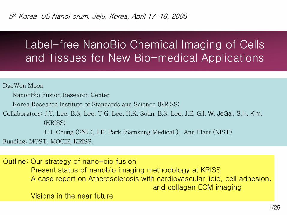
How to utilize NT to solve Biomedical Issues through noble methodologies
Today90 nm Node
Strain Enhanced Mobility
Tomorrow
New Materials
CMOSpMOS
FINFET
Future15 years
Non-classical CMOS
Beyond CMOSsource
Gate
drain
source
Gate
drain
Molecular Switches ? Nanowire Transistor ?
1948 First Transistor
STM/AFM, TEM/SEM, XRD,PES/AES, SIMS, RBS/MEIS,
Raman, ALD, QD, FIB, …..
Solving Bio Issues with NTHigh throughput
Noble analysis & manipulation
Nano-Bio Fusion
3/25nm mmmμm
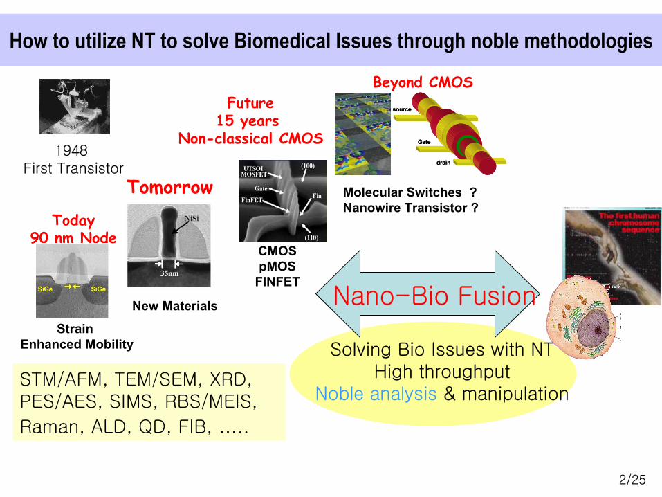
Label-free single cells/tissue biochemical imagingfor medical & pharmaceutical applications
Analysis Demands from BioAnalysis Demands from Bio--Medical R&DMedical R&D: : inin--vivo/invivo/in--vitro, biochemical imaging, dynamicsvitro, biochemical imaging, dynamics
sensitivity & selectivity, general methodologysensitivity & selectivity, general methodology
Large Gap between Molecular Biology and Medical Applications
Genomics
Proteomics
Phenomics
Systems biology
Cellomics Organomics
Physiomics
Tool Box
RNA
Protein
CellOrgan
BioinformaticsDNA
Humanomics
Bottom-Up Top-Down
4/25
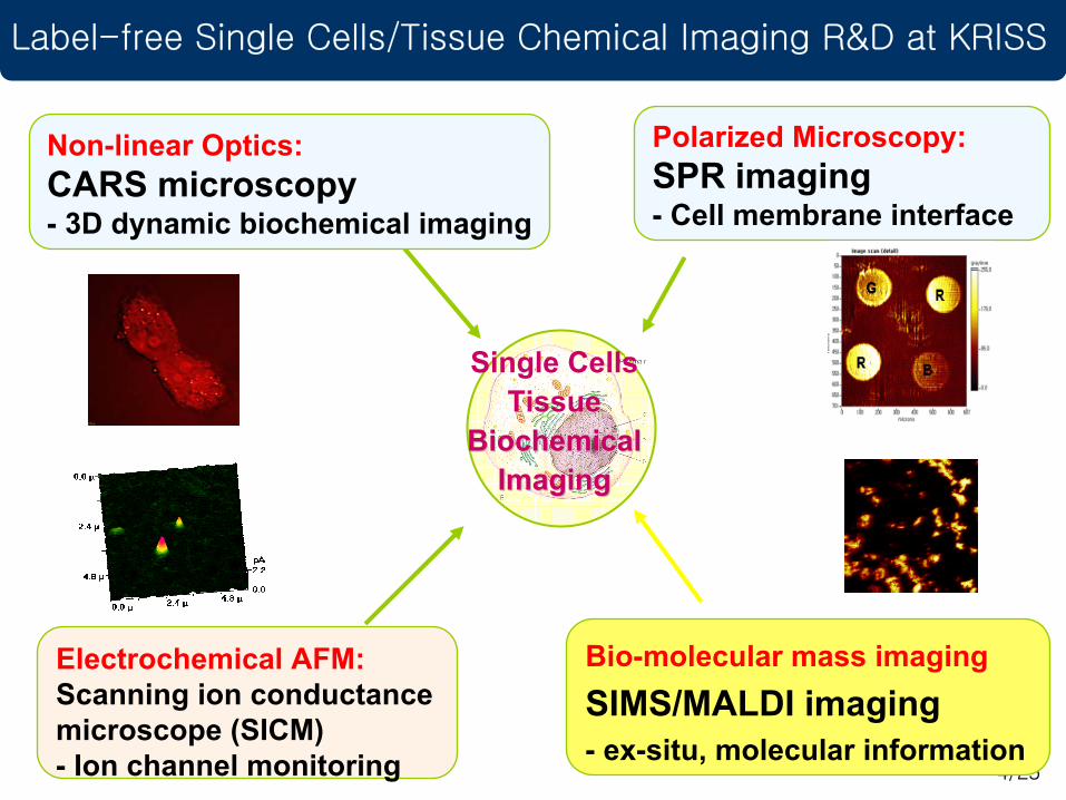
Label-free Single Cells/Tissue Chemical Imaging R&D at KRISS
Electrochemical AFM:Scanning ion conductance microscope (SICM)- Ion channel monitoring
Bio-molecular mass imagingSIMS/MALDI imaging- ex-situ, molecular information
Non-linear Optics:CARS microscopy- 3D dynamic biochemical imaging
Single CellsTissue
BiochemicalBiochemicalImagingImaging
Polarized Microscopy:SPR imaging- Cell membrane interface
5/25
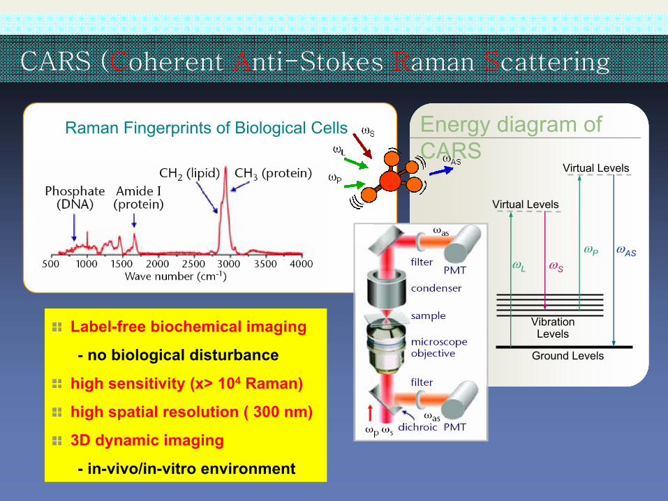
CARS (Coherent Anti-Stokes Raman Scattering
Raman Fingerprints of Biological Cells
ωPωS
Virtual Levels
ωASωL
Virtual Levels
Vibration Levels
Ground Levels
Energy diagram of CARS
Label-free biochemical imaging
- no biological disturbance
high sensitivity (x> 104 Raman)
high spatial resolution ( 300 nm)
3D dynamic imaging
- in-vivo/in-vitro environment
6/25
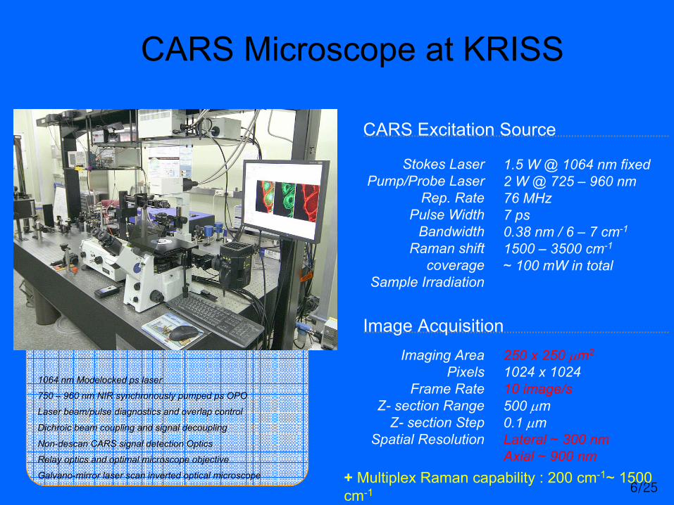
CARS Microscope at KRISS
Image Acquisition
1064 nm Modelocked ps laser
750 – 960 nm NIR synchronously pumped ps OPO
Laser beam/pulse diagnostics and overlap control
Non-descan CARS signal detection Optics
Galvano-mirror laser scan inverted optical microscope
Relay optics and optimal microscope objective
Dichroic beam coupling and signal decoupling
250 x 250 µm2
1024 x 102410 image/s500 µm0.1 µmLateral ~ 300 nmAxial ~ 900 nm
Imaging AreaPixels
Frame RateZ- section Range
Z- section StepSpatial Resolution
CARS Excitation Source
1.5 W @ 1064 nm fixed 2 W @ 725 – 960 nm76 MHz7 ps0.38 nm / 6 – 7 cm-1
1500 – 3500 cm-1
~ 100 mW in total
Stokes LaserPump/Probe Laser
Rep. RatePulse Width
BandwidthRaman shift
coverageSample Irradiation
+ Multiplex Raman capability : 200 cm-1~ 1500 cm-1
7/25
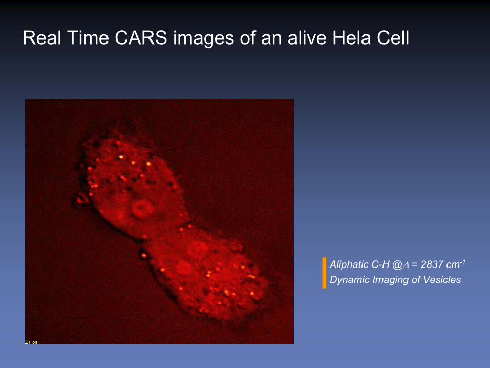
Real Time CARS images of an alive Hela Cell
Aliphatic C-H @∆ = 2837 cm-1
Dynamic Imaging of Vesicles
8/25
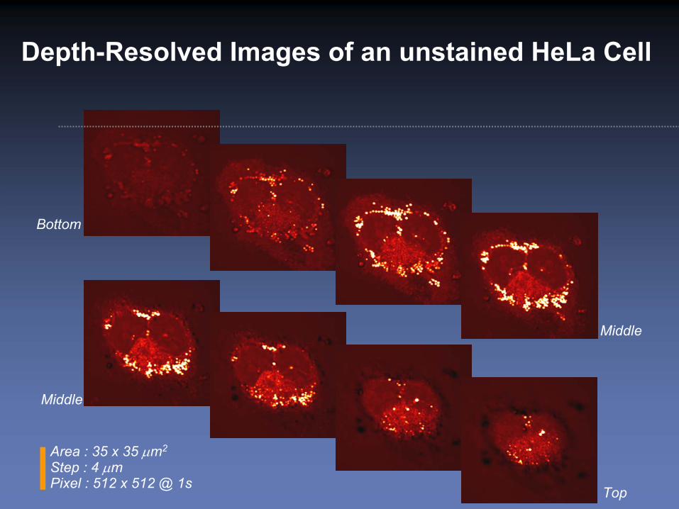
Depth-Resolved Images of an unstained HeLa Cell
Bottom
Top
Middle
Middle
Area : 35 x 35 µm2
Step : 4 µmPixel : 512 x 512 @ 1s
9/25
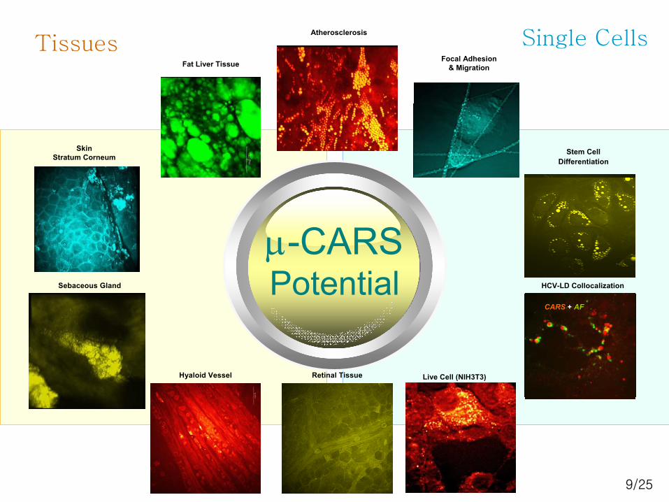
CARS 바이오 현미경의 현재 @KRISS
µ-CARS Potential HCV-LD Collocalization
Stem CellDifferentiation
Atherosclerosis
Fat Liver Tissue
Retinal TissueHyaloid Vessel
Focal Adhesion & Migration
Live Cell (NIH3T3)
SkinStratum Corneum
Sebaceous Gland
Tissues Single Cells
CARS + AF
10/25
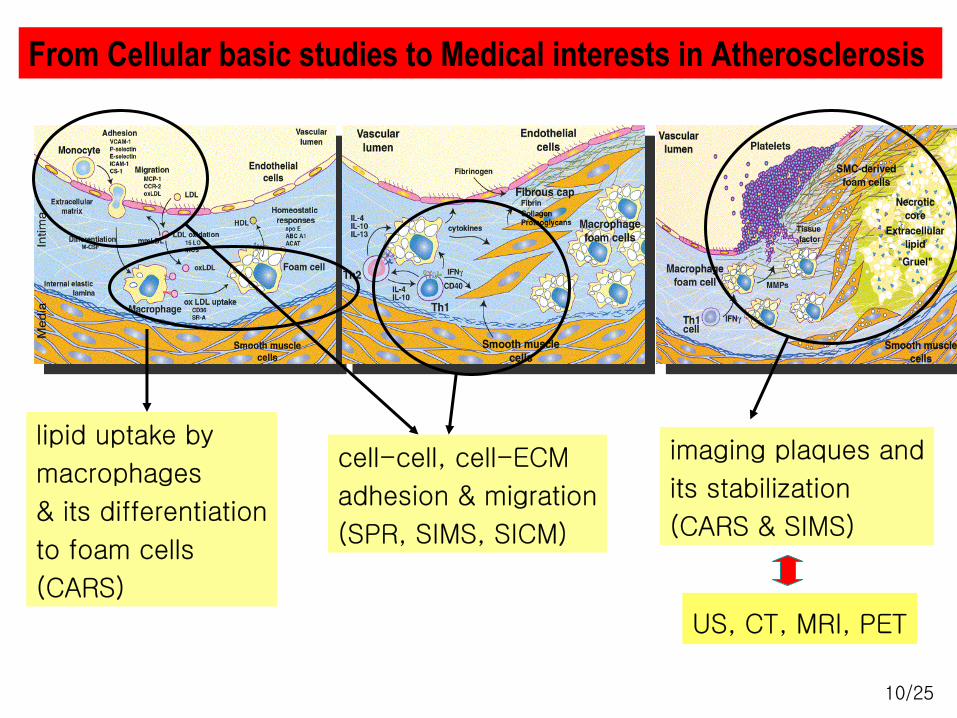
From Cellular basic studies to Medical interests in Atherosclerosis
imaging plaques and
its stabilization
(CARS & SIMS)
lipid uptake by
macrophages
& its differentiation
to foam cells
(CARS)
cell-cell, cell-ECM
adhesion & migration
(SPR, SIMS, SICM)
US, CT, MRI, PET
11/25
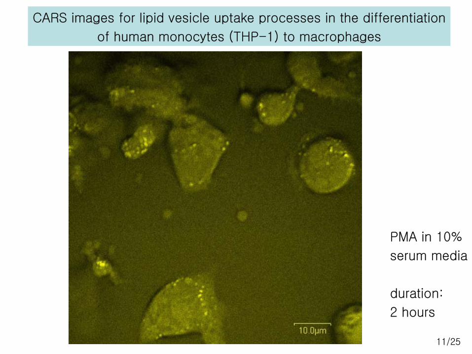
CARS images for lipid vesicle uptake processes in the differentiation
of human monocytes (THP-1) to macrophages
PMA in 10%
serum media
duration:
2 hours
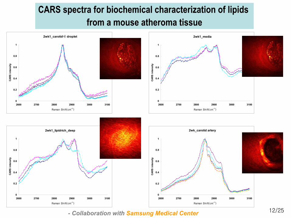
12/25
2wk1_carotid내 droplet
0
0.2
0.4
0.6
0.8
1
2600 2700 2800 2900 3000 3100
Raman Shift(cm-1)
CAR
S in
tens
ity
2wk1_media
0
0.2
0.4
0.6
0.8
1
2600 2700 2800 2900 3000 3100
Raman Shift(cm-1)
CA
RS
inte
nsity
2wk1_lipidrich_deep
0
0.2
0.4
0.6
0.8
1
2600 2700 2800 2900 3000 3100
Raman Shift(cm-1)
CARS
inte
nsity
2wk_carotid artery
0
0.2
0.4
0.6
0.8
1
2600 2700 2800 2900 3000 3100
Raman Shift(cm-1)
CAR
S in
tens
ity
CARS spectra for biochemical characterization of lipids from a mouse atheroma tissue
- Collaboration with Samsung Medical Center
13/25
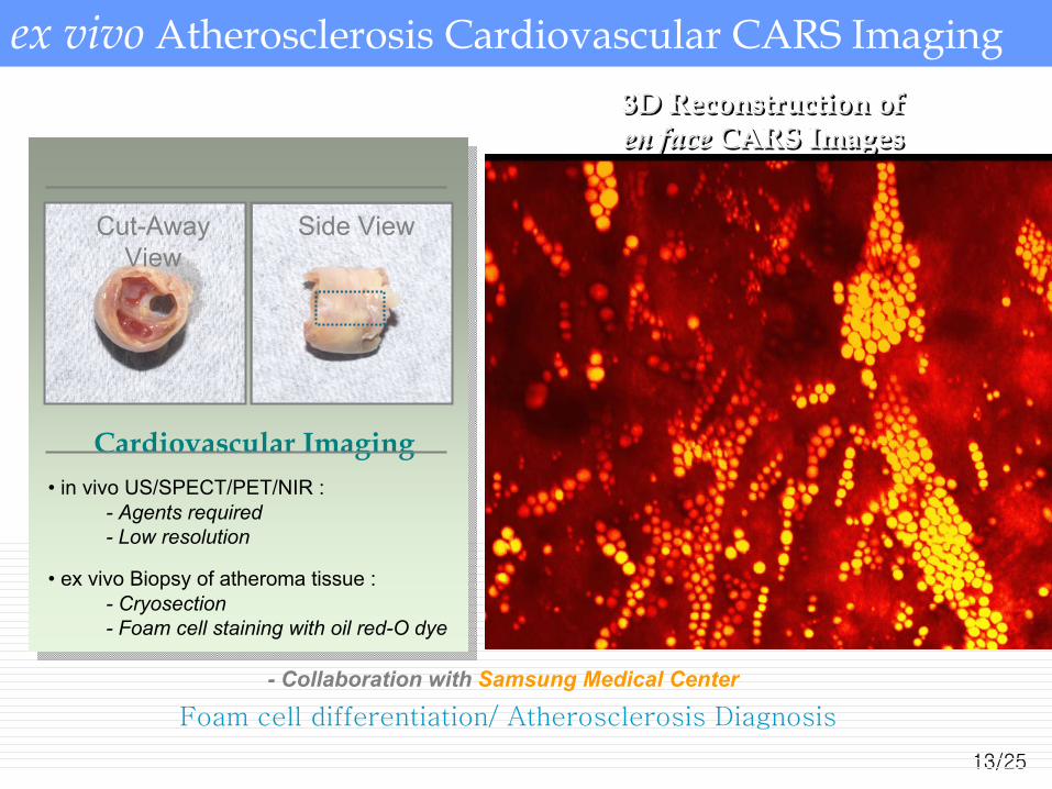
ex vivo Atherosclerosis Cardiovascular CARS Imaging
- Collaboration with Samsung Medical Center
Foam cell differentiation/ Atherosclerosis Diagnosis
3D Reconstruction of en face CARS Images3D Reconstruction of 3D Reconstruction of en faceen face CARS ImagesCARS Images
Side ViewCut-Away View
Cardiovascular Imaging• in vivo US/SPECT/PET/NIR :
• ex vivo Biopsy of atheroma tissue :- Cryosection- Foam cell staining with oil red-O dye
- Agents required- Low resolution
14/25
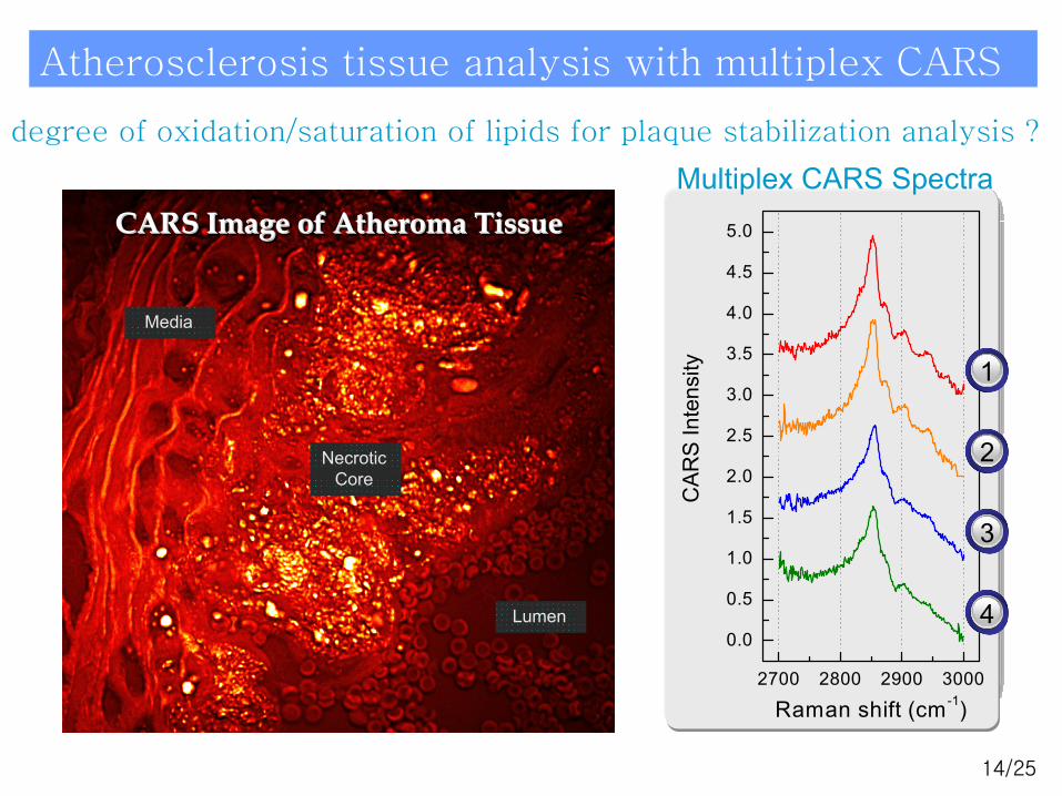
2700 2800 2900 3000
0.0
0.5
1.0
1.5
2.0
2.5
3.0
3.5
4.0
4.5
5.0
CA
RS
Inte
nsity
Raman shift (cm-1)
Atherosclerosis tissue analysis with multiplex CARS
Necrotic CoreNecrotic CoreNecrotic Core Fatty Streakin the Media Region
Fatty StreakFatty Streakin the Media Regionin the Media Region
Large Dropletin the Adventitia
Large DropletLarge Dropletin the Adventitiain the Adventitia
Pin-shaped Lump(Unidentified)
PinPin--shaped Lumpshaped Lump(Unidentified)(Unidentified)
1
2
3
4
1
2
34
CARS Image of Atheroma TissueCARS Image of CARS Image of AtheromaAtheroma TissueTissue
Necrotic Core
Necrotic Core
LumenLumen
MediaMedia
Multiplex CARS Spectradegree of oxidation/saturation of lipids for plaque stabilization analysis ?
15/25
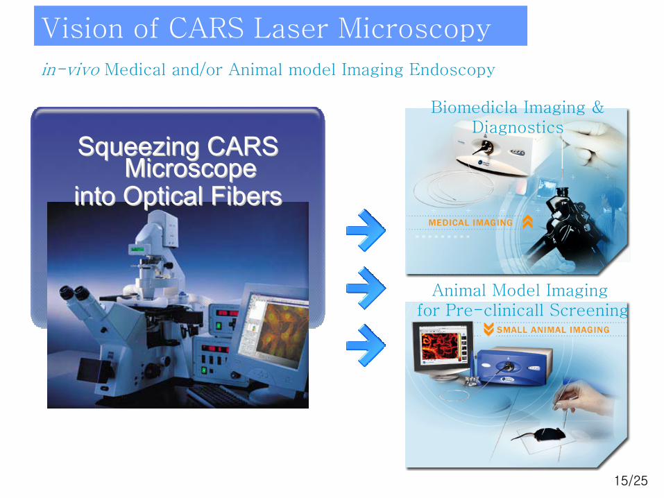
Vision of CARS Laser Microscopy
Biomedicla Imaging & Diagnostics
Animal Model Imagingfor Pre-clinicall Screening
in-vivo Medical and/or Animal model Imaging Endoscopy
Squeezing CARS Squeezing CARS Microscope Microscope
into Optical Fibersinto Optical Fibers
16/25
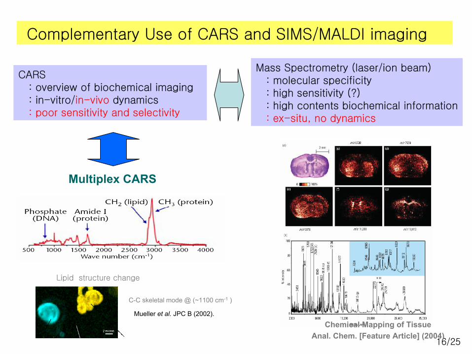
Complementary Use of CARS and SIMS/MALDI imaging
CARS: overview of biochemical imaging: in-vitro/in-vivo dynamics: poor sensitivity and selectivity
Mass Spectrometry (laser/ion beam): molecular specificity: high sensitivity (?): high contents biochemical information: ex-situ, no dynamics
Multiplex CARS
Chemical Mapping of TissueAnal. Chem. [Feature Article] (2004)
Lipid structure change
C-C skeletal mode @ (~1100 cm-1 )
Mueller et al. JPC B (2002).
17/25
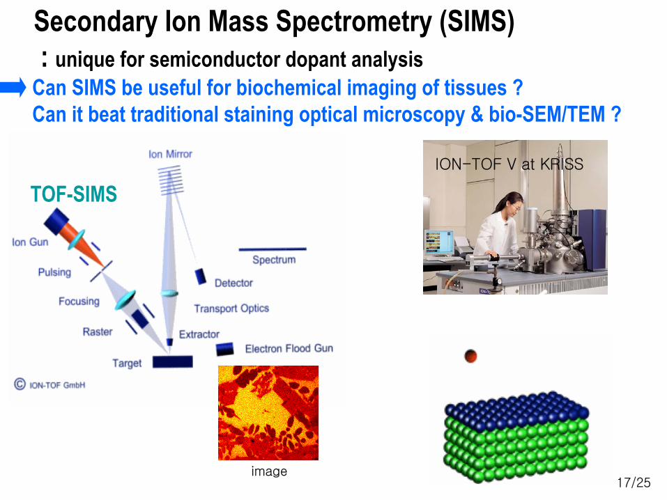
Secondary Ion Mass Spectrometry (SIMS): unique for semiconductor dopant analysis
Can SIMS be useful for biochemical imaging of tissues ? Can it beat traditional staining optical microscopy & bio-SEM/TEM ?
TOF-SIMSION-TOF V at KRISS
image
18/25
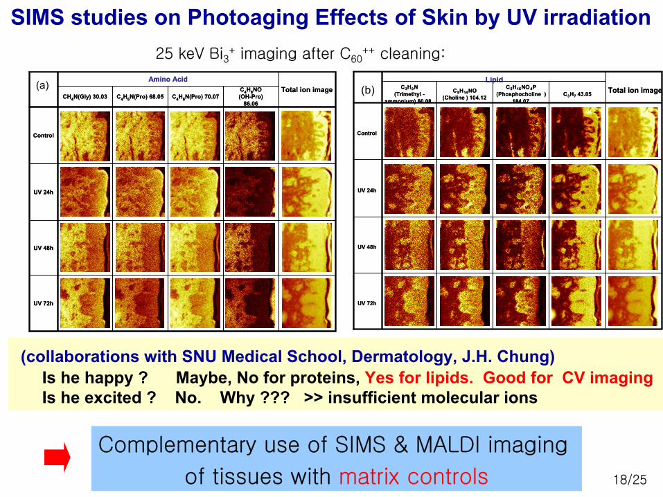
Total ion imageC4H8NO(OH-Pro)
86.06C4H8N(Pro) 70.07C4H6N(Pro) 68.05
Amino Acid
CH4N(Gly) 30.03
Control
UV 72h
UV 48h
UV 24h
Total ion imageC4H8NO(OH-Pro)
86.06C4H8N(Pro) 70.07C4H6N(Pro) 68.05
Amino Acid
CH4N(Gly) 30.03
Control
UV 72h
UV 48h
UV 24h
(a)
SIMS studies on Photoaging Effects of Skin by UV irradiation 25 keV Bi3
+ imaging after C60++ cleaning:
(collaborations with SNU Medical School, Dermatology, J.H. Chung) Is he happy ? Maybe, No for proteins, Yes for lipids. Good for CV imagingIs he excited ? No. Why ??? >> insufficient molecular ions
Total ion imageC3H7 43.05C5H15NO4P
(Phosphocholine ) 184.07
C5H14NO (Choline ) 104.12
LipidC3H9N
(Trimethyl -ammonium) 60.08
Control
UV 72h
UV 48h
UV 24h
Total ion imageC3H7 43.05C5H15NO4P
(Phosphocholine ) 184.07
C5H14NO (Choline ) 104.12
LipidC3H9N
(Trimethyl -ammonium) 60.08
Control
UV 72h
UV 48h
UV 24h
(b)
Complementary use of SIMS & MALDI imaging
of tissues with matrix controls
19/25
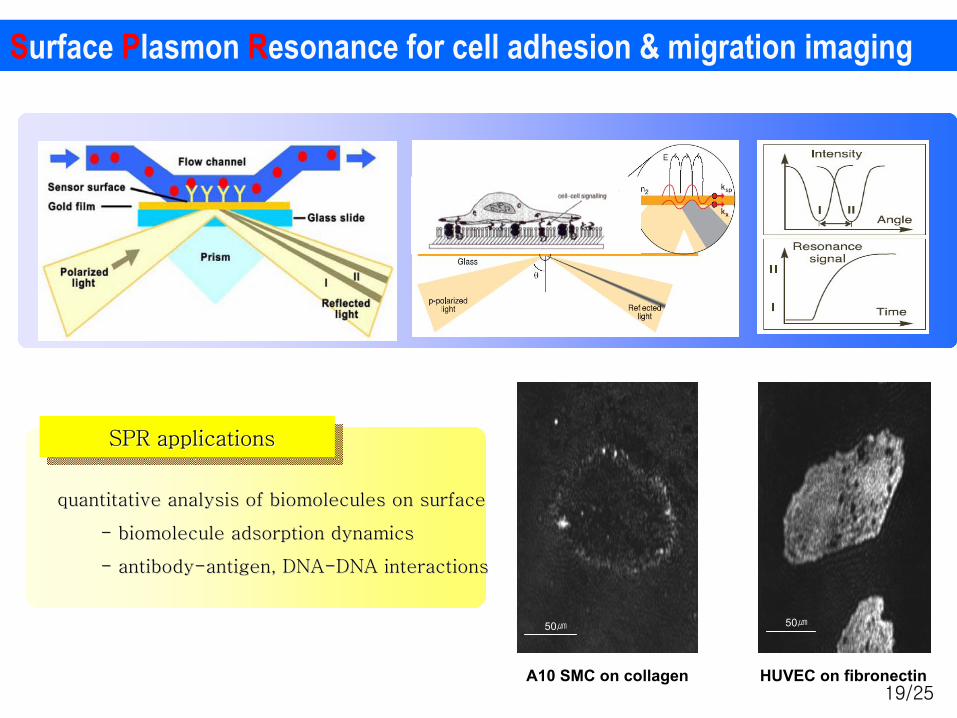
Surface Plasmon Resonance for cell adhesion & migration imaging
quantitative analysis of quantitative analysis of biomoleculesbiomolecules on surfaceon surface
-- biomoleculebiomolecule adsorption dynamicsadsorption dynamics
-- antibodyantibody--antigen, DNAantigen, DNA--DNA interactionsDNA interactions
SPR applicationsSPR applicationsSPR applications
HUVEC on fibronectinA10 SMC on collagen
50㎛50㎛
20/25
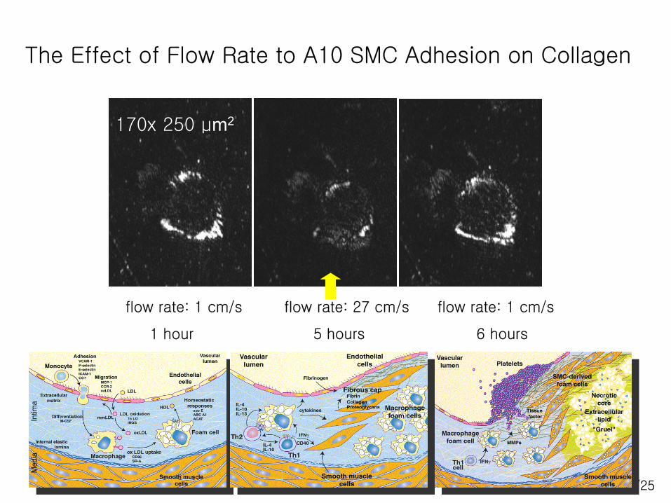
The Effect of Flow Rate to A10 SMC Adhesion on Collagen
flow rate: 27 cm/s
5 hours
flow rate: 1 cm/s
6 hours
flow rate: 1 cm/s
1 hour
170x 250 μm2
21/25
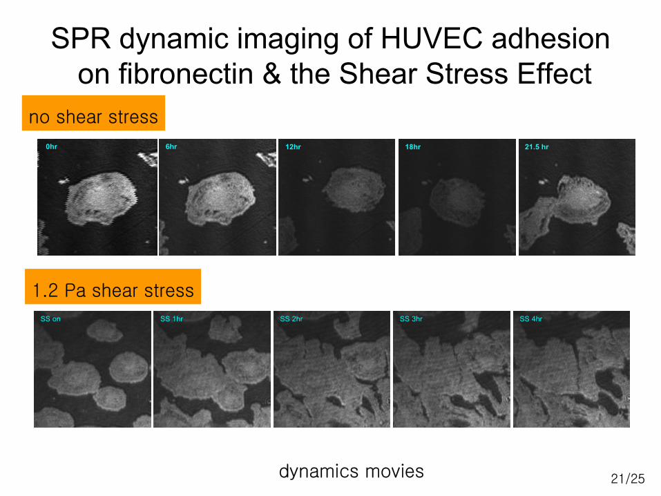
SPR dynamic imaging of HUVEC adhesionon fibronectin & the Shear Stress Effect
SS on SS 1hr SS 2hr SS 3hr SS 4hr
6hr 12hr 18hr 21.5 hr0hr
no shear stress
1.2 Pa shear stress
dynamics movies
22/25
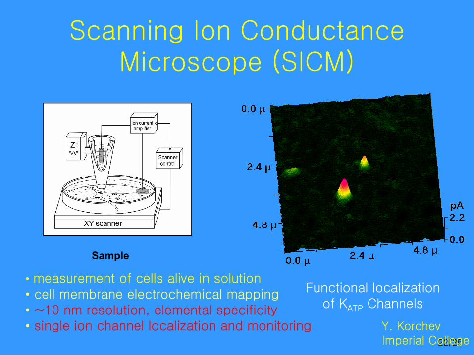
Scanning Ion Conductance Microscope (SICM)
Sample
Functional localization of KATP Channels
• measurement of cells alive in solution• cell membrane electrochemical mapping• ~10 nm resolution, elemental specificity• single ion channel localization and monitoring Y. Korchev
Imperial College
23/25
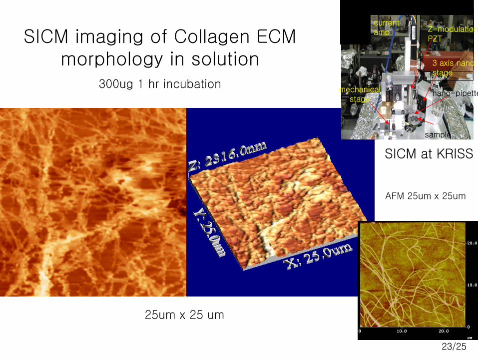
25um x 25 um
SICM imaging of Collagen ECM morphology in solution
300ug 1 hr incubation
AFM 25um x 25um
3 axis nanostage
mechanicalstage
nano-pipette
sample
Z-modulation PZT
current amp
SICM at KRISS
24/25
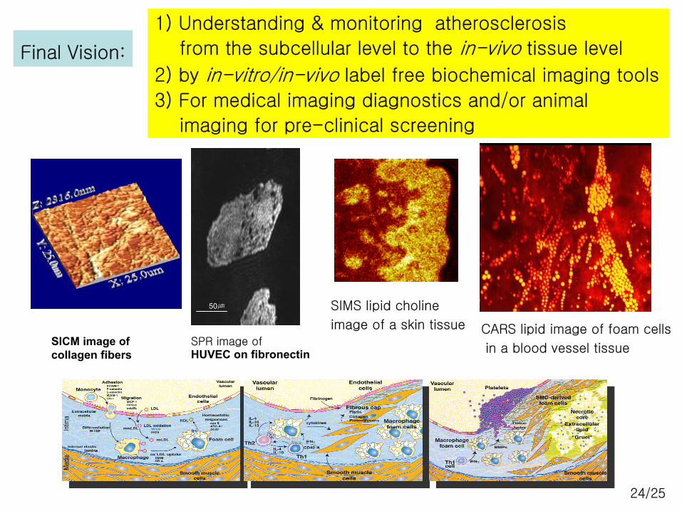
1) Understanding & monitoring atherosclerosis
from the subcellular level to the in-vivo tissue level
2) by in-vitro/in-vivo label free biochemical imaging tools
3) For medical imaging diagnostics and/or animal
imaging for pre-clinical screening
SPR image ofHUVEC on fibronectin
SICM image ofcollagen fibers
50㎛
Final Vision:
CARS lipid image of foam cells
in a blood vessel tissue
SIMS lipid choline
image of a skin tissue
25/25
1. Label1. Label--free tools such as CARS, biofree tools such as CARS, bio--SIMS, SPR, SICM can be used SIMS, SPR, SICM can be used as noble and complementary tools in biochemical imaging of as noble and complementary tools in biochemical imaging of single cells/tissues for cell biology and medical diagnostics.single cells/tissues for cell biology and medical diagnostics.
2. If it works nicely for 2. If it works nicely for atherosclerosis, it can be extended to study other diseases and to understanding EHS issues of nanomaterialsfor improvement of the quality of life.
3. To tackle these issues, global collaborations are mandatory a3. To tackle these issues, global collaborations are mandatory and nd beneficial to all of us. beneficial to all of us.
Why not between Korea and USA ! Why not between Korea and USA !
Conclusions Conclusions

![DOI: 10.1002/cphc.201100890 Label-Free Live-Cell Imaging of … · Label-Free Live-Cell Imaging of Nucleic Acids Using Stimulated Raman Scattering Microscopy Xu Zhang,[a, b] Maarten](https://img.pdfslide.net/doc/110x75/5c80d89f09d3f2f3348b90d7/doi-101002cphc201100890-label-free-live-cell-imaging-of-label-free-live-cell.jpg)