Embed Size (px)
Citation preview
LCH - III(2nd Version: January 2002)
Treatment Protocol of the
Third International Study
for
LANGERHANSCELL HISTIOCYTOSIS
START OF THE STUDY: April 2001
2
STUDY REFER CENTER
Gadner Helmut, MD, chairman
Grois Nicole, MD, study coordinator
Minkov Milen, MD, study coordinator
Pötschger Ulrike, MSc, statistician
Thiem Elfriede, data manager
St. Anna Children’s Hospital
Kinderspitalgasse 6
A -1090 Vienna, Austria
Tel.: (43-1)-40170/250 (H. Gadner)
(43-1)-40170/476 (LCH - office)
Fax: (43-1)-40170/430
E-mail: [email protected]
STUDY SUBCENTERS STUDY COORDINATORS
Argentina Braier Jorge, MD
Austria, Germany, Netherlands, Switzerland Gadner Helmut, MD,
Grois Nicole, MD,
Minkov Milen, MD,
Canada Weitzman Sheila, MD
France Donadieu Jean, MD, Ph.D.
Great Britain, Ireland Windebank Kevin, MD
Italy Arico Maurizio, MD
Scandinavia Henter Jan-Inge, MD
U.S.A. McClain Ken, MD
3
PATHOLOGY REFERENCE CENTER
Jaffe Ron, MD, head of the pathologist’s panel
Regional Pathology Centers Local Pathology Coordinators
Austria Simonitsch Ingrid, MD
Argentina Goldberg Julio, MD
France Emile Jean Francoise MD, Ph.D.
Germany Harms Dieter, MD, Schmidt Dieter, MD
Italy Paulli Marco, MD
Scandinavia Abiel Orrego, MD
U.S.A. Jaffe Ron, MD
DATA SAFETY MONITORING BOARD
Faldum Andreas, Ph.D., statistician
Michaelis Jörg, Ph.D., statistician
Otten Jaques, MD, clincian
Tubergan David, MD, clinician
STUDY COMMITTEE
Arico, Maurizio Pavia Italy
Braier, Jorge Buenos Aires Argentina
Donadieu, Jean Paris France
Gadner, Helmut Vienna Austria
Henter, Jan-Inge Stockholm Sweden
Janka-Schaub, Gritta Hamburg Germany
Komp, Diane New Haven USA
Ladisch, Stephan Washington D.C. USA
McClain, Kenneth Houston USA
Weitzmann, Sheila Toronto Canada
Windebank, Kevin Newcastle upon Tyne Great Britain
4
TABLE OF CONTENTS1 ADDRESSES..................................................................................................... 6
2 BACKGROUND............................................................................................... 12
2.1 CONCLUSIONS OF LCH II.............................................................................. 19
2.2 RATIONALE FOR THE USE OF METHOTREXATE IN “RISK” PATIENTS..... 20
2.3 SITUATION IN PATIENTS WITH MULTIFOCAL BONE DISEASE AND
“SPECIAL SITES” OF DISEASE...................................................................... 20
3 PATIENT’S ELIGIBILITY FOR LCH III ............................................................ 22
4 LCH III STUDY REQUIREMENTS................................................................... 22
4.1 HISTOPATHOLOGICAL DIAGNOSTIC CRITERIA ......................................... 22
4.2 BASELINE DIAGNOSTIC EVALUATIONS ...................................................... 23
4.3 EVALUATIONS REQUIRED UPON SPECIFIC INDICATION.......................... 24
4.4 DEFINITION OF ORGAN INVOLVEMENT ...................................................... 25
5 STRATIFICATION ........................................................................................... 26
5.1 GROUP 1 - MULTISYSTEM “RISK” PATIENTS .............................................. 26
5.2 GROUP 2 - MULTISYSTEM “LOW RISK” PATIENTS .................................... 26
5.3 GROUP 3 - SINGLE SYSTEM “MULTIFOCAL BONE DISEASE” and
LOCALIZED “SPECIAL SITE” INVOLVEMENT ............................................... 27
6 GOALS FOR LCH III ...................................................................................... 27
6.1 Group 1: MULTISYSTEM “RISK” Patients ....................................................... 27
6.2 Group 2: MULTISYSTEM “LOW RISK” Patients .............................................. 28
6.3 GROUP 3: SINGLE SYSTEM MULTIFOCAL BONE and LOCALIZED
“SPECIAL SITES” ............................................................................................ 28
7 STUDY DESIGN .............................................................................................. 28
8 REGISTRATION AND RANDOMIZATION...................................................... 29
8.1 REGISTRATION .............................................................................................. 29
5
8.2 RANDOMISATION........................................................................................... 29
9 TREATMENT ................................................................................................... 29
9.1 GROUP 1: MULTISYSTEM “RISK” PATIENTS .............................................. 29
9.2 GROUP 2: “LOW RISK” GROUP ..................................................................... 32
9.3 GROUP 3: “MULTIFOCAL BONE DISEASE” AND “SPECIAL SITES” ............ 33
9.4 SUPPORTIVE CARE GUIDELINES ................................................................ 33
9.5 TOXICITY......................................................................................................... 34
9.6 Therapy modifications ...................................................................................... 35
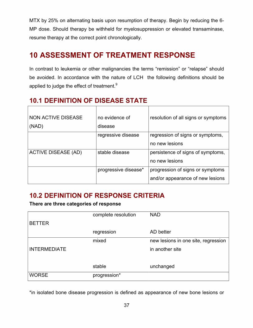
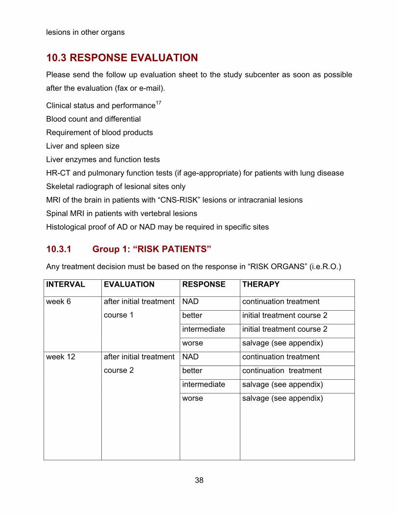
10 ASSESSMENT OF TREATMENT RESPONSE............................................... 37
10.1 DEFINITION OF DISEASE STATE.................................................................. 37
10.2 DEFINITION OF RESPONSE CRITERIA ........................................................ 37
10.3 RESPONSE EVALUATION.............................................................................. 38
11 OFF STUDY CRITERIA................................................................................... 41
12 AUTOPSY........................................................................................................ 41
13 FOLLOW UP INVESTIGATIONS AFTER STOP OF THERAPY..................... 42
14 DATA COLLECTION AND EVALUATION...................................................... 43
15 DATA SAFETY MONITORING BOARD (DSMB)............................................ 44
16 STATISTICAL CONSIDERATIONS ................................................................ 44
16.1 DESIGN ........................................................................................................... 45
16.2 ENDPOINTS .................................................................................................... 45
16.3 ANALYSES ...................................................................................................... 47
16.4 INTERIM-ANALYSES ...................................................................................... 49
16.5 POWER CONSIDERATION............................................................................. 51
16.6 STOPPING RULES.......................................................................................... 52
17 PUBLICATION................................................................................................. 54
18 REFERENCES................................................................................................. 54
6
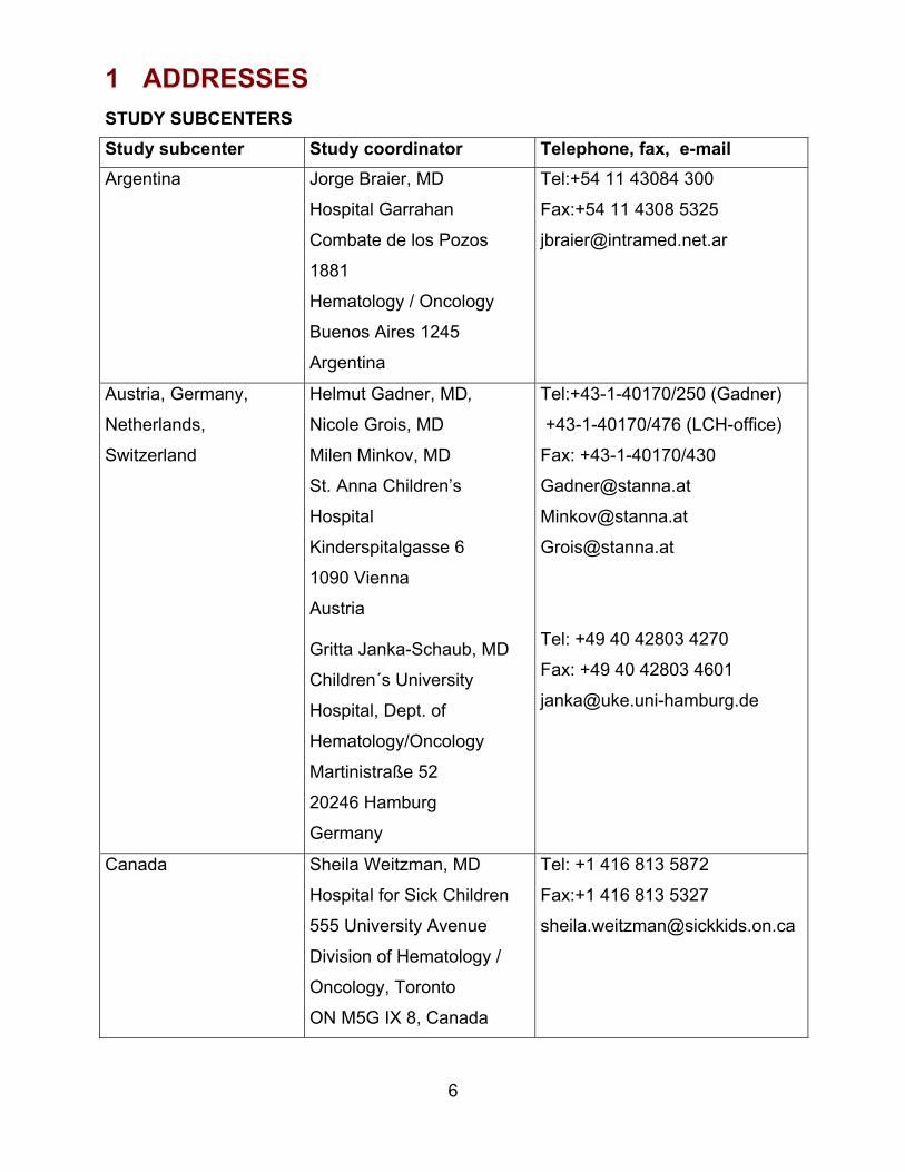
1 ADDRESSESSTUDY SUBCENTERSStudy subcenter Study coordinator Telephone, fax, e-mailArgentina Jorge Braier, MD
Hospital Garrahan
Combate de los Pozos
1881
Hematology / Oncology
Buenos Aires 1245
Argentina
Tel:+54 11 43084 300
Fax:+54 11 4308 5325
Austria, Germany,
Netherlands,
Switzerland
Helmut Gadner, MD,
Nicole Grois, MD
Milen Minkov, MD
St. Anna Children’s
Hospital
Kinderspitalgasse 6
1090 Vienna
Austria
Gritta Janka-Schaub, MD
Children´s University
Hospital, Dept. of
Hematology/Oncology
Martinistraße 52
20246 Hamburg
Germany
Tel:+43-1-40170/250 (Gadner)
+43-1-40170/476 (LCH-office)
Fax: +43-1-40170/430
Tel: +49 40 42803 4270
Fax: +49 40 42803 4601
Canada Sheila Weitzman, MD
Hospital for Sick Children
555 University Avenue
Division of Hematology /
Oncology, Toronto
ON M5G IX 8, Canada
Tel: +1 416 813 5872
Fax:+1 416 813 5327
7
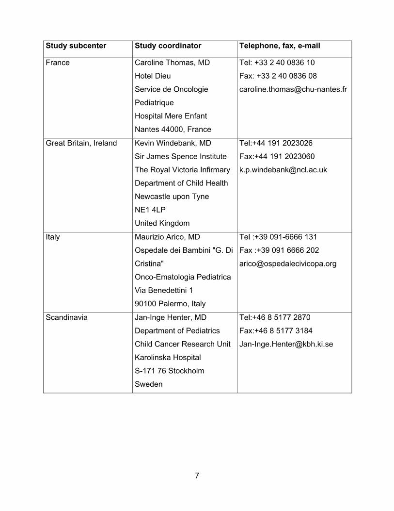
Study subcenter Study coordinator Telephone, fax, e-mail
France Caroline Thomas, MD
Hotel Dieu
Service de Oncologie
Pediatrique
Hospital Mere Enfant
Nantes 44000, France
Tel: +33 2 40 0836 10
Fax: +33 2 40 0836 08
Great Britain, Ireland Kevin Windebank, MD
Sir James Spence Institute
The Royal Victoria Infirmary
Department of Child Health
Newcastle upon Tyne
NE1 4LP
United Kingdom
Tel:+44 191 2023026
Fax:+44 191 2023060
Italy Maurizio Arico, MD
Ospedale dei Bambini "G. Di
Cristina"
Onco-Ematologia Pediatrica
Via Benedettini 1
90100 Palermo, Italy
Tel :+39 091-6666 131
Fax :+39 091 6666 202
Scandinavia Jan-Inge Henter, MD
Department of Pediatrics
Child Cancer Research Unit
Karolinska Hospital
S-171 76 Stockholm
Sweden
Tel:+46 8 5177 2870
Fax:+46 8 5177 3184
8
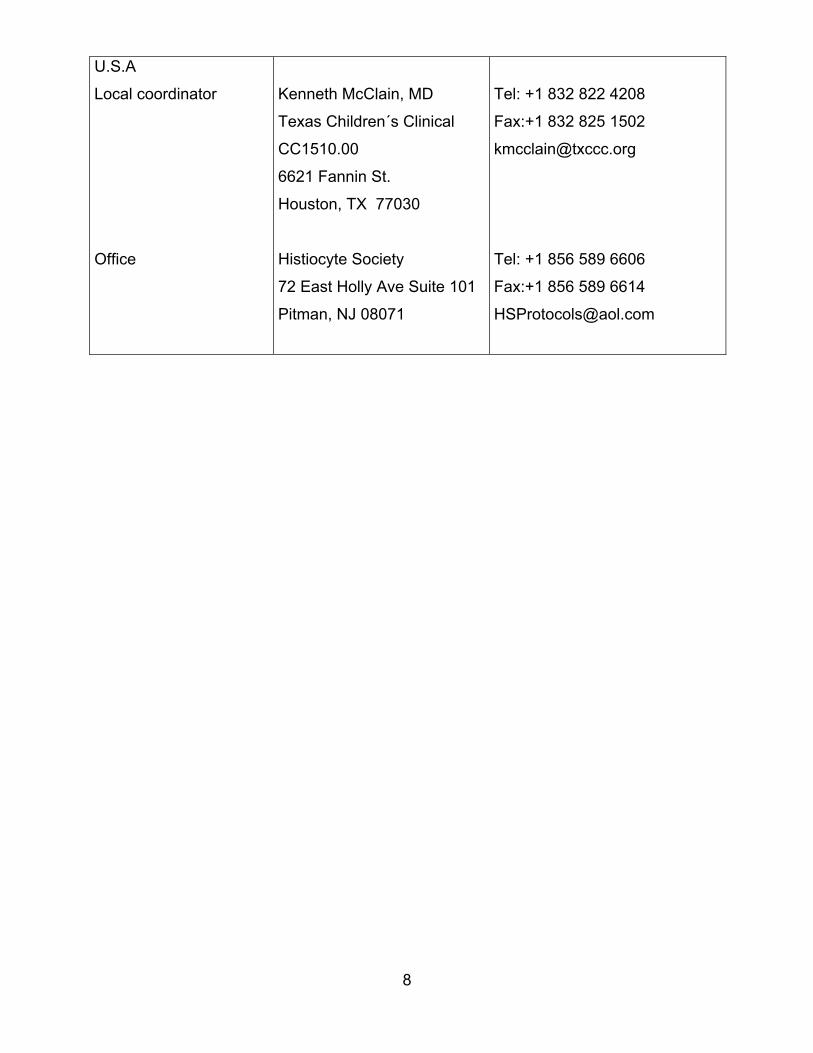
U.S.A
Local coordinator
Office
Kenneth McClain, MD
Texas Children´s Clinical
CC1510.00
6621 Fannin St.
Houston, TX 77030
Histiocyte Society
72 East Holly Ave Suite 101
Pitman, NJ 08071
Tel: +1 832 822 4208
Fax:+1 832 825 1502
Tel: +1 856 589 6606
Fax:+1 856 589 6614
9
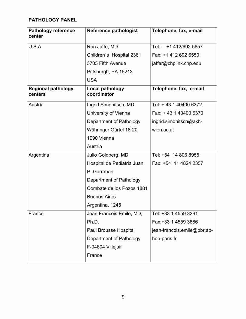
PATHOLOGY PANEL
Pathology referencecenter
Reference pathologist Telephone, fax, e-mail
U.S.A Ron Jaffe, MD
Children´s Hospital 2361
3705 Fifth Avenue
Pittsburgh, PA 15213
USA
Tel.: +1 412/692 5657
Fax: +1 412 692 6550
Regional pathologycenters
Local pathologycoordinator
Telephone, fax, e-mail
Austria Ingrid Simonitsch, MD
University of Vienna
Department of Pathology
Währinger Gürtel 18-20
1090 Vienna
Austria
Tel: + 43 1 40400 6372
Fax: + 43 1 40400 6370
ingrid.simonitsch@akh-
wien.ac.at
Argentina Julio Goldberg, MD
Hospital de Pediatria Juan
P. Garrahan
Department of Pathology
Combate de los Pozos 1881
Buenos Aires
Argentina, 1245
Tel: +54 14 806 8955
Fax: +54 11 4824 2357
France Jean Francois Emile, MD,
Ph.D.
Paul Brousse Hospital
Department of Pathology
F-94804 Villejuif
France
Tel: +33 1 4559 3291
Fax:+33 1 4559 3886
hop-paris.fr
10
Regional pathologycenters
Local pathologycoordinator
Telephone, fax, e-mail
Germany Dieter Harms, MD
University of Kiel
Department of Pediatric
Pathology
Michaelisstraße 11
24105 Kiel, Germany
Dietmar Schmidt, MD
Institute of Pathology
A 2,2
68159 Mannheim
Germany
Tel: +49 431 597 3450
Fax:+49 434 597 3486
Tel: +49 621 2277 9
Fax: +49 621 15328 8
Italy Marco Paulli, MD
Istituto di Anatomia
Patologica
IRCCS Policlinico San
Matteo
27100 Pavia
Italy
Tel: +39 0382 501241
Fax: +39 0382
Scandinavia Abiel Orrego, MD
Karolinska Hospital
Division of Pathology and
Cytology
Paediatric section
S – 171 76 Stockholm
Sweden
Tel: +46 8 5177 5147
Fax: +46 8 5177 4524
11
DATA SAFETY MONITORING BOARD
Study statistician Ulrike Pötschger, M.Sc.
St. Anna Children´s Hospital
Kinderspitalgasse 6
A-1090 Vienna
Tel: +43 1 40 170 477
Fax: +43 1 40 170 430
External biometrical
monitoring
Jörg Michaelis, Ph.D.
Andreas Faldum, Ph.D.
Institut für Medizinische
Statistik und Dokumentation
Klinikum der Johannes
Gutenberg-Universität Mainz
Obere Zahlbacher Str. 69
D-55101 Mainz
Germany
Tel. + 49 6131-17-3938
Fax + 49 6131-17-473938
mainz.de
Clinician Prof. Jaques Otten, M.D.
Professor of Pediatrics
101 Laarbeeklaan
Free University of Brussels
Hospital
B- 1090 Brussels
Belgium
Tel: + 32 2477 5775
Fax + 32 2477 5783
Clinician David Tubergan, M.D
Professor of Pediatrics
Anderson Cancer
Center
1515 Holcombe, Box 087
Houston, Texas 77030
Tel: + 713-745-0886
Fax +713-745-1549
12
2 BACKGROUND
Langerhans cell histiocytosis (LCH) is a rare disease that may affect any age group. It is
regarded as a clonal accumulation and proliferation of abnormal bone marrow derived
Langerhans cells. These dendritic cells along with lymphocytes, eosinophils and normal
histiocytes form infiltrates typical for the disease which may be found in various organs
and at different extent1. LCH includes a wide range of clinical presentations comprising
the clinical pictures of eosinophilic granuloma, Hand-Schüller-Christian syndrome or
Letterer-Siwe disease. The course of disease is unpredictable, varying from
spontaneous regression and resolution to rapid progression and death or repeated
recurrence and recrudescence with the risk of permanent consequences, defined as
irreversible long-term disabilities which are directly linked, predictable and permanent
results of the disease upon the patient2.
Patients with disease that is localized (skin, bone or lymph node) have a good prognosis
and are felt to need minimum or even no treatment. In contrast, multiple organ
involvement, which is particularly frequent in young children under 2 years, carries the
risk of a poor outcome3-6. Patients with multi-system disease benefit from therapy with
cytotoxic drugs and/or steroids, either alone or in combination as demonstrated in early
prospective multicentric studies for disseminated LCH7,8. On 1st April 1991, the
Histiocyte Society initiated LCH I - the first international clinical trial for the treatment of
multisystem LCH. It was the goal of this randomized prospective study to compare the
efficacy of monotherapy with vinblastine and etoposide (VP-16) with respect to
response, failure and morbidity. Therapy response was assessed according to the
following newly defined criteria: complete resolution of disease (no active disease,
NAD), disease regression (active disease, AD-better), intermediate response with
regression of some and reappearance of other lesions (AD-intermediate, mixed) or
unchanged disease (AD- intermediated, stable) and progression of the disease (AD-
worse). By the end of the study on August 15th, 1995 447 patients with LCH were
registered onto LCH I. 143 patients with multi-system disease were randomized on the
clinical trial, 74 patients were assigned to treatment arm A (VBL), 69 patients to
treatment arm B (VP-16). After 6 weeks of treatment (i.e. 2 treatment courses) 53% of
the patients were judged as responders (NAD or AD-better), 17% showed a progression
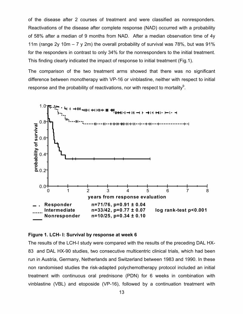
13
of the disease after 2 courses of treatment and were classified as nonresponders.
Reactivations of the disease after complete response (NAD) occurred with a probability
of 58% after a median of 9 months from NAD. After a median observation time of 4y
11m (range 2y 10m – 7 y 2m) the overall probability of survival was 78%, but was 91%
for the responders in contrast to only 34% for the nonresponders to the initial treatment.
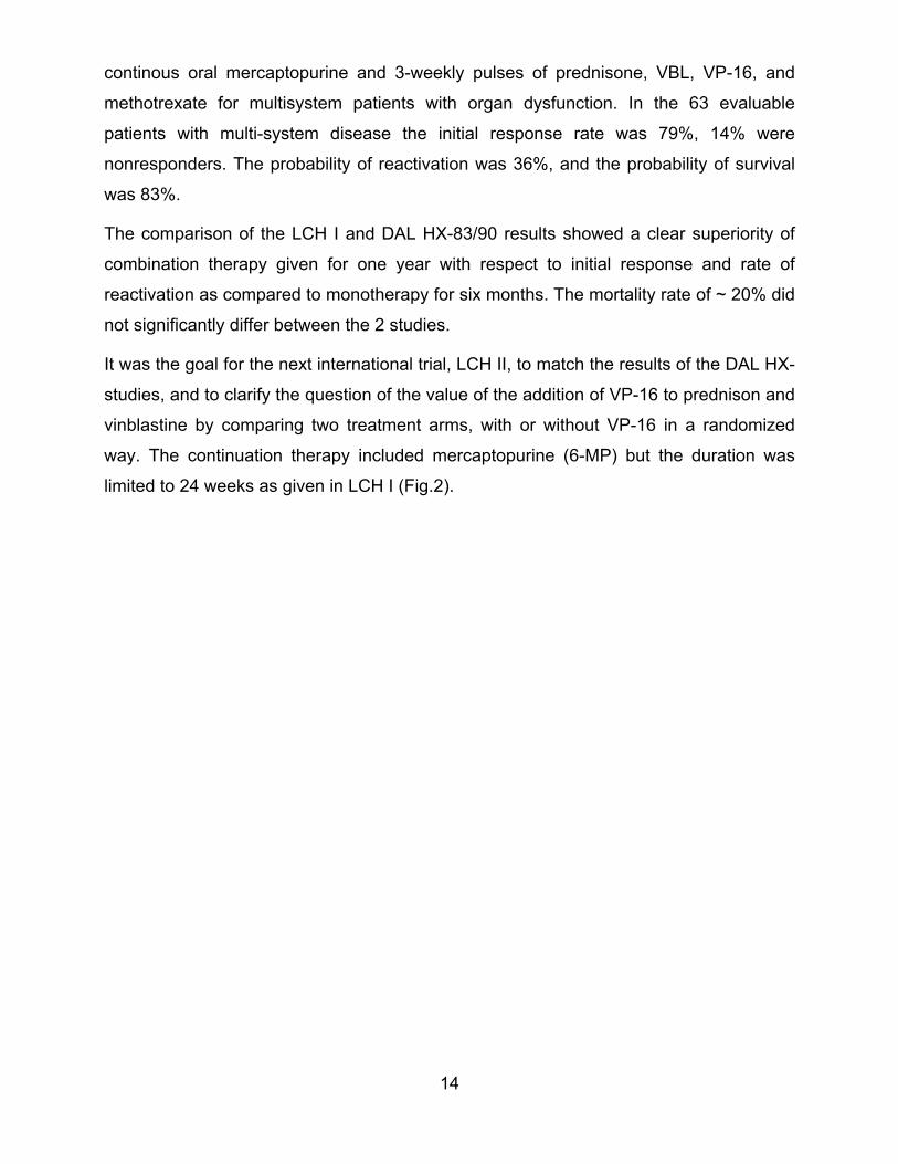
This finding clearly indicated the impact of response to initial treatment (Fig.1).
The comparison of the two treatment arms showed that there was no significant
difference between monotherapy with VP-16 or vinblastine, neither with respect to initial
response and the probability of reactivations, nor with respect to mortality9.
0 1 2 3 4 5 6 7 80.0
0.2
0.4
0.6
0.8
1.0
prob
abili
ty o
f sur
viva
l
years from response evaluationResponder n=71/76, p=0.91 ± 0.04Intermediate n=33/42, p=0.77 ± 0.07 log rank-test p<0.001Nonresponder n=10/25, p=0.34 ± 0.10
Figure 1. LCH- I: Survival by response at week 6The results of the LCH-I study were compared with the results of the preceding DAL HX-
83 and DAL HX-90 studies, two consecutive multicentric clinical trials, which had been
run in Austria, Germany, Netherlands and Switzerland between 1983 and 1990. In these
non randomised studies the risk-adapted polychemotherapy protocol included an initial
treatment with continuous oral prednisone (PDN) for 6 weeks in combination with
vinblastine (VBL) and etoposide (VP-16), followed by a continuation treatment with
14
continous oral mercaptopurine and 3-weekly pulses of prednisone, VBL, VP-16, and
methotrexate for multisystem patients with organ dysfunction. In the 63 evaluable
patients with multi-system disease the initial response rate was 79%, 14% were
nonresponders. The probability of reactivation was 36%, and the probability of survival
was 83%.
The comparison of the LCH I and DAL HX-83/90 results showed a clear superiority of
combination therapy given for one year with respect to initial response and rate of
reactivation as compared to monotherapy for six months. The mortality rate of ~ 20% did
not significantly differ between the 2 studies.
It was the goal for the next international trial, LCH II, to match the results of the DAL HX-
studies, and to clarify the question of the value of the addition of VP-16 to prednison and
vinblastine by comparing two treatment arms, with or without VP-16 in a randomized
way. The continuation therapy included mercaptopurine (6-MP) but the duration was
limited to 24 weeks as given in LCH I (Fig.2).
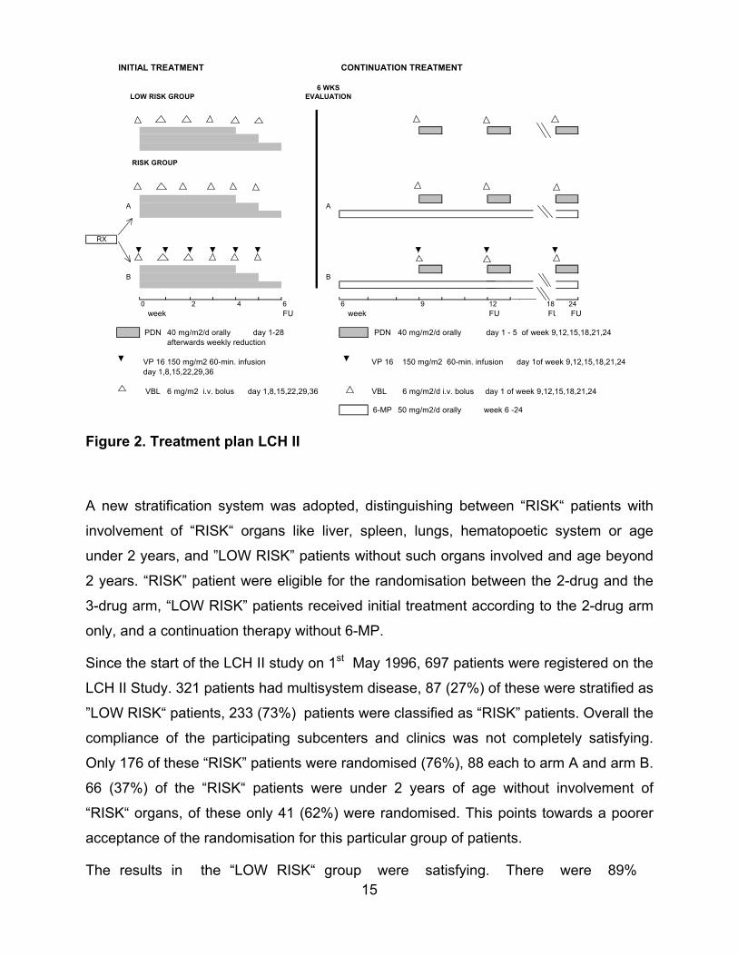
15
INITIAL TREATMENT CONTINUATION TREATMENT
6 WKS LOW RISK GROUP EVALUATION
RISK GROUP
A A
RX
B B
0 2 4 6 6 9 12 18 24 week FU week FU FU FU
PDN 40 mg/m2/d orally day 1-28 PDN 40 mg/m2/d orally day 1 - 5 of week 9,12,15,18,21,24 afterwards weekly reduction
VP 16 150 mg/m2 60-min. infusion VP 16 150 mg/m2 60-min. infusion day 1of week 9,12,15,18,21,24 day 1,8,15,22,29,36
VBL 6 mg/m2 i.v. bolus day 1,8,15,22,29,36 VBL 6 mg/m2/d i.v. bolus day 1 of week 9,12,15,18,21,24
6-MP 50 mg/m2/d orally week 6 -24
Figure 2. Treatment plan LCH II
A new stratification system was adopted, distinguishing between “RISK“ patients with
involvement of “RISK“ organs like liver, spleen, lungs, hematopoetic system or age
under 2 years, and ”LOW RISK” patients without such organs involved and age beyond
2 years. “RISK” patient were eligible for the randomisation between the 2-drug and the
3-drug arm, “LOW RISK” patients received initial treatment according to the 2-drug arm
only, and a continuation therapy without 6-MP.
Since the start of the LCH II study on 1st May 1996, 697 patients were registered on the
LCH II Study. 321 patients had multisystem disease, 87 (27%) of these were stratified as
”LOW RISK“ patients, 233 (73%) patients were classified as “RISK” patients. Overall the
compliance of the participating subcenters and clinics was not completely satisfying.
Only 176 of these “RISK” patients were randomised (76%), 88 each to arm A and arm B.
66 (37%) of the “RISK“ patients were under 2 years of age without involvement of
“RISK“ organs, of these only 41 (62%) were randomised. This points towards a poorer
acceptance of the randomisation for this particular group of patients.
The results in the “LOW RISK“ group were satisfying. There were 89%
responders, only one nonresponder at week 6, and no fatalities. Among 170 randomized
“RISK” patients, in whom the response at week 6 was available, 113 (66%) were judged
as responders. This compares favourably to the 6-week responder rate of 44% in the
LCH I study, but is less than the 76% rate of responders in the DAL HX studies.
Interestingly, the overall probability of survival of the multisystem patients did not differ
significantly between the 3 studies - DAL HX, LCH I and II, and was around 80%. This
observation indicates that there is a “High RISK” population of about 20% of the
multisystem patients which cannot be rescued with standard treatment including VBL
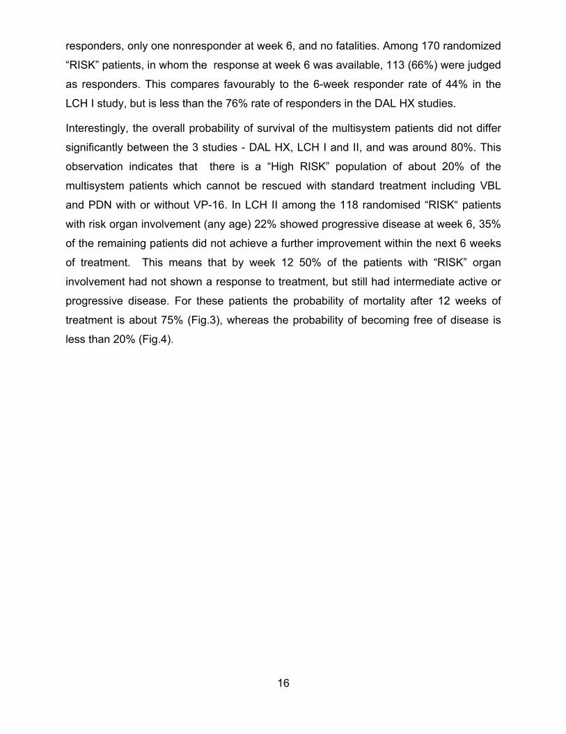
and PDN with or without VP-16. In LCH II among the 118 randomised “RISK“ patients
with risk organ involvement (any age) 22% showed progressive disease at week 6, 35%
of the remaining patients did not achieve a further improvement within the next 6 weeks
of treatment. This means that by week 12 50% of the patients with “RISK” organ
involvement had not shown a response to treatment, but still had intermediate active or
weeks of
disease is
progressive disease. For these patients the probability of mortality after 12
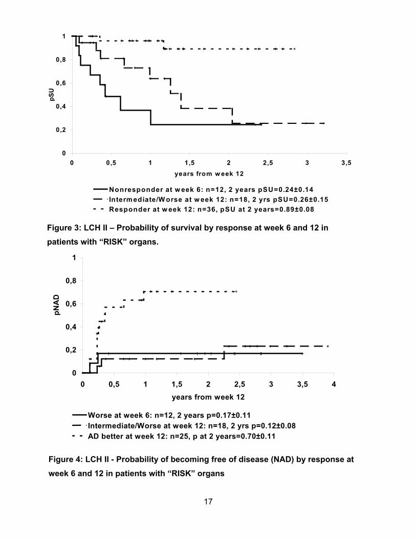
treatment is about 75% (Fig.3), whereas the probability of becoming free of
less than 20% (Fig.4).
16
17
0
0,2
0,4
0,6
0,8
1
0 0,5 1 1,5 2 2,5 3 3,5 4years from week 12
pNA
D
Worse at week 6: n=12, 2 years p=0.17±0.11Intermediate/Worse at week 12: n=18, 2 yrs p=0.12±0.08AD better at week 12: n=25, p at 2 years=0.70±0.11
Figure 3: LCH II – Probability of survival by response at week 6 and 12 inpatients with “RISK” organs.
Figure 4: LCH II - Probability of becoming free of disease (NAD) by response atweek 6 and 12 in patients with “RISK” organs
0
0,2
0,4
0,6
0,8
1
0 0,5 1 1,5 2 2,5 3 3,5years from w eek 12
pSU
Nonresponder at w eek 6: n=12, 2 years pSU=0.24±0.14Intermediate/W orse at w eek 12: n=18, 2 yrs pSU=0.26±0.15Responder at w eek 12: n=36, pSU at 2 years=0.89±0.08
18
Thus, patients with involvement of “RISK” organs, who do not show disease regression
by week 12 of therapy have high risk of poor outcome. This is the patient group we need
to focus on to improve their outcome in the next study. These patients may benefit from
new agents in the initial treatment and obviously rapidly need to be switched to
alternative salvage treatment strategies.
Notably, all of the patients who died in LCH II and in LCH I had involvement of “RISK“
organs. Therefore, it seems justified to regard risk organ involvement and response to
initial treatment as the most important prognostic factors, whereas young age under 2
years did not prove not anymore considered to be of independent prognostic
importance.
Overall the probability to become free of disease (NAD) was 84% for the “LOW RISK“
patients, and 57% for the “RISK” patients. Interestingly, the speed of response was
equal in both groups. The reactivation rate after complete response to therapy (NAD)
was 56% in the “LOW RISK“ patients and 64% in the “RISK” patients after 2 years.
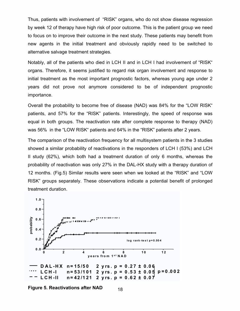
The comparison of the reactivation frequency for all multisystem patients in the 3 studies
showed a similar probability of reactivations in the responders of LCH I (53%) and LCH
II study (62%), which both had a treatment duration of only 6 months, whereas the
probability of reactivation was only 27% in the DAL-HX study with a therapy duration of
12 months. (Fig.5) Similar results were seen when we looked at the “RISK” and “LOW
RISK” groups separately. These observations indicate a potential benefit of prolonged
treatment duration.
D A L -H X n = 15 /5 0 2 y rs . p = 0 .2 7 ± 0 .0 6L C H - I n = 53 /1 01 2 y rs . p = 0 .5 3 ± 0 .0 5L C H - I I n = 42 /1 21 2 y rs . p = 0 .6 2 ± 0 .0 7
prob
abili
ty
l o g ran k -te s t p = 0.00 4
y e a r s fr o m 1 s t N A D
p = 0 .0 0 2
0 2 4 6 8 1 0 1 20 .0
0 .2
0 .4
0 .6
0 .8
1 .0
Figure 5. Reactivations after NAD
19
So far, the comparison of the two treatment arms of LCH II, i.e, the 2-drug arm A with
PDN and VBL and the 3-drug arm B with PDN, VBL and VP-16 has not shown any
significant difference with respect to initial response, survival and reactivation free
survival.
In the LCH I study, toxicity was seen in about 50% of the multisystem patients, and was
almost equal in both the treatment arms with vinblastine and VP16. Mild to moderate
leukopenia (WHO score I-II) was the most frequently observed event. Severe
thrombocytopenia or anemia, as well as hepatic dysfunction was seen only in patients
with initial involvement of “RISK” organs, and it was not possible to differentiate
treatment related toxicity from disease related dysfunction. Importantly, none of the
patients had to be withdrawn of the study because of toxicity.
Preliminary analysis revealed toxic events in 15/89 treatment courses in LCH II,
including mild to moderate leukopenia, nausea or vomiting.
The data on permanent consequences in LCH II will be evaluated within the next year.
So far, it can be stated that the probability to develop diabetes insipidus is 14% which is
about the same as in the previous study.
2.1 CONCLUSIONS OF LCH II
Risk organ involvement and poor response to initial treatment proved to be the most
important prognostic factors.
Patients with “RISK“ organ involvement who do not achieve a response to initial
treatment and are AD worse or intermediate by week 12 carry an about 75% risk of fatal
outcome. The probability of becoming free of disease is with standard therapy is less
than 20% for such patients with standard therapy.
Age under two years at diagnosis without “RISK” organ involvement is not associated
with a poor outcome, and will therefore not be considered for the initial stratification.
So far, VP16 has not shown any additional therapeutic benefit with respect to response,
survival or reactivation frequency, neither as monotherapy nor in combination with VBL
and PDN. Therefore, VP-16 will not be included in the standard initial treatment of LCH
III, considering its potential leukemogenicity.
20
The fatality rate was around 20% in all 3 studies which were using combination therapy,
monotherapy, or 2-drug and 3-drug regimen including PDN, VBL, and VP-16. This
observation points towards a need of new agents in the treatment for patients with
“RISK” organ involvement.
The retrospective comparison of the DAL HX, LCH I and II studies indicates that
prolonged duration of treatment may reduce the rate of reactivations.
2.2 RATIONALE FOR THE USE OF METHOTREXATE IN“RISK” PATIENTS
For years methotrexate (MTX) has proven to be an effective agent in the treatment of
LCH10,11. In 1974 Jones et al were comparing combination therapy with vincristine
(2mg/m2/week i.v.) and PDN versus MTX (30mg/m2 twice weekly orally) and PDN for a
minimum of 4 weeks in a randomized way and found a superior response rate in the
MTX group, even at this low dose. Their study also showed a clear benefit of prolonged
maintenance therapy12.
In the prospective DAL HX-83 study 14/21 Arm C multisystem patients (with organ
dysfunction) who had responded to the initial therapy, were given intermediate dose
MTX (500mg/m2 q 3 weeks) during the continuation therapy8. 7 pts experienced a
reactivation, 2 of these a fatal disease progression. 12 (86%) patients finally became
free of disease. The small number of patients in this study precludes final conclusions on
the effect of MTX in LCH.
2.3 SITUATION IN PATIENTS WITH MULTIFOCAL BONEDISEASE AND “SPECIAL SITES” OF DISEASE
2.3.1 MULTIFOCAL BONE DISEASE (MFB)
The LCH II Study Protocol did not include recommendations for treatment of single
system patients with multiple bone lesions. In the past there has been a controversy on
how to treat MFB. During the last two years information on 104 patients with MFB was
collected in a retrospective survey at the study center. Thirty-six patients initially did not
receive any systemic therapy, but were only observed after biopsy (n=13), or treated
with surgery (n=20), irradiation (n=4), or intralesional steroids (n=1). Sixty-eight patients
21
were treated with systemic therapy, which was monotherapy in 17 patients (PDN n=3,
VBL n=10, VP16 n=3, 2-CDA n=1). Twenty-two patients received either a combination of
VBL + PDN (n=17) or VP16 + PDN n=5. Combination therapy according to the DAL-HX
83/90 protocol was applied in 25 patients, and one patient received a combination
therapy consisting of cytosin-arabinoside, VCR and PDN13. Independent of the initial
treatment strategy, regression or resolution of the disease was seen in about 90% of the
patients. However, there was a significant difference with respect to the frequency of
reactivation between the different treatment groups. The probability of remaining free of
reactivation was 48% for local treatment and 55% for monotherapy, whereas it was 80%
for the 2-drug regimen, and 91% for combination therapy. These data confirm the
observation of the DAL-HX 83 study, in which only 18% of reactivations were observed
in multifocal disease, which was equal to unifocal bone disease and much less than
reported in the literature14. No statistical impact could be detected with request to the
duration of treatment.
Based on these observations, it was decided to offer an initial treatment according to the
“LOW RISK” arm of LCH III consisting of PDN and VBL with a treatment duration of 6
months to patients with multifocal bone disease.
2.3.2 “SPECIAL SITES”
2.3.2.1 “CNS-RISK” lesions
A retrospective analysis based on 1524 patients registered in the DAL HX-83/90, the
LCH I and LCH II studies revealed that involvement of the facial bones or anterior or
middle cranial fossa (temporal, sphenoidal, ethmoidal, cygomatic bone, orbital bones)
with intracranial tumour extension carry an about 3-fold risk for the development of
diabetes insipidus (DI) which is the hallmark of central nervous system involvement in
LCH and therefore are called “CNS-RISK” lesions. This was not true for vault lesions.
Based on these data it was concluded that patients with “CNS-RISK” lesions as the only
site of disease activity should not be regarded as simple single system disease, because
there is usually bone disease with soft tissue tumor and sometimes infiltration of the
meninges. Local therapy is usually problematic in such location and these patients
should rather receive systemic treatment. The LCH III study protocol offers therapy with
22
PDN and VBL to these patients.
2.3.2.2 VERTEBRAL LESIONS
Vertebral lesions sometimes present with significant soft tissue masses that may lead to
spinal cord compression, which can be now adequately assessed by MRI. Also in such
locations surgery might be too risky. Irradiation may be considered or systemic therapy
as offered in the LCH III study protocol should be initiated immediately even if the
lesions represent the only site of disease.
3 PATIENT’S ELIGIBILITY FOR LCH III
All newly diagnosed patients who meet the following criteria are eligible to be enrolled
and followed in the study:
• Definitive diagnosis of LCH
• Age under 18 years
• No prior treatment for LCH
4 LCH III STUDY REQUIREMENTS
Confirmation of a definitive histopathological diagnosis according to the criteria defined
by the Histiocyte Society. Mandatory review by the local reference pathologist in case of
presumptive diagnosis or provisional diagnosis.
Adoption of uniform clinical, laboratory and radiographic baseline and follow up
evaluations as given in the study protocol.
4.1 HISTOPATHOLOGICAL DIAGNOSTIC CRITERIA(modified according to the Writing Group of the Histiocyte Society15.)
4.1.1 Definitive diagnosis
requires the demonstration of CD1a antigenic determinants on the surface of lesional
cells (by immunocytology or immunohistology) or the finding of Birbeck granules in
lesional cells by electron microscopy.
23
4.1.2 Provisional diagnosis
is justified when the lesion has characteristic morphology and phenotype to an
experienced pathologist and the cells express S100 and at least one of the following:
ATPase, alpha-D-mannosidase, peanut lectin. Unstained slides from a provisional
diagnosis should be immediately sent before treatment is instituted to the regional study
pathologist for definitive diagnosis.
4.2 BASELINE DIAGNOSTIC EVALUATIONS (modified according to Clinical Writing Group of the Histiocyte Society16)
4.2.1 Clinical evaluation
4.2.1.1 Complete history:
Fever, pain, irritability, failure to thrive, loss of appetite, diarrhea, polydipsia, polyuria,
recurrent otitis, skin rashes, activity level, behavioural changes, neurological changes
4.2.1.2 Complete physical examination:
Measurement of temperature, height, weight, head circumference, pubertal status
Skin and skalp rashes, purpura, bleeding
Jaundice, pallor
Aural discharge
Orbital abnormalities
Gum and palatal lesions, dentition
Soft tissue swelling, lymphadenopathies
Dyspnea, tachypnea, intercostal retractions
Liver and spleen size, ascites, edema
Neurological examination (including papilledema, cranial nerve abnormalities, cerebellar
dysfunction)
4.2.2 Laboratory and radiographic evaluation
4.2.2.1 Mandatory minimum baseline evaluations for all patients:
Hemoglobin and/or hematocrit
24
Ferritin, iron, transferrin
White blood count and differential
Platelet count
Erythocyte sedimentation rate (ESR)
Renal function test (including creatine clearance, mandatory in “Risk” patients
randomized on Arm B, prior to MTX infusion)
Liver enzymes and function tests (SGOT, SGPT, γ-GT, alkaline phosphatase, bilirubin,
total protein, albumin)
Coagulation studies (PT, PTT, fibrinogen)
Chest radiograph, p.a. and lateral
Skeletal radiograph survey (radionuclide bone scan is not as sensitive as the skeletal
radiograph survey in most patients)
Urine osmolality (measurement after overnight water deprivation)
4.2.2.2 Mandatory for multi-system patients
Bone marrow aspiration and trephine with CD1a staining
HLA-typing (for “RISK” patients only, as soon as possible)
4.3 EVALUATIONS REQUIRED UPON SPECIFIC INDICATION
(modified according to the Clinical Writing Group of the Histiocyte Sciety16)
Indication Test
Abnormal chest radiograph,
tachypnea, intercostal retractions
High resolution – CT,
Pulmonary function test (if age
appropriate)
Patients with abnormal pulmonary
high resolution-CT:
to yield a diagnosis in case of isolated
lung involvement or to exclude infection
Lung biopsy,
Bronchoalveolar lavage
Unexplained chronic diarrhea or failure
to thrive, evidence of malabsorption
Endoscopic biopsy
25
Indication Test
Liver dysfunction:
to differentiate active LCH of the liver
from sclerosing cholangitis
Sonography,
Liver biopsy
Visual or neurologic abnormalities MRI of brain with i.v. gadolinium – DTPA,
Neurological evaluation, psychological
tests
Polyuria, polydipsia,
short stature, growth failure,
hypothalamic syndromes, galactorrhea,
precocious or delayed puberty
Endocrine evaluation including water
deprivation test, dynamic tests of the
anterior pituitary,
MRI of brain with i.v. gadolinium - DTPA
Gingiva involvement, loose teeth Panoramic dental radiography and
computed tomography of mandible and
maxilla,
oral surgery consultation
Aural discharge, deafness Otolaryngology consultation and
audiogram,
MRI of brain with i.v. gadolinium - DTPA
4.4 DEFINITION OF ORGAN INVOLVEMENT
4.4.1 “RISK” organs
Hematopoetic involvement:
With or without bone marrow
Involvement*
Anemia: hemoglobin <10 g/dl,
infants <9 g/dl (exclusion of iron deficiency)
Leukocytopenia: leukocytes <4,0 x 10⁹/l,
Thrombocytopenia: platelets < 100 x 109/l
*Bone marrow involvement is defined as demonstration of CD1a positive cells on bone
marrow smears. The clinical significance of CD1a positivity in the bone marrow remains
to be proven. Hypocellularity, hemophagocytosis, myelodysplasia, and/or myelofibrosis
may be regarded as secondary phenomena. Hemophagocytosis may be prominent in
severe progressive cases.
26
Spleen involvement: enlargement > 2 cm below costal
margin (proven by sonography)
Liver involvement: enlargement > 3 cm below costal
margin (proven by sonography) and/or
liver dysfunction (hyperbilirubinemia,
hypoproteinemia, hypalbuminemia,
elevated γGT, alkaline phosphatase,
elevated transaminases, ascites, edema)
and/or histopathological diagnosis
Lung involvement: typical changes on high resolution
computed tomography (HR-CT) and/or
histopathological diagnosis
4.4.2 “CNS RISK” lesions
Lesions in the orbital, temporal/ mastoid, sphenoidal, zygomatical, ethmoidal bones,
maxilla, sinuses or anterior or middle cranial fossa, with intracranial soft tissue extension
demonstrated on magnetic resonance imaging (MRI). Vault lesions are not regarded as
“CNS Risk” lesions.
5 STRATIFICATION
5.1 GROUP 1 - MULTISYSTEM “RISK” PATIENTS Multisystem patients WITH involvement of one or more “RISK” organs i.e. hematopoetic
system, liver, spleen or lungs
Patients with single system lung involvement are not eligible for randomisation
5.2 GROUP 2 - MULTISYSTEM “LOW RISK” PATIENTSMultisystem patients with multiple organs involved but WITHOUT involvement of “RISK”
27
organs
5.3 GROUP 3 - SINGLE SYSTEM “MULTIFOCAL BONEDISEASE” AND LOCALIZED “SPECIAL SITE”INVOLVEMENT
Patients with multifocal bone disease, i.e. lesions in 2 or more different bones;
Patients with localized special site involvement, like “CNS-RISK” lesions with intracranial
soft tissue extension or vertebral lesions with intraspinal soft tissue extension;
6 GOALS FOR LCH IIILike in the LCH II study the overall aims of this study are:
• to deliver risk-adapted therapy according to the extent and severity of the disease
• to evaluate the response in the different patient groups
• to evaluate the rate of failure in the different treatment groups, i.e. nonresponse to
therapy or disease reactivation during therapy
• to assess morbidity, i.e. evaluation of therapy toxicity and evaluation of the
incidence of permanent consequences in the different treatment groups.
The specific goals for the 3 patient groups are as follows:
6.1 GROUP 1: MULTISYSTEM “RISK” PATIENTS
6.1.1 To decrease mortality
• by decreasing the rate of nonresponders to initial treatment at week 6 by
introducing MTX as a new agent. The effect of MTX in addition to the initial
standard therapy with VBL and PDN will be assessed in a randomized way.
• by decreasing the rate of patients who do not achieve a response (NAD or AD
better) in “RISK” organs at week 12 by applying a second course of initial therapy.
• by encouraging an early switch to salvage therapy for nonresponders at week 6
or 12.
28
6.1.2 To decrease morbidity
• i.e. the rate of reactivation and permanent consequences by prolonging the
continuation treatment to one year.
6.1.3 To assess acute and late treatment toxicity
6.2 GROUP 2: MULTISYSTEM “LOW RISK” PATIENTS
6.2.1 To decrease morbidity
• by reducing the rate of reactivation and permanent consequences. The value of
prolonged continuation therapy for responders to initial treatment and the efficacy
of 6 versus 12 months continuation treatment will be tested in a randomized way.
6.2.2 To increase the response rate
• by applying a second course of initial treatment course according to arm A of the
“RISK” group in patients with an insufficient response (intermediate and worse)
to initial treatment at week 6.
6.2.3 To assess acute and late treatment toxicity
6.3 GROUP 3: SINGLE SYSTEM MULTIFOCAL BONE ANDLOCALIZED “SPECIAL SITES”
6.3.1 To reduce morbidityi.e. the rate of reactivation and permanent consequences (as compared to the historical
control group of the DAL HX 83/90 and LCH I and II studies).
7 STUDY DESIGNLCH III is an international, multicentric, prospective clincial study comprising
• a randomized clincial trial for multisystem “RISK” patients and
29
• a randomized clincial trial for multisystem “LOW RISK” patients and
• a pilot study for patients with single system MFB and localized “SPECIAL SITES”
8 REGISTRATION AND RANDOMIZATION
8.1 REGISTRATION After confirmation of the diagnosis of a new patient, the registration form and the
diagnostic evaluation forms have to be sent to the study subcenter or study reference
center by fax or e-mail without delay together with the completed and signed consent
form.
8.2 RANDOMISATION
8.2.1 Eligibility for randomization group 1 “RISK” patients
• eligibility for the study
• multisystem disease with involvement of “RISK” organs
8.2.2 Eligibility for randomization 2 “LOW RISK” patients
• eligibility for the study
• multisystem disease without involvement of “RISK” organs
• response to initial therapy, i.e. regression (AD better) or resolution (NAD) after 6
weeks of initial treatment course 1
Randomisation will be performed by the study subcenter or study reference center
promptly after the reception of the registration, diagnostic evaluation or follow up and
consent forms. The information on the assigned treatment arm and the randomisation
number will be forwarded to the participant by e-mail or fax without delay.
9 TREATMENT
9.1 GROUP 1: MULTISYSTEM “RISK” PATIENTSconsists of an initial treatment of one or two 6 week courses (according to
30
response) and a continuation treatment. The overall therapy duration is 12 months.
9.1.1 Treatment arm A
9.1.1.1 Initial reatment course 1
Continuous oral prednisone (PDN) 40mg/m2 daily in 3 doses as a 4-week course,
tapering over a period of 2 weeks.
Vinblastine (VBL) 6 mg/m2 i.v. bolus, day 1 of week 1, 2, 3, 4, 5, 6.
9.1.1.2 Initial treatment course 2
(starting without delay after course 1 for patients who are AD better or intermediate after
course 1. Patients who are NAD after course 1 proceed to continuation treatment.).
Oral prednisone (PDN) 40mg/m2 in 3 divided doses for 3 days every week, from week
7-12.
Vinblastine (VBL) 6 mg/m2 i.v. bolus, day 1 of week 7, 8, 9, 10, 11, 12.
9.1.1.3 Continuation treatment
(starting after initial treatment at day 1 of week 7 in pts who are NAD after course 1 of
initial treatment, or at day 1 of week 13 in pts who are NAD or AD-better after course 2
of initial treatment.
Continuous oral 6-mercaptopurine (6-MP) 50mg/m2 daily until the end of month 12 from
therapy start.
Pulses of oral prednisone PDN 40mg/m2 in 3 doses, day 1-5 q 3 weeks, starting at day 1
of week 7 in patients NAD after course 1 or at day 1 of week 13 in patients NAD or AD
better after course 2 until the end of month 12.
Vinblastine (VBL) 6mg/m2 i.v. bolus, day 1 q 3 weeks, starting at day 1 of week 7 in
patients NAD after course 1 or at day 1 of week 13 in patients NAD or AD better after
course 2 until the end of month 12.
31
9.1.2 Treatment arm B
9.1.2.1 Initial treatment course 1
Continuous oral prednisone (PDN) 40mg/m2 daily in 3 divided doses as a 4-week
course, tapering over a period of 2 weeks.
Vinblastine (VBL) 6 mg/m2 i.v. bolus, day 1 of week 1, 2, 3, 4, 5, 6 given before the MTX
infusion.
Methotrexate 500 mg/m2 24 hours-infusion with folinic acid (leucovorin) rescue, day 1 of
week 1, 3, 5. 1/10 of the dose as i.v. bolus over 30 min, followed by 9/10 of the dose as
23.5 hours infusion with 2000ml/m2 hydration.
Folinic acid 12mg/m2 orally is given 24 hours and 30 hours after the stop of the MTX
infusion (id est: 48 and 54 hours after start of MTX therapy).
9.1.2.2 Initial treatment course 2
(starting without delay after course 1 for patients who are AD better or intermediate after
course 1. Patients who are NAD after course 1 proceed to continuation treatment).
Oral prednisone (PDN) 40mg/m2 in 3 divided doses for 3 days every week, from week
7-12.
Vinblastine (VBL) 6 mg/m2 i.v. bolus, day 1 of week 7, 8, 9, 10, 11, 12 given before the
MTX infusion.
Methotrexate 500 mg/m2 24 hours-infusion with folinic acid (leucovorin) rescue, day 1 of
week 7, 9, and 11. 1/10 of the dose as i.v. bolus over 30 min, followed by 9/10 of the
dose as 23.5 hours infusion with 2000ml/m2 hydration.
Folinic acid 12mg/m2 orally is given 24 hours and 30 hours after the stop of the MTX
infusion (id est: 48 and 54 hours after start of MTX therapy).
9.1.2.3 Continuation treatment
Continuous oral 6-mercaptopurine (6-MP) 50mg/m2 until the end of month 12 from
therapy start.
Pulses of oral prednisone PDN 40mg/m2 in 3 doses, day 1-5 q 3 weeks, starting at day 1
of week 7 in patients NAD after course 1 or at day 1 of week 13 in patients NAD or
32
AD better after course 2 until the end of month 12.
Methotrexate 20mg/m2 orally, once weekly until the end of month 12.
Vinblastine (VBL) 6mg/m2 i.v. bolus, day 1 q 3 weeks, starting at day 1 of week 7 in
patients NAD after course 1 or at day 1 of week 13 in patients NAD or AD better after
course 2 until the end of month 12.
9.2 GROUP 2: “LOW RISK” GROUPTreatment consists of an initial treatment of 6 weeks (a second course is given only to
patients with persistent or progressive disease) and a continuation treatment.
The overall therapy duration is 6 or 12 months as randomly assigned.
9.2.1.1 Initial treatment course 1
Continuous oral prednisone (PDN) 40mg/m2 daily in 3 doses as a 4-week course,
tapering over a period of 2 weeks.
Vinblastine (VBL) 6 mg/m2 i.v. bolus, day 1 of week 1, 2, 3, 4, 5, 6.
9.2.1.2 Initial treatment course 2
(Only in patients with intermediate or worse response after course 1)
Oral prednisone (PDN) 40mg/m2 in 3 divided doses for 3 days every week , from week
7-12.
Vinblastine (VBL) 6 mg/m2 i.v. bolus, day 1 of week 7, 8, 9, 10, 11, 12.
9.2.1.3 Continuation treatment
The overall therapy duration is 6 months for Arm LR 6 or 12 months for Arm LR 12 .
Pulses of oral prednisone PDN 40mg/m2 in 3 doses, day 1-5 q 3 weeks, starting at day 1
of week 7 in patients NAD after course 1 or at day 1 of week 13 in patients NAD or AD
better after course 2 until the end of month 6 or 12 from therapy start.
Vinblastine (VBL) 6mg/m2 i.v. bolus, day 1 q 3 weeks, starting at day 1 of week 7 in
patients NAD after course 1 or at day 1 of week 13 in patients NAD or AD better after
course 2 until the end of month 6 or 12 from therapy start.
33
9.3 GROUP 3: “MULTIFOCAL BONE DISEASE” AND“SPECIAL SITES”
Treatment consists of an initial treatment of 6 weeks and a continuation treatment. A
second course is given only to patients with progressive disease. The overall therapy
duration is 6 months.
9.3.1 Initial treatment
Continuous oral prednisone (PDN) 40mg/m2 daily in 3 doses as a 4-week course,
tapering over a period of 2 weeks.
Vinblastine (VBL) 6 mg/m2 i.v. bolus, day 1 of week 1, 2, 3, 4, 5, 6.
9.3.2 Continuation treatment
(starting after initial treatment at day 1 of week 7 in pts who are NAD after course 1 of
initial treatment or at day 1 of week 13 in patients who are NAD or AD-better after
course 2 of initial treatment)
Pulses of oral prednisone PDN 40mg/m2 in 3 doses, day 1-5 q 3 weeks, starting at day 1
of week 7 in patients NAD after course 1 or at day 1 of week 13 in patients NAD or AD
better after course 2 until the end of month 6 from therapy start.
Vinblastine (VBL) 6mg/m2 i.v. bolus, day 1 q 3 weeks, starting at day 1 of week 7 in
patients NAD after course 1 or at day 1 of week 13 in patients NAD or AD better after
course 2 until the end of month 6 from therapy start.
9.4 SUPPORTIVE CARE GUIDELINES
Pneumocystis carinii prophylaxis
Oral sulphamethoxazole/trimethoprime, 5 mg/kg/day of the trimethoprime, divided into
2 doses/day, on 3 days per week throughout the study period and for 12 weeks
thereafter (must be stopped during MTX-infusion).
Antiemetics should be given as necessary.
34
Transfusions of red cells and platelets
Blood cell components should be filtered blood products and preferably irradiated (25
Gy), for prevention of GvHD.
G-CSF
In case of prolonged neutropenia, G-CSF may be given subcutaneously or
intravenously. The use of GM-CSF is not recommended.
Intravenous immunoglobulin
may be given in cases of hypoimmunoglobulinemia.
9.5 TOXICITY (please fill in Toxicity sheet, Appendix)
6-Mercaptopurine
Myelosuppression, hepatic dysfunction (elevated transaminases and cholestatic
jaundice), mucositis, dermatological manifestations, interaction with allopurinol, nausea,
vomiting
Methotrexate
Myelosuppression (leukopenia, anemia, thrombocytopenia), nausea, vomiting, mucositis
(ulcerative stomatitis, diarrhea), alopecia, skin rashes, nephrotoxicity, hepatic
dysfunction, liver fibrosis, encephalopathy, pneumonitis
Prednisone
Increased appetite, centripedal obesity, fluid retention, hyperglycemia,
immunosuppression, myopathy, osteoporosis, aseptic necrosis, peptic ulceration,
pancreatitis, mental alteration, cataracts, hypertension, precipitation of diabetes, growth
failure, amenorrhea, impaired wound healing, atrophy of subcutaneous tissue
Vinblastine
Peripheral neuropathy: paresthesia, dysphagia, hoarseness, bone pain (esp. mandible),
constipation, paralytic ileus, convulsions, myelosuppression (leukopenia, anemia,
thrombocytopenia), alopecia, inappropriate ADH secretion, local pain and necrosis if
35
extravasated, nausea, vomiting.
9.5.1 Serious Adverse Events
Any serious adverse event (death or grade III - IV non-haematological life threatening
toxicity) must be reported immediately (i.e. within the next working day) by the treating
institution to the the study reference center (and relayed to the local subcenters and
DSMB), for further reporting according to local practice (use form in appendix).
The toxicity criteria will be the same for all participating groups (WHO-score) and appear
in Appendix.
9.6 THERAPY MODIFICATIONSTry to avoid dose-reductions or delays.
Pancytopenia
at presentation may be disease related and is then not an indication for reduction of the
initial dosage.
Infants with body weight under 10 kg:
Drug doses are calculated based on body surface area (BSA) only and dose is adjusted
for age as follows:
< 6 months 50% of dose calculated form BSA
> 6 months < 12 months 75% of dose calculated from BSA
> 12 months 100% of dose calculated from BSA
Bone marrow toxicity:
In case of good response to therapy it is recommended to wait for hemapoetic recovery.
An absolute neutrophil count greater than 1.0 x 10⁹/l and a platelet count greater than
100 x 109/l are essential before starting each course of therapy except for the first dose.
In case of persistent disease activity it is recommended to continue protocol regardless
of the hematologic values.
Hepatotoxicity:
Samples for determination of liver enzymes (ALT/GOT, AST/GPT) must be
36
drawn immediately prior to a course of i.v. MTX. If values are 10-20N (i.e. 10-20 times
higher than normal values) wait 48 hrs and recheck to ensure that the levels are
decreasing. Discontinue TMP/SMZ if the transaminase elevation persists or increases,
and withhold chemotherapy until the value is <10N, then resume full dose
chemotherapy. Resumption of TMP/SMZ prophylaxis is left to the investigator’s
discretion. Should therapy be withheld for elevated transaminases during initial therapy,
resume therapy at point of interruption. Transaminase values of 20N mandate holding
therapy until the level returns to <10N. Persistence of values >20N for >2 weeks
requires an evaluation including: bilirubin, alkaline phosphatase, coagulation tests,
albumin, total protein and hepatitis serologies. A liver biopsy should be considered
before additional therapy is given to help to distinguish hepatic toxicity from LCH
involvement or sclerosing cholangitis. Under such circumstances, please contact the
local coordinator.
Nephrotoxicity
For patients with a GFR by creatinine clearance of less than 60 ml/min/1.73m2 delay
MTX and repeat clearance after hydration.
Gastrointestinal toxicity
In case of severe mucositis or diarrhea, therapy should be discontinued until recovery
and then reinstituted without dose reduction. To prevent constipation in patients treated
with vinblastine regular administration of mild laxatives is recommended. Serious
constipation with paralytic ileus requires cessation of vinblastine administration.
Neurotoxicity
In the event of significant toxicity (extensive weakness, severe paresthesia, severe
ileus), VBL may be temporarily discontinued and resumed at 50% dose when toxicity
resolves. Contact the local study coordinator before discontinuing VBL. Increase to
maximum tolerated dose (not exceed protocol dose) as soon as possible.
Oral 6-mercaptopurine and oral methotrexate
if neutrophil count falls below 500/µl, treatment will be held until recovery above these
levels and then resumed as tolerated. If neutrophil count falls below 500/µl on >2
occasions during continuation, discontinue TMP/SMZ and decrease dose of 6-MP or
37
MTX by 25% on alternating basis upon resumption of therapy. Begin by reducing the 6-
MP dose. Should therapy be withheld for myelosuppression or elevated transaminase,
resume therapy at the correct point chronologically.
10 ASSESSMENT OF TREATMENT RESPONSE In contrast to leukemia or other malignancies the terms “remission” or “relapse” should
be avoided. In accordance with the nature of LCH the following definitions should be
applied to judge the effect of treatment.9
10.1 DEFINITION OF DISEASE STATE
NON ACTIVE DISEASE
(NAD)
no evidence of
disease
resolution of all signs or symptoms
regressive disease regression of signs or symptoms,
no new lesions
ACTIVE DISEASE (AD) stable disease persistence of signs of symptoms,
no new lesions
progressive disease* progression of signs or symptoms
and/or appearance of new lesions
10.2 DEFINITION OF RESPONSE CRITERIAThere are three categories of response
BETTER
complete resolution NAD
regression AD better
INTERMEDIATE
mixed new lesions in one site, regression
in another site
stable unchanged
WORSE progression*
*in isolated bone disease progression is defined as appearance of new bone lesions or
38
lesions in other organs
10.3 RESPONSE EVALUATIONPlease send the follow up evaluation sheet to the study subcenter as soon as possible
after the evaluation (fax or e-mail).
Clinical status and performance17
Blood count and differential
Requirement of blood products
Liver and spleen size
Liver enzymes and function tests
HR-CT and pulmonary function tests (if age-appropriate) for patients with lung disease
Skeletal radiograph of lesional sites only
MRI of the brain in patients with “CNS-RISK” lesions or intracranial lesions
Spinal MRI in patients with vertebral lesions
Histological proof of AD or NAD may be required in specific sites
10.3.1 Group 1: “RISK PATIENTS”
Any treatment decision must be based on the response in “RISK ORGANS” (i.e.R.O.)
INTERVAL EVALUATION RESPONSE THERAPY
NAD continuation treatment
better initial treatment course 2
intermediate initial treatment course 2
week 6 after initial treatment
course 1
worse salvage (see appendix)
NAD continuation treatment
better continuation treatment
intermediate salvage (see appendix)
week 12 after initial treatment
course 2
worse salvage (see appendix)
39
NAD,
better
continuation treatment
intermediate
without R.O.
intermediate
with R.O.
continuation treatment
salvage (see appendix)
worse without
R.O.
arm A: re-start initial treatment
arm B
arm B: salvage (see appendix)
week 24 or
any time in
case of event
during continuation
treatment
worse with R.O. salvage (see appendix)
month 12 end of continuation
treatment
NAD STOP (in case of persistent
signs of disease activity, please
discuss with local coordinator)
10.3.2 Group 2 “LOW RISK” patients
Please send the follow up evaluation sheet to the study subcenter IMMEDIATELY after
the evaluation at week 6 (fax or e-mail) to have the randomization for the duration of
continuation treatment performed!
Interval Evaluation RESPONSE CONTINUE
NAD
better
continuation treatment
randomization: LR 6 or LR 12
intermediate initial treatment course 2
week 6 after initial treatment
course 1
worse initial treatment course 2
NAD
better
continuation treatment
intermediate continuation treatment
week 12 after initial treatment
course 2
worse discuss with local coordinator
NAD,
better
continuation treatment
intermediate continuation treatment
week 7 or 13-
23
during continuation
treatment
worse discuss with local coordinator
40
month 6 end of continuation
treatment
NAD
better
LR 6 STOP
LR 12 continuation treatment
intermediate continuation treatment
worse re-start initial treatment arm B
NAD
better
continuation treatment
intermediate continuation treatment
month 6-12 during continuation
treatment LR 12
worse re-start initial treatment arm B
month 12 end of continuation
therapy LR 12
STOP
(in case of persistent signs of
disease activity, please discuss with
local coordinator)
10.3.3 Group 3 patients with “MFB” or “SPECIAL SITES”
Brain MRI (and/or CT scans) or spinal MRI must be performed to assess the treatment
response in these critical sites. In case of residual “CNS-RISK” lesions on MRI
treatment must not be stopped. A biopsy of such lesions to rule out residual active
disease should be considered.
Interval Evaluation RESPONSE CONTINUE
NAD
better
continuation treatment
week 6 after initial treatment
course 1
worse initial treatment course 2
(risk patients arm A)
NAD
better
continuation treatmentweek 12 after initial treatment
course 2
worse discuss with local
coordinator
NAD
better
continuation treatment week 7 or 13-23 during continuation
treatment
worse discuss with local
coordinator
41
month 6 after continuation
treatment
NAD
better
STOP
(in case of clear
regression stop therapy,
repeat MRI after 3
months, in case of
significant residual tumor
consider biopsy and
discuss with local
coordinator)
11 OFF STUDY CRITERIA
In case of progression in “RISK organs” after initial treatment course 1 or course 2
switch to salvage (see appendix).
In case of evidence of residual active disease at the end of protocol therapy a biopsy of
residual lesions to rule out persistent active disease is recommended, and the individual
case should be discussed with the local co-ordinator.
If you have any questions regarding salvage therapy, please contact the local
coordinator or the study reference center.
12 AUTOPSY
Autopsy is to be encouraged for fatal cases. A set of unstained slides (5-10) or blocks
should be submitted to the regional study pathologist.

42
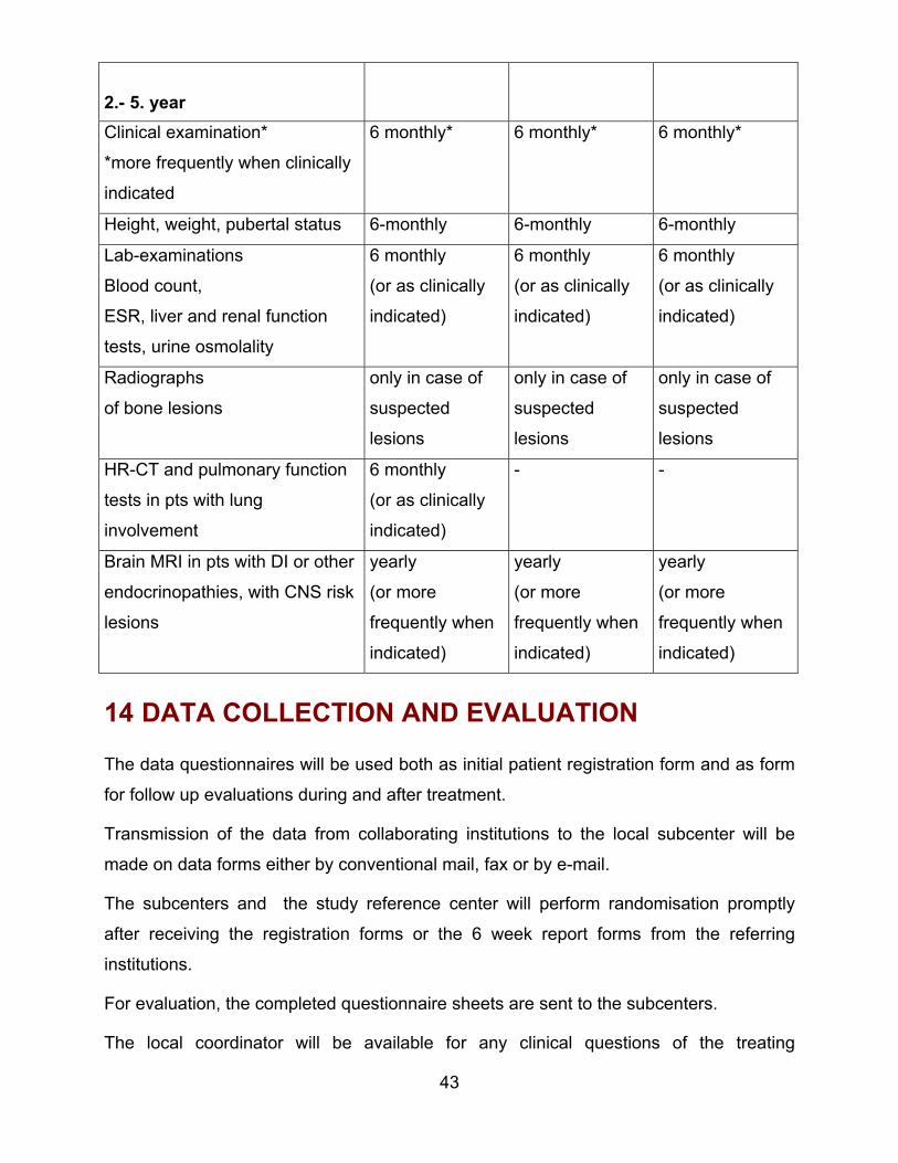
13 FOLLOW UP INVESTIGATIONS AFTER STOP OFTHERAPY
FOLLOW UP With RiskOrganinvolvement
Without RiskOrganinvolvement
Multifocal boneinvolvement,special site
1. year
Clinical examination 6 weekly 6 weekly 3 monthly
Height, weight, pubertal status 6 monthly 6 monthly 6 monthly
Lab-examinations
Blood count,
ESR, liver (renal function )tests,
urine osmolality
3 monthly 3 monthly 6 monthly
Radiographs
of bone lesions
3 monthly
until signs of
regression (or as
clinically
indicated)
3 monthly
until signs of
regression (or as
clinically
indicated)
3 monthly
until signs of
regression (or as
clinically
indicated)
HR-CT, pulmonary function
tests
(in case of pulmonary
involvement)
6-monthly
Sonography in pts with liver
involvement
6-monthly
Brain MRI
in pts with DI or other
endocrinopathies,
in pts with CNS risk lesions
yearly,
3 monthly until no residual mass lesions or stable
findings
Neuropsychometric assessment
(in case of CNS involvement)
yearly yearly yearly
43
2.- 5. yearClinical examination*
*more frequently when clinically
indicated
6 monthly* 6 monthly* 6 monthly*
Height, weight, pubertal status 6-monthly 6-monthly 6-monthly
Lab-examinations
Blood count,
ESR, liver and renal function
tests, urine osmolality
6 monthly
(or as clinically
indicated)
6 monthly
(or as clinically
indicated)
6 monthly
(or as clinically
indicated)
Radiographs
of bone lesions
only in case of
suspected
lesions
only in case of
suspected
lesions
only in case of
suspected
lesions
HR-CT and pulmonary function
tests in pts with lung
involvement
6 monthly
(or as clinically
indicated)
- -
Brain MRI in pts with DI or other
endocrinopathies, with CNS risk
lesions
yearly
(or more
frequently when
indicated)
yearly
(or more
frequently when
indicated)
yearly
(or more
frequently when
indicated)
14 DATA COLLECTION AND EVALUATION
The data questionnaires will be used both as initial patient registration form and as form
for follow up evaluations during and after treatment.
Transmission of the data from collaborating institutions to the local subcenter will be
made on data forms either by conventional mail, fax or by e-mail.
The subcenters and the study reference center will perform randomisation promptly
after receiving the registration forms or the 6 week report forms from the referring
institutions.
For evaluation, the completed questionnaire sheets are sent to the subcenters.
The local coordinator will be available for any clinical questions of the treating
44
physicians, and review the data sheets for completeness and correctness.
The data input into the uniform LCH III database will be performed at the local
subcenter, if possible, or the revised data sheets will be transferred to the study
reference center in Vienna.
For the regular study evaluations the local data bases will be transferred to the study
reference center.
Access to common data from the study data base will be given only with the approval of
the Scientific Committee and the permission of the Board of the Histiocyte Society.
15 DATA SAFETY MONITORING BOARD (DSMB)
An independant Data Safety Monitoring Board composed of 4 international experts will
monitor the progress of the study on ethical and scientific grounds. The role of the
DSMB will be
- to review accrual rate and
- to be involved in all interim analysis. Five sequential analysies are planned, the results
will remain confidential. Bases on the results of the interim analysis the DSMB will
recommentd whether the study can continue of whether it must be amended or stopped
prematurely.
- to monitor toxicity- biannually the DSMB will review the toxicity reports together with
the study committee.
The DSMB will be asked to review any major modification to the study proposed by the
study committee prior to its implementation.
16 STATISTICAL CONSIDERATIONS
The study reference center in Vienna will carry out all analyses. The results and the data
will be send to the Institute of Medical statistics in Mainz for an independent statistical
review after the interim-analyses and three weeks before a study committee meeting.
The randomisation lists will be provided by the statistical center in Mainz.
45
16.1 DESIGN
16.1.1 Group 1 “RISK” patients
The Group 1-Study of LCH-III is a randomized controlled clinical trial for patients with
multi-system LCH WITH involvement of “RISK” organs (hematopoetic system, liver,
spleen or lungs). The random assignment to the treatment arms will be done in blocks,.
stratified according to local subcenters to ensure a balance within the local subcenters
and within each pre-specified number of treatment assignments (=block). The
randomization will be done in the local subcenter without delay after diagnosis.
16.1.2 Group 2 “LOW RISK” patients:
The Group 2-Study of LCH-III is a randomized controlled clinical trial for patients with
multi-system LCH WITHOUT involvement of “RISK” organs (hematopoetic system, liver,
spleen or lungs) and initial response at week 6 (NAD/AD better).
The random assignment to the treatment arms will be done in blocks stratified according
to age at diagnosis (≤ 2 years, > 2 years) and local subcenters to ensure a balance
within age groups, the local subcenters and within each pre-specified number of
treatment assignments (=block). The randomization will be done in the local subcenter
without delay after the first response evaluation 6 weeks after therapy start. Only
patients with therapy response, i.e. regression (AD better) or resolution (NAD) after 6
weeks of initial treatment, will be eligible for randomization.
In addition, the impact of a prolonged initial treatment (= course2) for those patients who
are not eligible for randomization is examined and compared with the historical control
group of LCH-II patients.
16.2 ENDPOINTS
16.2.1 Primary endpoints
16.2.1.1 Group 1 “RISK” patients:
The primary aim of the study is to compare the therapeutic efficacy of control arm A(PDN+VBL) with the experimental arm B (PDN+VBL+MTX). The primary endpoint is
46
the proportion of non-responder in risk organs to the initial treatment.
Non-response to initial therapy is defined as:
• death within 12 weeks of initial treatment or
• progression (worse) in risk organs at week 6
• lack of response (=intermediate response or progression) in risk organs at week
12 as compared to the status of disease at week 6
If the null hypothesis is true, the two randomized treatment arms are equally effective in
terms of non-response. If the alternative hypotheses is true, there is a difference
between the two randomized arms in terms of efficacy.
16.2.1.2 Group 2 “LOW RISK” patients:
The primary aim of the study is to compare the reactivation free survival rate in initial
responders at week 6 with continuation treatment for 6 months (Arm LR 6) versus 12
months (Arm LR 12) in those patients without disease reactivation within the first 6
months.
If the null hypothesis is true, the reactivation rate of both randomized arms are equal. If
the alternative hypothesis is true, there is a difference between the two arms in terms of
reactivation frequency.
16.2.2 Secondary endpoints
• Overall survival
• Proportion of responders (overall and in risk organs) at week 6
• Proportion of responders (overall and in risk organs) at week 12
• Reactivation free survival after response at week 12
• Time to NAD
• Incidence of permanent consequences
• Toxicity
• Reactivation free survival after NAD (overall and in risk organs) and for
“RISK” patients a historical comparison of Arm A of LCH III with Arm A of LCH-II
with respect to the frequency of reactivation to evaluate the impact of the

47
prolonged study duration in risk patients
• Vaule of a prolonged initial treatment (course 2) of non-responding “LOW RISK”
patients compared to a historical control of LCH-II patients without “RISK” organ
involvement
16.3 ANALYSESThe analyses of the primary and secondary endpoints will be done according to the
intention-to-treat principle, i.e. the patients will be analyzed in their allocated treatment
group, even in case of non-compliance or protocol violations.
The statistical analyses of the primary endpoint will be done with a two-sided
significance level of 5 %. The statistical analyses of the secondary endpoints are
exploratory. A separate analyses will be performed for each group (“RISK” and “LOW
RISK”). In addition to the intention-to-treat analyses a secondary per protocol analyses
will be done including all patients who were treated according to the originally assigned
treatment arm without protocol violations (= unjustified dose modification, therapy delay
and/or improper switch to another therapy within the first 12 months).
16.3.1 Analysis of primary endpoints
16.3.1.1 Group 1 “RISK” patients:
The Fisher’s exact test will be used to compare the proportion of non-responders in risk
organs and the proportion of responders at week 6 and week 12.
16.3.1.2 Group 2 “LOW RISK” patients:
Reactivation (=progression in any organs) and death will be considered as events for the
calculation of Reactivation Free Survival. The interval will start 6 months after therapy
start, i.e. the time point when half of the patients are intended stop the continuation
therapy (Arm LR6) whereas patients from the other randomized arm (Arm LR12) will
further receive 6 months of continuation therapy. This means, randomized patients with
reactivations within the first 6 months after randomization will not be included in the
analyses. For censored patients the interval will be calculated until the date of the last
response evaluation.
48
The proportion of reactivation free survival will be estimated according to the method of
Kaplan-Meier and confidence intervals according to Dorey and Korn will be given for the
reactivation free survival rate after 2 and 3 years.18,19 The primary statistical evaluation
of the treatment effect will be done by log rank-test.
As a secondary aim the question whether a prolonged therapy can slow down the speed
of reactivation will be considered. Retrospective data from LCH-I and LCH-II and the
DAL-studies indicate that the hazards between 6 and 12 months of continuation therapy
are proportional and constant in time (i.e. exponentially distributed reactivation free
survival times). Therefore, a Weibull accelerated failure time model will be fitted to
evaluate an acceleration factor.20 Moreover, a time dependent Cox regression model
will be performed.21
16.3.2 Analysis of secondary endpoints
The overall survival time will be calculated from the date of randomization to death or the
last response evaluation.
The reactivation free survival will be calculated from the date of initial response
evaluation at week 12. Reactivation (=progression overall and in risk organs after
response at week 12) and death will be considered as events. For censored patients the
interval will be calculated until the date of the last response evaluation.
The time to non active disease (NAD) will be calculated from randomization to the date
of NAD. For censored observation the interval will be calculated until the date of the last
follow up information.
The reactivation free survival after NAD will be calculated from the date of NAD.
Reactivation (reappearance or progression in any organ) and death will be considered
as events. For censored patients the interval will be calculated until the date of last
response evaluation.
The time to permanent consequences will be calculated from the date of randomization
to the diagnoses of permanent consequences. Deaths without permanent
consequences will be censored at the time of death. For all other censored patients the
interval will be calculated until the date of the last response evaluation. For the
comparison of treatment arms patients with permanent consequences which are already
present at therapy start will not be considered in the analyses.
The proportion of survival, reactivation free survival after response in week 12,
reactivation after NAD, the time to NAD and the incidence of permanent consequences
will be estimated by the method of Kaplan Meier. The comparison of the randomized
arms will be done by log rank-tests.
The proportion of patients with severe organ toxicity (WHO score grade III-IV) within the
first 12 weeks of treatment will be compared with Fisher’s exact test.
To study the effect of prolonged initial therapy in Low Risk patients without response at
week 6. the response rate at week 12 (compared to the status of disease at week 6) will
be compared to the response rate at week 12 of the corresponding LCH-II patients. This
will be done with Fisher’s exact test.
1T
1
T
r
s
E
b
s
b
1
T
n
a
E
b
s
6.4 INTERIM-ANALYSES
49
he primary aims of the two trials will be monitored according to a group sequential plan.
6.4.1 Group 1 “RISK” patients
he data will be monitored a total of 5 times in equally spaced intervals, i.e. after the
esponse evaluation at week 12 of 20%, 40%, 60%, 80% and 100% of the total sample
ize.
arly stopping will be implemented either to reject the null hypothesis of no difference
etween the two randomized arms, or to retain the null hypotheses .22 The design of the
topping boundaries is due to O’Brien-Fleming using the normal approximation of the
inomial distribution.
6.4.2 Group 2 “LOW RISK” patients
he data will be monitored 5 times i.e. after 20%, 40%, 60%, 80% and 100% of the total
umber of events (see 13.4.1.2: Power Considerations) This schedule of interim
nalyses allows an equal distribution of the information between the interim analyses.
arly stopping will be implemented either to reject the null hypothesis of no difference
etween the two randomized arms or to retain the null hypotheses22. The design of the
topping boundaries is according to O’Brien-Fleming.
50
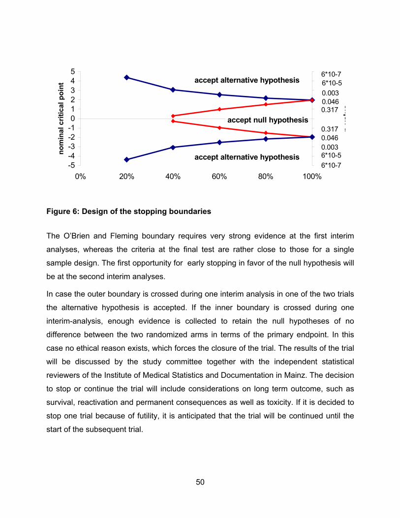
The O’Brien and Fleming boundary requires very strong evidence at the first interim
analyses, whereas the criteria at the final test are rather close to those for a single
sample design. The first opportunity for early stopping in favor of the null hypothesis will
be at the second interim analyses.
In case the outer boundary is crossed during one interim analysis in one of the two trials
the alternative hypothesis is accepted. If the inner boundary is crossed during one
interim-analysis, enough evidence is collected to retain the null hypotheses of no
difference between the two randomized arms in terms of the primary endpoint. In this
case no ethical reason exists, which forces the closure of the trial. The results of the trial
will be discussed by the study committee together with the independent statistical
reviewers of the Institute of Medical Statistics and Documentation in Mainz. The decision
to stop or continue the trial will include considerations on long term outcome, such as
survival, reactivation and permanent consequences as well as toxicity. If it is decided to
stop one trial because of futility, it is anticipated that the trial will be continued until the
start of the subsequent trial.
-5-4-3-2-1012345
0% 20% 40% 60% 80% 100%
nom
inal
crit
ical
poi
nt
pva
lue
accept null hypothesis
accept alternative hypothesis
accept alternative hypothesis
6*10-76*10-50.0030.0460.317
0.3170.0460.0036*10-56*10-7
Figure 6: Design of the stopping boundaries
51
16.5 POWER CONSIDERATION
16.5.1 Group 1 “RISK” patients:
The estimated proportion of non-responder in the control group is 50 %. With Arm B
(with the addition of MTX) we aim to reduce the rate of non-response to 30%. To
achieve a power above 80% we will need a total sample size of 202 patients to show
this 20%-difference (two-sided α=5%. NQuery 3.0, Fisher’s exact test). The group
sequential design of the study will raise the maximum sample size to 228. If the null
hypothesis is true, the expected sample size will be reduced to 143 patients. If the
alternative hypothesis is true (i.e. a 20% difference in the proportion of non-responders
between the two arms) the expected sample size is 158 patients23.
In LCH-II we observed an average annual recruitment of 70 – 80 multi-system patients,
of whom 60 % had risk organ involvement. This means that 42 – 48 patients will be
eligible for randomization of arm A versus arm B . If the randomization rate is similar to
LCH II i.e. 85% we will have approximately 35-40 randomizations a year. We assume
that we will not be able to evaluate response at week 12 in 10 % of the patients because
they will be lost to follow up. This means that we expect to reach the maximum sample
size of 228 evaluable patients after a period of approximately 6 years. If the null
hypothesis is true, the expected duration of the study will be 4.3 years. If the alternative
hypothesis is true, this will be 4.7 years.
16.5.2 Group 2 “LOW RISK” patients:
Data from the previous LCH-II study indicate that the reactivation free survival rate of the
control group (LR 6) is approximately exponential distributed with a 1-year reactivation
free survival rate of about 65%. With this study we aim to show a 20% difference in the
1-year reactivation free survival rate (i.e. a 1-year reactivation free survival rate of 85%
in patients with 12 month continuation treatment) with sufficient power.
To show a 20% difference at the 5% significance level with a power of 80% we would
need a total of 36 events following the group sequential design (Jennison Turnbull).
85% of the patients with multi-system LCH without involvement of risk organs had an
initial response. That means that with the expected annual recruitment of 40 multi-
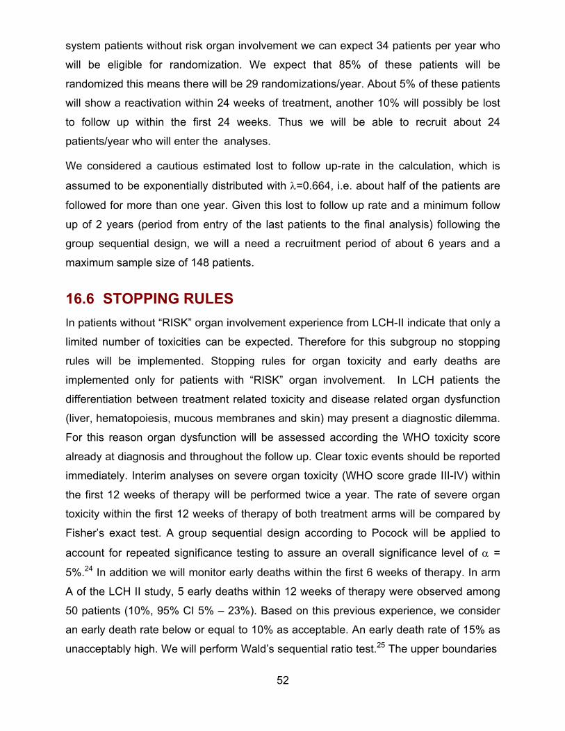
52
system patients without risk organ involvement we can expect 34 patients per year who
will be eligible for randomization. We expect that 85% of these patients will be
randomized this means there will be 29 randomizations/year. About 5% of these patients
will show a reactivation within 24 weeks of treatment, another 10% will possibly be lost
to follow up within the first 24 weeks. Thus we will be able to recruit about 24
patients/year who will enter the analyses.
We considered a cautious estimated lost to follow up-rate in the calculation, which is
assumed to be exponentially distributed with λ=0.664, i.e. about half of the patients are
followed for more than one year. Given this lost to follow up rate and a minimum follow
up of 2 years (period from entry of the last patients to the final analysis) following the
group sequential design, we will a need a recruitment period of about 6 years and a
maximum sample size of 148 patients.
16.6 STOPPING RULES In patients without “RISK” organ involvement experience from LCH-II indicate that only a
limited number of toxicities can be expected. Therefore for this subgroup no stopping
rules will be implemented. Stopping rules for organ toxicity and early deaths are
implemented only for patients with “RISK” organ involvement. In LCH patients the
differentiation between treatment related toxicity and disease related organ dysfunction
(liver, hematopoiesis, mucous membranes and skin) may present a diagnostic dilemma.
For this reason organ dysfunction will be assessed according the WHO toxicity score
already at diagnosis and throughout the follow up. Clear toxic events should be reported
immediately. Interim analyses on severe organ toxicity (WHO score grade III-IV) within
the first 12 weeks of therapy will be performed twice a year. The rate of severe organ
toxicity within the first 12 weeks of therapy of both treatment arms will be compared by
Fisher’s exact test. A group sequential design according to Pocock will be applied to
account for repeated significance testing to assure an overall significance level of α =
5%.24 In addition we will monitor early deaths within the first 6 weeks of therapy. In arm
A of the LCH II study, 5 early deaths within 12 weeks of therapy were observed among
50 patients (10%, 95% CI 5% – 23%). Based on this previous experience, we consider
an early death rate below or equal to 10% as acceptable. An early death rate of 15% as
unacceptably high. We will perform Wald’s sequential ratio test.25 The upper boundaries
53
we will use will be shown in figure 7.
Simulations show that with this boundary the risk to wrongly conclude that there is an
excess of early deaths (whereas the real event rate is below or equal to 10%) is 13%.
On the other hand, the power to detect an excess of early deaths of 15% or more will be
78%.
If we find significant differences in toxicities or if the number of early deaths observed,
reaches the boundary defined by the sequential plan, a full analysis will be performed,
and the results will be discussed among the members of the study committee together
with the independent biometrical reviewers. The study committee together with the
independent biometrical reviewers will decide, whether and how the study will proceed.
0
5
10
15
20
25
30
35
0 40 80 120 160 200 240
number of patients with 12 week evaluation
num
ber o
f dea
ths
with
in 1
2 w
eeks
of t
hera
py
upper boundary (y = 3.457 + 0.124x)
unacceptable high death rate
acceptable death rate
Figure 7: Boundaries for deaths within 12 weeks
54
17 PUBLICATION
Publication of overall study data or projects arisen from the overall study population may
be undertaken only with the agreement of the study committee.
Every subcenter or participating clinic may publish their own observations related to LCH
patients or data on specific research projects not concerning questions of the overall
study.
18 REFERENCES
1. Arceci,R.J. The histiocytoses: the fall of the Tower of Babel. Eur J Cancer 35, 747-
767 (1999).
2. The French Langerhans cell Histiocytosis Group. A multicentric retrospective survey
of Langerhans' cell histiocytosis: 348 cases observed between 1983 and 1993. Arch
Dis Child 75, 17-24 (1996).
3. Komp,D.M., Herson,J., Starling,K.A., Vietti,T.J. & Hvizdala,E. A staging system for
Histiocytosis X: a Southwest Oncology Group Study. Cancer 47, 798-800 (1981).
4. Ladisch,S. & Jaffe,E.S., pp. 617-6301995).
5. Lahey,E. Histiocytosis X : an analysis of prognostic factors. J Pediatr 184-189
(1975).
6. Nesbit,M.E. Cancer in Children, Clinical Management., pp. 176-184 (Springer
Verlag,1986).
7. Ceci,A. et al. Etoposide in Recurrent Childhood Langerhans' Cell Histiocytosis: An
Italian Cooperative Study. Cancer 62, 2528-2531 (1988).
8. Gadner,H., Heitger,A., Grois,N., Gatterer-Menz,I. & Ladisch,S. for the DAL HX 83
Study Group). Treatment strategy for disseminated Langerhans cell histiocytosis.
Med Ped Oncol 23, 72-80 (1994).
9. Gadner H et al. LCH-I: A randomized trial of treatment of multisystem Langerhans
cell histiocytosis. J Pediatr (2001).
10. Womer,R.B., Anunciato,K.R. & Chehrenama,M. Oral methotrexate and alternate-day
prednisone for low-risk Langerhans cell histiocytosis. Med Ped Oncol 25, 70-73
(1995).
55
11. Komp,D.M., Trueworthy,R., Hvizdala,E. & Sexauer,C. Prednisolone, methotrexate,
and 6-mercaptopurine in the treatment of histiocytosis X [letter]. Cancer Treat Rep
63, 2125-2126 (1979).
12. Jones,B., Kung,F., Chevalier,L. & al.,e. Chemotherapy of reticuloendotheliosis,
Comparison of methotrexate plus prednisone vs vincristine plus prednisone. Cancer
34 , 1011-1017 (1974).
13. Egeler,R.M., Kraker,J.d. & Voute,P.A. Cytosine-arabinoside, vincristine, and
prednisolone in the treatment of children with disseminated Langerhans cell
histiocytosis with organ dysfunction: experience at a single institution. Med Ped
Oncol 21, 265-270 (1993).
14. Titgemeyer C et al. Pattern and course of single-system disease in Langerhans cell
histiocytosis. Med Ped Oncol 37, 1-7 (2001).
15. Writing Group of the Histiocyte Society. Histiocytosis syndromes in children. Lancet
1, 208-209 (1987).
16. Writing Group of the Histiocyte Society. Histiocytosis syndromes in children: II.
Approach to the clinical and laboratory evaluation of children with Langerhans Cell
Histiocytosis. Med Ped Oncol 17, 492-495 (1989).
17. Lansky,S.B., List,M.A., Lansky,L.L., Ritter-Sterr,C. & Miller,D.R. The measurement of
performance in childhood cancer patients. Cancer 60, 1651-1656 (1987).
18. Dorey FJ & Korn EL. Effective sample sizes for confidence intervals for survival
probabilities. Statist Med 6, 679-687 (1987).
19. Kaplan,E.L. & P.Meier. Nonparametric estimation from incomplete observations. J.
Am Stat Assoc 53, 457-481 (1958).
20. Cox DR & Oakes D. Analysis of Survival Data. Chapman & Hall, London (1984).
21. Cox DR. Regression models and life-tables. J Roy Statist Soc B 34, 187-220 (1972).
22. Pampallona S & Tsiatis AA. Group sequential designs for one-sided and two-sided
hypothesis testing with provision for early stopping in favor of the null hypothesis. J
Statist Planning and Inference 42, 19-35 (1994).
23. Jennison CH & Turnbull BW. Group Sequential methods with applications to clinical
trials. 114. Chapman & Hall, London (2000).

56
24. Pocock SJ. Group sequential methods in the design and analysis of clinical trials.
Biometrika 64, 191-199 (1977).
25. Wald A. A sequential analysis. Wiley, New York (1947).