Embed Size (px)
Citation preview

Lhx9: A Novel LIM-Homeodomain Gene Expressed in theDeveloping Forebrain
Sylvie Retaux,1 Monique Rogard,1 Ingolf Bach,2 Vieri Failli,1 and Marie-Jo Besson2
1Laboratoire de Neurochimie-Anatomie, Institut des Neurosciences, 75005 Paris, France, and 2Howard Hughes MedicalInstitute, University of California, School of Medicine, La Jolla, California 92093
A novel LIM-homeodomain gene, Lhx9, was isolated by degen-erate RT-PCR followed by mouse embryonic library screening.Lhx9 cDNA encodes a protein that is most closely related toDrosophila apterous and rodent Lhx2 proteins. The Lhx9 spa-tiotemporal pattern of expression during embryogenesis wassimilar but distinct from Lhx2. Highest expression levels werefound in the diencephalon, telencephalic vesicles, and dorsalmesencephalon. Domains of expression respected the pro-posed neuromeric boundaries (Puelles and Rubenstein, 1993).Lhx9 was also expressed in the spinal cord, forelimb andhindlimb mesenchyme, and urogenital system. Although Lhx9expression was sustained in diencephalon and mesencephalonfrom embryonic day 10.5 (E10.5) to postnatal stages, it wastransient in the future cerebral cortex, where it was turned off
between E14.5 and E16.5. Lhx9 expression was highest if notexclusively located (depending on the region of interest) in theintermediate and mantle zones, as opposed to the mitoticventricular zone. Lhx9 protein was tested for interaction withthe recently discovered cofactors of LIM-homeodomain pro-teins and was found to interact strongly both with CLIM1 andCLIM2. The expression pattern and structural characteristics ofLhx9 suggest that it encodes a transcription factor that mightbe involved in the control of cell differentiation of several neuralcell types. Furthermore, Lhx9 protein could act in a combina-torial manner with other LIM-homeodomain factors expressedin overlapping pattern.
Key words: LIM-homeodomain; Lhx9; Lhx2; forebrain; devel-opment; neuromeres
A major issue in developmental neurobiology is the understand-ing of forebrain development and patterning. During embryogen-esis, the anterior neural tube generates complex and highly orga-nized structures, such as the cerebral cortex, the basal ganglia,and the thalamus. In adults, these structures are interconnected,almost always topographically.
Homeodomain genes play decisive roles in the establishment ofcerebral structures and/or the generation of cell types. Amongthem, LIM-homeodomain (LIM-hd) genes encode developmen-tally expressed transcription factors that contain a homeodomainand two cysteine-rich LIM domains involved in protein–proteininteractions (Sanchez-Garcia and Rabbitts, 1994; Dawid et al.,1995). In the current view, the LIM domains inhibit DNA bindingby the homeodomain. After fixation of cofactors, transcription isactivated, and synergism with other transcription factors is pro-moted. Indeed, cofactors for LIM-hd proteins have been isolated:NLI/Ldb1/CLIM2 and CLIM1 interact with LIM domains ofLIM homeoproteins to potentiate transactivation of downstreamgenes (Agulnick et al., 1996; Jurata et al., 1996; Bach et al., 1997).These cofactors probably confer time and space specificity to theregulatory action of LIM-hd proteins.
LIM-hd genes were first implicated in cell fate determination ininvertebrates; Caenorhabditis elegans mec-3 and lin-11 (Way and
Chalfie, 1988; Freyd et al., 1990) and Drosophila apterous (Lund-gren et al., 1995; Thor and Thomas, 1997) determine cell typesand axonal pathfinding. In vertebrates, the spinal cord motorneurons give an example for similar roles of LIM-hd genes: motorneurons express a set of four LIM-hd genes, in a combinatorialmanner that is correlated to the motor neuron position in thespinal cord and to the type of peripheral innervated target(Tsuchida et al., 1994). Thus, a LIM-hd combinatorial code mightdefine pathfinding phenotypes. If similar combinations ofLIM-hd genes were expressed in the forebrain, they could par-ticipate in the establishment of its highly organized circuits.Moreover, Drosophila islet governs aminergic phenotypes (Thorand Thomas, 1997). Forebrain LIM-hd genes could govern neu-rotransmitter expression in neurons as well.
Homeodomain genes expression patterns in the developingCNS have led to the theory of the prosomeric forebrain (Puellesand Rubenstein, 1993; Rubenstein et al., 1994; Puelles, 1995). Inthis model, regions and boundaries of expression of developmen-tal factors are seen as landmarks for the determination of cerebralareas according to longitudinal and transverse subdivisions thatform segmented structures. LIM-hd genes integrate this modelwell. Lhx1/lim-1 (Barnes et al., 1994), Lhx2/LH-2 (Xu et al.,1993), Lhx3/pLIM (Seidah et al., 1994; Bach et al., 1995), L3/Lhx8(Matsumoto et al., 1996), Lhx4/Gsh4 (Li et al., 1994), Lhx5(Sheng et al., 1997), Lhx6, and Lhx7 (Grigoriou et al., 1998) areall expressed in the developing rodent CNS, and their expressionpatterns respect neuromeric boundaries (present paper). The roleof LIM-hd genes in cellular specification, combined with theirapparent function in the coding of positional information, makesthem important factors to be studied during brain regionalizationand wiring.
In a search to isolate more LIM-hd involved in forebrain
Received May 11, 1998; revised Oct. 13, 1998; accepted Nov. 4, 1998.This work was supported by the Association Franco-Israelienne pour la Recher-
che Scientifique et Technique, Fondation pour la Recherche Medicale, and CentreNational de la Recherche Scientifique. We are grateful to Dr. A. Joyner for the En-2in situ probe and J. L. Duband and Y. Bassaglia for discussions about Lhx9 in thecaelomic cavity.
Correspondence should be addressed to Dr. Sylvie Retaux, Laboratoire deNeurochimie-Anatomie, Institut des Neurosciences, 9 quai St. Bernard, 75005 Paris,France.Copyright © 1999 Society for Neuroscience 0270-6474/99/190783-11$05.00/0
The Journal of Neuroscience, January 15, 1999, 19(2):783–793
development, we hypothesized the existence of a subfamily ofLhx2-related LIM-hd genes. An RT-PCR cloning strategy al-lowed the isolation of mouse Lhx9, which is similar to but distinctfrom Lhx2 in sequence and expression pattern, the two genesthereby forming a new subfamily of forebrain LIM-hd genes.
MATERIALS AND METHODSRT-PCR cloning. Total RNA from embryonic day 15 (E15) or E18forebrain were reverse-transcribed to cDNA with avian myeloblastosisvirus reverse transcriptase (Boehringer Mannheim, Mannheim,Germany) and used as templates for PCR reactions (Qiagen, Hilden,Germany) using the following degenerate oligonucleotide primers:51, ATGCGIACITCITTYAARCAYCAYCARCT; 52, ATGAGRAC-ITCITTYAARCAYCARCT; 31, MAYTTIGCIGCIGCRTTYTGRA-ACCA; and 32, MAYTTIGCYCTIGCRTTYTGRAACCA, where I isan inosine residue. 59 (51 and 52) and 39 (31 and 32) primers weredesigned to isolate genes of the Lhx2 subfamily and were chosen toamplify a stretch of DNA in the homeodomain. Any combination of 39and 59 primers led to the amplification of a single band of 160 bp asexpected. After cloning of this 160 bp fragment into pGEM-T (Promega,Madison, WI), sequencing of 80 independent clones revealed the pres-ence of three different fragments. One was mouse Lhx2, and the twoothers were unknown. We concentrated on one that we named Lhx9. The160 bp DNA fragment was used to screen a lgt11 E14.5 mouse headcDNA library under high stringency. The Lhx9 cDNA was isolated usingstandard molecular biology techniques and sequenced on both strandswith Amersham (Les Ulis, France) thermosequenase. The 1.016 kb PCRproduct of Lhx9 cDNA was subcloned into pGEM-T (Promega) forsubsequent experiments.
Tissue preparation. Timed pregnant C57/Black6 mice were anesthe-tized with 0.1 ml/100 gm body weight pentobarbital. Embryos wereharvested, fixed in 0.1 M 4-morpholinepropanesulfonic acid, pH 7.4, 2 mM
EGTA, 1 mM MgSO4 , and 3.7% formaldehyde at room temperature,cryoprotected in 30% sucrose, and frozen by immersion in isopentane ondry ice. Noon on the day after the night of mating was considered as E0.5.Postnatal day 1 (P1)–P7 brains were processed identically, except thatsucrose was 20%. Whole embryos or dissected brains were cryostat-sectioned at 20 mm, and sections were stored at 280°C until use.
In situ hybridization and image processing. The pGEM-Lhx9 plasmidwas linearized with NdEI or NcoI and used as template for RNAsynthesis with T7 or SP6 polymerase in the presence of [ 35S]UTP (10mCi/mmol; ICN Biochemicals, Costa Mesa, CA) for antisense and sensecontrol probes, respectively. The probe thus included the 59 noncodingregion, the two LIM domains, the linker region, and the beginning of thehomeodomain. The Bluescript-Lhx2 plasmid including the 1.1 kb Lhx2insert was linearized with EcoRI, and T3 polymerase was used forantisense probe synthesis as described (Bach et al., 1997). TheBluescript-En2 39 untranslated region plasmid was linearized with ClaI,and T7 was used to generate an 800 bp En-2 probe. Restriction enzymeswere purchased from Appligene (Heidelberg, Germany) or Promega, andRNA polymerases were from Stratagene (La Jolla, CA). Briefly, slideswere acetylated, dehydrated progressively, and hybridized 16 hr at 60°Cin the presence of 5 3 10 5–10 6 cpm of probe/slide. They were thenwashed under high stringency, treated 1 hr with RNase A (20 mg/ml), anddehydrated. Sections were exposed 2–3 d to Eastman Kodak (Rochester,NY) Biomax MR film for autoradiography and then dipped in KodakNTB-2 emulsion. Emulsions were developed after 10–12 d of exposure,and sections were counterstained with methylene blue. Photographs weretaken on a Leica (Nussloch, Germany) DMBL microscope, scanned on aCanon (Tokyo, Japan) 2700 scanner, and mounted for figures withAdobe (Mountain View, CA) Photoshop. Images were corrected forcolor balance, contrast, brightness, or cropping, but no other correctionswere made.
In vitro protein–protein interaction assays. PCR products of full-lengthCLIM1a and CLIM2 were ligated in frame into the XhoI–XbaI sites ofthe bacterial expression vector pGEX-KG to yield a glutathioneS-transferase (GST) fusion protein. The PCR product of the 1.016 kbLhx9 cDNA was subcloned into the pGEM-T vector (Promega), and[ 35S]methionine-labeled Lhx9 protein was produced using the Promegain vitro transcription–translation kit, SP6 RNA polymerase, and[ 35S]methionine. The in vitro protein–protein interaction assays wereperformed as described previously (Bach et al., 1995).
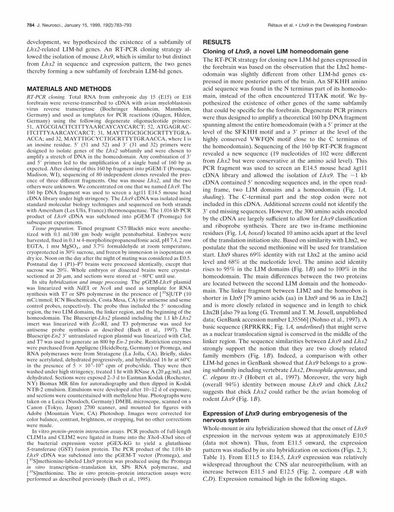
RESULTSCloning of Lhx9, a novel LIM homeodomain geneThe RT-PCR strategy for cloning new LIM-hd genes expressed inthe forebrain was based on the observation that the Lhx2 home-odomain was slightly different from other LIM-hd genes ex-pressed in more posterior parts of the brain. An SFKHH aminoacid sequence was found in the N terminus part of its homeodo-main, instead of the often encountered TITAK motif. We hy-pothesized the existence of other genes of the same subfamilythat could be specific for the forebrain. Degenerate PCR primerswere thus designed to amplify a theoretical 160 bp DNA fragmentspanning almost the entire homeodomain (with a 59 primer at thelevel of the SFKHH motif and a 39 primer at the level of thehighly conserved VWFQN motif close to the C terminus ofthe homeodomain). Sequencing of the 160 bp RT-PCR fragmentrevealed a new sequence (19 nucleotides of 102 were differentfrom Lhx2 but were conservative at the amino acid level). ThisPCR fragment was used to screen an E14.5 mouse head lgt11cDNA library and allowed the isolation of Lhx9. The ;1 kbcDNA contained 59 noncoding sequences and, in the open read-ing frame, two LIM domains and a homeodomain (Fig. 1A,shading). The C-terminal part and the stop codon were notincluded in this cDNA. Additional screens could not identify the39 end missing sequences. However, the 300 amino acids encodedby the cDNA are largely sufficient to allow for Lhx9 classificationand riboprobe synthesis. There are two in-frame methionineresidues (Fig. 1A, boxed) located 10 amino acids apart at the levelof the translation initiation site. Based on similarity with Lhx2, wepostulate that the second methionine will be used for translationstart. Lhx9 shares 69% identity with rat Lhx2 at the amino acidlevel and 68% at the nucleotide level. The amino acid identityrises to 95% in the LIM domains (Fig. 1B) and to 100% in thehomeodomain. The main differences between the two proteinsare located between the second LIM domain and the homeodo-main. The linker fragment between LIM2 and the homeobox isshorter in Lhx9 [79 amino acids (aa) in Lhx9 and 96 aa in Lhx2]and is more closely related in sequence and in length to chickLhx2B [also 79 aa long (G. Tremml and T. M. Jessell, unpublisheddata; GenBank accession number L35566] (Nohno et al., 1997). Abasic sequence (RPRKRK; Fig. 1A, underlined) that might serveas a nuclear translocation signal is conserved in the middle of thelinker region. The sequence similarities between Lhx9 and Lhx2strongly support the notion that they are two closely relatedfamily members (Fig. 1B). Indeed, a comparison with otherLIM-hd genes in GenBank showed that Lhx9 belongs to a grow-ing subfamily including vertebrate Lhx2, Drosophila apterous, andC. elegans ttx-3 (Hobert et al., 1997). Moreover, the very high(overall 94%) identity between mouse Lhx9 and chick Lhx2suggests that chick Lhx2 could rather be the avian homolog ofrodent Lhx9 (Fig. 1B).
Expression of Lhx9 during embryogenesis of thenervous systemWhole-mount in situ hybridization showed that the onset of Lhx9expression in the nervous system was at approximately E10.5(data not shown). Thus, from E11.5 onward, the expressionpattern was studied by in situ hybridization on sections (Figs. 2, 3;Table 1). From E11.5 to E14.5, Lhx9 expression was relativelywidespread throughout the CNS alar neuroepithelium, with anincrease between E11.5 and E12.5 (Fig. 2, compare A,B withC,D). Expression remained high in the following stages.
784 J. Neurosci., January 15, 1999, 19(2):783–793 Retaux et al. • Lhx9 in the Developing Forebrain
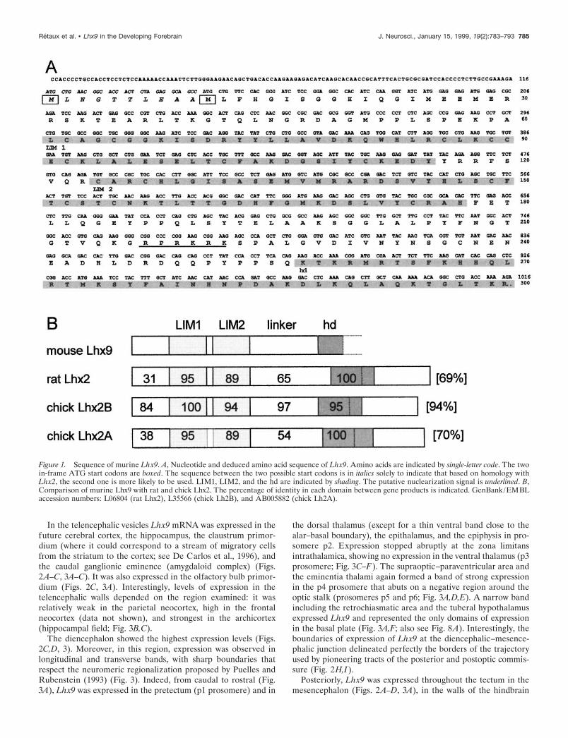
In the telencephalic vesicles Lhx9 mRNA was expressed in thefuture cerebral cortex, the hippocampus, the claustrum primor-dium (where it could correspond to a stream of migratory cellsfrom the striatum to the cortex; see De Carlos et al., 1996), andthe caudal ganglionic eminence (amygdaloid complex) (Figs.2A–C, 3A–C). It was also expressed in the olfactory bulb primor-dium (Figs. 2C, 3A). Interestingly, levels of expression in thetelencephalic walls depended on the region examined: it wasrelatively weak in the parietal neocortex, high in the frontalneocortex (data not shown), and strongest in the archicortex(hippocampal field; Fig. 3B,C).
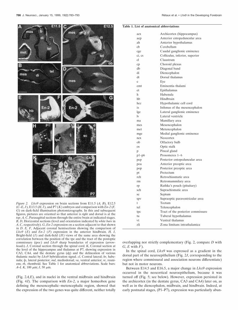
The diencephalon showed the highest expression levels (Figs.2C,D, 3). Moreover, in this region, expression was observed inlongitudinal and transverse bands, with sharp boundaries thatrespect the neuromeric regionalization proposed by Puelles andRubenstein (1993) (Fig. 3). Indeed, from caudal to rostral (Fig.3A), Lhx9 was expressed in the pretectum (p1 prosomere) and in
the dorsal thalamus (except for a thin ventral band close to thealar–basal boundary), the epithalamus, and the epiphysis in pro-somere p2. Expression stopped abruptly at the zona limitansintrathalamica, showing no expression in the ventral thalamus (p3prosomere; Fig. 3C–F). The supraoptic–paraventricular area andthe eminentia thalami again formed a band of strong expressionin the p4 prosomere that abuts on a negative region around theoptic stalk (prosomeres p5 and p6; Fig. 3A,D,E). A narrow bandincluding the retrochiasmatic area and the tuberal hypothalamusexpressed Lhx9 and represented the only domains of expressionin the basal plate (Fig. 3A,F; also see Fig. 8A). Interestingly, theboundaries of expression of Lhx9 at the diencephalic–mesence-phalic junction delineated perfectly the borders of the trajectoryused by pioneering tracts of the posterior and postoptic commis-sure (Fig. 2H,I).
Posteriorly, Lhx9 was expressed throughout the tectum in themesencephalon (Figs. 2A–D, 3A), in the walls of the hindbrain
Figure 1. Sequence of murine Lhx9. A, Nucleotide and deduced amino acid sequence of Lhx9. Amino acids are indicated by single-letter code. The twoin-frame ATG start codons are boxed. The sequence between the two possible start codons is in italics solely to indicate that based on homology withLhx2, the second one is more likely to be used. LIM1, LIM2, and the hd are indicated by shading. The putative nuclearization signal is underlined. B,Comparison of murine Lhx9 with rat and chick Lhx2. The percentage of identity in each domain between gene products is indicated. GenBank/EMBLaccession numbers: L06804 (rat Lhx2), L35566 (chick Lh2B), and AB005882 (chick Lh2A).
Retaux et al. • Lhx9 in the Developing Forebrain J. Neurosci., January 15, 1999, 19(2):783–793 785
(Fig. 2A,E), and in nuclei in the ventral midbrain and hindbrain(Fig. 4 I). The comparison with En-2, a major homeobox genedefining the mesencephalic–metencephalic region, showed thatthe expression of the two genes was quite different, neither totally
overlapping nor strictly complementary (Fig. 2, compare D withG, E with F).
In the spinal cord, Lhx9 was expressed as a gradient in thedorsal part of the neuroepithelium (Fig. 2J, corresponding to theregion where commissural and association neurons differentiate)but not in motor neurons.
Between E14.5 and E16.5, a major change in Lhx9 expressionoccurred in the neocortical neuroepithelium, because it wasturned off (Fig. 5; see below). However, expression persisted inthe archicortex (in the dentate gyrus, CA3 and CA4) later on, aswell as in the diencephalon, midbrain, and hindbrain. Indeed, atearly postnatal stages, (P1–P7), expression was particularly abun-
Figure 2. Lhx9 expression on brain sections from E11.5 (A, B), E12.5(C–E, J ), E13.5 (H, I ), and P7 (K ) embryos and comparison with En-2 (F,G) on dark-field illumination photomicrographs. In this and subsequentfigures, pictures are oriented so that anterior is right and dorsal is at thetop. A, C, Parasagittal sections through the entire brain at indicated stages.B, D, Horizontal sections (level and orientation indicated by white bars inA, C, respectively). G, En-2 expression on a section adjacent to that shownin D. E, F, Adjacent coronal hemisections showing the comparison ofLhx9 (E) and En-2 (F) expression in the anterior hindbrain. H, I,Bright-field ( I ) and dark-field (H ) views of the same area showing thecorrelation between the position of the tpc and the tract of the postopticcommissure (tpoc) and Lhx9 sharp boundaries of expression (arrow-heads). J, Coronal section through the spinal cord. K, Coronal section atthe level of the hippocampus and thalamus at P7, showing expression inCA3, CA4, and the dentate gyrus (dg) and the delineation of variousthalamic nuclei by Lhx9 hybridization signal. cl, Central lateral; hr, habe-nula; lp, lateral posterior; md, mediodorsal; va, ventral anterior; re, reuni-ens; rh, rhomboid. See Table 1 for anatomical abbreviations. Scale bars:A–I, K, 100 mm; J, 50 mm.
Table 1. List of anatomical abbreviations
acx Archicortex (hippocampus)aep Anterior entopeduncular areaah Anterior hypothalamuscb Cerebellumcge Caudal ganglionic eminenceci, cs Colliculus, inferior, superiorcl Claustrumcp Choroid plexusdb Diagonal banddi Diencephalondt Dorsal thalamuse Eyeemt Eminentia thalamiet Epithalamush Habenulahb Hindbrainhcc Hypothalamic cell cordis Isthmus of the mesencephalonlge Lateral ganglionic eminencelv Lateral ventriclema Mamillary areames Mesencephalonmet Metencephalonmge Medial ganglionic eminencencx Neocortexob Olfactory bulbos Optic stalkp Pineal glandp1–p6 Prosomeres 1–6pep Posterior entopeduncular areapoa Anterior preoptic areapop Posterior preoptic areapt Pretectumrch Retrochiasmatic arearm Retromammilary arearp Rathke’s pouch (pituitary)sch Suprachismatic arease Septumspv Supraoptic paraventricular areat Tectumtel Telencephalontpc Tract of the posterior commissuretu Tuberal hypothalamusvt Ventral thalamuszli Zona limitans intrathalamica
786 J. Neurosci., January 15, 1999, 19(2):783–793 Retaux et al. • Lhx9 in the Developing Forebrain
dant in the dorsal thalamus where it allowed the delineation ofvarious nuclei (Fig. 2K). Lhx9 marked a sharp boundary betweenthe very-high-expressing inferior colliculus and the high-expressing superior colliculus (Fig. 4L). A number of mesence-phalic and pons nuclei, including deep cerebellar nuclei, stillexpressed Lhx9 at these postnatal stages (data not shown).
In summary, Lhx9 expression was relatively widespread, mostlyin the alar region of the neuroepithelium. In this respect, it isnoteworthy that expression was absent from the large region ofthe ganglionic eminences in the ventral telencephalon (corre-sponding to the striatal–pallidal primordium).
Lhx9 and Lhx2 are expressed in overlapping butdistinct patternsBecause Lhx9 and Lhx2 are closely related family members, wenext compared directly the expression of the two genes.
Concerning regional expression, the two genes were expressed
mainly in overlapping patterns, with few noticeable exceptions:Lhx2 was expressed in the ganglionic eminences, the eyes, theoptic stalk, and the neuromeres surrounding the optic stalk,whereas Lhx9 was not (compare Figs. 4A,F, 3B, 4B). In theprosencephalic region, the two mRNAs were expressed in com-plementary patterns, showing bands of expression with sharpboundaries, separated by expression-negative zones. Moreprecisely, Lhx2 was not expressed in the eminentia thalami–supraoptic paraventricular areas, whereas Lhx9 was, but theLhx9-negative zone at the optic stalk, preoptic area, and anteriorhypothalamus was Lhx2 positive (compare Figs. 4A,F, 4D–G, 3E,4C). Expression of the two genes was perfectly overlapping in thedorsal thalamus and was absent for both genes in the ventralthalamus (Fig. 4, compare A–F). Thus, the addition of the twoexpression domains covered almost the entire diencephalic areawith the exception of the ventral thalamus (for summary, seeFig. 8A).
In the tectal epithelium, the expression patterns of the Lhxgenes were overlapping in the marginal layers (Fig. 4 I–J). Incontrast to engrailed, Lhx9 and Lhx2 were not expressed as agradient. Their expression domains spanned the mesencephalic–metencephalic junction, slightly thinning out at the isthmus (Fig.4 I–J, arrows). Furthermore, when compared with En-2, they werenot as largely expressed in the ventral mesencephalon (Fig. 4,compare I–K).
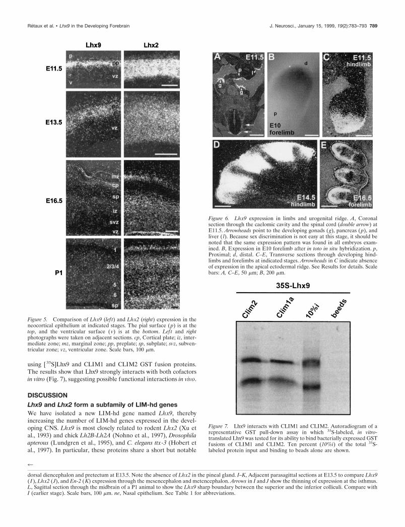
Interesting differences between the two family members wereobserved at the cellular level. In the telencephalic vesicles, Lhx9mRNA was exclusively located in the differentiating layers of theneuroepithelium, as opposed to the mitotic ventricular zone,whereas Lhx2 was expressed throughout the depth of the neocor-tical epithelium, including the ventricular and the differentiatingmantle zones (Fig. 5). Concerning the timing of Lhx9 and Lhx2expression, a major difference was again found in the neocortex.Whereas Lhx9 expression stopped between E14.5 and E16.5,Lhx2 was still expressed in the neocortex until P1 (Fig. 5) andlater (data not shown). Moreover, although Lhx9 stopped inhippocampal fields CA1 and CA2 around birth (Fig. 3K), Lhx2was still expressed in the whole hippocampus (data not shown).
Thus, Lhx9 and Lhx2 are expressed in patterns that are com-patible both for redundant and complementary roles during brainregionalization and neurogenesis.
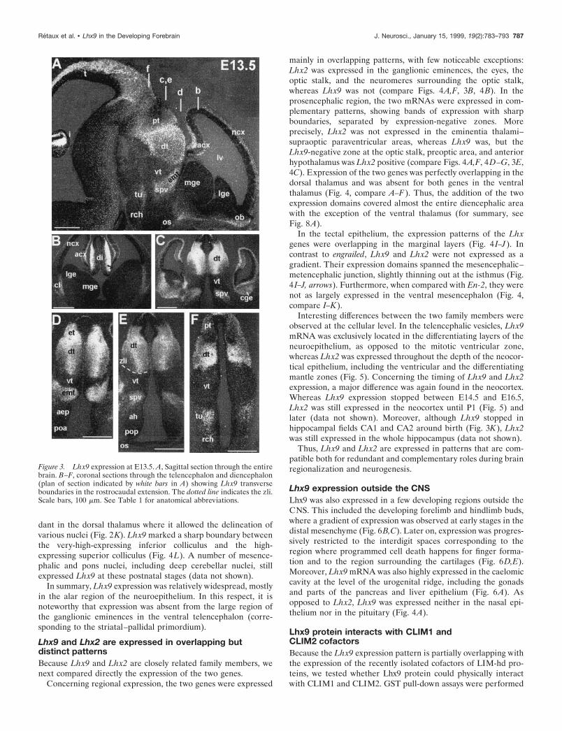
Lhx9 expression outside the CNSLhx9 was also expressed in a few developing regions outside theCNS. This included the developing forelimb and hindlimb buds,where a gradient of expression was observed at early stages in thedistal mesenchyme (Fig. 6B,C). Later on, expression was progres-sively restricted to the interdigit spaces corresponding to theregion where programmed cell death happens for finger forma-tion and to the region surrounding the cartilages (Fig. 6D,E).Moreover, Lhx9 mRNA was also highly expressed in the caelomiccavity at the level of the urogenital ridge, including the gonadsand parts of the pancreas and liver epithelium (Fig. 6A). Asopposed to Lhx2, Lhx9 was expressed neither in the nasal epi-thelium nor in the pituitary (Fig. 4A).
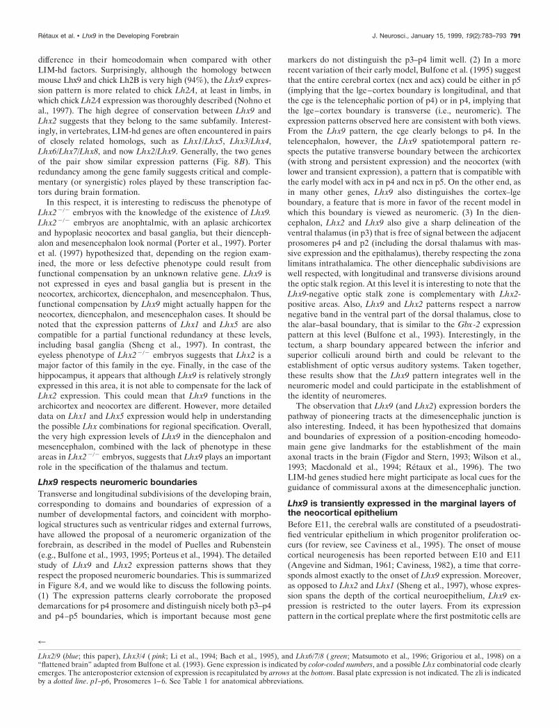
Lhx9 protein interacts with CLIM1 andCLIM2 cofactorsBecause the Lhx9 expression pattern is partially overlapping withthe expression of the recently isolated cofactors of LIM-hd pro-teins, we tested whether Lhx9 protein could physically interactwith CLIM1 and CLIM2. GST pull-down assays were performed
Figure 3. Lhx9 expression at E13.5. A, Sagittal section through the entirebrain. B–F, coronal sections through the telencephalon and diencephalon(plan of section indicated by white bars in A) showing Lhx9 transverseboundaries in the rostrocaudal extension. The dotted line indicates the zli.Scale bars, 100 mm. See Table 1 for anatomical abbreviations.
Retaux et al. • Lhx9 in the Developing Forebrain J. Neurosci., January 15, 1999, 19(2):783–793 787

Figure 4. Comparison of Lhx2 (A–E, J), Lhx9 (F–I, L), and En-2 (K) expression on sections. A, F, Parasagittal adjacent sections through the entire brainat E14.5, comparing expression of Lhx2 (A) and Lhx9 (F). B, C, Coronal sections at E13.5 showing Lhx2 expression in the telencephalon anddiencephalon. Sections are adjacent to those shown in Figure 3, B and E, respectively, for comparison of transverse boundaries. D, G, Horizontal adjacentsections showing Lhx2 (D) and Lhx9 (G) expression in the eye and optic stalk at E14.5. The asterisk in G points to the nonspecific signal attributableto the presence of the eye pigmented epithelium. E, H, Adjacent parasagittal section showing Lhx2 (E) and Lhx9 (H ) in the (Figure legend continues)
788 J. Neurosci., January 15, 1999, 19(2):783–793 Retaux et al. • Lhx9 in the Developing Forebrain
using [35S]Lhx9 and CLIM1 and CLIM2 GST fusion proteins.The results show that Lhx9 strongly interacts with both cofactorsin vitro (Fig. 7), suggesting possible functional interactions in vivo.
DISCUSSIONLhx9 and Lhx2 form a subfamily of LIM-hd genesWe have isolated a new LIM-hd gene named Lhx9, therebyincreasing the number of LIM-hd genes expressed in the devel-oping CNS. Lhx9 is most closely related to rodent Lhx2 (Xu etal., 1993) and chick Lh2B-Lh2A (Nohno et al., 1997), Drosophilaapterous (Lundgren et al., 1995), and C. elegans ttx-3 (Hobert etal., 1997). In particular, these proteins share a short but notable
Figure 6. Lhx9 expression in limbs and urogenital ridge. A, Coronalsection through the caelomic cavity and the spinal cord (double arrow) atE11.5. Arrowheads point to the developing gonads ( g), pancreas ( p), andliver ( l). Because sex discrimination is not easy at this stage, it should benoted that the same expression pattern was found in all embryos exam-ined. B, Expression in E10 forelimb after in toto in situ hybridization. p,Proximal; d, distal. C–E, Transverse sections through developing hind-limbs and forelimbs at indicated stages. Arrowheads in C indicate absenceof expression in the apical ectodermal ridge. See Results for details. Scalebars: A, C–E, 50 mm; B, 200 mm.
Figure 7. Lhx9 interacts with CLIM1 and CLIM2. Autoradiogram of arepresentative GST pull-down assay in which 35S-labeled, in vitro-translated Lhx9 was tested for its ability to bind bacterially expressed GSTfusions of CLIM1 and CLIM2. Ten percent (10%i) of the total 35S-labeled protein input and binding to beads alone are shown.
4
dorsal diencephalon and pretectum at E13.5. Note the absence of Lhx2 in the pineal gland. I–K, Adjacent parasagittal sections at E13.5 to compare Lhx9( I ), Lhx2 ( J), and En-2 (K) expression through the mesencephalon and metencephalon. Arrows in I and J show the thinning of expression at the isthmus.L, Sagittal section through the midbrain of a P1 animal to show the Lhx9 sharp boundary between the superior and the inferior colliculi. Compare withI (earlier stage). Scale bars, 100 mm. ne, Nasal epithelium. See Table 1 for abbreviations.
Figure 5. Comparison of Lhx9 (lef t) and Lhx2 (right) expression in theneocortical epithelium at indicated stages. The pial surface (p) is at thetop, and the ventricular surface (v) is at the bottom. Left and rightphotographs were taken on adjacent sections. cp, Cortical plate; iz, inter-mediate zone; mz, marginal zone; pp, preplate; sp, subplate; svz, subven-tricular zone; vz, ventricular zone. Scale bars, 100 mm.
Retaux et al. • Lhx9 in the Developing Forebrain J. Neurosci., January 15, 1999, 19(2):783–793 789
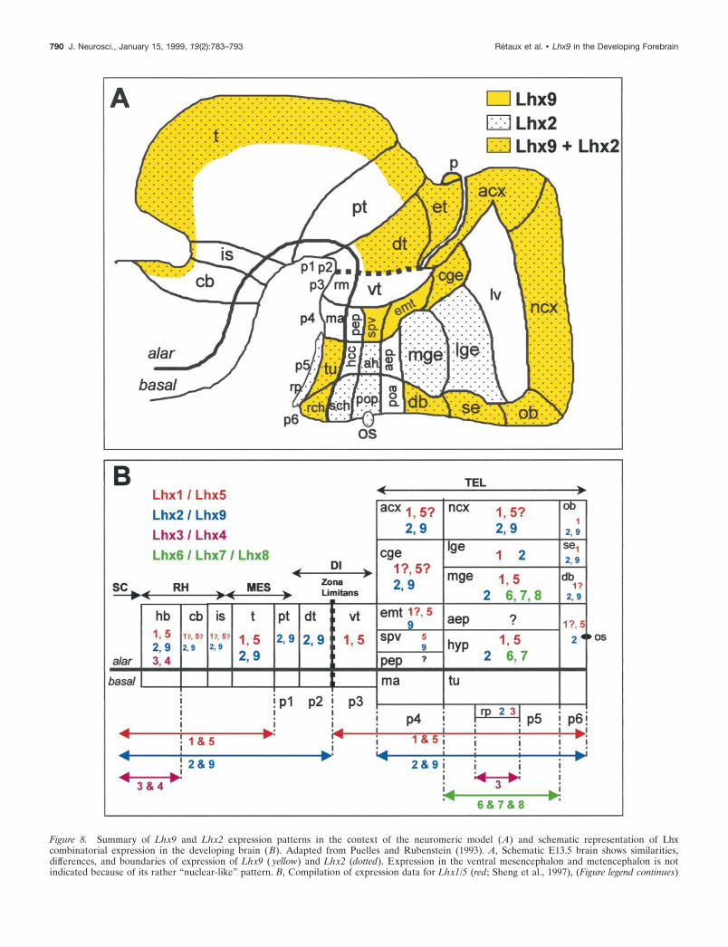
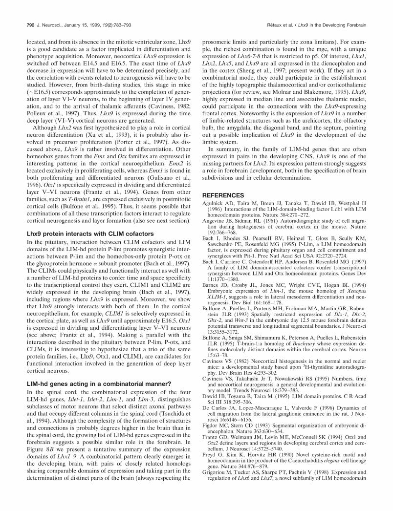
Figure 8. Summary of Lhx9 and Lhx2 expression patterns in the context of the neuromeric model (A) and schematic representation of Lhxcombinatorial expression in the developing brain ( B). Adapted from Puelles and Rubenstein (1993). A, Schematic E13.5 brain shows similarities,differences, and boundaries of expression of Lhx9 ( yellow) and Lhx2 (dotted). Expression in the ventral mesencephalon and metencephalon is notindicated because of its rather “nuclear-like” pattern. B, Compilation of expression data for Lhx1/5 (red; Sheng et al., 1997), (Figure legend continues)
790 J. Neurosci., January 15, 1999, 19(2):783–793 Retaux et al. • Lhx9 in the Developing Forebrain
difference in their homeodomain when compared with otherLIM-hd factors. Surprisingly, although the homology betweenmouse Lhx9 and chick Lh2B is very high (94%), the Lhx9 expres-sion pattern is more related to chick Lh2A, at least in limbs, inwhich chick Lh2A expression was thoroughly described (Nohno etal., 1997). The high degree of conservation between Lhx9 andLhx2 suggests that they belong to the same subfamily. Interest-ingly, in vertebrates, LIM-hd genes are often encountered in pairsof closely related homologs, such as Lhx1/Lhx5, Lhx3/Lhx4,Lhx6/Lhx7/Lhx8, and now Lhx2/Lhx9. Generally, the two genesof the pair show similar expression patterns (Fig. 8B). Thisredundancy among the gene family suggests critical and comple-mentary (or synergistic) roles played by these transcription fac-tors during brain formation.
In this respect, it is interesting to rediscuss the phenotype ofLhx22/2 embryos with the knowledge of the existence of Lhx9.Lhx22/2 embryos are anophtalmic, with an aplasic archicortexand hypoplasic neocortex and basal ganglia, but their dienceph-alon and mesencephalon look normal (Porter et al., 1997). Porteret al. (1997) hypothesized that, depending on the region exam-ined, the more or less defective phenotype could result fromfunctional compensation by an unknown relative gene. Lhx9 isnot expressed in eyes and basal ganglia but is present in theneocortex, archicortex, diencephalon, and mesencephalon. Thus,functional compensation by Lhx9 might actually happen for theneocortex, diencephalon, and mesencephalon cases. It should benoted that the expression patterns of Lhx1 and Lhx5 are alsocompatible for a partial functional redundancy at these levels,including basal ganglia (Sheng et al., 1997). In contrast, theeyeless phenotype of Lhx22/2 embryos suggests that Lhx2 is amajor factor of this family in the eye. Finally, in the case of thehippocampus, it appears that although Lhx9 is relatively stronglyexpressed in this area, it is not able to compensate for the lack ofLhx2 expression. This could mean that Lhx9 functions in thearchicortex and neocortex are different. However, more detaileddata on Lhx1 and Lhx5 expression would help in understandingthe possible Lhx combinations for regional specification. Overall,the very high expression levels of Lhx9 in the diencephalon andmesencephalon, combined with the lack of phenotype in theseareas in Lhx22/2 embryos, suggests that Lhx9 plays an importantrole in the specification of the thalamus and tectum.
Lhx9 respects neuromeric boundariesTransverse and longitudinal subdivisions of the developing brain,corresponding to domains and boundaries of expression of anumber of developmental factors, and coincident with morpho-logical structures such as ventricular ridges and external furrows,have allowed the proposal of a neuromeric organization of theforebrain, as described in the model of Puelles and Rubenstein(e.g., Bulfone et al., 1993, 1995; Porteus et al., 1994). The detailedstudy of Lhx9 and Lhx2 expression patterns shows that theyrespect the proposed neuromeric boundaries. This is summarizedin Figure 8A, and we would like to discuss the following points.(1) The expression patterns clearly corroborate the proposeddemarcations for p4 prosomere and distinguish nicely both p3–p4and p4–p5 boundaries, which is important because most gene
markers do not distinguish the p3–p4 limit well. (2) In a morerecent variation of their early model, Bulfone et al. (1995) suggestthat the entire cerebral cortex (ncx and acx) could be either in p5(implying that the lge–cortex boundary is longitudinal, and thatthe cge is the telencephalic portion of p4) or in p4, implying thatthe lge–cortex boundary is transverse (i.e., neuromeric). Theexpression patterns observed here are consistent with both views.From the Lhx9 pattern, the cge clearly belongs to p4. In thetelencephalon, however, the Lhx9 spatiotemporal pattern re-spects the putative transverse boundary between the archicortex(with strong and persistent expression) and the neocortex (withlower and transient expression), a pattern that is compatible withthe early model with acx in p4 and ncx in p5. On the other end, asin many other genes, Lhx9 also distinguishes the cortex–lgeboundary, a feature that is more in favor of the recent model inwhich this boundary is viewed as neuromeric. (3) In the dien-cephalon, Lhx2 and Lhx9 also give a sharp delineation of theventral thalamus (in p3) that is free of signal between the adjacentprosomeres p4 and p2 (including the dorsal thalamus with mas-sive expression and the epithalamus), thereby respecting the zonalimitans intrathalamica. The other diencephalic subdivisions arewell respected, with longitudinal and transverse divisions aroundthe optic stalk region. At this level it is interesting to note that theLhx9-negative optic stalk zone is complementary with Lhx2-positive areas. Also, Lhx9 and Lhx2 patterns respect a narrownegative band in the ventral part of the dorsal thalamus, close tothe alar–basal boundary, that is similar to the Gbx-2 expressionpattern at this level (Bulfone et al., 1993). Interestingly, in thetectum, a sharp boundary appeared between the inferior andsuperior colliculi around birth and could be relevant to theestablishment of optic versus auditory systems. Taken together,these results show that the Lhx9 pattern integrates well in theneuromeric model and could participate in the establishment ofthe identity of neuromeres.
The observation that Lhx9 (and Lhx2) expression borders thepathway of pioneering tracts at the dimesencephalic junction isalso interesting. Indeed, it has been hypothesized that domainsand boundaries of expression of a position-encoding homeodo-main gene give landmarks for the establishment of the mainaxonal tracts in the brain (Figdor and Stern, 1993; Wilson et al.,1993; Macdonald et al., 1994; Retaux et al., 1996). The twoLIM-hd genes studied here might participate as local cues for theguidance of commissural axons at the dimesencephalic junction.
Lhx9 is transiently expressed in the marginal layers ofthe neocortical epitheliumBefore E11, the cerebral walls are constituted of a pseudostrati-fied ventricular epithelium in which progenitor proliferation oc-curs (for review, see Caviness et al., 1995). The onset of mousecortical neurogenesis has been reported between E10 and E11(Angevine and Sidman, 1961; Caviness, 1982), a time that corre-sponds almost exactly to the onset of Lhx9 expression. Moreover,as opposed to Lhx2 and Lhx1 (Sheng et al., 1997), whose expres-sion spans the depth of the cortical neuroepithelium, Lhx9 ex-pression is restricted to the outer layers. From its expressionpattern in the cortical preplate where the first postmitotic cells are
4
Lhx2/9 (blue; this paper), Lhx3/4 ( pink; Li et al., 1994; Bach et al., 1995), and Lhx6/7/8 ( green; Matsumoto et al., 1996; Grigoriou et al., 1998) on a“flattened brain” adapted from Bulfone et al. (1993). Gene expression is indicated by color-coded numbers, and a possible Lhx combinatorial code clearlyemerges. The anteroposterior extension of expression is recapitulated by arrows at the bottom. Basal plate expression is not indicated. The zli is indicatedby a dotted line. p1–p6, Prosomeres 1–6. See Table 1 for anatomical abbreviations.
Retaux et al. • Lhx9 in the Developing Forebrain J. Neurosci., January 15, 1999, 19(2):783–793 791
located, and from its absence in the mitotic ventricular zone, Lhx9is a good candidate as a factor implicated in differentiation andphenotype acquisition. Moreover, neocortical Lhx9 expression isswitched off between E14.5 and E16.5. The exact time of Lhx9decrease in expression will have to be determined precisely, andthe correlation with events related to neurogenesis will have to bestudied. However, from birth-dating studies, this stage in mice(;E16.5) corresponds approximately to the completion of gener-ation of layer VI–V neurons, to the beginning of layer IV gener-ation, and to the arrival of thalamic afferents (Caviness, 1982;Polleux et al., 1997). Thus, Lhx9 is expressed during the timedeep layer (VI–V) cortical neurons are generated.
Although Lhx2 was first hypothesized to play a role in corticalneuron differentiation (Xu et al., 1993), it is probably also in-volved in precursor proliferation (Porter et al., 1997). As dis-cussed above, Lhx9 is rather involved in differentiation. Otherhomeobox genes from the Emx and Otx families are expressed ininteresting patterns in the cortical neuroepithelium: Emx2 islocated exclusively in proliferating cells, whereas Emx1 is found inboth proliferating and differentiated neurons (Gulisano et al.,1996). Otx1 is specifically expressed in dividing and differentiatedlayer V–VI neurons (Frantz et al., 1994). Genes from otherfamilies, such as T-Brain1, are expressed exclusively in postmitoticcortical cells (Bulfone et al., 1995). Thus, it seems possible thatcombinations of all these transcription factors interact to regulatecortical neurogenesis and layer formation (also see next section).
Lhx9 protein interacts with CLIM cofactorsIn the pituitary, interaction between CLIM cofactors and LIMdomains of the LIM-hd protein P-lim promotes synergistic inter-actions between P-lim and the homeobox-only protein P-otx onthe glycoprotein hormone a subunit promoter (Bach et al., 1997).The CLIMs could physically and functionally interact as well witha number of LIM-hd proteins to confer time and space specificityto the transcriptional control they exert. CLIM1 and CLIM2 arewidely expressed in the developing brain (Bach et al., 1997),including regions where Lhx9 is expressed. Moreover, we showthat Lhx9 strongly interacts with both of them. In the corticalneuroepithelium, for example, CLIM1 is selectively expressed inthe cortical plate, as well as Lhx9 until approximately E16.5. Otx1is expressed in dividing and differentiating layer V–VI neurons(see above; Frantz et al., 1994). Making a parallel with theinteractions described in the pituitary between P-lim, P-otx, andCLIMs, it is interesting to hypothesize that a trio of the sameprotein families, i.e., Lhx9, Otx1, and CLIM1, are candidates forfunctional interaction involved in the generation of deep layercortical neurons.
LIM-hd genes acting in a combinatorial manner?In the spinal cord, the combinatorial expression of the fourLIM-hd genes, Islet-1, Islet-2, Lim-1, and Lim-3, distinguishessubclasses of motor neurons that select distinct axonal pathwaysand that occupy different columns in the spinal cord (Tsuchida etal., 1994). Although the complexity of the formation of structuresand connections is probably degrees higher in the brain than inthe spinal cord, the growing list of LIM-hd genes expressed in theforebrain suggests a possible similar role in the forebrain. InFigure 8B we present a tentative summary of the expressiondomains of Lhx1–9. A combinatorial pattern clearly emerges inthe developing brain, with pairs of closely related homologssharing comparable domains of expression and taking part in thedetermination of distinct parts of the brain (always respecting the
prosomeric limits and particularly the zona limitans). For exam-ple, the richest combination is found in the mge, with a uniqueexpression of Lhx6-7-8 that is restricted to p5. Of interest, Lhx1,Lhx2, Lhx5, and Lhx9 are all expressed in the diencephalon andin the cortex (Sheng et al., 1997; present work). If they act in acombinatorial mode, they could participate in the establishmentof the highly topographic thalamocortical and/or corticothalamicprojections (for review, see Molnar and Blakemore, 1995). Lhx9,highly expressed in median line and associative thalamic nuclei,could participate in the connections with the Lhx9-expressingfrontal cortex. Noteworthy is the expression of Lhx9 in a numberof limbic-related structures such as the archicortex, the olfactorybulb, the amygdala, the diagonal band, and the septum, pointingout a possible implication of Lhx9 in the development of thelimbic system.
In summary, in the family of LIM-hd genes that are oftenexpressed in pairs in the developing CNS, Lhx9 is one of themissing partners for Lhx2. Its expression pattern strongly suggestsa role in forebrain development, both in the specification of brainsubdivisions and in cellular determination.
REFERENCESAgulnick AD, Taira M, Breen JJ, Tanaka T, Dawid IB, Westphal H
(1996) Interactions of the LIM-domain-binding factor Ldb1 with LIMhomeodomain proteins. Nature 384:270–272.
Angevine JB, Sidman RL (1961) Autoradiographic study of cell migra-tion during histogenesis of cerebral cortex in the mouse. Nature192:766–768.
Bach I, Rhodes SJ, PearseII RV, Heinzel T, Gloss B, Scully KM,Sawchenko PE, Rosenfeld MG (1995) P-Lim, a LIM homeodomainfactor, is expressed during pituitary organ and cell commitment andsynergizes with Pit-1. Proc Natl Acad Sci USA 92:2720–2724.
Bach I, Carriere C, Ostendorff HP, Andersen B, Rosenfeld MG (1997)A family of LIM domain-associated cofactors confer transcriptionalsynergism between LIM and Otx homeodomain proteins. Genes Dev11:1370–1380.
Barnes JD, Crosby JL, Jones MC, Wright CVE, Hogan BL (1994)Embryonic expression of Lim-1, the mouse homolog of XenopusXLIM-1, suggests a role in lateral mesoderm differentiation and neu-rogenesis. Dev Biol 161:168–178.
Bulfone A, Puelles L, Porteus MH, Frohman MA, Martin GR, Ruben-stein JLR (1993) Spatially restricted expression of Dlx-1, Dlx-2,Gbx-2, and Wnt-3 in the embryonic day 12.5 mouse forebrain definespotential transverse and longitudinal segmental boundaries. J Neurosci13:3155–3172.
Bulfone A, Smiga SM, Shimamura K, Peterson A, Puelles L, RubensteinJLR (1995) T-brain-1:a homolog of Brachyury whose expression de-fines molecularly distinct domains within the cerebral cortex. Neuron15:63–78.
Caviness VS (1982) Neocortical histogenesis in the normal and reelermice: a developmental study based upon 3H-thymidine autoradiogra-phy. Dev Brain Res 4:293–302.
Caviness VS, Takahashi Jr T, Nowakowski RS (1995) Numbers, timeand neocortical neurogenesis: a general developmental and evolution-ary model. Trends Neurosci 18:379–383.
Dawid IB, Toyama R, Taira M (1995) LIM domain proteins. C R AcadSci III 318:295–306.
De Carlos JA, Lopez-Mascaraque L, Valverde F (1996) Dynamics ofcell migration from the lateral ganglionic eminence in the rat. J Neu-rosci 16:6146–6156.
Figdor MC, Stern CD (1993) Segmental organization of embryonic di-encephalon. Nature 363:630–634.
Frantz GD, Weimann JM, Levin ME, McConnell SK (1994) Otx1 andOtx2 define layers and regions in developing cerebral cortex and cere-bellum. J Neurosci 14:5725–5740.
Freyd G, Kim K, Horvitz HR (1990) Novel cysteine-rich motif andhomeodomain in the product of the Caenorhabditis elegans cell lineagegene. Nature 344:876–879.
Grigoriou M, Tucker AS, Sharpe PT, Pachnis V (1998) Expression andregulation of Lhx6 and Lhx7, a novel subfamily of LIM homeodomain
792 J. Neurosci., January 15, 1999, 19(2):783–793 Retaux et al. • Lhx9 in the Developing Forebrain
encoding genes, suggests a role in mammalian head development.Development 125:2063–2074.
Gulisano M, Broccoli V, Pardini C, Boncinelli E (1996) Emx1 and Emx2show different patterns of expression during proliferation and differen-tiation of the developing cerebral cortex in the mouse. Eur J Neurosci8:1037–1050.
Hobert O, Mori I, Yamashita Y, Honda H, Ohshima Y, Liu Y, Ruvkun G(1997) Regulation of interneuron function in the C. elegans thermo-regulatory pathway by the ttx-3 LIM homeobox gene. Neuron19:345–357.
Jurata LW, Kenny DA, Gill GN (1996) Nuclear LIM interactor, a rhom-botin and LIM homeodomain interacting protein, is expressed early inneuronal development. Proc Natl Acad Sci USA 93:11693–11698.
Li H, Witte DP, Branford WW, Aronow JB, Weinstein M, Kaur S, WertS, Singh G, Schreiner CM, Whitsett JA, Scott Jr WJ, Potter SS (1994)Gsh-4 encodes a LIM-type homeodomain, is expressed in the develop-ing central nervous system, and is required for early postnatal survival.EMBO J 12:2876–2895.
Lundgren SE, Callahan CA, Thor S, Thomas JB (1995) Control ofneuronal pathway selection by the Drosophila LIM homeodomain geneapterous. Development 121:1769–1773.
Macdonald R, Xu Q, Barth A, Mikkola I, Holder N, Fjose A, Krauss S,Wilson SW (1994) Regulatory gene expression boundaries demarcatesites of neuronal differentiation in the embryonic zebrafish forebrain.Neuron 13:1039–1053.
Matsumoto K, Tanaka T, Furuyama T, Kashihara Y, Ishii N, Tohyama M,Kitanaka J, Takemura M, Mori T, Wanaka A (1996) Differential ex-pression of LIM-homeodomain genes in the embryonic murine brain.Neurosci Lett 211:147–150.
Molnar Z, Blakemore C (1995) How do thalamic axons find their way tothe cortex? Trends Neurosci 18:389–397.
Nohno T, Kawakami Y, Wada N, Ishikawa T, Ohuchi H, Sumihare N(1997) Differential expression of the two closely related LIM-classhomeobox genes LH-2A and LH-2B during limb development. BiochemBiophys Res Commun 238:506–511.
Polleux F, Dehay C, Kennedy H (1997) The timetable of laminar neu-rogenesis contributes to the specification of cortical areas in mouseisocortex. J Comp Neurol 385:96–116.
Porter FD, Drago J, Xu Y, Cheema SS, Wassif C, Huang SP, Lee E,Grinberg A, Massalas JS, Bodine D, Alt F, Westphal H (1997) Lhx-2,
a LIM homeobox gene, is required for eye, forebrain, and definitiveerythrocyte development. Development 124:2935–2944.
Porteus MH, Bulfone A, Liu J-K, Puelles L, Lo L-C, John LR (1994)DLX-2, MASH-1, and MAP-2 expression and bromodeoxyuridine in-corporation define molecularly distinct cell populations in the embry-onic mouse forebrain. J Neurosci 14:6370–6383.
Puelles L (1995) A segmental morphological paradigm for understand-ing vertebrate forebrain. Brain Behav Evol 46:319–337.
Puelles L, Rubenstein JLR (1993) Expression patterns of homeobox andother putative regulatory genes in the embryonic forebrain suggest aneuromeric organization. Trends Neurosci 11:472–479.
Retaux S, McNeill L, Harris WA (1996) Engrailed, retinotectal target-ing, and axonal patterning in the midbrain during Xenopus develop-ment: an antisense study. Neuron 16:63–75.
Rubenstein JLR, Martinez S, Shimamura K, Puelles L (1994) The em-bryonic vertebrate forebrain: the prosomeric model. Science266:578–580.
Sanchez-Garcia I, Rabbitts TH (1994) The LIM domain: a new struc-tural motif found in zinc-finger-like proteins. Trends Genet 9:315–320.
Seidah NG, Barale JC, Marcinkiewicz M, Mattei MG, Day R, Chretien M(1994) The mouse homeoprotein mLIM-3 is expressed early in cellsderived from the neuroepithelium and persists in adult pituitary. DNACell Biol 13:1163–1180.
Sheng HZ, Bertuzzi S, Chiang C, Shawlot W, Taira M, Dawid IB,Westphal H (1997) Expression of murine Lhx-5 suggest a role inspecifying the forebrain. Dev Dyn 208:266–277.
Thor S, Thomas JB (1997) The Drosophila islet gene governs axon path-finding and neurotransmitter identity. Neuron 18:397–409.
Tsuchida T, Ensini M, Morton SB, Baldassare M, Edlund T, Jessell TM,Pfaff SL (1994) Topographic organization of embryonic motor neu-rons defined by expression of LIM homeobox genes. Cell 79:957–970.
Way JC, Chalfie M (1988) mec-3, a homeobox-containing gene thatspecifies differentiation of the touch receptor neurons in C. elegans. Cell54:5–16.
Wilson SW, Placzek M, Furley AJ (1993) Border disputes: do bound-aries play a role in growth cone guidance? Trends Neurosci 16:316–323.
Xu Y, Baldassare M, Fisher P, Rathbun G, Oltz EM, Yancopoulos GD,Jessell TM, Alt FW (1993) LH-2: a LIM/homeodomain gene ex-pressed in developing lymphocytes and neural cells. Proc Natl Acad SciUSA 90:227–231.
Retaux et al. • Lhx9 in the Developing Forebrain J. Neurosci., January 15, 1999, 19(2):783–793 793