Embed Size (px)
Citation preview
Linfoma de
Hodgkin
Juan F García
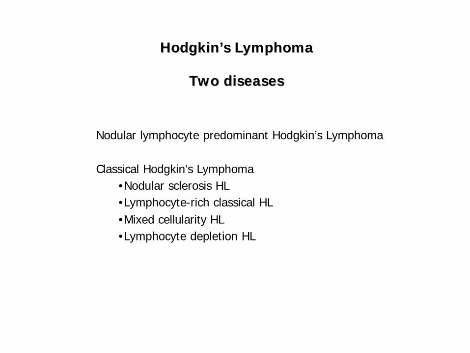
Hodgkin’s Hodgkin’s LymphomaLymphoma
Two diseasesTwo diseases
Nodular lymphocyte predominant Hodgkin’s Lymphoma
Classical Hodgkin’s Lymphoma•Nodular sclerosis HL•Lymphocyte-rich classical HL•Mixed cellularity HL•Lymphocyte depletion HL
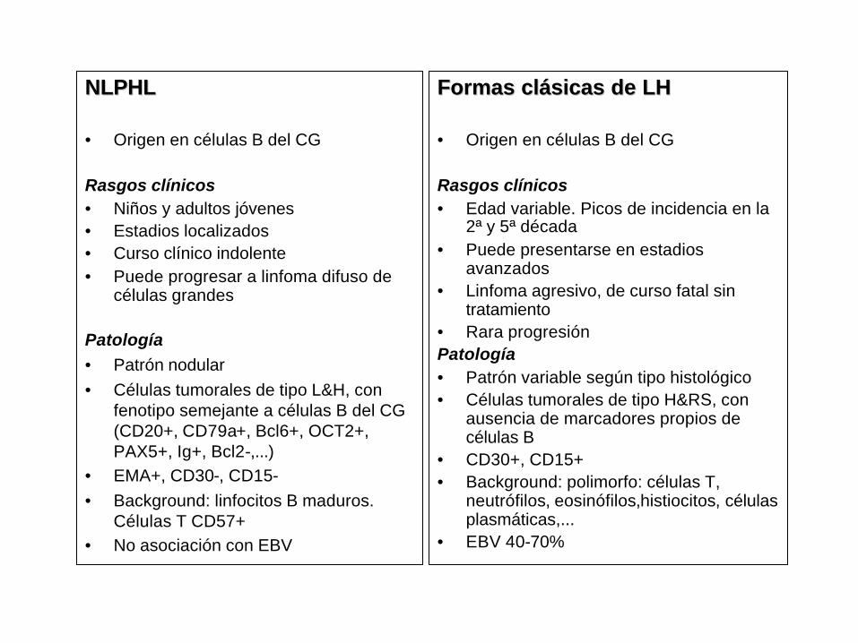
NLPHLNLPHL
• Origen en células B del CG
Rasgos clínicos• Niños y adultos jóvenes• Estadios localizados• Curso clínico indolente• Puede progresar a linfoma difuso de
células grandes
Patología• Patrón nodular• Células tumorales de tipo L&H, con
fenotipo semejante a células B del CG (CD20+, CD79a+, Bcl6+, OCT2+, PAX5+, Ig+, Bcl2-,...)
• EMA+, CD30-, CD15-• Background: linfocitos B maduros.
Células T CD57+• No asociación con EBV
Formas clásicas de LHFormas clásicas de LH
• Origen en células B del CG
Rasgos clínicos• Edad variable. Picos de incidencia en la
2ª y 5ª década• Puede presentarse en estadios
avanzados• Linfoma agresivo, de curso fatal sin
tratamiento• Rara progresiónPatología• Patrón variable según tipo histológico• Células tumorales de tipo H&RS, con
ausencia de marcadores propios de células B
• CD30+, CD15+ • Background: polimorfo: células T,
neutrófilos, eosinófilos,histiocitos, células plasmáticas,...
• EBV 40-70%
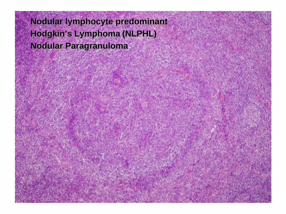
Nodular lymphocyte predominant Nodular lymphocyte predominant Hodgkin’s Lymphoma Hodgkin’s Lymphoma (NLPHL)(NLPHL)Nodular ParagranulomaNodular Paragranuloma
Rasgos clínicos
• Niños y adultos jóvenes, estadios localizados, curso clínico indolente
• ...pero puede progresar a linfoma difuso de células grandes (confrecuencia morfología tipo TCRBCL)
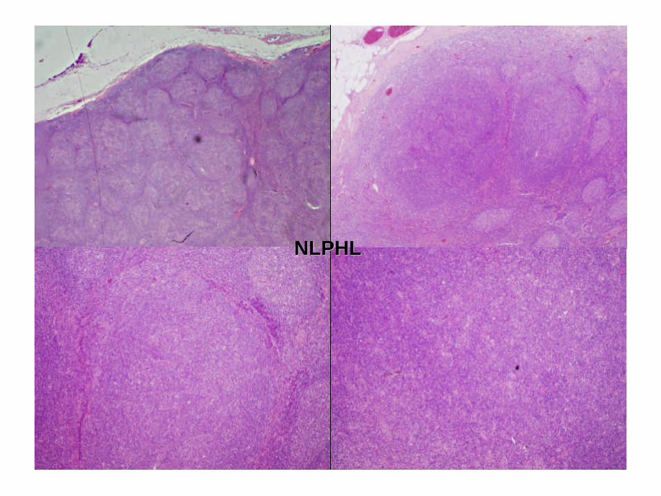
Morfología y fenotipo
• Patrón nodular: folículos irregulares con una reacción anormal del centro germinal (transformación progresiva)
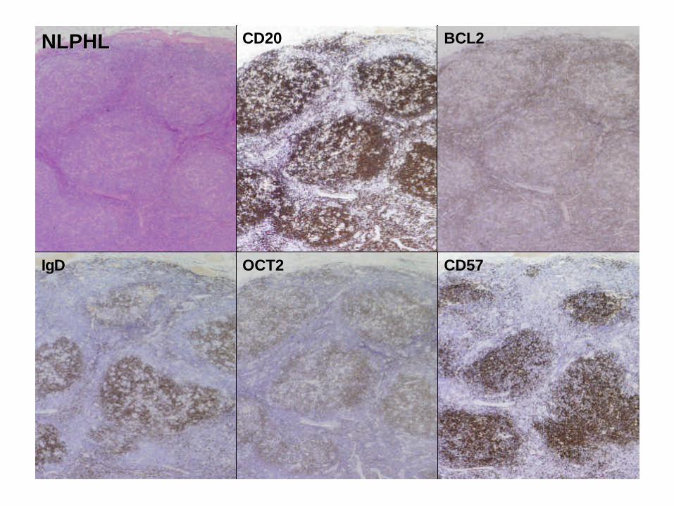
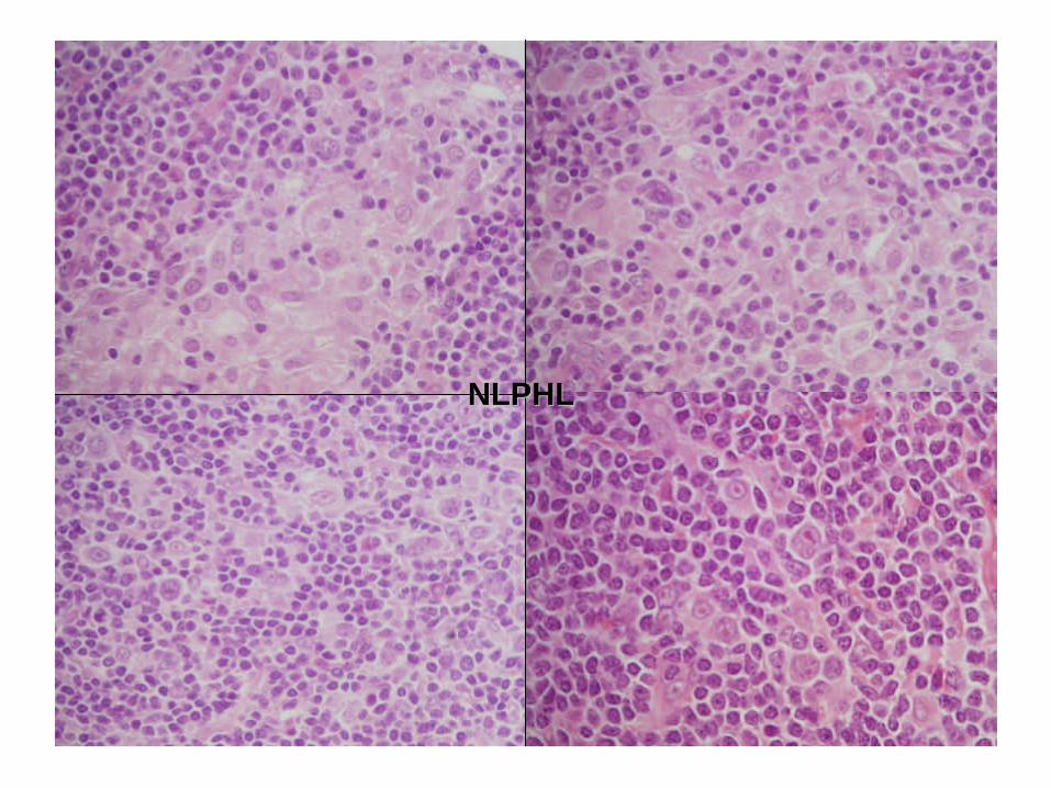
• Células tumorales de tipo L&H, con fenotipo semejante a células B del CG: CD20+, CD79a+, Bcl6+, OCT2+, PAX5+, MUM1-/+, Ig+, Bcl2-
• EMA+, CD30-, CD15-
• Background: nódulos de linfocitos B maduros. Histiocitos. Células T CD57+
• No asociación con EBV
NLPHLNLPHL
NLPHLNLPHL
IgDIgD
CD20CD20 BCL2BCL2
CD57CD57OCT2OCT2
NLPHLNLPHL
NLPHLNLPHL
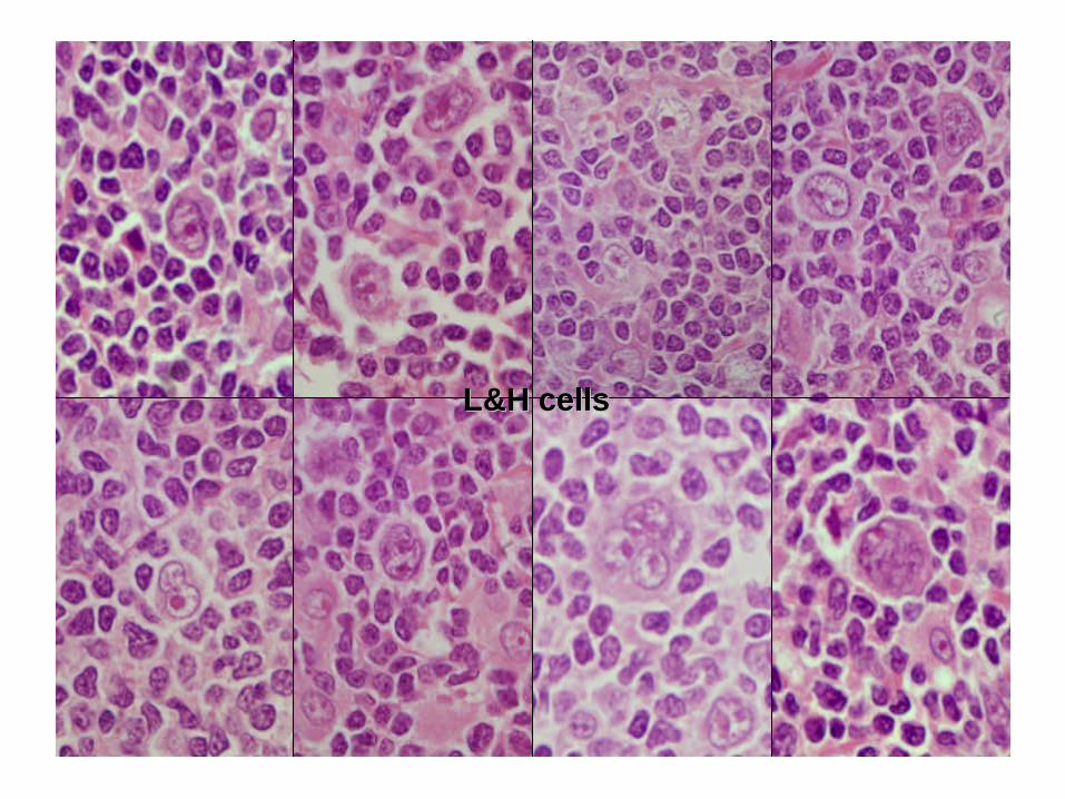
L&H L&H cellscells
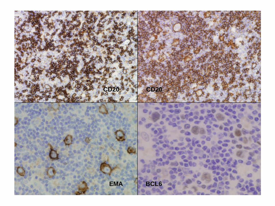
BCL6BCL6EMAEMA
CD20CD20CD20CD20
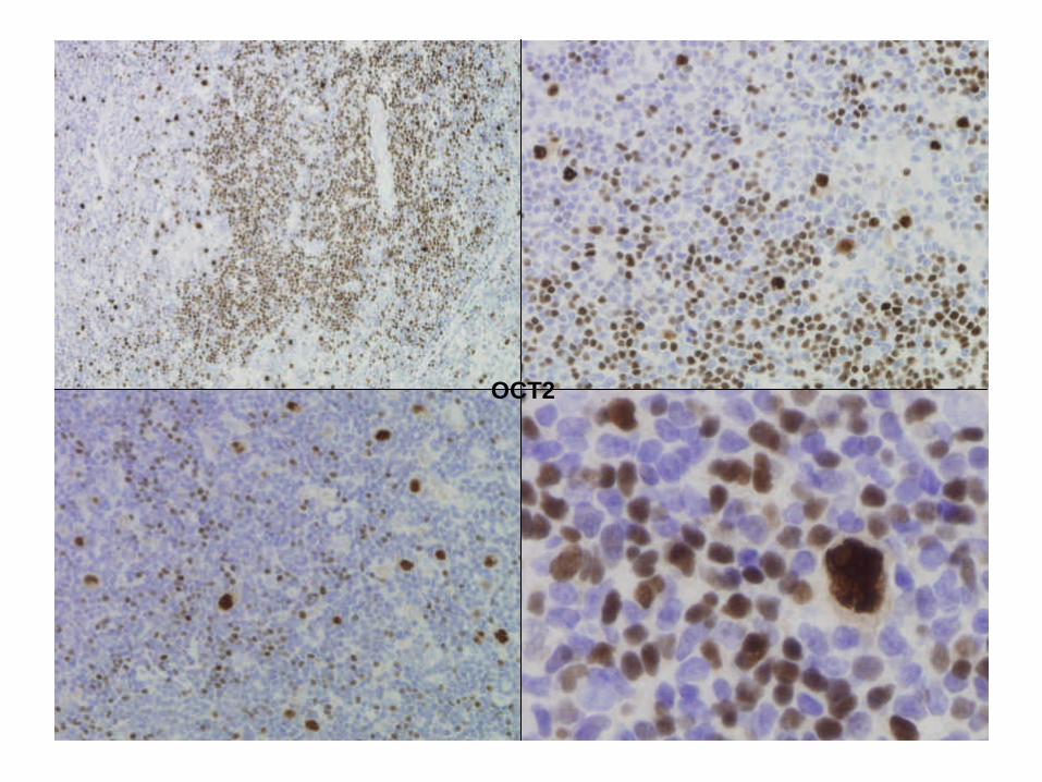
OCT2OCT2
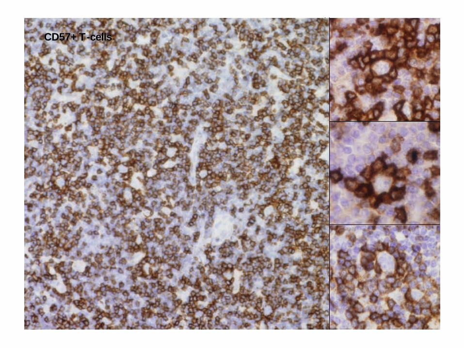
CD57+ TCD57+ T--cellscells
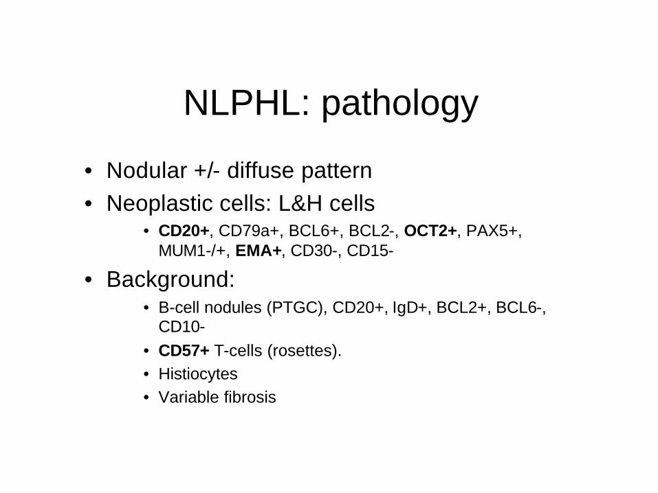
NLPHL: pathology
• Nodular +/- diffuse pattern• Neoplastic cells: L&H cells
• CD20+, CD79a+, BCL6+, BCL2-, OCT2+, PAX5+, MUM1-/+, EMA+, CD30-, CD15-
• Background:• B-cell nodules (PTGC), CD20+, IgD+, BCL2+, BCL6-,
CD10-• CD57+ T-cells (rosettes).• Histiocytes• Variable fibrosis
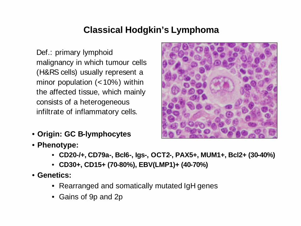
Classical Hodgkin’s Lymphoma
Def.: primary lymphoid malignancy in which tumour cells (H&RS cells) usually represent a minor population (<10%) within the affected tissue, which mainly consists of a heterogeneous infiltrate of inflammatory cells.
• Origin: GC B-lymphocytes• Phenotype:
• CD20-/+, CD79a-, Bcl6-, Igs-, OCT2-, PAX5+, MUM1+, Bcl2+ (30-40%)• CD30+, CD15+ (70-80%), EBV(LMP1)+ (40-70%)
• Genetics: • Rearranged and somatically mutated IgH genes• Gains of 9p and 2p
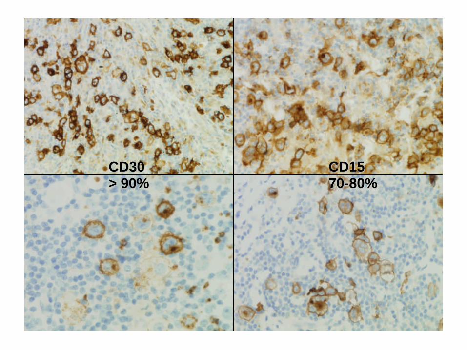
CD30CD30> 90%> 90%
CD15CD157070--80%80%
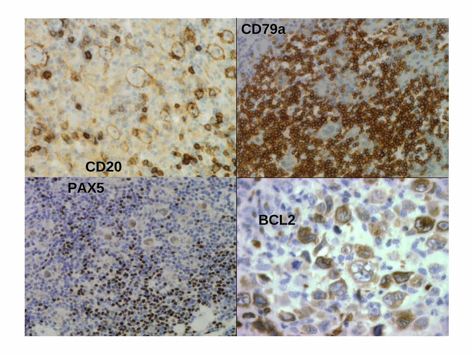
CD20CD20
CD79aCD79a
PAX5PAX5
BCL2BCL2
Bcl2Bcl2
Months
140120100806040200
Ove
rall S
urvival (Bcl2
)
1,1
1,0
,9
,8
,7
,6
,5
,4
,3
,2
,1
0,0
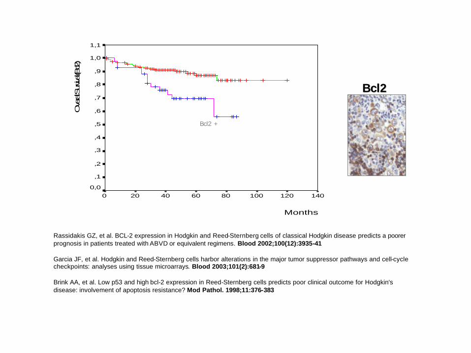
Bcl2 +
Rassidakis GZ, et al. BCL-2 expression in Hodgkin and Reed-Sternberg cells of classical Hodgkin disease predicts a poorerprognosis in patients treated with ABVD or equivalent regimens. Blood 2002;100(12):3935-41
Garcia JF, et al. Hodgkin and Reed-Sternberg cells harbor alterations in the major tumor suppressor pathways and cell-cycle checkpoints: analyses using tissue microarrays. Blood 2003;101(2):681-9
Brink AA, et al. Low p53 and high bcl-2 expression in Reed-Sternberg cells predicts poor clinical outcome for Hodgkin's disease: involvement of apoptosis resistance? Mod Pathol. 1998;11:376-383
Classical HL: pathology• Variable histological features:
subtypes NS, MC, LRCHL, LD• Neoplastic cells: H&RS cells, and
variants: mononuclear (Hodgkin’s) cells, lacunar cells, mummy cells.
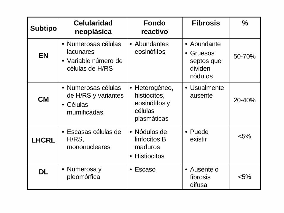
<5%• Ausente o
fibrosis difusa
• Escaso• Numerosa y pleomórfica
DL
<5%• Puede
existir• Nódulos de
linfocitos B maduros
• Histiocitos
• Escasas células de H/RS, mononucleares
LHCRL
20-40%
50-70%
%
• Usualmente ausente
• Heterogéneo, histiocitos, eosinófilos y células plasmáticas
• Numerosas célulasde H/RS y variantes
• Células mumificadas
CM
• Abundante• Gruesos
septos que dividen nódulos
• Abundantes eosinófilos
• Numerosas células lacunares
• Variable número de células de H/RS
EN
FibrosisFondo reactivo
Celularidad neoplásicaSubtipo
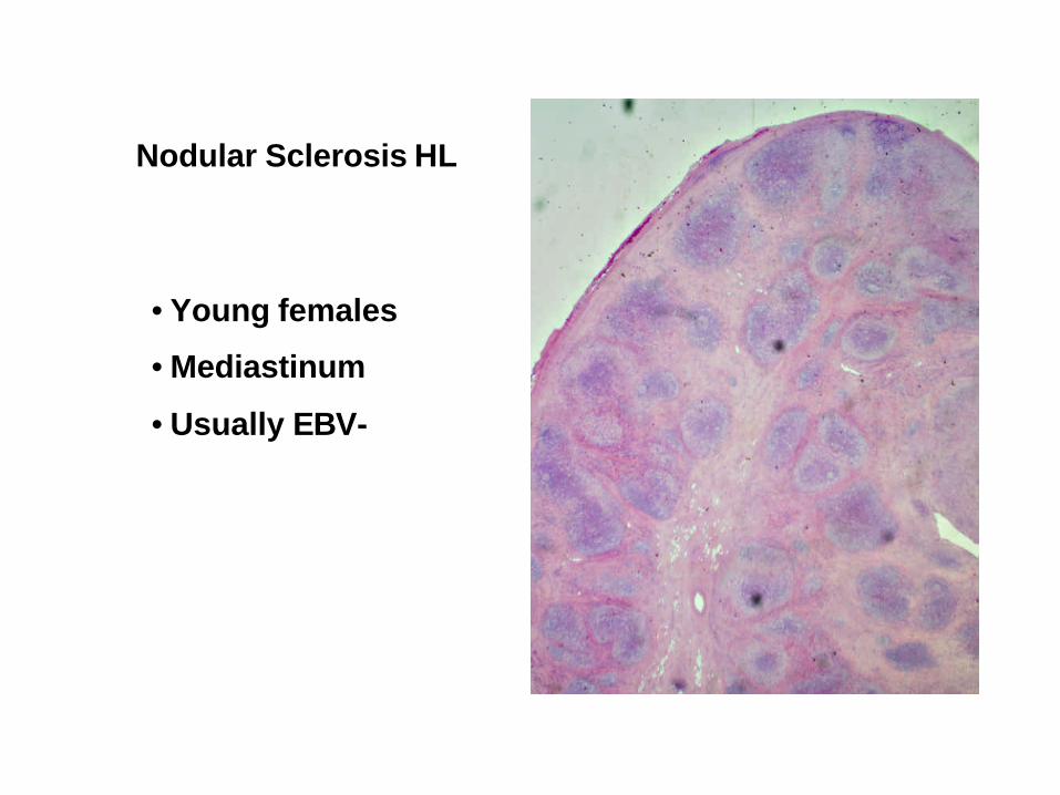
Nodular Sclerosis HL
• Young females
• Mediastinum
• Usually EBV-
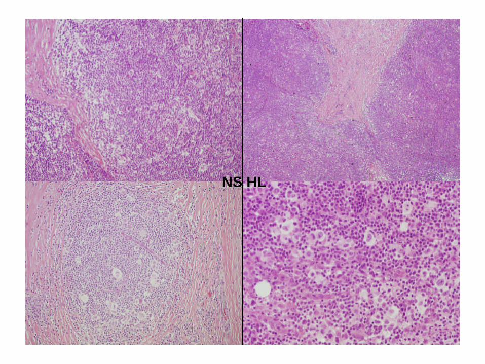
NS HLNS HL
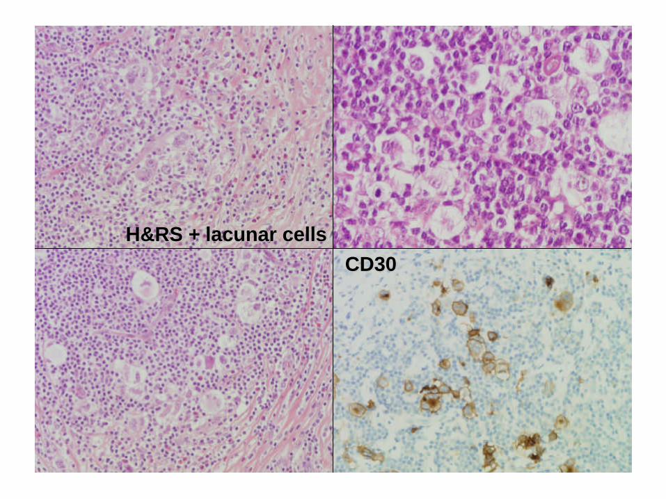
H&RS + H&RS + lacunar cellslacunar cellsCD30CD30
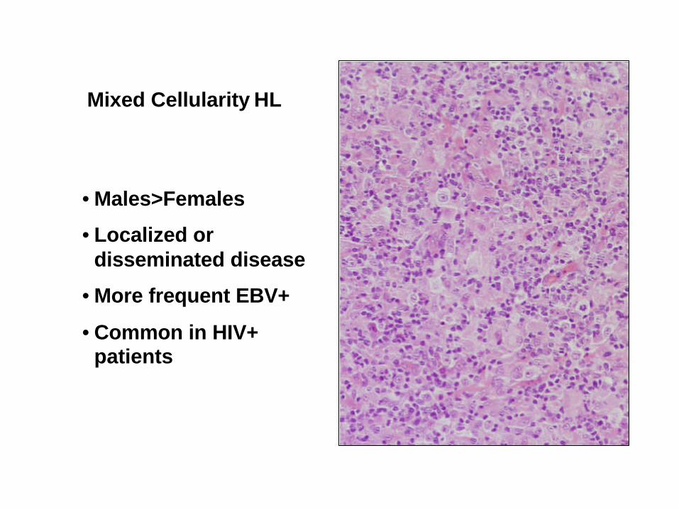
Mixed Cellularity HL
• Males>Females
• Localized or disseminated disease
• More frequent EBV+
• Common in HIV+ patients
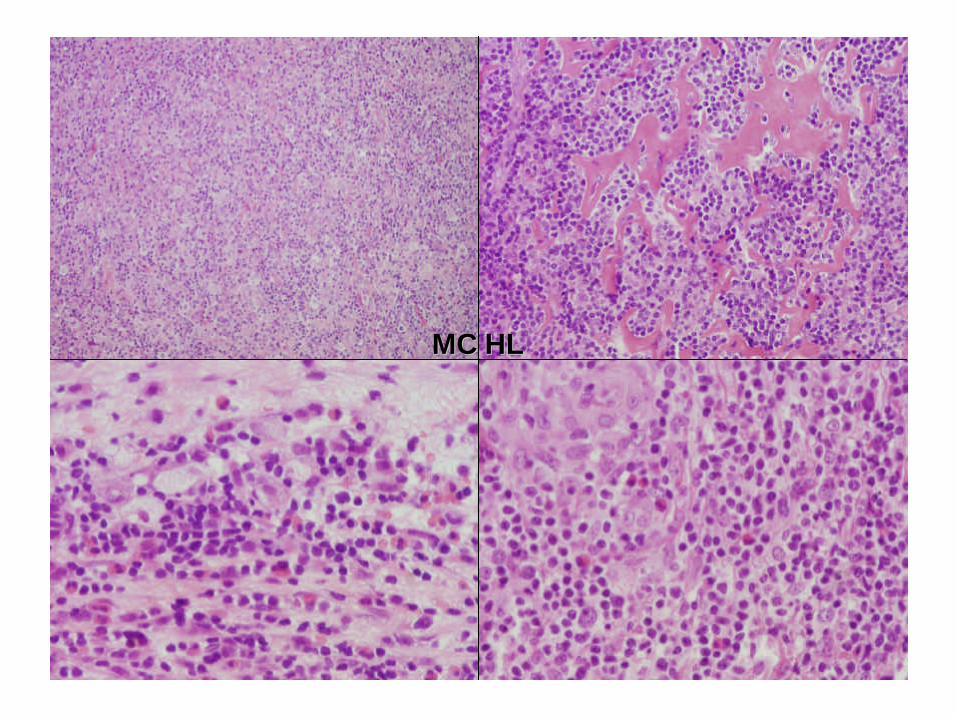
MC HLMC HL
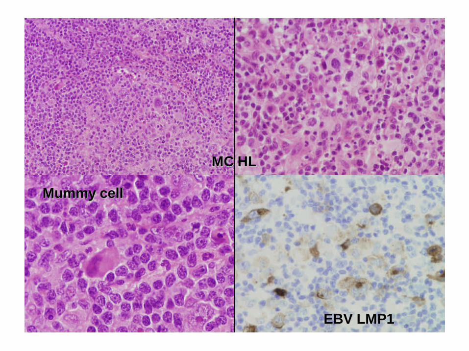
MC HLMC HL
MummyMummy cellcell
EBV LMP1EBV LMP1
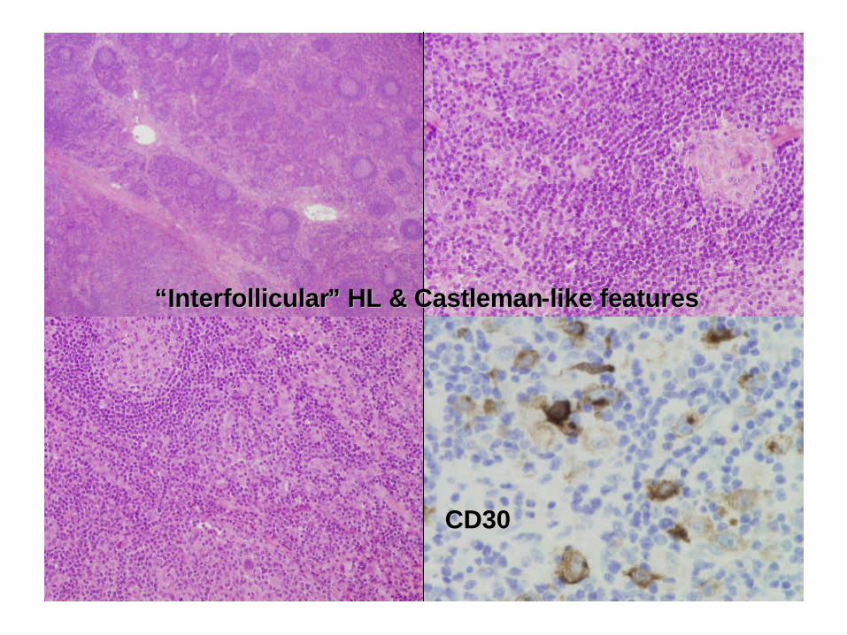
““InterfollicularInterfollicular” HL & ” HL & CastlemanCastleman--like featureslike features
CD30CD30
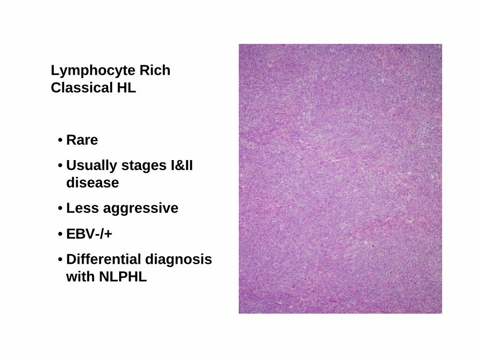
Lymphocyte Rich Classical HL
• Rare
• Usually stages I&II disease
• Less aggressive
• EBV-/+
• Differential diagnosis with NLPHL
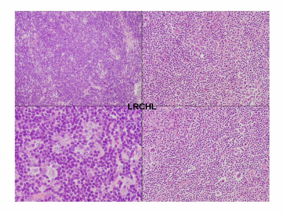
LRCHL
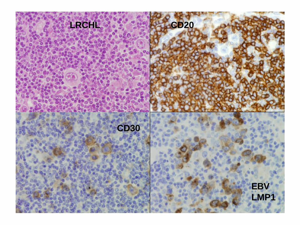
LRCHL CD20
CD30
EBVLMP1
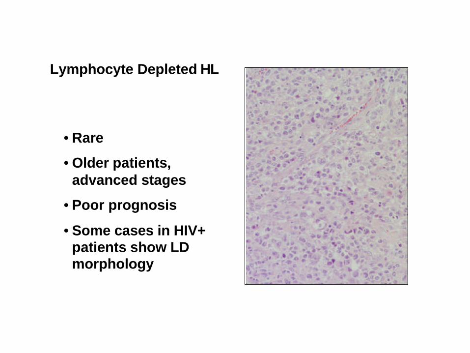
Lymphocyte Depleted HL
• Rare
• Older patients, advanced stages
• Poor prognosis
• Some cases in HIV+ patients show LD morphology
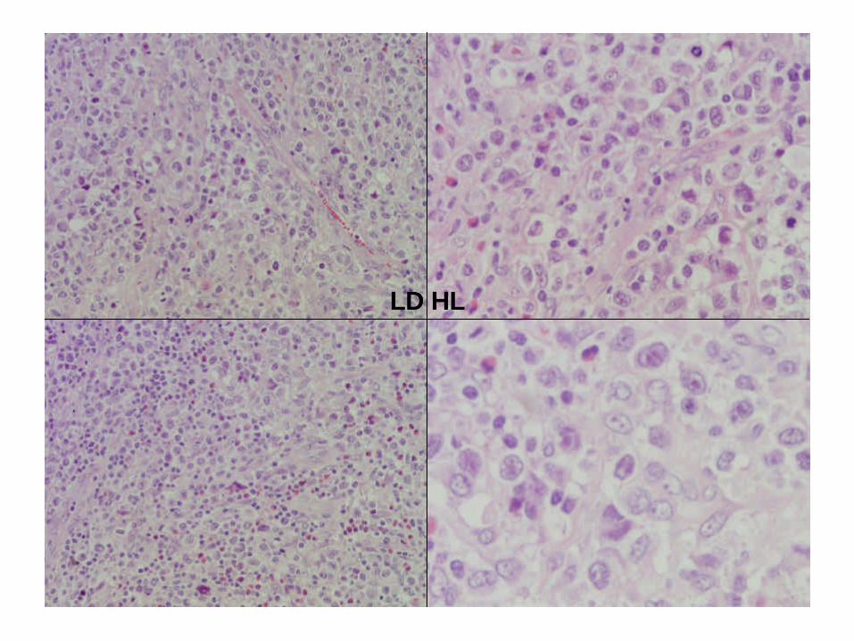
LD HL
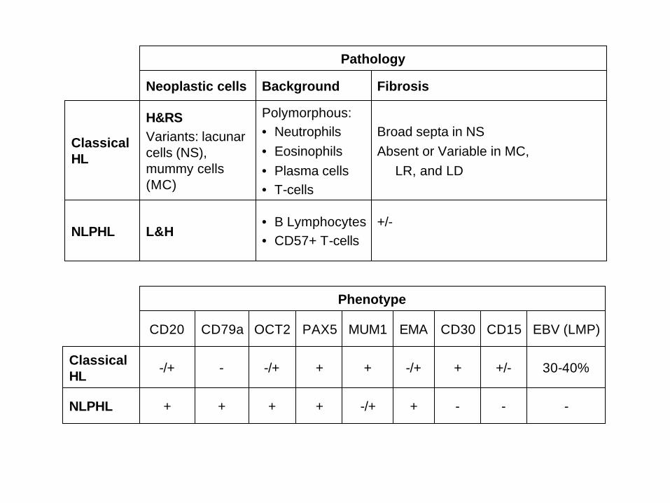
Pathology
+/-
Broad septa in NSAbsent or Variable in MC,
LR, and LD
Fibrosis
NLPHL
Classical HL
• B Lymphocytes• CD57+ T-cells
L&H
Polymorphous:• Neutrophils• Eosinophils• Plasma cells• T-cells
H&RSVariants: lacunarcells (NS), mummy cells (MC)
BackgroundNeoplastic cells
Phenotype
NLPHL
Classical HL
+
-/+
EMA
-
30-40%
EBV (LMP)
-
+/-
CD15
-
+
CD30
+
-/+
OCT2
+
+
PAX5
-/+++
+--/+
MUM1CD79aCD20
+/-
-/+
BCL2
+
-
BCL6
Phenotype
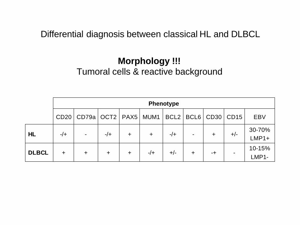
DLBCL
HL
10-15%LMP1-
30-70%LMP1+
EBV
-
+/-
CD15
-+
+
CD30
+
-/+
OCT2
+
+
PAX5
-/+++
+--/+
MUM1CD79aCD20
Differential diagnosis between classical HL and DLBCL
Morphology !!!Tumoral cells & reactive background
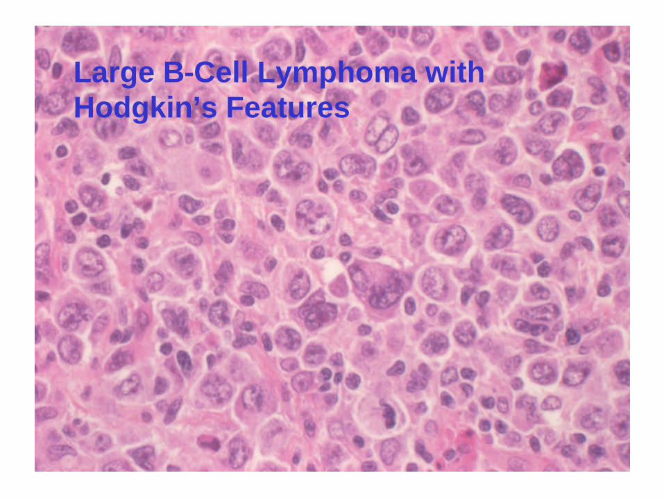
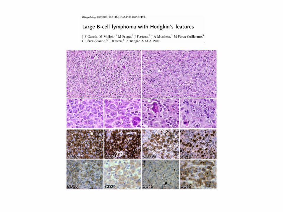
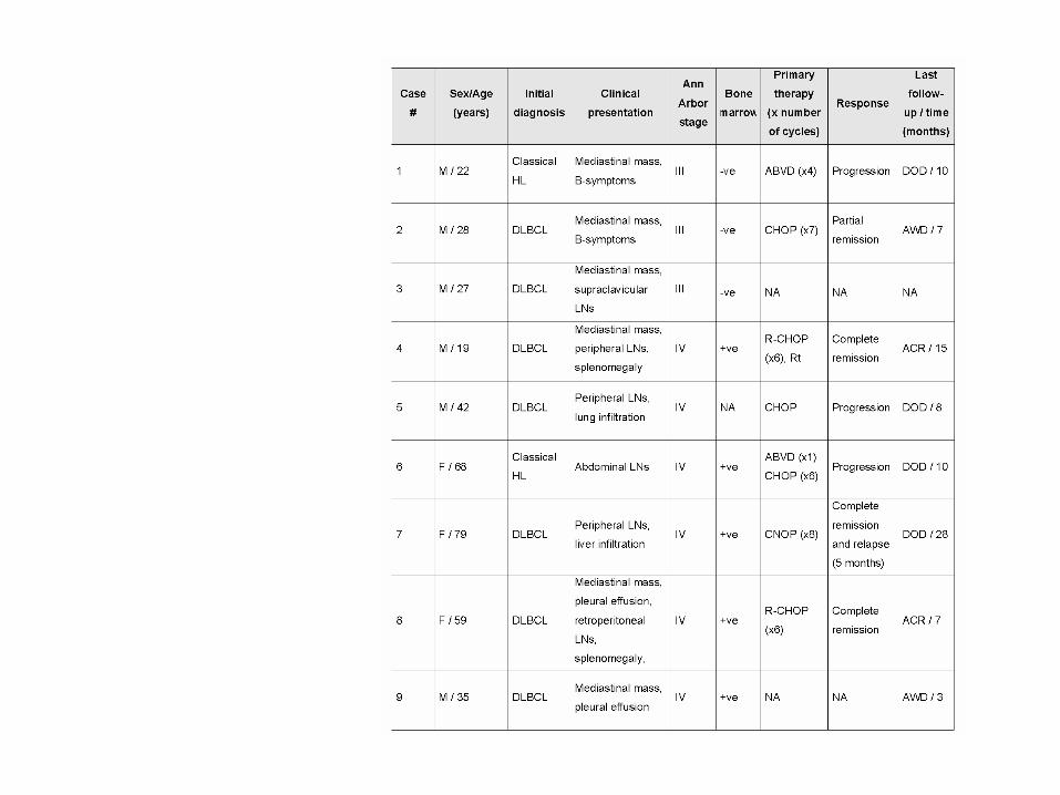
Large B-Cell Lymphoma with Hodgkin’s Features
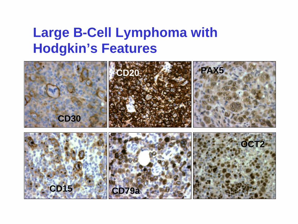
CD30
CD20
OCT2
PAX5
CD79aCD15
Large B-Cell Lymphoma with Hodgkin’s Features
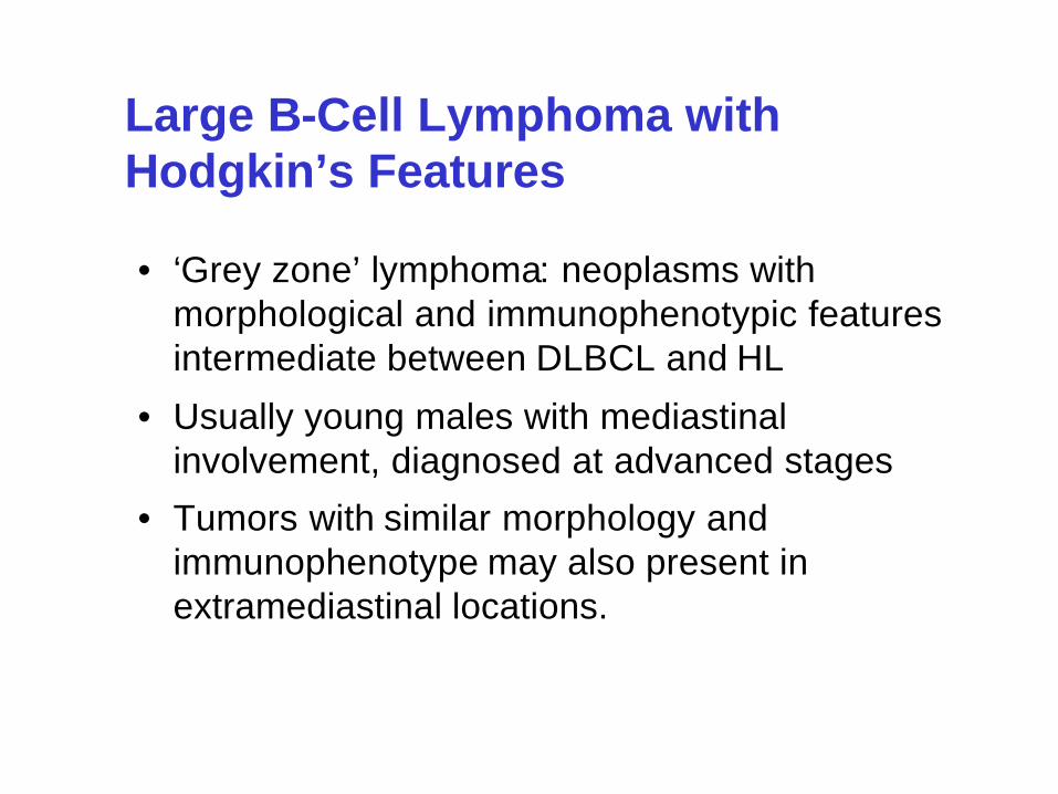
• ‘Grey zone’ lymphoma: neoplasms with morphological and immunophenotypic features intermediate between DLBCL and HL
• Usually young males with mediastinalinvolvement, diagnosed at advanced stages
• Tumors with similar morphology and immunophenotype may also present in extramediastinal locations.
Large B-Cell Lymphoma with Hodgkin’s Features
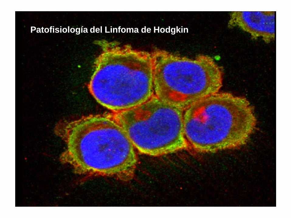
Patofisiología del Linfoma de Hodgkin
•• Origen celular: Origen celular: •• linfocitos B del centro germinallinfocitos B del centro germinal
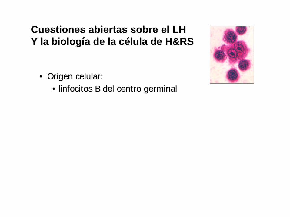
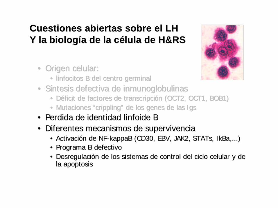
Cuestiones abiertas sobre el LHCuestiones abiertas sobre el LHY la bY la biologiologííaa de la cde la céélula de H&RSlula de H&RS
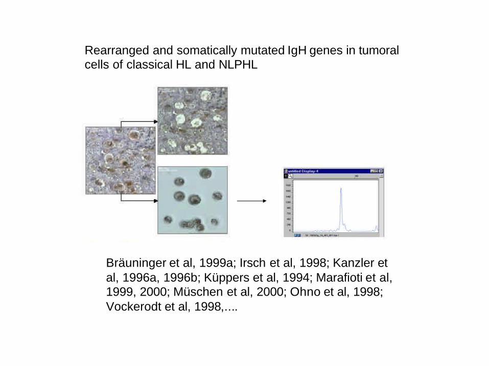
Rearranged and somatically mutated IgH genes in tumoral cells of classical HL and NLPHL
Bräuninger et al, 1999a; Irsch et al, 1998; Kanzler etal, 1996a, 1996b; Küppers et al, 1994; Marafioti et al,1999, 2000; Müschen et al, 2000; Ohno et al, 1998;Vockerodt et al, 1998,....
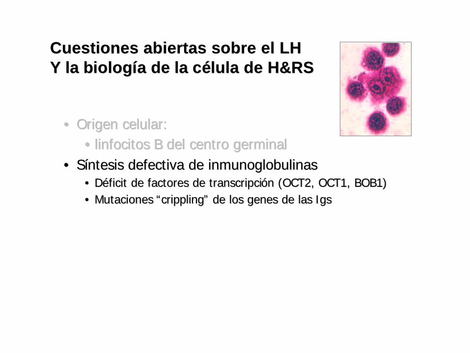
•• Origen celular: Origen celular: •• linfocitos B del centro germinallinfocitos B del centro germinal
•• SSííntesis defectiva de inmunoglobulinasntesis defectiva de inmunoglobulinas•• DDééficit de factores de transcripcificit de factores de transcripcióón (OCT2, OCT1, BOB1)n (OCT2, OCT1, BOB1)•• Mutaciones Mutaciones ““cripplingcrippling”” de los genes de las de los genes de las IgsIgs
Cuestiones abiertas sobre el LHCuestiones abiertas sobre el LHY la bY la biologiologííaa de la cde la céélula de H&RSlula de H&RS
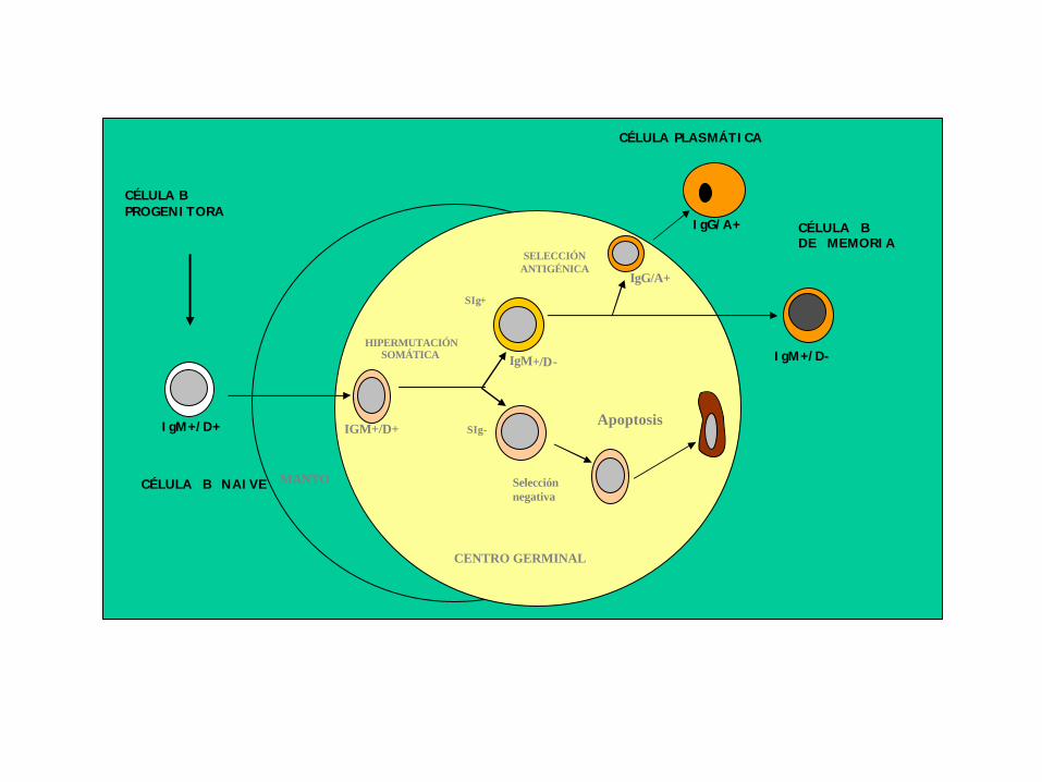
CÉLULA B NAIVE
CÉLULA BPROGENITORA
IgM+/D+
MANTO
IGM+/D+
SELECCIÓNANTIGÉNICA
SIg+
IgM+/D-
CÉLULA B DE MEMORIA
IgG/A+
CÉLULA PLASMÁTICA
IgG/A+
HIPERMUTACIÓNSOMÁTICA
CENTRO GERMINAL
Apoptosis
Selecciónnegativa
IgM+/D-
SIg-
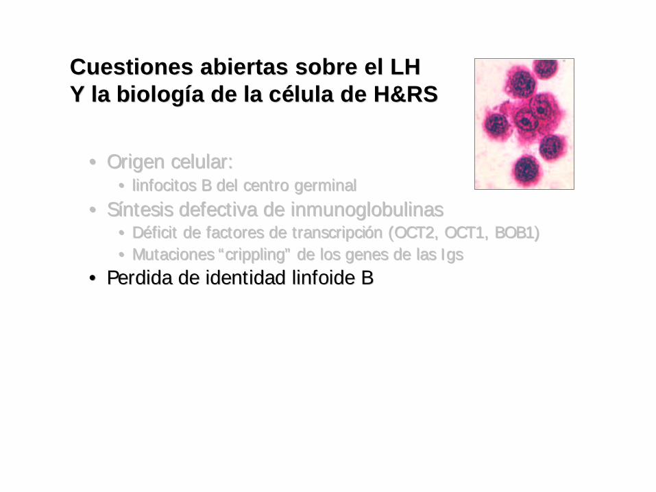
•• Origen celular: Origen celular: •• linfocitos B del centro germinallinfocitos B del centro germinal
•• SSííntesis defectiva de inmunoglobulinasntesis defectiva de inmunoglobulinas•• DDééficit de factores de transcripcificit de factores de transcripcióón (OCT2, OCT1, BOB1)n (OCT2, OCT1, BOB1)•• Mutaciones Mutaciones ““cripplingcrippling”” de los genes de las de los genes de las IgsIgs
•• Perdida de identidad linfoide BPerdida de identidad linfoide B
Cuestiones abiertas sobre el LHCuestiones abiertas sobre el LHY la bY la biologiologííaa de la cde la céélula de H&RSlula de H&RS
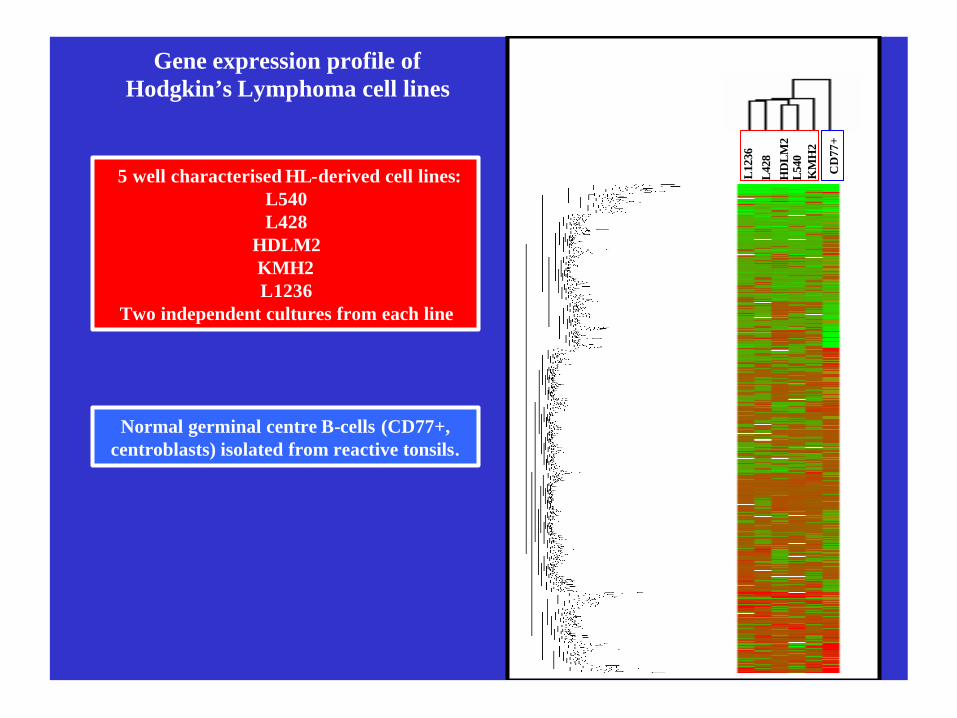
Gene expression profile ofHodgkin’s Lymphoma cell lines
5 well characterised HL-derived cell lines:L540L428
HDLM2KMH2L1236
Two independent cultures from each line
Normal germinal centre B-cells (CD77+, centroblasts) isolated from reactive tonsils.
L12
36L
428
HD
LM
2L
540
KM
H2
CD
77+
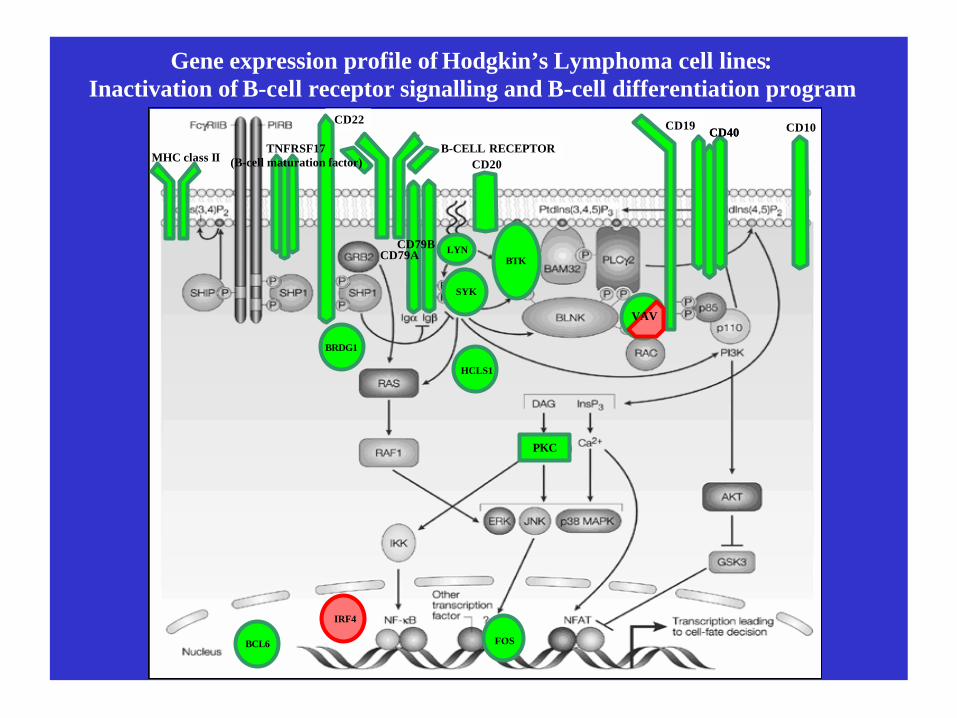
Gene expression profile of Hodgkin’s Lymphoma cell lines:Inactivation of B-cell receptor signalling and B-cell differentiation program
BCL6 FOS
IRF4
B-CELL RECEPTOR
CD19
CD20
CD22
CD79ACD79B
CD10
MHC class II
CD40CD40
BRDG1
HCLS1
TNFRSF17(B-cell maturation factor)
LYN
SYK
PKC
VAV
BTK
•• Origen celular: Origen celular: •• linfocitos B del centro germinallinfocitos B del centro germinal
•• SSííntesis defectiva de inmunoglobulinasntesis defectiva de inmunoglobulinas•• DDééficit de factores de transcripcificit de factores de transcripcióón (OCT2, OCT1, BOB1)n (OCT2, OCT1, BOB1)•• Mutaciones Mutaciones ““cripplingcrippling”” de los genes de las de los genes de las IgsIgs
•• Perdida de identidad linfoide BPerdida de identidad linfoide B•• Diferentes mecanismos de supervivenciaDiferentes mecanismos de supervivencia
•• ActivaciActivacióón de NFn de NF--kappaB kappaB (CD30, EBV, JAK2, (CD30, EBV, JAK2, STATsSTATs, , IkBaIkBa,...),...)•• Programa B defectivoPrograma B defectivo•• DesregulaciDesregulacióón de los sistemas de control del ciclo celular y de n de los sistemas de control del ciclo celular y de
la apoptosis la apoptosis
Cuestiones abiertas sobre el LHCuestiones abiertas sobre el LHY la bY la biologiologííaa de la cde la céélula de H&RSlula de H&RS
Identification of a gene expression signature associated with unfavourable treatment
response in Hodgkin’s Lymphoma
Abel Sánchez-AguileraLymphoma GroupMolecular Pathology ProgrammeSpanish National Cancer Centre (CNIO)
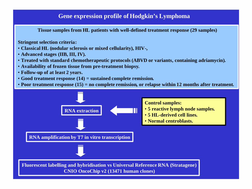
Gene expression profile of Hodgkin’s Lymphoma
Tissue samples from HL patients with well-defined treatment response (29 samples)
Stringent selection criteria:• Classical HL (nodular sclerosis or mixed cellularity), HIV-, • Advanced stages (IIB, III, IV).• Treated with standard chemotherapeutic protocols (ABVD or variants, containing adriamycin).• Availability of frozen tissue from pre-treatment biopsy.• Follow-up of at least 2 years.• Good treatment response (14) = sustained complete remission.• Poor treatment response (15) = no complete remission, or relapse within 12 months after treatment.
Fluorescent labelling and hybridisation vs Universal Reference RNA (Stratagene)CNIO OncoChip v2 (13471 human clones)
RNA amplification by T7 in vitro transcription
RNA extraction
Control samples:• 5 reactive lymph node samples.• 5 HL-derived cell lines.• Normal centroblasts.
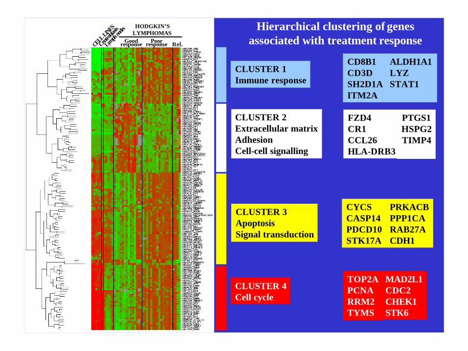
Hierarchical clustering of genes associated with treatment response
CLUSTER 1Immune response
CLUSTER 2Extracellular matrixAdhesionCell-cell signalling
CLUSTER 3ApoptosisSignal transduction
CLUSTER 4Cell cycle
CD8B1CD3DSH2D1AITM2A
ALDH1A1LYZSTAT1
MAD2L1CDC2CHEK1STK6
TOP2APCNARRM2TYMS
CYCSCASP14PDCD10STK17A
PRKACBPPP1CARAB27ACDH1
FZD4 CR1CCL26HLA-DRB3
CELL LINES
Centrob
lasts
Lymph
nodes
Goodresponse Poorresponse
HODGKIN’S LYMPHOMAS
Rel.
PTGS1HSPG2TIMP4
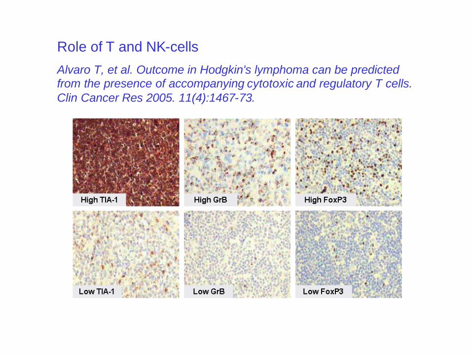
Role of T and NK-cells
Alvaro T, et al. Outcome in Hodgkin's lymphoma can be predicted from the presence of accompanying cytotoxic and regulatory T cells. Clin Cancer Res 2005. 11(4):1467-73.
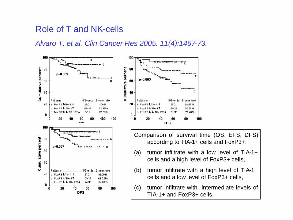
Comparison of survival time (OS, EFS, DFS) according to TIA-1+ cells and FoxP3+:
(a) tumor infiltrate with a low level of TIA-1+ cells and a high level of FoxP3+ cells,
(b) tumor infiltrate with a high level of TIA-1+ cells and a low level of FoxP3+ cells,
(c) tumor infiltrate with intermediate levels of TIA-1+ and FoxP3+ cells.
Role of T and NK-cells
Alvaro T, et al. Clin Cancer Res 2005. 11(4):1467-73.

Hierarchical clustering of genes associated with treatment response
CLUSTER 1Immune response
CLUSTER 2Extracellular matrixAdhesionCell-cell signalling
CLUSTER 3ApoptosisSignal transduction
CLUSTER 4Cell cycle
CD8B1CD3DSH2D1AITM2A
ALDH1A1LYZSTAT1
MAD2L1CDC2CHEK1STK6
TOP2APCNARRM2TYMS
CYCSCASP14PDCD10STK17A
PRKACBPPP1CARAB27ACDH1
FZD4 CR1CCL26HLA-DRB3
CELL LINES
Centrob
lasts
Lymph
nodes
Goodresponse Poorresponse
HODGKIN’S LYMPHOMAS
Rel.
PTGS1HSPG2TIMP4
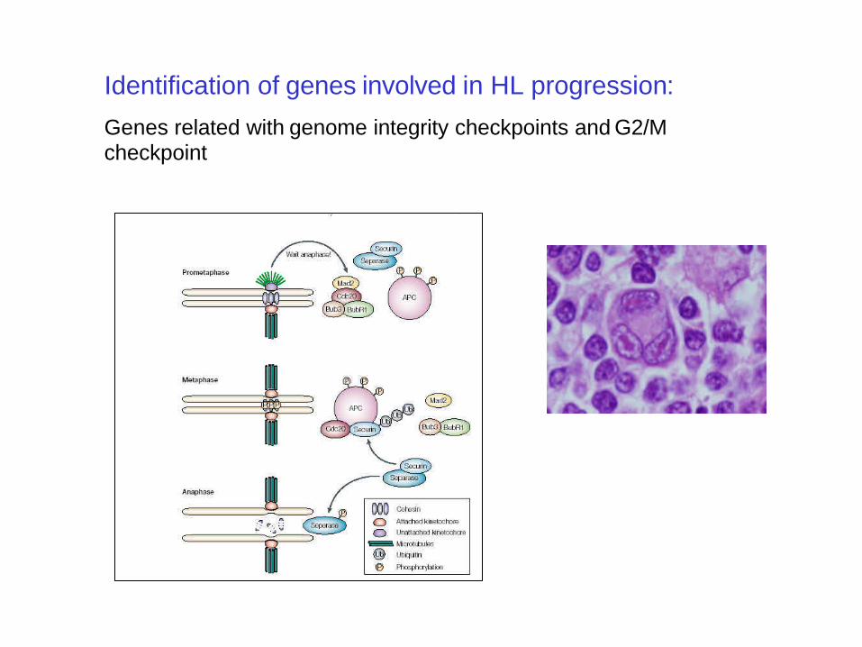
Identification of genes involved in HL progression:
Genes related with genome integrity checkpoints and G2/M checkpoint
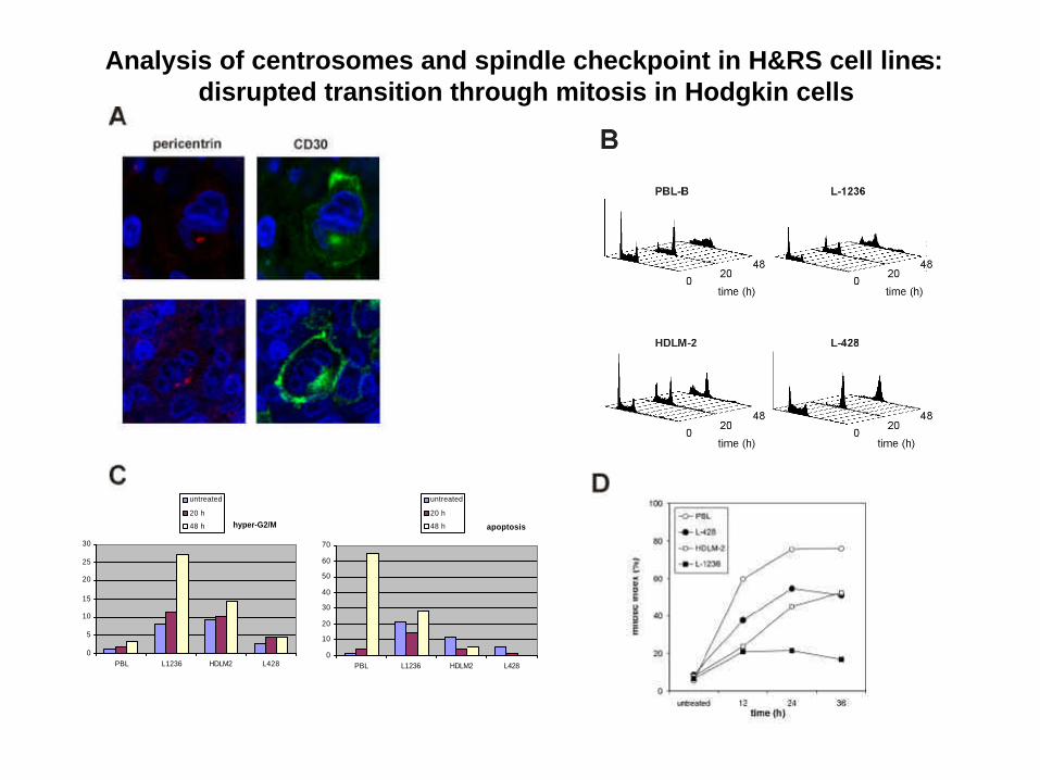
Analysis of centrosomes and spindle checkpoint in H&RS cell lines: disrupted transition through mitosis in Hodgkin cells
hyper-G2/M
0
5
10
15
20
25
30
PBL L1236 HDLM2 L428
apoptosis
0
10
20
30
40
50
60
70
PBL L1236 HDLM2 L428
untreated
20 h
48 h
untreated
20 h
48 h
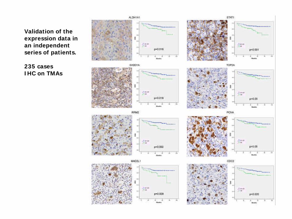
Validation of the expression data in an independentseries of patients.
235 cases IHC on TMAs
Linfoma de Hodgkin
Dr. Juan F García