Embed Size (px)
Citation preview
Eur Resplr J 1991, 4, 114Q-1142 CASE REPORT
Lobar hypoplasia
C. Della Pona, G. Rocco, A. Rizzi, M. Robustellini, G. Rossi, B. Crasti*
Lobar hypoplasia. C. Della Pona, G. Rocco, A. Rizzi, M. Robustellini, G. Rossi, B. Crasti. ABSTRACT: Lung tissue developmental abnormalities are seldom re· ported. According to the classification of ScHNEIDER (1900) [1], which was amended by BoYDEN (1955) [2], they Include pulmonary agenesis, aplasia and hypoplasia. Due to the early onset of symptoms, lung agen· esis and aplasia are usually detected soon after birth. Conversely, lung or lobar hypoplasia may remain clinically silent for a long time.
A single case of left lower lobe hypoplasia is reported. A recurring and unrelenting septic fever was the presenting symptom, whilst the radiological picture showed a left lower lobe consolidation. On the surgical specimen gross pathology revealed a lobar hypoplasia. At a short· term follow-up (nine months) the patient shows good overall condition, being free from further complications. Eur Respir J., 1991, 4, 1140-1142.
"E. Morelli" Regional Hospital, Division of Thoracic Surgery, and • Service of Radiology, Sondalo, Italy.
Correspondence: A. Rizzi, Divisione di Chirurgia Toracica, Via Zubiani 33, 23039 Sondalo (SO), Italy.
Keywords: Lobar hypoplasia; lung.
Received: February 25, 1991; accepted after revision May 8, 1991.
Case report Laboratory findings included leucocytosis (12.7x10⁹·Z-1)
and an increase in Katz's formula. The patient was a farmer, aged 60 yrs, with a history
of heavy smoking (300 packs a year for 40 yrs) and chronic obstructive pulmonary disease (COPD). Following pneumological evaluation for recurring septic fever, not subsiding after medical treatment, the patient was admitted to our Thoracic Division in order to assess surgical eligibility. The physical examination revealed a reduced ventilation in the left pulmonary field without extrathoracic pathological involvement except for a low-degree hepatomegaly.
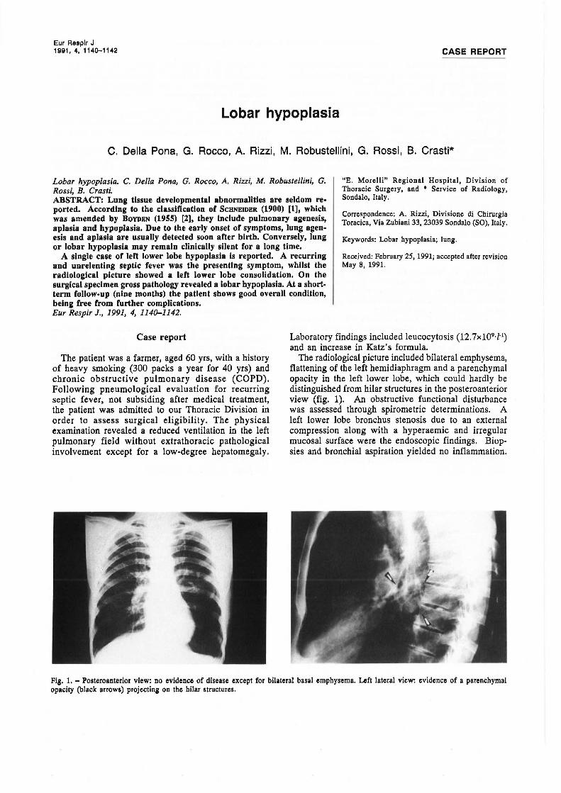
The radiological picture included bilateral emphysema, flattening of the left hemidiaphragm and a parenchyma} opacity in the left lower lobe, which could hardly be distinguished from hilar structures in the posteroanterior view (fig. 1). An obstructive functional disturbance was assessed through spirometric determinations. A left lower lobe bronchus stenosis due to an external compression along with a hyperaemic and irregular mucosal surface were the endoscopic findings. Biop· sies and bronchial aspiration yielded no inflammation.
Fig. 1. - Posteroanterior view: no evidence of disease except for bilateral basal emphysema. Left lateral view: evidence of a parenchyma! opacity (black arrows) projecting on the hilar structures.

LOBAR HYPOPLASIA 1141
No bacterial growth was detected at cultural examination. A staging procedure for a pulmonary neoplasm was started.
Chest computed tomography (CT) revealed a posterior parenchyma! consolidation in the left lower lobe showing contrast enhancement in its caudal portion and an air bronchogram in its core (fig. 2). Several emphysematous blebs in the surrounding parenchyma and no mediastinal nodal involvement were detected. Mediastinal dislocation the left side concurred. Brain CT and abdominal ultrasonography did not show recurrences.
Fig. 2. - Computed tomographic (Cf) evidence of a well-defined mass, showing air bronchogram and contrast-enhancement in its caudal portion, located at D9 level and indwelt in the costovertebral groove. Gross pathology demonstrated a hypoplastic left lower lobe.
Surgery was scheduled on account of the controversial diagnosis and the poor yield of medical treatment. A left lower lobectomy through a left posterolateral thoracotomy was performed. Intra-operatively, the left lower lobe was hardly detected, due to its small size and its posterior situation. The little parenchyma surrounded the bronchus at the hilum where all the pulmonary fibrotic vessels merged. On the tenth postoperative day the patient was dismissed after an uneventful course. A short-term follow-up (nine months) showed good postsurgical results.
Pathology
The resected lobe measured 6x3.5x4 cm and presented a consolidated structure with diffuse anthracotic subpleural streaks. The ectatic lobar bronchus was filled with creamy and yellowish contents; some hypoplastic and thickened bronchial and vascular branches were demonstrated distally. Histology showed bronchitis and peribronchitis and a greater than normal bronchii/ alveoli ratio. Furthermore, alveolar adenomatosis, fetal pulmonary alveoli and blood vessel muscular layer
hypertrophy concurred. These findings suggested the diagnosis of left lower lobe hypoplasia.
Discussion
According to ScHNEIDER (1900) [1] and, later, BoYDEN (1955) [2], three groups of developmental abnormalities can be recognized: 1) agenesis of the lung, i.e. absence of one or both lungs with no bronchial or vascular remnants observed; 2) aplasia, when a rudimental bronchus is detected without blood vessels or surrounding parenchyma; 3) hypoplasia, when an ill-formed bronchus supplies a poorly-developed alveolar tissue consolidated in a fleshy and unlobulated structure. The overall frequency of these anomalies is low: up to one case of pulmonary aplasia out of 10,000 radiological investigations are demonstrated at routine screenings [3]. Neither side nor sex prevalence has been documented [3]. Congenital hypoplasia could be related to extrathoracic compressions or to pressures exerted on the diaphragm, on the thoracic cage or, again, inside the chest, yielding reduction of the available thoracic volume. In addition, a primitive hypoplasia resulting from a diverted development of the alveolar structure during the period of rapid growth in the last two months of gestation has been reported. Hypoplasia may occur as an isolated abnormality or may be associated with others as in prune-belly syndrome, oligohydramnios or Potter's syndrome [4].
Although clinically silent in most patients, lobar hypoplasia may predispose to recurring infections causing respiratory distress. Indirect diagnosis is often possible through bronchography, endoscopy and selective pulmonary angiography [5, 6]; in our case diagnosis was made in the process of an oncological staging procedure. Surgery is indicated for removal of anatomical anomalies yielding repeated infections or when a neoplasm is suspected [7]. In our patient the combination of surgical exploration and pathological confirmation has allowed the clarification of the clinical picture.
References
1. Schneider P. - Die missbindungen der atmungsorgane. In: Die morphologie der missbindungen des menschen und der tiere. E. Schwalbe ed., Gustav Fisher, Jena, Vol. 3, 1900-1913, pp. 817-822. 2. Boyden EA. - Developmental anomalies of the lung. Am J Surg, 1955, 89, 79-89. 3. Valle AR. - Agenesis of the lung. Am J Surg, 1955, 89, 90-96. 4. Currarino G, Williams B. - Causes of unilateral pulmonary hypoplasia: a study of 33 cases. Pediatr Radio/, 1985, 15, 15-21. 5. Steiner HA. - Aplasia of the lung: a case report. Radiology, 1956, 67, 751-753. 6. K.ihara F, Usui K, Masuda Y. - A case of aplasia of left lung diagnosed during life. Jap J Chest Dis, 1964, 23, 61~16.
1142 C. DELLA PONA ET AL.
7. Ferguson TB Jr, Ferguson TB. Congenital lesions of the lung and emphysema. In: Surgery of the Chest. D.C. Sabiston Jr, F.C. Spencer eds, W.B. Saunders Co., Philadelphia, 1990, Vol. 1, pp. 76~14.
Hypoplasie lobaire. C. Della Pona, G. Rocco, A. Rizzi, M. Robustellini, G. Rossi, B. Crasti. RESUME: Les anomalies du d6veloppement du tissu pulmonaire sont rares. Selon la classification de Schneider (1900), modifi6e par Boyden (1955), elles incluent l'ag6nesie pulmonaire, l'aplasie et l'hypoplasie. En raison de
!'apparition rapide des symptOmes, l'agenesie et l'aplasie pulmonaire sont habituellement d6tect6es peu apr~s la naissance. Par contre, l'hypoplasie pulmonaire ou lobaire peut rester cliniquement silencieuse pendant longtemps.
Nous faisons etat d'un cas isole d'hypoplasie du lobe inferieur gauche. Le symptOme clinique de presentation fut une fi~vre septique r6cidivante en remission, alors qu'une densification du lobe pulmonaire inf6rieur gauche apparaissait au cliche thoracique. L'examen macroscopique de la pi~ce d'ex6r~se a montre une hypoplasie lobaire. Au follow-up de neuf mois, le patient est dans un etat general satisfaisant, sans aucune complication. Eur Respir J., 1991, 4, 1140-1142.