Embed Size (px)
Citation preview
6/2/2019
1
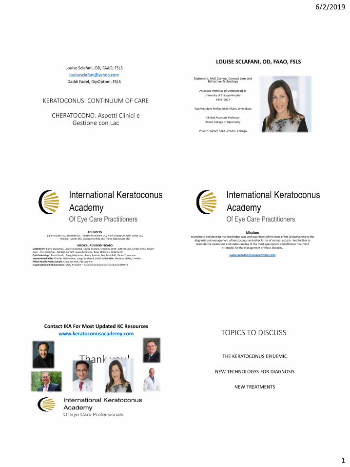
KERATOCONUS: CONTINUUM OF CARE
CHERATOCONO: Aspetti Clinici eGestione con Lac
Louise Sclafani, OD, FAAO, FSLS
Daddi Fadel, DipOptom, FSLS
LOUISE SCLAFANI, OD, FAAO, FSLS
Diplomate, AAO Cornea, Contact Lens and Refractive Technology
Associate Professor of Ophthalmology
University of Chicago Hospital
1993- 2017
Vice President Professional Affairs, SynergEyes
Clinical Associate Professor
Illinois College of Optometry
Private Practice SoLo EyeCare, Chicago
Presented by
Louise A. Sclafani, OD, FAAO
International Keratoconus Academy
FOUNDERSS.Barry Eiden OD, Joe Barr OD, Timothy McMahon OD, Clark Chang OD, John Gelles OD,
William Trattler MD, Eric Donnenfeld MD, Yaron Rabinowitz MD
MEDICAL ADVISORY BOARDOptometry: Barry Weissman, Loretta Szczotka, Louise Sclafani, Christine Sindt , Jeff Sonsino, Lynett Johns, Robert Davis , Tim Edrington, Melissa Barnett, Susan Gromacki, Ryan McKinnis, Ed Bennett Ophthalmology: Peter Hersh, Parag Majmudar, Randy Epstein, Roy Rubinfeld, Vance ThompsonInternational: ODs: Charles McMonnies, Langis Michaud, Daddi Fadel MDs: Florence Malet, F. HefeziAllied Health Professionals: Craig Norman, Pat CarolineOrganizational Collaboration: Mary Prudden – National Keratoconus Foundation (NKCF)
International Keratoconus Academy
Mission:to promote and develop the knowledge base and awareness of the state of the art pertaining to the
diagnosis and management of keratoconus and other forms of corneal ectasia. And further to promote the awareness and understanding of the most appropriate and effective treatment
strategies for the management of these diseases.
www.keratoconusacademy.com
Thank you!
Contact IKA For Most Updated KC Resources
www.keratoconusacademy.com TOPICS TO DISCUSS
THE KERATOCONUS EPIDEMIC
NEW TECHNOLOGYS FOR DIAGNOSIS
NEW TREATMENTS
6/2/2019
2
ETIOLOGY AND PREVALENCE
Eziologia e Prevalenza
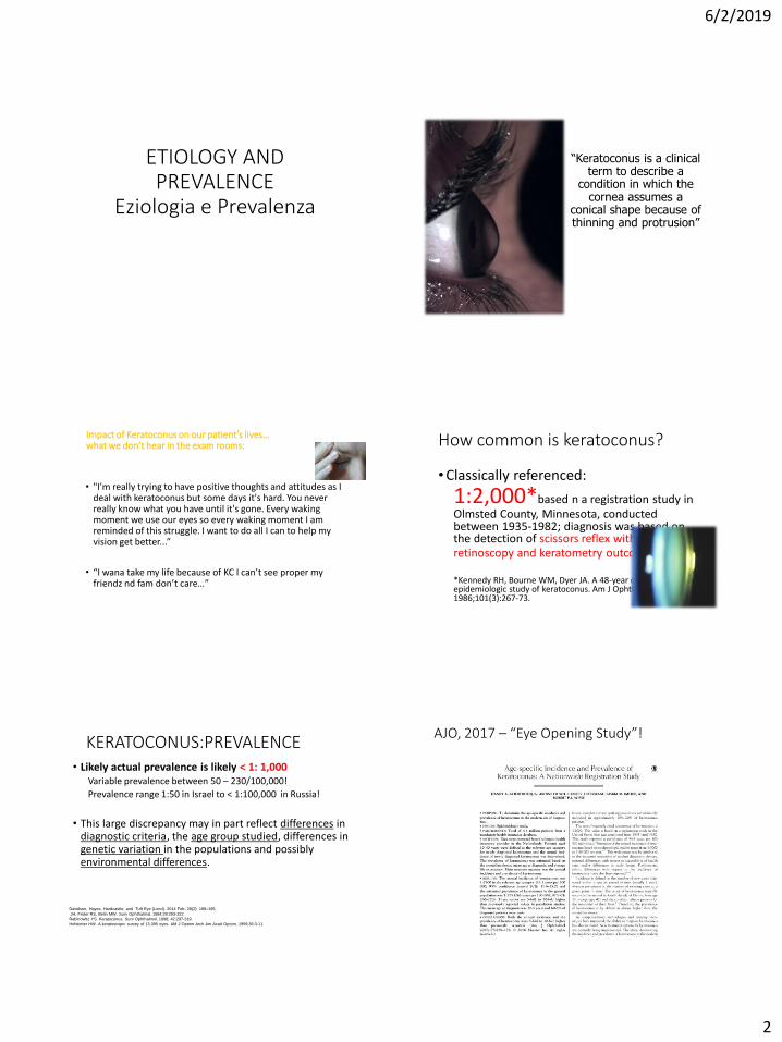
“Keratoconus is a clinical term to describe a
condition in which the cornea assumes a
conical shape because of thinning and protrusion”
Impact of Keratoconus on our patient’s lives… what we don’t hear in the exam rooms:
• "I'm really trying to have positive thoughts and attitudes as I deal with keratoconus but some days it's hard. You never really know what you have until it's gone. Every waking moment we use our eyes so every waking moment I am reminded of this struggle. I want to do all I can to help my vision get better...”
• “I wana take my life because of KC I can’t see proper my friendz nd fam don’t care…”
How common is keratoconus?
•Classically referenced:
1:2,000*based n a registration study in Olmsted County, Minnesota, conducted between 1935-1982; diagnosis was based on the detection of scissors reflex with retinoscopy and keratometry outcomes!
*Kennedy RH, Bourne WM, Dyer JA. A 48-year clinical and epidemiologic study of keratoconus. Am J Ophthalmol1986;101(3):267-73.
KERATOCONUS:PREVALENCE• Likely actual prevalence is likely < 1: 1,000
Variable prevalence between 50 – 230/100,000!
Prevalence range 1:50 in Israel to < 1:100,000 in Russia!
• This large discrepancy may in part reflect differences in diagnostic criteria, the age group studied, differences in genetic variation in the populations and possibly environmental differences.
Davidson, Hayes, Hardcastle, and Tuft Eye (Lond). 2014 Feb; 28(2): 189–195.
JH, Feder RS, Belin MW. Surv Ophthalmal, 1984;28:293-322.
Rabinowitz YS. Keratoconus. Surv Ophthalmol, 1998; 42:297-319.
Hofstetter HW. A keratoscopic survey of 13,395 eyes. AM J Optom Arch Am Acad Optom, 1959;36:3-11.
AJO, 2017 – “Eye Opening Study”!
6/2/2019
3
AJO – 2017: Age-specific Incidence and Prevalence of Keratoconus: A Nationwide Registration Study
• Netherlands study: 4.4 million patients from a mandatory health insurance data base
• Prevalence of keratoconus in the general population was 1:375
• Annual incidence: (new cases) of keratoconus was 1:7,500
• Conclusion: “Both the annual incidence and the prevalence of keratoconus were five-fold to ten-fold higher than previously reported.”
Basic Science Research
• Christina Kenney, MD, PhD
• KCN have higher # of mitochondrial DNA deletions that leads to decrease oxidative phosphorylation… increase H202
• Causes leakage, damages proteins, and results in oxidative stress
• Leads to • apoptosis,
• abnormal healing,
• inflammation.
Basic Science Research
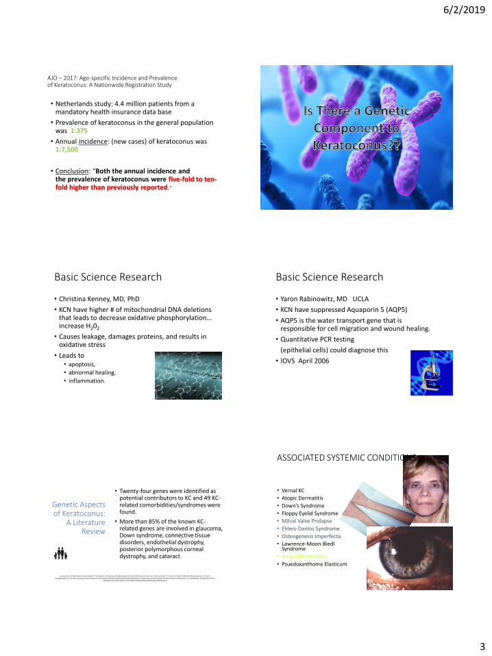
• Yaron Rabinowitz, MD UCLA
• KCN have suppressed Aquaporin 5 (AQP5)
• AQP5 is the water transport gene that is responsible for cell migration and wound healing.
• Quantitative PCR testing
(epithelial cells) could diagnose this
• IOVS April 2006
Genetic Aspects of Keratoconus:
A Literature Review
• Twenty-four genes were identified as potential contributors to KC and 49 KC-related comorbidities/syndromes were found.
• More than 85% of the known KC-related genes are involved in glaucoma, Down syndrome, connective tissue disorders, endothelial dystrophy, posterior polymorphous corneal dystrophy, and cataract.
ASSOCIATED SYSTEMIC CONDITIONS
• Vernal KC
• Atopic Dermatitis
• Down’s Syndrome
• Floppy Eyelid Syndrome
• Mitral Valve Prolapse
• Ehlers-Danlos Syndrome
• Osteogenesis Imperfecta
• Lawrence-Moon-BiedlSyndrome
• Neurofibromatosis
• Psuedoxanthoma Elasticum
6/2/2019
4
ASSOCIATED SYSTEMIC CONDITIONS
• Vernal KC
• Atopic Dermatitis
• Down’s Syndrome
• Floppy Eyelid Syndrome• PKP >31 lbs. *, 8.7x morbid obese
• Kristinson, IOVS 2003
• Mitral Valve Prolapse
• Ehlers-Danlos Syndrome
• Osteogenesis Imperfecta
• Lawrence-Moon-Biedl
• Neurofibromatosis
• PXE
ASSOCIATED SYSTEMIC CONDITIONS
• Vernal KC
• Atopic Dermatitis
• Down’s Syndrome
• Floppy Eyelid Syndrome
• Mitral Valve Prolapse
• Ehlers-Danlos Syndrome
• Osteogenesis Imperfecta
• Lawrence-Moon-Biedl Syndrome
• Neurofibromatosis
• Psuedoxanthoma Elasticum
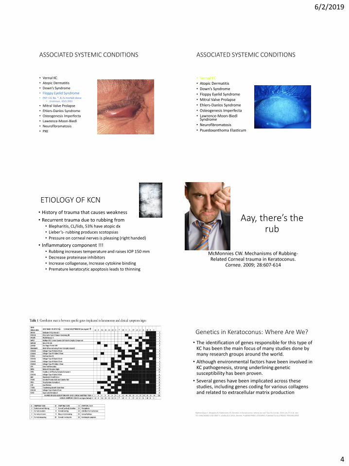
ETIOLOGY OF KCN
• History of trauma that causes weakness
• Recurrent trauma due to rubbing from• Blepharitis, CL/lids, 53% have atopic dx
• Lieber’s- rubbing produces scotopsias
• Pressure on corneal nerves is pleasing (right handed)
• Inflammatory component !!!• Rubbing increases temperature and raises IOP 150 mm
• Decrease proteinase inhibitors
• Increase collagenase, Increase cytokine binding
• Premature keratocytic apoptosis leads to thinning
Aay, there’s the rub
McMonnies CW. Mechanisms of Rubbing-Related Corneal trauma in Keratoconus.
Cornea. 2009; 28:607-614
Genetics in Keratoconus: Where Are We?
• The identification of genes responsible for this type of KC has been the main focus of many studies done by many research groups around the world.
• Although environmental factors have been involved in KC pathogenesis, strong underlining genetic susceptibility has been proven.
• Several genes have been implicated across these studies, including genes coding for various collagens and related to extracellular matrix production
Bykhovskaya Y, Margines B, Rabinowitz YS. Genetics in Keratoconus: where are we? Eye Vis (Lond). 2016 Jun 27;3:16. doi:
10.1186/s40662-016-0047-5. eCollection 2016. Review. PubMed PMID: 27350955; PubMed Central PMCID: PMC4922054.
6/2/2019
5
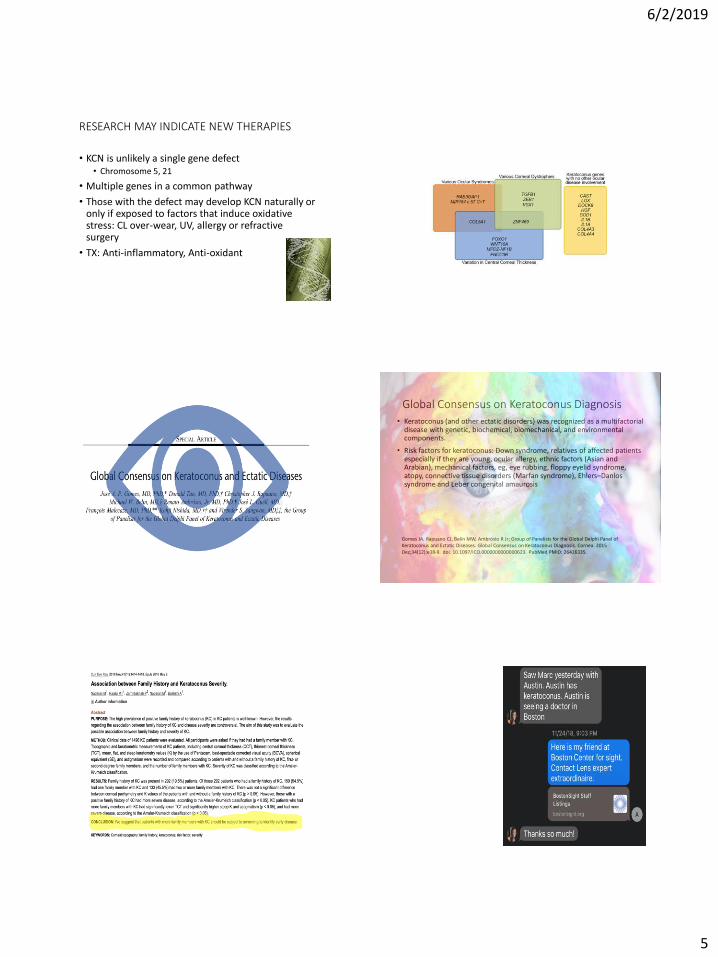
RESEARCH MAY INDICATE NEW THERAPIES
• KCN is unlikely a single gene defect• Chromosome 5, 21
• Multiple genes in a common pathway
• Those with the defect may develop KCN naturally or only if exposed to factors that induce oxidative stress: CL over-wear, UV, allergy or refractive surgery
• TX: Anti-inflammatory, Anti-oxidant
Keratoconus Genes and Their Involvement in Ocular Diseases
Global Consensus on Keratoconus Diagnosis
• Keratoconus (and other ectatic disorders) was recognized as a multifactorial disease with genetic, biochemical, biomechanical, and environmental components.
• Risk factors for keratoconus: Down syndrome, relatives of affected patients especially if they are young, ocular allergy, ethnic factors (Asian and Arabian), mechanical factors, eg, eye rubbing, floppy eyelid syndrome, atopy, connective tissue disorders (Marfan syndrome), Ehlers–Danlos syndrome and Leber congenital amaurosis
Gomes JA, Rapuano CJ, Belin MW, Ambrósio R Jr; Group of Panelists for the Global Delphi Panel of Keratoconus and Ectatic Diseases. Global Consensus on Keratoconus Diagnosis. Cornea. 2015 Dec;34(12):e38-9. doi: 10.1097/ICO.0000000000000623. PubMed PMID: 26426335.
Naderan M, Rajabi MT, Zarrinbakhsh P, Naderan M, Bakhshi A. Associationbetween Family History and Keratoconus Severity. Curr Eye Res . 2016Nov;41(11):1414-1418. Epub 2016 May 9. PubMed PMID: 27158890.
23 NOV 2018
6/2/2019
6
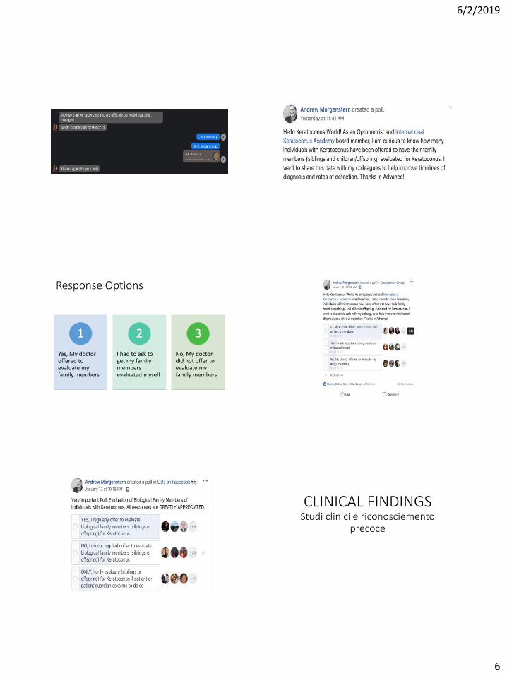
Response Options
Yes, My doctor offered to evaluate my family members
1
I had to ask to get my family members evaluated myself
2
No, My doctor did not offer to evaluate my family members
3
CLINICAL FINDINGSStudi clinici e riconosciemento
precoce
6/2/2019
7
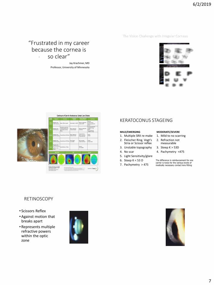
“Frustrated in my career because the cornea is
so clear”Jay Krachmer, MD
Professor, University of Minnesota
“
The Vision Challenge with Irregular Corneas
• Monocular Diplopia
• “Ghost” images
• “Asthenopia”
• Polyopia
• Photophobia
• Halos around lights
KERATOCONUS STAGEING
MILD/EMERGING
1. Multiple SRX re-make
2. Fleischer Ring, Vogt’s Stria or Scissor reflex
3. Unstable topography
4. No scar
5. Light Sensitivity/glare
6. Steep K < 53 D
7. Pachymetry > 475
MODERATE/SEVERE
1. Mild to no scarring
2. Refraction not measurable
3. Steep K > 53D
4. Pachymetry <475
The difference in reimbursement for one carrier is twice for the various levels of medically necessary contact lens fitting
RETINOSCOPY
• Scissors Reflex
•Against motion that breaks apart
•Represents multiple refractive powers within the optic zone
KERATOCONUS FINDINGS
• VOGT’S STRIAE
• FLEISCHER RING
• STROMAL THINNING
• STROMAL SCARS
• SWIRL-LIKE PATTERN
• ENLARGED CORNEAL NERVES
• ACUTE HYDROPS
• EPITHELIAL THINNING *
• POSTERIOR STEEPENING *
• CORNEAL BIOMECHANICAL PROPERTIES*
6/2/2019
8
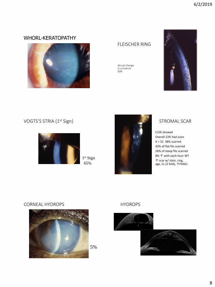
WHORL-KERATOPATHYFLEISCHER RING
abrupt change in curvature50%
VOGTS’S STRIA (1st Sign)
1st Sign
65%
STROMAL SCAR
CLEK showed
Overall 13% had scars
K > 52 38% scarred
43% of flat fits scarred
26% of steep fits scarred
8% ↑ with each hour WT
↑ scar w/ stain, ring, age, CL (2 fold), ↑FDACL
CORNEAL HYDROPS
5%
HYDROPS
6/2/2019
9
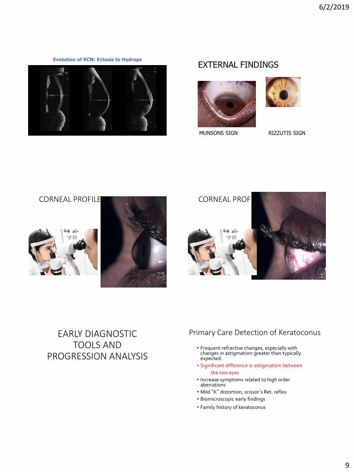
Evolution of KCN: Ectasia to Hydrops
image courtesy of Prof. G. Baikoff
EXTERNAL FINDINGS
MUNSONS SIGN RIZZUTIS SIGN
CORNEAL PROFILE CORNEAL PROFILE
EARLY DIAGNOSTIC TOOLS AND
PROGRESSION ANALYSIS
Primary Care Detection of Keratoconus
• Frequent refractive changes, especially with changes in astigmatism greater than typically expected.
• Significant difference in astigmatism between
the two eyes
• Increase symptoms related to high order aberrations
• Mild “K” distortion, scissor’s Ret. reflex
• Biomicroscopic early findings
• Family history of keratoconus
6/2/2019
10
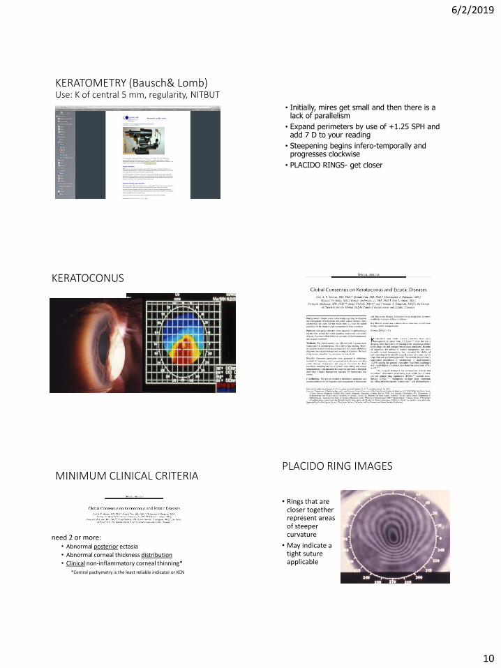
KERATOMETRY (Bausch& Lomb)Use: K of central 5 mm, regularity, NITBUT
Keratoconus- Keratometry
• Initially, mires get small and then there is a lack of parallelism
• Expand perimeters by use of +1.25 SPH and add 7 D to your reading
• Steepening begins infero-temporally and progresses clockwise
• PLACIDO RINGS- get closer
KERATOCONUS
MINIMUM CLINICAL CRITERIA
need 2 or more: • Abnormal posterior ectasia
• Abnormal corneal thickness distribution
• Clinical non-inflammatory corneal thinning*
*Central pachymetry is the least reliable indicator or KCN
PLACIDO RING IMAGES
• Rings that are closer together represent areas of steeper curvature
• May indicate a tight suture applicable
6/2/2019
11
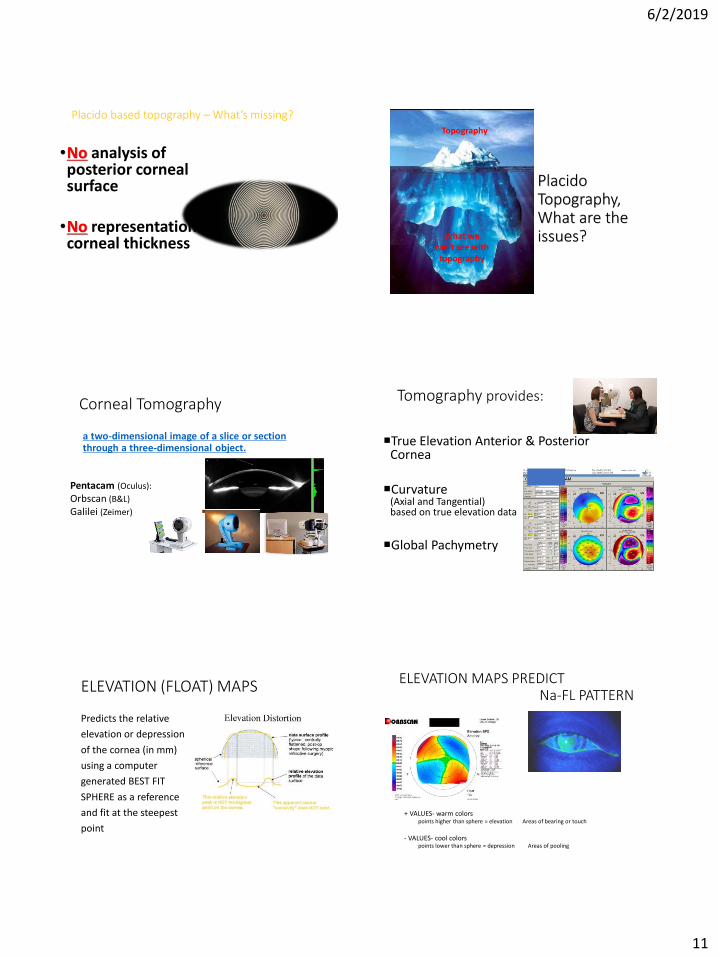
Placido based topography – What’s missing?
•No analysis of posterior corneal surface
•No representation of corneal thickness
With courtesy from Prof. Michael Belin
PlacidoTopography,What are the issues?
Topography
What we can’t see with
topography
Corneal Tomography
a two-dimensional image of a slice or section through a three-dimensional object.
Pentacam (Oculus):
Orbscan (B&L)
Galilei (Zeimer)
Tomography provides:
True Elevation Anterior & Posterior Cornea
Curvature (Axial and Tangential) based on true elevation data
Global Pachymetry
ELEVATION (FLOAT) MAPS
Predicts the relative
elevation or depression
of the cornea (in mm)
using a computer
generated BEST FIT
SPHERE as a reference
and fit at the steepest
point
ELEVATION MAPS PREDICT Na-FL PATTERN
+ VALUES- warm colors points higher than sphere = elevation Areas of bearing or touch
- VALUES- cool colorspoints lower than sphere = depression Areas of pooling
6/2/2019
12
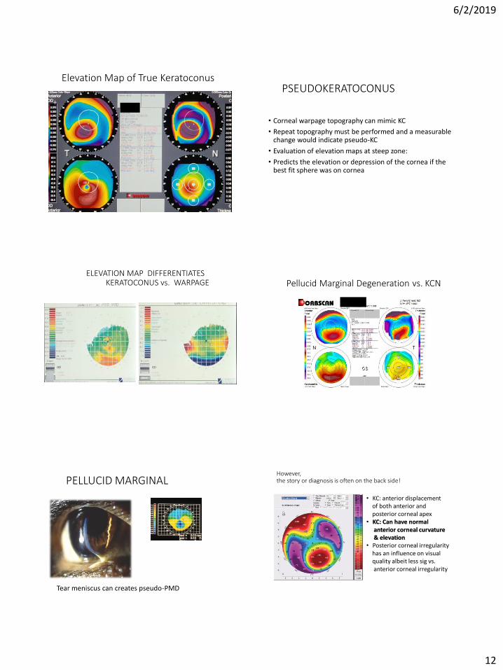
Elevation Map of True KeratoconusPSEUDOKERATOCONUS
• Corneal warpage topography can mimic KC
• Repeat topography must be performed and a measurable change would indicate pseudo-KC
• Evaluation of elevation maps at steep zone:
• Predicts the elevation or depression of the cornea if the best fit sphere was on cornea
ELEVATION MAP DIFFERENTIATESKERATOCONUS vs. WARPAGE Pellucid Marginal Degeneration vs. KCN
PELLUCID MARGINAL
Tear meniscus can creates pseudo-PMD
However, the story or diagnosis is often on the back side!
Back surface elevation map – Pentacam Kcns
• KC: anterior displacement of both anterior and posterior corneal apex
• KC: Can have normal anterior corneal curvature& elevation
• Posterior corneal irregularity has an influence on visual quality albeit less sig vs. anterior corneal irregularity
6/2/2019
13
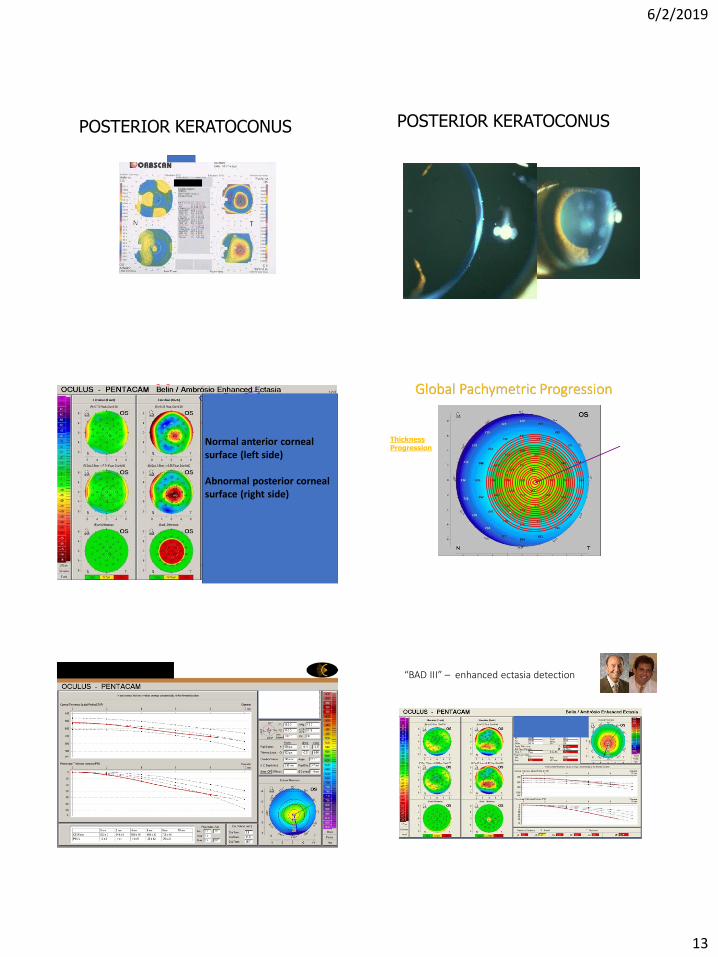
POSTERIOR KERATOCONUS POSTERIOR KERATOCONUS
How Would You Know ??
Normal by Curvature - Clearly Abnormal by Tomography (N. Ant. Elevation but abnl. Post elevation)
Normal anterior corneal surface (left side)
Abnormal posterior corneal surface (right side)
Global Pachymetric Progression
Evaluation of the
Thickness Progression on concentric rings from thinnest spot towards periphery
Thinnest
spot
***Central pachymetry is the least reliable indicator or KCN
PTI: percentage of increased thickness
CTSP: corneal thickness spatial profile
“BAD III” – enhanced ectasia detection
6/2/2019
14
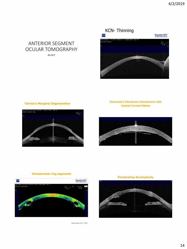
ANTERIOR SEGMENT OCULAR TOMOGRAPHY
AS-OCT
KCN- Thinning
Terrien‘s Marginal Degeneration
image courtesy of Dr. M. Packer
Descemet‘s Membrane Detachment with
Central Corneal Edema
image courtesy of Dr. M. Packer
Intrastromal ring segments
image courtesy of Dr. J. Güell
Penetrating Keratoplasty
image courtesy of Dr. M. Packer
6/2/2019
15
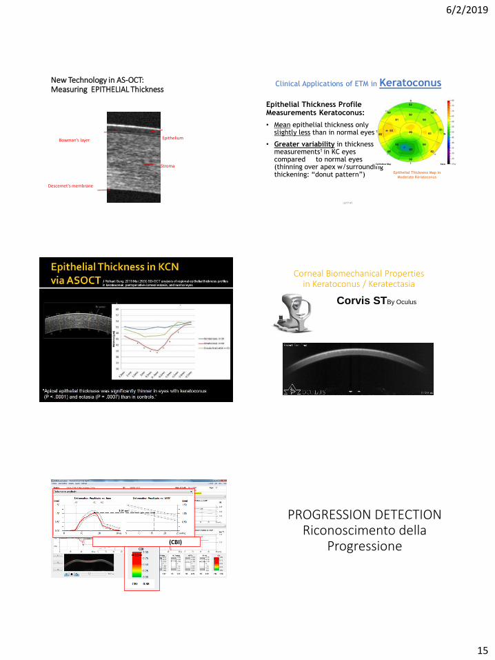
New Technology in AS-OCT:Measuring EPITHELIAL Thickness
EpitheliumBowman’s layer
Stroma
Descemet's membrane
Clinical Applications of ETM in Keratoconus
Epithelial Thickness Profile Measurements Keratoconus:
• Mean epithelial thickness only slightly less than in normal eyes1
• Greater variability in thickness measurements1 in KC eyes compared to normal eyes (thinning over apex w/surrounding thickening: “donut pattern”)
1. Kanellopoulos A, Asimellis G. OCT corneal epithelial topographic asymmetry as a sensitive diagnostic tool for early and advancing keratoconus. Clin Ophthalmol. 2014 Nov 18;8:2277-87.
Epithelial Thickness Map in
Moderate Keratoconus
Corneal Biomechanical Propertiesin Keratoconus / Keratectasia
Corvis STBy Oculus
Captures 140 images in the 31 ms after air pulse
Biomechanical Screening Report
Corvis Biomechanical Index (CBI)
PROGRESSION DETECTIONRiconoscimento della
Progressione
6/2/2019
16
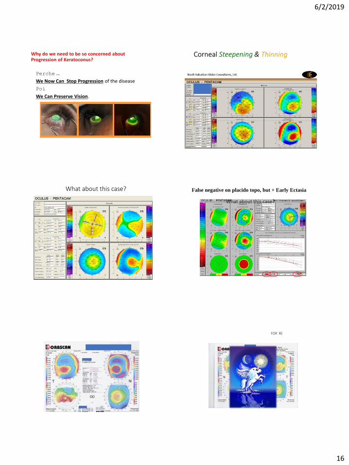
Why do we need to be so concerned about Progression of Keratoconus?
Perche …
We Now Can Stop Progression of the disease
Poi
We Can Preserve Vision.
Corneal Steepening & Thinning
• In KCN: Both anterior and posterior surfaces move forward but the posterior surface moves
MORE = therefore
Anterior Steepening and Corneal Thinning
What about this case?
With courtesy from RenatoAmbrosio
What about this case?
False negative on placido topo, but + Early Ectasia
With courtesy from Renato Ambrosio
24 yo AA Male, graduate studentGradual ↓VA 20/40 : unsuccessful CL fit @ Duke
+1.00 -4.00 x 055 51.6/44.4@064 -7.20@064 478um
24 yo AA Male, graduate studentLE plano 20/20 45.8/45.1@168 536um
CL OPTIONS: GP, HYBRID, SOFT FOR RE
6/2/2019
17
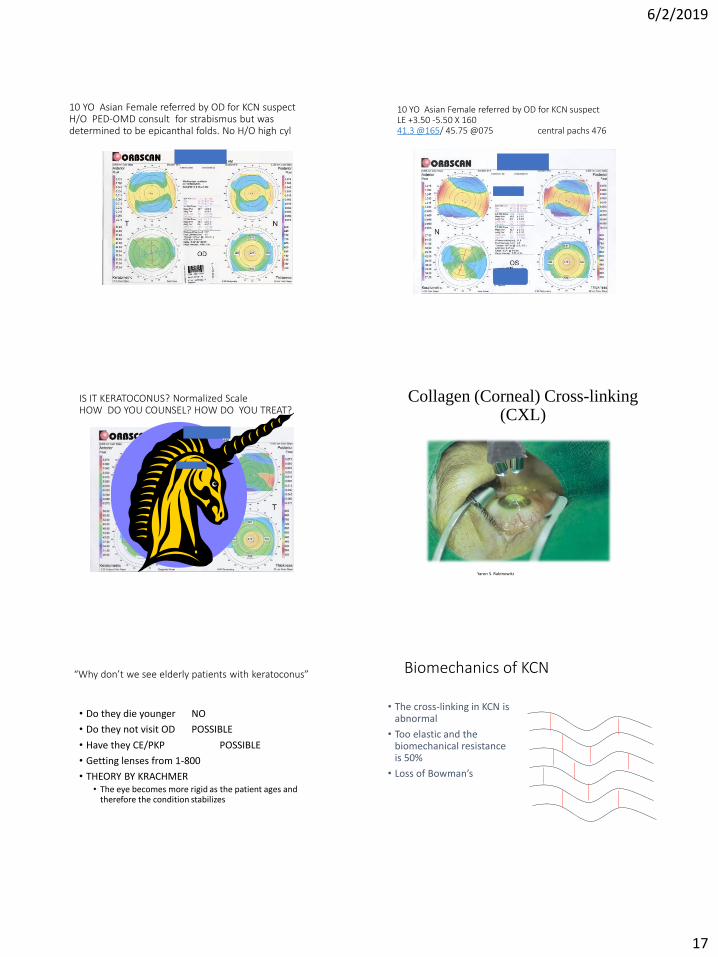
10 YO Asian Female referred by OD for KCN suspectH/O PED-OMD consult for strabismus but was determined to be epicanthal folds. No H/O high cyl
10 YO Asian Female referred by OD for KCN suspectLE +3.50 -5.50 X 16041.3 @165/ 45.75 @075 central pachs 476
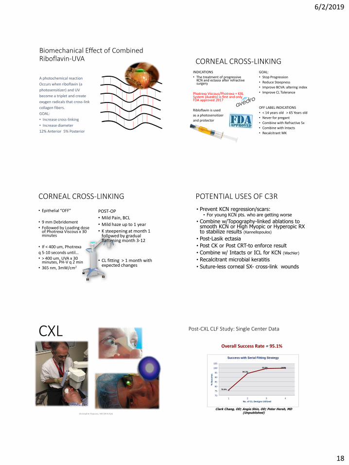
IS IT KERATOCONUS? Normalized ScaleHOW DO YOU COUNSEL? HOW DO YOU TREAT?
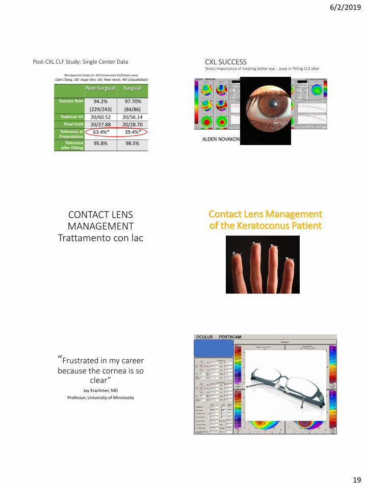
Collagen (Corneal) Cross-linking (CXL)
Yaron S. Rabinowitz
“Why don’t we see elderly patients with keratoconus”
• Do they die younger NO
• Do they not visit OD POSSIBLE
• Have they CE/PKP POSSIBLE
• Getting lenses from 1-800
• THEORY BY KRACHMER• The eye becomes more rigid as the patient ages and
therefore the condition stabilizes
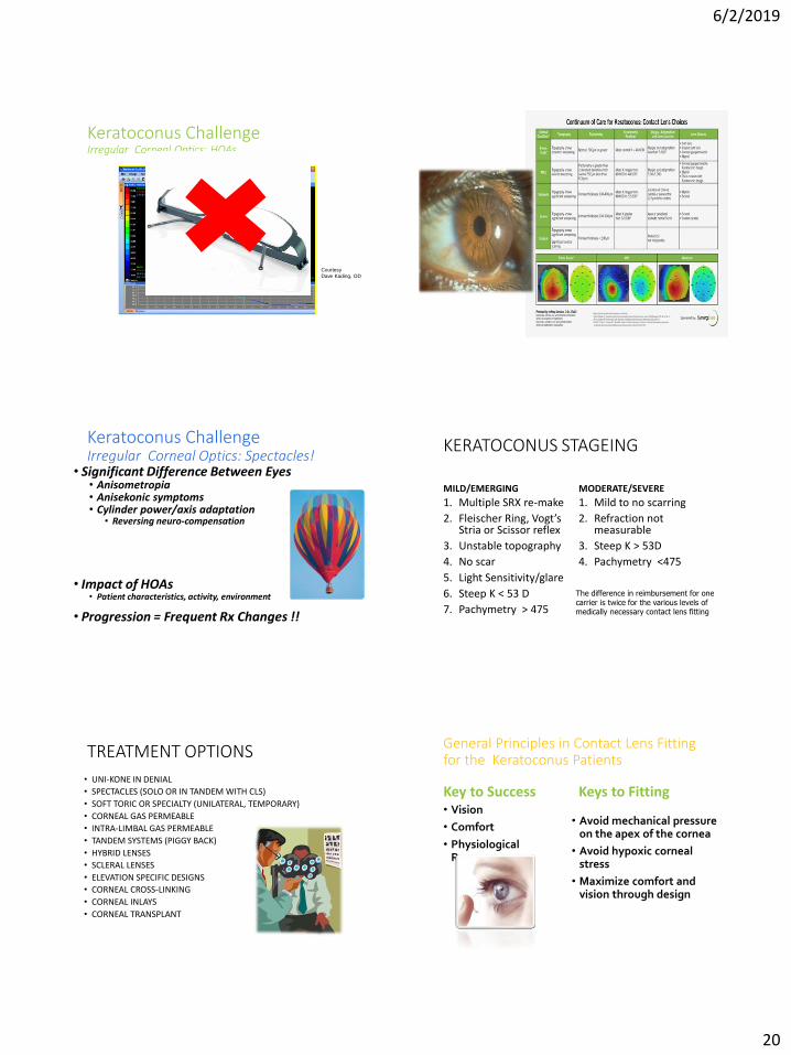
Biomechanics of KCN
• The cross-linking in KCN is abnormal
• Too elastic and the biomechanical resistance is 50%
• Loss of Bowman’s
6/2/2019
18
Biomechanical Effect of Combined Riboflavin-UVA
A photochemical reaction
Occurs when riboflavin (a
photosensitizer) and UV
become a triplet and create
oxygen radicals that cross-link
collagen fibers.
GOAL:
• Increase cross-linking
• Increase diameter
12% Anterior 5% Posterior
CORNEAL CROSS-LINKINGINDICATIONS
• The treatment of progressive KCN and ectasia after refractive surgery
Photrexa Viscous/Photrexa + KXL System (Avedro) is first and only FDA approved 2017
Ribloflavin is used
as a photosensitizer
and protector
GOAL:
• Stop Progression
• Reduce Steepness
• Improve BCVA: altering index
• Improve CL Tolerance
OFF LABEL INDICATIONS
• < 14 years old > 65 Years old
• Never for pregant
• Combine with Refractive Sx
• Combine with Intacts
• Recalcitrant MK
CORNEAL CROSS-LINKING
• Epithelial “OFF”
• 9 mm Debridement
• Followed by Loading dose of Photrexa Viscous x 30 minutes
• If < 400 um, Photrexa
q 5-10 seconds until…
• > 400 um, UVA x 30 minutes, PH-V q 2 min
• 365 nm, 3mW/cm2
POST-OP
• Mild Pain, BCL
• Mild haze up to 1 year
• K steepening at month 1 followed by gradual flattening month 3-12
• CL fitting > 1 month with expected changes
POTENTIAL USES OF C3R
• Prevent KCN regression/scars: • For young KCN pts. who are getting worse
• Combine w/Topography-linked ablations to smooth KCN or High Myopic or Hyperopic RX to stabilize results (Kannellopoulos)
• Post-Lasik ectasia
• Post CK or Post CRT-to enforce result
• Combine w/ Intacts or ICL for KCN (Wachler)
• Recalcitrant microbial keratitis
• Suture-less corneal SX- cross-link wounds
CXL
Christopher Rapuano, MD (Wills Eye)
Post-CXL CLF Study: Single Center Data
70
75
80
85
90
95
100
105
1 2 3 4
% S
uccess
No. of CL Designs Utilized
Success with Serial Fitting Strategy
75.9%
94.3%
99.6% 100%
Overall Success Rate = 95.1%
Clark Chang, OD; Angie Shin, OD; Peter Hersh, MD (Unpublished)
6/2/2019
19
Post-CXL CLF Study: Single Center Data
Non-Surgical Surgical
Success Rate 94.2%
(229/243)
97.70%
(84/86)
Habitual VA 20/60.52 20/56.14Final CLVA 20/27.88 20/28.70
Tolerance at Presentation
63.4%* 39.4%*
Tolerance after Fitting
95.8% 98.5%
Retrospective Study (n= 329 Consecutive KC/Ectasia eyes)
Clark Chang, OD; Angie Shin, OD; Peter Hersh, MD (Unpublished)
CXL SUCCESS Stress importance of treating better eye …ease in fitting CLS after
ALDEN NOVAKONE IT FACTOR 2
CONTACT LENS MANAGEMENT
Trattamento con lac
Contact Lens Management of the Keratoconus Patient
“Frustrated in my career because the cornea is so
clear”Jay Krachmer, MD
Professor, University of Minnesota
6/2/2019
20
Keratoconus Challenge Irregular Corneal Optics: HOAs
81.0 Diopters
30.0 Diopters
Courtesy
Dave Kading, OD
Keratoconus Challenge Irregular Corneal Optics: Spectacles!
• Significant Difference Between Eyes• Anisometropia• Anisekonic symptoms• Cylinder power/axis adaptation
• Reversing neuro-compensation
• Impact of HOAs• Patient characteristics, activity, environment
• Progression = Frequent Rx Changes !!
KERATOCONUS STAGEING
MILD/EMERGING
1. Multiple SRX re-make
2. Fleischer Ring, Vogt’s Stria or Scissor reflex
3. Unstable topography
4. No scar
5. Light Sensitivity/glare
6. Steep K < 53 D
7. Pachymetry > 475
MODERATE/SEVERE
1. Mild to no scarring
2. Refraction not measurable
3. Steep K > 53D
4. Pachymetry <475
The difference in reimbursement for one carrier is twice for the various levels of medically necessary contact lens fitting
TREATMENT OPTIONS
• UNI-KONE IN DENIAL
• SPECTACLES (SOLO OR IN TANDEM WITH CLS)
• SOFT TORIC OR SPECIALTY (UNILATERAL, TEMPORARY)
• CORNEAL GAS PERMEABLE
• INTRA-LIMBAL GAS PERMEABLE
• TANDEM SYSTEMS (PIGGY BACK)
• HYBRID LENSES
• SCLERAL LENSES
• ELEVATION SPECIFIC DESIGNS
• CORNEAL CROSS-LINKING
• CORNEAL INLAYS
• CORNEAL TRANSPLANT
General Principles in Contact Lens Fitting for the Keratoconus Patients
Key to Success• Vision
• Comfort
• Physiological Response
Keys to Fitting
• Avoid mechanical pressure on the apex of the cornea
• Avoid hypoxic corneal stress
• Maximize comfort and vision through design
6/2/2019
21
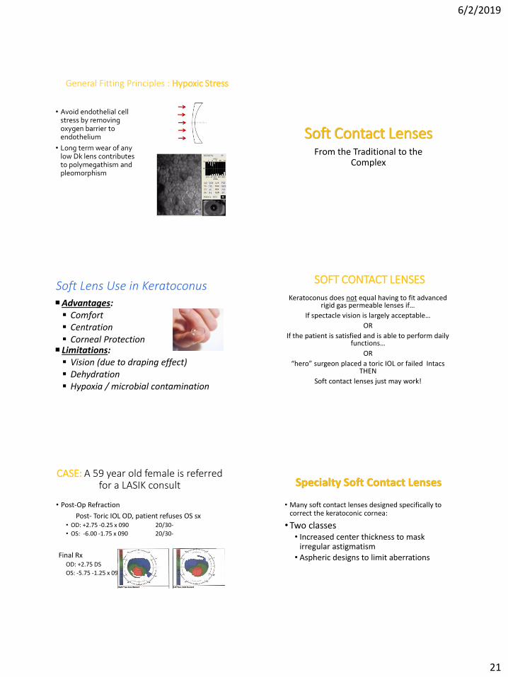
• Avoid endothelial cell stress by removing oxygen barrier to endothelium
• Long term wear of any low Dk lens contributes to polymegathism and pleomorphism
General Fitting Principles : Hypoxic Stress
Soft Contact LensesFrom the Traditional to the
Complex
Soft Lens Use in KeratoconusAdvantages: ▪ Comfort ▪ Centration▪ Corneal Protection
Limitations: ▪ Vision (due to draping effect)▪ Dehydration ▪ Hypoxia / microbial contamination
SOFT CONTACT LENSES
Keratoconus does not equal having to fit advanced rigid gas permeable lenses if…
If spectacle vision is largely acceptable…
OR
If the patient is satisfied and is able to perform daily functions…
OR
“hero” surgeon placed a toric IOL or failed IntacsTHEN
Soft contact lenses just may work!
CASE: A 59 year old female is referred for a LASIK consult
• Post-Op Refraction
Post- Toric IOL OD, patient refuses OS sx• OD: +2.75 -0.25 x 090 20/30-
• OS: -6.00 -1.75 x 090 20/30-
Final RxOD: +2.75 DS Air Optix Aqua 20/30-
OS: -5.75 -1.25 x 090 Air Optix Astg. 20/30-
Specialty Soft Contact Lenses
• Many soft contact lenses designed specifically to correct the keratoconic cornea:
• Two classes• Increased center thickness to mask
irregular astigmatism• Aspheric designs to limit aberrations
6/2/2019
22
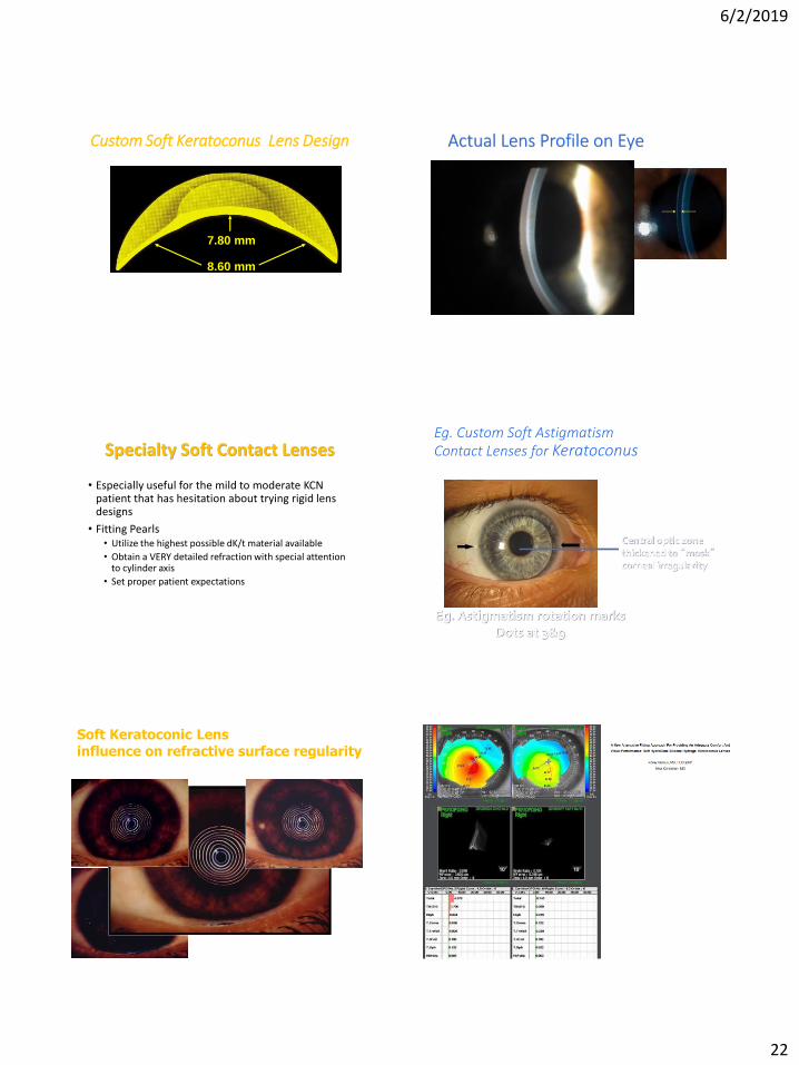
Custom Soft Keratoconus Lens Design
7.80 mm
8.60 mm
“Thick” portion of lens is limited to the central optical zone
Actual Lens Profile on Eye
Specialty Soft Contact Lenses
• Especially useful for the mild to moderate KCN patient that has hesitation about trying rigid lens designs
• Fitting Pearls• Utilize the highest possible dK/t material available
• Obtain a VERY detailed refraction with special attention to cylinder axis
• Set proper patient expectations
Eg. Custom Soft Astigmatism Contact Lenses for Keratoconus
Eg. Astigmatism rotation marksDots at 3&9
Central optic zonethickened to “mask”corneal irregularity
Soft Keratoconic Lens influence on refractive surface regularity (photokeratoscopy) Courtesy Mark Andre
6/2/2019
23
Is Hypoxia an Issue with these Lenses???
• Thickness limited to the central optic zone
• Thinning via lenticularization peripherally (protects the limbal stem cell area)
• Lenses are fit with significantly greater movement (.5 to .75mm w/blink)
• Designs are now available in higher Dk materials (Contamac’s Definitive, Lagado)
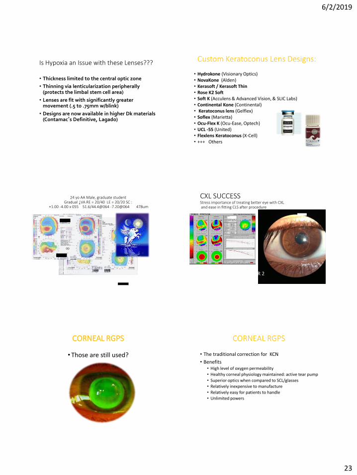
Custom Keratoconus Lens Designs:
• Hydrokone (Visionary Optics)• NovaKone (Alden)• Kerasoft / Kerasoft Thin• Rose K2 Soft • Soft K (Acculens & Advanced Vision, & SLIC Labs)• Continental Kone (Continental)• Keratoconus lens (Gelflex)• Soflex (Marietta) • Ocu-Flex K (Ocu-Ease, Optech)• UCL -55 (United)• Flexlens Keratoconus (X-Cell)• +++ Others
24 yo AA Male, graduate studentGradual ↓VA RE = 20/40 LE = 20/20 SC :
+1.00 -4.00 x 055 51.6/44.4@064 -7.20@064 478um
TX OPTIONS FOR RESOFT TORIC, SPECIALTY HYBRID, GP,CXL
CXL SUCCESS Stress importance of treating better eye with CXLand ease in fitting CLS after procedure
ALDEN NOVAKONE IT FACTOR 2
CORNEAL RGPS
• Those are still used?
CORNEAL RGPS
• The traditional correction for KCN
• Benefits• High level of oxygen permeability
• Healthy corneal physiology maintained: active tear pump
• Superior optics when compared to SCL/glasses
• Relatively inexpensive to manufacture
• Relatively easy for patients to handle
• Unlimited powers
6/2/2019
24
CORNEAL RGPS
• “Negatives”• Patient discomfort
• Increased adaptation time
• Lack of face to face education on proper fitting with new practitioners
• The Underlying Question• CLEK Study: they cause corneal scarring?
• Flat fitting RGPs appear to cause an increased likelihood of apical scarring
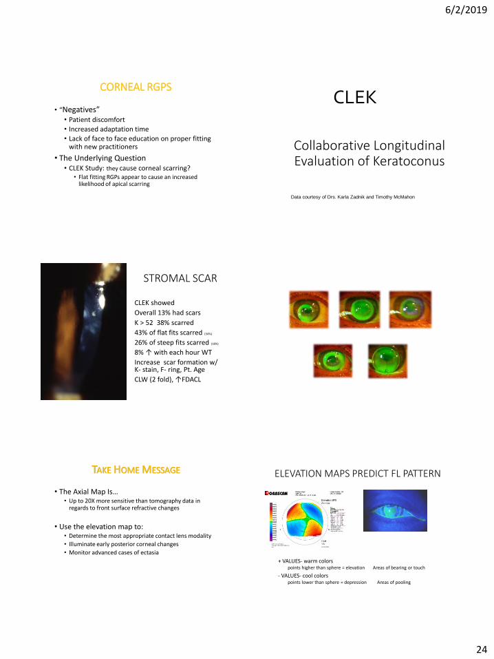
Collaborative Longitudinal Evaluation of Keratoconus
CLEK
Data courtesy of Drs. Karla Zadnik and Timothy McMahon
STROMAL SCAR
CLEK showed
Overall 13% had scars
K > 52 38% scarred
43% of flat fits scarred (34%)
26% of steep fits scarred (18%)
8% ↑ with each hour WT
Increase scar formation w/ K- stain, F- ring, Pt. Age
CLW (2 fold), ↑FDACL
Corneal Lens Design Options
Courtesy of Christine Sindt, OD
Apex Cone Comfort Cone Rose K
Jupiter Cone Soper Cone
TAKE HOME MESSAGE
• The Axial Map Is…• Up to 20X more sensitive than tomography data in
regards to front surface refractive changes
• Use the elevation map to:• Determine the most appropriate contact lens modality
• Illuminate early posterior corneal changes
• Monitor advanced cases of ectasia
ELEVATION MAPS PREDICT FL PATTERN
+ VALUES- warm colors points higher than sphere = elevation Areas of bearing or touch
- VALUES- cool colorspoints lower than sphere = depression Areas of pooling
6/2/2019
25
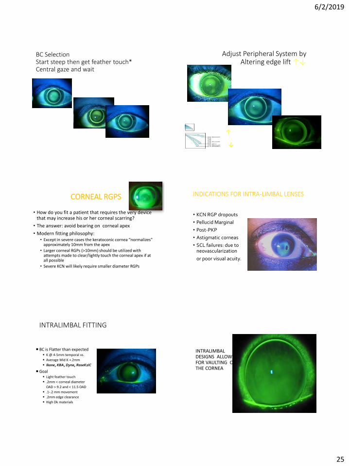
BC Selection Start steep then get feather touch* Central gaze and wait
Tight –go flatter
Increase EL
IDEAL EL
Loose- go tighter
Decrease EL
Adjust Peripheral System byAltering edge lift ↑↓
↓
↓
CORNEAL RGPS
• How do you fit a patient that requires the very device that may increase his or her corneal scarring?
• The answer: avoid bearing on corneal apex
• Modern fitting philosophy:• Except in severe cases the keratoconic cornea “normalizes”
approximately 10mm from the apex
• Larger corneal RGPs (>10mm) should be utilized with attempts made to clear/lightly touch the corneal apex if at all possible
• Severe KCN will likely require smaller diameter RGPs
INDICATIONS FOR INTRA-LIMBAL LENSES
• KCN RGP dropouts
• Pellucid Marginal
• Post-PKP
• Astigmatic corneas
• SCL failures: due to neovascularization
or poor visual acuity.
INTRALIMBAL FITTING
BC is Flatter than expected▪ K @ 4-5mm temporal vs.
▪ Average Mid K +.2mm
▪ Ikone, KBA, Dyna, RoseK2IC
Goal▪ Light feather touch
▪ .2mm < corneal diameter
OAD > 9.2 and < 11.5 OAD
▪ .1-.2 mm movement
▪ .2mm edge clearance
▪ High Dk materials
INTRALIMBAL DESIGNS ALLOW FOR VAULTING OF THE CORNEA
6/2/2019
26
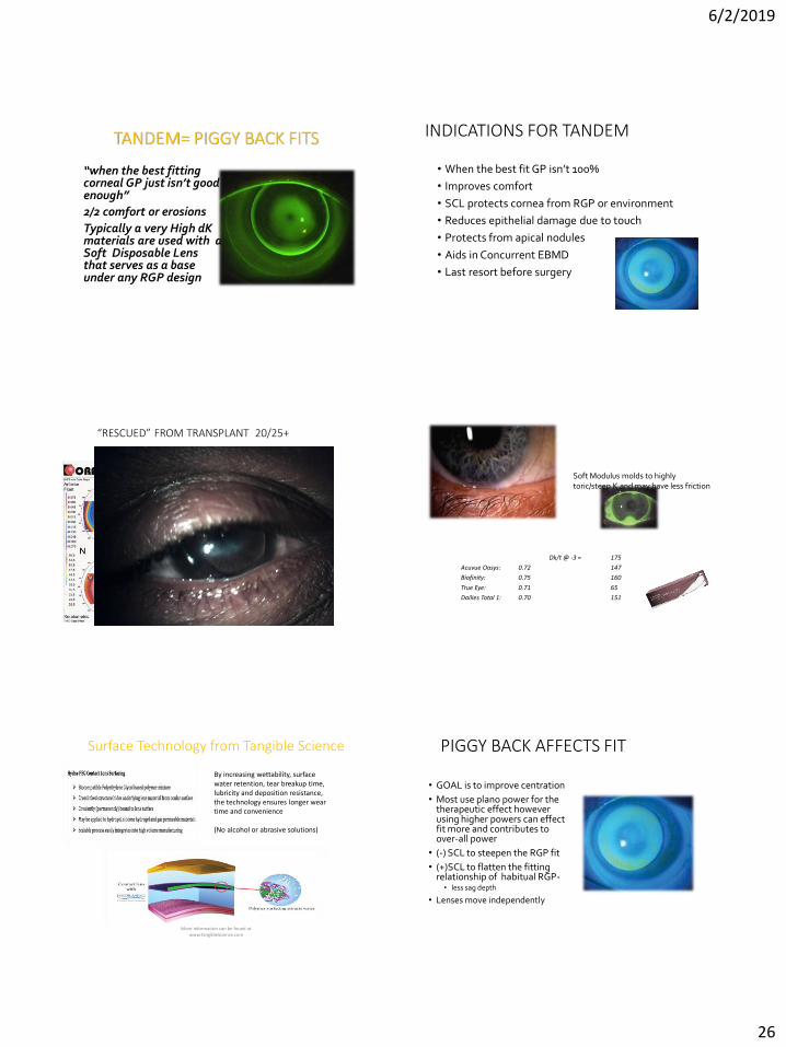
TANDEM= PIGGY BACK FITS
“when the best fitting corneal GP just isn’t good enough”
2/2 comfort or erosions
Typically a very High dKmaterials are used with a Soft Disposable Lens that serves as a base under any RGP design
INDICATIONS FOR TANDEM
• When the best fit GP isn’t 100%
• Improves comfort
• SCL protects cornea from RGP or environment
• Reduces epithelial damage due to touch
• Protects from apical nodules
• Aids in Concurrent EBMD
• Last resort before surgery
“RESCUED” FROM TRANSPLANT 20/25+Modulus and Dk
Night & Day: 1.50 Dk/t @ -3 = 175
Acuvue Oasys: 0.72 147
Biofinity: 0.75 160
True Eye: 0.71 65
Dailies Total 1: 0.70 151
Soft Modulus molds to highly toric/steep K and may have less friction
Surface Technology from Tangible Science
More information can be found at www.TangibleScience.com
By increasing wettability, surface water retention, tear breakup time, lubricity and deposition resistance, the technology ensures longer wear time and convenience
(No alcohol or abrasive solutions)
PIGGY BACK AFFECTS FIT
• GOAL is to improve centration
• Most use plano power for the therapeutic effect however using higher powers can effect fit more and contributes to over-all power
• (-) SCL to steepen the RGP fit
• (+)SCL to flatten the fitting relationship of habitual RGP-• less sag depth
• Lenses move independently
6/2/2019
27
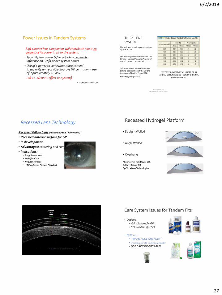
Power Issues in Tandem Systems
Soft contact lens component will contribute about 20 percent of its power in air to the system.
• Typically low power (+/- 0.50) – has negligible influence on GP fit or net system power
• Use of + power to somewhat mask corneal irregularity and possibly improve GP centration - use of approximately +6.00 D
(+6 = 1.2D net + effect on system)• Daniel Brazeau,OD
THICK LENS SYSTEM
The soft lens is no longer a thin lens system in “air”
The Tear Layer created between the GP and Hydrogel “negates” some of the SCL power… but not all..
Calculate power between the area behind back surface of the GP and the cornea AKA the TL and SCL.
BVP= F1/(1-t/n)F1 +F2
MINHEE WOO OD AND BARRY WEISMAN OD PhD
EFFECTIVE POWERS OF SCL UNDER GP IN TANDEM DESIGN IS ABOUT 20% OF ORIGINAL
POWER (10-30%)
Recessed Lens Technology
Recessed Pillow Lens (Fusion & EyeVisTechnologies)
• Recessed anterior surface for GP
• In development
• Advantages: centering and comfort
• Indications: • Irregular corneas
• Multifocal GP
• Regular corneas
• *Other Recess: Flexlens Piggyback
Recessed Hydrogel Platform
• Straight Walled
• Angle Walled
• Overhang
*Courtesy of Rob Davis, OD,
S. Barry Eiden, OD
EyeVis Vision Technologies
Courtesy of Rob Davis, OD
Care System Issues for Tandem Fits
• Option 1:
• GP solutions for GP
• SCL solutions for SCL
• Option 2:
• “One for all & all for one!”• (multipurpose SCL solution or peroxide)
• USE DAILY DISPOSABLE!
6/2/2019
28
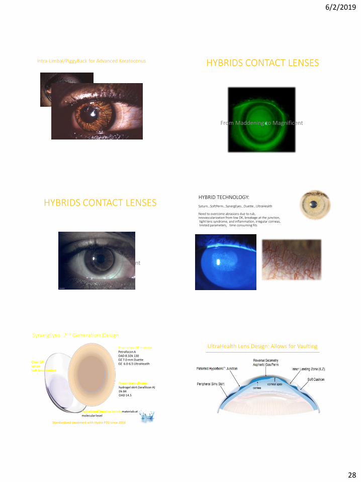
Intra-Limbal/PiggyBack for Advanced Keratoconus HYBRIDS CONTACT LENSES
From Maddening to Magnificent
HYBRIDS CONTACT LENSES
From Maddening to Magnificent
HYBRID TECHNOLOGY:
Saturn…SoftPerm…SynergEyes…Duette…UltraHealth
Need to overcome abrasions due to rub, neovascularization from low DK, breakage at the junction,tight lens syndrome, and inflammation, irregular corneas,limited parameters, time consuming fits
Proprietary GP material Petrafocon AOAD 8.5Dk 130OZ 7.0 mm DuetteOZ 6.0-6.5 UltraHeatlh
SynergEyes 2nd Generation Design
Proprietary silicone hydrogel skirt (larafilcon A)Dk 84OAD 14.5
Hyperbond®junction bonds materials at molecular level
Clear GP vision Soft lens comfort
Standardized treatment with Hydra-PEG since 2016
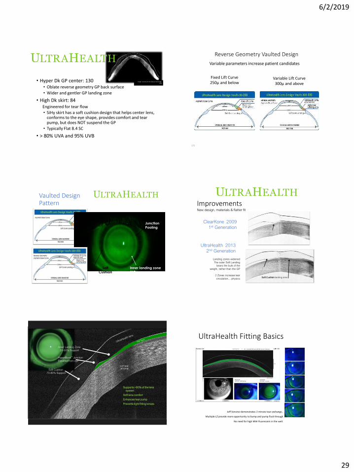
UltraHealth Lens Design: Allows for Vaulting
6/2/2019
29
• Hyper Dk GP center: 130• Oblate reverse geometry GP back surface
• Wider and gentler GP landing zone
• High Dk skirt: 84Engineered for tear flow
• SiHy skirt has a soft cushion design that helps center lens, conforms to the eye shape, provides comfort and tear pump, but does NOT suspend the GP
• Typically Flat 8.4 SC
• > 80% UVA and 95% UVB
Image courtesy of Lens Optical Technology, UZA
172
Fixed Lift Curve 250µ and below
Variable Lift Curve 300µ and above
Variable parameters increase patient candidates
Reverse Geometry Vaulted Design
Vaulted DesignPattern
Junction
Pooling
Inner landing zone
(ILZ)
Soft
Cushion
ImprovementsNew design, materials & flatter fit
ClearKone 2009
1st Generation
Soft Cushion landing zone
Landing zones widened
The outer Soft Landing
bears the bulk of the
weight, rather than the GP
2 Zones increase tear
circulation… physics
GP inner landing zone
UltraHealth 2013
2nd Generation
GP Inner landing zone
corneal stroma
Supports >80% of the lens
system
Soft lens comfort
Enhances tear pump
Prevents tight fitting lenses
Inner Landing Zone
20-30% Support
Soft Cushion
70-80% Support
UltraHealth Fitting Basics
Jeff Sonsino demonstrates 2 minute tear exchange..
Multiple LZ provide more opportunity to bump and pump fluid through
No need for high MW Fluorescein in the well.
6/2/2019
30
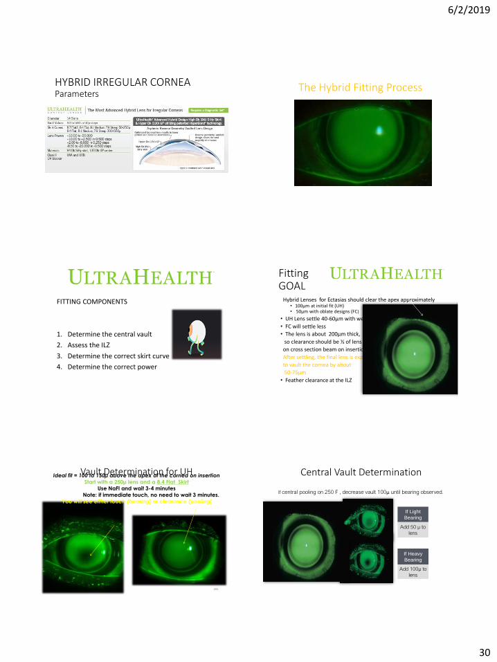
HYBRID IRREGULAR CORNEA Parameters
The Hybrid Fitting Process
Well fit UltraHealth Hybrid per NaFL evaluation
FITTING COMPONENTS
1. Determine the central vault
2. Assess the ILZ
3. Determine the correct skirt curve
4. Determine the correct power
FittingGOAL
Hybrid Lenses for Ectasias should clear the apex approximately • 100μm at initial fit (UH) • 50µm with oblate designs (FC)
• UH Lens settle 40-60μm with wear
• FC will settle less
• The lens is about 200μm thick,
so clearance should be ½ of lens thickness
on cross section beam on insertion
After settling, the final lens is expected
to vault the cornea by about
50-75μm
• Feather clearance at the ILZ
Vault Determination for UH
181
Ideal fit = 100 to 150µ above the apex of the cornea on insertion
Start with a 250µ lens and a 8.4 Flat Skirt Use NaFl and wait 3-4 minutes
Note: if immediate touch, no need to wait 3 minutes.
You will see either touch (bearing) or clearance (pooling)
Central Vault Determination
If central pooling on 250 F , decrease vault 100µ until bearing observed.
If Light
Bearing
Add 50 µ to
lens
If Heavy
Bearing
Add 100µ to
lens
6/2/2019
31
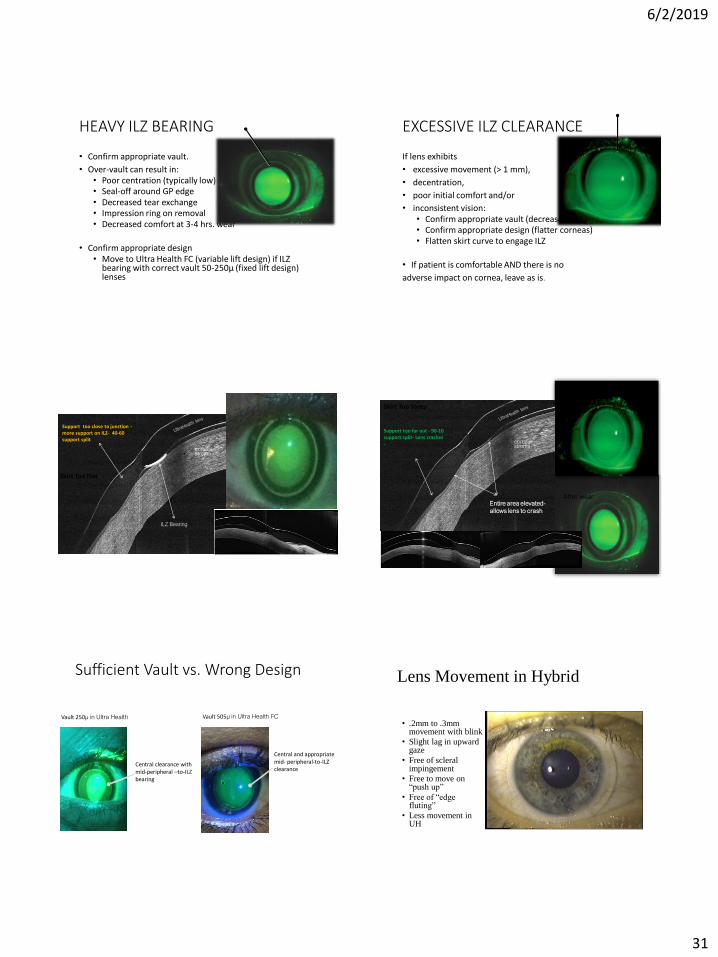
HEAVY ILZ BEARING
• Confirm appropriate vault.
• Over-vault can result in:• Poor centration (typically low)• Seal-off around GP edge• Decreased tear exchange• Impression ring on removal• Decreased comfort at 3-4 hrs. wear
• Confirm appropriate design• Move to Ultra Health FC (variable lift design) if ILZ
bearing with correct vault 50-250µ (fixed lift design) lenses
EXCESSIVE ILZ CLEARANCE
If lens exhibits
• excessive movement (> 1 mm),
• decentration,
• poor initial comfort and/or
• inconsistent vision:• Confirm appropriate vault (decrease vault)• Confirm appropriate design (flatter corneas)• Flatten skirt curve to engage ILZ
• If patient is comfortable AND there is no
adverse impact on cornea, leave as is.
corneal stroma
ILZ Bearing
Support too close to junction -more support on ILZ- 40-60 support split
Skirt Too Flat
corneal stroma
Entire area elevated-
allows lens to crash
Support too far out - 90-10 support split- Lens crashes -
At fit
After wear
Skirt Too Steep
Sufficient Vault vs. Wrong Design
Vault 250µ in Ultra Health
Central clearance with mid-peripheral –to-ILZ bearing
Vault 505µ in Ultra Health FC
Central and appropriate mid- peripheral-to-ILZ clearance
Lens Movement in Hybrid
• .2mm to .3mm movement with blink
• Slight lag in upward gaze
• Free of scleral impingement
• Free to move on “push up”
• Free of “edge fluting”
• Less movement in UH
6/2/2019
32
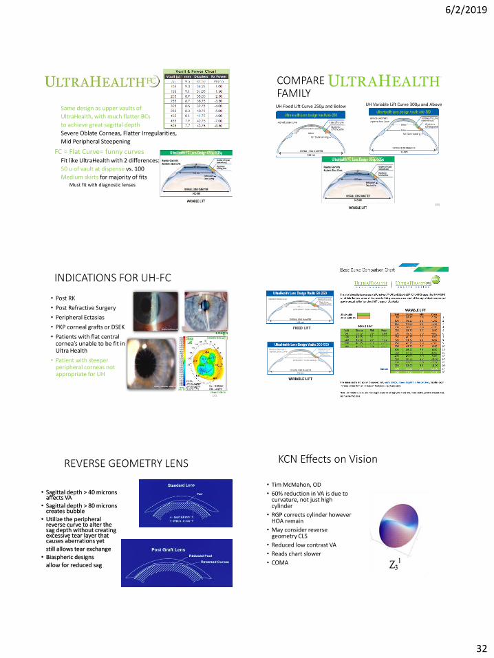
Same design as upper vaults of
UltraHealth, with much flatter BCs
to achieve great sagittal depth
Severe Oblate Corneas, Flatter Irregularities,
Mid Peripheral Steepening
FC = Flat Curve= funny curvesFit like UltraHealth with 2 differences:
50 u of vault at dispense vs. 100
Medium skirts for majority of fitsMust fit with diagnostic lenses
UH Fixed Lift Curve 250µ and Below UH Variable Lift Curve 300µ and Above
COMPAREFAMILY
190
INDICATIONS FOR UH-FC
• Post RK
• Post Refractive Surgery
• Peripheral Ectasias
• PKP corneal grafts or DSEK
• Patients with flat central cornea’s unable to be fit in Ultra Health
• Patient with steeper peripheral corneas not appropriate for UH
191
REVERSE GEOMETRY LENS
• Sagittal depth > 40 microns affects VA
• Sagittal depth > 80 microns creates bubble
• Utilize the peripheral reverse curve to alter the sag depth without creating excessive tear layer that causes aberrations yet
still allows tear exchange
• Biaspheric designs allow for reduced sag
KCN Effects on Vision
• Tim McMahon, OD
• 60% reduction in VA is due to curvature, not just high cylinder
• RGP corrects cylinder however HOA remain
• May consider reverse geometry CLS
• Reduced low contrast VA
• Reads chart slower
• COMA
6/2/2019
33
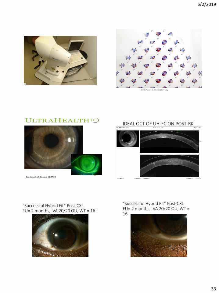
Zernike Polynomials :Wavefront Technology
197Courtesy of Jeff Sonsino, OD,FAAO
IDEAL OCT OF UH-FC ON POST-RK
“Successful Hybrid Fit” Post-CXLFU= 2 months, VA 20/20 OU, WT = 16 !
“Successful Hybrid Fit” Post-CXLFU= 2 months, VA 20/20 OU, WT = 16
6/2/2019
34
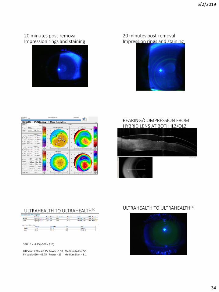
20 minutes post-removal Impression rings and staining
20 minutes post-removal Impression rings and staining
BEARING/COMPRESSION FROM HYBRID LENS AT BOTH ILZ/OLZ
Consider Flatter SkirtGOAL 50-100 um clearance(ct= 200 um)
RE changed from steep to flat skirtLE skirt change didn’t improve….
ULTRAHEALTH TO ULTRAHEALTHFC
SPH LE = -1.25 (-500 x 115)
UH Vault 200 = 46.25 Power -6.50 Medium to Flat SCFK Vault 450 = 42.75 Power -.25 Medium Skirt = 8.1
ULTRAHEALTH TO ULTRAHEALTHFC
6/2/2019
35
Trouble Shooting Tips
• At recheck, if poor lens movement, verify tear exchange by instilling fluorescein• Active tear pump should move NaFL under lens even w/o lens movement
• Discomfort at 3-4 hour mark, low-riding lens, and/or difficult removal may indicate over-vault
• If excessive movement with blink and/or bubble uptake after insertion, steepen skirt after confirming appropriate vault
• Dry lens/dry fingers or tissue are key for removal
• Impression ring may be visible after wear and is acceptable as long as there is no epithelial disruption• If there is epithelial disruption recheck the fit for over-vault or ILZ bearing.
Personal TIPS and Techniques
• During fit, FIND FIRST TOUCH; it is easy to over-vault
• Remember to assess ILZ with each lens
• Set proper patient expectations; reassure there will be initial lens “awareness” that will go away in a few days with gradual build-up of wear time
• Avoid making changes at lens dispense visit
• Have patient review insertion, removal and lens care video PRIOR TO DISPENSE APPOINTMENT
• Train staff on I&R; Assure patient that removal is “Different ” then what they are used to, but not “Difficult ”
• Have patient remove lenses during office hours first several days
Hybrid Contact Lenses Fitting Tips
• Insert initial lens (250 vault/flat skirt)
• Obtain OCT clearance data
• Steepen the skirt to improve centration
• Flatten the skirt to promote lens movement
• Lens moves in/out
• Patients who will be more of a challenge
• Chronic dry eye … however now there is THP
• Patients with intracorneal ring segments … FC
• Patients likely to require toric optics… 1D RA
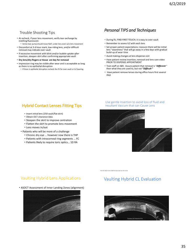
Use gentle Insertion to avoid loss of fluid and resultant Vaccum that can Cause Lens to tighten
Vaulting Hybrid Lens Applications
• ASOCT Assessment of Inner Landing Zones (alignment)
Vaulting Hybrid CL Evaluation
•Use the caliper tool to label the exact vault over the cornea
Courtesy of J Sonsino O.D.
Adequate Clearance Central GP Zone & Inner Landing Zone Mild Clearance
6/2/2019
36
corneal stroma
ILZ Bearing
Support too close to junction -more support on ILZ- 40-60 support split
Skirt Too Flat
SCLERAL CONTACT LENSES
What’s Old is New Again!
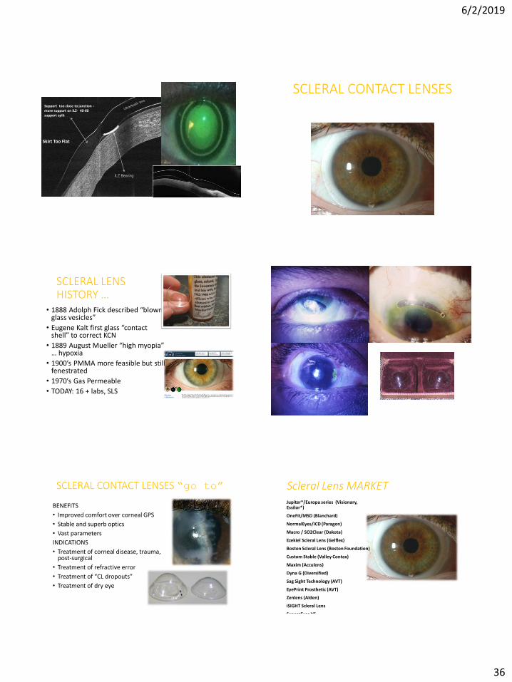
SCLERAL LENS HISTORY …
• 1888 Adolph Fick described “blown glass vesicles”
• Eugene Kalt first glass “contact shell” to correct KCN
• 1889 August Mueller “high myopia” … hypoxia
• 1900’s PMMA more feasible but still fenestrated
• 1970’s Gas Permeable
• TODAY: 16 + labs, SLS
SCLERAL CONTACT LENSES “go to”
BENEFITS
• Improved comfort over corneal GPS
• Stable and superb optics
• Vast parameters
INDICATIONS
• Treatment of corneal disease, trauma, post-surgical
• Treatment of refractive error
• Treatment of “CL dropouts”
• Treatment of dry eye
Scleral Lens MARKET Jupiter*/Europa series (Visionary, Essilor*)
OneFit/MSD (Blanchard)
NormalEyes/ICD (Paragon)
Macro / SO2Clear (Dakota)
Ezekiel Scleral Lens (Gelflex)
Boston Scleral Lens (Boston Foundation)
Custom Stable (Valley Contax)
Maxim (Acculens)
Dyna G (Diversified)
Sag Sight Technology (AVT)
EyePrint Prosthetic (AVT)
Zenlens (Alden)
iSIGHT Scleral Lens
SynergEyes VSPat Caroline, FAAO & Pacific University
6/2/2019
37
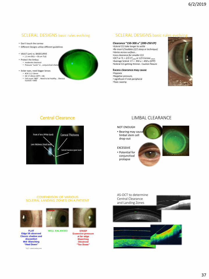
SCLERAL DESIGNS basic rules evolving
• Don’t touch the cornea
• Different Designs utilize different guidelines
• VAULT (um) vs. BASECURVE • (.1 mm BCΔ = 50 um TLΔ)
• Protect the limbus• moderate clearance• Pressure “sucks” in …conjunctival-chelasis
• Sicker eyes, need bigger lenses• KCN 11.2-16mm • DX 17-20mm (EPP = 18)• Cell count “800” .. Need to be healthy… Obvious
nucleoli = BAD
Pat Caroline, FAAO & Pacific UniversityEsther Simone Vissar, Netherlands
SCLERAL DESIGNS basic rules evolving
Clearance “150-300 u” (200-250 EP)•Scleral CLS take longer to settle•Re-insert if bubbles (2/2 steep or technique)•Varies across surface… •Less clearance for smaller CLS•OCT or TL = 2/3 CTLENS or 1/3 Cornea THICK
•Average Scleral CT = 350 u – 450 u (EPP) •Scleral CLS getting thinner.. Caution flexure
Excess clearance may cause •Hypoxia•Negative pressure,• significant if mid-peripheral•Toxic swamp
Pat Caroline, FAAO & Pacific UniversityEsther Simone Vissar, Netherlands
Central Clearance LIMBAL CLEARANCE
NOT ENOUGH
• Bearing may cause limbal stem cell drop-out
EXCESSIVE
• Potential for conjunctivalprolapse
COMPARISON OF VARIOUS SCLERAL LANDING ZONES ON A PATIENT
WELL BALANCEDFLAT
Edge lift observed
Classic shadow and
discomfort
Mid- Blanching
“Heel Down”
STEEP
Excessive pressure
at far edge
Blanching
Observed
“Toe Down”
* SLZ = scleral landing zone
AS-OCT to determine Central Clearance and Landing Zones
6/2/2019
38
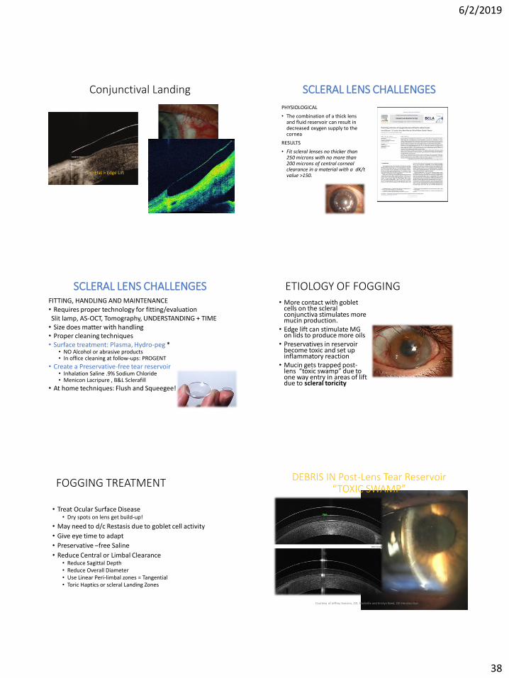
Conjunctival Landing
Too Flat = Edge Lift Too Steep = Conjunctival Impingement
SCLERAL LENS CHALLENGES
PHYSIOLOGICAL
• The combination of a thick lens and fluid reservoir can result in decreased oxygen supply to the cornea
RESULTS
• Fit scleral lenses no thicker than 250 microns with no more than 200 microns of central corneal clearance in a material with a dK/t value >150.
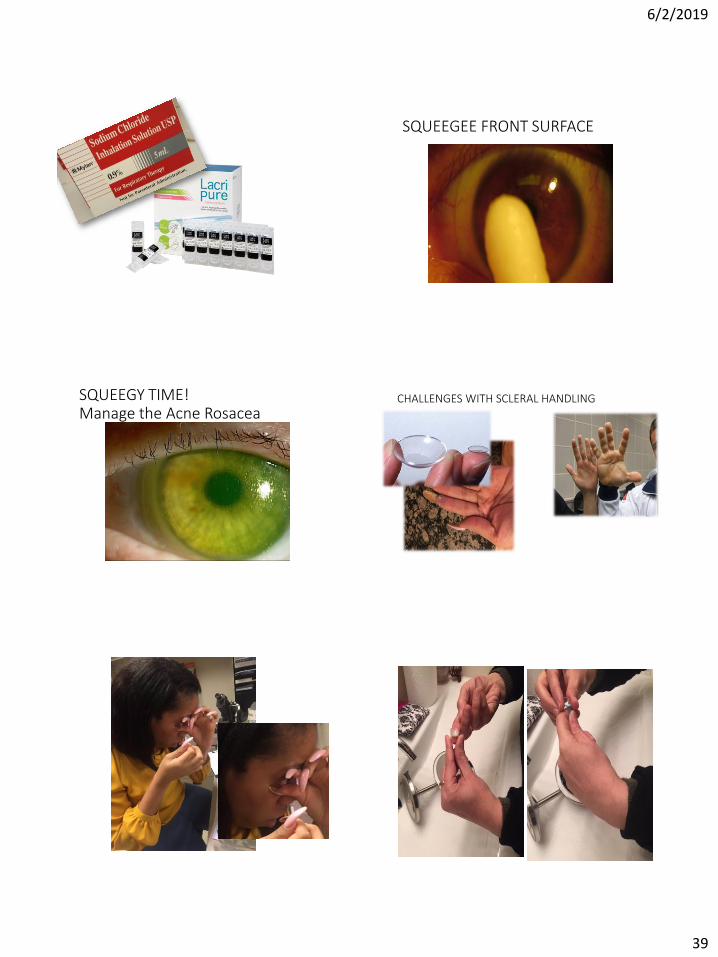
SCLERAL LENS CHALLENGESFITTING, HANDLING AND MAINTENANCE• Requires proper technology for fitting/evaluationSlit lamp, AS-OCT, Tomography, UNDERSTANDING + TIME• Size does matter with handling• Proper cleaning techniques• Surface treatment: Plasma, Hydro-peg *
• NO Alcohol or abrasive products• In office cleaning at follow-ups: PROGENT
• Create a Preservative-free tear reservoir• Inhalation Saline .9% Sodium Chloride• Menicon Lacripure , B&L Sclerafill
• At home techniques: Flush and Squeegee!
ETIOLOGY OF FOGGING• More contact with goblet
cells on the scleral conjunctiva stimulates more mucin production.
• Edge lift can stimulate MG on lids to produce more oils
• Preservatives in reservoir become toxic and set up inflammatory reaction
• Mucin gets trapped post-lens “toxic swamp” due to one way entry in areas of lift due to scleral toricity
FOGGING TREATMENT
• Treat Ocular Surface Disease• Dry spots on lens get build-up!
• May need to d/c Restasis due to goblet cell activity
• Give eye time to adapt
• Preservative –free Saline
• Reduce Central or Limbal Clearance • Reduce Sagittal Depth• Reduce Overall Diameter• Use Linear Peri-limbal zones = Tangential• Toric Haptics or scleral Landing Zones
DEBRIS IN Post-Lens Tear Reservoir“TOXIC SWAMP”
Courtesy of Jeffrey Sonsino, OD, Nashville and Kristyn Reed, OD Houston Eye
6/2/2019
39
SQUEEGEE FRONT SURFACE
SQUEEGY TIME! Manage the Acne Rosacea
CHALLENGES WITH SCLERAL HANDLING
6/2/2019
40
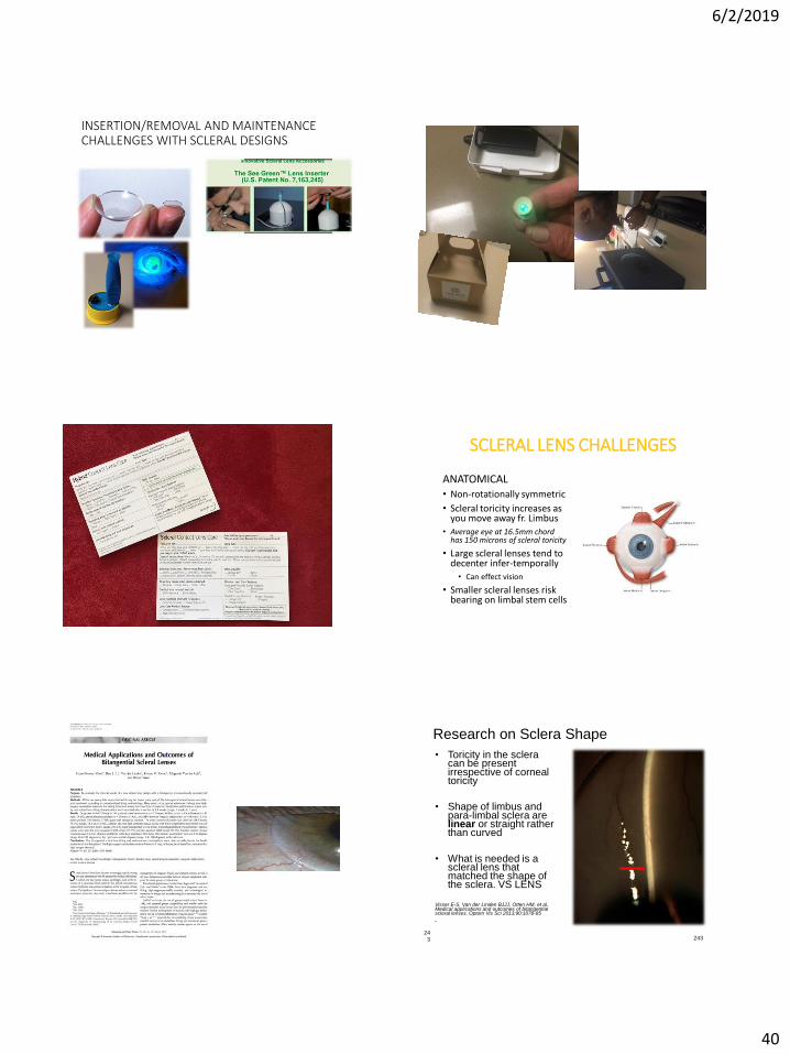
INSERTION/REMOVAL AND MAINTENANCE CHALLENGES WITH SCLERAL DESIGNS
SCLERAL LENS CHALLENGES
ANATOMICAL• Non-rotationally symmetric
• Scleral toricity increases as you move away fr. Limbus
• Average eye at 16.5mm chord has 150 microns of scleral toricity
• Large scleral lenses tend to decenter infer-temporally
• Can effect vision
• Smaller scleral lenses risk bearing on limbal stem cells
• Toricity in the sclera can be present irrespective of corneal toricity
• Shape of limbus and para-limbal sclera are linear or straight rather than curved
• What is needed is a scleral lens that matched the shape of the sclera. VS LENS
Visser E-S, Van der Linden BJJJ, Otten HM, et al. Medical applications and outcomes of bitangential scleral lenses. Optom Vis Sci 2013;90:1078-85
•
Research on Sclera Shape
24324
3
6/2/2019
41
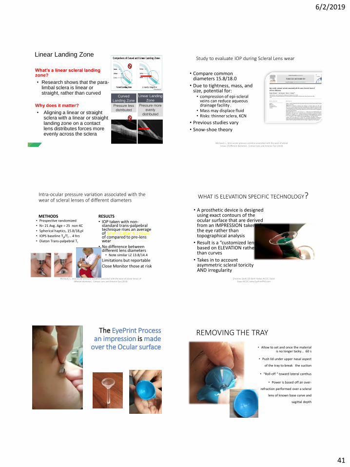
What’s a linear scleral landing zone?
• Research shows that the para-limbal sclera is linear or straight, rather than curved
Why does it matter?
• Aligning a linear or straight sclera with a linear or straight landing zone on a contact lens distributes forces more evenly across the sclera
Linear Landing Zone
Curved
Landing Zone
Pressure less
distributed
Linear Landing
Zone
Pressure more
evenly
distributed
Study to evaluate IOP during Scleral Lens wear
• Compare common diameters 15.8/18.0
• Due to tightness, mass, and size, potential for:• compression of epi-scleral
veins can reduce aqueous drainage facility .
• Mass may displace fluid• Risks: thinner sclera, KCN
• Previous studies vary
• Snow-shoe theory
Michaud, L., Intra-ocular pressure variation associated with the wear of scleral lenses of different diameters. Contact Lens and Anterior Eye (2018)
Intra-ocular pressure variation associated with the wear of scleral lenses of different diameters
METHODS• Prospective randomized
• N= 21 Avg. Age = 25 non-KC
• Spherical haptics, 15.8/18,pl
• IOPS baseline Tg/Tt .. 4 hrs
• Diaton Trans-palpebral Tt
RESULTS• IOP taken with non-
standard trans-palpebral technique rises an average of 5mmHg after 4.3 hours of compared to pre-lens wear
• No difference between different lens diameters• Note similar LZ 13.8/14.4
• Limitations but reportable
• Close Monitor those at risk
Michaud, L., Intra-ocular pressure variation associated with the wear of scleral lenses of different diameters. Contact Lens and Anterior Eye (2018)
WHAT IS ELEVATION SPECIFIC TECHNOLOGY?
• A prosthetic device is designed using exact contours of the ocular surface that are derived from an IMPRESSION taken of the eye rather than topographical analysis
• Result is a “customized lens” based on ELEVATION rather than curves
• Takes in to account asymmetric scleral toricityAND irregularity
Christine Sindt, OD Keith Parker, NCLEC David Slater NCLEC www.EyePrintPRO.com
The EyePrint Processan impression is made
over the Ocular surface
REMOVING THE TRAY
• Allow to set and once the material is no longer tacky… 60 s
• Push lid under upper nasal aspect
of the tray to break the suction
• “Roll-off “ toward lateral canthus
• Power is based off an over-
refraction performed over a scleral
lens of known base curve and
sagittal depth
6/2/2019
42
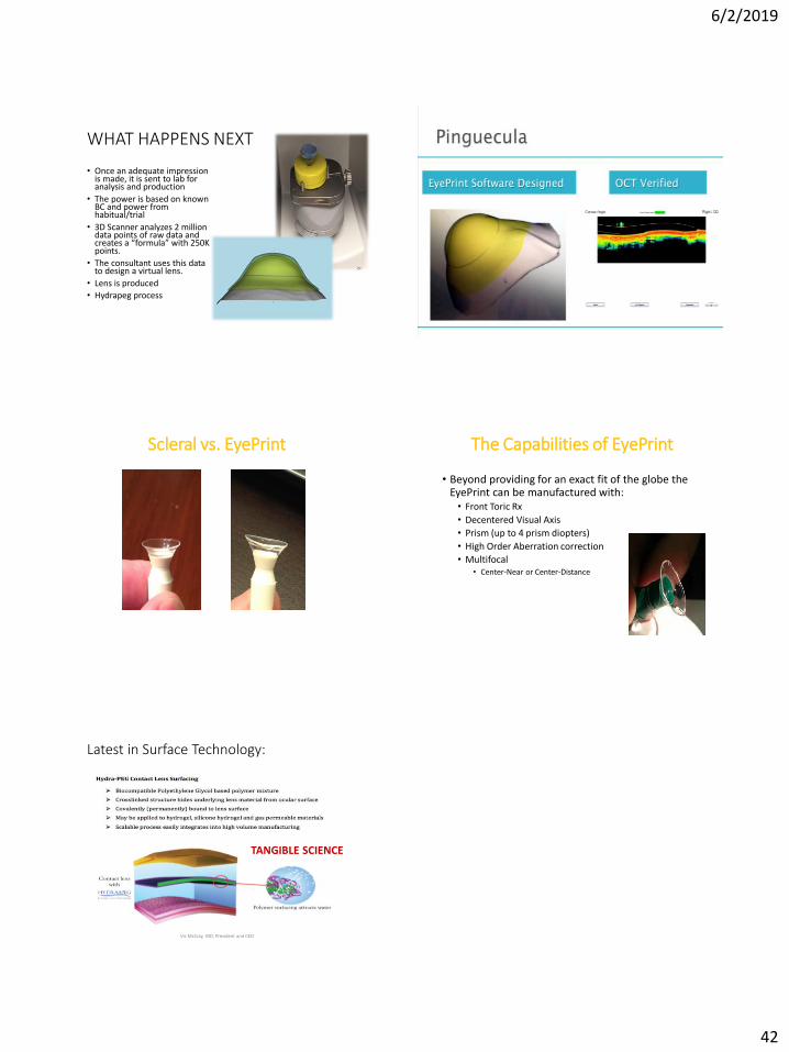
WHAT HAPPENS NEXT
• Once an adequate impression is made, it is sent to lab for analysis and production
• The power is based on known BC and power from habitual/trial
• 3D Scanner analyzes 2 million data points of raw data and creates a “formula” with 250K points.
• The consultant uses this data to design a virtual lens.
• Lens is produced
• Hydrapeg process
Scleral vs. EyePrint The Capabilities of EyePrint
• Beyond providing for an exact fit of the globe the EyePrint can be manufactured with:• Front Toric Rx
• Decentered Visual Axis
• Prism (up to 4 prism diopters)
• High Order Aberration correction
• Multifocal• Center-Near or Center-Distance
Latest in Surface Technology:
Vic McCray, MD, President and CEO
TANGIBLE SCIENCE