Embed Size (px)
Citation preview
Low Gradient Severe (?) Aortic Stenosis
Philippe Pibarot, DVM, PhD, FACC, FAHA, FESC, FASECanada Research Chair in Valvular Heart Diseases
UniversitéLAVAL
Institut Universitaire de Cardiologieet de Pneumologie de Québec / Québec Heart & Lung Institute

Disclosure: Philippe PibarotFinancial relationship with industry:
Edwards Lifesciences: Echo CoreLab for PARTNER 2–
SAPIEN 3, PARTNER 3, TAVR-UNLOAD trials
V-Wave Ltd: Echo CoreLab for FIM Study
Cardiac Phoenix: Echo CoreLab for BACE FIM Study
Other financial disclosure:
Research Grants from Canadian Institutes of Health
Research and Heart & Stroke Foundation of Quebec
Off label Use: None

LVEF >50%<50%
Low Gradient ASAVA≤1.0 cm2 MG<40 mmHg
«CLASSICAL»LOW-FLOW
LOW-GRADIENTD2 Stage
«PARADOXICAL»LOW-FLOW
LOW-GRADIENTD3 Stage
NORMAL-FLOWLOW-GRADIENT
D? Stage
SVi<35 mL/m2 >35 mL/m2
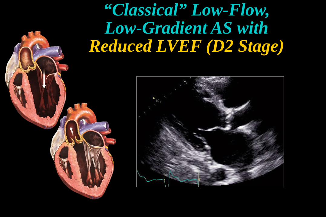
“Classical” Low-Flow, Low-Gradient AS with
Reduced LVEF (D2 Stage)
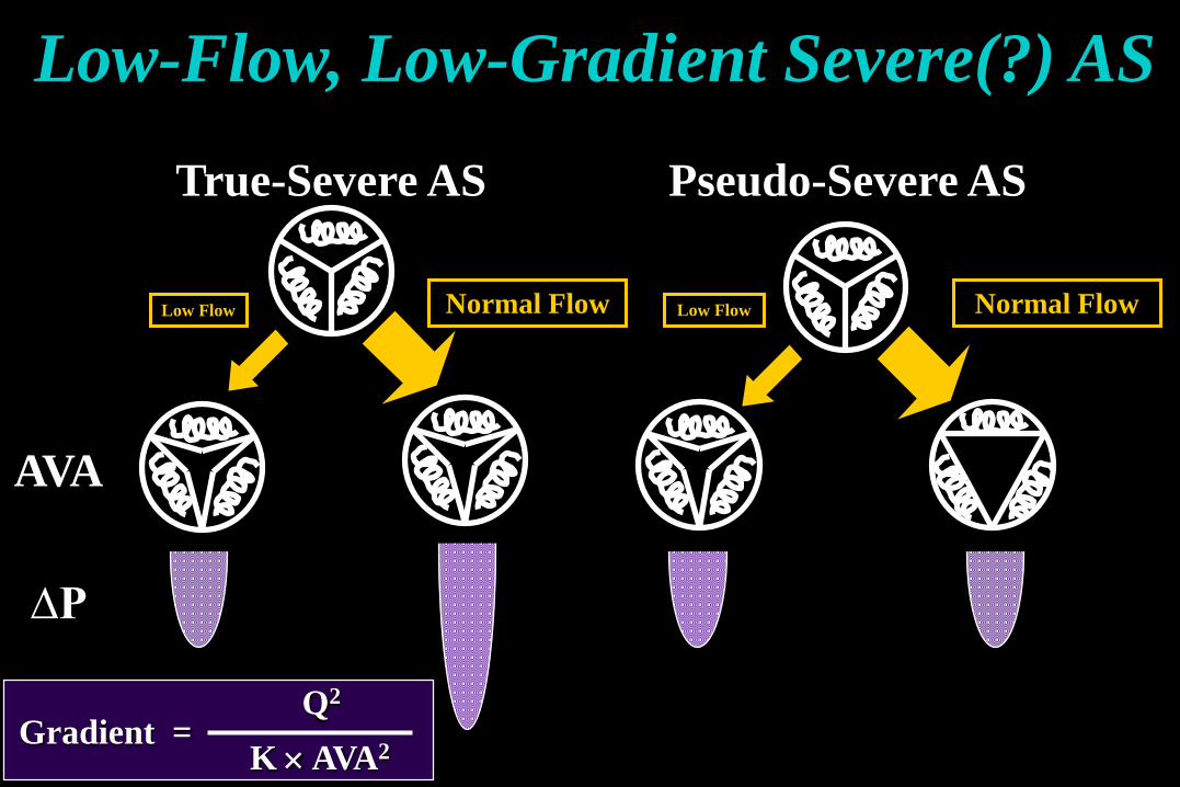
Low Flow Low FlowNormal Flow Normal Flow
True-Severe AS Pseudo-Severe AS
Low-Flow, Low-Gradient Severe(?) AS
∆P
AVA
Gradient = Q2
K ×AVA2
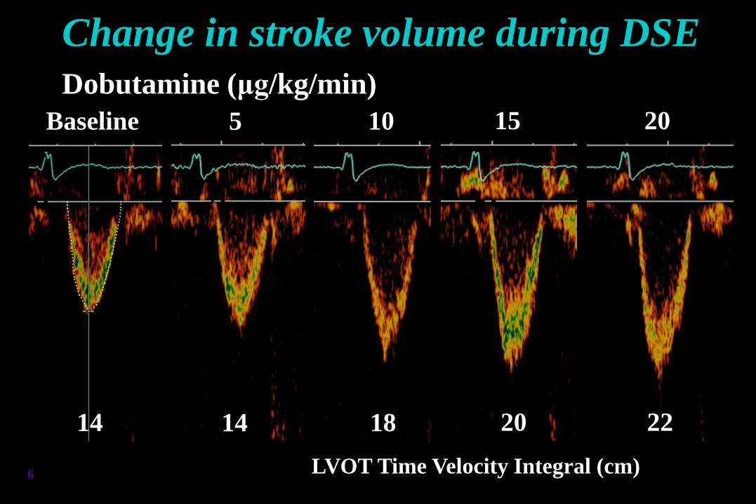
Change in stroke volume during DSEDobutamine (μg/kg/min)
LVOT Time Velocity Integral (cm)
Baseline
14
5
14 20
15
22
20
18
10
6
Mean Pressure Gradient (mm Hg)
Baseline
21
5
19
10
36
15
39
20
42
7
Change in Gradient during DSEDobutamine (μg/kg/min)
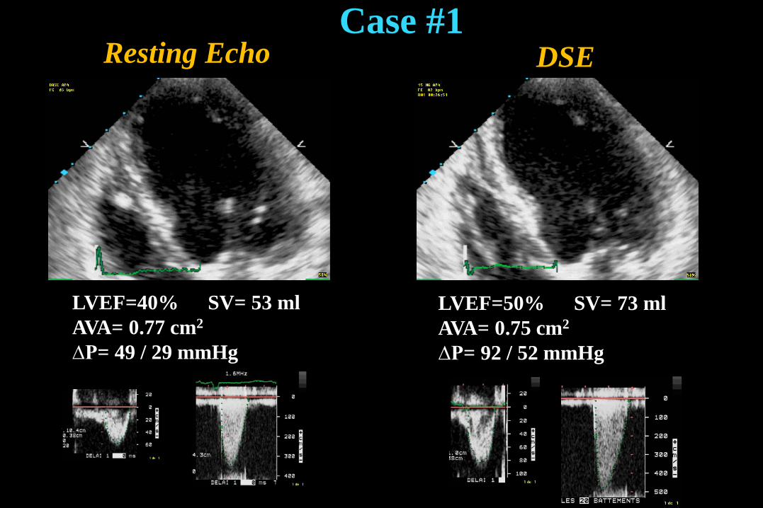
LVEF=40% SV= 53 mlAVA= 0.77 cm2
∆P= 49 / 29 mmHg
LVEF=50% SV= 73 mlAVA= 0.75 cm2
∆P= 92 / 52 mmHg
DSEResting EchoCase #1
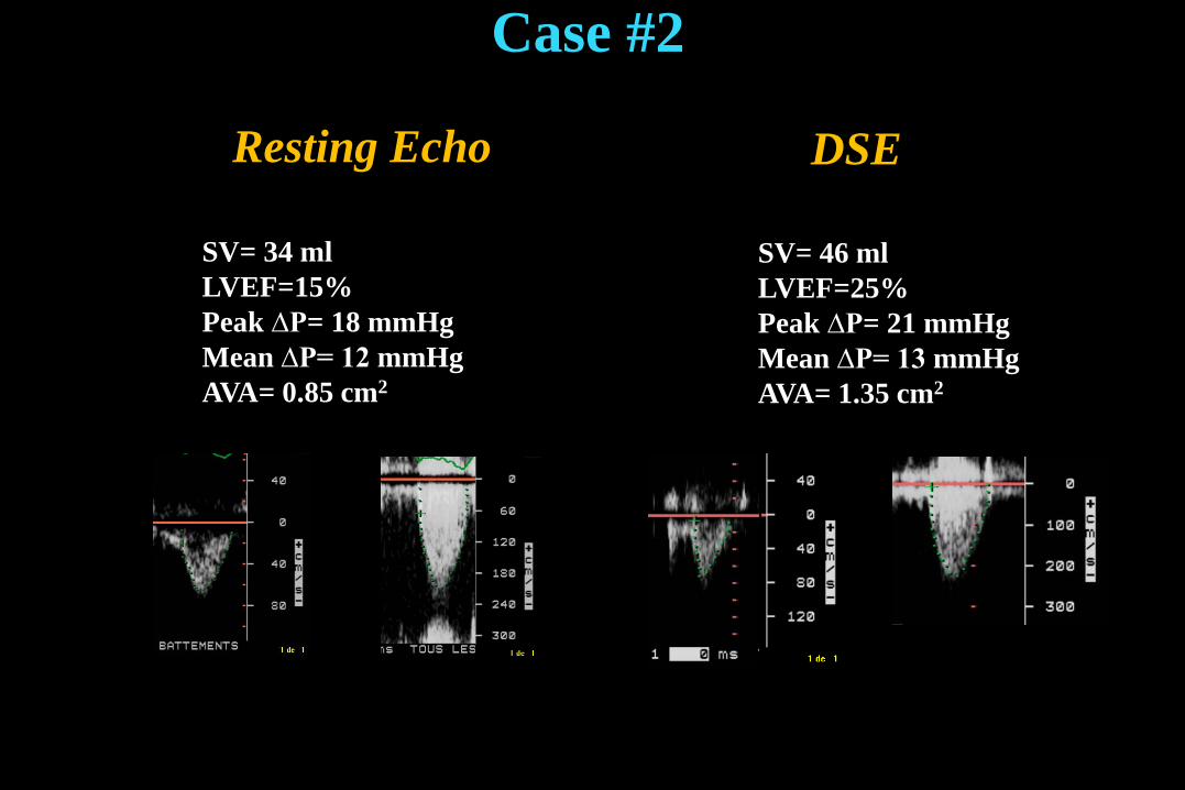
DSEResting Echo
Case #2
SV= 46 mlLVEF=25%Peak ∆P= 21 mmHgMean ∆P= 13 mmHgAVA= 1.35 cm2
SV= 34 mlLVEF=15%Peak ∆P= 18 mmHgMean ∆P= 12 mmHgAVA= 0.85 cm2
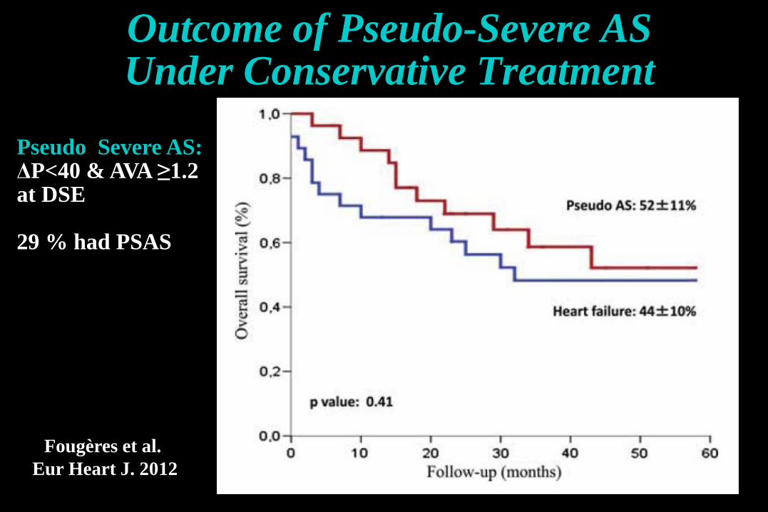
Outcome of Pseudo-Severe AS Under Conservative Treatment
Fougères et al. Eur Heart J. 2012
Pseudo Severe AS:ΔP<40 & AVA ≥1.2 at DSE
29 % had PSAS
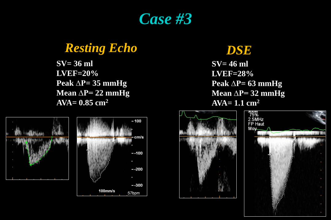
DSEResting EchoSV= 46 mlLVEF=28%Peak ∆P= 63 mmHgMean ∆P= 32 mmHgAVA= 1.1 cm2
SV= 36 mlLVEF=20%Peak ∆P= 35 mmHgMean ∆P= 22 mmHgAVA= 0.85 cm2
Case #3
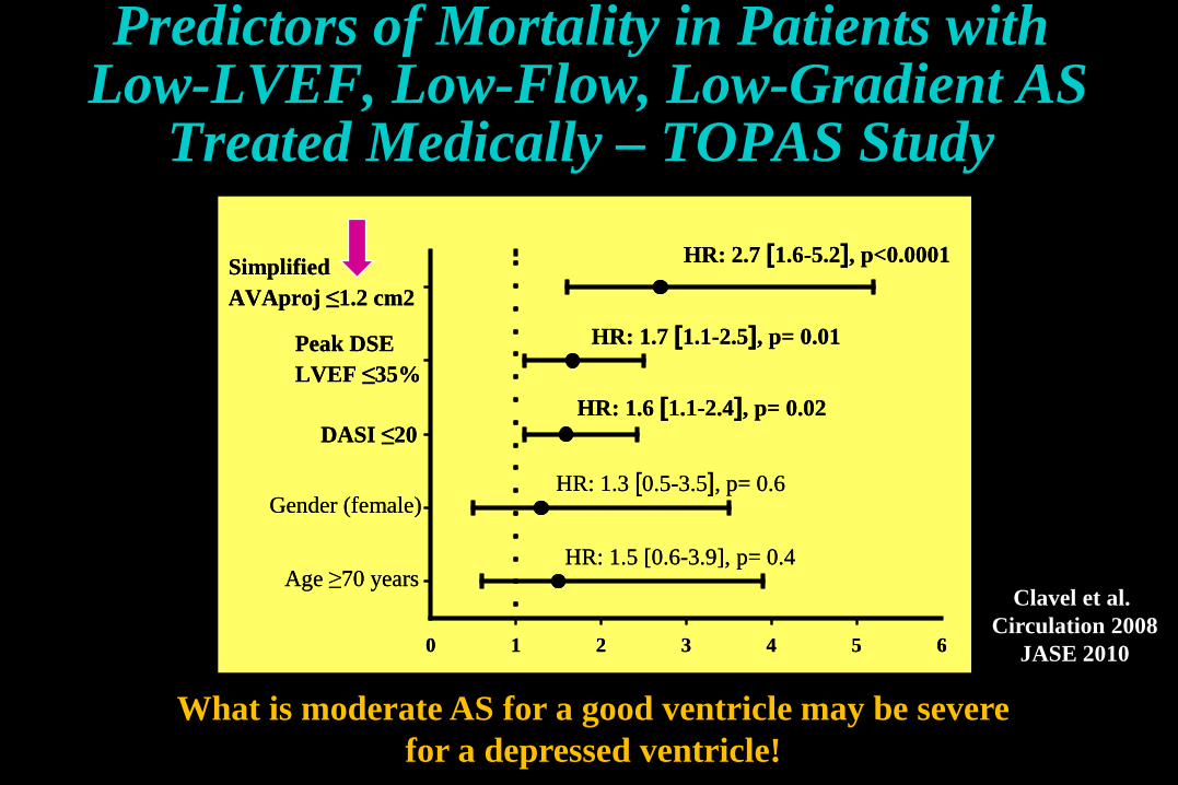
0 1 2 3 4 5 6
Age ≥70 years
Gender (female)
DASI ≤20
Peak DSE LVEF ≤35%
SimplifiedAVAproj ≤1.2 cm2
HR: 1.5 [0.6-3.9], p= 0.4
HR: 1.3 [0.5-3.5], p= 0.6
HR: 1.6 [1.1-2.4], p= 0.02
HR: 1.7 [1.1-2.5], p= 0.01
HR: 2.7 [1.6-5.2], p<0.0001
0 1 2 3 4 5 6
Age ≥70 years
Gender (female)
DASI ≤20
Peak DSE LVEF ≤35%
SimplifiedAVAproj ≤1.2 cm2
HR: 1.5 [0.6-3.9], p= 0.4
HR: 1.3 [0.5-3.5], p= 0.6
HR: 1.6 [1.1-2.4], p= 0.02
HR: 1.7 [1.1-2.5], p= 0.01
HR: 2.7 [1.6-5.2], p<0.0001
Predictors of Mortality in Patients withLow-LVEF, Low-Flow, Low-Gradient AS
Treated Medically – TOPAS Study
Clavel et al. Circulation 2008
JASE 2010
What is moderate AS for a good ventricle may be severe for a depressed ventricle!
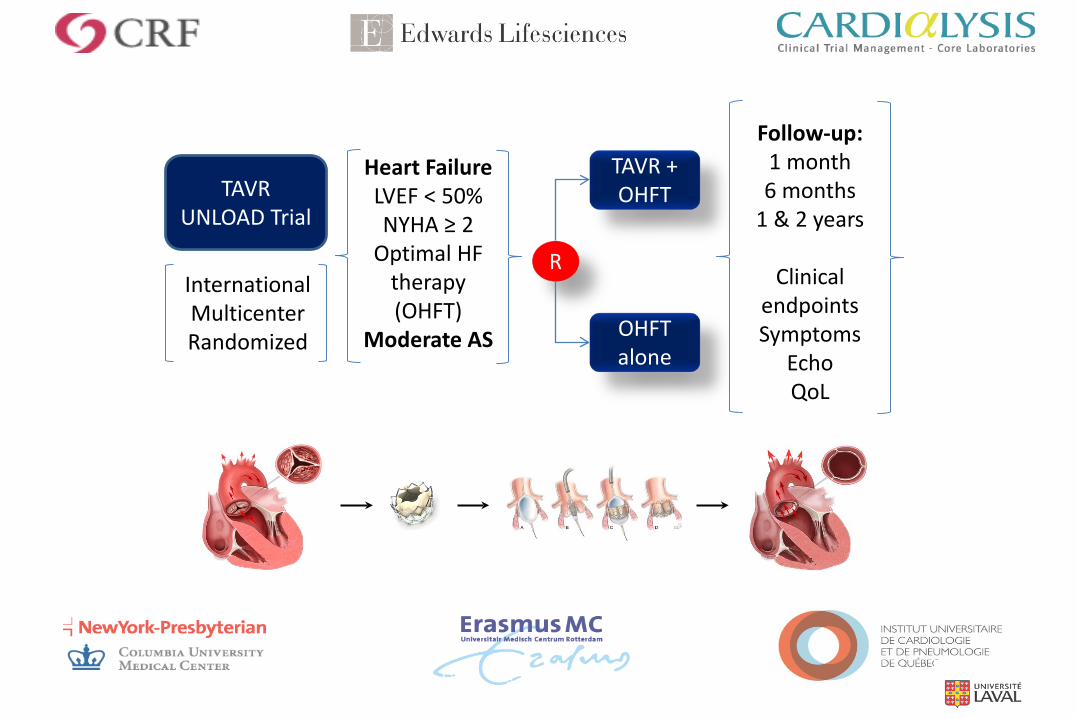
InternationalMulticenterRandomized
TAVR UNLOAD Trial
Heart FailureLVEF < 50%NYHA ≥ 2
Optimal HF therapy(OHFT)
Moderate AS
R
TAVR + OHFT
OHFT alone
Follow-up:1 month6 months
1 & 2 years
ClinicalendpointsSymptoms
EchoQoL
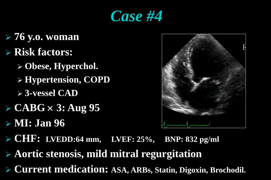
Case #4 76 y.o. woman Risk factors:Obese, Hyperchol.Hypertension, COPD 3-vessel CAD
CABG × 3: Aug 95 MI: Jan 96 CHF: LVEDD:64 mm, LVEF: 25%, BNP: 832 pg/ml
Aortic stenosis, mild mitral regurgitation Current medication: ASA, ARBs, Statin, Digoxin, Brochodil.
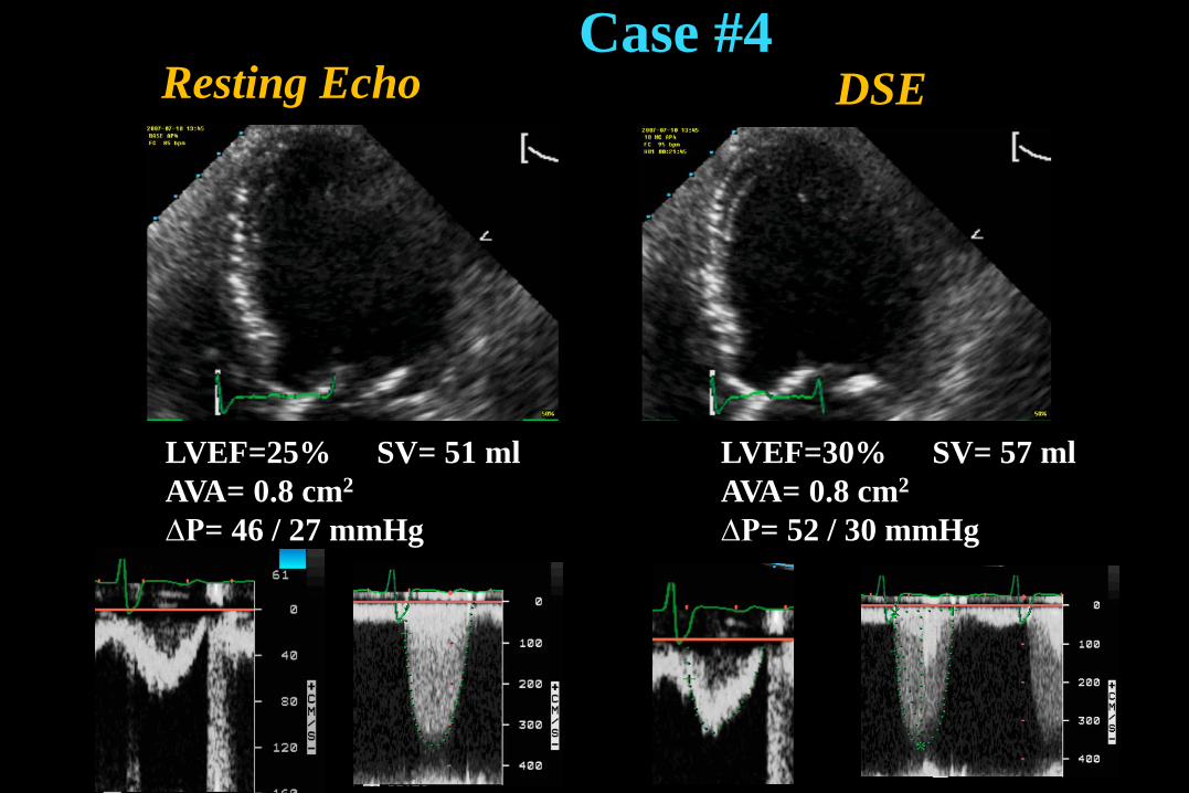
LVEF=25% SV= 51 mlAVA= 0.8 cm2
∆P= 46 / 27 mmHg
LVEF=30% SV= 57 mlAVA= 0.8 cm2
∆P= 52 / 30 mmHg
DSEResting EchoCase #4
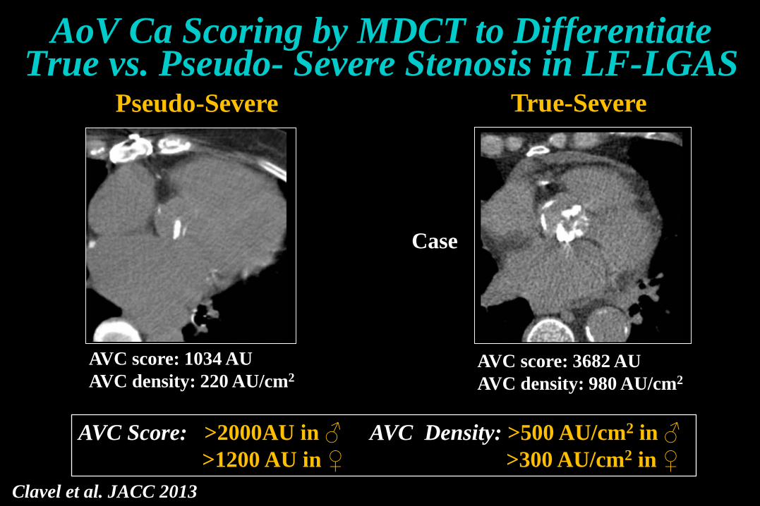
Fig. 4AoV Ca Scoring by MDCT to Differentiate True vs. Pseudo- Severe Stenosis in LF-LGAS
Pseudo-Severe True-Severe
AVC score: 1034 AU AVC density: 220 AU/cm2
AVC score: 3682 AUAVC density: 980 AU/cm2
AVC Score: >2000AU in ♂ AVC Density: >500 AU/cm2 in ♂>1200 AU in ♀ >300 AU/cm2 in ♀
Clavel et al. JACC 2013
Case
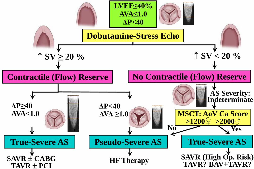
LVEF≤40%AVA≤1.0ΔP<40
No Contractile (Flow) Reserve
↑ SV < 20 %
AS Severity:Indeterminate
Yes
SAVR (High Op. Risk)TAVR? BAV+TAVR?
True-Severe AS
↑ SV ≥ 20 %
Contractile (Flow) Reserve
SAVR ± CABGTAVR ± PCI
ΔP≥40AVA<1.0
True-Severe AS
No
Pseudo-Severe AS
ΔP<40AVA ≥1.0
Dobutamine-Stress Echo
MSCT: AoV Ca Score >1200♀ >2000♂
HF Therapy
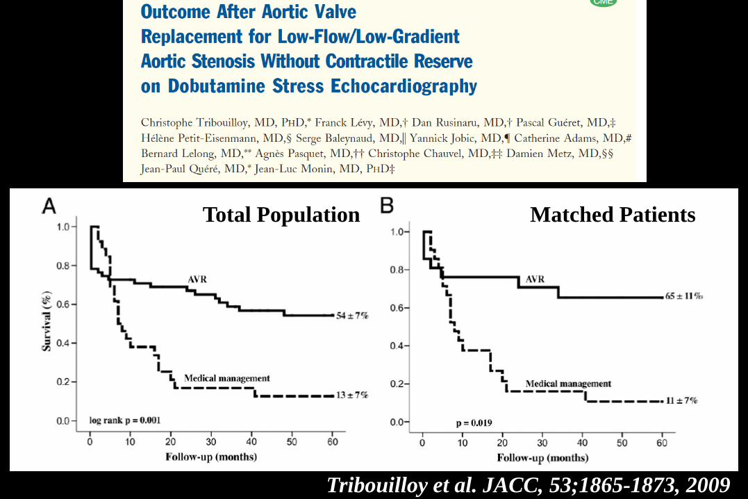
Total Population Matched Patients
Tribouilloy et al. JACC, 53;1865-1873, 2009
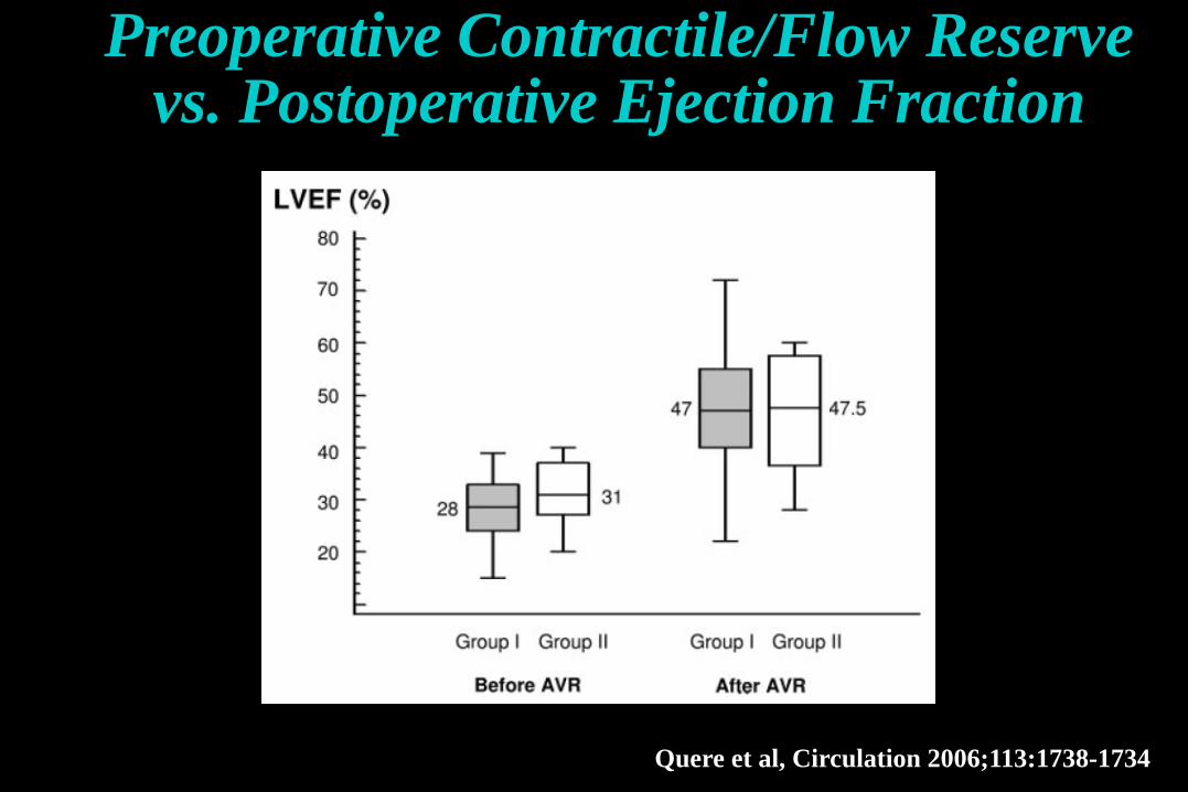
Preoperative Contractile/Flow Reserve vs. Postoperative Ejection Fraction
Quere et al, Circulation 2006;113:1738-1734
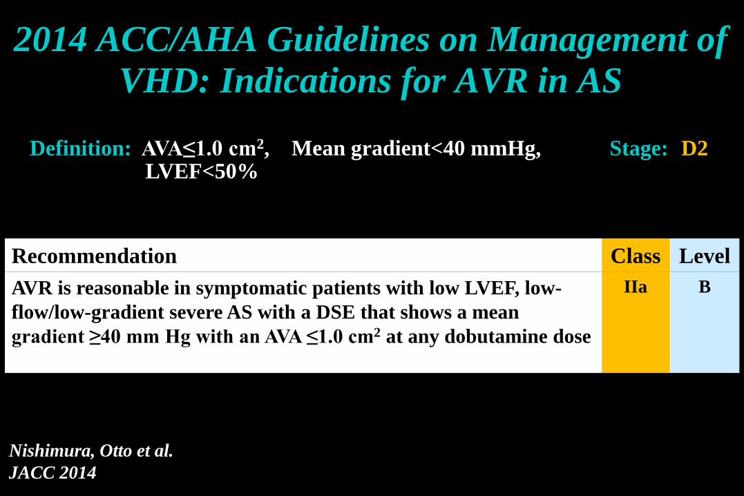
Recommendation Class LevelAVR is reasonable in symptomatic patients with low LVEF, low-flow/low-gradient severe AS with a DSE that shows a mean gradient ≥40 mm Hg with an AVA ≤1.0 cm2 at any dobutamine dose
IIa B
2014 ACC/AHA Guidelines on Management of VHD: Indications for AVR in AS
Nishimura, Otto et al.JACC 2014
Definition: AVA≤1.0 cm2, Mean gradient<40 mmHg,LVEF<50%
Stage: D2

2012 ESC/EACTS Guidelines on Management of VHD: Indications for AVR in AS
Vahanian et al. EHJ 2012
Severe AS on DSE: Increase in AVA <0.2 cm2 with final AVA <1 cm2; mean gradient >40 mmHg
Flow reserve: >20% increase in stroke volume
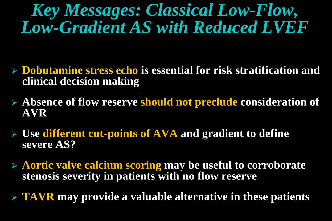
Key Messages: Classical Low-Flow, Low-Gradient AS with Reduced LVEF
Dobutamine stress echo is essential for risk stratification and clinical decision making
Absence of flow reserve should not preclude consideration of AVR
Use different cut-points of AVA and gradient to define severe AS?
Aortic valve calcium scoring may be useful to corroborate stenosis severity in patients with no flow reserve
TAVR may provide a valuable alternative in these patients

Hachicha Z et al., Circulation, 2007Dumesnil et al. Eur Heart J, 2009Pibarot & Dumesnil JACC, in press, 2012
↑AgeWomenHypertensionMetS – Diabetes
“Paradoxical” Low-Flow, Low-Gradient AS with
Preserved LVEF (Stage D3)
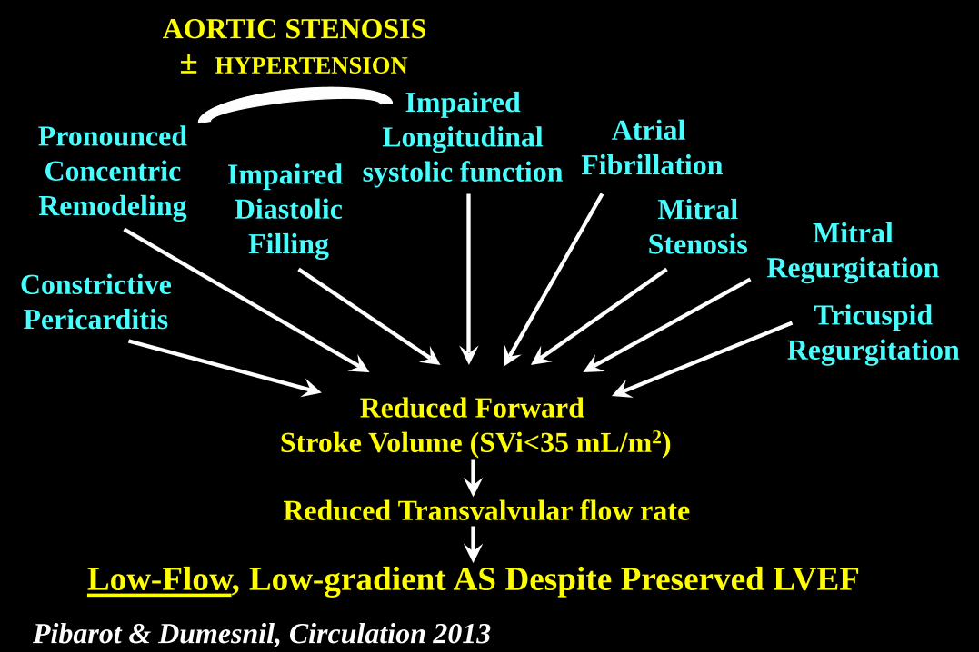
Pronounced ConcentricRemodeling
Impaired DiastolicFilling
Impaired Longitudinal
systolic function Mitral
Stenosis
TricuspidRegurgitation
Reduced Forward Stroke Volume (SVi<35 mL/m2)
Reduced Transvalvular flow rate
Low-Flow, Low-gradient AS Despite Preserved LVEF
MitralRegurgitation
Pibarot & Dumesnil, Circulation 2013
Atrial Fibrillation
ConstrictivePericarditis
AORTIC STENOSIS± HYPERTENSION
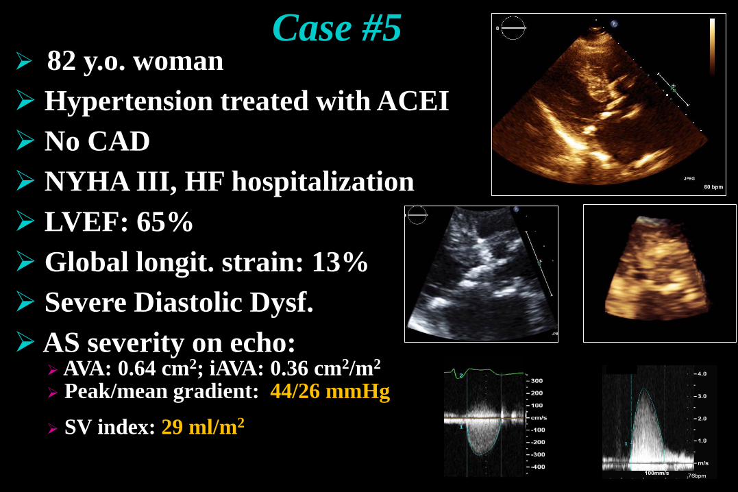
82 y.o. woman Hypertension treated with ACEI No CAD NYHA III, HF hospitalization LVEF: 65% Global longit. strain: 13% Severe Diastolic Dysf.AS severity on echo:
AVA: 0.64 cm2; iAVA: 0.36 cm2/m2
Peak/mean gradient: 44/26 mmHg SV index: 29 ml/m2
Case #5
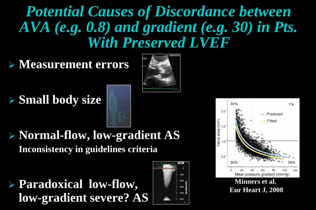
Potential Causes of Discordance betweenAVA (e.g. 0.8) and gradient (e.g. 30) in Pts.
With Preserved LVEF Measurement errors
Small body size
Normal-flow, low-gradient ASInconsistency in guidelines criteria
Paradoxical low-flow, low-gradient severe? AS
Minners et al. Eur Heart J, 2008
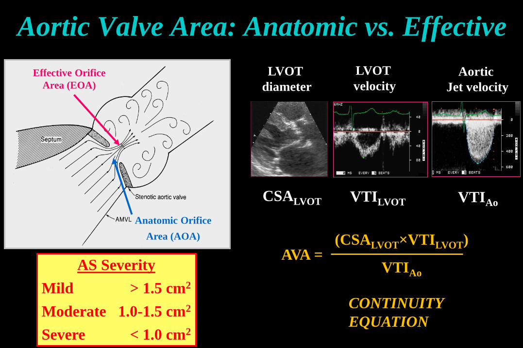
LVOT diameter
Aortic Jet velocity
LVOT velocity
CONTINUITY EQUATION
Aortic Valve Area: Anatomic vs. Effective
(CSALVOT×VTILVOT)
VTIAoAVA =
CSALVOT VTILVOT VTIAo
Effective Orifice Area (EOA)
Anatomic Orifice Area (AOA)
AS SeverityMild > 1.5 cm2
Moderate 1.0-1.5 cm2
Severe < 1.0 cm2
ProximalLVOT18 mm
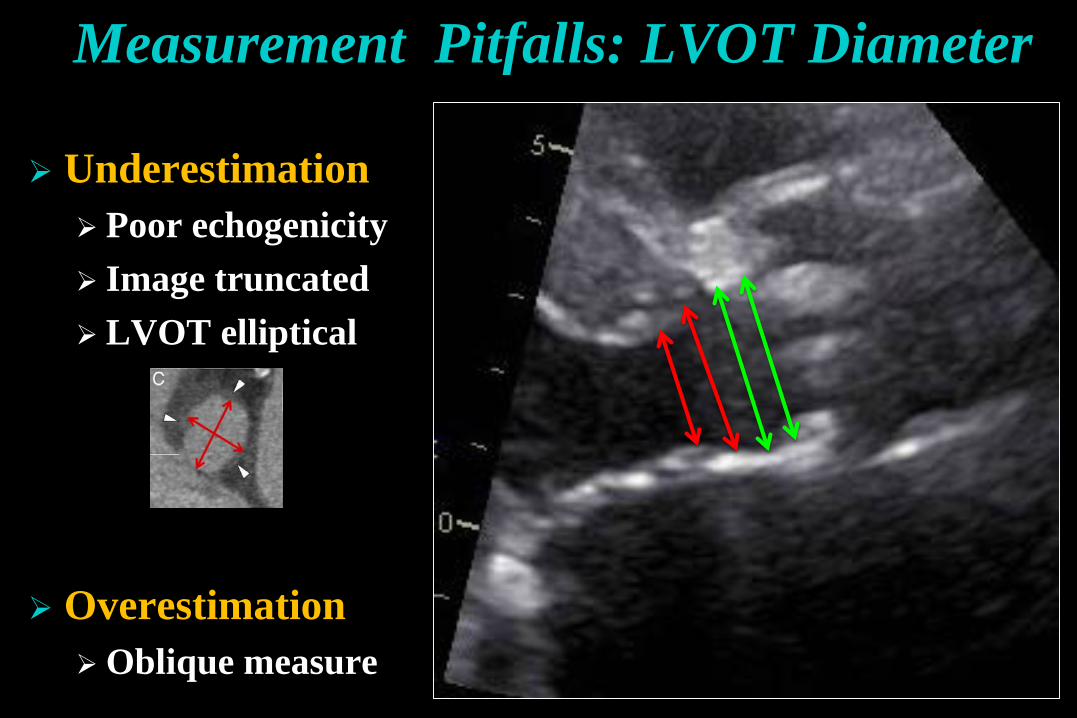
Measurement Pitfalls: LVOT Diameter
Underestimation Poor echogenicity Image truncated LVOT elliptical
OverestimationOblique measure
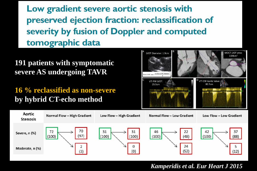
Kamperidis et al. Eur Heart J 2015
191 patients with symptomatic severe AS undergoing TAVR
16 % reclassified as non-severe by hybrid CT-echo method
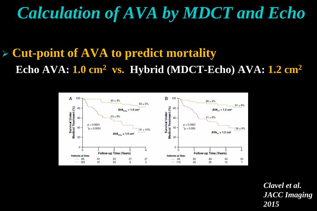
Calculation of AVA by MDCT and Echo
Cut-point of AVA to predict mortalityEcho AVA: 1.0 cm2 vs. Hybrid (MDCT-Echo) AVA: 1.2 cm2
Clavel et al. JACC Imaging 2015
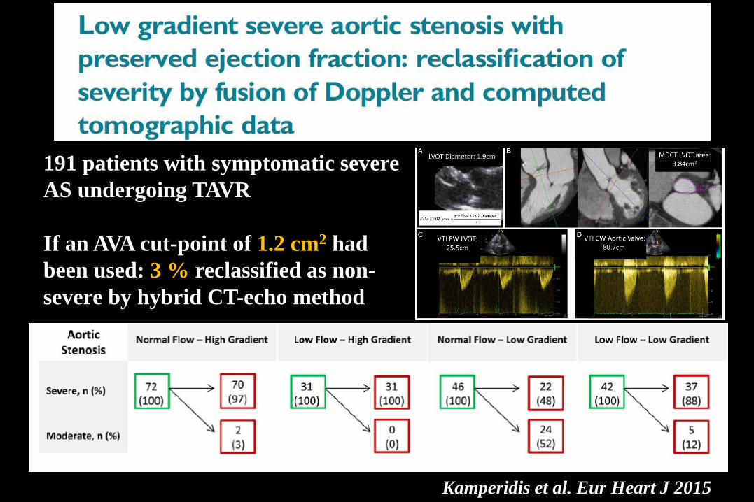
Kamperidis et al. Eur Heart J 2015
191 patients with symptomatic severe AS undergoing TAVR
If an AVA cut-point of 1.2 cm2 had been used: 3 % reclassified as non-severe by hybrid CT-echo method
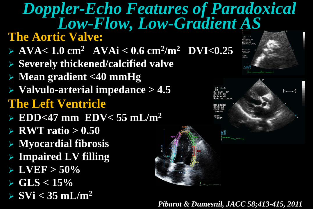
The Aortic Valve: AVA< 1.0 cm2 AVAi < 0.6 cm2/m2 DVI<0.25 Severely thickened/calcified valve Mean gradient <40 mmHg Valvulo-arterial impedance > 4.5The Left Ventricle EDD<47 mm EDV< 55 mL/m2
RWT ratio > 0.50 Myocardial fibrosis Impaired LV filling LVEF > 50% GLS < 15% SVi < 35 mL/m2
Pibarot & Dumesnil, JACC 58;413-415, 2011
Doppler-Echo Features of ParadoxicalLow-Flow, Low-Gradient AS
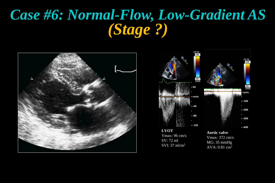
Case #6: Normal-Flow, Low-Gradient AS(Stage ?)
LVOTVmax: 96 cm/sSV: 72 mlSVI: 37 ml/m2
Aortic valveVmax: 372 cm/sMG: 35 mmHgAVA: 0.81 cm2
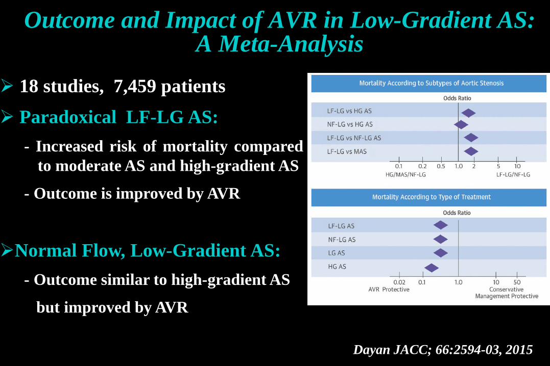
Outcome and Impact of AVR in Low-Gradient AS: A Meta-Analysis
Dayan JACC; 66:2594-03, 2015
18 studies, 7,459 patients Paradoxical LF-LG AS:
- Increased risk of mortality comparedto moderate AS and high-gradient AS
- Outcome is improved by AVR
Normal Flow, Low-Gradient AS:- Outcome similar to high-gradient AS
but improved by AVR
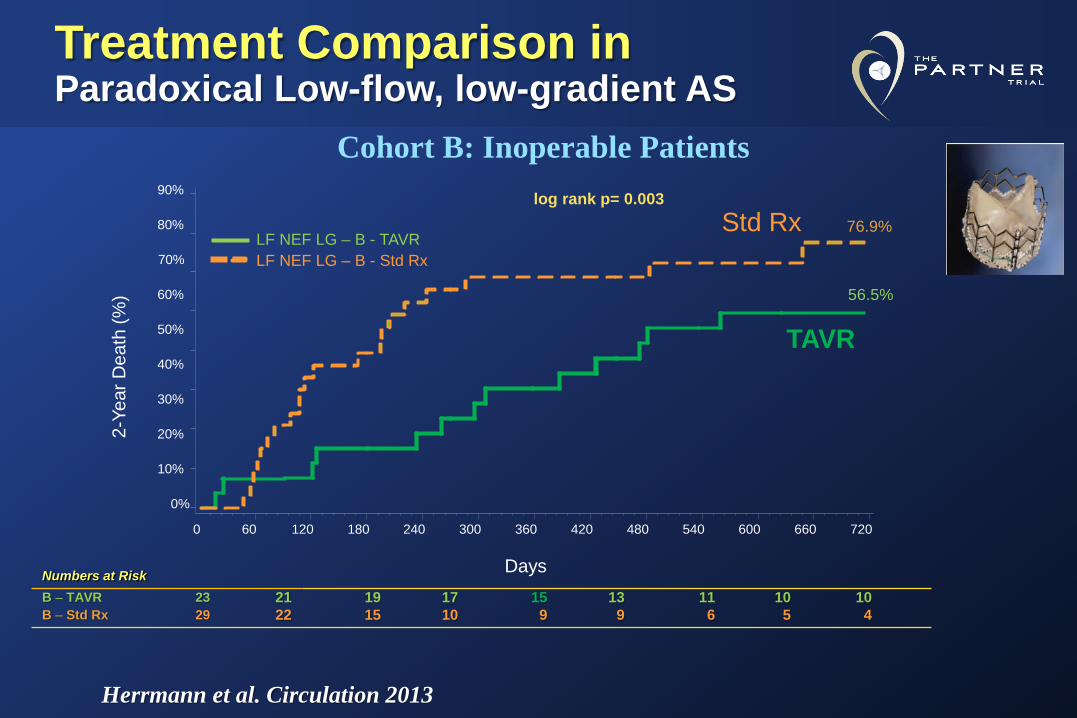
Treatment Comparison inParadoxical Low-flow, low-gradient AS
0%
10%
20%
30%
40%
50%
60%
80%
90%
60 120 180 240 300 360 420 480 540 600 660 720
70%
2-Ye
ar D
eath
(%) 56.5%
76.9%
0
log rank p= 0.003
Numbers at RiskB – TAVR 23 21 19 17 15 13 11 10 10B – Std Rx 29 22 15 10 9 9 6 5 4
LF NEF LG – B - TAVRLF NEF LG – B - Std Rx
Days
Herrmann et al. Circulation 2013
Cohort B: Inoperable Patients
TAVR
Std Rx
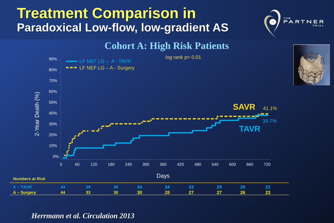
Treatment Comparison inParadoxical Low-flow, low-gradient AS
0%
10%
20%
30%
40%
50%
60%
80%
90%
60 120 180 240 300 360 420 480 540 600 660 720
70%
2-Ye
ar D
eath
(%)
39.7%
41.1%
0
log rank p= 0.01
Numbers at Risk
A – TAVR 43 39 38 34 34 33 29 26 22A – Surgery 44 33 30 30 28 27 27 26 23
LF NEF LG – A - TAVRLF NEF LG – A - Surgery
Days
Herrmann et al. Circulation 2013
TAVR
SAVR
Cohort A: High Risk Patients
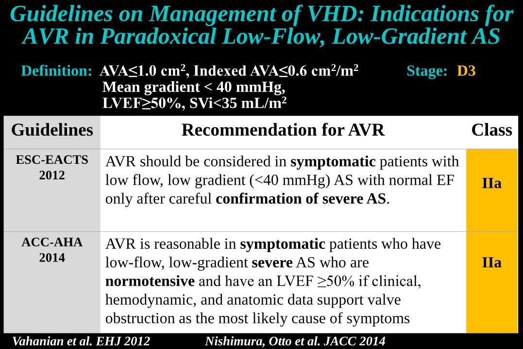
Guidelines Recommendation for AVR ClassESC-EACTS
2012AVR should be considered in symptomatic patients with low flow, low gradient (<40 mmHg) AS with normal EF only after careful confirmation of severe AS.
IIa
ACC-AHA2014
AVR is reasonable in symptomatic patients who have low-flow, low-gradient severe AS who are normotensive and have an LVEF ≥50% if clinical, hemodynamic, and anatomic data support valve obstruction as the most likely cause of symptoms
IIa
Guidelines on Management of VHD: Indications for AVR in Paradoxical Low-Flow, Low-Gradient AS
Vahanian et al. EHJ 2012 Nishimura, Otto et al. JACC 2014
Definition: AVA≤1.0 cm2, Indexed AVA≤0.6 cm2/m2
Mean gradient < 40 mmHg,LVEF≥50%, SVi<35 mL/m2
Stage: D3

AVC Score:3200 AU
Case #5: Aortic Valve Calcium Scoring by MDCT
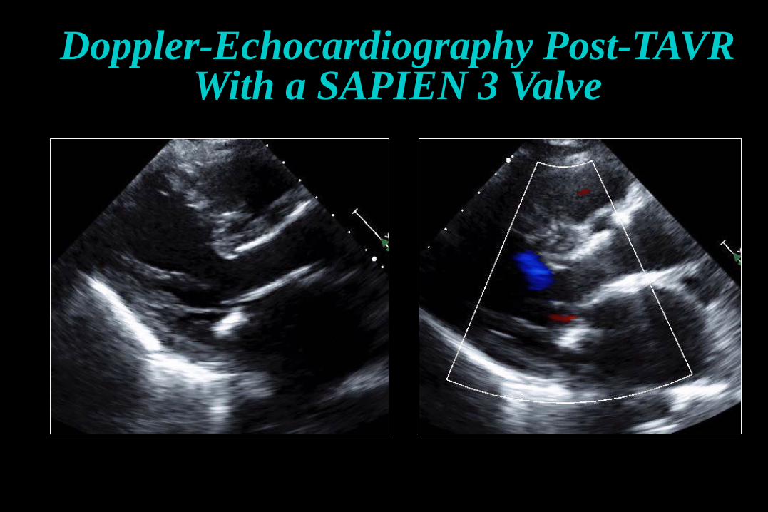
Doppler-Echocardiography Post-TAVRWith a SAPIEN 3 Valve
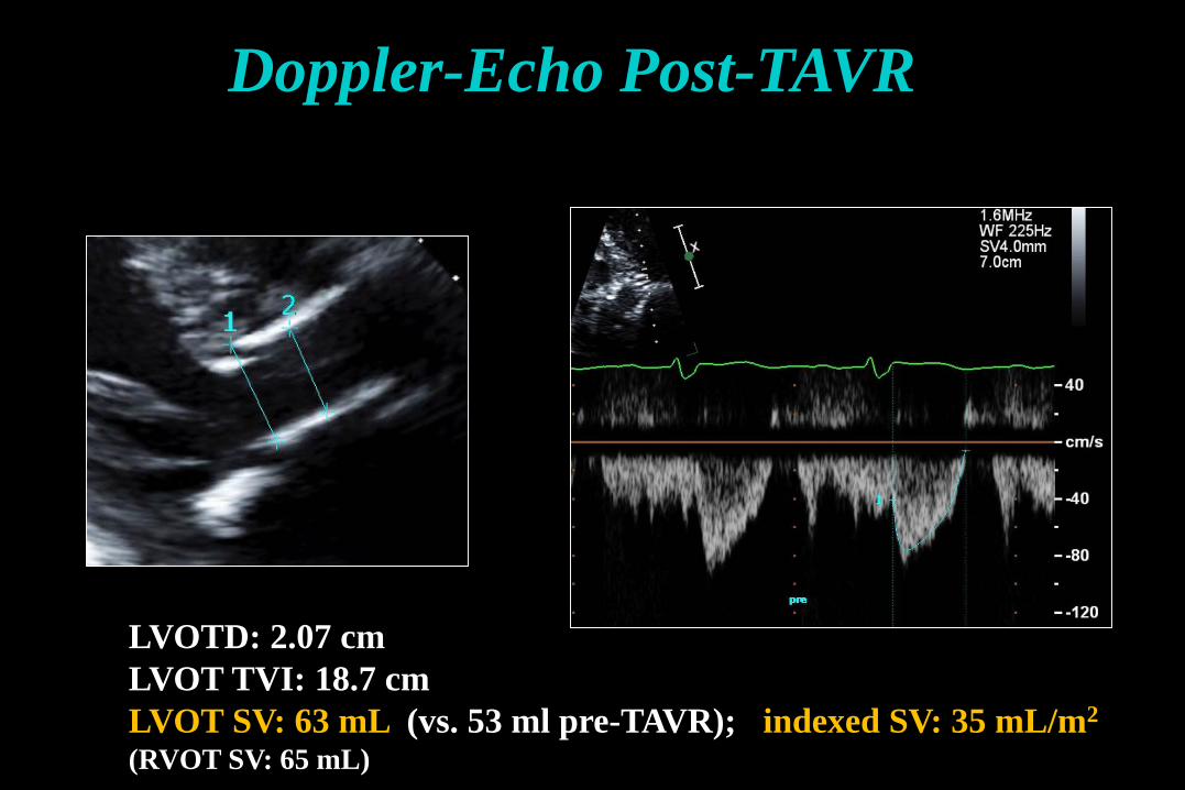
LVOTD: 2.07 cmLVOT TVI: 18.7 cmLVOT SV: 63 mL (vs. 53 ml pre-TAVR); indexed SV: 35 mL/m2
(RVOT SV: 65 mL)
Doppler-Echo Post-TAVR
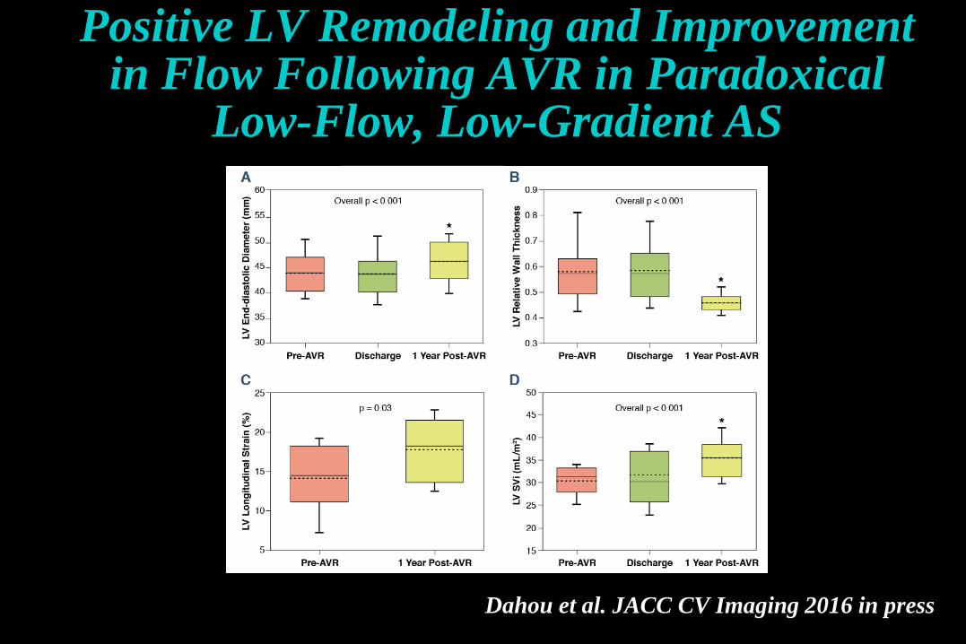
Positive LV Remodeling and Improvement in Flow Following AVR in Paradoxical
Low-Flow, Low-Gradient AS
Dahou et al. JACC CV Imaging 2016 in press
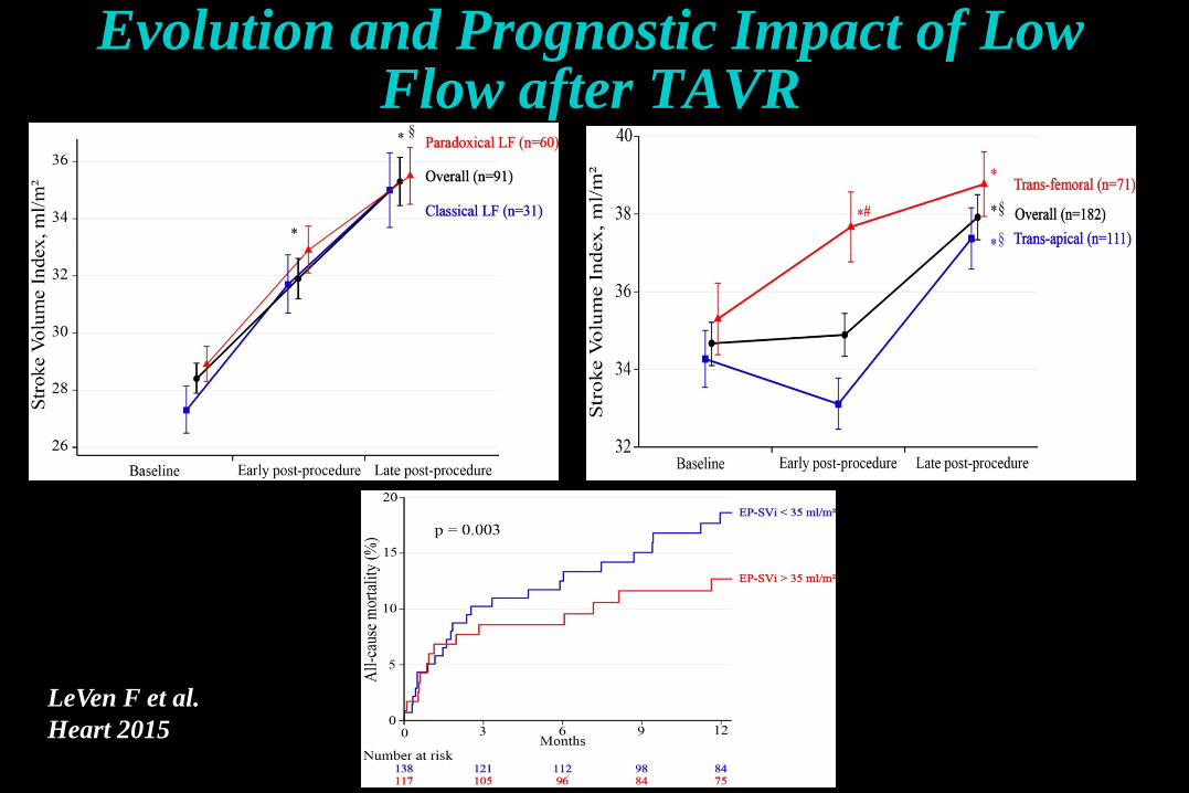
Evolution and Prognostic Impact of Low Flow after TAVR
LeVen F et al.Heart 2015
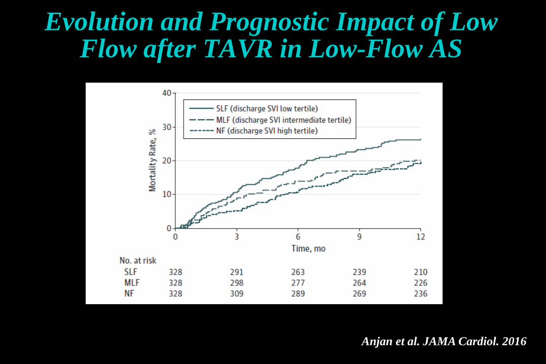
Evolution and Prognostic Impact of Low Flow after TAVR in Low-Flow AS
Anjan et al. JAMA Cardiol. 2016
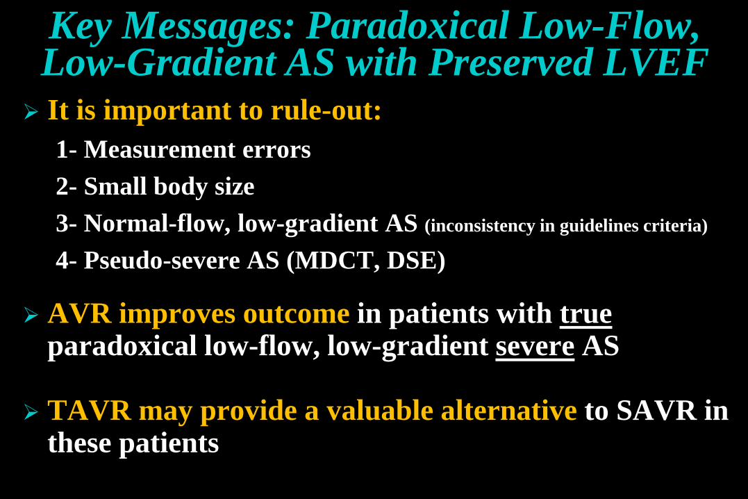
It is important to rule-out:1- Measurement errors2- Small body size3- Normal-flow, low-gradient AS (inconsistency in guidelines criteria)
4- Pseudo-severe AS (MDCT, DSE)
AVR improves outcome in patients with trueparadoxical low-flow, low-gradient severe AS
TAVR may provide a valuable alternative to SAVR in these patients
Key Messages: Paradoxical Low-Flow, Low-Gradient AS with Preserved LVEF