Low Gradient Severe (?) Aortic Stenosis
Philippe Pibarot, DVM, PhD, FACC, FAHA, FESC, FASECanada Research Chair in Valvular Heart Diseases
UniversitéLAVAL
Institut Universitaire de Cardiologieet de Pneumologie de Québec / Québec Heart & Lung Institute

Disclosure: Philippe PibarotFinancial relationship with industry:
Edwards Lifesciences: Echo CoreLab for PARTNER 2–
SAPIEN 3, PARTNER 3, TAVR-UNLOAD trials
V-Wave Ltd: Echo CoreLab for FIM Study
Cardiac Phoenix: Echo CoreLab for BACE FIM Study
Other financial disclosure:
Research Grants from Canadian Institutes of Health
Research and Heart & Stroke Foundation of Quebec
Off label Use: None

LVEF >50%<50%
Low Gradient ASAVA≤1.0 cm2 MG<40 mmHg
«CLASSICAL»LOW-FLOW
LOW-GRADIENTD2 Stage
«PARADOXICAL»LOW-FLOW
LOW-GRADIENTD3 Stage
NORMAL-FLOWLOW-GRADIENT
D? Stage
SVi<35 mL/m2 >35 mL/m2
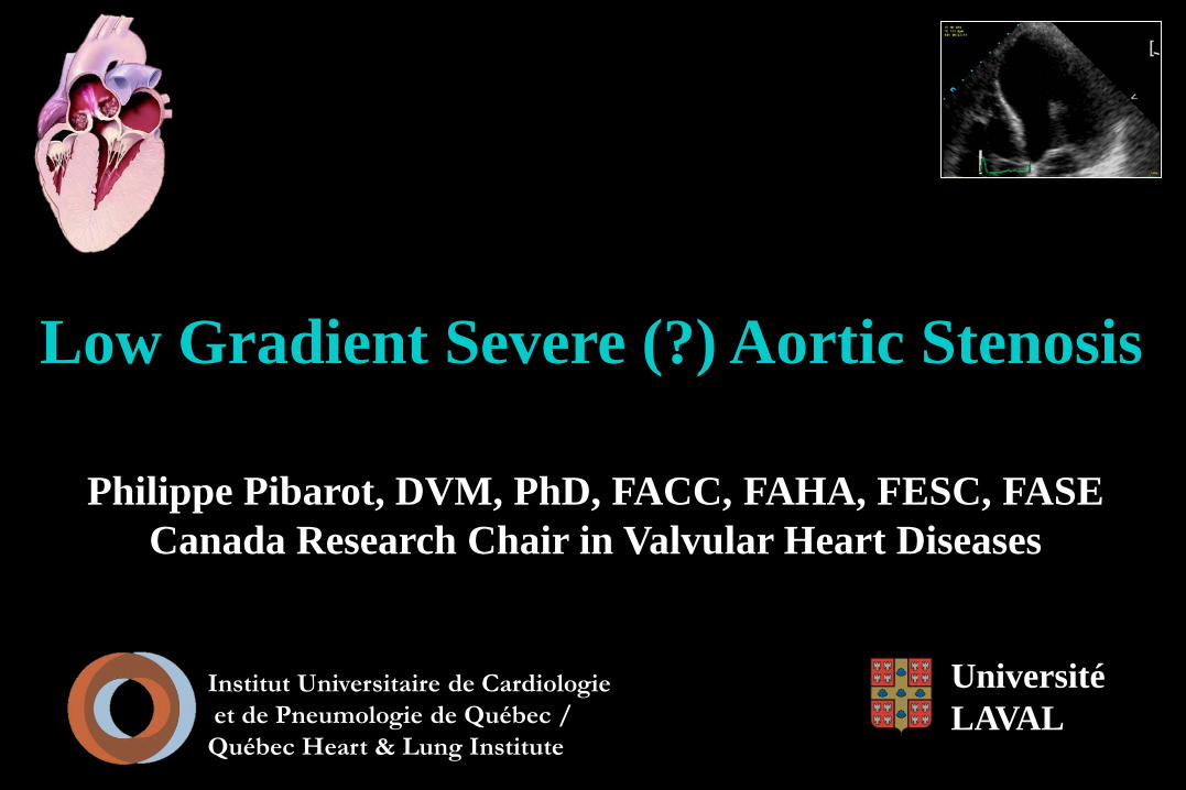
“Classical” Low-Flow, Low-Gradient AS with
Reduced LVEF (D2 Stage)
Low Flow Low FlowNormal Flow Normal Flow
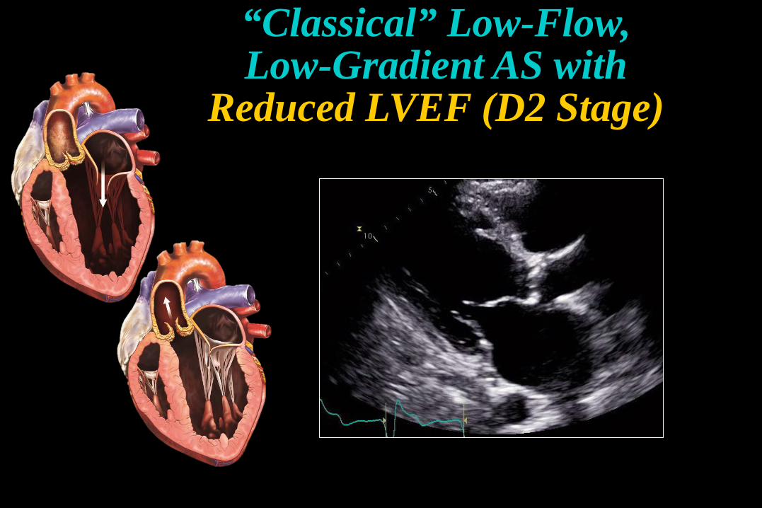
True-Severe AS Pseudo-Severe AS
Low-Flow, Low-Gradient Severe(?) AS
∆P
AVA
Gradient = Q2
K ×AVA2
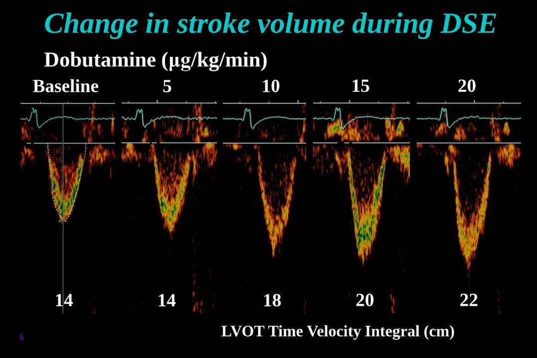
Change in stroke volume during DSEDobutamine (μg/kg/min)
LVOT Time Velocity Integral (cm)
Baseline
14
5
14 20
15
22
20
18
10
6
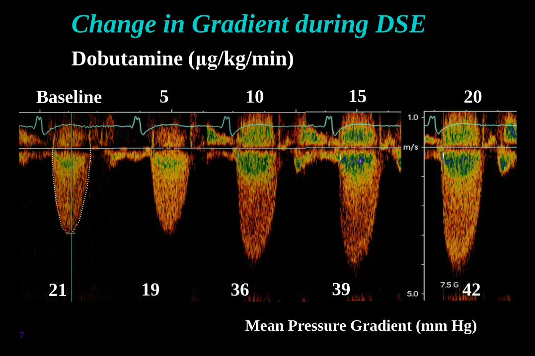
Mean Pressure Gradient (mm Hg)
Baseline
21
5
19
10
36
15
39
20
42
7
Change in Gradient during DSEDobutamine (μg/kg/min)
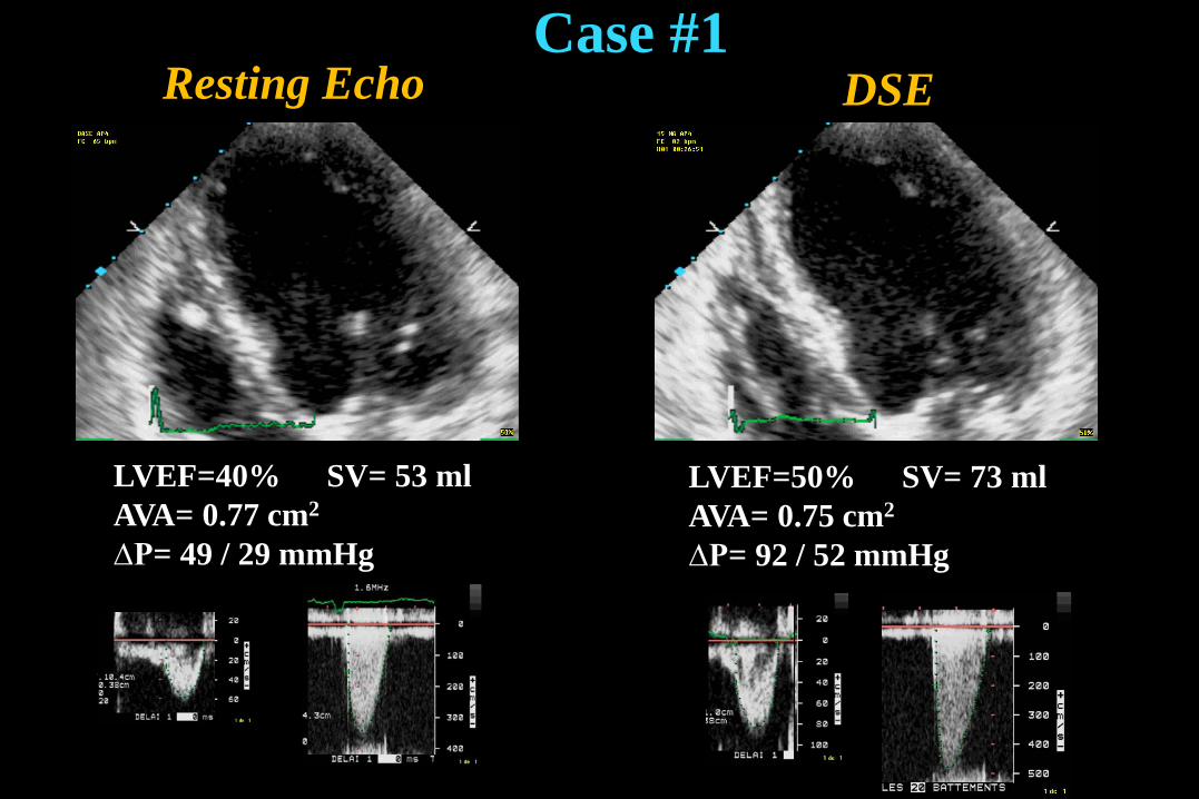
LVEF=40% SV= 53 mlAVA= 0.77 cm2
∆P= 49 / 29 mmHg
LVEF=50% SV= 73 mlAVA= 0.75 cm2
∆P= 92 / 52 mmHg
DSEResting EchoCase #1
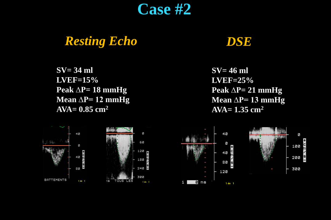
DSEResting Echo
Case #2
SV= 46 mlLVEF=25%Peak ∆P= 21 mmHgMean ∆P= 13 mmHgAVA= 1.35 cm2
SV= 34 mlLVEF=15%Peak ∆P= 18 mmHgMean ∆P= 12 mmHgAVA= 0.85 cm2
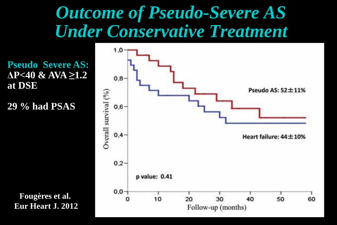
Outcome of Pseudo-Severe AS Under Conservative Treatment
Fougères et al. Eur Heart J. 2012
Pseudo Severe AS:ΔP<40 & AVA ≥1.2 at DSE
29 % had PSAS
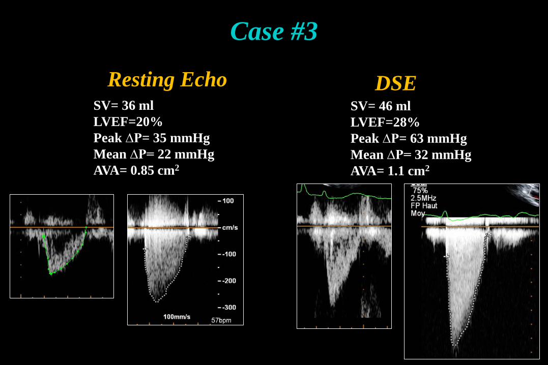
DSEResting EchoSV= 46 mlLVEF=28%Peak ∆P= 63 mmHgMean ∆P= 32 mmHgAVA= 1.1 cm2
SV= 36 mlLVEF=20%Peak ∆P= 35 mmHgMean ∆P= 22 mmHgAVA= 0.85 cm2
Case #3
0 1 2 3 4 5 6
Age ≥70 years
Gender (female)
DASI ≤20
Peak DSE LVEF ≤35%
SimplifiedAVAproj ≤1.2 cm2
HR: 1.5 [0.6-3.9], p= 0.4
HR: 1.3 [0.5-3.5], p= 0.6
HR: 1.6 [1.1-2.4], p= 0.02
HR: 1.7 [1.1-2.5], p= 0.01
HR: 2.7 [1.6-5.2], p<0.0001
0 1 2 3 4 5 6
Age ≥70 years
Gender (female)
DASI ≤20
Peak DSE LVEF ≤35%
SimplifiedAVAproj ≤1.2 cm2
HR: 1.5 [0.6-3.9], p= 0.4
HR: 1.3 [0.5-3.5], p= 0.6
HR: 1.6 [1.1-2.4], p= 0.02
HR: 1.7 [1.1-2.5], p= 0.01
HR: 2.7 [1.6-5.2], p<0.0001
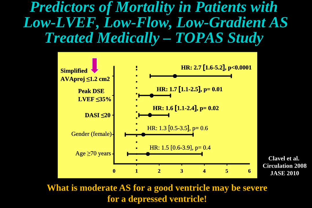
Predictors of Mortality in Patients withLow-LVEF, Low-Flow, Low-Gradient AS
Treated Medically – TOPAS Study
Clavel et al. Circulation 2008
JASE 2010
What is moderate AS for a good ventricle may be severe for a depressed ventricle!
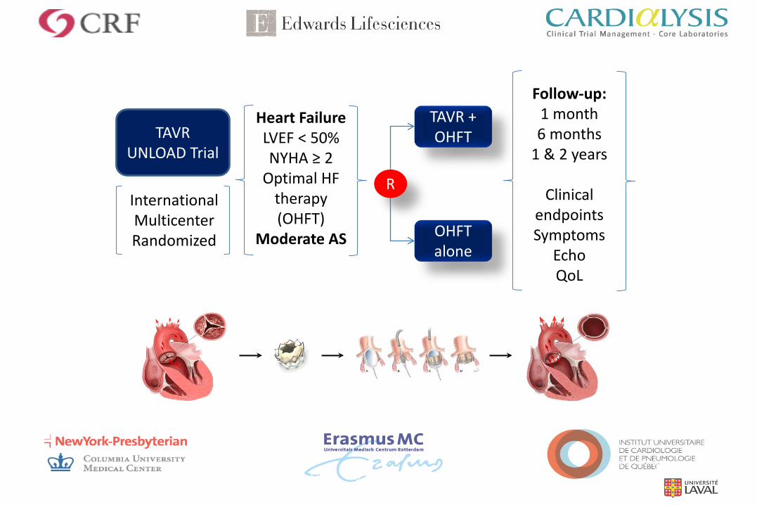
InternationalMulticenterRandomized
TAVR UNLOAD Trial
Heart FailureLVEF < 50%NYHA ≥ 2
Optimal HF therapy(OHFT)
Moderate AS
R
TAVR + OHFT
OHFT alone
Follow-up:1 month6 months
1 & 2 years
ClinicalendpointsSymptoms
EchoQoL
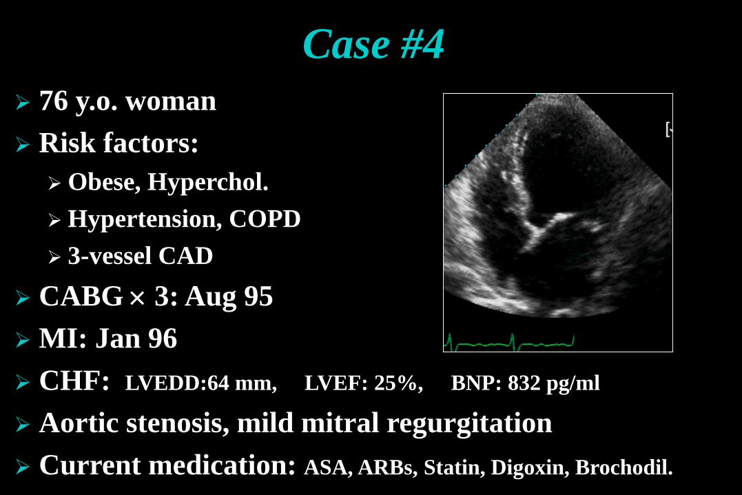
Case #4 76 y.o. woman Risk factors:Obese, Hyperchol.Hypertension, COPD 3-vessel CAD
CABG × 3: Aug 95 MI: Jan 96 CHF: LVEDD:64 mm, LVEF: 25%, BNP: 832 pg/ml
Aortic stenosis, mild mitral regurgitation Current medication: ASA, ARBs, Statin, Digoxin, Brochodil.
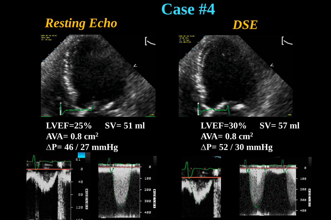
LVEF=25% SV= 51 mlAVA= 0.8 cm2
∆P= 46 / 27 mmHg
LVEF=30% SV= 57 mlAVA= 0.8 cm2
∆P= 52 / 30 mmHg
DSEResting EchoCase #4
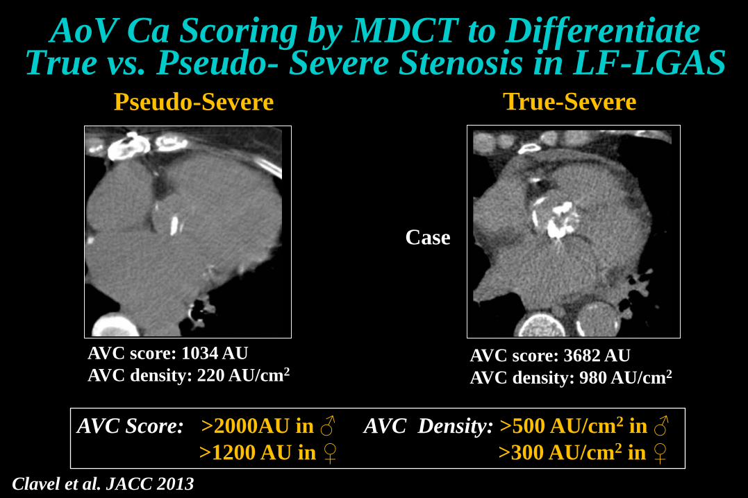
Fig. 4AoV Ca Scoring by MDCT to Differentiate True vs. Pseudo- Severe Stenosis in LF-LGAS
Pseudo-Severe True-Severe
AVC score: 1034 AU AVC density: 220 AU/cm2
AVC score: 3682 AUAVC density: 980 AU/cm2
AVC Score: >2000AU in ♂ AVC Density: >500 AU/cm2 in ♂>1200 AU in ♀ >300 AU/cm2 in ♀
Clavel et al. JACC 2013
Case
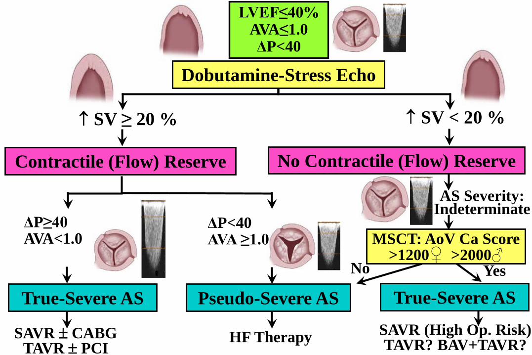
LVEF≤40%AVA≤1.0ΔP<40
No Contractile (Flow) Reserve
↑ SV < 20 %
AS Severity:Indeterminate
Yes
SAVR (High Op. Risk)TAVR? BAV+TAVR?
True-Severe AS
↑ SV ≥ 20 %
Contractile (Flow) Reserve
SAVR ± CABGTAVR ± PCI
ΔP≥40AVA<1.0
True-Severe AS
No
Pseudo-Severe AS
ΔP<40AVA ≥1.0
Dobutamine-Stress Echo
MSCT: AoV Ca Score >1200♀ >2000♂
HF Therapy
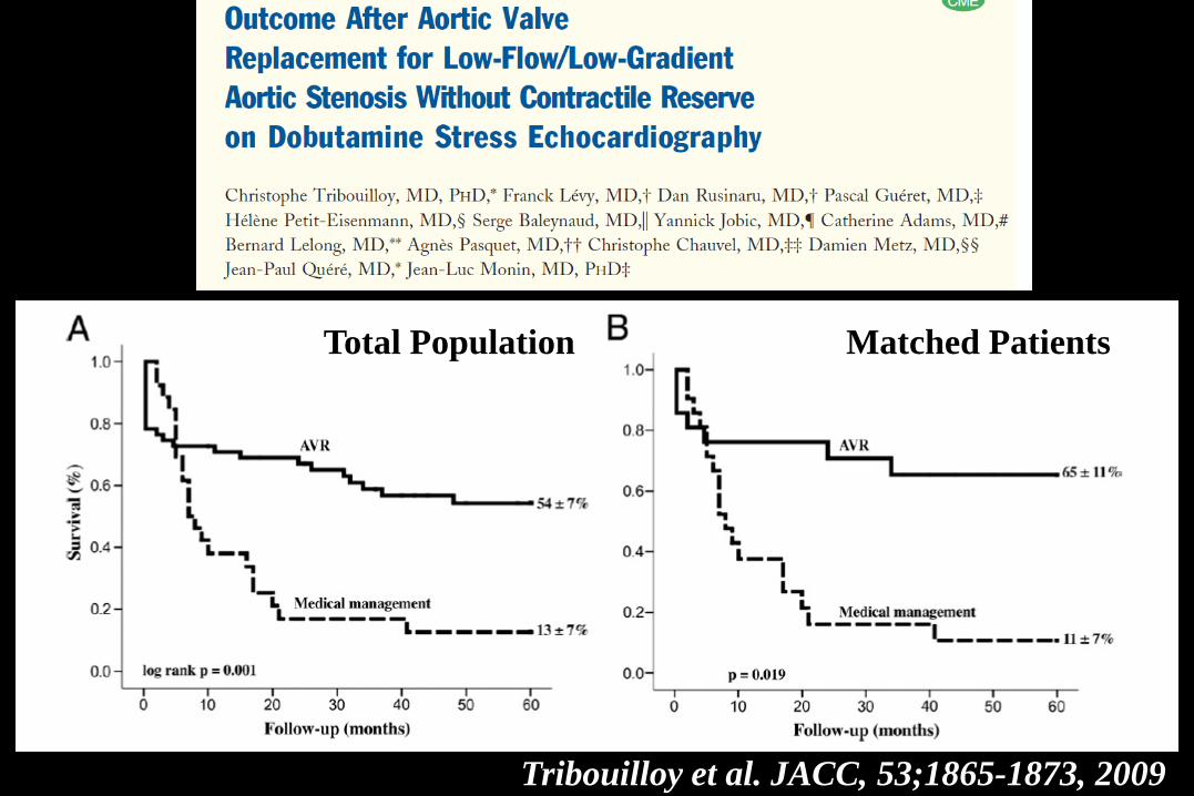
Total Population Matched Patients
Tribouilloy et al. JACC, 53;1865-1873, 2009
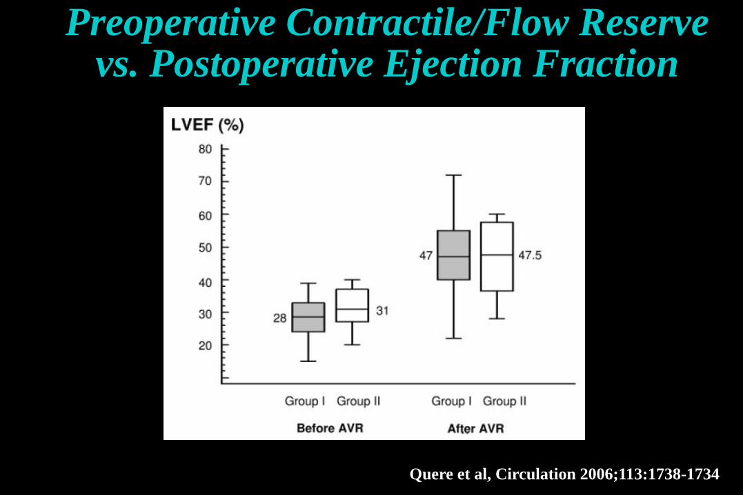
Preoperative Contractile/Flow Reserve vs. Postoperative Ejection Fraction
Quere et al, Circulation 2006;113:1738-1734
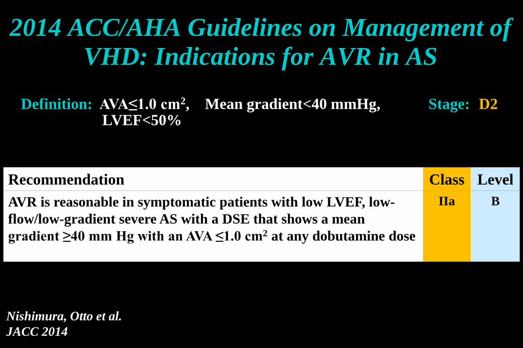
Recommendation Class LevelAVR is reasonable in symptomatic patients with low LVEF, low-flow/low-gradient severe AS with a DSE that shows a mean gradient ≥40 mm Hg with an AVA ≤1.0 cm2 at any dobutamine dose
IIa B
2014 ACC/AHA Guidelines on Management of VHD: Indications for AVR in AS
Nishimura, Otto et al.JACC 2014
Definition: AVA≤1.0 cm2, Mean gradient<40 mmHg,LVEF<50%
Stage: D2

2012 ESC/EACTS Guidelines on Management of VHD: Indications for AVR in AS
Vahanian et al. EHJ 2012
Severe AS on DSE: Increase in AVA <0.2 cm2 with final AVA <1 cm2; mean gradient >40 mmHg
Flow reserve: >20% increase in stroke volume
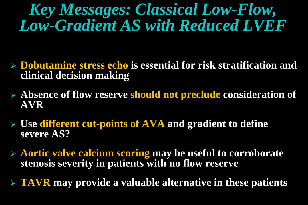
Key Messages: Classical Low-Flow, Low-Gradient AS with Reduced LVEF
Dobutamine stress echo is essential for risk stratification and clinical decision making
Absence of flow reserve should not preclude consideration of AVR
Use different cut-points of AVA and gradient to define severe AS?
Aortic valve calcium scoring may be useful to corroborate stenosis severity in patients with no flow reserve
TAVR may provide a valuable alternative in these patients

Hachicha Z et al., Circulation, 2007Dumesnil et al. Eur Heart J, 2009Pibarot & Dumesnil JACC, in press, 2012
↑AgeWomenHypertensionMetS – Diabetes
“Paradoxical” Low-Flow, Low-Gradient AS with
Preserved LVEF (Stage D3)
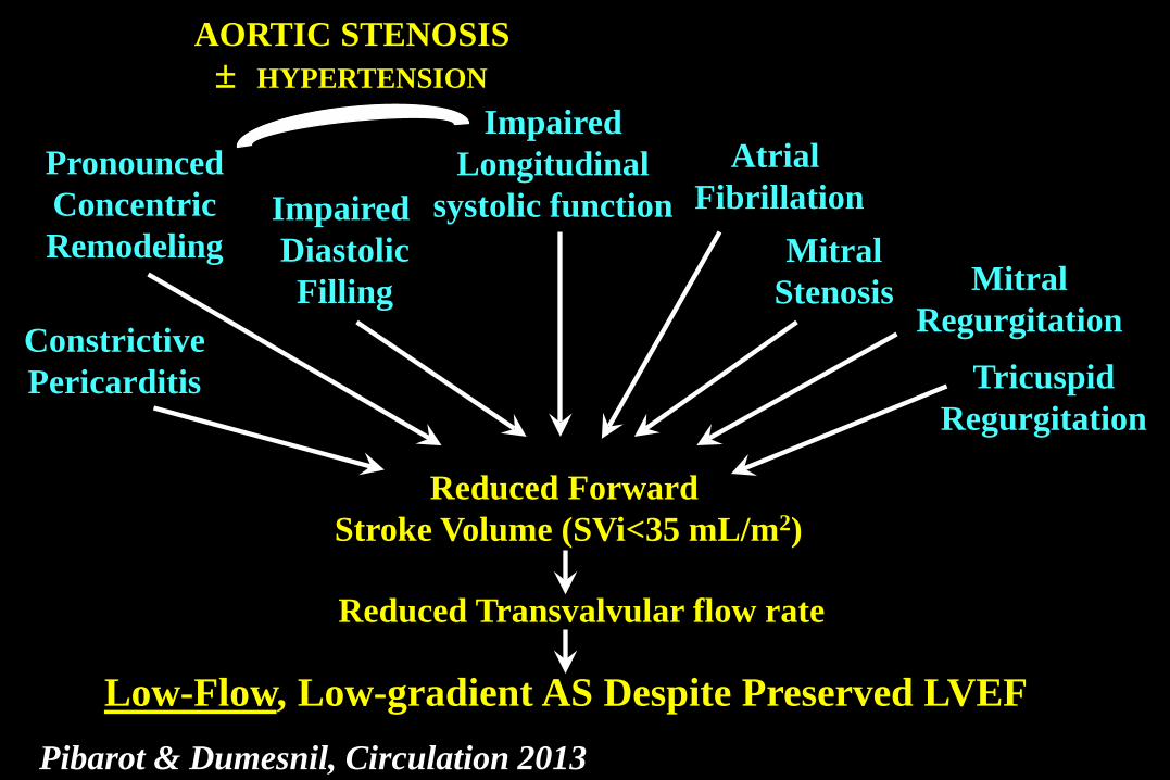
Pronounced ConcentricRemodeling
Impaired DiastolicFilling
Impaired Longitudinal
systolic function Mitral
Stenosis
TricuspidRegurgitation
Reduced Forward Stroke Volume (SVi<35 mL/m2)
Reduced Transvalvular flow rate
Low-Flow, Low-gradient AS Despite Preserved LVEF
MitralRegurgitation
Pibarot & Dumesnil, Circulation 2013
Atrial Fibrillation
ConstrictivePericarditis
AORTIC STENOSIS± HYPERTENSION
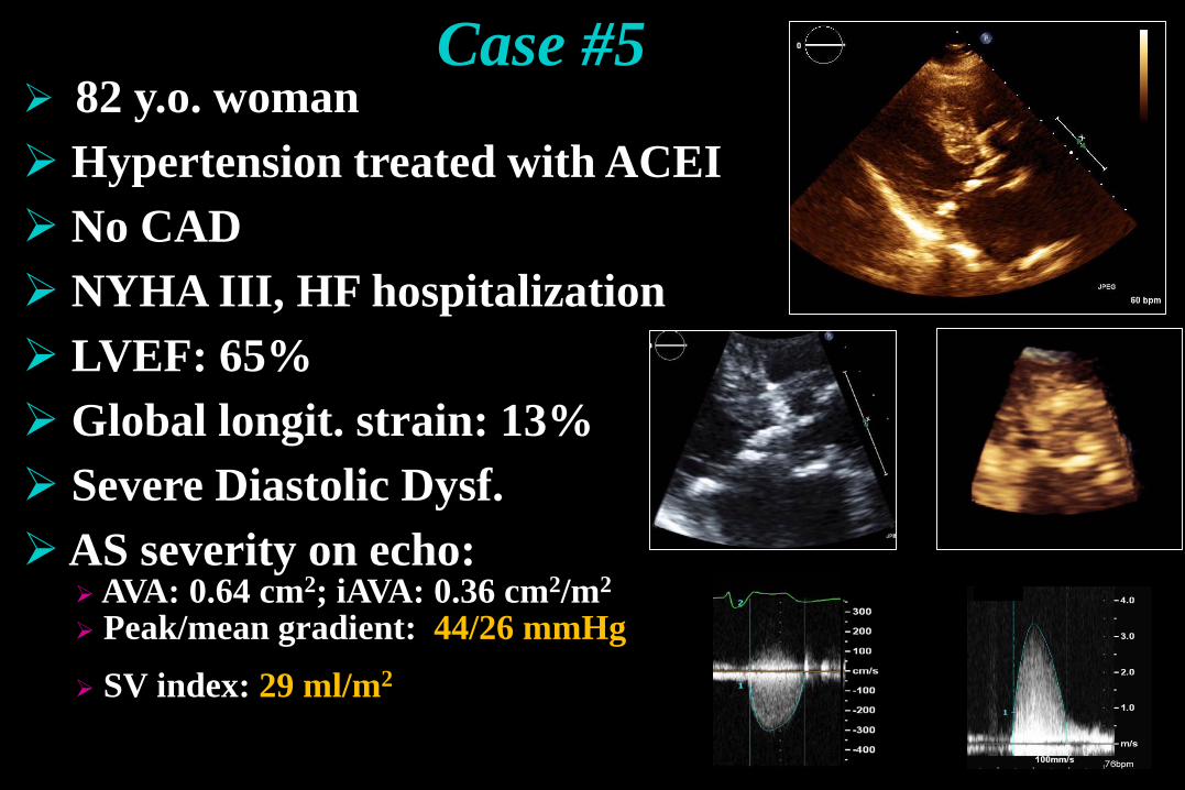
82 y.o. woman Hypertension treated with ACEI No CAD NYHA III, HF hospitalization LVEF: 65% Global longit. strain: 13% Severe Diastolic Dysf.AS severity on echo:
AVA: 0.64 cm2; iAVA: 0.36 cm2/m2
Peak/mean gradient: 44/26 mmHg SV index: 29 ml/m2
Case #5
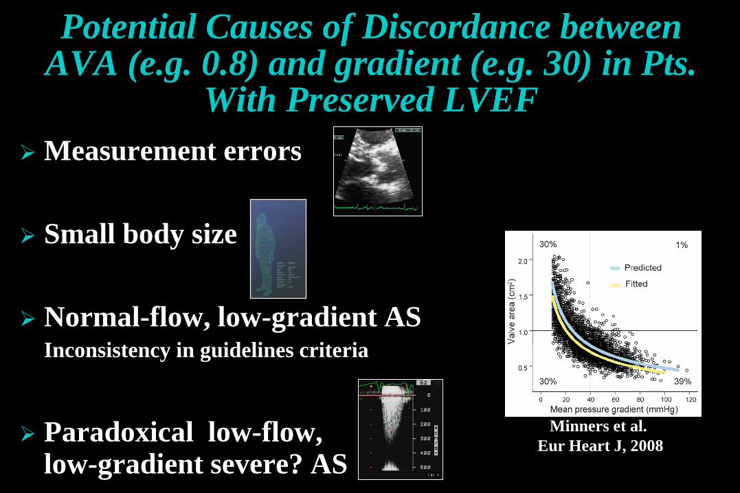
Potential Causes of Discordance betweenAVA (e.g. 0.8) and gradient (e.g. 30) in Pts.
With Preserved LVEF Measurement errors
Small body size
Normal-flow, low-gradient ASInconsistency in guidelines criteria
Paradoxical low-flow, low-gradient severe? AS
Minners et al. Eur Heart J, 2008
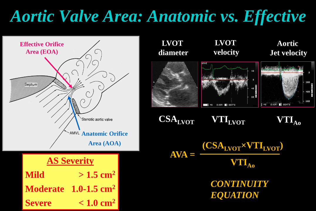
LVOT diameter
Aortic Jet velocity
LVOT velocity
CONTINUITY EQUATION
Aortic Valve Area: Anatomic vs. Effective
(CSALVOT×VTILVOT)
VTIAoAVA =
CSALVOT VTILVOT VTIAo
Effective Orifice Area (EOA)
Anatomic Orifice Area (AOA)
AS SeverityMild > 1.5 cm2
Moderate 1.0-1.5 cm2
Severe < 1.0 cm2
ProximalLVOT18 mm
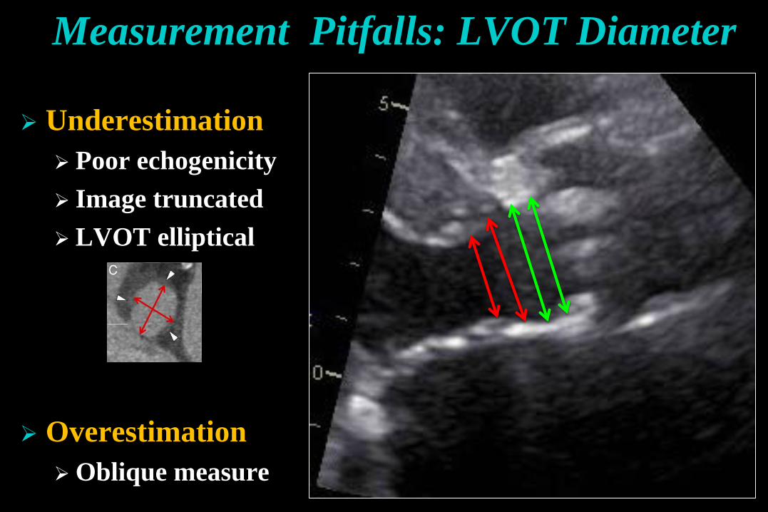
Measurement Pitfalls: LVOT Diameter
Underestimation Poor echogenicity Image truncated LVOT elliptical
OverestimationOblique measure
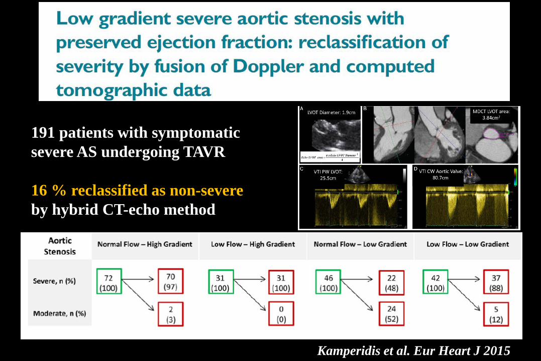
Kamperidis et al. Eur Heart J 2015
191 patients with symptomatic severe AS undergoing TAVR
16 % reclassified as non-severe by hybrid CT-echo method
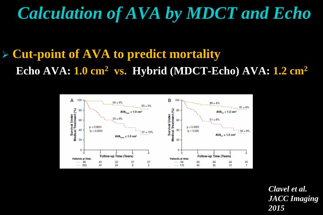
Calculation of AVA by MDCT and Echo
Cut-point of AVA to predict mortalityEcho AVA: 1.0 cm2 vs. Hybrid (MDCT-Echo) AVA: 1.2 cm2
Clavel et al. JACC Imaging 2015
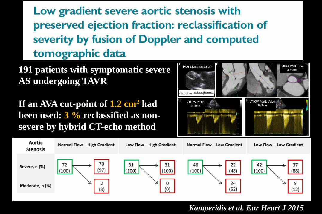
Kamperidis et al. Eur Heart J 2015
191 patients with symptomatic severe AS undergoing TAVR
If an AVA cut-point of 1.2 cm2 had been used: 3 % reclassified as non-severe by hybrid CT-echo method
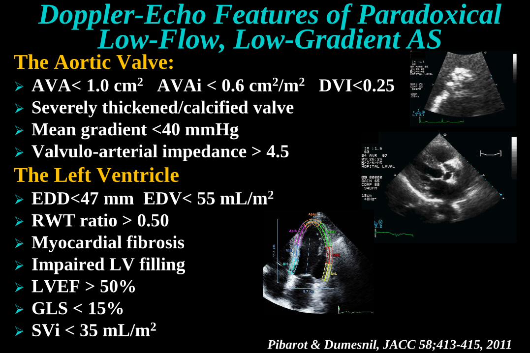
The Aortic Valve: AVA< 1.0 cm2 AVAi < 0.6 cm2/m2 DVI<0.25 Severely thickened/calcified valve Mean gradient <40 mmHg Valvulo-arterial impedance > 4.5The Left Ventricle EDD<47 mm EDV< 55 mL/m2
RWT ratio > 0.50 Myocardial fibrosis Impaired LV filling LVEF > 50% GLS < 15% SVi < 35 mL/m2
Pibarot & Dumesnil, JACC 58;413-415, 2011
Doppler-Echo Features of ParadoxicalLow-Flow, Low-Gradient AS
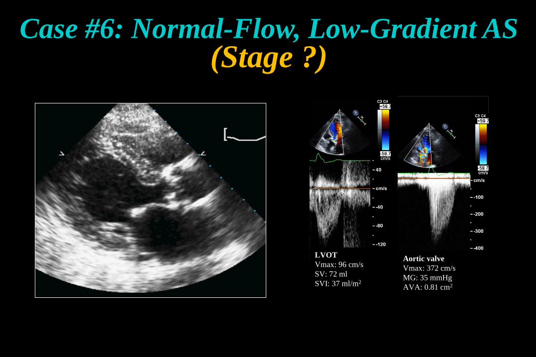
Case #6: Normal-Flow, Low-Gradient AS(Stage ?)
LVOTVmax: 96 cm/sSV: 72 mlSVI: 37 ml/m2
Aortic valveVmax: 372 cm/sMG: 35 mmHgAVA: 0.81 cm2
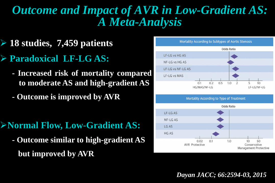
Outcome and Impact of AVR in Low-Gradient AS: A Meta-Analysis
Dayan JACC; 66:2594-03, 2015
18 studies, 7,459 patients Paradoxical LF-LG AS:
- Increased risk of mortality comparedto moderate AS and high-gradient AS
- Outcome is improved by AVR
Normal Flow, Low-Gradient AS:- Outcome similar to high-gradient AS
but improved by AVR
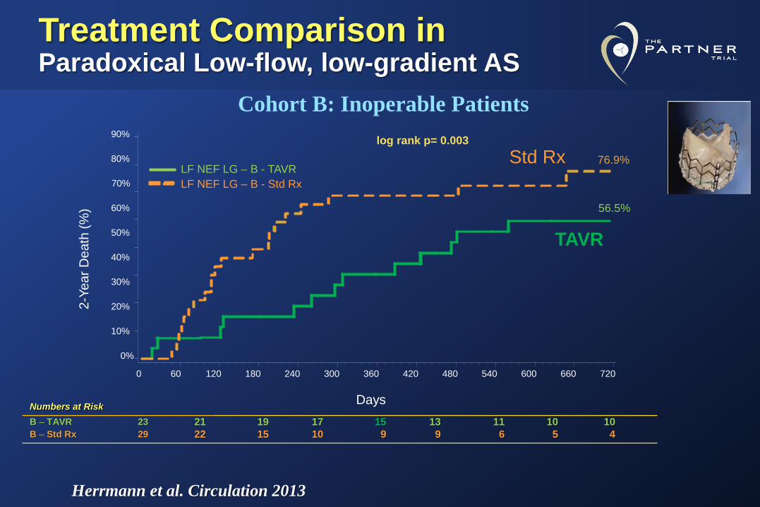
Treatment Comparison inParadoxical Low-flow, low-gradient AS
0%
10%
20%
30%
40%
50%
60%
80%
90%
60 120 180 240 300 360 420 480 540 600 660 720
70%
2-Ye
ar D
eath
(%) 56.5%
76.9%
0
log rank p= 0.003
Numbers at RiskB – TAVR 23 21 19 17 15 13 11 10 10B – Std Rx 29 22 15 10 9 9 6 5 4
LF NEF LG – B - TAVRLF NEF LG – B - Std Rx
Days
Herrmann et al. Circulation 2013
Cohort B: Inoperable Patients
TAVR
Std Rx
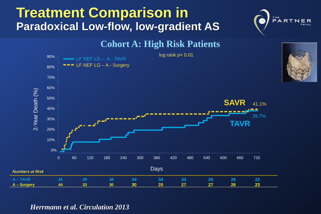
Treatment Comparison inParadoxical Low-flow, low-gradient AS
0%
10%
20%
30%
40%
50%
60%
80%
90%
60 120 180 240 300 360 420 480 540 600 660 720
70%
2-Ye
ar D
eath
(%)
39.7%
41.1%
0
log rank p= 0.01
Numbers at Risk
A – TAVR 43 39 38 34 34 33 29 26 22A – Surgery 44 33 30 30 28 27 27 26 23
LF NEF LG – A - TAVRLF NEF LG – A - Surgery
Days
Herrmann et al. Circulation 2013
TAVR
SAVR
Cohort A: High Risk Patients
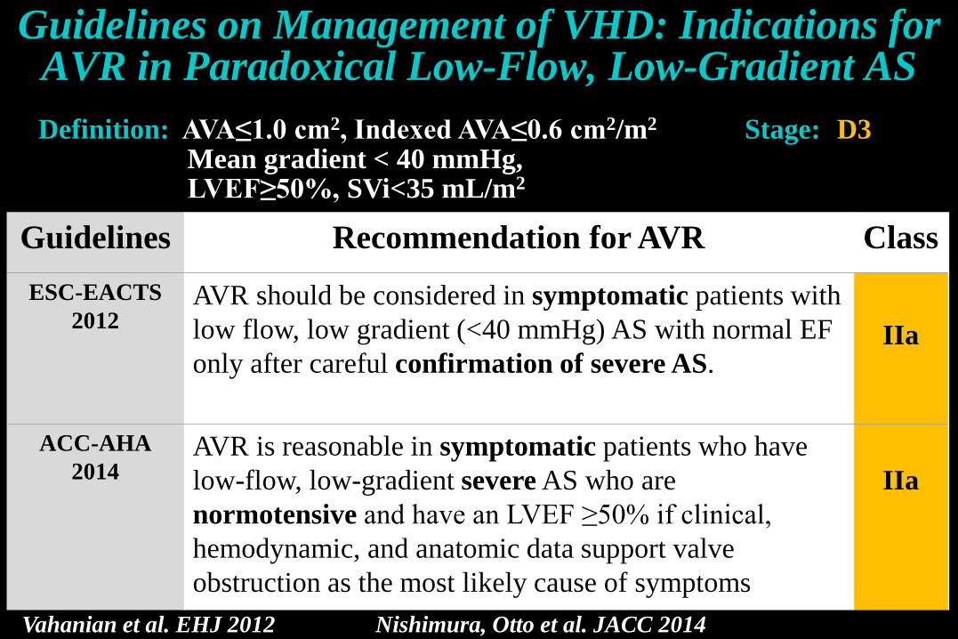
Guidelines Recommendation for AVR ClassESC-EACTS
2012AVR should be considered in symptomatic patients with low flow, low gradient (<40 mmHg) AS with normal EF only after careful confirmation of severe AS.
IIa
ACC-AHA2014
AVR is reasonable in symptomatic patients who have low-flow, low-gradient severe AS who are normotensive and have an LVEF ≥50% if clinical, hemodynamic, and anatomic data support valve obstruction as the most likely cause of symptoms
IIa
Guidelines on Management of VHD: Indications for AVR in Paradoxical Low-Flow, Low-Gradient AS
Vahanian et al. EHJ 2012 Nishimura, Otto et al. JACC 2014
Definition: AVA≤1.0 cm2, Indexed AVA≤0.6 cm2/m2
Mean gradient < 40 mmHg,LVEF≥50%, SVi<35 mL/m2
Stage: D3

AVC Score:3200 AU
Case #5: Aortic Valve Calcium Scoring by MDCT
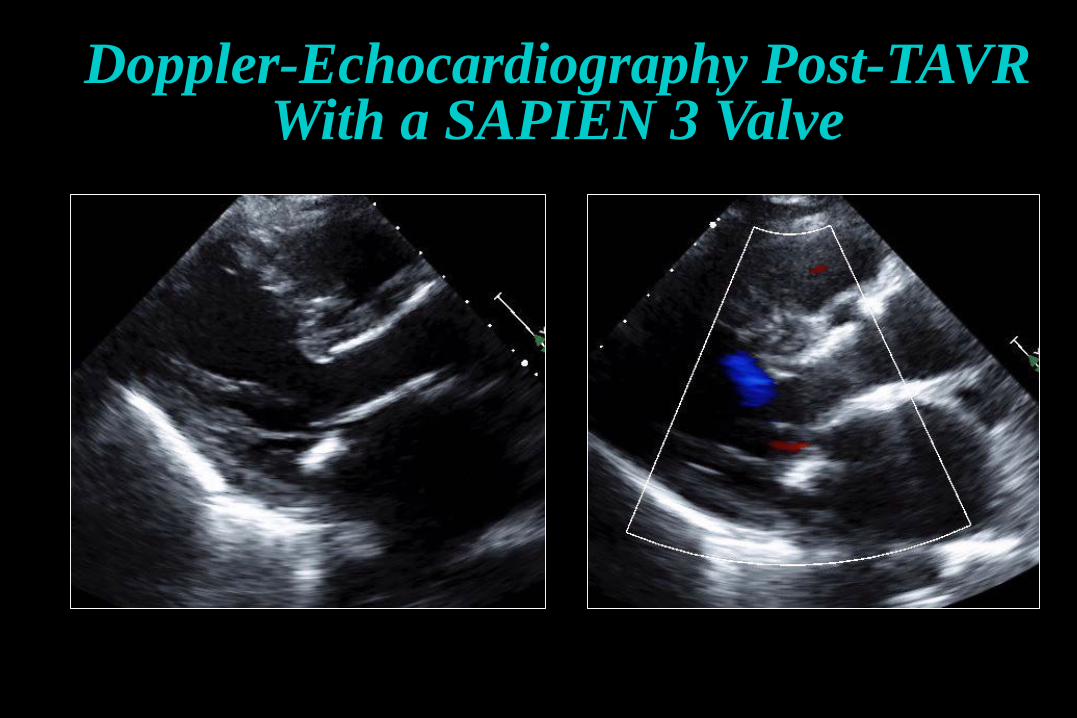
Doppler-Echocardiography Post-TAVRWith a SAPIEN 3 Valve
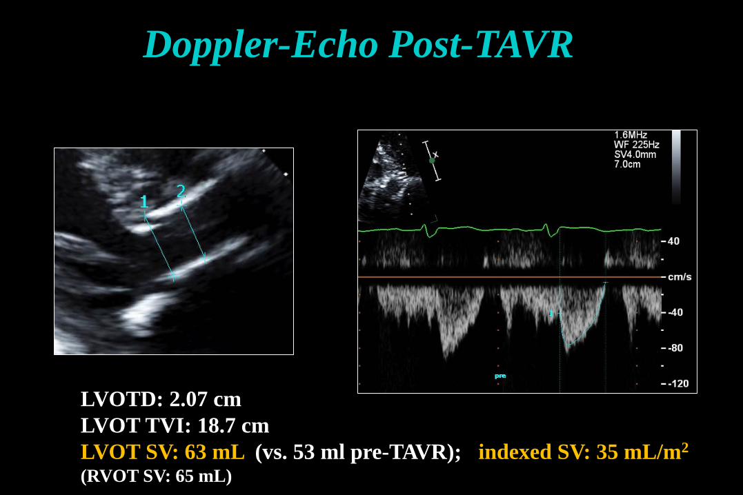
LVOTD: 2.07 cmLVOT TVI: 18.7 cmLVOT SV: 63 mL (vs. 53 ml pre-TAVR); indexed SV: 35 mL/m2
(RVOT SV: 65 mL)
Doppler-Echo Post-TAVR
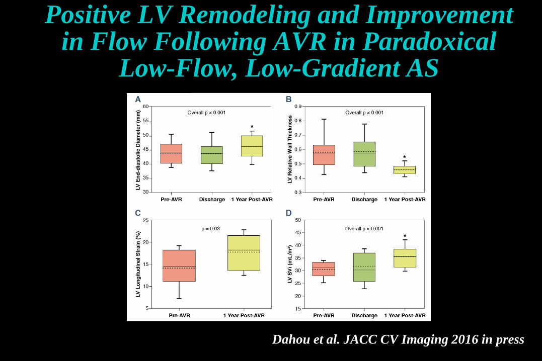
Positive LV Remodeling and Improvement in Flow Following AVR in Paradoxical
Low-Flow, Low-Gradient AS
Dahou et al. JACC CV Imaging 2016 in press
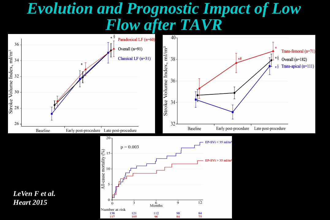
Evolution and Prognostic Impact of Low Flow after TAVR
LeVen F et al.Heart 2015
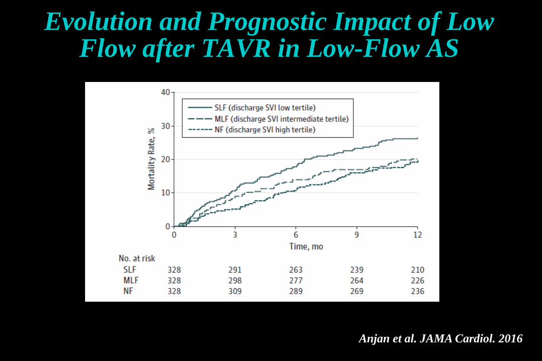
Evolution and Prognostic Impact of Low Flow after TAVR in Low-Flow AS
Anjan et al. JAMA Cardiol. 2016
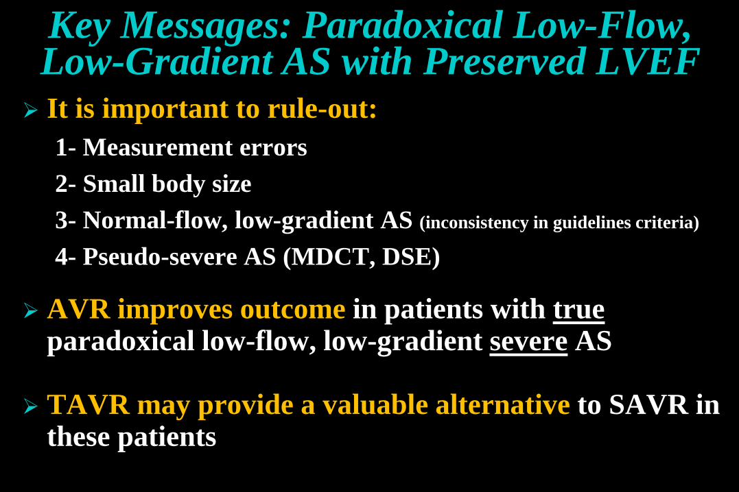
It is important to rule-out:1- Measurement errors2- Small body size3- Normal-flow, low-gradient AS (inconsistency in guidelines criteria)
4- Pseudo-severe AS (MDCT, DSE)
AVR improves outcome in patients with trueparadoxical low-flow, low-gradient severe AS
TAVR may provide a valuable alternative to SAVR in these patients
Key Messages: Paradoxical Low-Flow, Low-Gradient AS with Preserved LVEF
Recommended


![Original Article The extent of aortic lymphadenectomy in ... · computed tomography (PET-CT), are less invasive but have low sensitivity for evaluation of aortic LN [5]. On the other](https://img.pdfslide.net/doc/110x75/6113eed7b1e18257fb339f86/original-article-the-extent-of-aortic-lymphadenectomy-in-computed-tomography.jpg)