Embed Size (px)
Citation preview

10 SPINELINE JANUARY/FEBRUARY 2005
CURRENT CONCEPTS
Invited Review
Lumbar Zygapophysial Joint Evaluation and Treatment
Michael C. Geraci, MD, PTBuffalo Spine and Sports MedicineWilliamsville, NY
Ray M. Baker, MDWashington Interventional Spine AssociatesRedmond, WA
Rick C. Sasso, MDIndiana Spine GroupIndianapolis, IN
Whereas the existence
of “facet syndrome” is
debatable, most clinicians
currently agree that lumbar
z-joints are a common
source of low back pain.
This premise is strongly
supported by numerous
anatomic and clinical
studies.
BACKGROUNDThe concept of primary facet joint (zygapophysial or z-joint) pain has long been compelling. In 1911, Goldthwait1 proposed that the facet joints were responsible for a signifi cant amount of low back pain. Ghormley2 coined the term “facet syndrome” in 1933 and recommended surgical fusion to treat this disorder. Local anesthetic injection into the facet joint was fi rst described by Mooney3 in 1976 as a method to confi rm this diagnosis.
The 1988 Volvo Award in clinical sciences was awarded to Jackson et al4 for their prospective, statistical study regarding lumbar facet joint injections. They were not able to identify clinical parameters that predicted response to facet blocks. They refuted the existence of “facet joint syndrome” and concluded that the facet joint was not the single or primary source of low back pain. Additional prospective, randomized, controlled studies in the 1980s5,6 concluded that facet joint injections were a nonspecifi c method of treatment for low back pain and, diagnostically, were not correlated with the clinical diagnosis of facet joint syndrome.
Whereas the existence of “facet syndrome” is debatable, most clinicians currently agree that lumbar z-joints are a common source of low back pain. This premise is strongly supported by numerous anatomic and clinical studies.
Cadaveric studies have demonstrated that the z-joints receive innervation from the medial branch nerves of the primary dorsal rami. Neuroanatomic studies further prove the presence of an
EDITOR’S NOTEThis article will discuss the basic science, clinical evaluation and research data supporting the concept that the lumbar zygapophysial joint (z-joint) is a potential source of low back pain and can be effectively managed through various noninvasive or minimally invasive treatment options. It is an extension of a symposium from the recent NASS annual meeting in Chicago on Low Back Pain Treatment. Two of the authors of this article were speakers on that panel, Ray Baker, MD (radiofrequency neurotomy) and Rick Sasso, MD (surgery). Mike Geraci, MD, PT, graciously agreed to contribute some of his thoughts and ideas regarding the manual medicine approach to assessing and treating z-joint pain.
On reading this article, you will notice that the majority of rigorous science (utilizing controlled trials) is present only in the area of z-joint diagnostic injections and radiofrequency neurotomy. One could argue that the described manual treatments and exercises are not specifi c for z-joint pain and could apply to all chronic, nonspecifi c low back pain. While that would be diffi cult to deny, it is certainly not meant to diminish the role of the clinical evaluation and the importance of other nonsurgical treatment measures such as manual therapy and exercise. It is simply a refl ection of the state of the art of the science of low back pain in 2005. As a corollary, if the only tools accepted to treat chronic low back pain of an injection-proven z-joint origin were those demonstrated by randomized, controlled trials, then other than medication, the spine physician is left with either radiofrequency neurotomy or nothing. As always, the reader is encouraged to reach his or her own conclusion.

11JANUARY/FEBRUARY 2005 SPINELINE
CURRENT CONCEPTS
extensive distribution of small nerve fi bers and endings in the lum-bar z-joint.7-10 These nerves were high-threshold mechanoreceptors shown to contain substance P, typical of nociceptors. Sensitization and excitation of z-joint nerves and surrounding muscle were also shown when the nerves were exposed to infl ammatory or algesic chemicals. Together, these studies provide strong evidence that lumbar z-joints can produce pain.
DIAGNOSISSchwarzer,11-14 Dreyfuss,15 Bogduk,16,17 Kaplan18 and Manchikan-ti19,20 have extensively studied the z-joints. These studies focused on the ability of z-joints to produce pain, the clinical features associated with and prevalence of z-joint pain, and the ability of medial branch anesthetic injections to reliably diagnose patients with z-joint pain. The authors employed a variety of methods, including placebo controls, comparative anesthetic protocols and computed tomography (CT) confi rmation of anesthetic spread; one study even subjected patients to both z-joint injections and provocation discography to determine the relative prevalence of disc versus z-joint pain.12 In another randomized blinded placebo controlled study, lumbar z-joint capsular distension was shown to produce pain in normal volunteers. The pain was abolished with anesthetization of the medial branches with lidocaine (Xylocaine),but not with saline.18 Collectively, these and other studies have shown that it is clinically diffi cult, if not impossible, to diagnose lumbar z-joint pain without the use of controlled diagnostic (ie, diagnostic) injections. Although one study reported a collection of symptoms and signs that increased the probability of a patient having z-joint pain, this has been refuted recently.21-23
Common imaging techniques (ie, plain radiographs, magnetic resonance imaging [MRI], CT and bone single photon emission computed tomography [SPECT] scans) cannot discern which patients have z-joint pain24-29 either. SPECT scans select out a subgroup of patients who respond better to z-joint injections, but these tests suffer from low sensitivity and are not clinically useful as a screening tool.30
Although controlled diagnostic injections remain the best way of diagnosing z-joint pain, they are not infallible and care must be taken to assure a correct diagnosis. Schwarzer et al14 showed that a single z-joint injection resulted in a 38% false positive rate. Subsequent studies demonstrated that the specifi city improved by requiring 80% pain relief to secure the diagnosis, the false positive rate fell to 27% and the positive predictive value rose from 31% to 63%. Additionally, by performing placebo controlled or compara-tive anesthetic technique injections, the positive predictive value rose further.19,31
Comparative anesthetic techniques rely on the patient hav-ing a longer duration of pain relief with bupivicaine (Marcaine) than with lidocaine and require at least two separate injections for confi rmation. This increases specifi city greatly, but excludes two thirds of patients with true z-joint pain who exhibit a ‘discordant response,’ that is, a longer duration of relief with lidocaine than bupivacaine.31
To balance between the cost/ethical dilemmas of placebo controlled injections and the exclusion of appropriate patients fol-lowing a true comparative anesthetic protocol, a third approach has become popular: modifi ed comparative anesthetic protocol. With this protocol, a patient is required to have > 1-2 hours of pain relief with lidocaine, and > 2-3 hours of pain relief with bupivacaine.Using this protocol, Dreyfuss et al32 achieved a 90% success rate with subsequent radiofrequency neurotomy.
In summary, anatomic and clinical studies suggest the fol-lowing: Lumbar z-joints are anatomically capable of producing pain
mediated through the medial branch nerves. There are no clinical features or imaging techniques that are
pathognomonic for z-joint pain. 15% to 45% of patients presenting with low back pain have
z-joint pain. Controlled diagnostic injections are the best method of diag-
nosing z-joint pain. At least 80% pain relief is required after modifi ed compara-
tive anesthetic medial branch nerve injection to reliably select patients with z-joint pain.
MANUAL ASSESSMENT AND THERAPY: MIKE GERACI, MD, PTAs demonstrated by Schwarzer et al,12 z-joint pain is frequently as-sociated with degenerative discs and therefore, lumbar z-joint pain is not necessarily an isolated problem. Further, z-joint problems may progress along a continuum including synovitis, stiffness (hy-pomobility), capsular laxity (hypermobility), arthritis and instabil-ity, although not always predictably. Additionally, z-joint problems are frequently associated with musculoligamentous abnormalities including muscle fi brosis leading to localized segmental and, at times, more global infl exibilities.
Because of these recognized aggregate mechanical and func-tional impairments, the manual medicine approach to diagnosis and treatment has been used in various forms for thousands of years. Many practitioners of manual medicine exist (eg, osteopaths, chiropractors, physical therapists, massage therapists, medical doc-tors) but very few consistent guidelines have been developed for its application. Manual medicine has remained in the forefront of spinal treatment because patients continue to demand it and certain clinical subgroups derive a recognized benefi t.33,34
AssessmentAs indicated above, there are no clearly validated provocative trunk movements that correlate with injection-proven lumbar z-joint pain. Nevertheless, a comprehensive physical examination of the spine should include a segmental component (manually-based) and a functional component (movement-based), both of which can lead to a more specifi c treatment prescription. Overall range of motion of the lumbar spine is assessed on the “screening” examination and should include multiple planes of movement including fl exion,

12 SPINELINE JANUARY/FEBRUARY 2005
CURRENT CONCEPTS
extension, side bending, rotation or a combination of these move-ments. A skilled manual practitioner also performs a segmental examination, palpating for bony asymmetries and tissue texture abnormalities. Palpation of the transverse and spinous processes as the patient moves through an arc of fl exion, recovery, extension, lateral bending and rotation of the spine, allows identifi cation of asymmetry of these bony landmarks, suggesting abnormal segmen-tal motion. Tissue texture abnormalities, particularly of the deeper unisegmental muscles (ie, transverseres and rotatores) in the medial gutter lateral to the spinous processes, may indicate an underlying abnormal z-joint (ie, hypomobile or hypermobile).
The functional exam includes assessment of the functional kinetic chain. This may include identifying movement or range of motion abnormalities in other joint structures such as the foot and ankle, knee, hip, or pelvis that can contribute to dysfunctions of the lumbar z-joints. It also includes evaluation of dynamic muscle strength and fl exibilities, recognizing imbalances such as tight or facilitated muscles (such as the psoas, hip adductors, hamstrings and gastrocsoleus) and weak or inhibited muscles (such as the gluteus maximus and medius). Identifying these dysfunctions of the entire kinetic chain, will also lead to optimal exercise prescriptions.
Manual Therapy Manual therapy treatment varies considerably from specialty to specialty. However, some of the terminology is standardized. The term mobilization refers to graded, directional force applied to a specifi c segment usually performed in the side lying position. These are graded I through V, with the increasing grades of mobilization proportional to an increase in amplitude of force. Grades I and II are considered gentle oscillations and used primarily for pain control, with grade V, also known as a manipulation, consisting of a high velocity thrusting maneuver.
Physical therapists generally perform grades I through IV mobilizations, whereas osteopaths and chiropractors can apply manipulations. Other techniques such as muscle energy35,36 use the patient’s own muscle activation to correct joint mobility. Myo-fascial release can be used to release fascial and muscle tightness patterns in order to reduce z-joint dysfunction of the spine. Other osteopathic techniques include functional indirect, craniosacral, and strain-counterstrain,37 and joint play38 or motion-palpation39
that are used in treating joint dysfunction.
Exercise TherapyExercises have been used to improve lumbopelvic muscle con-trol and stability, to optimize multiplanar movement patterns, to maintain joint correction between visits and to ultimately reduce the need for manual treatments for z-joint dysfunction. While no single exercise regimen exists specifi cally for z-joint related prob-lems, there are two general principles of exercise therapy. The fi rst focuses on pain control and regaining local muscle control. The second focuses on the more global kinetic chain by stretching tight muscles, strengthening weak muscles and retraining whole body movement patterns, proprioception and task-specifi c function.
Exercises for pain control are often an extension of the physi-cal examination. Even if a distinct diagnosis of z-joint pain cannot be made based on physical examination, a reasonable exercise program can be started without advanced expertise. Directional exercises based on the centralization or reduction of pain during the screening examination can be applied to the early stages of treatment. For example, if low back and referred pain to the hip girdle are reduced with fl exion-biased movements and increased with extension-biased movements, fl exion is recommended fi rst. These are frequently prescribed as William’s fl exion exercises and can include posterior pelvic tilts and knee to chest maneuvers performed in a supine position. However, it is important to realize that fl exion exercises are not universally applied to z-joint pain and occasionally extension-biased exercises are preferred.
An early approach at regaining lumbopelvic control is the “pel-vic clock” technique as developed by Feldenkrais40 and modifi ed for manual evaluation and treatment by Bookhout et al.41 This can be performed in the supine, seated or standing position, the latter more representative of positions of daily function. In this evaluation and treatment strategy, an imaginary clock face is superimposed over the pelvis, so that the 12 o’clock position represents fl attening the back by rotating the pelvis into a posterior tilt; 6 o’clock represents rolling into lumbar extension by an anterior tilt; 3 o’clock represents rotation of the pelvis to the left; and 9 o’clock represents rotation of the pelvis to the right. Any point on the clock can be touched with the pelvis and brought back to the starting position. This has been successfully used for joint dysfunction with stiffness being the primary problem, both as a home exercise treatment program as well as an evaluation tool. Positions that provoke pain are eliminated in the very early stages of this exercise, but are added back as pain diminishes.
Another technique known as muscle rebalancing has been used to treat “group” dysfunctions or abnormalities that include three or more segments that do not move with proper coupling. This treatment focuses on the neurophysiologic phenomenon of muscle facilitation (or tightening) and muscle inhibition (or weak-ening). This pattern may predispose to altered segmental motion. In the lumbar spine, there is normally a strong coupling pattern of lateral bending and spinous process rotation to the opposite side. If uncoupling of motion is identifi ed over several levels, for example L1 through L4, in which side bending and spinous process rotation occur to the same side, there is often an associated iliopsoas muscle facilitation and gluteal inhibition on the concave side. Passive and then active stretching of the facilitated muscle may correct this joint dysfunction without the need for specifi c joint mobilization techniques. This will also allow progressive strengthening of the weakened hip abductor and extensor.
A new development in manual medicine treatment is functional joint mobilization, as developed by Lambert.42 Most of these tech-niques are done either standing or seated (as opposed to side lying) and integrate the use of diagonal proprioceptive neuromuscular facilitation (PNF) patterns. These PNF patterns more closely match the multiplanar z-joint dysfunctional movement patterns, either in
13JANUARY/FEBRUARY 2005 SPINELINE
CURRENT CONCEPTS
fl exion and rotation or extension and rotation. This has led to the development of functional exercises to maintain correction, often obviating the need for regular manual treatments.
The most important aspect of functional rehabilitation for z-joint dysfunction and pain may be identifying the repetitive patterns that people move in at home, work or athletically, and pinpointing defi cits in the functional kinetic chain. The lumbar spine must compensate for these mechanical defi cits, eg, lack of subtalar joint motion or limited hip internal range of motion. For example, the lumbar spine may be forced to excessively rotate or side bend to compensate for loss of hip internal rotation as the trunk rotates over an arthritic hip joint. This can overload the physiologically available rotation in the lumbar spine and mechanically overload the z-joints on the right. After many cycles, the z-joints may become stiff and painful and no longer accommodate a neutral posture, then progress to a state of hypermobility and eventually develop arthritis.
Once pain is reduced or eliminated and early muscular control of the lumbopelvic region is accomplished, more challenging core strengthening exercises are initiated. These exercises, however, are not exclusive for z-joint pain (see “Core Strengthening,” Spine-Line, November/December 2003, pp 9-19.) A lack of a consistent approach to exercise is a void in the treatment arsenals of many manual therapy practitioners. This may lead to excessive unimodal treatment. However, one should not underestimate the value (real or perceived) of therapeutic touch from the reasonable use of manual medicine techniques. The ideal treatment environment should allow a combination of manual therapy, exercise and when indicated, fl uoroscopically guided z-joint injection procedures.
LUMBAR MEDIAL BRANCH RADIOFREQUENCY NEUROTOMY: RAY BAKER, MDGeneral PrinciplesA certain percentage of patients with z-joint pain do not improve with exercise, manual therapy or other well accepted noninvasive treatments. Over the past 30 years, medial branch radiofrequency (RF) neurotomy has evolved as a minimally invasive treatment for patients with refractory z-joint pain. Patients are selected based on their response to properly performed medial branch blocks, often involving a modifi ed comparative anesthetic protocol (see p 11).
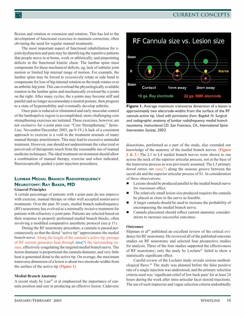
During the RF neurotomy procedure, a cannula is passed per-cutaneously so that the distal “active tip” approximates the medial branch nerve. Along the length of the cannula’s active tip, passage of RF current generates heat through into(?) the surrounding tis-sues, effectively coagulating the targeted medial branch nerve. The lesion diameter is proportional the cannula diameter, and very little heat is generated distal to the active tip. On average, the maximum transverse dimension of a lesion is about two electrode-widths from the surface of the active tip (Figure 1).
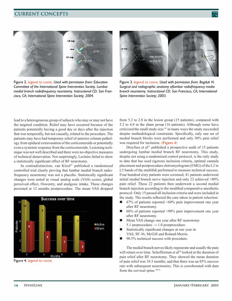
Medial Branch AnatomyA recent study by Lau43 et al emphasized the importance of can-nula position and size in producing an effective lesion. Cadaveric
dissections, performed as a part of the study, also extended our knowledge of the anatomy of the medial branch nerves. (Figure 2 & 3.) The L1 to L4 medial branch nerves were shown to run across the neck of the superior articular process, not at the base of the transverse process as was previously assumed. The L5 primary dorsal ramus ran runs(?) along the osseous groove between the sacral ala and the superior articular process of S1. In consideration of these observations: Lesions should be produced parallel to the medial branch nerve
for maximum effect. The relatively small lesion size produced requires the cannula
be placed as close to the nerve as feasible. A larger cannula should be used to increase the probability of
encompassing the medial branch nerve. Cannula placement should refl ect current anatomic consider-
ations to increase successful outcomes.
OutcomesSlipman et al44 published an excellent review of the critical evi-dence for RF neurotomy. He reviewed all of the published outcome studies on RF neurotomy and selected four prospective studies for analysis. Three of the four studies supported the effectiveness of RF neurotomy; only the study by Leclaire45 failed to show a statistically signifi cant effect.
Careful review of the Leclaire study reveals serious method-ological fl aws.46 The study was planned before the false positive rate of a single injection was understood, and the primary selection criteria used was ‘signifi cant relief of low back pain’ for at least 24 hours during the week after intra-articular facet steroid injections. The use of such imprecise and vague selection criteria undoubtedly
Figure 1. Average maximum transverse dimension of a lesion is approximately two electrode-widths from the surface of the RF cannula active tip. Used with permission from: Bogduk N. Surgical and radiographic anatomy of lumbar radiofrequency medial branch neurotomy. Instructional CD. San Francisco, CA.; International Spine Intervention Society; 2003.
14 SPINELINE JANUARY/FEBRUARY 2005
CURRENT CONCEPTS
lead to a heterogeneous group of subjects who may or may not have the targeted condition. Relief may have occurred because of the patients potentially having a good day or days after the injection that was temporally, but not causally, related to the procedure. The patients may have had temporary relief of anterior column pathol-ogy from epidural extravasation of the corticosteroids or potentially even a systemic response from the corticosteroids. Lesioning tech-nique was not well described and there were no objective measures of technical denervation. Not surprisingly, Leclaire failed to show a statistically signifi cant effect of RF neurotomy.
In contradistinction, van Kleef47In contradistinction, van Kleef47In contradistinction, van Kleef published a randomized controlled trial clearly proving that lumbar medial branch radio-frequency neurotomy was not a placebo. Statistically signifi cant changes were noted in visual analog scale (VAS) scores, global perceived effect, Oswestry, and analgesic intake. These changes persisted at 12 months postprocedure. The mean VAS dropped
from 5.2 to 2.8 in the lesion group (15 patients), compared with 5.2 to 4.8 in the sham group (16 patients). Although some have criticized the small study size,48 in many ways the study succeeded despite methodological constraints. Specifi cally, only one set of medial branch blocks were performed and only 50% pain relief was required for inclusion. (Figure 4)
Dreyfuss et al32 published a prospective audit of 15 patients undergoing lumbar medial branch RF neurotomy. This study, despite not using a randomized control protocol, is the only study to date that has used rigorous inclusion criteria, optimal cannula placement and postprocedure eletromyelogram (EMG) of the L2 to L5 bands of the multifi di performed to measure technical success. Four hundred sixty patients were screened; 41 patients underwent initial medial branch nerve injection and only 22 achieved >80% pain relief. These 22 patients then underwent a second medial branch injection according to the modifi ed comparative anesthetic protocol. Only 15 passed all inclusion criteria and were included in the study. The results refl ected the care taken in patient selection: 87% of patients reported >60% pain improvement one year
after RF neurotomy. 60% of patients reported >90% pain improvement one year
after RF neurotomy. Mean VAS change one year after RF neurotomy:. 5.1 preprocedure → 1.0 postprocedure. Statistically signifi cant changes at one year in:. VAS, SF-36, McGill and Roland-Morris. 90.5% technical success with procedure.
The medial branch nerves likely regenerate and usually the pain will return over time. Schofferman et al49 looked at the duration of pain relief after RF neurotomy. They showed the mean duration of pain relief was 10.5 months, and that there was an 85% success rate with subsequent neurotomies. This is corroborated with data from the cervical spine.50,51
Figure 2. legend to come. Used with permission from: Education Committee of the International Spine Intervention Society. Lumbar medial branch radiofrequency neurotomy. Instructional CD. San Fran-cisco, CA; International Spine Intervention Society; 2004.
Figure 3. legend to come. Used with permission from: Bogduk N. Surgical and radiographic anatomy ofl umbar radiofrequency media branch neurotomy. Instructional CD. San Francisco, CA; International Spine Intervention Society; 2003.
Figure 4. legend to come.

15JANUARY/FEBRUARY 2005 SPINELINE
CURRENT CONCEPTS
ComplicationsOnly one study has been published that specifi cally addresses the question of complications after RF neurotomy. Kornick et al52 pub-lished a study of 92 patients that underwent 616 RF lesions with only a 1% minor complication rate (0.3% to 1.7%) including: three cases of localized pain lasting > two weeks three cases of neuritic pain lasting < two weeks 0 infections or new sensory/motor defi cits or major complica-
tions.
RF Neurotomy ConclusionMore studies need to be performed using randomized control pro-tocols, larger study sizes, proper cannula position and currently accepted inclusion criteria. Despite this, enough evidence exists to support the following: Radiofrequency neurotomy is an effective treatment for well
selected patients with lumbar z-joint pain.selected patients with lumbar z-joint pain.selected Patient selection criteria using modifi ed comparative anesthetic
MBB protocol and >80% pain relief should be encouraged. RF neurotomy is a safe procedure with few associated com-
plications Proper electrode position and size optimize technical suc-
cess. Segmental EMG testing of the multifi dus muscle is helpful to
assess the technical adequacy of the procedure.
LUMBAR FUSION: RICK SASSO, MDThose patients with primary z-joint pain who do not respond to nonsurgical treatment may respond favorably to surgical fusion. The most important factor in a successful outcome is confi rming that the diagnosis is accurate. Thus, the description previously of the meticulous care necessary when planning and performing the medial branch anesthetic injections. Careful consideration to surgi-cal technique is also critical.
Surgical treatment of lumbar z-joint pain has been evaluated. Markwalder53 evaluated surgical fusion for facet syndrome in 119 patients. The best outcome occurred in the translaminar facet screw fi xation cohort who had 96% good-excellent results. The translaminar facet screws were postulated to have the lowest risk of irritation to the adjacent segments and the facet screws specifi cally immobilized the painful lumbar z-joints. Biomechanical evaluation has proven these screws to be as strong, and in some loading modes, stronger than pedicle screw and rod constructs (? REFERENCE). Moreover, transfacet screw fi xation was not compromised after repetitive cyclic loading.
Various techniques for lumbar fusion exist and several are reasonable alternatives to surgically treat primary z-joint pain. The variables to consider are: type of fusion: anterior versus posterior (facet or intertrans-
verse), with or without interbody fusion (anterior lumbar interbody fusion versus posterior lumbar interbody fusion
versus translaminar lumbar interbody fusion) type of posterior instrumentation: translaminar facet screws
versus pedicle screws and rods type of bone graft: posterior (autogenous iliac crest, local bone
graft, demineralized bone matrix, bone morphogenetic protein) or interbody (femoral ring allograft, cage)
Bone grafting across the intertransverse process region may produce a very strong lumbar fusion mass. However, the soft tissue dissection is extensive. Primary facet fusion entails minimal soft tissue dissection and allows complete removal of the facet capsule. The addition of an interbody fusion will increase the chance that a solid fusion will occur by stabilizing the anterior column. The question of whether this should be performed from an anterior or posterior approach is surgeon specifi c. The advantages of an anterior approach compared to a posterior interbody fusion are decreased blood loss, more complete removal of the disc and the ability to place a larger interbody graft. The disadvantage is that a separate incision is required. With a posterior lumbar interbody fusion or transforaminal lumbar interbody fusion, the same incision is used, but the exposure is much more extensive and requires retraction of the dura and nerve roots. Complications related to the neural structures are much higher with this approach because the cauda equina and exiting nerve roots are between the surgeon and the target disc space.
Although pedicle screw fi xation is a very common and familiar technique for all spine surgeons, it requires large and bulky im-plants. The cephalad juxta-level unfused z-joint is specifi cally at risk because of the position of the upper pedicle screw. The entry point requires exposure of this adjacent z-joint and frequently re-sults in destruction of the facet capsule and part of the joint. Even with “minimally invasive” pedicle screw placement, the screw head and upper part of the rod irritate the z-joint during normal motion. A great advantage of facet screws is that they are placed far away from the adjacent z-joint. They are not bulky and they directly ad-dress the painful z-joint, whereas pedicle screw constructs simply span across the painful joint and do not directly immobilize it. The issue of the type of bone graft is controversial, but what is very clear is the fact that a large percentage (up to 30%) of patients continue to have bone graft donor site pain if autogenous iliac crest bone is harvested. Thus, the trend is to avoid autogenous bone.
As long as the diagnosis of lumbar facet syndrome can be as-sured, then it appears reasonable to consider lumbar arthrodesis if nonoperative treatment is not successful. Truly minimally invasive posterior instrumentation with transfacet screws that are applied with minimal soft tissue disruption and spare the adjacent facet joints appear to be best supported by the literature to facilitate a successful lumbar fusion with the ultimate goal of pain resolution and minimization of adjacent level degeneration.

16 SPINELINE JANUARY/FEBRUARY 2005
CURRENT CONCEPTS
REFERENCES1. Goldthwait JE. The lumbosacral articulation: an explanation of
many cases of lumbago, sciatica, and paraplegia. Boston Med Surg J. 1911;164:365-372.
2. Ghormley RK. Low back pain with special reference to the articular facets with presentation of an operative procedure. JAMA. 1933;101: 1773-1777.
3. Mooney V, Robertson J. The facet syndrome. Clin Ortho. 1976;115: 149-156.
4. Jackson RP, Jacobs RR, Montesano PX. Facet joint injection in low-back pain. Spine. 1988;13: 966-971.
5. Lilius G, Laasonen EM, Myllynen P, Harilainen A, Gronlund G. Lumbar facet joint syndrome: a randomized clinical trial. J Bone Joint Surg Br. 1989;71B:681-684.
6. Moran R, O’Connell D, Walsh MG. The diagnostic value of facet joint injections. Spine. 1988;13:1407-1410.
7. Giles LG, Harvey AR. Immunohistochemical demonstration of no-ciceptors in the capsule and synovial folds of human zygapophyseal joints. Br J Rheumatol. 1987;26:362-364.
8. Beaman DN, Graziano GP, Glover RA, Wojtys EM, Chang V. Sub-stance P innervation of lumbar spine facet joints. Spine. 1993;18:1044-1049.
9. Ashton IK, Ashton BA, Gibson SJ, Polak JM, Jaffray DC, Eisenstein SM. Morphological basis for back pain: the demonstration of nerve fi bers and neuropeptides in the lumbar facet joint capsule but not in the ligamentum fl avum. J Orthop Res. 1992;10:72-78.
10. Cavanaugh JM, Ozaktay AC, Yamashita HT, King AI. Lumbar facet pain: biomechanics, neuroanatomy, and neurophysiology. J Biomech.1996;29:1117-1129.
11. Schwarzer AC, Aprill CN, Derby R, Fortin J, Kine G, Bogduk N. Clinical features of patients with pain stemming from the lumbar zygapophysial joints. Is the lumbar facet syndrome a clinical entity? Spine. 1994;19(10):1132-1137.
12. Schwarzer AC, Aprill CN, Derby R, Fortin J, Kine G, Bogduk N. The relative contributions of the disc and zygapophyseal joint in chronic low back pain. Spine. 1994;19(7): 801-806.
13. Schwarzer AC, Wang SC, Bogduk N, McNaught PJ, Laurent R. Prevalence and clinical features of lumbar zygapophysial joint pain: a study in an Australian population with chronic low back pain. Ann Rheum Dis. 1995;54(2):100-106.
14. Schwarzer AC, Aprill CN, Derby R, Fortin J. Kine G, Bogduk N. The false positive rate of uncontrolled diagnostic blocks of the lumbar zygapophysial joints. Pain. 1994;58:195-200.
15. Dreyfuss P, Schwarzer AC, Lau P, Bogduk N. Specifi city of lumbar medial branch and L5 dorsal ramus blocks. A computed tomography study. Spine. 1997;22(8):895-902.
16. Bogduk, N. The anatomical basis for spinal pain syndromes. J Ma-nipulative Physiol Ther. 1995;18(9):603-605.
17. Bogduk N, Wilson AS, Tynan W. The human lumbar dorsal rami. J Anat. 1982;134:383-397.
18. Kaplan M, Dreyfuss P, Halbrook B. Bogduk N. The ability of lumbar medial branch blocks to anesthetize the zygapophysial joint. A physi-ologic challenge. Spine. 1998;23(17):1847-1852.
19. Manchikanti L, Boswell MB, Singh V, Pampati V, Damron KS, Beyer CD. Prevalence of facet joint pain in chronic spinal pain of cervical, thoracic and lumbar regions. BMC Musculoskelet Disord.2004;5(1):15.
20. Manchikanti L, Pampati V, Fellows B, Bakhit CE. Prevalence of lumbar facet joint pain in chronic low back pain. Pain Physician.
1999; 2(3):59-64.21. Revel MS, Poiraudeau, Aulely GR, et al. Capacity of the clinical pic-
ture to characterize low back pain relieved by facet joint anesthesia. Proposed criteria to identify patients with painful facet joints. Spine.1998;23(18):1972-1976; discussion 1977.
22. Manchikanti L, Pampati V, Fellows B, Baha A. The inability of the clinical picture to characterize pain from facet joints. Pain Physician. 2000;3:158–166.
23. Laslett M, Oberg B, Aprill CN, McDonald B. Zygapophysial joint blocks in chronic low back pain: a test of Revel’s model as a screen-ing test. BMC Musculoskelet Disord. 2004;5(1):43.
24. Schwarzer AC, Wang SC, O’Driscoll D, Harrington T, Bogduk N, Laurent R. The ability of computed tomography to identify a painful zygapophysial joint in patients with chronic low back pain. Spine.1995;20:907-912.
25. Schwarzer AC, Scott AM, Wang SC, Hoschl R, Wiseman JC, Cop-per RA. The role of bone scintigraphy in chronic low back pain: a comparison of SPECT and planar images and zygapophysial joint injection. Aust NZJ Med. 1992;22:185.
26. Murtagh FR. Computed tomography and fl uoroscopy guided anaes-thesia and steroid injection in facet syndrome. Spine. 1988;13:686-689.
27. Haas M, Nyiendo J, Petersen C, et al. Lumbar motion trends and correlation with low back pain. Part I: a roentgenological evalua-tion of coupled motion in lateral bending. J Manipul Physiol Ther. 1992;15:145-158.
28. Haas M, Nyiendo J. Lumbar motion trends and correlation with low back pain. Part II: a roentgenological evaluation of quantitative seg-mental motion in lateral bending. J Manipul Physiol Ther. 1992;15: 224–2.
29. Raymond J, Dumas JM, Lisbona R. Nuclear imaging as a screening test for patients referred for intra-articular facet block. J Can Assoc Radiol. 1984;35:291-292.
30. Dolan AL, Ryan PJ, Arden NK, et al. The value of SPECT scans in identifying back pain likely to benefi t from facet joint injection. Br J Rheumatol. 1996;35(12):1269-1273.
31. Lord SM, Barnsley L, Bogduk N. The utility of comparative local anesthetic blocks versus placebo-controlled blocks for the diagnosis of cervical zygapophysial joint pain. Clin J Pain. 1995;11:208-213.
32. Dreyfuss P, Halbrook B, Pauza K, Joshi A, McLarty J, Bogduk. Ef-fi cacy and validity of radiofrequency neurotomy for chronic lumbar zygapophysial joint pain. Spine. 2000;25(10):1270-1277.
33. Bigos S, Bowyer O, Braen G, et al. Acute Low Back Pain Problems in Adults. Clinical Practice Guideline No. 14. Rockville, MD: Agency for Healthcare Research and Quality, Public Health Service, US Department of Health and Human Services; 1994.
34. Shekelle PG, Adams AH, Chassin MR, et al. Spinal manipulation for low back pain. Ann Intern Med. 1992;117:590-598.
35. Mitchell FL Jr, Moran PS, Pruzzo NA. An evalution and treatment manual of osteopathic muscle energy procedures. Valley Park, MO; Mitchell, Moran & Pruzzo Associates; 1979.
36. Greenman PE. Principles of Manual Medicine. 3rd ed. Philadelphia, rd ed. Philadelphia, rd
PA; Lippincott Williams & Wilkins; 2003.37. Jones LH. Strain and counterstrain. Colorado Springs, CO; American
Academy of Osteopathy; 1991.38. Mennell JM, Back Pain. Boston, MA: Little, Brown and Company;
1960.39. Harrison PE. Three-dimensional spinal coupling mechanics: part II.
implications for chiropractic theories and practice. J Manipulative

17JANUARY/FEBRUARY 2005 SPINELINE
CURRENT CONCEPTS
Physiol Ther. 1998;21(3):177-186. 40. Feldenkrais M. Awareness Through Movement: Health Exercises For
Personal Growth. New York, NY: Harper Collins; 1972.41. Bookhout MR, Geraci MC, Greenman PE. Exercise Prescription as
an Adjunct to Manual Medicine. Course Syllabus. East Lansing, MI; Michigan State University, College of Osteopathic Medicine; 2001.
42. Lambert M. Functional Joint Mobilizations. Course Notes. Williams-ville, NY; Buffalo Spine and Sports Institute; 2001.
43. Lau P, Mercer S, Govind J, Bogduk N. The surgical anatomy of lumbar medial branch neurotomy (facet denervation). Pain Med.2004;5(3):289-298.
44. Slipman CW, Bhat AL, Gilchrist RV, Issac Z, Chou L, Lenrow DA. A critical review of the evidence for the use of zygapophysial injections and radiofrequency denervation in the treatment of low back pain. Spine J. 2003; 3(4): 310-316.
45. Leclaire R, Fortin L, Lambert R, Bergeron YM, Rossignol M. Radio-frequency facet joint denervation in the treatment of low back pain: a placebo controlled trial to assess effi cacy. Spine. 2001;26(13):1411-1417.
46. Dreyfuss P, Baker R, Leclaire R, et al. Radiofrequency facet joint denervation in the treatment of low back pain: a placebo-controlled clinical trial to assess effi cacy. Spine. 2002;27(5):556-557.
47. van Kleef M, Barendse GA, Kessels A, voets HM, Weber WE de Lange S. Randomized trial of radiofrequency lumbar facet denervation for chronic low back pain. Spine. 1999;24(18):1937-1942.
48. Niemisto L, Kalso E, Malmivaara A, Seitsalo S, Hurri H. Radiofre-quency denervation for neck and back pain: a systematic review within the framework of the Cochrane collaboration back review group. Spine.2003;28(16):1877-1888.
49. Schofferman J, Kine G. Effectiveness of repeated radiofrequency neurotomy for lumbar facet pain. Spine. 2004;29(21):2471-2473.
50. Lord S, Barensley L, Wallis BJ, McDonald GJ, Bogduk N. Percutane-ous radiofrequency neurotomy for chronic cervical zygapophyseal-joint pain. N Engl J Med. 1996;335:1721-1726.
51. McDonald GJ, Lord SM, Bogduk N. Long-term follow-up of patients treated with cervical radiofrequency neurotomy for chronic neck pain. Neurosurg. 1999;45(1):61.
52. Kornick C, Kramarich SS, Lamer TJ, Todd Sitzman B. Complications of lumbar facet radiofrequency denervation. Spine. 2004;29(12): 1352-1354.
53. Markwalder TM, Merat M. The lumbar and lumbosacral facet syndro-me: diagnostic measures, surgical treatment and results in 119 patients. Acta Neurochir. 1994;128:40-46.
54. reference to come?