Embed Size (px)
Citation preview

Mahmoud ABU-ABEELEHMahmoud ABU-ABEELEHAssociate ProfessorAssociate Professor
Department of SurgeryDepartment of SurgeryDivision of Cardiothoracic SurgeryDivision of Cardiothoracic Surgery
School of Medicine School of MedicineUniversity Of JordanUniversity Of Jordan
Adult Cardiac SurgeryAdult Cardiac Surgery
Adult Cardiac Surgery: Ischemic Heart Disease (History)Adult Cardiac Surgery: Ischemic Heart Disease (History)
William Heberden- 1768- described angina pectoris.
Claude BeckClaude Beck
1930’s- sought to increase myocardial blood flow indirectly with pericardial fat and omentum.
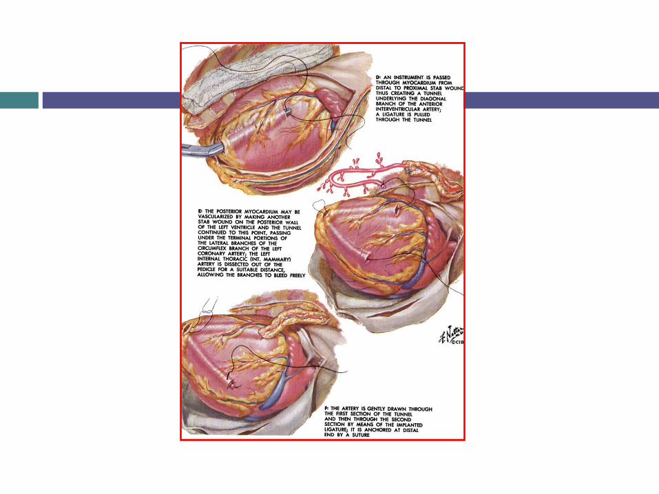
Arthur VinebergArthur Vineberg 1940’s- Mobilization of left internal mammary artery with implantation
of bleeding end into the left ventricle. 1964- follow-up study on 140 patients
33% mortality
85% relief from angina

Adult Cardiac Surgery: Ischemic Heart Disease (History)Adult Cardiac Surgery: Ischemic Heart Disease (History)
John H. Gibbon, Jr.John H. Gibbon, Jr.
Heart-lung machine May 1953- ASD closure
Adult Cardiac Surgery: Ischemic Heart Disease (History)Adult Cardiac Surgery: Ischemic Heart Disease (History)
KOLOSOV in Russia LIMA→LAD 1962- David C. Sabiston, Jr.-
Aortocoronary saphenous vein bypass
Rene Favaloro Cleveland Clinic
Frank Spencer/George Green Internal mammary artery
Adult Cardiac Surgery: Ischemic Heart Disease (CABG)Adult Cardiac Surgery: Ischemic Heart Disease (CABG)
Early and widespread acceptance of coronary bypass was
delayed.
Best known cooperative studies (1970-80’s) were the;VA
CCoronary AArtery SSurgery SStudy
European Coronary Surgery Study
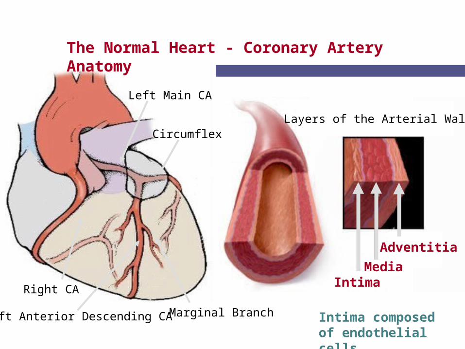
Intima
Adventitia
Media
The Normal Heart - Coronary Artery Anatomy
Left Main CA
Circumflex
Left Anterior Descending CA
Right CA
Marginal Branch
Layers of the Arterial Wall
Intima composed of endothelial cells
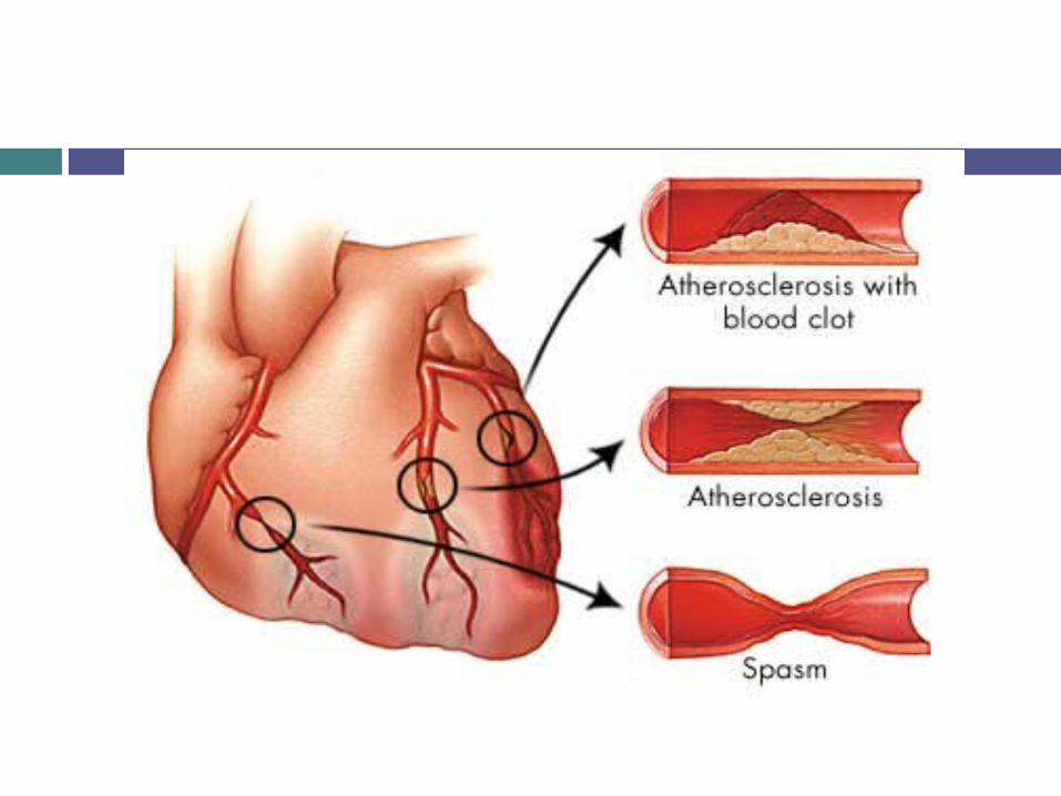

Pathogenesis of ACS

ATHEROSCLEROSIS

Risk Factors
UncontrollableUncontrollableUncontrollableUncontrollable
11
•Sex
•Hereditary
•Race
•Age
ControllableControllableControllableControllable
•High blood pressure
•High blood cholesterol
•Smoking
•Physical activity
•Obesity
•Diabetes
•Stress and anger
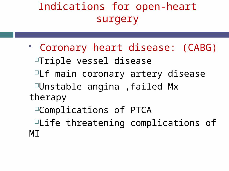
Indications for open-heart surgery
Coronary heart disease: (CABG)Triple vessel diseaseLf main coronary artery diseaseUnstable angina ,failed Mx therapyComplications of PTCALife threatening complications of MI

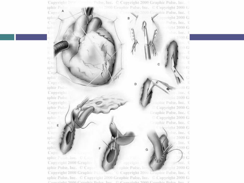
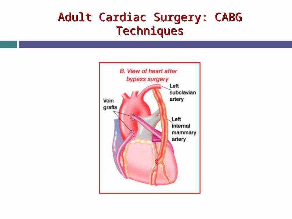
Adult Cardiac Surgery: CABG TechniquesAdult Cardiac Surgery: CABG Techniques
Median sternotomy Cardiopulmonary bypass Cardioplegic arrest Mammary artery, reversed saphenous vein, radial artery Minimally access incisions (Port Access) “Off-pump”
Adult Cardiac Surgery: CABG TechniquesAdult Cardiac Surgery: CABG Techniques
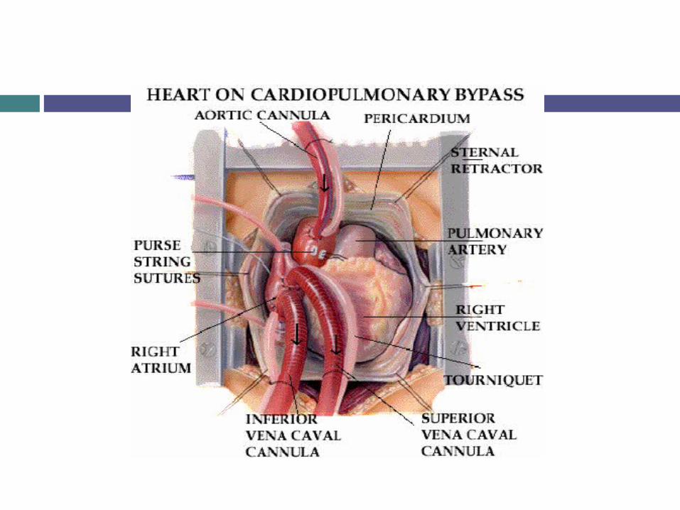

Heart Lung Machine


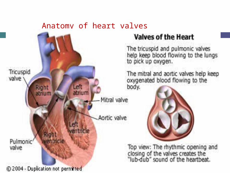
Anatomy of heart valves

Anatomy
MV:
2Cusps, Anterior and posterior The Ant is the larger Intervenes bet. A-V and aortic orifice AV: 3 semilunar cusps, ant (RT), post. Wall (LT
and post) TV; 3cusps, ant, septal ,post. PV; 3 semilunar cusps one post. (lt) two
ant( ant and rt)

AVS tricuspid and bicuspid calcifications

Adult Cardiac Surgery: Valvular Heart DiseaseAdult Cardiac Surgery: Valvular Heart Disease
Aortic stenosis-Aortic stenosis- Age-related degenerative Mild AS: AVA > 1.5cm2 ; Moderate 1-1.5cm2 ; Severe <1cm2
Indications for surgery largely based on symptoms Syncope, angina, dyspnea and CHF
Aortic regurgitation-Aortic regurgitation- Calcific aortic disease, idiopathic degenerative disease, endocarditis,
rheumatic disease, bicuspid valve, aortic dissection, Marfan, etc. Indications for surgery
Acute AR- inadequate time for ventricular compensation Chronic AR- symptoms, decreasing EF, LVEDD >75mm, LVESD >55mm
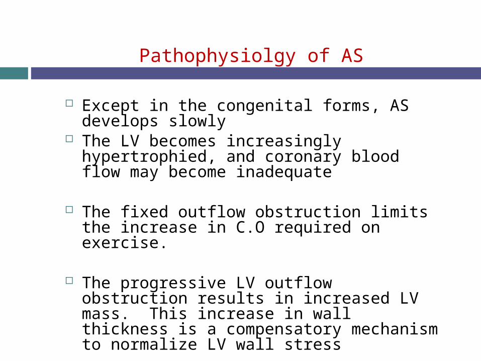
Pathophysiolgy of AS
Except in the congenital forms, AS develops slowly
The LV becomes increasingly hypertrophied, and coronary blood flow may become inadequate
The fixed outflow obstruction limits the increase in C.O required on exercise.
The progressive LV outflow obstruction results in increased LV mass. This increase in wall thickness is a compensatory mechanism to normalize LV wall stress

Symptoms of AS
Exertional dyspnea Angina Pulmonary edema Exertional syncope Sudden death

Signs of AS
Ejection systolic murmur Slow rising carotid pulse Reduce pulse pressure LV hypertrophy Signs of LV failure (crepitations,
pulmonary edema)
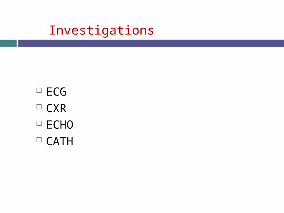
Investigations
ECG CXR ECHO CATH

ECHO criteria for assessment of aortic stenosis
severity Mean gradient(mmhg) Aortic valve area
(cm2)
mild <25 >1.5
moderate 25-50 1-1.5
severe >50 <1
critical >80 <0.7

Recommendations for Aortic Valve
Replacement in Aortic Stenosis
Symptomatic patients with severe AS
Patients with severe AS undergoing
coronary artery bypass surgery
Patients with severe AS undergoing surgery on the aorta
or other heart valves
Patients with moderate AS undergoing coronary artery bypass surgery or surgery on the aorta or other heart
valves
Asymptomatic patients with severe AS and the following;
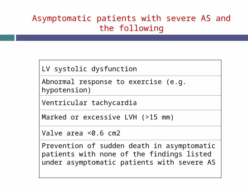
Asymptomatic patients with severe AS and the following
LV systolic dysfunction
Abnormal response to exercise (e.g. hypotension)
Ventricular tachycardia
Marked or excessive LVH (>15 mm)
Valve area <0.6 cm2
Prevention of sudden death in asymptomatic patients with none of the findings listed under asymptomatic patients with severe AS
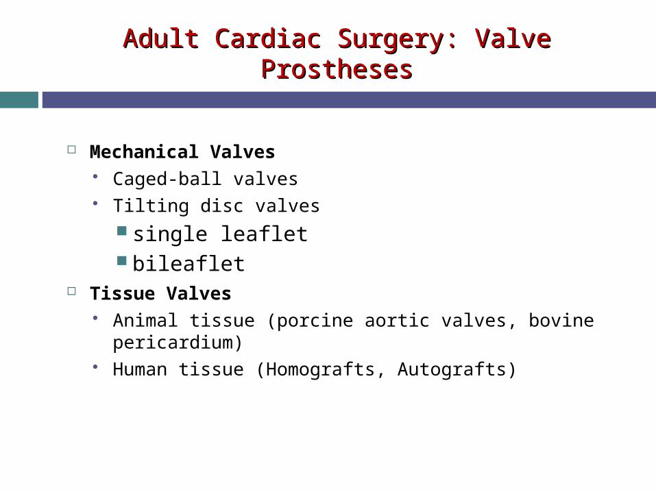
Adult Cardiac Surgery: Valve ProsthesesAdult Cardiac Surgery: Valve Prostheses
Mechanical Valves Caged-ball valves Tilting disc valves
single leaflet bileaflet
Tissue Valves Animal tissue (porcine aortic valves, bovine pericardium) Human tissue (Homografts, Autografts)

Mechanical valves
ball and cage bileaflet

Mechanical valves
tilting-disc valve

Bioprosthetic Valves Aortic homograft
Human tissue valves
autograft homograft
Animal tissue valves
Heterograft or xenograft

Adult Cardiac SurgeryAdult Cardiac Surgery

How to choose a valve
Mechanical valve in patients < 65years. Tissue valves in patients > 65 years Tissue valves in patients whose life expectancy is
< 10 year Tissue valve in patients who have problems
which are likely to cause life threatening bleeding.
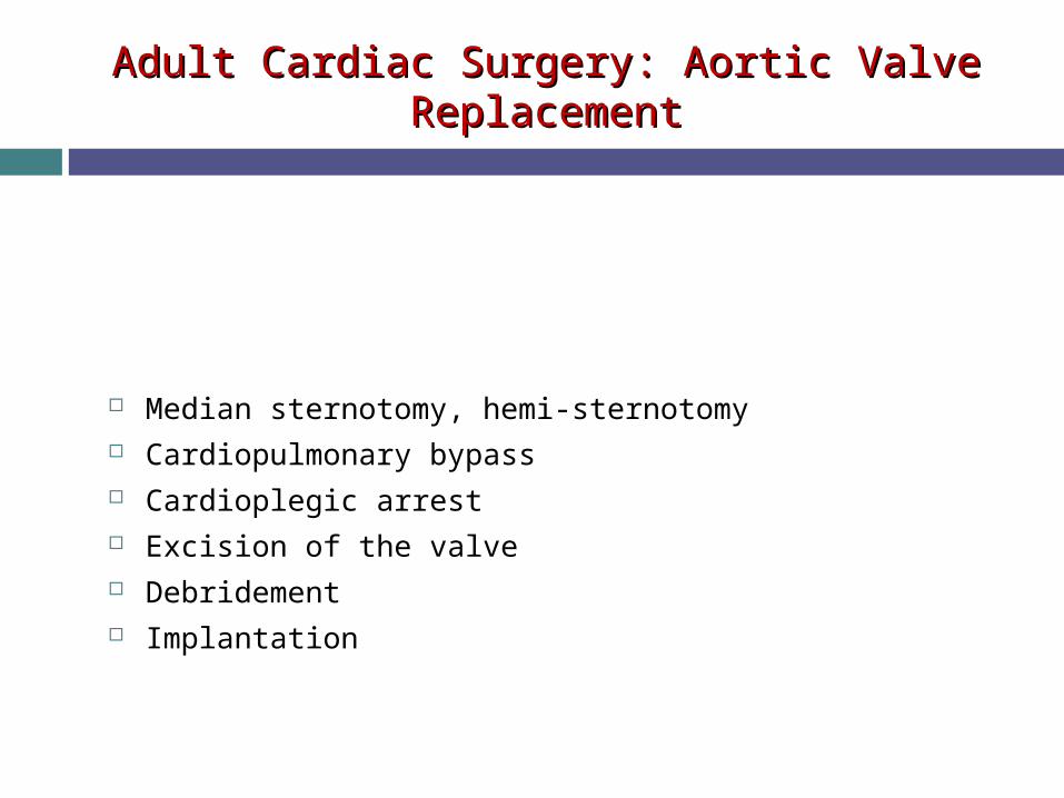
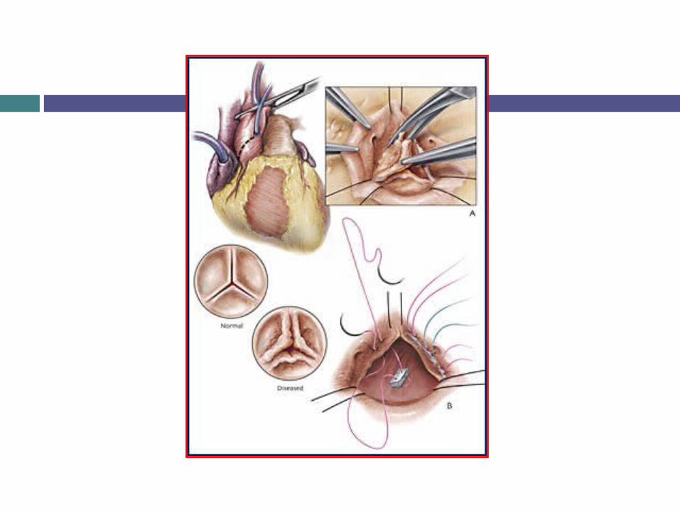
Adult Cardiac Surgery: Aortic Valve ReplacementAdult Cardiac Surgery: Aortic Valve Replacement
Median sternotomy, hemi-sternotomy Cardiopulmonary bypass Cardioplegic arrest Excision of the valve Debridement Implantation

Adult Cardiac Surgery: ACC/AHAAdult Cardiac Surgery: ACC/AHA
Aortic position Bileaflet- INR of 2-3 Other disk valves and Starr-Edwards- INR 2.5-3.5 In patients with higher risk of TE, INR 2.5-3.5 with addition of aspirin
80-100mg/d. (AF, ↓EF, prior TE, hypercoagulable state)
Mitral position All- INR 2.5-3.5

Adult Cardiac Surgery: ACC/AHAAdult Cardiac Surgery: ACC/AHA
Tissue prosthesis- Anticoagulation recommended in first 3
months, although aspirin alone in aortic position in some centers. INR 2.5-3.5
After 3 months, discontinue unless other circumstances

THANK YOU