Embed Size (px)
Citation preview

MALDI-TOF MS Analysisof Urinary Nucleosides
Bernd Kammerer, Antje Frickenschmidt, and Christoph H. GleiterInstitute of Pharmacology and Toxicology, Division of Clinical Pharmacology, University Hospital Tübingen,Tübingen, Germany
Stefan LauferInstitute of Pharmacy, University of Tübingen, Tübingen, Germany
Hartmut LiebichMedical Clinic, University Hospital Tübingen, Tübingen, Germany
As RNA turnover seems to be impaired in cancer patients, modified nucleosides havebeen evaluated as potential tumor markers. Modified nucleosides are mainly formed post-transcriptionally in tRNA, set free during RNA metabolism, and excreted in urine. Especiallymethylated nucleosides play an important role, as their levels are higher in urine from cancerpatients. For structural elucidation of known and unknown nucleosides from urine samples ofcancer patients, MALDI-TOF MS and MALDI-PSD were used for the first time. This techniquegenerally ensures high sensitivity, mass resolution, and accuracy. In our analytical approachwe prepurified nucleosides from urine by affinity chromatography and subsequently sepa-rated them by semipreparative high performance liquid chromatography. The differentfractions were collected separately and analyzed by MALDI-TOF MS and PSD-MALDI usinga mixture of six low molecular weight calibrants for internal or external calibration. Themolecular totals formulas based on a mass accuracy of 10 ppm and below were calculated anda systematic data base search was performed. The inherent problem of the MALDI-technique,the reduced sensitivity for low molecular weight substances caused by matrix suppressioneffects, was reduced by our technique. We identified several nucleosides in urine, which werepreviously identified via retention times and UV spectra of standards after HPLC analysis.Eight further nucleosides were observed. This work demonstrates for the first time thepotential of MALDI-TOF and PSD-MALDI in combination with semipreparative HPLC forassignment of nucleosides in urine. The particularly high mass accuracy of this massspectrometric method provides opportunities for identifying unknown compounds. (J AmSoc Mass Spectrom 2005, 16, 940–947) © 2005 American Society for Mass Spectrometry
More than 50 different nucleosides have beenidentified in urine from healthy people as wellas from patients suffering from different
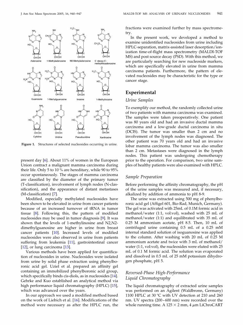
kinds of cancer; some have been shown to be elevatedin urine of cancer patients [1, 2]. They are degradationproducts of ribonucleic acid (RNA), particularly tRNA(transfer RNA), and show various modifications [1]. Anumber of nucleosides occurring in urine are shown inFigure 1. Transfer RNA contains a wide variety ofmodified nucleosides, which influence the translationalefficiency and precision as well as the sensitivity to thereading context and the reading frame maintenance.Nucleosides in precursor tRNA (pre-tRNA) are modi-fied by enzymes like tRNA-methyltransferases or -syn-thetases after the transcription. Their function and kind
Published online April 26, 2005Address reprint requests to Dr. B. Kammerer, Department of Clinical
Pharmacology, University of Tübingen, Otfried-Müller Strasse 45, D-72076Tübinigen, Germany. E-mail: [email protected]© 2005 American Society for Mass Spectrometry. Published by Elsevie1044-0305/05/$30.00doi:10.1016/j.jasms.2005.02.018
of modification (methylation, desaminization, conver-sion of uridine to pseuduridine etc.) depend on theirlocation in the tRNA chain [1, 3]. After having per-formed their task, the tRNA molecules are cleaved tonucleosides. Unmodified nucleosides are recycled forbiosynthesis of new RNA, while modified ones are notsubstrates to the salvage enzymes and thus are set freein the blood circulation and excreted with urine [1, 3]. Inthe meantime, 79 different modified nucleosides havebeen isolated from tRNA and have been characterized,all of which are derivatives of the nucleosides adeno-sine, guanosine, uridine, and cytosine [4]. Dudley et al.identified a new nucleoside, 5=-deoxycytidine, in theurine of a patient with terminal head and neck cancer[5]. In urine of head and neck cancer patients in otherstages of the disease, this nucleoside did not occur.There may be a relation between cancer stage and theappearance of 5=-deoxycytidine in urine.
In the case of breast cancer, no reliable tumor mark-
ers for diagnosis in an early stage are available to ther Inc. Received June 30, 2004Revised February 14, 2005
Accepted February 14, 2005
941J Am Soc Mass Spectrom 2005, 16, 940–947 MALDI-TOF MS ANALYSIS OF URINARY NUCLEOSIDES
present day [6]. About 11% of women in the EuropeanUnion contract a malignant mamma carcinoma duringtheir life. Only 5 to 10 % are hereditary, while 90 to 95%occur spontaneously. The stages of mamma carcinomaare classified by the diameter of the primary tumor(T-classification), involvement of lymph nodes (N-clas-sification), and the appearance of distant metastases(M-classification) [7].
Modified, especially methylated nucleosides havebeen shown to be elevated in urine from cancer patientsbecause of an increased turnover of tRNA in tumortissue [8]. Following this, the pattern of modifiednucleosides may be used in tumor diagnosis [9]. It wasshown that the levels of 1-methylinosine and N2,N2-dimethylguanosine are higher in urine from breastcancer patients [10]. Increased levels of modifiednucleosides were also observed in urine from patientssuffering from leukemia [11], gastrointestinal cancer[12], or lung carcinoma [13].
Various methods have been applied for quantifica-tion of nucleosides in urine. Nucleosides were isolatedfrom urine by solid phase extraction using phenylbo-ronic acid gel. Uziel et al. prepared an affinity gelcontaining an immobilized phenylboronic acid group,which specifically binds cis-diols, as in nucleosides [14].Gehrke and Kuo established an analytical method viahigh performance liquid chromatography (HPLC) [15],which was advanced over the years.
In our approach we used a modified method basedon the work of Liebich et al. [16]. Modifications of the
Figure 1. Structures of selected nucleosides occurring in urine.
method were necessary as after the HPLC run, the
fractions were examined further by mass spectrome-try.
In the present work, we developed a method toexamine unidentified nucleosides from urine includingHPLC-separation, matrix-assisted laser desorption/ion-ization time-of-flight mass spectrometry (MALDI-TOFMS) and post source decay (PSD). With this method, weare particularly searching for new nucleoside markers,which are specifically elevated in urine from mammacarcinoma patients. Furthermore, the pattern of ele-vated nucleosides may be characteristic for the type orcancer stage.
Experimental
Urine Samples
To exemplify our method, the randomly collected urineof two patients with mamma carcinoma was examined.The samples were taken preoperatively. One patientwas 80 years old and had an invasive ductal mammacarcinoma and a low-grade ductal carcinoma in situ(DCIS). The tumor was smaller than 2 cm and noinvolvement of the lymph nodes was diagnosed. Theother patient was 70 years old and had an invasivelobar mamma carcinoma. The tumor was also smallerthan 2 cm. Metastases were diagnosed in the lymphnodes. This patient was undergoing chemotherapyprior to the operation. For comparison, two urine sam-ples of healthy patients were also examined with HPLC.
Sample Preparation
Before performing the affinity chromatography, the pHof the urine samples was measured and, if necessary,alkalized by addition of ammonia to pH 8-9.
The urine was extracted using 500 mg of phenylbo-ronic acid gel (Affigel 601, Bio-Rad, Munich, Germany).The gel was activated with 25mL of 0.1M formic acid inmethanol/water (1:1, vol:vol), washed with 25 mL ofmethanol/water (1:1) and equilibrated with 35 mL of0.25 M ammonium acetate, pH 8.5. Then, 10 mL ofcentrifuged urine containing 0.5 mL of a 0.25 mMinternal standard solution of isoguanosine was appliedto the column. After washing with 20 mL of 0.25 Mammonium acetate and twice with 3 mL of methanol/water (1:1, vol:vol), the nucleosides were eluted with 25mL of 0.1 M formic acid. The solution was evaporatedand dissolved in 0.5 mL of 25 mM potassium dihydro-gen phosphate, pH 5.
Reversed-Phase High-PerformanceLiquid Chromatography
The liquid chromatography of extracted urine sampleswas performed on an Agilent (Waldbronn, Germany)1100 HPLC at 30 °C with UV detection at 210 and 260nm. UV spectra (200–600 nm) were recorded over the
whole running time. A 125 � 2 mm, 4 �m LiChroCART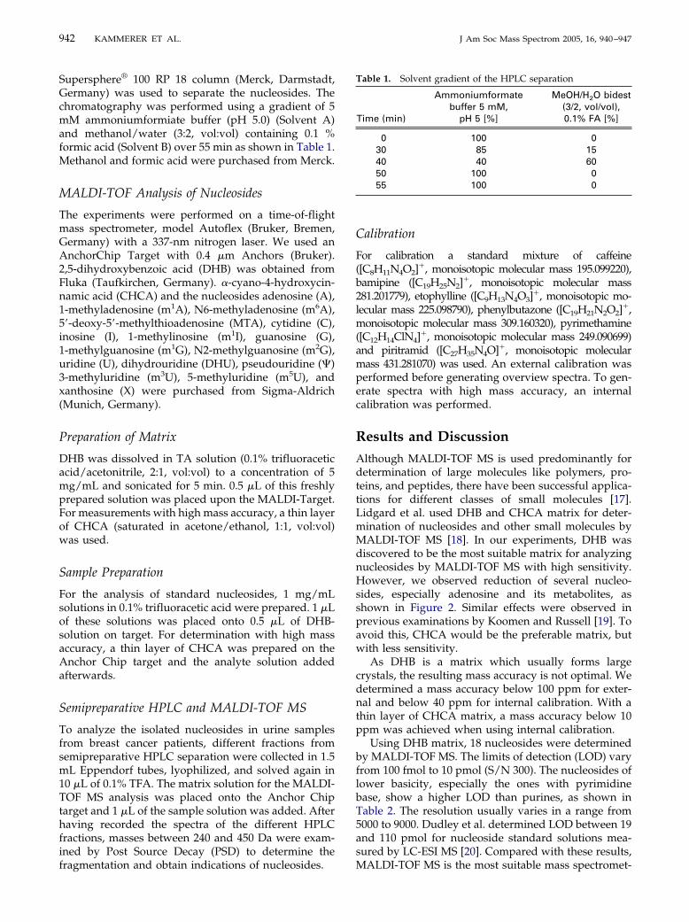
942 KAMMERER ET AL. J Am Soc Mass Spectrom 2005, 16, 940–947
Supersphere® 100 RP 18 column (Merck, Darmstadt,Germany) was used to separate the nucleosides. Thechromatography was performed using a gradient of 5mM ammoniumformiate buffer (pH 5.0) (Solvent A)and methanol/water (3:2, vol:vol) containing 0.1 %formic acid (Solvent B) over 55 min as shown in Table 1.Methanol and formic acid were purchased from Merck.
MALDI-TOF Analysis of Nucleosides
The experiments were performed on a time-of-flightmass spectrometer, model Autoflex (Bruker, Bremen,Germany) with a 337-nm nitrogen laser. We used anAnchorChip Target with 0.4 �m Anchors (Bruker).2,5-dihydroxybenzoic acid (DHB) was obtained fromFluka (Taufkirchen, Germany). �-cyano-4-hydroxycin-namic acid (CHCA) and the nucleosides adenosine (A),1-methyladenosine (m1A), N6-methyladenosine (m6A),5=-deoxy-5=-methylthioadenosine (MTA), cytidine (C),inosine (I), 1-methylinosine (m1I), guanosine (G),1-methylguanosine (m1G), N2-methylguanosine (m2G),uridine (U), dihydrouridine (DHU), pseudouridine (�)3-methyluridine (m3U), 5-methyluridine (m5U), andxanthosine (X) were purchased from Sigma-Aldrich(Munich, Germany).
Preparation of Matrix
DHB was dissolved in TA solution (0.1% trifluoraceticacid/acetonitrile, 2:1, vol:vol) to a concentration of 5mg/mL and sonicated for 5 min. 0.5 �L of this freshlyprepared solution was placed upon the MALDI-Target.For measurements with high mass accuracy, a thin layerof CHCA (saturated in acetone/ethanol, 1:1, vol:vol)was used.
Sample Preparation
For the analysis of standard nucleosides, 1 mg/mLsolutions in 0.1% trifluoracetic acid were prepared. 1 �Lof these solutions was placed onto 0.5 �L of DHB-solution on target. For determination with high massaccuracy, a thin layer of CHCA was prepared on theAnchor Chip target and the analyte solution addedafterwards.
Semipreparative HPLC and MALDI-TOF MS
To analyze the isolated nucleosides in urine samplesfrom breast cancer patients, different fractions fromsemipreparative HPLC separation were collected in 1.5mL Eppendorf tubes, lyophilized, and solved again in10 �L of 0.1% TFA. The matrix solution for the MALDI-TOF MS analysis was placed onto the Anchor Chiptarget and 1 �L of the sample solution was added. Afterhaving recorded the spectra of the different HPLCfractions, masses between 240 and 450 Da were exam-ined by Post Source Decay (PSD) to determine the
fragmentation and obtain indications of nucleosides.Calibration
For calibration a standard mixture of caffeine([C8H11N4O2]
�, monoisotopic molecular mass 195.099220),bamipine ([C19H25N2]
�, monoisotopic molecular mass281.201779), etophylline ([C9H13N4O3]
�, monoisotopic mo-lecular mass 225.098790), phenylbutazone ([C19H21N2O2]
�,monoisotopic molecular mass 309.160320), pyrimethamine([C12H14ClN4]
�, monoisotopic molecular mass 249.090699)and piritramid ([C27H35N4O]�, monoisotopic molecularmass 431.281070) was used. An external calibration wasperformed before generating overview spectra. To gen-erate spectra with high mass accuracy, an internalcalibration was performed.
Results and Discussion
Although MALDI-TOF MS is used predominantly fordetermination of large molecules like polymers, pro-teins, and peptides, there have been successful applica-tions for different classes of small molecules [17].Lidgard et al. used DHB and CHCA matrix for deter-mination of nucleosides and other small molecules byMALDI-TOF MS [18]. In our experiments, DHB wasdiscovered to be the most suitable matrix for analyzingnucleosides by MALDI-TOF MS with high sensitivity.However, we observed reduction of several nucleo-sides, especially adenosine and its metabolites, asshown in Figure 2. Similar effects were observed inprevious examinations by Koomen and Russell [19]. Toavoid this, CHCA would be the preferable matrix, butwith less sensitivity.
As DHB is a matrix which usually forms largecrystals, the resulting mass accuracy is not optimal. Wedetermined a mass accuracy below 100 ppm for exter-nal and below 40 ppm for internal calibration. With athin layer of CHCA matrix, a mass accuracy below 10ppm was achieved when using internal calibration.
Using DHB matrix, 18 nucleosides were determinedby MALDI-TOF MS. The limits of detection (LOD) varyfrom 100 fmol to 10 pmol (S/N 300). The nucleosides oflower basicity, especially the ones with pyrimidinebase, show a higher LOD than purines, as shown inTable 2. The resolution usually varies in a range from5000 to 9000. Dudley et al. determined LOD between 19and 110 pmol for nucleoside standard solutions mea-sured by LC-ESI MS [20]. Compared with these results,
Table 1. Solvent gradient of the HPLC separation
Time (min)
Ammoniumformatebuffer 5 mM,
pH 5 [%]
MeOH/H2O bidest(3/2, vol/vol),0.1% FA [%]
0 100 030 85 1540 40 6050 100 055 100 0
MALDI-TOF MS is the most suitable mass spectromet-
943J Am Soc Mass Spectrom 2005, 16, 940–947 MALDI-TOF MS ANALYSIS OF URINARY NUCLEOSIDES
ric method to achieve high sensitivity, resolution, andmass accuracy. Dudley et al. also showed characteristicfragmentation ions of nucleosides with LC-ESI MS/MS[20]. ESI MS and MALDI-TOF MS both are soft ioniza-tion techniques. With MALDI-TOF MS the mass accu-racy and resolution are usually better, while ESI MS canbe coupled with liquid chromatography. The MS/MSspectra of MALDI-TOF MS and ESI MS are similar, butnot identical. The major peak in the studies of Dudley etal. was that of the protonated nucleic base [20]. Analo-gous fragmentation was seen in our PSD spectra.
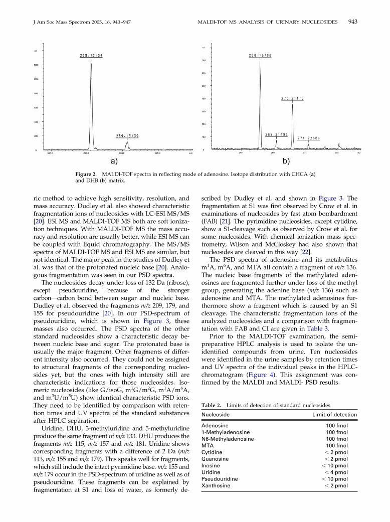
The nucleosides decay under loss of 132 Da (ribose),except pseudouridine, because of the strongercarbonOcarbon bond between sugar and nucleic base.Dudley et al. observed the fragments m/z 209, 179, and155 for pseudouridine [20]. In our PSD-spectrum ofpseudouridine, which is shown in Figure 3, thesemasses also occurred. The PSD spectra of the otherstandard nucleosides show a characteristic decay be-tween nucleic base and sugar. The protonated base isusually the major fragment. Other fragments of differ-ent intensity also occurred. They could not be assignedto structural fragments of the corresponding nucleo-sides yet, but the ones with high intensity still arecharacteristic indications for those nucleosides. Iso-meric nucleosides (like G/isoG, m1G/m2G, m1A/m6A,and m3U/m5U) show identical characteristic PSD ions.They need to be identified by comparison with reten-tion times and UV spectra of the standard substancesafter HPLC separation.
Uridine, DHU, 3-methyluridine and 5-methyluridineproduce the same fragment of m/z 133. DHU produces thefragments m/z 115, m/z 157 and m/z 181. Uridine showscorresponding fragments with a difference of 2 Da (m/z113, m/z 155 and m/z 179). This speaks well for fragments,which still include the intact pyrimidine base. m/z 155 andm/z 179 occur in the PSD-spectrum of uridine as well as ofpseudouridine. These fragments can be explained by
Figure 2. MALDI-TOF spectra in reflecting moand DHB (b) matrix.
fragmentation at S1 and loss of water, as formerly de-
scribed by Dudley et al. and shown in Figure 3. Thefragmentation at S1 was first observed by Crow et al. inexaminations of nucleosides by fast atom bombardment(FAB) [21]. The pyrimidine nucleosides, except cytidine,show a S1-cleavage such as observed by Crow et al. forsome nucleosides. With chemical ionization mass spec-trometry, Wilson and McCloskey had also shown thatnucleosides are cleaved in this way [22].
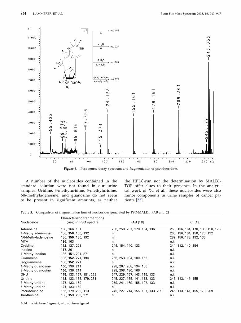
The PSD spectra of adenosine and its metabolitesm1A, m6A, and MTA all contain a fragment of m/z 136.The nucleic base fragments of the methylated aden-osines are fragmented further under loss of the methylgroup, generating the adenine base (m/z 136) such asadenosine and MTA. The methylated adenosines fur-thermore show a fragment which is caused by an S1cleavage. The characteristic fragmentation ions of theanalyzed nucleosides and a comparison with fragmen-tation with FAB and CI are given in Table 3.
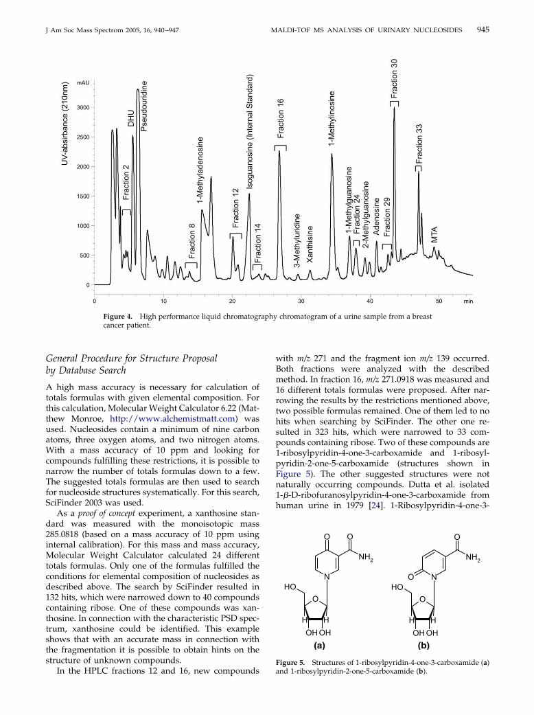
Prior to the MALDI-TOF examination, the semi-preparative HPLC analysis is used to isolate the un-identified compounds from urine. Ten nucleosideswere identified in the urine samples by retention timesand UV spectra of the individual peaks in the HPLC-chromatogram (Figure 4). This assignment was con-firmed by the MALDI and MALDI- PSD results.
Table 2. Limits of detection of standard nucleosides
Nucleoside Limit of detection
Adenosine 100 fmol1-Methyladenosine 100 fmolN6-Methyladenosine 100 fmolMTA 100 fmolCytidine � 2 pmolGuanosine � 2 pmolInosine � 10 pmolUridine � 4 pmolPseudouridine � 10 pmol
adenosine. Isotope distribution with CHCA (a)
de ofXanthosine � 2 pmol
m a
944 KAMMERER ET AL. J Am Soc Mass Spectrom 2005, 16, 940–947
A number of the nucleosides contained in thestandard solution were not found in our urinesamples. Uridine, 3-methyluridine, 5-methyluridine,N6-methyladenosine, and guanosine do not seemto be present in significant amounts, as neither
Figure 3. Post source decay spectru
Table 3. Comparison of fragmentation ions of nucleosides gene
NucleosideCharacteristic fragmentions
(m/z) in PSD spectra
Adenosine 136, 166, 181 2681-Methyladenosine 136, 150, 180, 192 n.i.N6-Methyladenosine 136, 150, 180, 192 n.i.MTA 136, 163 n.i.Cytidine 112, 137, 228 244Inosine 137, 261 n.i.1-Methylinosine 136, 151, 201, 271 n.i.Guanosine 136, 152, 271, 194 266Isoguanosine 136, 152, 271 n.i.1-Methylguanosine 166, 136, 211 2982-Methylguanosine 166, 136, 211 298DHU 115, 133, 157, 181, 229 247Uridine 113, 133, 155, 179, 231 2453-Methyluridine 127, 133, 169 2595-Methyluridine 127, 133, 169 n.i.Pseudouridine 155, 179, 209, 113 245Xanthosine 136, 153, 200, 271 n.i.
Bold: nucleic base fragment, n.i.: not investigated
the HPLC-run nor the determination by MALDI-TOF offer clues to their presence. In the analyti-cal work of Xu et al., these nucleosides were alsominor components in urine samples of cancer pa-tients [23].
nd fragmentation of pseudouridine.
by PSD-MALDI, FAB and CI
FAB [18] CI [19]
, 237, 178, 164, 136 268, 136, 164, 178, 135, 150, 176268, 136, 164, 150, 178, 192282, 150, 178, 192, 136n.i.
, 140, 133 244, 112, 140, 154n.i.n.i.
, 194, 180, 152 n.i.n.i.
, 208, 194, 166 n.i., 180, 166 n.i., 157, 143, 115, 133 n.i., 155, 141, 113, 133 245, 113, 141, 155, 169, 155, 127, 133 n.i.
n.i., 214, 155, 137, 133, 209 245, 113, 141, 155, 179, 209
n.i.
rated
, 250
, 154
, 253
, 267, 208, 229, 227, 241
, 227
945J Am Soc Mass Spectrom 2005, 16, 940–947 MALDI-TOF MS ANALYSIS OF URINARY NUCLEOSIDES
General Procedure for Structure Proposalby Database Search
A high mass accuracy is necessary for calculation oftotals formulas with given elemental composition. Forthis calculation, Molecular Weight Calculator 6.22 (Mat-thew Monroe, http://www.alchemistmatt.com) wasused. Nucleosides contain a minimum of nine carbonatoms, three oxygen atoms, and two nitrogen atoms.With a mass accuracy of 10 ppm and looking forcompounds fulfilling these restrictions, it is possible tonarrow the number of totals formulas down to a few.The suggested totals formulas are then used to searchfor nucleoside structures systematically. For this search,SciFinder 2003 was used.
As a proof of concept experiment, a xanthosine stan-dard was measured with the monoisotopic mass285.0818 (based on a mass accuracy of 10 ppm usinginternal calibration). For this mass and mass accuracy,Molecular Weight Calculator calculated 24 differenttotals formulas. Only one of the formulas fulfilled theconditions for elemental composition of nucleosides asdescribed above. The search by SciFinder resulted in132 hits, which were narrowed down to 40 compoundscontaining ribose. One of these compounds was xan-thosine. In connection with the characteristic PSD spec-trum, xanthosine could be identified. This exampleshows that with an accurate mass in connection withthe fragmentation it is possible to obtain hints on thestructure of unknown compounds.
Figure 4. High performance liquid chromatogrcancer patient.
In the HPLC fractions 12 and 16, new compounds
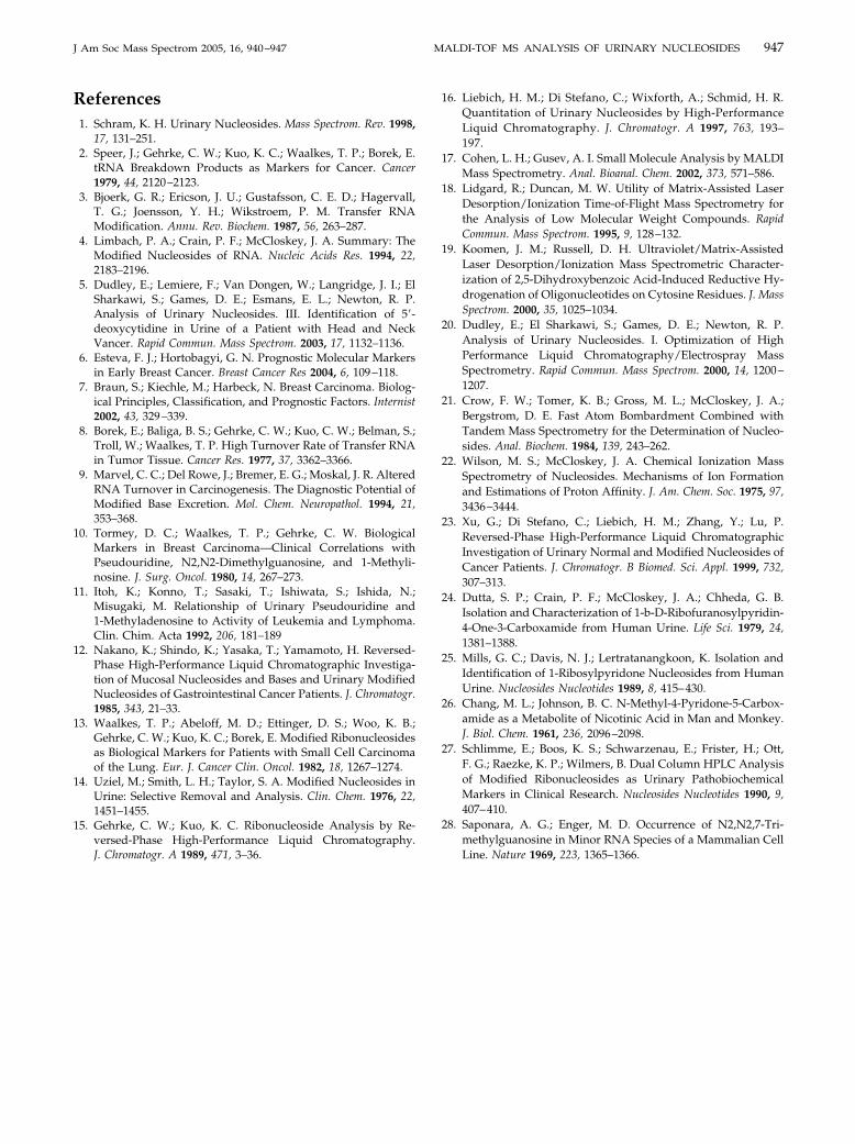
with m/z 271 and the fragment ion m/z 139 occurred.Both fractions were analyzed with the describedmethod. In fraction 16, m/z 271.0918 was measured and16 different totals formulas were proposed. After nar-rowing the results by the restrictions mentioned above,two possible formulas remained. One of them led to nohits when searching by SciFinder. The other one re-sulted in 323 hits, which were narrowed to 33 com-pounds containing ribose. Two of these compounds are1-ribosylpyridin-4-one-3-carboxamide and 1-ribosyl-pyridin-2-one-5-carboxamide (structures shown inFigure 5). The other suggested structures were notnaturally occurring compounds. Dutta et al. isolated1-�-D-ribofuranosylpyridin-4-one-3-carboxamide fromhuman urine in 1979 [24]. 1-Ribosylpyridin-4-one-3-
chromatogram of a urine sample from a breast
N
O
OHOHH H
OH
O O
NH2
NO
O
OHOHH H
OH
O
NH2
(a) (b)
Figure 5. Structures of 1-ribosylpyridin-4-one-3-carboxamide (a)
aphy
and 1-ribosylpyridin-2-one-5-carboxamide (b).

946 KAMMERER ET AL. J Am Soc Mass Spectrom 2005, 16, 940–947
carboxamide and 1-ribosylpyridin-2-one-5-carboxam-ide were isolated and identified from human urine in1989 by Mills et al. [25] Both compounds are likely to beingredients of the examined urine samples, as twoHPLC fractions contain m/z 271 producing the fragment139. The likely sources of these nucleosides are NMN orNAD [26].
Fraction 2 contained an unidentified m/z 255, whichwas analyzed with the described method as well, butonly synthetically prepared structures were proposed.Further investigations for elucidation are in progress.
Other nucleosides were observed. These are a meth-ylated cytidine (m/z 258), an acetylated cytidine (m/z286), another methylated guanosine (m/z 298), a doublemethylated guanosine (m/z 312), a triple methylatedguanosine (m/z 326), and a nucleoside with m/z of 413.This nucleoside is likely N6-threonylcarbamoyladen-osine (t6A).
As the two methylated guanosines, 1-methyl-guanosine and N2-methylguanosine, already wereidentified in urine with this chromatographic system,the most likely position for another methyl group is N7.N7-methylguanosine is another methylated nucleoside,which has been identified earlier in human urine.
N2,N2-dimethylguanosine and N6-carbamoylthreo-nyladenosine have been evaluated as tumor markers inbreast cancer patients in former studies [27]. As de-scribed, we could also identify the masses with thecorresponding fragments of these nucleosides in urinefrom breast cancer patients. Both nucleosides are majorcompounds in the urine samples, which is consistentwith the previous observations.
One peak contained m/z 326, PSD produced a m/zfragment 194, indicating the presence of a triple meth-ylated guanosine. We assume that it is N2,N2,7-tri-methylguanosine, the only known naturally occurringtrimethylguanosine with three methyl groups at thebase moiety. N2,N2,7-trimethylguanosine was de-scribed to be part of mRNA and snRNA [4, 28]. Theresults of the database search for the examined fractions
Table 4. MALDI-TOF MS and PSD MALDI results
HPLCfraction
RT[min:sec]
m/z [Da](mass accuracy [ppm])
C
2 5:00–6:00 255.09817 114,8 12:20–15:00 258.10966, (2.6,IC) 126,
12 19:30–21:30 271.09417, (4.3,IC) 139,14 23:00–24:20 298.11331, (6.2,IC) 166,16 26:00–27:40 271.09257, (1.6,IC) 139,24 37:30–38:40 308.08740a, (4.5,IC) 154,29 42:00–43:40 326.14514, (4.0,IC) 136,30 43:00–43:50 312.12905, (5.4,IC) 138,33 46:20–47:00 413.13964, (5.9,IC) 136,
Numbers in bold represent nucleic base fragmentsIC: internally calibratedasodium adduct
are shown in Table 4.
Conclusions
To the authors’ best knowledge this is the first time thatnucleosides from urine were analyzed by MALDI-TOFand PSD-MALDI. We achieved a very high sensitivityand mass accuracy for nucleosides measured by massspectrometric methods. It could be shown that nucleo-sides can be analyzed by MALDI-TOF MS using CHCAmatrix with limits of detection between 100 fmol and 10pmol. The mass accuracy was below 10 ppm whenusing internal calibration.
PSD spectra of twelve nucleosides show that purines aswell as pyrimidines decay under neutral loss of the sugarproducing the nucleic base as a fragment. Other fragmentsenhance the selectivity, as they are characteristic for thedifferent nucleosides, except those with isomeric nucleicbases, which produce similar PSD spectra.
After extraction from urine by affinity chromatographyand separation via reversed-phase HPLC, several smallmolecular masses were observed and determined byPSD-MALDI-TOF. Some compounds could be identifiedas nucleosides, while others seem to be nucleosides thatcould not yet be identified. A number of masses did notprovide any indication of being nucleosides.
With the described method including affinity chro-matography, semipreparative liquid chromatography,and MALDI-TOF MS, it is possible to propose struc-tures for nucleosides from urine. After having identi-fied new nucleosides, they should be examined for theirpotential as tumor markers by quantifying and compar-ing both the concentrations and the entire nucleosidepattern of cancer patients and healthy subjects.
AcknowledgmentsThe authors thank the DFG Graduiertenkolleg Analytische Che-mie of Tübingen University for the financial support of this work.They also thank Professor C. E. Müller and Dr. Burbiel, Depart-ment of Pharmaceutical Chemistry, University of Bonn, Germany,for providing synthetic nucleosides, and Professor J. H. Kim, Seoul
cteristic PSDagments Proposed nucleoside
Unidentified nucleoside154 Methylated cytidine (mC)164, 255 PCNR136, 150, 130, 147 7-Methylguanosine (m7G)164, 255 PCNR200, 112 N4-Acetylcytidin (ac4C)176, 194 Trimethylguanosine180, 196, 296 N2,N2-Dimethylguanosine (m2
2G)272, 282, 389 N6-Carbamoylthreonyladenosine (t6A)
harafr
123
138,152,281,152,136,158,165,214,
University, South Korea, for the donation of isoguanosine.
947J Am Soc Mass Spectrom 2005, 16, 940–947 MALDI-TOF MS ANALYSIS OF URINARY NUCLEOSIDES
References1. Schram, K. H. Urinary Nucleosides. Mass Spectrom. Rev. 1998,
17, 131–251.2. Speer, J.; Gehrke, C. W.; Kuo, K. C.; Waalkes, T. P.; Borek, E.
tRNA Breakdown Products as Markers for Cancer. Cancer1979, 44, 2120–2123.
3. Bjoerk, G. R.; Ericson, J. U.; Gustafsson, C. E. D.; Hagervall,T. G.; Joensson, Y. H.; Wikstroem, P. M. Transfer RNAModification. Annu. Rev. Biochem. 1987, 56, 263–287.
4. Limbach, P. A.; Crain, P. F.; McCloskey, J. A. Summary: TheModified Nucleosides of RNA. Nucleic Acids Res. 1994, 22,2183–2196.
5. Dudley, E.; Lemiere, F.; Van Dongen, W.; Langridge, J. I.; ElSharkawi, S.; Games, D. E.; Esmans, E. L.; Newton, R. P.Analysis of Urinary Nucleosides. III. Identification of 5=-deoxycytidine in Urine of a Patient with Head and NeckVancer. Rapid Commun. Mass Spectrom. 2003, 17, 1132–1136.
6. Esteva, F. J.; Hortobagyi, G. N. Prognostic Molecular Markersin Early Breast Cancer. Breast Cancer Res 2004, 6, 109–118.
7. Braun, S.; Kiechle, M.; Harbeck, N. Breast Carcinoma. Biolog-ical Principles, Classification, and Prognostic Factors. Internist2002, 43, 329–339.
8. Borek, E.; Baliga, B. S.; Gehrke, C. W.; Kuo, C. W.; Belman, S.;Troll, W.; Waalkes, T. P. High Turnover Rate of Transfer RNAin Tumor Tissue. Cancer Res. 1977, 37, 3362–3366.
9. Marvel, C. C.; Del Rowe, J.; Bremer, E. G.; Moskal, J. R. AlteredRNA Turnover in Carcinogenesis. The Diagnostic Potential ofModified Base Excretion. Mol. Chem. Neuropathol. 1994, 21,353–368.
10. Tormey, D. C.; Waalkes, T. P.; Gehrke, C. W. BiologicalMarkers in Breast Carcinoma—Clinical Correlations withPseudouridine, N2,N2-Dimethylguanosine, and 1-Methyli-nosine. J. Surg. Oncol. 1980, 14, 267–273.
11. Itoh, K.; Konno, T.; Sasaki, T.; Ishiwata, S.; Ishida, N.;Misugaki, M. Relationship of Urinary Pseudouridine and1-Methyladenosine to Activity of Leukemia and Lymphoma.Clin. Chim. Acta 1992, 206, 181–189
12. Nakano, K.; Shindo, K.; Yasaka, T.; Yamamoto, H. Reversed-Phase High-Performance Liquid Chromatographic Investiga-tion of Mucosal Nucleosides and Bases and Urinary ModifiedNucleosides of Gastrointestinal Cancer Patients. J. Chromatogr.1985, 343, 21–33.
13. Waalkes, T. P.; Abeloff, M. D.; Ettinger, D. S.; Woo, K. B.;Gehrke, C. W.; Kuo, K. C.; Borek, E. Modified Ribonucleosidesas Biological Markers for Patients with Small Cell Carcinomaof the Lung. Eur. J. Cancer Clin. Oncol. 1982, 18, 1267–1274.
14. Uziel, M.; Smith, L. H.; Taylor, S. A. Modified Nucleosides inUrine: Selective Removal and Analysis. Clin. Chem. 1976, 22,1451–1455.
15. Gehrke, C. W.; Kuo, K. C. Ribonucleoside Analysis by Re-versed-Phase High-Performance Liquid Chromatography.
J. Chromatogr. A 1989, 471, 3–36.16. Liebich, H. M.; Di Stefano, C.; Wixforth, A.; Schmid, H. R.Quantitation of Urinary Nucleosides by High-PerformanceLiquid Chromatography. J. Chromatogr. A 1997, 763, 193–197.
17. Cohen, L. H.; Gusev, A. I. Small Molecule Analysis by MALDIMass Spectrometry. Anal. Bioanal. Chem. 2002, 373, 571–586.
18. Lidgard, R.; Duncan, M. W. Utility of Matrix-Assisted LaserDesorption/Ionization Time-of-Flight Mass Spectrometry forthe Analysis of Low Molecular Weight Compounds. RapidCommun. Mass Spectrom. 1995, 9, 128–132.
19. Koomen, J. M.; Russell, D. H. Ultraviolet/Matrix-AssistedLaser Desorption/Ionization Mass Spectrometric Character-ization of 2,5-Dihydroxybenzoic Acid-Induced Reductive Hy-drogenation of Oligonucleotides on Cytosine Residues. J. MassSpectrom. 2000, 35, 1025–1034.
20. Dudley, E.; El Sharkawi, S.; Games, D. E.; Newton, R. P.Analysis of Urinary Nucleosides. I. Optimization of HighPerformance Liquid Chromatography/Electrospray MassSpectrometry. Rapid Commun. Mass Spectrom. 2000, 14, 1200–1207.
21. Crow, F. W.; Tomer, K. B.; Gross, M. L.; McCloskey, J. A.;Bergstrom, D. E. Fast Atom Bombardment Combined withTandem Mass Spectrometry for the Determination of Nucleo-sides. Anal. Biochem. 1984, 139, 243–262.
22. Wilson, M. S.; McCloskey, J. A. Chemical Ionization MassSpectrometry of Nucleosides. Mechanisms of Ion Formationand Estimations of Proton Affinity. J. Am. Chem. Soc. 1975, 97,3436–3444.
23. Xu, G.; Di Stefano, C.; Liebich, H. M.; Zhang, Y.; Lu, P.Reversed-Phase High-Performance Liquid ChromatographicInvestigation of Urinary Normal and Modified Nucleosides ofCancer Patients. J. Chromatogr. B Biomed. Sci. Appl. 1999, 732,307–313.
24. Dutta, S. P.; Crain, P. F.; McCloskey, J. A.; Chheda, G. B.Isolation and Characterization of 1-b-D-Ribofuranosylpyridin-4-One-3-Carboxamide from Human Urine. Life Sci. 1979, 24,1381–1388.
25. Mills, G. C.; Davis, N. J.; Lertratanangkoon, K. Isolation andIdentification of 1-Ribosylpyridone Nucleosides from HumanUrine. Nucleosides Nucleotides 1989, 8, 415–430.
26. Chang, M. L.; Johnson, B. C. N-Methyl-4-Pyridone-5-Carbox-amide as a Metabolite of Nicotinic Acid in Man and Monkey.J. Biol. Chem. 1961, 236, 2096–2098.
27. Schlimme, E.; Boos, K. S.; Schwarzenau, E.; Frister, H.; Ott,F. G.; Raezke, K. P.; Wilmers, B. Dual Column HPLC Analysisof Modified Ribonucleosides as Urinary PathobiochemicalMarkers in Clinical Research. Nucleosides Nucleotides 1990, 9,407–410.
28. Saponara, A. G.; Enger, M. D. Occurrence of N2,N2,7-Tri-methylguanosine in Minor RNA Species of a Mammalian Cell
Line. Nature 1969, 223, 1365–1366.