Embed Size (px)
Citation preview
CHAPTER 48
MALIGNANT SOFT TISSUE LESIONSOF THE FOOT AND ANKLE,
Tbm Jordan, PMSWilliam Bowman, MDClay Ballinger, DPMDonald Creen, DPM
INTRODUCTION
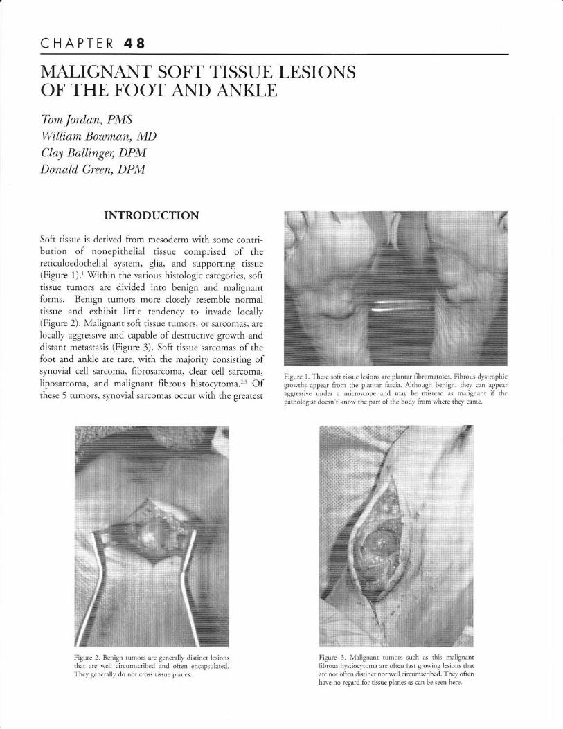
Soft tissue is derived from mesoderm with some conrri-bution of nonepithelial tissue comprised of thereticuloedothelial system, glia, and supporting tissue(Figure i).'\X/ithin the various histologic categories, softtissue tumors are divided into benign and malignantforms. Benign tumors more ciosely resemble normaltissue and exhibit little tendency to invade locally(Figure 2). Malignant soft tissue tumors, or sarcomas, are
locally aggressive and capable of destructive growth anddistant metastasis (Figure 3). Soft tissue sarcomas of thefoot and ankle are rare, with the majority consisting ofsynovial cell sarcoma, fibrosarcoma, clear cell sarcoma,
liposarcoma, and malignant fibrous histocytoma.''r Ofthese 5 tumors, synovial sarcomas occur with the greatest
Figure 2. Benign tumors are generallv distinct Iesions
that are weil circumscribed and often encapsulated.Thev generallv do nor cross tissrLe planes.
Figure 1.'Ihese soft tissue lesions are plantar fibromatoses. Fibrous dystrophicgrou.ths appear lrom the plantar fascia. Although benign, they can appear
aggressive under :r microscope and may be misread as malignant if thepathologist doesn't knolv the part of the body from where the,v came.
Figure 3. Malignant tumors such as this malignantfibrous hl,stiocl.toma are often fast grorving lesions thatare not often distinct nor well circumscribed. They oftenharre no regard for tissue planes as can be seen here.

28O CHAPTER48
frequency. Leiomyosarcoma, rhabdomyosarcoma, angis-
arcoma, and malignant schwannoma are also found in the
foot and ankle, but with much less incidence.3't Foot and
ankle sarcomas have a more benign course than sarcomas
at other locations.
INCIDENCE
Soft tissue tumors occur everl'where in the human body,
however most arise in the trunk and proximal extremities
because of the high tissue volume in these areas. In a
rerrospecrive review of 39,179 soft tissue lesions seen
from 1980 to 1989 at the Armed Forces Institute ofpathology, 8o/o of all benign soft tissue tumors and 5o/o
of all malignant soft tissue tumors occurred in the footand ankle.t
Kirby et al retrospectively analyzed 83 patients whohad a soft tissue tumor in the foot and 72 (87o/o) of thelesions were benign, while the remaining 11 (13%) were
malignant.' Five of the 11 malignant tumors were
synovial sarcomas. The age of the patient and the location
of the lesions were the most important factors that
characterized the malignant tumors in this study.
Duration of symptoms, history of trauma, and the size ofthe lesion were not useful discriminators between
malignant tumors and benign lesions.o
In a more recent study, 252 cases of malignant soft
tissue neoplasms of the foot were examined from a major
medical center specializing in the treatment of cancer. The
most common malignant soft tissue tumors were synovial
sarcoma (24o/o) followed by clear cell sarcoma (l2o/o),
fibrosarcoma (11olo), malignant fibrous histocytoma (9%)
and liposarco ma (5o/o) .2
The differential diagnosis for synovial sarcomas,
clear cell sarcomas, fibrosarcomas, malignant fibrous
histocytoma and liposarcomas are quite extensive and can
include any subcutaneous soft tissue mass of the foot or
ankle. The differential diagnosis includes but is notIimited to ganglion cyst (the most common soft tissue
lesion in the foot and ankle), lipoma, fibroma, synovial
cyst, giant cell tumor of the tendon sheath, pigmentedvillonodular synovitis, neurofibroma, epidermal inclusioncyst, Ieiomyoma, leiomyosarcoma, rhabdomyoma, and
rhabdomyosarcoma.6 A detailed history, thorough exam,
and adjunctive tests including radiographs, magnetic
resonance imaging (MRI), and computed tomography(CT) are important for an accurate diagnosis. A definitivediagnosis is made by histological studies from a fine
needle aspiration, core needle aspiration, or biopsy.l'
SYNOVIAL SARCOMA
Synovial sarcomas are the most common sarcoma of the
foot representing 5o/o of all soft tissue tumors, and
compromising25 to 560/o of al| malignant lesions.e''o The
most common site of presentation is the knee followed by
the foot and ankle region (Figure 4).'o'" Synovial sarcomas
are most commonly identified over the dorsum of the
foot along the course of the extensor tendons, or near a
joint, and are therefore often mistaken for a commonlyoccurring ganglion cysts upon first presentation.r' Thistumor does not originate from synovial structures, butrather has a true epithelial component distinguished by its
histological keratin expression. A highly organized
relationship exists between epithelial and spindle cell
components.ll The biphasic histological type contains
both epithelial and spindle components, while the
monophasic type is a spindle cell tumor with no
epithelial component. ro
Synovial sarcomas have a tendency to arise in young
adults between the ages of 15 and 40 years.' On average,
the delay between the onset of symptoms and subsequent
treatment is 1B to 24 months because they usualiy remain
quiescent for a long period of time." As with all other soft
tissue neoplasms of the foot or ankle, the most common
clinical signs and symptoms are pain, tenderness, edema,
and an enlarging mass. A careful sensory examination ofthe cutaneous innervation of the foot may provide the
extent of neoplasm spread.'
Figure 4. This synovial sarcoma is a solid lesior on the
lateral midfoot rearfoot area. The patient rv,ras havingdifficulry wearing shoes.

CHAPTER48 281
Immunohistochemistry studies are necessary in order
to make a definitive diagnosis. Most poorlv differentiated
synovial sarcomas are immunoreactive for cytokeratin,
epithelial membrane antigen, vimentin, CD99 and S100;
negative for CD34, and negative for desmin." Reactivity ofCD99 is an important indicator for a poorly differentiatedsynovial sarcoma.''
Indications for tumor severiry are not uniformamong different studies. In a study by Scully et al of the 14
patients with synovial sarcoma of the foot or ankle, those
with biphasic histological features of epithelial and spindle
components had better outcomes than those withmonophasic types.s Also, patients with prolonged
symptoms before diagnosis had better outcomes. It was
hypothesizes that patients with prolonged symptoms had
slower growing, less aggressive tumors. In a different study
where a prospective analysis was performed on 48 patients
with extremiq. and truncal synovial sarcomas, tumor size,
margin of resection and mitotic activity were prognostic
factors for survival while grade, histologic subrype and
tumor site were not.' The tumor will most often
metastasize to the lungs followed by the lymph nodes and
the bone marrow.o Once the lesion matastasises, the
outcome is often fatal.t The five-year survival rate forsynovial sarcoma is between 360/o and 650/o and has been
found to metastasize or recur in 50o/o to 70o/o of cases."
CLEAR CELL SARCOMA
Clear cell sarcoma (CCS) is a slow growing soft tissue
tumor commonly associated with tendons and aponeuroses
in the extremities of young adults between the ages of 20
and 40 years (Figure 5)." CCS represented 23 our of 189
(I2o/o) of all soft rissue sarcomas of the foot and ankle in
Figure 5. This clear ccll sarcoma is somewhat unusual as it involved bone. This
is often referred to as the rnalignant melanoma of soft tissue.
one study.'About 40o/o of all diagnosed clear cell sarcomas
occur in the foot and ankle.'3 The mass is lobulated or
multinodular, usually 2 rc 6 cm in diameter and rarely
encapsulated. CCS is melanocl'tic or mesenchymal inorigin with the mesenchymal type closely resembling
synovial sarcoma.'' It is differentiated from synovial
sarcoma by histopathologic criteria including the presence
of spindle cells and clear cells, with abundant intracellular
glycogen and occasional multinucleated giant cells.'' The
five-year survival rates are estimated between 48 and
670/o.'3 Clear cell sarcoma is also referred to as malignant
melanoma of soft parts.
FIBROSARCOMA
Fibrosarcomas represent about 10% of all sarcomas in the
foot and occur at any age but most commonly between the
ages of 30 and 55 years.' This tumor occurs in fibrous
tissue of fascial envelopes, aponeuroses, intramuscular sePta
and in tendons.'a They arise from a slow growing painless
mass more so than a painful rapidly growing mass.
Malignant transformation of plantar fibroma to
fibrosarcoma is rare, however distinction between a
fibromatosis and low-grade fibrosarcoma may be difficult
to make histologically (Figure 1). All fibrosarcomas have
microscopic characteristics of fibroblastic cells in different
Ievels of differentiation and coliagen fibers.'* High-grade
fibrosarcomas have an irregular arrangement of spindle
shaped fibroblastic cells and high mitotic actiYity
as opposed to fibromas, which have differentiatedfibroblast cells with rare mitosis.'' The 2 most imPortant
determinants for five-year survival after tumor resection are
histologic grade and size of the tumor''a
LIPOSARCOMA
Liposarcomas represent about 5olo of all sarcomas in the
foot.''a Peak incidence is between the ages of 40 and 60
years with greater propensity for males.u Liposarcomas are
usually large slow growing tumors in the intermuscular or
periarticular planes, most frequently found in the thigh.u
Grossly, liposarcomas are generally divided into distinct
lobules formed by fibrous septa. They are composed ofproliferating lipoblasts, a vascular nenvork and a matrix ofglycosaminoglycans and mucopolysaccharides.'5 Irregular
calcifications may be seen on plain radiographs. MRI stud-
ies may display a 1ow signal on T1 in some types oflipomsarcomas (mlxoid Iiposarcomas) because these
lesions contain less than l0o/o mature fatu Poorly
differentiated tumors are highly aggressive and tend to

282 CHAPTER 48
produce matastases in a high percentage of cases. The five-year survival rate of liposarcomas of the extremities is upto 70o/o with less aggressive types (myxoid and well-differentiated) having a much better prognosis than moreaggressive (round cell and pleomorphic) types.'5
MALIGNANT FIBROUS HISTOCYTOMA
Malignant fibrous histocl.toma (MFH) are the mosrcommon soft tissue tumors in adult life, most commonlyeffecting individuals 50-70 years of age (Figure l).6 Theyrepresent about 4-9o/o of all sarcomas in the foot andankle.''a Men are affected twice as frequently as women.The lesions are solitary and multilobulated masses
between 5 and 10 cm in diameter spreading along fascialplanes.'6 MFH has a wide spectrum of cellular and tissuealterations. The histopathic features will display a mixtureof fibroblastic and histiocytic elemenrs with a cartwheelpattern of streaming spindle cells.'6 Because the tumor has
a broad range of histological appearances, it is dividedinto the following subtypes: storiform-pleomorphic,mlxoid, giant cell, inflammatory, and angiomatiod.'6
IMAGING STUDIES
Radiographs may be useful by indicating the extent ofbony involvement or calcifications occurring within thelesion. The soft-tissue mass and its relationship to bonystructures can aid in the selection of further imagingmodalities. \fhiie plain radiographs are generally negative
Figure 6. MRI of Sl.novial Sarcoma in the midfoot/rearfbot area.
for sarcomas, in l5o/o to 20o/o of cases of synovialsarcomas, periosteal reactions, superficial bone erosion orfocal calcifications are seen.rT To diagnose bone invasionby a soft tissue mass, the combination of piain film andMR imaging is adequate.
MRI is the examination of choice for soft tissue
masses due to its ability to evaluate size, Iocation, tumormargin, signal homogeneity, and signal changes inadjacent soft tissue (Figure 6)." Both benign andmalignant tumors usually show a low signal intensiry onT1-weighted images, therefore signal intensiry provideslittle information about the histological grade of a givenmass. Exceptions include lipomas, some liposarcomasand hemangiomas which all have high signal intensity onT1-weighted sequences because they contain either fator blood.'' Signal intensities on T2-weighted images are
usually heterogeneous for sarcomas because of thevariable amount of hemorrhage and necrosis occurringwithin the lesion.' " A mass with a well-circumscribedsmooth border is not necessarily indicative of benignbecause sarcomas tend to grow in a centripetal fashionalong the path of least resistance and a smooth margin is
usually seen.' Computed tomography provides excellentbony detail and show thinned cortices or calcificationsthat tend to occur in synovial sarcomas.S However, CTis usually not warranted in the case of soft tissue
tumors. Angiograms or MRA studies may be helpful todifferentiate vascular tumors (Figure 7) and bone scans
may be helpful to determine metastasis (Figure B).
Figure 7. Full body bone scan.

CHAPTER 48 283
Figure 8. MRA study of the synovial sarcoma.
BIOPSY TECHNIQUES
Indications for the biopsy of soft tissue tumors include a
size of greater than 5cm, tumors exhibiting growth, and
masses present for longer than 4 weeks in duration.'tX/hen a lesion is questionable and a biopsy is needed, itis best to have the biopsy performed at a center that
specializes in musculoskeletal tumors where the surgeon
performing the biopsy will also perform the definitive
care. Over a L5-year period, The Musculoskeletal Tumor
Sociery noted a much higher incidence of biopsy related
complications when the biopsy was not performed at the
treating center.'
Fine needle aspiration involves using a fine-gauge
needle to aspirate individual cells from a mass (Figure 9).
The technique is relatively atraumatic and can be used to
sample deep tumors minimizing the potential risk for
,r-t, spillage. However, due to the limited amount ofaspirate obtained it is sometimes difficult to discern the
grade and histological type of sarcoma from the aspirate':
Core needle biopsy is commonly performed with a
Tiu-Cut or a Craig needle obtaining a thin sliver of tissue
(1 by 10 mm). If properly done, determination of both
histological type and grade of a sarcoma has been
correctly determined in over 90%o of cases.'
Using the open biopsy technique, an incision is made
to obtain the specimen. An excisional biopsy involves
the removal of the entire mass and is best used for
subcutaneous benign lesions. An incisional biopsy involves
removing a portion of the tumor for pathological studies
and leaving the remaining mass in situ. This procedure
may be performed for masses suspected of being malignant
because less tumor spillage will occur and the subsequent
definitive tumor resection will be easier with the majoriry
of the rumor in place.
Figure 9. CT directed needle biopsy of trLmor
Table 1
AMERICAN JOINT COMMITTEEON CANCER FOR THE STAGING
OF SOFT:TISSUE SARCOMA
Stage Grade Tirmor Nodal Metastasis-
Size (T) Involve-and Location ment
IAIB
IiAIIBIIC
IVAI\B
A.yAny
A.yA.y
Low <5cmLow >5cm,
superficial
No NoNo No
Low >5cm, deep NoHigh <5cm NoHigh >5cm, No
superficial
NoNoNo
III High >5cm, deeP No No
Yes NoNo Yes
STAGING
The purpose of staging a tumor is to determine the
pati.r-rt's progress and to direct treatment' The
Musculoskeletal Tumor Society system of staging is based
on grade, site and metastasis (Table 1)." Enneking
.orl.id... the grade of a lesion to be an assessment of the
biologic aggressiveness of the lesion requiring histologic,
radiographic and clinical evaluation.'e The site of the lesion
is either within the borders of its compartment of origin,

284 CHAPTER 48
contained within a compartment with extracapsularextensions, or extracompartmental. A superficial lesion inthe skin and/or subcutaneous tissue that has not penetratedthe deep fascia is considered intracompartmental.The extension of the tumor into adjacent anatomicalcompartments is considered a prognostic factor by some,but is not universally agreed upon.''"'
The American ]oint Committee on Cancer for theStaging of Soft-Tissue Sarcoma focuses on three mainevents in the life cycle of a tumor: growth, as indicated bysize of the tumor (T); spread to the lymph nodes (N);and distant metastasis (M).' In this system the sarcoma
grade is determined according to the number of mitoticfigures, and the extent of the necrosis. High-grade tumorshave a higher likelihood of metastatic spread thanlow-grade tumors.
TREAIMENT
The four general types of procedures used to excise
tumors are: intralesional, marginal, wide, and radical.-Intralesional involves incising the tumor capsule andremoving the lesion in sections. This approach is onlyappropriate for benign lesions. tWhen performing a mar-ginal excision, the tumor is shelled out around thereactive zone. This technique is also only appropriate forbenign lesions because tumor cells can be left behind.-,'o Awide excision involves the removal of viable tissue aroundthe lesion and a radical surgery involves the removal of an
entire anatomical compartment, which usually entails an
amputation when dealing with the foot or ankle.'- Forhigh-grade sarcomas, only a radical approach with normaltissue at the reactive zone can be considered tumor-free.-Because of their tendency to recur, amputations of theinvolved part followed by radiation and or chemotherapy
Figure 10. A 52-year old lemale u,ith soft tissue mass in the first intcrmetararsalspacc right foot.
is the best treatment for aggressive soft tissue tumors ofthe foot and ankle. Chemotherapy with doxorubicin,cyclophosphamide and methotrexate prolongs both theabsence of disease and survival in patients with high-grade soft tissue sarcomas of the extremities.'n The use ofchemotherapy and or radiation is especially crucial incases of limb-sparing surgery such as ray resections, whichhave lead to functionally good results for less aggressive
pseudoencapsulated sarcomas.:'o Patients undergoinglimb-sparing surgery with wide resection of the compro-mised soft tissue followed by radiation have success rates
ranging from 600/o to 100o/o for all soft tissue sarcomas ofthe extremities.'o Overall, margin of resection andadjunctive radiation and or chemotherapy are associated
with survival outcome.
CASE PRESENTAIION
A 52-year-old female was referred by her primary care
physician for evaluation of a long-standing "bump" overthe dorsum and plantar aspect of the first interspace ofher left foot which increased in size rapidly over the last
several months. She described mild tenderness over thesite of the mass. Her past medical history was significantfor hypertension. Lisinopril was her only medication. Past
surgeries included a cholecystectomy and a cesarean
section, Her only allergy is penicillin that causes her tobreak out in a rash. No family members have cancer
history and the review of systems was unremarkable.Physical exam revealed a7 cm soft and tender mass
extending between the first and second metatarsalspalpable on both the dorsal and the plantar aspect of theforefoot (Figure 10). Vascular and neurological systems
were intact. Inguinal adenopathy was not appreciated.Systemic examination was normal. Diagnostic studies
consisting of radiographs, fine needle aspiration (FNA)and MRI were then preformed. The radiographs revealed
a partially calcified plantar soft tissue mass with mild
Figure 11A, B. MRI of the synovial sarcoma of the first intermetarsal space.

CHAPTER 48 285
Figure 12A. Disarticulation of the loot at
Symmes amputation. Figure 128. lt{edialalong rvith the tibial cartilage. Figure 1 2C.
over the distal tibia.
the ankle is the first step in theand lateral malleolli are remoyedThe plantar flap of tissue is closed
erosion of the shaft of the second metatarsal. The MRIrevealed a heterogeneous soft tissue mass with some calci-
fications (Figure 11). Multiple gray to Pale elongated
tissue fragments of tissue were visible from the gross
FNA. The microscopic hematoxylin and eosin stain ofthe FNA displayed highly cellular spindle cells, S-shaped
nuclei and brisk mitotic activity. The results of the FNAwarranted a full body bone scan and a CT of the chest,
abdomen, and pelvis, all which were negative. Pathology
reported the tumor to be a monophasic synovial sarcoma.
A Symes amputation (disarticulation at the ankle
joint) was performed because a wide excision would notallow for a functional foot due to the significant size ofthe tumor (Figure 12). The lesion was viewed with the
pathologist and appeared to be solid and lobulated, and
did cross tissue planes (Figure 13). The Patient was then
consulted to oncology and began radiation therapy'
Postoperatively the patient was progressing well with a
prosthesis (Figure 14).
2
REFERENCES
Cheng EY, Thompson RC. Nerv devclopntent in the staging and
imaging of soft tissue sarcomas. ,I Bone Joint Surg Anz 1 999;8 1:882-92.
Bakotic BV, Borkowski P. Primaq' soft-tissue neoplasms of the foot:'['he clinicopathologic featurcs of 401 cases. J Foot Anhle Surg
200 1:40:28-35.Zel,toonjian T, N'fankin HJ, Gebirardt MC, Hornicek FJ. Distal lorver
extremjty sarcomas: frequencl' of occurrence and patient surviva] rate.
Foot Anhle Int 2004:25:325-30.Kirby Ef, Shercff lv{J, Lervis Mlr4. Soft-tissue trLmors and tumor-liLeIcsiorrs of the foot. J Bone Joint Sru'g Aru 1989;71:621-6.
Kransdorf J\4J: Malignant soft-tissue tumors in a larse referral
population: Distribution of diagnoses b1' age, sex, and location.
AJRl 995r 1 64:129-34.\{erd M, L)eFronzo DJ, Landsman AS, Sr4rrenant M, Sakoff M Mvxoidliposarcoma of the ank1e. ./ Fb ot Ankle Sury 199534:465'73.
Figure 1 3. \flith the pathologisc thc lesion is exposed to reveal a solid hbulated
rubbery t1'pe ofmatcrial that has clearll'crossed the local tissue planes. There is
no encapsulation of the lesion.
Figure 14. (iood prostheses are available for the
prr'enr. lollowirrg: S1 mme' rmpu.:tio,t.
7.Bos GD, Esther RJ, -ffoll TS. Foot tumors: Diagnosis and treatrnent.
J Aru Acad Orthop Sttrg 2002:10:259-70.8. Scully SP, t'emple H'f, Harrelson: Synovial sarcoma of the foot ancl
anlde. Clin Orthol, Relat Rr 1999;364:220-6.
9. Singer S, Baldini EH, Demetri GD, Fletcher JA, Corson JN{. S1'novial
sarmrna: Prognostic significancc of tumor size, margin of resecrion, and
mirotic actir.ity. / Clin Oncologt 1996;14: 1201-8.
10. Sobel E, Giorgini R, Oropeza R, Bapat K, Richardson H. Limb salvage
in recurrent synovial sarcoma of the right ankle and lowcr leg. ./ Am
Podidu'ic Med Asoc 2002:92: 90-6.11. Mietrinen N{, Virtanen I. S1'novial sarcoma-a misnomer. lP/
1 984;1 17:1 B-25.
12. Enzinger FM, Veiss S\W. S,vnovial Sarcoma. In. Enzinser FM. Weiss
SW. Soft Tissue Tumors. St. Louis: Mosby-Year Book; 1995. p. 757-85.

286 CFIAPTER 48
13.
14
Chung EB, Enzinger FM: Malignant melanoma of soft parts: Aassessment of clear cell sarcoma. Am J Surg Path 1983;7:405.Blume PA, Niemi V{, Courtright DJ, Gorecki GA. Fibrosarcoma of thefoot: A cme presentation and review ofthe literature. r/ Am Podianic Med-4ssoc 1997;36:57-4.Enzinger FM, \Zeiss S'W. Liposarcoma. In. Enzinger FM, IX/eiss SlM.Soft Tissue Tumors. St. Louis: Mosby-Year Book; 1995. p.431-66.Enzinger FM, Veiss S\M. Malignant Fibrohistiocytic Tumors. In In.Enzinger FM, \7eiss S\X/. Soft Tissue Tumors. St. Louis: Mosby-YearBook; 1995. p.351-79.
Chou LB, Malawer MM. Synovial sarcoma presenting as posterior tibialtendon dysfunction: A report of two cases and review of the literature.Foot Anlzle Int 2003;25: 810-4.Sundaram M, Mcleod RA. MR imaging of tumor and tumorlike lesions
ofbone and soft tissue. AJR 1990:155:817-24.Enneking '$7F: A System of staging musculoskeletal neoplasms. C/lzOrthop Reht Res 1986:204:9-24.
17.
18.
19.r5
16,