Embed Size (px)
Citation preview
pbtasttaaf
ISaihtpsleasoitott
1mh2t3h4STgd(G
#2e
1
ORIGINAL RESEARCH
ROLE OF MAST CELLS IN ACUPUNCTURE EFFECT: A PILOT STUDYDi Zhang, PhD,1 Guanghong Ding, MS,1# Xueyong Shen, MD,2 Wei Yao, PhD,1 Zhiying Zhang, MD,3
Yuqing Zhang, MS,1 Jun Lin, MS,1 and Quanbao Gu4
pitteus
Kt
To better understand the therapeutic effectiveness of acu-uncture, questions about the underlying mechanisms need toe addressed. Here we describe the impact of manual stimula-ion by an acupuncture needle of zusanli (stomach 36 [ST36]) onnalgesia in rats. The analgesic effect was more pronounced aftertimulation of ST36 than after stimulation of a sham point nearhe acupuncture point. At the same time, we determined inissue slices the density of mast cells in the acupuncture pointsnd nearby points, as well as the degree of degranulation beforend after stimulation. We found that the density of mast cells
rom the ST36 of rats was higher than that from a nearby sham (mmboainsa
tstluhnmomattdeltttat
sa(a-mail: [email protected].
70 © 2008 by Elsevier Inc. Printed in the United States. All Rights ReserISSN 1550-8307/08/$34.00
oint. In addition, acupuncture resulted in a remarkable increasen degranulation of the mast cells. Pretreatment of the acupunc-ure point with disodium chromoglycate not only counteractedhe phenomenon of degranulation but also reduced analgesicffect of acupuncture. Our experiments on inhibition of degran-lation of mast cells in tissue from acupuncture points demon-trates the possible role of mast cells in acupuncture effects.
ey words: Acupoint, mast cells, DSCG, analgesia, degranula-ion ratio
Explore 2008; 4:170-177. © Elsevier Inc. 2008)
NTRODUCTIONeveral thousand years of medical practice have revealed thatcupuncture, one of the elements in Traditional Chinese Med-cine, is an effective therapeutic technique to maintain goodealth and to treat various diseases.1,2 A growing body of scien-ific literature provides strong evidence for the efficacy of acu-uncture in analgesia.3,4 Analgesic effects of acupuncture areystemic, whereas acupuncture manipulations themselves areocal.5-7 However, the physiological mechanisms underlying theffects following stimulation of acupuncture points (acupoints)re still unclear; this is one of the problems which needs to beolved to establish a scientific basis of acupuncture and is alsone of the important problems in meridian research. Meridiansndicate pathways along which the Qi (energy) is spread throughhe human body. These pathways form the basis for the spreadf the signals elicited by acupuncture. Meridian research inves-igates the physiological and physical mechanisms underlyinghe signal distribution.8-11 It is believed that during the needling
Shanghai Research Center of Acupuncture and Meridians, Depart-ent of Mechanics and Engineering Science, Fudan University, Shang-
ai, ChinaCollege of Acupuncture-Moxibustion, Shanghai University of Tradi-
ional Chinese Medicine, Shanghai, ChinaDepartment of Anatomy, Second Military Medical University, Shang-ai, ChinaShanghai Institute of Cell Biology, Chinese Academy of Sciences,hanghai, Chinahis work was supported by grants from National Basic Research Pro-ram of China (973 Program, No: 2005CB523306, GD), Science Foun-ation of Shanghai Municipal Commission of Science and TechnologyNo: 05DZ19745, 06DZ19732, 064319053, 07DZ19722, 07DZ19733,D) and Fudan University Doctorate Innovation Foundation (DZ).
Corresponding Author. Address: 199 Guo Shou Jing Road, Shanghai,01203, China
anipulation process, the application of lift and thrust and twistanipulation pulls the surrounding tissue around the needle
ody, delivering the cellular signal conducted along the pathwayf channels (meridians) and leading to downstream effects thatctivate certain cellular pathways and facilitate healing.9 Exam-nation of tissue morphology using techniques including mag-etic resonance imaging and computed tomography have alsoupported the possibility that the physical basis of meridians andcupoints is a complex system based on connective tissue.12
As one of the resident cells in the loose connective tissue fromhe human body, mast cells have attracted the interest of re-earchers since the 1980s. One working hypothesis has been thathe so-called De Qi sensation in response to acupuncture is re-ated to the response of local mast cells.13 De Qi sensation is anique needle sensation (grasp), including tingling, numbness,eaviness, and other feelings that occur after an acupunctureeedle has properly been placed in the acupoint.4,9 Throughechanical stimulation, acupuncture can cause degranulation
f the local mast cells in the acupoint and promote release ofediators, including arachidonic acid products, biogenic
mines, chemoattractants, cytokines, growth factors, neuropep-ides, proteoglycans, and proteolytic enzymes.14 These media-ors act in several biological ways.15 For example, histamine canilate capillary vessels and venules, which act on the vascularndothelium to expose the basal membrane and exude the tissueiquid, resulting in a change of electric potential gradient in theissue along meridians.13 Another example of how the release ofhese mediators can effect systemic change is the way in whichhe neuropeptide serotonin, involved in analgesia, body temper-ture regulation, and nerve activity, can affect endocrine func-ion in the body.16
In the present study, our aim was to investigate the relation-hip between degranulation of mast cells in acupoints and thenalgesic effect of acupuncture. The change of pain thresholdPT) in Spraque-Dawley rat tails was adopted as a measure for the
nalgesic effect.17,18 The acupoint commonly used for analgesia,ved EXPLORE May/June 2008, Vol. 4, No. 3doi:10.1016/j.explore.2008.02.002
zaarpp(tfocp
MAWrtfla2mpumecsioNiaGN
NTlhtu3lttTsprtbew
ASelsMSawde0Rtbrhte
Fcd
Fat
M
usanli (stomach 36, [ST36]), was selected to apply the liftingnd thrusting and twisting stimulation19-21 under the control ofclinic needle real-time force monitor.22,23 To investigate the
ole of mast cell degranulation in the process of generation ofain analgesia in response to acupuncture, we treated the acu-oint with the mast cell stabilizer disodium chromoglycateDSCG)24 and compared the effects of acupuncture in thereated points with those not treated with DSCG. In addition,rom in vitro skin and tissue specimens, we estimated the densityf mast cells in acupoints and nearby sham points, and also thehange of degree of degranulation of mast cells from the acu-oints in response to acupuncture.
ETHODSnimals and Groupse used 52 male and female laboratory-born Spraque-Dawley
ats provided by the Shanghai Experimental Animal Centre ofhe Chinese Academy of Science. They all exhibited normal tailick latency and weighed 200 � 20 g (at age of six weeks). Thenimals were housed in cages at controlled temperature (20 �°C) with a 14/10 hour light/dark cycle. Food and water wereade available ad lib. All animals were handled with care to
revent infection and to minimize stress. Two rats each weresed, for control and acupuncture, and for transmission electronicroscopy. The remaining rats (48) were randomly divided into
ight groups using a random-number table, six rats each forontrol group (A); acupuncture to ST36 (B1); acupuncture toham point nearby ST36 (B2); injection of DSCG to ST36 (C1);njection of NaCl to ST36 (C2); acupuncture after pretreatmentf DSCG to ST36 (D1); acupuncture after pretreatment withaCl to ST36 (D2); and acupuncture after DSCG pretreatment
n the sham point (D3). All the experiments were performed inccordance with the principles and procedures outlined in theuide for the Care and Use of Laboratory Animals issued by the U.S.
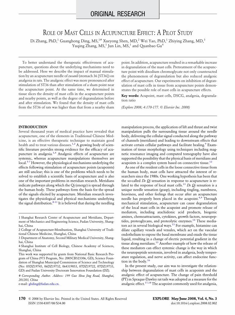
igure 1. The structure and principle of force detection. The me-hanical sensor was used to detect the real-time force on the needleuring acupuncture manipulation.
ational Academy of Sciences. n
ast Cells in Acupuncture
ociceptive Testing Modelhe rat tail was exposed to radiation heat, and the tail flick
atency was measured to detect the heat sensitivity. This modelas been extensively applied in previous research on acupunc-ure analgesia.25 The method is characterized by simplicity, easyse, and reproducibility of results. In our experiments, a model3T tail stimulator analgesia meter (IITC Life Sciences, Wood-and Hills, Calif) was used to apply the heat stimulation. Eachime, the skin of the rat tail, with an area of 4 mm2 and 2 cm fromhe tail tip, was stimulated with 40% of maximum light strength.he room temperature was controlled within 22 � 1°C. A 20-econd cutoff maximum was programmed into the timer torevent tissue damage. Rats were habituated to the testing appa-atus for 10 minutes prior to testing. Then, we successively de-ermined the temperature for tail flick three times to obtain theasic PT as average (five-minute intervals were allowed betweenach test). Ten minutes after testing the basic PT, the experimentas started.
cupuncture Stimulationince zusanli is a popular acupoint for analgesia studies in animalxperiments20,21 as well as for clinical treatment,3,4 it was se-ected as the acupoint in our experiments. Sterilized stainlessteel acupuncture needles (0.25 mm, 1 inch, Suzhou Kangnian
edical Devices Co, Lt, Suzhou, China) were inserted intoT36 of the right hind leg, located 5-mm lateral and distal to thenterior tubercle of the tibia.26 (The acupuncture point dosageas as follows: from Table 6-1 of Li (2003),26 the equivalentosage was calculated according to the surface area ratio betweenxperimental animal and the human being, which in our case is.018, according to conversion formula: DB � (DA � WA) �ATE/WB. [Note: DB is the dosage for each kilogram weight of
he rat. DA is the dosage for each kilogram weight of the humaneing. WA and WB are the weight of the human being and theat. RATE is the ratio coefficients of the surface area from rat anduman being, RATE � 0.018]. DA � WA � 1 mL, WB � 200 g;hus, DB � 0.09 mL/kg. It is about 20 �L of DSCG solution forach injection with a concentration of DSCG of 0.02 g/mL). The
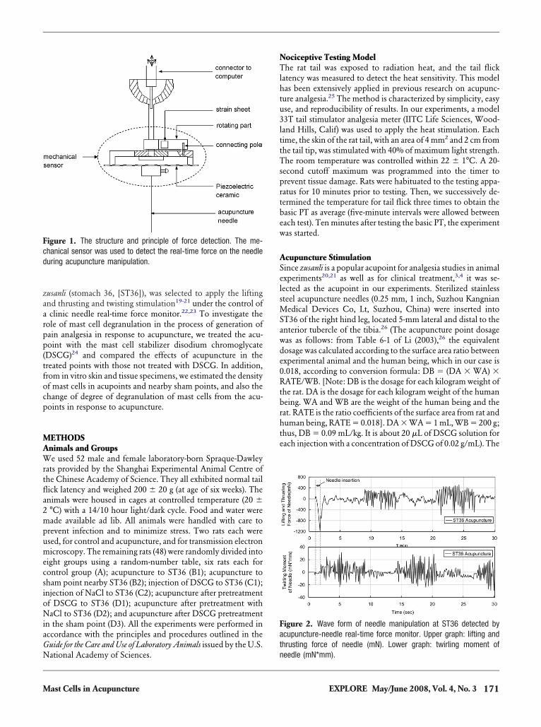
igure 2. Wave form of needle manipulation at ST36 detected bycupuncture-needle real-time force monitor. Upper graph: lifting andhrusting force of needle (mN). Lower graph: twirling moment of
eedle (mN*mm).171EXPLORE May/June 2008, Vol. 4, No. 3
sap
tnad
Qt
FtmrPraS
F2tsrtsC
FmaDDbt
T
ABB
C
C
D
D
D
S
1
ite 3 mm nearby ST36 (in the direction of the fibula) was takens the sham point. The locations of acupoint and nearby shamoint were clearly separated as shown previously by adding gen-
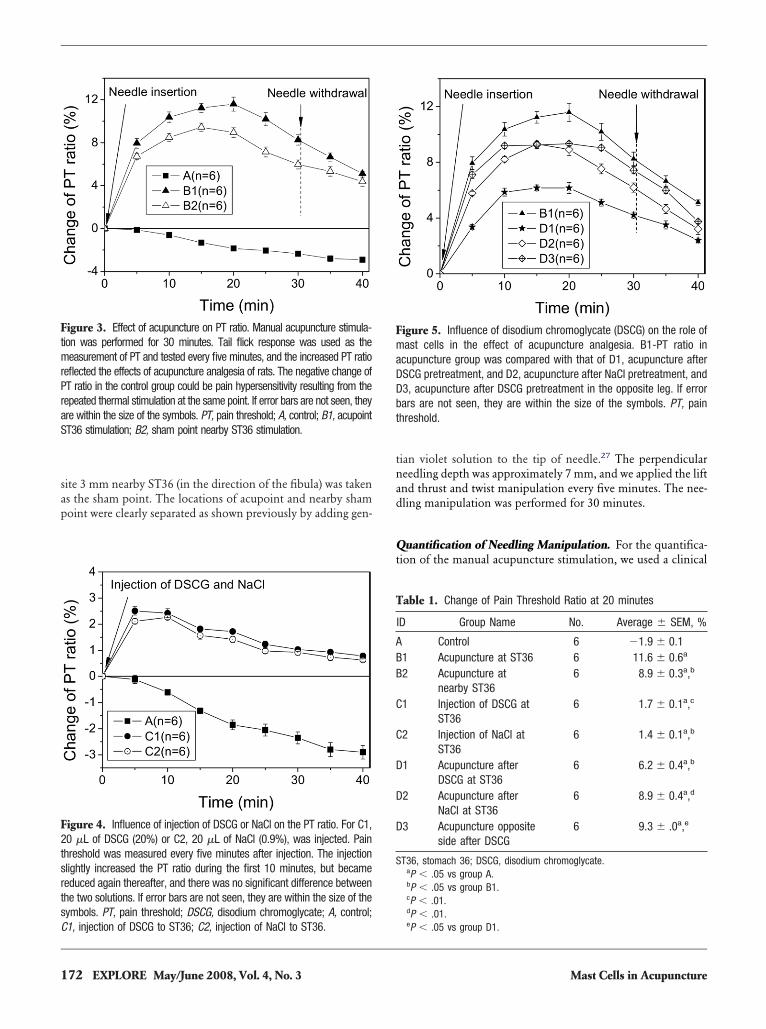
igure 3. Effect of acupuncture on PT ratio. Manual acupuncture stimula-ion was performed for 30 minutes. Tail flick response was used as theeasurement of PT and tested every five minutes, and the increased PT ratio
eflected the effects of acupuncture analgesia of rats. The negative change ofT ratio in the control group could be pain hypersensitivity resulting from theepeated thermal stimulation at the same point. If error bars are not seen, theyre within the size of the symbols. PT, pain threshold; A, control; B1, acupointT36 stimulation; B2, sham point nearby ST36 stimulation.
igure 4. Influence of injection of DSCG or NaCl on the PT ratio. For C1,0 �L of DSCG (20%) or C2, 20 �L of NaCl (0.9%), was injected. Painhreshold was measured every five minutes after injection. The injectionlightly increased the PT ratio during the first 10 minutes, but becameeduced again thereafter, and there was no significant difference betweenhe two solutions. If error bars are not seen, they are within the size of theymbols. PT, pain threshold; DSCG, disodium chromoglycate; A, control;
1, injection of DSCG to ST36; C2, injection of NaCl to ST36.72 EXPLORE May/June 2008, Vol. 4, No. 3
ian violet solution to the tip of needle.27 The perpendiculareedling depth was approximately 7 mm, and we applied the liftnd thrust and twist manipulation every five minutes. The nee-ling manipulation was performed for 30 minutes.
uantification of Needling Manipulation. For the quantifica-ion of the manual acupuncture stimulation, we used a clinical
igure 5. Influence of disodium chromoglycate (DSCG) on the role ofast cells in the effect of acupuncture analgesia. B1-PT ratio in
cupuncture group was compared with that of D1, acupuncture afterSCG pretreatment, and D2, acupuncture after NaCl pretreatment, and3, acupuncture after DSCG pretreatment in the opposite leg. If errorars are not seen, they are within the size of the symbols. PT, painhreshold.
able 1. Change of Pain Threshold Ratio at 20 minutes
ID Group Name No. Average � SEM, %
Control 6 �1.9 � 0.11 Acupuncture at ST36 6 11.6 � 0.6a
2 Acupuncture atnearby ST36
6 8.9 � 0.3a,b
1 Injection of DSCG atST36
6 1.7 � 0.1a,c
2 Injection of NaCl atST36
6 1.4 � 0.1a,b
1 Acupuncture afterDSCG at ST36
6 6.2 � 0.4a,b
2 Acupuncture afterNaCl at ST36
6 8.9 � 0.4a,d
3 Acupuncture oppositeside after DSCG
6 9.3 � .0a,e
T36, stomach 36; DSCG, disodium chromoglycate.aP � .05 vs group A.bP � .05 vs group B1.cP � .01.dP � .01.
eP � .05 vs group D1.Mast Cells in Acupuncture
M
173ast Cells in Acupuncture EXPLORE May/June 2008, Vol. 4, No. 3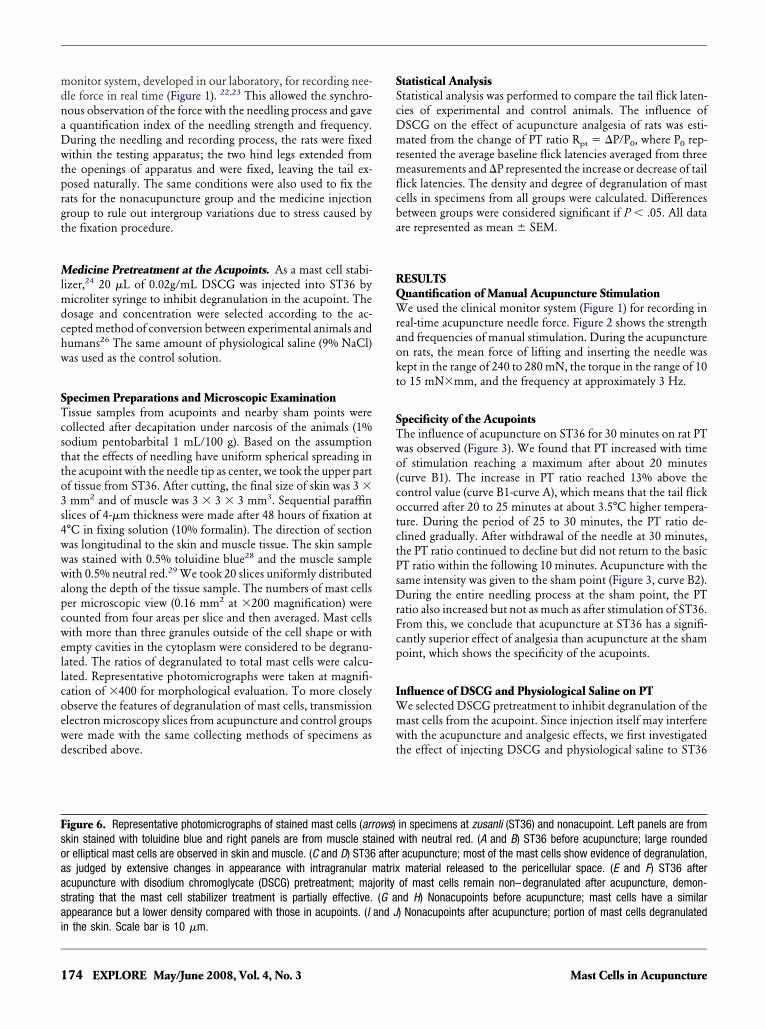
mdnaDwtprgt
Mlmdchw
STcstto3s4wwwapcwellcoewd
SScDmrmflcba
RQWraokt
STwo(cotctPsDrFcp
IWmwt
Fsoaasai
1
onitor system, developed in our laboratory, for recording nee-le force in real time (Figure 1). 22,23 This allowed the synchro-ous observation of the force with the needling process and gavequantification index of the needling strength and frequency.uring the needling and recording process, the rats were fixedithin the testing apparatus; the two hind legs extended from
he openings of apparatus and were fixed, leaving the tail ex-osed naturally. The same conditions were also used to fix theats for the nonacupuncture group and the medicine injectionroup to rule out intergroup variations due to stress caused byhe fixation procedure.
edicine Pretreatment at the Acupoints. As a mast cell stabi-izer,24 20 �L of 0.02g/mL DSCG was injected into ST36 by
icroliter syringe to inhibit degranulation in the acupoint. Theosage and concentration were selected according to the ac-epted method of conversion between experimental animals andumans26 The same amount of physiological saline (9% NaCl)as used as the control solution.
pecimen Preparations and Microscopic Examinationissue samples from acupoints and nearby sham points wereollected after decapitation under narcosis of the animals (1%odium pentobarbital 1 mL/100 g). Based on the assumptionhat the effects of needling have uniform spherical spreading inhe acupoint with the needle tip as center, we took the upper partf tissue from ST36. After cutting, the final size of skin was 3 �mm2 and of muscle was 3 � 3 � 3 mm3. Sequential paraffin
lices of 4-�m thickness were made after 48 hours of fixation at°C in fixing solution (10% formalin). The direction of sectionas longitudinal to the skin and muscle tissue. The skin sampleas stained with 0.5% toluidine blue28 and the muscle sampleith 0.5% neutral red.29 We took 20 slices uniformly distributedlong the depth of the tissue sample. The numbers of mast cellser microscopic view (0.16 mm2 at �200 magnification) wereounted from four areas per slice and then averaged. Mast cellsith more than three granules outside of the cell shape or withmpty cavities in the cytoplasm were considered to be degranu-ated. The ratios of degranulated to total mast cells were calcu-ated. Representative photomicrographs were taken at magnifi-ation of �400 for morphological evaluation. To more closelybserve the features of degranulation of mast cells, transmissionlectron microscopy slices from acupuncture and control groupsere made with the same collecting methods of specimens asescribed above.
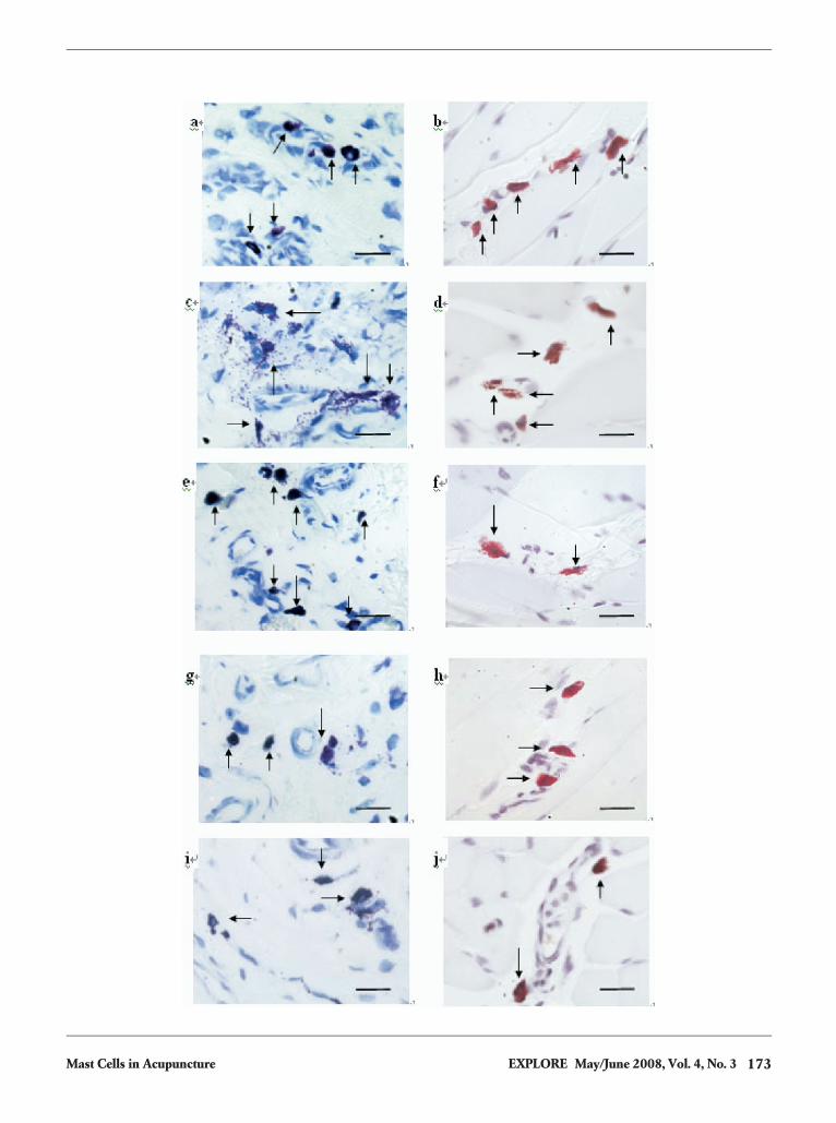
igure 6. Representative photomicrographs of stained mast cells (arrkin stained with toluidine blue and right panels are from muscle star elliptical mast cells are observed in skin and muscle. (C and D) ST36s judged by extensive changes in appearance with intragranular mcupuncture with disodium chromoglycate (DSCG) pretreatment; matrating that the mast cell stabilizer treatment is partially effective.ppearance but a lower density compared with those in acupoints. (I
n the skin. Scale bar is 10 �m.74 EXPLORE May/June 2008, Vol. 4, No. 3
tatistical Analysistatistical analysis was performed to compare the tail flick laten-ies of experimental and control animals. The influence ofSCG on the effect of acupuncture analgesia of rats was esti-ated from the change of PT ratio Rpt � �P/P0, where P0 rep-
esented the average baseline flick latencies averaged from threeeasurements and �P represented the increase or decrease of tailick latencies. The density and degree of degranulation of mastells in specimens from all groups were calculated. Differencesetween groups were considered significant if P � .05. All datare represented as mean � SEM.
ESULTSuantification of Manual Acupuncture Stimulatione used the clinical monitor system (Figure 1) for recording in
eal-time acupuncture needle force. Figure 2 shows the strengthnd frequencies of manual stimulation. During the acupuncturen rats, the mean force of lifting and inserting the needle wasept in the range of 240 to 280 mN, the torque in the range of 10o 15 mN�mm, and the frequency at approximately 3 Hz.
pecificity of the Acupointshe influence of acupuncture on ST36 for 30 minutes on rat PTas observed (Figure 3). We found that PT increased with timef stimulation reaching a maximum after about 20 minutescurve B1). The increase in PT ratio reached 13% above theontrol value (curve B1-curve A), which means that the tail flickccurred after 20 to 25 minutes at about 3.5°C higher tempera-ure. During the period of 25 to 30 minutes, the PT ratio de-lined gradually. After withdrawal of the needle at 30 minutes,he PT ratio continued to decline but did not return to the basicT ratio within the following 10 minutes. Acupuncture with theame intensity was given to the sham point (Figure 3, curve B2).uring the entire needling process at the sham point, the PT
atio also increased but not as much as after stimulation of ST36.rom this, we conclude that acupuncture at ST36 has a signifi-antly superior effect of analgesia than acupuncture at the shamoint, which shows the specificity of the acupoints.
nfluence of DSCG and Physiological Saline on PTe selected DSCG pretreatment to inhibit degranulation of theast cells from the acupoint. Since injection itself may interfereith the acupuncture and analgesic effects, we first investigated
he effect of injecting DSCG and physiological saline to ST36
in specimens at zusanli (ST36) and nonacupoint. Left panels are fromwith neutral red. (A and B) ST36 before acupuncture; large roundedacupuncture; most of the mast cells show evidence of degranulation,material released to the pericellular space. (E and F) ST36 after
of mast cells remain non–degranulated after acupuncture, demon-nd H) Nonacupoints before acupuncture; mast cells have a similar) Nonacupoints after acupuncture; portion of mast cells degranulated
ows)inedafteratrix
jority(G a
and J
Mast Cells in Acupuncture
wtPcsi
IABabF(Arotitttomp
NMaob
atdbamcftbc
DTtsd
Ffga(bo(
T
AN
T
CAAA
M
M
ithout acupuncture (Figure 4). It is shown that injection ofhese two solutions had similar effects. After a slight increase ofT ratio within the first 10 minutes, a slow gradual decreaseould be observed that may be attributed to sensitization as alsoeen in the control group. Hence, the influence from injectiontself became very weak after 20 minutes.
nfluence of DSCG and Physiological Saline oncupuncture Effectased on the above result on the influence of injection, wepplied the needling 20 minutes after injection. The PT valueefore injection was taken as the basal PT and subtracted.igure 5 and Table 1 demonstrate that pretreatment of DSCGcurve D1) attenuated the analgesic effects of acupuncture.fter 20 minutes of acupuncture, when the maximum PT
atio was obtained, the sites with DSCG pretreatment dem-nstrated a reduction of the ratio by 5.4 (D1-B1), which waswice the decrease (2.7) seen in the sites pretreated with phys-ological saline 2.7 (D2-B1). A similarly small effect (2.3) onhe PT ratio was obtained after 20 minutes of acupuncture athe opposite leg (D3-B1). We concluded from these findingshat the three kinds of pretreatment all weakened the effectsf acupuncture on PT, but to different degrees. Stabilizing theast cells in acupoint with DSCG clearly made the most
ronounced attenuating difference.
umber of Mast Cells and the Degranulation Phenomenonast cell density and degranulation was investigated by light
nd electron microscopy. In the stained tissue slices, we couldbserve the basophilic granules of the metachromia in mast cellsy light microscopy (Figure 6A-J). The density of mast cells in
able 2. Comparison of Mast Cells in Acupoints and Nonacupoints
Group Name
No. of Mast Cellsa
Skin Muscle
cupoint (n � 20) 34.2 � 2.5 12.4 � 1.4onacupoint (n � 20) 22.1 � 1.7b 8.5 � 1.0b
aAverage � SEM; 0.16 mm2/microscopic view (�200).bP � .01 vs acupoint group.
able 3. Degranulation Ratio of Mast Cells in Acupoints
Group Name
MC Degranulation Ratioa
Skin, % Muscle, %
ontrol (n � 6) 29.2 � 1.2 26.3 � 1.8cupuncture (n � 6) 48.9 � 3.9b 32.2 � 3.1c
cupuncture after DSCG (n � 6) 33.7 � 2.8d 28.5 � 2.3cupuncture after NaCl (n � 6) 50.4 � 2.5b 29.7 � 2.3
C, mast cell; DSCG, disodium chromoglycate.aAverage � SEM.bP � .01.cP � .05 vs control group.
bdP � .01 vs acupuncture group.
ast Cells in Acupuncture
cupoint skin and muscle tissues was significantly higher thanhat from the sham point (Table 2). The acupuncture raised theegree of degranulation of the mast cells in the acupoint area ofoth skin and muscle (Table 3). After DSCG pretreatment, thecupuncture could not significantly facilitate degranulation ofast cells in skin and muscle of the acupoints. In electron mi-
roscopy (Figure 7), we observed more closely the degranulationeature in tissue slices after acupuncture; the cell membranehinned and extensive intragranular matrix material appeared toe released to the pericellular space with empty cavities in theytoplasm.
ISCUSSIONhis article presents results that suggest a role of mast cells from
he acupoints in the analgesic effects of acupuncture. The mea-urements of the analgesia and the observations of mast cellensity and degranulation in tissue slices indicate a correlation
igure 7. Transmission electron microscopy photomicrographs ofeatured mast cells at zusanli (ST36) in control and acupunctureroup. (A) Skin mast cell without acupuncture, rounded in shape withcomplete intact cytomembrane densely packing the intragranules
arrow). (B) Skin mast cell after acupuncture, which has a thin androken cytomembrane, with signs of degranulation, granules outsidef the cell shape (arrowhead) and empty cavity in the cytoplasmasterisk).
etween them.
175EXPLORE May/June 2008, Vol. 4, No. 3
bpon
atiaii
tdWtwefedict
stsaptgmptbsmt
iiia6slat
tArinmd
flmtfcdttnat
AWPpt
R
Fmntcmr
1
First, with the same stimulation intensity during acupuncture,etter effects of analgesia were obtained by stimulation at acu-oints than at the nearby sham points. In addition, the densitiesf mast cells in acupoints in skin was 54.9% higher than inearby points, and in muscle was 45.3% higher.Second, along with the increase in PT caused by acupuncture,
facilitation in degranulation of the mast cells was observed inhe acupoints. After the inhibition of degranulation of mast cellsn acupoints by DSCG, this analgesia was significantly attenu-ted. This suggests that mast cells from the acupoint area partic-pate in the acupuncture effect, and degranulation of mast cellss an important link in producing this effect.
Third, there was also an increase of PT in response to acupunc-ure of nonacupoints, and the mast cells in nonacupoints alsoegranulated, but the effects were significantly less pronounced.e hypothesize that, although acupuncturing at nonacupoints,
he manipulation of lifting and thrusting, twisting and twirlingill be partially transmitted to the neighboring acupoint. Nev-rtheless, compared with ST36, the nearby sham point uni-ormly displays weaker analgesia than the real point during thentire acupuncture procedure. Based on the observation that theensities of mast cells in acupoints are significantly higher thann nonacupoints, the influence from degranulation in acupointsan be expected to be stronger than from the degranulation inhe nonacupoints.
Our results confirm previous findings that the mast cell den-ities are about 50% higher in acupoints than in nonacupoints;his was shown previously for several other acupoints, includinganyinjiao (SP6), yanglingquan (GB34), hegu (LI4), quchi (LI11),nd neiguan (PC6).13 In this study, we also demonstrate that thehenomenon of mast cell degranulation may be correlated withhe analgesic effect of acupuncture. The reason for greater anal-esic effectiveness in acupoints compared with nonacupointsay be the result of the higher densities of mast cells in acu-
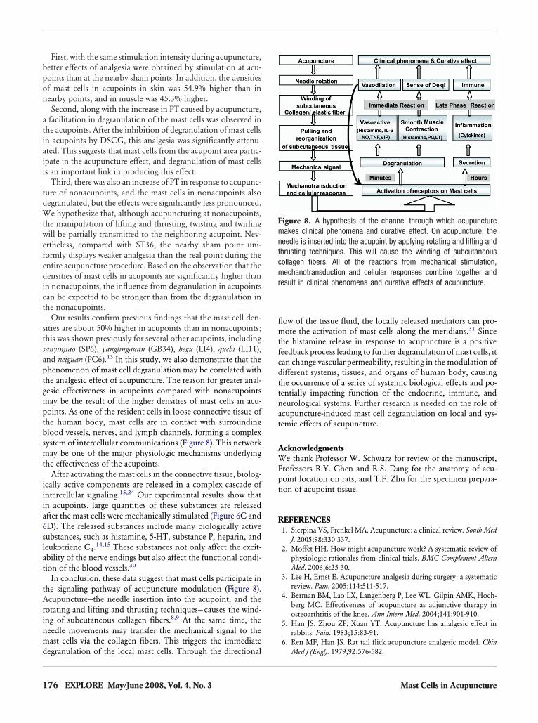
oints. As one of the resident cells in loose connective tissue ofhe human body, mast cells are in contact with surroundinglood vessels, nerves, and lymph channels, forming a complexystem of intercellular communications (Figure 8). This networkay be one of the major physiologic mechanisms underlying
he effectiveness of the acupoints.After activating the mast cells in the connective tissue, biolog-
cally active components are released in a complex cascade ofntercellular signaling.15,24 Our experimental results show thatn acupoints, large quantities of these substances are releasedfter the mast cells were mechanically stimulated (Figure 6C andD). The released substances include many biologically activeubstances, such as histamine, 5-HT, substance P, heparin, andeukotriene C4.
14,15 These substances not only affect the excit-bility of the nerve endings but also affect the functional condi-ion of the blood vessels.30
In conclusion, these data suggest that mast cells participate inhe signaling pathway of acupuncture modulation (Figure 8).cupuncture—the needle insertion into the acupoint, and the
otating and lifting and thrusting techniques—causes the wind-ng of subcutaneous collagen fibers.8,9 At the same time, theeedle movements may transfer the mechanical signal to theast cells via the collagen fibers. This triggers the immediate
egranulation of the local mast cells. Through the directional
76 EXPLORE May/June 2008, Vol. 4, No. 3
ow of the tissue fluid, the locally released mediators can pro-ote the activation of mast cells along the meridians.31 Since
he histamine release in response to acupuncture is a positiveeedback process leading to further degranulation of mast cells, itan change vascular permeability, resulting in the modulation ofifferent systems, tissues, and organs of human body, causinghe occurrence of a series of systemic biological effects and po-entially impacting function of the endocrine, immune, andeurological systems. Further research is needed on the role ofcupuncture-induced mast cell degranulation on local and sys-emic effects of acupuncture.
cknowledgmentse thank Professor W. Schwarz for review of the manuscript,
rofessors R.Y. Chen and R.S. Dang for the anatomy of acu-oint location on rats, and T.F. Zhu for the specimen prepara-ion of acupoint tissue.
EFERENCES1. Sierpina VS, Frenkel MA. Acupuncture: a clinical review. South Med
J. 2005;98:330-337.2. Moffet HH. How might acupuncture work? A systematic review of
physiologic rationales from clinical trials. BMC Complement AlternMed. 2006;6:25-30.
3. Lee H, Ernst E. Acupuncture analgesia during surgery: a systematicreview. Pain. 2005;114:511-517.
4. Berman BM, Lao LX, Langenberg P, Lee WL, Gilpin AMK, Hoch-berg MC. Effectiveness of acupuncture as adjunctive therapy inosteoarthritis of the knee. Ann Intern Med. 2004;141:901-910.
5. Han JS, Zhou ZF, Xuan YT. Acupuncture has analgesic effect inrabbits. Pain. 1983;15:83-91.
6. Ren MF, Han JS. Rat tail flick acupuncture analgesic model. Chin
igure 8. A hypothesis of the channel through which acupunctureakes clinical phenomena and curative effect. On acupuncture, the
eedle is inserted into the acupoint by applying rotating and lifting andhrusting techniques. This will cause the winding of subcutaneousollagen fibers. All of the reactions from mechanical stimulation,echanotransduction and cellular responses combine together and
esult in clinical phenomena and curative effects of acupuncture.
Med J (Engl). 1979;92:576-582.
Mast Cells in Acupuncture
1
1
1
1
1
1
1
1
1
1
2
2
2
2
2
2
2
2
2
2
3
3
M
7. Han JS. Acupuncture and stimulation-produced analgesia. In: HerzA, ed. Opioids II Handbook of Experimental Pharmacology. New York,NY: Springer-Verlag; 1993.
8. Langevin HM, Yandow JA. Relationship of acupuncture points andmeridians to connective tissue points. Anatom Rec. 2002;269:257-265.
9. Langevin HM, Churchill DL, Cipolla MJ. Mechanical signalingthrough connective tissue: a mechanism for the therapeutic effect ofacupuncture. FASEB J. 2001;15:2275-2282.
0. Langevin DL, Churchill HM, Fox JR, Badger GJ, Garra BS, Krag MH.Biomechanical response to acupuncture needling in humans. J ApplPhysiol. 2001;91:2471-2478.
1. Zhang D, Ding GH, Shen XY, Wang L, Liu C. Development ofresearches on the connective tissues and acupuncture [in Chinese].Acupunct Res. 2004;29:77-81.
2. Fei L, Cheng HS, Cai DH, et al. Experimental exploration andresearch prospect of physical bases and functional characteristics ofmeridians. Chin Sci Bull (Engl). 1998;43:1233-1252.
3. Zhong AM, Wu JL, Hu YL. Study on correlation between the mast celland the acupoint. World J Acupunc Mox (Engl). 1994;4:53-58.
4. Theoharides TC, Bielory L. Mast cells and mast cell mediators astargets of dietary supplements. Ann Allergy Asthma Immunol.2004;93 (suppl 1):S24-S34.
5. Dean DM, Dana B, Yoseph AM. Mast cells. Physiol Rev. 1997;77:1033-1079.
6. Petti F, Bangrazi A, Liguori A, Reale G, Ippoliti F. Effects of acu-puncture on immune response related to opioid-like peptides. J TradChin Med. 1998;18:55-63.
7. Liu X, Zhu B, Zhang SX. Relationship between electroacupunctureanalgesia and descending pain inhibitory mechanism of nucleusraphe magnus. Pain. 1986;24:383-396.
8. Uom ES, Min BI, Kim JH, Cho YW. Analgesic effect of the acu-puncture using the method of quick insertion and withdrawal of theneedle in rats. Neurosci Lett. 2001;298:21-24.
9. Yin L, Jin X, Qiao W, et al. PET imaging of brain function whilepuncturing the acupoint ST36. Chin Med J (Engl). 2003;116:1836-1839.
0. Kim JH, Min BI, Schmidt D, Lee HJ, Park DS. The differencebetween electroacupuncture only and electroacupuncture with ma-
nipulation on analgesia in rats. Neurosci Lett. 2000;279:149-152.ast Cells in Acupuncture
1. Chang FC, Tsai HY, Yu MC, et al. The central serotonergic systemmediates the analgesic effect of electroacupuncture on zusanli(ST36) acupoints. J Biomed Sci. 2004;11:179-185.
2. Ding GH, Shen XY, Dai JH. The difference of acupuncture manip-ulation frequencies of lifting-thrusting and twirling-rotating at deqiand non-deqi [in Chinese]. Chin Acupunc Mox. 2002;22:679-681.
3. Ding GH, Shen XY, Dai JH, Liu H, Yao W, Li XA. Research anddevelopment on the dynamic system for detecting the force of acu-puncture needle during the acupuncture process in the clinical prac-tice of traditional Chinese medicine [in Chinese]. J Biomed Eng.2003;20:121-124.
4. Aldenborg F, Enerback L. Thymus dependence of connective tissuemast cells: a quantitative cytofluorometric study of the growth ofperitoneal mast cell in normal and athymic rats. Int Arch AllergyImmunol. 1985;78:277-282.
5. Hargreaves K, Dubner R, Brown F, Flores C, Joris J. A new andsensitive method for measuring thermal nociception in cutaneoushyperalgesia. Pain. 1988;32:77-88.
6. Li ZR. Experimental Acupuncture Science [in Chinese]. Beijing, China:Traditional Chinese Medicine Press; 2003:327.
7. Lin J, Huang H, Ding GH, Zhang D. Relation of mast cell functionand acupuncture analgesia on adjuvant arthritis rat. Acupunc Res.2007;32:16-19.
8. ElSayed SO, Dyson M. Effect of laser pulse repetition rate and pulseduration on mast cell number and degranulation. Lasers Surg Med.1996;19:433-437.
9. Muller T. Supravital uptake of cationic dyes by mast cell granules–alight and electronmicroscope study. Biotech Histochem. 1994;69:171-176.
0. Theoharides TC, Singh LK, Boucher W, et al. Corticotropin-releas-ing hormone induces skin mast cell degranulation and increasedvascular permeability, a possible explanation for its pro-inflamma-tory effects. Endocrinology. 1998;139:403-413.
1. Ding GH, Shen XY, Yao W, Dang RS, Yang J, Chen EY. Dynamicmechanism in directional flow of tissue fluid and human meridian
phenomenon [in Chinese]. Prog Nat Sci. 2005;15:61-70.177EXPLORE May/June 2008, Vol. 4, No. 3
![Synovial mast cells in osteoarthritis - MedDocs Online · to 3% of all cells within the synovium [15]. These cells exhibit a typical mast cell morphology and range in diameter from](https://img.pdfslide.net/doc/110x75/5f09e3977e708231d428fc54/synovial-mast-cells-in-osteoarthritis-meddocs-online-to-3-of-all-cells-within.jpg)