Embed Size (px)
Citation preview
Filali et al. BMC Genomics 2012, 13:399http://www.biomedcentral.com/1471-2164/13/399
RESEARCH ARTICLE Open Access
Medulla oblongata transcriptome changes duringpresymptomatic natural scrapie and theirassociation with prion-related lesionsHicham Filali1, Inmaculada Martin-Burriel2,3, Frank Harders4, Luis Varona5, Carmen Serrano2, Cristina Acín1,Juan J Badiola1, Alex Bossers4 and Rosa Bolea1*
Abstract
Background: The pathogenesis of natural scrapie and other prion diseases is still poorly understood. Determiningthe variations in the transcriptome in the early phases of the disease might clarify some of the molecularmechanisms of the prion-induced pathology and allow for the development of new biomarkers for diagnosis andtherapy. This study is the first to focus on the identification of genes regulated during the preclinical phases ofnatural scrapie in the ovine medulla oblongata (MO) and the association of these genes with prion deposition,astrocytosis and spongiosis.
Results: A custom microarray platform revealed that 86 significant probes had expression changes greater than2-fold. From these probes, we identified 32 genes with known function; the highest number of regulated geneswas included in the phosphoprotein-encoding group. Genes encoding extracellular marker proteins and thoseinvolved in the immune response and apoptosis were also differentially expressed. In addition, we investigated therelationship between the gene expression profiles and the appearance of the main scrapie-associated brain lesions.Quantitative Real-time PCR was used to validate the expression of some of the regulated genes, thus showing thereliability of the microarray hybridization technology.
Conclusions: Genes involved in protein and metal binding and oxidoreductase activity were associated with priondeposition. The expression of glial fibrillary acidic protein (GFAP) was associated with changes in the expression ofgenes encoding proteins with oxidoreductase and phosphatase activity, and the expression of spongiosis wasrelated to genes encoding extracellular matrix components or transmembrane transporters. This is the firstgenome-wide expression study performed in naturally infected sheep with preclinical scrapie. As in previousstudies, our findings confirm the close relationship between scrapie and other neurodegenerative diseases.
Keywords: Natural scrapie, Preclinical sheep, Microarray, Genetic expression, Real time PCR, Prion
BackgroundScrapie is a prion-associated encephalopathy that occursnaturally in sheep and goats. It is characterized by the ac-cumulation of a pathological agent, the prion protein(PrPSc), mainly in the central nervous system [1]. PrPSc
differs from the endogenous normal form (PrPc) in itsconformation, partial resistance to proteolytic degrad-ation and insolubility in the presence of detergents [2,3].
* Correspondence: [email protected] de Investigación en Encefalopatías y Enfermedades TransmisiblesEmergentes. Facultad de Veterinaria, Universidad de Zaragoza, Zaragoza,SpainFull list of author information is available at the end of the article
© 2012 Filali et al.; licensee BioMed Central LtCommons Attribution License (http://creativecreproduction in any medium, provided the or
Scrapie is included in transmissible spongiform enceph-alopathies (TSEs), a disease class that also affects humans(e.g., Creutzfeldt-Jakob disease and Kuru) and cattle (e.g.,bovine spongiform encephalopathy [BSE]) [4-6].The incubation period of the disease is long and
asymptomatic. PrPSc can be detected in VRQ/VRQsheep, genotype for the PRNP gene, two months afterinfection [7]. Three to six months after infection, thepathological agent is detected in the lymphoid forma-tions associated with the gastrointestinal tract [8,9].From six to nine months, the secondary lymphoidorgans are also infected, and finally, at the tenth
d. This is an Open Access article distributed under the terms of the Creativeommons.org/licenses/by/2.0), which permits unrestricted use, distribution, andiginal work is properly cited.
Filali et al. BMC Genomics 2012, 13:399 Page 2 of 15http://www.biomedcentral.com/1471-2164/13/399
month after infection, the central nervous system isaffected [10-12].The neuropathological events in prion diseases occur
at different times depending on the disease. High levelsof PrPSc exist without clinical disease in Gerstmann-Sträussler syndrome [13]; conversely, PrPSc is present invery low levels in fatal familiar insomnia [14]. The de-gree of prion accumulation in specific brain regions doesnot correlate with the clinical features (reviewed in [15]).In addition to prion deposition, other molecularmechanisms act early during the disease. For example,the brain undergoes oxidative stress in the early stagesof prion invasion into the brain and may predispose thebrain to neurodegenerative mechanisms [16]. Genomicanalysis confirmed the induction of cellular stress (oxi-dative stress and ER stress) and the activation of othermolecular pathways in a murine model of prion disease[17]. Other functional genomic studies performed in ani-mal models of scrapie infection have indicated that sev-eral genes are misregulated in the early phases of theinfection [18-24].To date, very few genomic approaches have focused
on the analysis of the early molecular events in priondiseases and, to a lesser extent, studies dealing with thenatural disease. The identification of the genes involvedin the preclinical changes of the disease can help in thediscovery of new biomarkers and targets for future diag-nosis tests or treatments. In an earlier published work,we presented the differentially expressed genes in thebrains of scrapie-symptomatic sheep and the relationshipbetween scrapie-related neuropathological changes andthe transcriptional activities of the identified genes [25].The objectives of the present study were to identify thegenes that are differentially expressed during naturalpreclinical scrapie infection in sheep using a CVI customdesigned 4x44K ovine microarray and to determine therelationship between their expression patterns andprion-associated lesions. In this way, we discuss the vari-ation in gene expression and its association with scrapieneuropathology during the progression of the disease.
MethodsEthics statementThis study was performed in strict accordance with therecommendations for the care and use of experimentalanimals of the University of Zaragoza, in accordancewith the law (R.D. 1201/2005). The protocol wasapproved by the Committee on the Ethics of AnimalExperiments (Permit Number: PI02/08).
AnimalsA total of 10 Rasa Aragonesa female sheep aged 1–5 yearswere included in this study. Six of them were selected
from flocks located in areas free of scrapie and were usedas controls.Four of the animals exhibited preclinical signs of scra-
pie, and the diagnoses were made by third eyelid biopsies[26] and confirmed using the rapid test (TeEsE, Bio-Rad)and immunohistochemistry to detect PrPSc using the6 H4 monoclonal antibody [27]. This characterizationwas performed considering the presence of the clinicalsigns associated with the disease as per previouslyreported criteria [26]. All of the animals belonged toflocks that had been previously characterized as scrapie-affected flocks and were located in different geographicalareas. The animals were genotyped for PRNP poly-morphisms via full Open Reading Frame sequencing aspreviously reported [28], and the sheep chosen for thisstudy displayed the ARQ/ARQ genotype without othercoding mutations outside the 136, 154 and 171 codons,which is the most susceptible genotype in this ovinebreed [28]. The presence of the prion protein was con-firmed by immunohistochemical methods and westernblotting [27].
Tissue collection and RNA isolationAnimals were sacrificed by intravenous injection of sodiumpentobarbital and exsanguination. Necropsy was per-formed immediately, and the physical examination of thescrapie-infected and control animals did not reveal anyadditional pathological signs. The samples were rapidlypreserved and processed according to established guide-lines regarding safety. The lesion pattern in scrapie is bilat-eral; therefore, one-half of the caudal medulla oblongata,including the obex, was snap-frozen in liquid nitrogenprior to long-term storage at −80°C until RNA extraction.The other half was formalin-fixed and paraffin-embeddedfor further histopathological analysis. Total RNA was iso-lated from a tissuemizer-disrupted medulla oblongata induplicate using TRIzolW (Invitrogen AG) followed by aphenol and chloroform extraction and subjected to a puri-fication step with the NucleoSpinW RNA clean-up kitRNAII (Macherey-Nagel GmbH & Co. KG). The quality ofthe total RNA was assessed using the RNA 6000 NanoAssay kit and the 2100 Bioanalyzer (Agilent Technologies).The RNA integrity number (RIN) index for each samplewas estimated using the Agilent 2100 Expert software. TheRIN provides a numerical assessment of the integrity ofRNA that facilitates the standardization of the quality in-terpretation. Only high quality RNA samples with an RINnumber equal to or higher than 7 were further processedfor microarray analysis.
Histology and prion immunohistochemical detectionA histopathological study of the medulla oblongata atthe level of the obex was performed in HE-stained slices
Filali et al. BMC Genomics 2012, 13:399 Page 3 of 15http://www.biomedcentral.com/1471-2164/13/399
(one from each individual control and each positiveanimal).Immunohistochemical (IHC) studies were performed
on adjacent sections. For every antibody, positive andnegative controls (the omission of primary antibodiesfrom the control and scrapie slides) were performed.Detection of the prion proteins was performed following
pretreatment as previously described [29]. Briefly, sectionswere pretreated with 98% formic acid, hydrated and thenautoclaved to enhance antigen retrieval. To block en-dogenous peroxidase activity, the sections were incubatedwith blocking reagent (DAKO) for 10 min after proteinaseK digestion (Roche, 4 g/ml). Next, the sections wereincubated with the monoclonal primary antibody L42(R-Biopharm, dilution 1:500) at RT for 30 min. Endogen-ous peroxidase blocking was used to process sections. Theenzyme-conjugated polymer Envision (DAKO, 30 min)was used as the visualization system and DAB (DAKO,10 min) as the chromogen. The sections were counter-stained with hematoxylin.Astrocytosis was evaluated based on glial fibrillary
acidic protein (GFAP) immunostaining, as previouslydescribed [30,31]. Briefly, after heat-induced epitope re-trieval pretreatment with citrate buffer (pH 6.0), the sec-tions were incubated for 1 h at RT with a rabbitpolyclonal anti-GFAP antibody (DAKO, dilution 1:400).The omission of the primary antibodies from the controland scrapie slides served as negative controls in the rou-tine immunoreactions.The preparations were examined with a Zeiss Axios-
kop 40 optical microscope (Carl Zeiss AG) and a40 ×−magnification objective lens (Carl Zeiss AG). Theimages were captured with a digital camera (AxioCamMRc5, Zeiss AG) that was coupled to the microscopeand a computer and were analyzed using the ImageJ1.4.3.67 image-analysis software package (Psion Image,NIH) to determine the areas occupied by PrPSc depos-ition, astrocytosis and spongiosis. For the evaluation ofthe IHC and HE slides, captured images were opened inNIH Image/ImageJ using the area method to evaluatethe indices of positivity. The total area occupied bybrown markers (PrP and GFAP) or by white spaces(spongiosis) was estimated by setting a “threshold” usingthe thresholding tool for the selection of these areas,and the positive IHC/HE index for that image was calcu-lated. Using the Student´s t test, significant differencesbetween the control and scrapie groups were detected.
Custom sheep oligo-DNA microarrayThe custom CVI 4x44K microarrays contained customeArray-designed 60-mer probes on previously sequencednormalized and subtracted cDNA libraries of ovinePeyer's Patch, obex and tonsil, supplemented by the pub-licly available Ovis aries transcripts from the NCBI/EBI
databases and by the Agilent O. aries transcript catalog.All of the arrays were printed using Sureprint technol-ogy (Agilent Technologies).
Preparation of the labeled cDNA and microarrayhybridizationAll of the procedures for the preparation of the labeledcRNA probes and subsequent Genechip hybridizationswere performed according to the Agilent TechnologiesOne-Color Microarray-Based Gene Expression Analysisguidelines. First, cDNA was synthesized using 1 μgtotal RNA as a template and the T7 Promoter Primerof Agilent One-Color RNA Spike-In (Quick Amp Kit,One-Color, Agilent Technologies). The cDNA was thentranscribed and labeled using T7 RNA Polymerase andcyanine 3-CTP. Finally, the labeled cRNAs werecleaned up using Qiagen RNeasy mini spin columns.The samples were then hybridized to custom CVI-
Agilent 4x44K chips for 17 h at 65°C and 6 rpm. Follow-ing the manufacturer’s protocol, the chips were thenwashed and incubated with wash buffers and scannedusing the GenePix 4200AL Scanner (Axon Instruments)in conjunction with GenePix Pro 6.0 software.The hybridizations of each sample were performed in
duplicate, resulting in 8 microarrays for the preclinicalscrapie animals and 12 for the negative control animals.
Microarray data analysisThe hybridization data were extracted with the AgilentFeature Extraction, version 9.5.3.1, image analysis appli-cation (Agilent Technologies) before processing withGeneSpring GX 10.0.2 (Agilent Technologies). Usingthe 75th percentile method intensity, the chip valueswere normalized, and the expression values were calcu-lated. The global medulla oblongata gene expressionprofiles from the clinical scrapie-infected animals werecompared to the negative controls, through a linearmodel that accounts for both technical (random animaleffects) and biological replicates (disease effects). Inaddition, a multiple testing correction proposed byBenjamini-Hochberg was applied. Further, only geneswith a P-value ≤ 0.05 for the difference between healthyand preclinical individuals and a 2-fold change (FC) asthe lower limit were selected. Although 2-FC is used ascutoff value, according to our experience, we considerthat the major conclusion of our work is not changingeven if we would have used a different threshold, butalways higher than 1.5. These genes were clustered bytheir Euclidean distance coefficient using the Permut-Matrix software [32]. A BLAST search of the GenBankdatabase was performed to identify the genes that weresimilar to the differentially expressed probes. The mo-lecular functions of the genes were classified accordingto Gene Ontology (GO) using an on-line functional
Filali et al. BMC Genomics 2012, 13:399 Page 4 of 15http://www.biomedcentral.com/1471-2164/13/399
annotation of DAVID Bioinformatics Resources 2008[33,34] (NIAID/NIH, USA).
The relationship between neuropathology and geneexpressionUsing a Mixed Model Analysis under a Bayesian approachby the Gene Expression Analysis with Mixed Models(GEAMM) software [35], the relationship between neuro-pathological lesions and gene expression was analyzed.The statistical model assumed the following Bayesian likeli-hood of logarithm of gene expression data provided by theoligo-DNA microarray: p y a; b;Rj ÞeN Xaþ cb; I���Rð Þ�
,where a is the array effect, b is the vector regressionslope associated with the numerical valuation of theneuropathological changes (c: prion deposition, spongi-osis or astrocytosis), X is the incidence matrix thatrelates the array effects to the logarithm of gene ex-pression data (y), and R is the matrix of residual (co)variances with probe-specific residual variance andnull residual covariances. Prior distributions wereassumed to be flat for a, b and R. A more detaileddescription of the statistical procedure was describedby Casellas et al. (2008) [35].The Bayesian analysis was performed using a Gibbs
sampler approach [36] with a single chain of 500,000iterations after discarding the first 50,000. The resultswith a posterior probability below 0.01 for a regressionslope associated with a neuropathological lesion greater(or lower) than zero were selected.
Quantitative real-time PCRQuantitative real-time PCR (qRT-PCR) was performed toconfirm the expression of the 12 genes/sequences involvedin the mechanisms related to neurodegenerative or repar-ation processes and/or had a high level of differential ex-pression in the scrapie group compared to the controls inthe oligo-DNA microarray expression analysis. Eight ofthese genes also displayed the highest significance in theMixed Model Analysis. The PCR primer sequences usedfor the quantification of the genes encoding amyloid beta(A4) precursor (APP), aquaporin 4 (AQP4), calcineurin-likephosphoesterase domain-containing 1 (CPPED1), golgi gol-gin subfamily 4 (GOLGA4), maguk p55 subfamily member7 (MPP7), nell2 (NELL2), CD3 gamma chain (CD3G), gran-ulysin (GNLY), lysosomal protein transmembrane 4 beta(LAPTM4B) and serine/arginine-rich splicing factor 3(SRSF3) and the two ovine scrapie related sequences(OSRS1) and (OSRS2) are shown in Table 1. RNA samplesused for qRT-PCR were the same used for microarrayexperiments, the qRT-PCR assays were designed with Pri-mer Express 2.0 software (Applied Biosystems) to select ap-propriate primer sequences from known sheep or bovinesequences. Whenever possible, the exon-exon border was
included to prevent the amplification of genomic DNA inthe PCR reaction. Complementary DNA (cDNA) wassynthesized from 1 μg RNA using random hexamer pri-mers with the Superscript First Standard Synthesis Systemfor RT-PCR (Invitrogen). To confirm the elimination of anyremaining DNA, reverse transcription with and without theenzyme was performed.qRT-PCR was performed using SYBRW Green (PE
Applied Biosystems) assays. PCR amplification was per-formed in an ABI-Prism fast 7500 Sequence DetectionSystem (PE Applied Biosystems). All qRT-PCR reac-tions were run in triplicate in total reaction volumes of10 μl with 10–20 ng of cDNA as the template and a300 nM final primer concentration. Universal condi-tions were used with an initial 10 min activation anddenaturation step at 95°C, followed by 40 cycles of15 s at 95°C and 30 s at 60°C. The baseline andthreshold for the Ct calculations were set automaticallywith the ABI-Prism 7500 software Version 2.0.1. Thelevels of gene expression were determined using thecomparative Ct method.To improve the normalization accuracy, the geometric
mean of three housekeeping genes was used to calculatethe normalization factor (NF), which was used tonormalize the expression level of each gene in each sam-ple [37]. The NF was calculated from the GAPDH,G6PDH and RPL32 expression data. These are the threemost stable reference housekeeping genes in the sheepmedulla oblongata, and they have been used as in-ternal references for expression studies in scrapie[38]. The primers and PCR conditions for the ampli-fication of these housekeeping genes have beendescribed previously [38,39].The quantitative results obtained from the qRT-
PCR assays were expressed as the fold-change. Stu-dent’s t test analyses were used to determine if thedifferences observed between the groups were statis-tically significant (P < 0.05).
ResultsPreclinical scrapie-related lesionsThe neuropathological features of scrapie were evaluatedin the medulla oblongata tissues of 6 control and 4 pre-clinical scrapie-infected sheep. Spongiosis, PrPSc depos-ition and GFAP immunoreactivity were consistent withthe features of classical scrapie [40]. PrPSc deposition andspongiosis were only detected in the affected animals(Figure 1). Particular medullary areas in the obex, such asthe nucleus dorsal motor of the vagus, the spinal tract ofthe trigeminal nerve and the solitary tract nucleus, wereseverely affected in the infected group. Even with thehigh variability observed in the scrapie group, the differ-ences between the groups were statistically significant(P < 0.01 and P < 0.05).
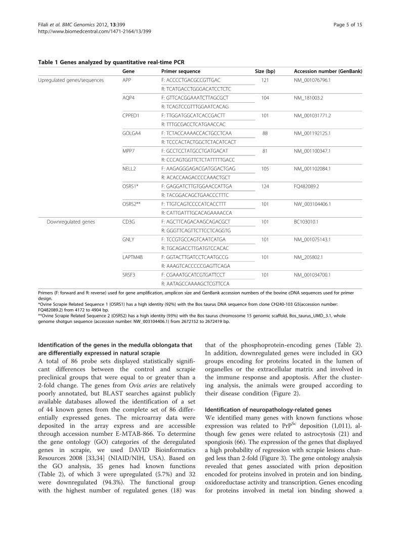
Table 1 Genes analyzed by quantitative real-time PCR
Gene Primer sequence Size (bp) Accession number (GenBank)
Upregulated genes/sequences APP F: ACCCCTGACGCCGTTGAC 121 NM_001076796.1
R: TCATGACCTGGGACATCCTCTC
AQP4 F: GTTCACGGAAATCTTAGCGCT 104 NM_181003.2
R: TCAGTCCGTTTGGAATCACAG
CPPED1 F: TTGGATGGCATCACCGACTT 101 NM_001031771.2
R: TTTGCGACCTCATGAACCAC
GOLGA4 F: TCTACCAAAACCACTGCCTCAA 88 NM_001192125.1
R: TCCCACTACTGGCTCTACATCACT
MPP7 F: GCCTCCTATGCCTGATGACAT 81 NM_001100347.1
R: CCCAGTGGTTCTCTATTTTTGACC
NELL2 F: AAGAGGGAGACGATGGACTGAG 105 NM_001102084.1
R: ACACCAAGACCCCAAACTGCT
OSRS1* F: GAGGATCTTGTGGAACCATTGA 124 FQ482089.2
R: TACGGACAGCTGAACCCTTTC
OSRS2** F: TTGTCAGTCCCCATCACCTTT 101 NW_003104406.1
R: CATTGATTTGCACAGAAAACCA
Downregulated genes CD3G F: AGCTTCAGACAAGCAGACGCT 101 BC103010.1
R: GGGTTCAGTTCTTCCTCAGGTG
GNLY F: TCCGTGCCAGTCAATCATGA 101 NM_001075143.1
R: TGCAGACCTTGATGTCCACAC
LAPTM4B F: GGTACTTGATCCTCAATGCCG 101 NM_205802.1
R: AAAGTCACCCCCGAGTTCAGA
SRSF3 F: CGAAATGCATCGTGATTCCT 101 NM_001034700.1
R: AATAGCCAAAAGCTCGTTCCA
Primers (F: forward and R: reverse) used for gene amplification, amplicon size and GenBank accession numbers of the bovine cDNA sequences used for primerdesign.*Ovine Scrapie Related Sequence 1 (OSRS1) has a high identity (92%) with the Bos taurus DNA sequence from clone CH240-103 G5(accession number:FQ482089.2) from 4172 to 4904 bp.**Ovine Scrapie Related Sequence 2 (OSRS2) has a high identity (93%) with the Bos taurus chromosome 15 genomic scaffold, Bos_taurus_UMD_3.1, wholegenome shotgun sequence (accession number: NW_003104406.1) from 2672152 to 2672419 bp.
Filali et al. BMC Genomics 2012, 13:399 Page 5 of 15http://www.biomedcentral.com/1471-2164/13/399
Identification of the genes in the medulla oblongata thatare differentially expressed in natural scrapieA total of 86 probe sets displayed statistically signifi-cant differences between the control and scrapiepreclinical groups that were equal to or greater than a2-fold change. The genes from Ovis aries are relativelypoorly annotated, but BLAST searches against publiclyavailable databases allowed the identification of a setof 44 known genes from the complete set of 86 differ-entially expressed genes. The microarray data weredeposited in the array express and are accessiblethrough accession number E-MTAB-866. To determinethe gene ontology (GO) categories of the deregulatedgenes in scrapie, we used DAVID BioinformaticsResources 2008 [33,34] (NIAID/NIH, USA). Based onthe GO analysis, 35 genes had known functions(Table 2), of which 3 were upregulated (5.7%) and 32were downregulated (94.3%). The functional groupwith the highest number of regulated genes (18) was
that of the phosphoprotein-encoding genes (Table 2).In addition, downregulated genes were included in GOgroups encoding for proteins located in the lumen oforganelles or the extracellular matrix and involved inthe immune response and apoptosis. After the cluster-ing analysis, the animals were grouped according totheir disease condition (Figure 2).
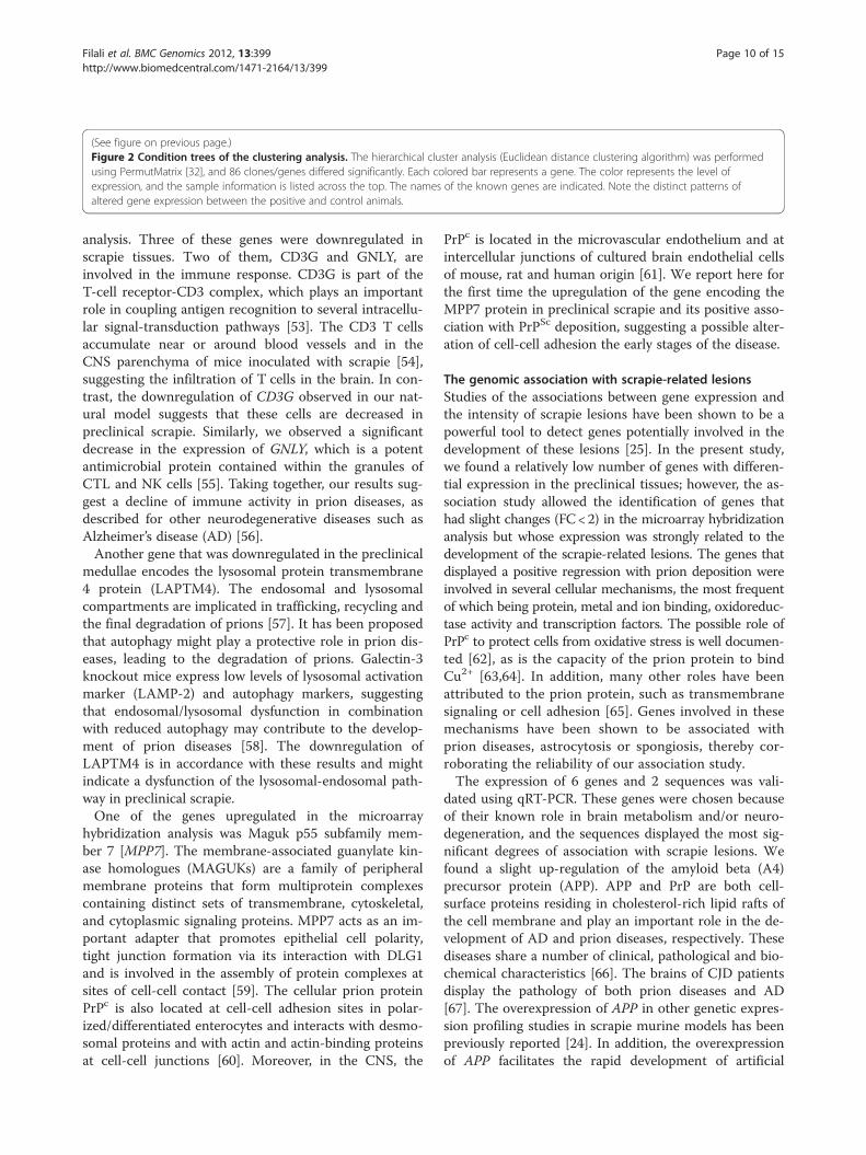
Identification of neuropathology-related genesWe identified many genes with known functions whoseexpression was related to PrPSc deposition (1,011), al-though few genes were related to astrocytosis (21) andspongiosis (66). The expression of the genes that displayeda high probability of regression with scrapie lesions chan-ged less than 2-fold (Figure 3). The gene ontology analysisrevealed that genes associated with prion depositionencoded for proteins involved in protein and ion binding,oxidoreductase activity and transcription. Genes encodingfor proteins involved in metal ion binding showed a
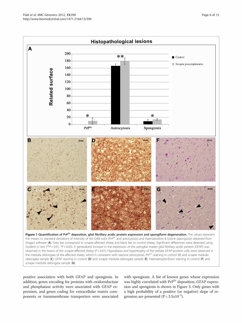
Figure 1 Quantification of PrPSc deposition, glial fibrillary acidic protein expression and spongiform degeneration. The values representthe means (± standard deviation) of intensity of the DAB color (PrPSc and astrocytosis) and Haematoxiline & Eosine (spongiosis) obtained fromImageJ software (A). Grey bar correspond to scrapie-affected sheep and black bar to control sheep. Significant differences were detected usingStudent´s t test (**P< 0.01, *P< 0.05). A generalized increase in the expression of the astroglial marker glial fibrillary acidic protein (GFAP) wasobserved in the brains of the scrapie-affected sheep (P< 0.01). Hyperplasia and hypertrophy of the stellate GFAP-positive cells were observed inthe medulla oblongata of the affected sheep, which is consistent with reactive astrocytosis. PrPSc staining in control (B) and scrapie medullaoblongata sample (C). GFAP staining in control (D) and scrapie medulla oblongata sample (E). Haematoxylin/Eosin staining in control (F) andscrapie medulla oblongata sample (G).
Filali et al. BMC Genomics 2012, 13:399 Page 6 of 15http://www.biomedcentral.com/1471-2164/13/399
positive association with both GFAP and spongiosis. Inaddition, genes encoding for proteins with oxidoreductaseand phosphatase activity were associated with GFAP ex-pression, and genes coding for extracellular matrix com-ponents or transmembrane transporters were associated
with spongiosis. A list of known genes whose expressionwas highly correlated with PrPSc deposition, GFAP expres-sion and spongiosis is shown in Figure 3. Only genes witha high probability of a positive (or negative) slope of re-gression are presented (P< 3.5x10⁻³).
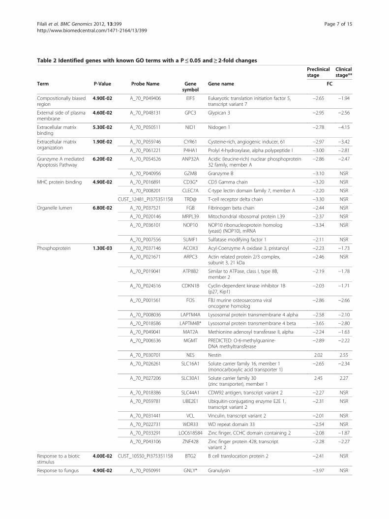
Table 2 Identified genes with known GO terms with a P≤0.05 and≥ 2-fold changes
Preclinicalstage
Clinicalstage**
Term P-Value Probe Name Genesymbol
Gene name FC
Compositionally biasedregion
4.90E-02 A_70_P049406 EIF5 Eukaryotic translation initiation factor 5,transcript variant 7
−2.65 −1.94
External side of plasmamembrane
4.60E-02 A_70_P048131 GPC3 Glypican 3 −2.95 −2.56
Extracellular matrixbinding
5.30E-02 A_70_P050511 NID1 Nidogen 1 −2.78 −4.15
Extracellular matrixorganization
1.90E-02 A_70_P059746 CYR61 Cysteine-rich, angiogenic inducer, 61 −2.97 −3.42
A_70_P061221 P4HA1 Prolyl 4-hydroxylase, alpha polypeptide I −3.00 −2.81
Granzyme A mediatedApoptosis Pathway
6.20E-02 A_70_P054526 ANP32A Acidic (leucine-rich) nuclear phosphoprotein32 family, member A
−2.86 −2.47
A_70_P040956 GZMB Granzyme B −3.10 NSR
MHC protein binding 4.90E-02 A_70_P016891 CD3G* CD3 Gamma chain −3.20 NSR
A_70_P008201 CLEC7A C-type lectin domain family 7, member A −2.20 NSR
CUST_12481_PI375351158 TRD@ T-cell receptor delta chain −3.30 NSR
Organelle lumen 6.80E-02 A_70_P037521 FGB Fibrinogen beta chain −2.44 NSR
A_70_P020146 MRPL39 Mitochondrial ribosomal protein L39 −2.37 NSR
A_70_P036101 NOP10 NOP10 ribonucleoprotein homolog(yeast) (NOP10), mRNA
−3.34 NSR
A_70_P007556 SUMF1 Sulfatase modifying factor 1 −2.11 NSR
Phosphoprotein 1.30E-03 A_70_P037146 ACOX3 Acyl-Coenzyme A oxidase 3, pristanoyl −2.23 −1.73
A_70_P021671 ARPC3 Actin related protein 2/3 complex,subunit 3, 21 kDa
−2.46 NSR
A_70_P019041 ATP8B2 Similar to ATPase, class I, type 8B,member 2
−2.19 −1.78
A_70_P024516 CDKN1B Cyclin-dependent kinase inhibitor 1B(p27, Kip1)
−2.03 −1.71
A_70_P001561 FOS FBJ murine osteosarcoma viraloncogene homolog
−2.86 −2.66
A_70_P008036 LAPTM4A Lysosomal protein transmembrane 4 alpha −2.58 −2.10
A_70_P018586 LAPTM4B* Lysosomal protein transmembrane 4 beta −3.65 −2.80
A_70_P049041 MAT2A Methionine adenosyl transferase II, alpha −2.24 −1.63
A_70_P006536 MGMT PREDICTED: O-6-methylguanine-DNA methyltransferase
−2.89 −2.22
A_70_P030701 NES Nestin 2.02 2.55
A_70_P026261 SLC16A1 Solute carrier family 16, member 1(monocarboxylic acid transporter 1)
−2.65 −2.34
A_70_P027206 SLC30A1 Solute carrier family 30(zinc transporter), member 1
2.45 2.27
A_70_P018386 SLC44A1 CDW92 antigen, transcript variant 2 −2.27 NSR
A_70_P059781 UBE2E1 Ubiquitin-conjugating enzyme E2E 1,transcript variant 2
−2.31 NSR
A_70_P031441 VCL Vinculin, transcript variant 2 −2.01 NSR
A_70_P022731 WDR33 WD repeat domain 33 −2.54 NSR
A_70_P033291 LOC618584 Zinc finger, CCHC domain containing 2 −2.08 −1.87
A_70_P043106 ZNF428 Zinc finger protein 428, transcriptvariant 2
−2.28 −2.27
Response to a bioticstimulus
4.00E-02 CUST_10550_PI375351158 BTG2 B cell translocation protein 2 −2.41 NSR
Response to fungus 4.90E-02 A_70_P050991 GNLY* Granulysin −3.97 NSR
Filali et al. BMC Genomics 2012, 13:399 Page 7 of 15http://www.biomedcentral.com/1471-2164/13/399
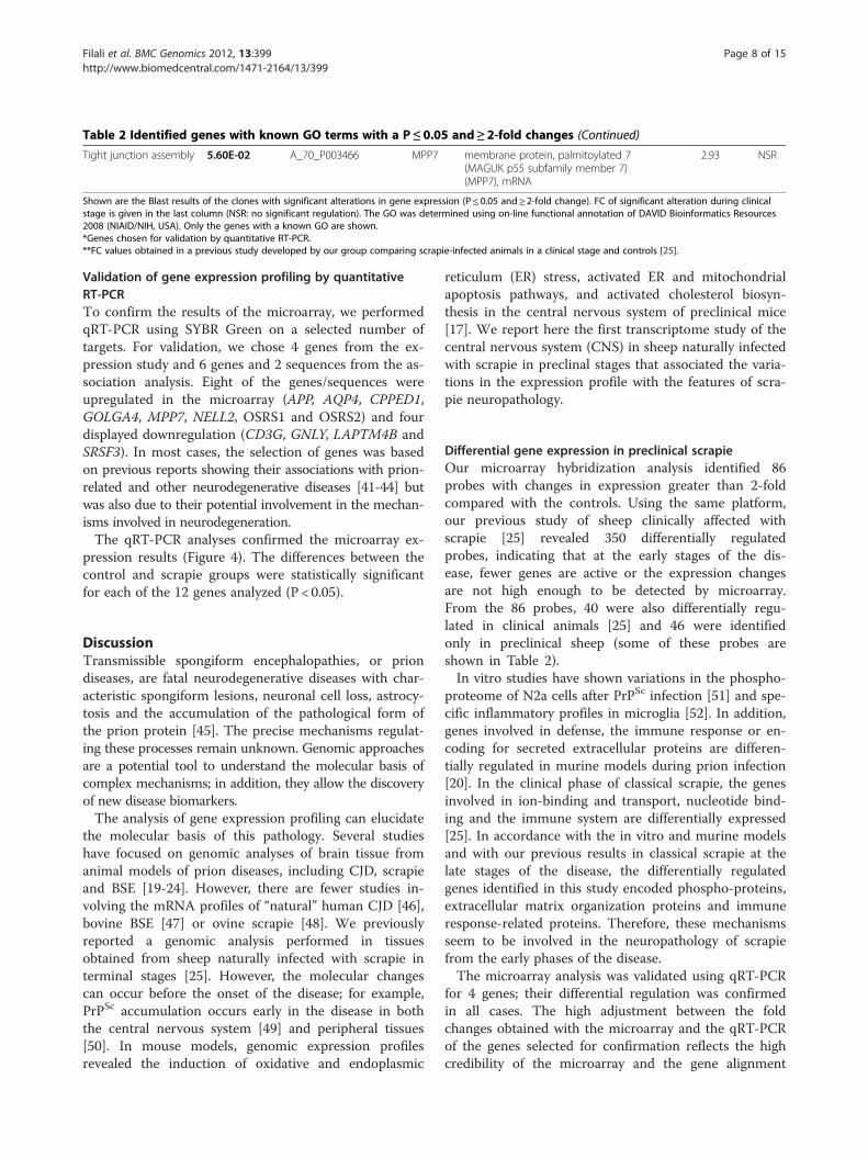
Table 2 Identified genes with known GO terms with a P≤0.05 and≥ 2-fold changes (Continued)
Tight junction assembly 5.60E-02 A_70_P003466 MPP7 membrane protein, palmitoylated 7(MAGUK p55 subfamily member 7)(MPP7), mRNA
2.93 NSR
Shown are the Blast results of the clones with significant alterations in gene expression (P≤ 0.05 and≥ 2-fold change). FC of significant alteration during clinicalstage is given in the last column (NSR: no significant regulation). The GO was determined using on-line functional annotation of DAVID Bioinformatics Resources2008 (NIAID/NIH, USA). Only the genes with a known GO are shown.*Genes chosen for validation by quantitative RT-PCR.**FC values obtained in a previous study developed by our group comparing scrapie-infected animals in a clinical stage and controls [25].
Filali et al. BMC Genomics 2012, 13:399 Page 8 of 15http://www.biomedcentral.com/1471-2164/13/399
Validation of gene expression profiling by quantitativeRT-PCRTo confirm the results of the microarray, we performedqRT-PCR using SYBR Green on a selected number oftargets. For validation, we chose 4 genes from the ex-pression study and 6 genes and 2 sequences from the as-sociation analysis. Eight of the genes/sequences wereupregulated in the microarray (APP, AQP4, CPPED1,GOLGA4, MPP7, NELL2, OSRS1 and OSRS2) and fourdisplayed downregulation (CD3G, GNLY, LAPTM4B andSRSF3). In most cases, the selection of genes was basedon previous reports showing their associations with prion-related and other neurodegenerative diseases [41-44] butwas also due to their potential involvement in the mechan-isms involved in neurodegeneration.The qRT-PCR analyses confirmed the microarray ex-
pression results (Figure 4). The differences between thecontrol and scrapie groups were statistically significantfor each of the 12 genes analyzed (P < 0.05).
DiscussionTransmissible spongiform encephalopathies, or priondiseases, are fatal neurodegenerative diseases with char-acteristic spongiform lesions, neuronal cell loss, astrocy-tosis and the accumulation of the pathological form ofthe prion protein [45]. The precise mechanisms regulat-ing these processes remain unknown. Genomic approachesare a potential tool to understand the molecular basis ofcomplex mechanisms; in addition, they allow the discoveryof new disease biomarkers.The analysis of gene expression profiling can elucidate
the molecular basis of this pathology. Several studieshave focused on genomic analyses of brain tissue fromanimal models of prion diseases, including CJD, scrapieand BSE [19-24]. However, there are fewer studies in-volving the mRNA profiles of “natural” human CJD [46],bovine BSE [47] or ovine scrapie [48]. We previouslyreported a genomic analysis performed in tissuesobtained from sheep naturally infected with scrapie interminal stages [25]. However, the molecular changescan occur before the onset of the disease; for example,PrPSc accumulation occurs early in the disease in boththe central nervous system [49] and peripheral tissues[50]. In mouse models, genomic expression profilesrevealed the induction of oxidative and endoplasmic
reticulum (ER) stress, activated ER and mitochondrialapoptosis pathways, and activated cholesterol biosyn-thesis in the central nervous system of preclinical mice[17]. We report here the first transcriptome study of thecentral nervous system (CNS) in sheep naturally infectedwith scrapie in preclinal stages that associated the varia-tions in the expression profile with the features of scra-pie neuropathology.
Differential gene expression in preclinical scrapieOur microarray hybridization analysis identified 86probes with changes in expression greater than 2-foldcompared with the controls. Using the same platform,our previous study of sheep clinically affected withscrapie [25] revealed 350 differentially regulatedprobes, indicating that at the early stages of the dis-ease, fewer genes are active or the expression changesare not high enough to be detected by microarray.From the 86 probes, 40 were also differentially regu-lated in clinical animals [25] and 46 were identifiedonly in preclinical sheep (some of these probes areshown in Table 2).In vitro studies have shown variations in the phospho-
proteome of N2a cells after PrPSc infection [51] and spe-cific inflammatory profiles in microglia [52]. In addition,genes involved in defense, the immune response or en-coding for secreted extracellular proteins are differen-tially regulated in murine models during prion infection[20]. In the clinical phase of classical scrapie, the genesinvolved in ion-binding and transport, nucleotide bind-ing and the immune system are differentially expressed[25]. In accordance with the in vitro and murine modelsand with our previous results in classical scrapie at thelate stages of the disease, the differentially regulatedgenes identified in this study encoded phospho-proteins,extracellular matrix organization proteins and immuneresponse-related proteins. Therefore, these mechanismsseem to be involved in the neuropathology of scrapiefrom the early phases of the disease.The microarray analysis was validated using qRT-PCR
for 4 genes; their differential regulation was confirmedin all cases. The high adjustment between the foldchanges obtained with the microarray and the qRT-PCRof the genes selected for confirmation reflects the highcredibility of the microarray and the gene alignment
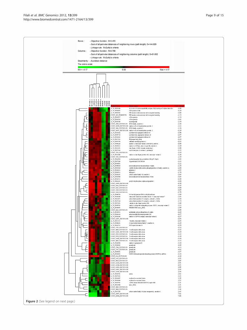
Figure 2 (See legend on next page.)
Filali et al. BMC Genomics 2012, 13:399 Page 9 of 15http://www.biomedcentral.com/1471-2164/13/399
(See figure on previous page.)Figure 2 Condition trees of the clustering analysis. The hierarchical cluster analysis (Euclidean distance clustering algorithm) was performedusing PermutMatrix [32], and 86 clones/genes differed significantly. Each colored bar represents a gene. The color represents the level ofexpression, and the sample information is listed across the top. The names of the known genes are indicated. Note the distinct patterns ofaltered gene expression between the positive and control animals.
Filali et al. BMC Genomics 2012, 13:399 Page 10 of 15http://www.biomedcentral.com/1471-2164/13/399
analysis. Three of these genes were downregulated inscrapie tissues. Two of them, CD3G and GNLY, areinvolved in the immune response. CD3G is part of theT-cell receptor-CD3 complex, which plays an importantrole in coupling antigen recognition to several intracellu-lar signal-transduction pathways [53]. The CD3 T cellsaccumulate near or around blood vessels and in theCNS parenchyma of mice inoculated with scrapie [54],suggesting the infiltration of T cells in the brain. In con-trast, the downregulation of CD3G observed in our nat-ural model suggests that these cells are decreased inpreclinical scrapie. Similarly, we observed a significantdecrease in the expression of GNLY, which is a potentantimicrobial protein contained within the granules ofCTL and NK cells [55]. Taking together, our results sug-gest a decline of immune activity in prion diseases, asdescribed for other neurodegenerative diseases such asAlzheimer’s disease (AD) [56].Another gene that was downregulated in the preclinical
medullae encodes the lysosomal protein transmembrane4 protein (LAPTM4). The endosomal and lysosomalcompartments are implicated in trafficking, recycling andthe final degradation of prions [57]. It has been proposedthat autophagy might play a protective role in prion dis-eases, leading to the degradation of prions. Galectin-3knockout mice express low levels of lysosomal activationmarker (LAMP-2) and autophagy markers, suggestingthat endosomal/lysosomal dysfunction in combinationwith reduced autophagy may contribute to the develop-ment of prion diseases [58]. The downregulation ofLAPTM4 is in accordance with these results and mightindicate a dysfunction of the lysosomal-endosomal path-way in preclinical scrapie.One of the genes upregulated in the microarray
hybridization analysis was Maguk p55 subfamily mem-ber 7 [MPP7]. The membrane-associated guanylate kin-ase homologues (MAGUKs) are a family of peripheralmembrane proteins that form multiprotein complexescontaining distinct sets of transmembrane, cytoskeletal,and cytoplasmic signaling proteins. MPP7 acts as an im-portant adapter that promotes epithelial cell polarity,tight junction formation via its interaction with DLG1and is involved in the assembly of protein complexes atsites of cell-cell contact [59]. The cellular prion proteinPrPc is also located at cell-cell adhesion sites in polar-ized/differentiated enterocytes and interacts with desmo-somal proteins and with actin and actin-binding proteinsat cell-cell junctions [60]. Moreover, in the CNS, the
PrPc is located in the microvascular endothelium and atintercellular junctions of cultured brain endothelial cellsof mouse, rat and human origin [61]. We report here forthe first time the upregulation of the gene encoding theMPP7 protein in preclinical scrapie and its positive asso-ciation with PrPSc deposition, suggesting a possible alter-ation of cell-cell adhesion the early stages of the disease.
The genomic association with scrapie-related lesionsStudies of the associations between gene expression andthe intensity of scrapie lesions have been shown to be apowerful tool to detect genes potentially involved in thedevelopment of these lesions [25]. In the present study,we found a relatively low number of genes with differen-tial expression in the preclinical tissues; however, the as-sociation study allowed the identification of genes thathad slight changes (FC<2) in the microarray hybridizationanalysis but whose expression was strongly related to thedevelopment of the scrapie-related lesions. The genes thatdisplayed a positive regression with prion deposition wereinvolved in several cellular mechanisms, the most frequentof which being protein, metal and ion binding, oxidoreduc-tase activity and transcription factors. The possible role ofPrPc to protect cells from oxidative stress is well documen-ted [62], as is the capacity of the prion protein to bindCu2+ [63,64]. In addition, many other roles have beenattributed to the prion protein, such as transmembranesignaling or cell adhesion [65]. Genes involved in thesemechanisms have been shown to be associated withprion diseases, astrocytosis or spongiosis, thereby cor-roborating the reliability of our association study.The expression of 6 genes and 2 sequences was vali-
dated using qRT-PCR. These genes were chosen becauseof their known role in brain metabolism and/or neuro-degeneration, and the sequences displayed the most sig-nificant degrees of association with scrapie lesions. Wefound a slight up-regulation of the amyloid beta (A4)precursor protein (APP). APP and PrP are both cell-surface proteins residing in cholesterol-rich lipid rafts ofthe cell membrane and play an important role in the de-velopment of AD and prion diseases, respectively. Thesediseases share a number of clinical, pathological and bio-chemical characteristics [66]. The brains of CJD patientsdisplay the pathology of both prion diseases and AD[67]. The overexpression of APP in other genetic expres-sion profiling studies in scrapie murine models has beenpreviously reported [24]. In addition, the overexpressionof APP facilitates the rapid development of artificial
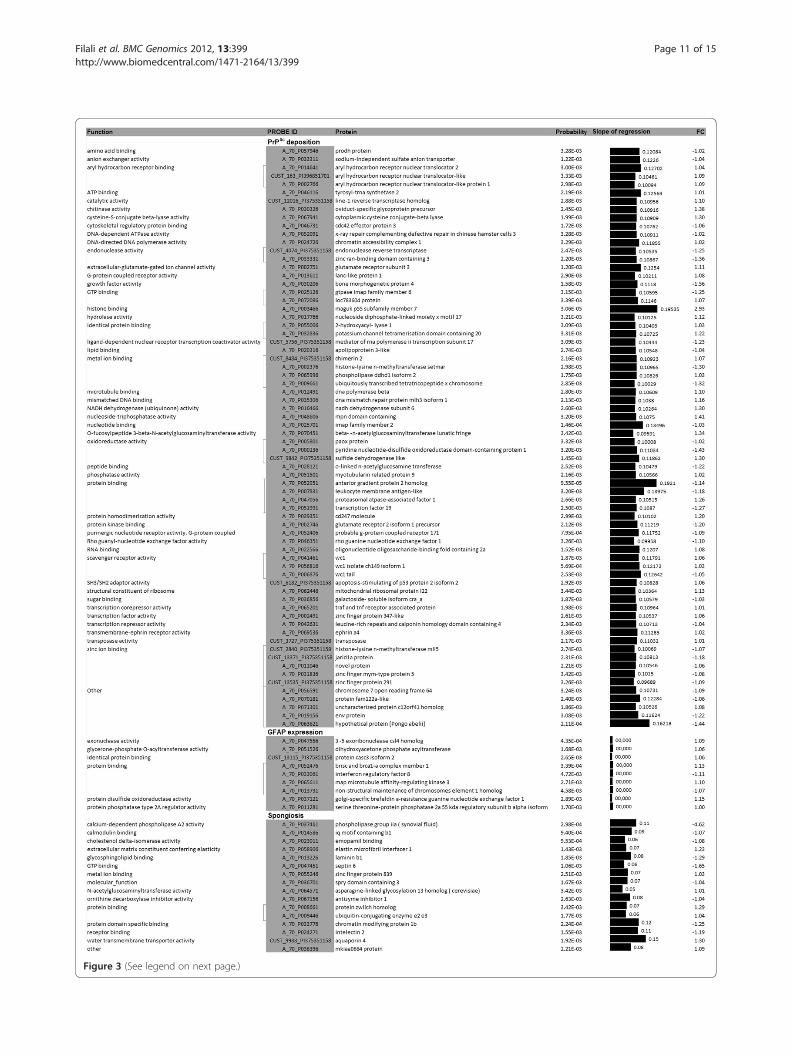
Figure 3 (See legend on next page.)
Filali et al. BMC Genomics 2012, 13:399 Page 11 of 15http://www.biomedcentral.com/1471-2164/13/399
(See figure on previous page.)Figure 3 Relationship between gene expression profiles and scrapie histopathological lesions. Proteins encoded by genes whoseexpression is associated with PrPSc deposition, glial fibrillary acidic protein expression and spongiosis. Only the highly significant related genes areshown (P < 3.5x10⁻³). The slope of regression between histopathological lesions and gene expression was obtained under a Mixed Modelapproach.
Filali et al. BMC Genomics 2012, 13:399 Page 12 of 15http://www.biomedcentral.com/1471-2164/13/399
scrapie [68]. The overexpression of APP in the preclin-ical naturally infected animals found in our study are inaccordance with these previous studies and highlight thepossible early interaction between APP and PrP.Water metabolism is of major importance in a number
of physiological processes in the CNS. Alterations in thedistribution of water and cerebrospinal fluid in the brainare a common occurrence in multiple neuropathologicalconditions, including brain edema, brain tumors, stroke,hyponatremia, head injuries and hydrocephalus. Aquaporin4 (AQP4) is most likely expressed by activated glial cells,and an increase in its level is indicative of ongoing astrocy-tosis [69]. The increase in the expression levels of AQP4has been reported in Creutzfeldt-Jakob disease, bovinespongiform encephalopathy and scrapie-infected transgenicmice [22,43,44,70]. Our work confirms the upregulation ofAQP4 in the preclinical phases of natural ovine scrapie.
Figure 4 Real-time RT-PCR confirmation of the microarray results. Diffmicroarray and quantitative RT-PCR: amyloid beta (A4) precursor (APP), aquphosphoesterase domain-containing 1 (CPPED1), granulysin (GNLY), golgi g(LAPTM4B), maguk p55 subfamily member 7 (MPP7), nell2 (NELL2), ovine scr(OSRS2) and serine/arginine-rich splicing factor 3 (SRSF3).
Genes that have never been associated with prion dis-eases or other neurodegenerative diseases were shown tobe significantly regulated. Our microarray data indicatedan increase in the expression of a gene similar tocalcineurin-like phosphoesterase domain-containing 1[CPPED1] that was confirmed by quantitative RT-PCR.The CPPED1 protein has hydrolase and metal ion-binding activities. To date, no studies have reported thedifferential regulation of this gene in neurodegenerativediseases. However, PrPc interacts with a range of divalentmetal ions and maintains the their homeostasis, and theconformational change that occurs in the formation ofPrPSc is induced by the interaction with ions (see [71]for review). This gene has a positive association withprion deposition (GEAMM analysis), suggesting a pos-sible role in early scrapie development. However, furtheranalysis will be essential to confirm this conclusion.
erential expression of selected sequences/genes analyzed byaporin 4 (AQP4), CD3 gamma chain (CD3G), calcineurin-likeolgin subfamily 4 (GOLGA4), lysosomal protein transmembrane 4 betaapie related sequence 1 (OSRS1), ovine scrapie related sequence 2
Filali et al. BMC Genomics 2012, 13:399 Page 13 of 15http://www.biomedcentral.com/1471-2164/13/399
Golgi golgin subfamily 4 [GOLGA4] may play a role inthe delivery of transport vesicles containing GPI-linkedproteins from the trans-Golgi network through its inter-action with microtubule-actin crosslinking factor 1(MACF1). The prion protein is attached to the outer leafletof the plasma membrane by a glycosyl-phosphatidyl-inositol (GPI) anchor (reviewed in [72]). Our resultsdemonstrate the upregulation of GOLGA4 is positivelyassociated with PrPSc deposition, suggesting that thisprotein might have a role in PrP trafficking.We previously reported that anti-apoptotic genes are
overexpressed in terminal scrapie, which suggested theactivation of neuroprotective mechanisms during thedisease [30]. In accordance with this, we found here thatneural tissue-specific epidermal growth factor-like repeatdomain-containing protein) NELL2 is overexpressed inpreclinical scrapie. NELL2 is a secreted glycoproteinthat is predominantly expressed in neural tissues andincreases in vitro cell survival under cell death-inducing conditions [73]. In addition, NELL2 mayplay an important role in the maintenance of neuralfunctions by regulating the intracellular machineryinvolving Ca2+ signaling, synaptic transport and/orvesicle release [74]. In our study, NELL2 displayed apositive association with PrPSc deposition and spon-giosis, which suggests a possible role in the patho-genesis of the disease related to the role of PrP inCa2+ homeostasis [75].Finally, we observed the downregulation of a serine/
arginine-rich splicing factor 3 [SRSF3], which seems to beinvolved in the differential splicing of the low-densitylipoprotein receptor (LDLR), a major apolipoprotein E(APOE) receptor that has been associated with choles-terol homeostasis and, possibly, AD development [76].This splicing factor is a proto-oncogene [77] and is antia-poptotic [78]. To our knowledge, our work is the firststudy to describe the differential regulation of this genein prion diseases. The downregulation of SRSF3 is in ac-cordance with its probable protective activity againstneuronal cell death. Further studies will be necessary toinvestigate the possible role of SRSF3 in the disease.In addition to the regulation of known genes, several
non-annotated sequences were differentially expressed inthe preclinical medullae and associated with scrapielesions. We confirmed the upregulation of two sequences(OSRS1 and OSRS2) that were associated with astrocyto-sis and spongiosis, respectively. These sequences did notdisplay homology with any known genes (Table 1), butthey show a high homology with parts of two publishedbovine sequences (FQ482089.2 and NW_003104406.1,respectively). These sequences come from an ovinecDNA library generated from the brain and lymphoid tis-sue of scrapie- and control-infected sheep [25]. Al-though further analyses are necessary to confirm
their differential regulation in a wider number of ani-mals or in different prion animal models, these cus-tom sequences can represent potential unknownbiomarkers useful for the diagnosis of presympto-matic prion disease.
ConclusionsIn summary, this is the first genome-wide expression studyperformed in naturally infected sheep with preclinical scra-pie and shows the induction of a reduced number of genescompared with the changes shown in clinical scrapie sheep.Differentially regulated genes confirmed the involvement ofthe immune system, alterations in the extracellular matrixand changes in the ion binding in the neuropathology ofprion diseases. In addition, changes in the levels of genesencoding for proteins related to cell-cell contact and traf-ficking and re-cycling pathways could play an importantrole in the development of the disease. The association ofgenomic changes with scrapie lesions allowed the identifi-cation of a higher number of candidate genes to be used asbiomarkers and could be useful to develop biotools for theearly diagnosis of the disease. In addition to their inclusionin the previous functional groups, the identified genes wererelated to water metabolism or apoptosis. As in previousstudies, our findings confirm the close relationship betweenscrapie and other neurodegenerative diseases. Moreover,the reported association analysis contributes to the know-ledge of the molecular mechanisms underlying the patho-genesis of prion diseases. Further studies are required tolocate the cellular proteins encoded by these differentiallyregulated genes and to study the expression of the identi-fied genes in other brain areas and, in this manner, contrib-ute to the knowledge of their role in the disease.
AbbreviationsAPP: Amyloid beta (A4) precursor; AQP4: Aquaporin 4; ARQ: Alanine arginineglutamine; BSE: Bovine spongiform encephalopathy; CD3G: Cluster ofdifferentiation 3 gamma chain; CPPED1: Calcineurin-like phosphoesterasedomain-containing 1; DAB: 3,3'-diaminobenzidine; G6PDH: Glucose-6-phosphate dehydrogenase; GAPDH: Glyceraldehyde-3-phosphatedehydrogenase; GEAMM: Gene expression analysis with mixed models;GNLY: Granulysin; GO: Gene ontology; GOLGA4: Golgi golgin subfamily 4;HE: Hematoxylin eosine; IHC: Immunohistochemistry; LAPTM4B: Lysosomalprotein transmembrane 4 beta; MO: Medulla oblongata; MPP7: Maguk p55subfamily member 7; OSRS: Ovine scrapie related sequences; PRNP: Prionprotein; PrPC: Cellular prion protein; PrPSc: Scrapie prion protein; RIN: RNAintegrity number; RPL32: Ribosomal protein l32; SRSF3: Serine/arginine-richsplicing factor 3; TSEs: Transmissible spongiform encephalopathies;VRQ: Valine arginine glutamine.
Competing interestsThe authors declare that they have no competing interests.
Authors’ contributionsHF performed the experiments and drafted the manuscript. IMB participatedin the design of the study, participated in the molecular genetic studies, inthe sequence alignment and drafted the manuscript. FH participated incarrying out the microarray analysis. LV performed the association analysis.CS participated in carrying out of the genetic studies. CA participated in thedesign of the study and drafted the manuscript. JJB participated in its design
Filali et al. BMC Genomics 2012, 13:399 Page 14 of 15http://www.biomedcentral.com/1471-2164/13/399
and coordination and helped to draft the manuscript. AB participated in thedesign of the microarray study and sequence alignment. RB conceived ofthe study, and participated in its design and coordination and helped todraft the manuscript. All authors read and approved the final manuscript.
AcknowledgementsThe authors thank Belen Marin, Silvia Ruiz and Yolanda Gracia for theirtechnical assistance. This work was performed as part of the AGL2008-0256project, financed by MICINN-FEDER. Hicham Filali was supported by adoctoral grant from the Spanish MAEC/AECID. The arrays and sequencedlibraries were made available from projects at CVI supported by grants fromthe Dutch Ministry of Agriculture, Nature Management, and Fisheries (LNV).
Author details1Centro de Investigación en Encefalopatías y Enfermedades TransmisiblesEmergentes. Facultad de Veterinaria, Universidad de Zaragoza, Zaragoza,Spain. 2Laboratorio de Genética Bioquímica (LAGENBIO), Facultad deVeterinaria, Universidad de Zaragoza, Zaragoza, Spain. 3Instituto Aragonés deCiencias de la Salud, Zaragoza, Spain. 4Central Veterinary Institute ofWageningen UR (CVI), Lelystad, The Netherlands. 5Unidad de GenéticaCuantitativa y Mejora Animal. Facultad de Veterinaria, Universidad deZaragoza, Zaragoza, Spain.
Received: 13 March 2012 Accepted: 6 August 2012Published: 16 August 2012
References1. Prusiner SB: Prions. Proc Natl Acad Sci U S A 1998, 95:13363–13383.2. Griffith JS: Self-replication and scrapie. Nature 1967, 215:1043–1044.3. Prusiner SB: Novel proteinaceous infectious particles cause scrapie.
Science 1982, 216:136–144.4. Aguzzi A, Weissmann C, Brandner S, Raeber AJ, Klein MA, Voigtlander T: PrP-
expressing tissue required for transfer of scrapie infectivity from spleento brain. Nature 1997, 389:69–73.
5. Chesebro B: Human TSE disease–viral or protein only? Nat Med 1997,3:491–492.
6. Bons N, Mestre-Frances N, Belli P, Cathala F, Gajdusek DC, Brown P: Naturaland experimental oral infection of nonhuman primates by bovinespongiform encephalopathy agents. Proc Natl Acad Sci U S A 1999,96:4046–4051.
7. Andreoletti O, Berthon P, Marc D, Sarradin P, Grosclaude J, van Keulen L,Schelcher F, Elsen J, Lantier F: Early accumulation of PrPsc in gut-associated lymphoid and nervous tissues of susceptible sheep from aRomanov flock with natural scrapie. J Gen Virol 2000, 81:3115–3126.
8. van Keulen LJ, Schreuder BE, Vromans ME, Langeveld JP, Smits MA:Pathogenesis of natural scrapie in sheep. Arch Virol Suppl 2000, (16):57–71.
9. van Keulen LJ, Bossers A, van Zijderveld F: TSE pathogenesis in cattle andsheep. Vet Res 2008, 39:24.
10. Elsen JM, Amigues Y, Schelcher F, Ducrocq V, Andreoletti O, Eychenne F,Khang JV, Poivey JP, Lantier F, Laplanche JL: Genetic susceptibility andtransmission factors in scrapie: detailed analysis of an epidemic in aclosed flock of Romanov. Arch Virol 1999, 144:431–445.
11. Diaz C, Vitezica ZG, Rupp R, Andreoletti O, Elsen JM: Polygenic variationand transmission factors involved in the resistance/susceptibility toscrapie in a Romanov flock. J Gen Virol 2005, 86:849–857.
12. Aguzzi A: Peripheral prion pursuit. J Clin Invest 2001, 108:661–662.13. Collinge J, Owen F, Poulter M, Leach M, Crow TJ, Rossor MN, Hardy J,
Mullan MJ, Janota I, Lantos PL: Prion dementia without characteristicpathology. Lancet 1990, 336:7–9.
14. Manetto V, Medori R, Cortelli P, Montagna P, Tinuper P, Baruzzi A, RancurelG, Hauw JJ, Vanderhaeghen JJ, Mailleux P, et al: Fatal familial insomnia:clinical and pathologic study of five new cases. Neurology 1992,42:312–319.
15. NC, Mallucci GR: Rescuing neurons in prion disease. Biochem J, 433:19–29.16. Yun SW, Gerlach M, Riederer P, Klein MA: Oxidative stress in the brain at
early preclinical stages of mouse scrapie. Exp Neurol 2006,201:90–98.
17. Brown AR, Rebus S, McKimmie CS, Robertson K, Williams A, Fazakerley JK:Gene expression profiling of the preclinical scrapie-infectedhippocampus. Biochem Biophys Res Commun 2005, 334:86–95.
18. Tortosa R, Castells X, Vidal E, Costa C, Ruiz De Villa Mdel C, Sanchez A,Barcelo A, Torres JM, Pumarola M, Arino J: Central nervous system geneexpression changes in a transgenic mouse model for bovine spongiformencephalopathy. Vet Res 2011, 42:109.
19. Dandoy-Dron F, Guillo F, Benboudjema L, Deslys JP, Lasmezas C,Dormont D, Tovey MG, Dron M: Gene expression in scrapie. Cloningof a new scrapie-responsive gene and the identification ofincreased levels of seven other mRNA transcripts. J Biol Chem 1998,273:7691–7697.
20. Booth S, Bowman C, Baumgartner R, Sorensen G, Robertson C, Coulthart M,Phillipson C, Somorjai RL: Identification of central nervous system genesinvolved in the host response to the scrapie agent during preclinicaland clinical infection. J Gen Virol 2004, 85:3459–3471.
21. Brown AR, Webb J, Rebus S, Williams A, Fazakerley JK: Identification ofup-regulated genes by array analysis in scrapie-infected mouse brains.Neuropathol Appl Neurobiol 2004, 30:555–567.
22. Riemer C, Neidhold S, Burwinkel M, Schwarz A, Schultz J, Kratzschmar J,Monning U, Baier M: Gene expression profiling of scrapie-infected braintissue. Biochem Biophys Res Commun 2004, 323:556–564.
23. Xiang W, Windl O, Wunsch G, Dugas M, Kohlmann A, Dierkes N, Westner IM,Kretzschmar HA: Identification of differentially expressed genes inscrapie-infected mouse brains by using global gene expressiontechnology. J Virol 2004, 78:11051–11060.
24. Skinner PJ, Abbassi H, Chesebro B, Race RE, Reilly C, Haase AT: Geneexpression alterations in brains of mice infected with three strains ofscrapie. BMC Genomics 2006, 7:114.
25. Filali H, Martin-Burriel I, Harders F, Varona L, Lyahyai J, Zaragoza P, PumarolaM, Badiola JJ, Bossers A, Bolea R: Gene expression profiling andassociation with prion-related lesions in the medulla oblongata ofsymptomatic natural scrapie animals. PLoS One 2011, 6:e19909.
26. Vargas F, Lujan L, Bolea R, Monleon E, Martin-Burriel I, Fernandez A, De BlasI, Badiola JJ: Detection and clinical evolution of scrapie in sheep by 3rdeyelid biopsy. J Vet Intern Med 2006, 20:187–193.
27. Bolea R, Monleon E, Schiller I, Raeber AJ, Acin C, Monzon M, Martin-Burriel I,Struckmeyer T, Oesch B, Badiola JJ: Comparison of immunohistochemistryand two rapid tests for detection of abnormal prion protein in differentbrain regions of sheep with typical scrapie. J Vet Diagn Invest 2005,17:467–469.
28. Acin C, Martin-Burriel I, Monleon E, Rodellar C, Badiola JJ, Zaragoza P: PrPpolymorphisms in Spanish sheep affected with natural scrapie. Vet Rec2004, 155:370–372.
29. Monleon E, Monzon M, Hortells P, Vargas A, Acin C, Badiola JJ: Detection ofPrPsc on lymphoid tissues from naturally affected scrapie animals:comparison of three visualization systems. J Histochem Cytochem 2004,52:145–151.
30. Serrano C, Lyahyai J, Bolea R, Varona L, Monleon E, Badiola JJ, Zaragoza P,Martin-Burriel I: Distinct spatial activation of intrinsic and extrinsicapoptosis pathways in natural scrapie: association with prion-relatedlesions. Vet Res 2009, 40:42.
31. Vidal E, Acin C, Foradada L, Monzon M, Marquez M, Monleon E, PumarolaM, Badiola JJ, Bolea R: Immunohistochemical characterisation ofclassical scrapie neuropathology in sheep. J Comp Pathol 2009,141:135–146.
32. Caraux G, Pinloche S: PermutMatrix: a graphical environment to arrangegene expression profiles in optimal linear order. Bioinformatics 2005,21:1280–1281.
33. Dennis G Jr, Sherman BT, Hosack DA, Yang J, Gao W, Lane HC, Lempicki RA,DAVID: Database for annotation, visualization, and integrated discovery.Genome Biol 2003, 4:P3.
34. da Huang W, Sherman BT, Lempicki RA: Systematic and integrativeanalysis of large gene lists using DAVID bioinformatics resources. NatProtoc 2009, 4:44–57.
35. Casellas J, Ibanez-Escriche N, Martinez-Giner M, Varona L: GEAMM v.1.4: aversatile program for mixed model analysis of gene expression data.Anim Genet 2008, 39:89–90.
36. Gelfand AE, Smith AFM: Sampling-Based Approaches to CalculatingMarginal Densities. J Am Stat Assoc 1990, 85:398–409.
37. Vandesompele J, De Preter K, Pattyn F, Poppe B, Van Roy N, De Paepe A,Speleman F: Accurate normalization of real-time quantitative RT-PCRdata by geometric averaging of multiple internal control genes. GenomeBiol 2002, 3:RESEARCH0034.
Filali et al. BMC Genomics 2012, 13:399 Page 15 of 15http://www.biomedcentral.com/1471-2164/13/399
38. Lyahyai J, Serrano C, Ranera B, Badiola JJ, Zaragoza P, Martin-Burriel I: Effectof scrapie on the stability of housekeeping genes. Anim Biotechnol 2010,21:1–13.
39. Garcia-Crespo D, Juste RA, Hurtado A: Selection of ovine housekeepinggenes for normalisation by real-time RT-PCR; analysis of PrP geneexpression and genetic susceptibility to scrapie. BMC Vet Res 2005, 1:3.
40. Vidal E, Bolea R, Tortosa R, Costa C, Domenech A, Monleon E, Vargas A,Badiola JJ, Pumarola M: Assessment of calcium-binding proteins(Parvalbumin and Calbindin D-28 K) and perineuronal nets in normaland scrapie-affected adult sheep brains. J Virol Methods 2006,136:137–146.
41. Endres K, Mitteregger G, Kojro E, Kretzschmar H, Fahrenholz F: Influence ofADAM10 on prion protein processing and scrapie infectiosity in vivo.Neurobiol Dis 2009, 36:233–241.
42. Parkin ET, Watt NT, Hussain I, Eckman EA, Eckman CB, Manson JC, BaybuttHN, Turner AJ, Hooper NM: Cellular prion protein regulates beta-secretasecleavage of the Alzheimer's amyloid precursor protein. Proc Natl Acad SciU S A 2007, 104:11062–11067.
43. Costa C, Tortosa R, Rodriguez A, Ferrer I, Torres JM, Bassols A, Pumarola M:Aquaporin 1 and aquaporin 4 overexpression in bovine spongiformencephalopathy in a transgenic murine model and in cattle field cases.Brain Res 2007, 1175:96–106.
44. Rodriguez A, Perez-Gracia E, Espinosa JC, Pumarola M, Torres JM, Ferrer I:Increased expression of water channel aquaporin 1 and aquaporin 4 inCreutzfeldt-Jakob disease and in bovine spongiform encephalopathy-infected bovine-PrP transgenic mice. Acta Neuropathol 2006,112:573–585.
45. Prusiner SB, Bolton DC, Groth DF, Bowman KA, Cochran SP, McKinley MP:Further purification and characterization of scrapie prions. Biochemistry1982, 21:6942–6950.
46. Xiang W, Windl O, Westner IM, Neumann M, Zerr I, Lederer RM, KretzschmarHA: Cerebral gene expression profiles in sporadic Creutzfeldt-Jakobdisease. Ann Neurol 2005, 58:242–257.
47. Khaniya B, Almeida L, Basu U, Taniguchi M, Williams JL, Barreda DR, MooreSS, Guan LL: Microarray analysis of differentially expressed genes fromPeyer's patches of cattle orally challenged with bovine spongiformencephalopathy. J Toxicol Environ Health A 2009, 72:1008–1013.
48. Cosseddu GM, Andreoletti O, Maestrale C, Robert B, Ligios C, Piumi F,Agrimi U, Vaiman D: Gene expression profiling on sheep brain revealsdifferential transcripts in scrapie-affected/not-affected animals. Brain Res2007, 1142:217–222.
49. Andreoletti O, Simon S, Lacroux C, Morel N, Tabouret G, Chabert A, Lugan S,Corbiere F, Ferre P, Foucras G, et al: PrPSc accumulation in myocytes fromsheep incubating natural scrapie. Nat Med 2004, 10:591–593.
50. Casalone C, Corona C, Crescio MI, Martucci F, Mazza M, Ru G, Bozzetta E,Acutis PL, Caramelli M: Pathological prion protein in the tonguesof sheep infected with naturally occurring scrapie. J Virol 2005,79:5847–5849.
51. Wagner W, Ajuh P, Lower J, Wessler S: Quantitative phosphoproteomicanalysis of prion-infected neuronal cells. Cell Commun Signal 2010, 8:28.
52. Baker CA, Manuelidis L: Unique inflammatory RNA profiles of microglia inCreutzfeldt-Jakob disease. Proc Natl Acad Sci U S A 2003, 100:675–679.
53. Flanagan BF, Wotton D, Tuck-Wah S, Owen MJ: DNase hypersensitivity andmethylation of the human CD3G and D genes during T-celldevelopment. Immunogenetics 1990, 31:13–20.
54. Lewicki H, Tishon A, Homann D, Mazarguil H, Laval F, Asensio VC, CampbellIL, DeArmond S, Coon B, Teng C, et al: T cells infiltrate the brain in murineand human transmissible spongiform encephalopathies. J Virol 2003,77:3799–3808.
55. Hogg AE, Bowick GC, Herzog NK, Cloyd MW, Endsley JJ: Induction ofgranulysin in CD8+ T cells by IL-21 and IL-15 is suppressed by humanimmunodeficiency virus-1. J Leukoc Biol 2009, 86:1191–1203.
56. Richartz-Salzburger E, Batra A, Stransky E, Laske C, Kohler N, Bartels M,Buchkremer G, Schott K: Altered lymphocyte distribution in Alzheimer'sdisease. J Psychiatr Res 2007, 41:174–178.
57. Heiseke A, Aguib Y, Schatzl HM: Autophagy, prion infection and theirmutual interactions. Curr Issues Mol Biol 2010, 12:87–97.
58. Mok SW, Riemer C, Madela K, Hsu DK, Liu FT, Gultner S, Heise I, Baier M:Role of galectin-3 in prion infections of the CNS. Biochem Biophys ResCommun 2007, 359:672–678.
59. Stucke VM, Timmerman E, Vandekerckhove J, Gevaert K, Hall A: The MAGUKprotein MPP7 binds to the polarity protein hDlg1 and facilitatesepithelial tight junction formation. Mol Biol Cell 2007, 18:1744–1755.
60. Morel E, Fouquet S, Strup-Perrot C, Pichol Thievend C, Petit C, Loew D,Faussat AM, Yvernault L, Pincon-Raymond M, Chambaz J, et al: The cellularprion protein PrP(c) is involved in the proliferation of epithelial cells and inthe distribution of junction-associated proteins. PLoS One 2008, 3:e3000.
61. Viegas P, Chaverot N, Enslen H, Perriere N, Couraud PO, Cazaubon S:Junctional expression of the prion protein PrPC by brain endothelialcells: a role in trans-endothelial migration of human monocytes. J Cell Sci2006, 119:4634–4643.
62. Milhavet O, Lehmann S: Oxidative stress and the prion protein intransmissible spongiform encephalopathies. Brain Res Brain Res Rev 2002,38:328–339.
63. Vassallo N, Herms J: Cellular prion protein function in copper homeostasisand redox signalling at the synapse. J Neurochem 2003, 86:538–544.
64. Millhauser GL: Copper and the prion protein: methods, structures,function, and disease. Annu Rev Phys Chem 2007, 58:299–320.
65. Westergard L, Christensen HM, Harris DA: The cellular prion protein (PrP(C)): its physiological function and role in disease. Biochim Biophys Acta2007, 1772:629–644.
66. Barnham KJ, Cappai R, Beyreuther K, Masters CL, Hill AF: Delineatingcommon molecular mechanisms in Alzheimer's and prion diseases.Trends Biochem Sci 2006, 31:465–472.
67. Hainfellner JA, Wanschitz J, Jellinger K, Liberski PP, Gullotta F, Budka H:Coexistence of Alzheimer-type neuropathology in Creutzfeldt-Jakobdisease. Acta Neuropathol 1998, 96:116–122.
68. Baier M, Apelt J, Riemer C, Gultner S, Schwarz A, Bamme T, Burwinkel M,Schliebs R: Prion infection of mice transgenic for human APPSwe: increasedaccumulation of cortical formic acid extractable Abeta(1–42) and rapidscrapie disease development. Int J Dev Neurosci 2008, 26:821–824.
69. Nielsen S, Nagelhus EA, Amiry-Moghaddam M, Bourque C, Agre P, OttersenOP: Specialized membrane domains for water transport in glial cells:high-resolution immunogold cytochemistry of aquaporin-4 in rat brain.J Neurosci 1997, 17:171–180.
70. Riemer C, Queck I, Simon D, Kurth R, Baier M: Identification of upregulatedgenes in scrapie-infected brain tissue. J Virol 2000, 74:10245–10248.
71. Rana A, Gnaneswari D, Bansal S, Kundu B: Prion metal interaction: is prionpathogenesis a cause or a consequence of metal imbalance? Chem BiolInteract 2009, 181:282–291.
72. Nunziante M, Gilch S, Schatzl HM: Prion diseases: from molecular biologyto intervention strategies. ChemBioChem 2003, 4:1268–1284.
73. Choi EJ, Kim DH, Kim JG, Kim DY, Kim JD, Seol OJ, Jeong CS, Park JW, ChoiMY, Kang SG, et al: Estrogen-dependent transcription of the NEL-like 2(NELL2) gene and its role in protection from cell death. J Biol Chem 2010,285:25074–25084.
74. Kim H, Ha CM, Choi J, Choi EJ, Jeon J, Kim C, Park SK, Kang SS, Kim K, LeeBJ: Ontogeny and the possible function of a novel epidermal growthfactor-like repeat domain-containing protein, NELL2, in the rat brain.J Neurochem 2002, 83:1389–1400.
75. Brini M, Miuzzo M, Pierobon N, Negro A, Sorgato MC: The prion proteinand its paralogue Doppel affect calcium signaling in Chinese hamsterovary cells. Mol Biol Cell 2005, 16:2799–2808.
76. Ling IF, Estus S: Role of SFRS13A in low-density lipoprotein receptorsplicing. Hum Mutat 2010, 31:702–709.
77. Jia R, Li C, McCoy JP, Deng CX, Zheng ZM: SRp20 is a proto-oncogenecritical for cell proliferation and tumor induction and maintenance.Int J Biol Sci 2010, 6:806–826.
78. He X, Arslan AD, Pool MD, Ho TT, Darcy KM, Coon JS, Beck WT: Knockdownof splicing factor SRp20 causes apoptosis in ovarian cancer cells and itsexpression is associated with malignancy of epithelial ovarian cancer.Oncogene 2011, 30:356–365.
doi:10.1186/1471-2164-13-399Cite this article as: Filali et al.: Medulla oblongata transcriptome changesduring presymptomatic natural scrapie and their association with prion-related lesions. BMC Genomics 2012 13:399.