Embed Size (px)
Citation preview
MEIOTIC DRIVE
Spindle asymmetry drives non-Mendelian chromosome segregationTakashi Akera,1 Lukáš Chmátal,1 Emily Trimm,1 Karren Yang,1 Chanat Aonbangkhen,2
David M. Chenoweth,2 Carsten Janke,3,4 Richard M. Schultz,1 Michael A. Lampson1*
Genetic elements compete for transmission through meiosis, when haploid gametes arecreated from a diploid parent. Selfish elements can enhance their transmission through aprocess known as meiotic drive. In female meiosis, selfish elements drive by preferentiallyattaching to the egg side of the spindle.This implies some asymmetry between the two sidesof the spindle, but the molecular mechanisms underlying spindle asymmetry are unknown.Here we found that CDC42 signaling from the cell cortex regulatedmicrotubule tyrosinationto induce spindle asymmetry and that non-Mendelian segregation depended on thisasymmetry. Cortical CDC42 depends on polarization directed by chromosomes, which arepositioned near the cortex to allow the asymmetric cell division.Thus, selfish meiotic driversexploit the asymmetry inherent in female meiosis to bias their transmission.
Genetic conflict is inherent in any haploid-diploid life cycle because genetic elementscompete for transmission through meio-sis. Mendel’s law of segregation states thatalleles of a gene are transmitted with equal
probability, but this law can be violated by selfishgenetic elements through meiotic drive—for ex-ample, by eliminating competing gametes (e.g.,sperm killing or spore killing) or by exploiting theasymmetry in female meiosis to increase trans-mission to the egg. Despite the impact of meiotic
drive on evolution and genetics (1–4), the under-lying mechanisms are largely unknown.Female meiosis provides a clear opportunity
for selfish elements to cheat because only chro-mosomes that segregate to the egg can be trans-mitted to offspring, whereas the rest are degradedin polar bodies. Conceptually, femalemeiotic drivedepends on three conditions: asymmetry in cellfate, a functional difference between homologouschromosomes that influences their segregation,and asymmetry within the meiotic spindle (5).
The asymmetry in cell fate is well established(6), and chromosomal rearrangements and am-plifications of repetitive sequences (e.g., centro-meres) are associated with biased segregation(7–10). Asymmetry within the meiotic spindlewas noted in grasshopper in 1976 (11) but notstudied further.Oocyte spindles are positioned close to the cor-
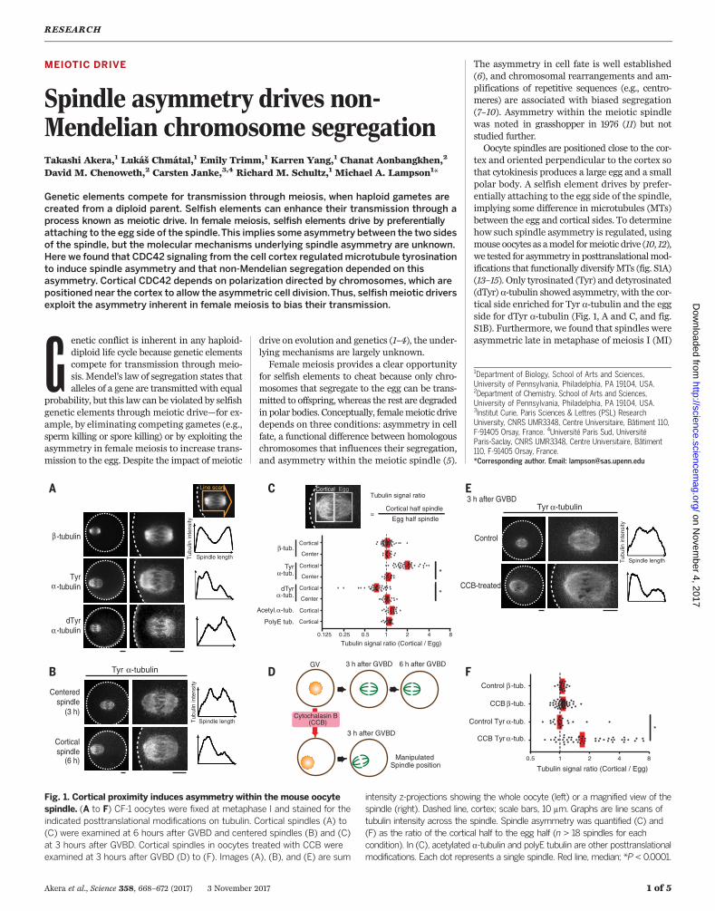
tex and oriented perpendicular to the cortex sothat cytokinesis produces a large egg and a smallpolar body. A selfish element drives by prefer-entially attaching to the egg side of the spindle,implying some difference in microtubules (MTs)between the egg and cortical sides. To determinehow such spindle asymmetry is regulated, usingmouse oocytes as amodel formeiotic drive (10, 12),we tested for asymmetry in posttranslationalmod-ifications that functionally diversifyMTs (fig. S1A)(13–15). Only tyrosinated (Tyr) and detyrosinated(dTyr) a-tubulin showed asymmetry, with the cor-tical side enriched for Tyr a-tubulin and the eggside for dTyr a-tubulin (Fig. 1, A and C, and fig.S1B). Furthermore, we found that spindles wereasymmetric late in metaphase of meiosis I (MI)
RESEARCH
Akera et al., Science 358, 668–672 (2017) 3 November 2017 1 of 5
1Department of Biology, School of Arts and Sciences,University of Pennsylvania, Philadelphia, PA 19104, USA.2Department of Chemistry, School of Arts and Sciences,University of Pennsylvania, Philadelphia, PA 19104, USA.3Institut Curie, Paris Sciences & Lettres (PSL) ResearchUniversity, CNRS UMR3348, Centre Universitaire, Bâtiment 110,F-91405 Orsay, France. 4Université Paris Sud, UniversitéParis-Saclay, CNRS UMR3348, Centre Universitaire, Bâtiment110, F-91405 Orsay, France.*Corresponding author. Email: [email protected]
Control
CCB-treated
3 h after GVBD
*
Cytochalasin B(CCB)
ManipulatedSpindle position
3 h after GVBD 6 h after GVBDGV
3 h after GVBD
β -tubulin
Tyrα -tubulin
dTyrα -tubulin
Cortical half spindle
Egg half spindle
Tubulin signal ratioLine scan
=
Tubulin signal ratio (Cortical / Egg)
Cortical
Center
Cortical
Center
Cortical
Center
Cortical
Cortical
β-tub.
Tyrα-tub.
dTyrα-tub.
Acetyl. α-tub.
PolyE tub.
Cortical Egg
0.5 1 2 4 8
CCB Tyr α-tub.
Control Tyr α-tub.
CCB β-tub.
Control β-tub.
Tubulin signal ratio (Cortical / Egg)
0.125 0.25 0.5 1 2 84
Corticalspindle
(6 h)
Centeredspindle
(3 h)
**
Spindle lengthTub
ulin
inte
nsity
Spindle lengthTub
ulin
inte
nsity
Spindle lengthTub
ulin
inte
nsity
Tyr α-tubulin
Tyr α-tubulin
Fig. 1. Cortical proximity induces asymmetry within the mouse oocytespindle. (A to F) CF-1 oocytes were fixed at metaphase I and stained for theindicated posttranslational modifications on tubulin. Cortical spindles (A) to(C) were examined at 6 hours after GVBD and centered spindles (B) and (C)at 3 hours after GVBD. Cortical spindles in oocytes treated with CCB wereexamined at 3 hours after GVBD (D) to (F). Images (A), (B), and (E) are sum
intensity z-projections showing the whole oocyte (left) or a magnified view of thespindle (right). Dashed line, cortex; scale bars, 10 mm. Graphs are line scans oftubulin intensity across the spindle. Spindle asymmetry was quantified (C) and(F) as the ratio of the cortical half to the egg half (n > 18 spindles for eachcondition). In (C), acetylated a-tubulin and polyE tubulin are other posttranslationalmodifications. Each dot represents a single spindle. Red line, median; *P < 0.0001.
on Novem
ber 4, 2017
http://science.sciencemag.org/
Dow
nloaded from
when positioned near the cortex, but not earlierwhen positioned in the center of the oocyte (Fig.1, B toD, and fig. S2). Because theMI spindle firstforms in the center and then migrates towardthe cortex (16–20), asymmetry might depend oneither cortical proximity or time, or both. To dis-tinguish between these possibilities, we manip-ulated spindle position by treating oocytes withcytochalasin B (CCB) before maturation to inhibitactin polymerization. The nucleus drifted to thecortex in 24% of these oocytes, with the spindle
positioned near the cortex by 3 hours after ger-minal vesicle breakdown (GVBD) versus migra-tion at 6 hours under normal conditions (Fig. 1Dand fig. S3A). Cortical spindles in CCB-treatedoocytes showed asymmetric Tyr a-tubulin stain-ing at 3 hours after GVBD, whereas b-tubulinstaining remained symmetric (Fig. 1, E and F, andfig. S3B). Similar results were obtained with cy-tochalasinD (fig. S3C). Asymmetry could be createdif the spindle pole closer to the cortex generatedmoreTyra-tubulin.However,misoriented spindles
parallel to the cortex also had stronger Tyra-tubulin signals on the cortical side, inconsistentwith a difference between spindle poles (fig. S4).Thus, the cortex directly regulates MTs to induceasymmetry within the spindle.The cortex overlying the spindle is polarized
through a chromatin-based gradient of guano-sine triphosphate–bound RAN (RANGTP) (21, 22)(fig. S5A) and enriched in multiple signaling fac-tors, including active CDC42 and RAC GTPases,and in polymerized actin (called the actin cap)
Akera et al., Science 358, 668–672 (2017) 3 November 2017 2 of 5
eDHFR-CDC42 Q61L
Halo-PACTTargeted405 nm laser
Active CDC42 recruited to one spindle pole
Light-induceddimerization
Incubate for 30 min
Fix and stain for Tyr α-tubulin
Add dimerizer
Tyr α-tubulin signal ratio (Recruited / Unrecruited)
0.5
CDC42T17N
RACT17N
RANT24N
RANQ69L
Control
21 4 8
Tyr α-tubulin signal ratio (Cortical / Egg)
** ****
*
CDC42Q61L
Anchorprotein
Recruited protein
PACT domain
Cage
Photocaged small moleculedimerizer
Spindle lengthTub
ulin
inte
nsity
Tyr α-tubulin
TMP HaloligandHalotageDHFR
eDHFR
eDHFR-CDC42Q61L
eDHFRconstructsTyr α-tubulin
eDHFRconstructs
Tyr α -tubulin
eDHFR
eDHFRCdc42Q61L
Spindle lengthTub
ulin
inte
nsity
Control
RANQ69L
RACT17N
CDC42T17N
RANT24N
0.5 1 2
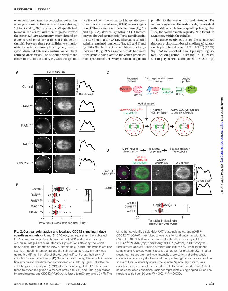
Fig. 2. Cortical polarization and localized CDC42 signaling inducespindle asymmetry. (A and B) CF-1 oocytes expressing the indicatedGTPase mutant were fixed 6 hours after GVBD and stained for Tyra-tubulin. Images are sum intensity z-projections showing the wholeoocyte (left) or a magnified view of the spindle (right), and graphs are linescans of tubulin intensity across the spindle. Spindle asymmetry wasquantified (B) as the ratio of the cortical half to the egg half (n > 17spindles for each condition). (C) Schematics of the light-induced dimeriza-tion experiment.The dimerizer is composed of a HaloTag ligand linked to theeDHFR ligand trimethoprim (TMP), which is photocaged.The PACTdomain,fused to enhanced green fluorescent protein (EGFP) and HaloTag, localizesto spindle poles, and CDC42Q61LDCAAX is fused to mCherry and eDHFR.The
dimerizer covalently binds Halo-PACTat spindle poles, and eDHFR-CDC42Q61LDCAAX is recruited to one pole by local uncaging with light.(D) Halo-EGFP-PACTwas coexpressed with either mCherry-eDHFR-CDC42Q61LDCAAX (top) or mCherry-eDHFR (bottom) in CF-1 oocytes.Recruitment of eDHFR fusion proteins was induced by uncaging at onespindle pole. Oocytes were fixed and stained for Tyr a-tubulin 30 min afteruncaging. Images are maximum intensity z-projections showing wholeoocytes (left) or magnified views of the spindle (right), and graphs are linescans of tubulin intensity across the spindle. Spindle asymmetry wasquantified as the ratio of the recruited side to the unrecruited side (n > 39spindles for each condition). Each dot represents a single spindle. Red line,median; scale bars, 10 mm; *P < 0.01; **P < 0.0001.
RESEARCH | REPORTon N
ovember 4, 2017
http://science.sciencem
ag.org/D
ownloaded from
(6, 23, 24) (fig. S5A). To determinewhether spindleasymmetry depends on cortical polarization, weexpressed either constitutively active (RANQ69L;Q, glutamine; L, leucine) or dominant-negative(RANT24N; T, threonine; N, asparagine) RANmu-tants. In each case, loss of polarization led to lossof spindle asymmetry (Fig. 2, A and B, and fig.S5B).We next tested CDC42 andRACGTPases byexpressing dominant-negativemutants. CDC42T17N
diminished the Tyr a-tubulin signal overall andprevented asymmetry, whereas RACT17N did notaffect asymmetry (Fig. 2, A and B, and fig. S5, Band C). Furthermore, expressing a constitutivelyactive CDC42mutantwith the plasmamembrane–targetingCAAXmotif removed (CDC42Q61LDCAAX)(25) significantly increased Tyr a-tubulin sig-nal (fig. S7) (CAAX; C, cysteine; A, any aliphaticamino acid residue; X, any amino acid residue).We next tested whether the actin cap, whichdepends on CDC42 activity (24) (fig. S5A), con-tributes to spindle asymmetry. Inhibiting the actin-
nucleating actin-related protein 2/3 (Arp2/3) com-plex with the small-molecule inhibitor CK-666abolished actin cap formation (26) but did notaffect spindle asymmetry (fig. S6). Thus, activeCDC42 is sufficient to increase a-tubulin tyrosi-nation and is required for spindle asymmetryindependent of actin cap formation.Our observations suggest that asymmetric lo-
calization of active CDC42 relative to the spindleis the mechanism underlying spindle asymmetry.To test this hypothesis, we developed an opto-genetic strategy to target active CDC42 to one poleof a centered spindle, which is normally sym-metric, using a photocaged small molecule thatheterodimerizes HaloTag and Escherichia colidihydrofolate reductase (eDHFR) fusion proteins(27, 28) (Fig. 2C). We used HaloTag fused to aPACT domain, which localizes to spindle poles(29), to recruit eDHFR fusion proteins specificallyto one pole by local uncaging of the dimerizer(fig. S8A). Recruiting the constitutively active
CDC42Q61LDCAAXmutant induced spindle asym-metry by increasing Tyr a-tubulin signals on therecruited side, whereas recruiting eDHFR alonehad no effect (Fig. 2D and fig. S8B). These resultsstrongly support our model that cortically local-ized CDC42 activity induces asymmetry withinthe spindle. Several factors may contribute to theweaker asymmetry induced by our optogeneticapproach, compared to that observed normallyon spindles near the cortex. CDC42Q61LDCAAX ex-pression increased Tyr a-tubulin overall (fig. S7),leaving less opportunity to create asymmetry bya further increase on one side. In addition, ex-perimentally inducedamounts of CDC42at spindlepoles may be lower than normal amounts at thecortex, and other cortical factors may also con-tribute to the asymmetry.To determine the importance of spindle asym-
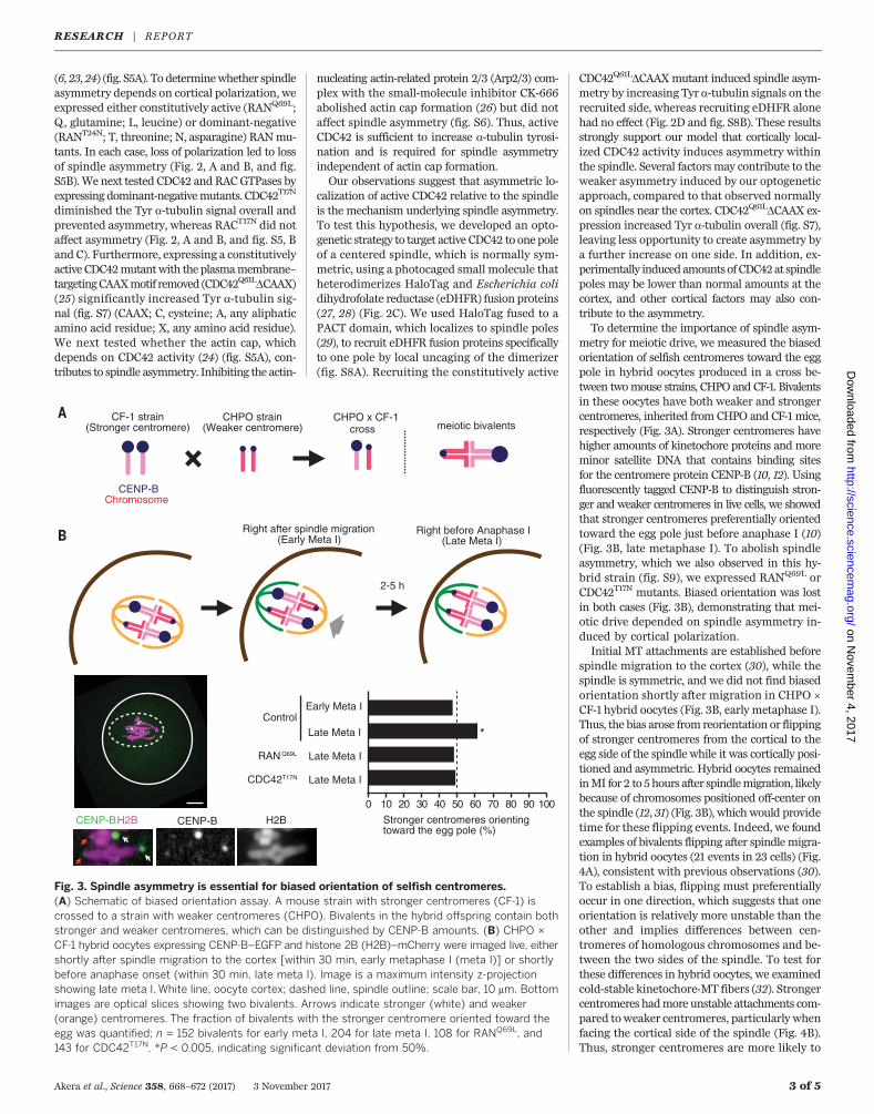
metry for meiotic drive, we measured the biasedorientation of selfish centromeres toward the eggpole in hybrid oocytes produced in a cross be-tween twomouse strains, CHPO and CF-1. Bivalentsin these oocytes have both weaker and strongercentromeres, inherited from CHPO and CF-1 mice,respectively (Fig. 3A). Stronger centromeres havehigher amounts of kinetochore proteins and moreminor satellite DNA that contains binding sitesfor the centromere protein CENP-B (10, 12). Usingfluorescently tagged CENP-B to distinguish stron-ger and weaker centromeres in live cells, we showedthat stronger centromeres preferentially orientedtoward the egg pole just before anaphase I (10)(Fig. 3B, late metaphase I). To abolish spindleasymmetry, which we also observed in this hy-brid strain (fig. S9), we expressed RANQ69L orCDC42T17N mutants. Biased orientation was lostin both cases (Fig. 3B), demonstrating that mei-otic drive depended on spindle asymmetry in-duced by cortical polarization.Initial MT attachments are established before
spindle migration to the cortex (30), while thespindle is symmetric, and we did not find biasedorientation shortly after migration in CHPO ×CF-1 hybrid oocytes (Fig. 3B, early metaphase I).Thus, the bias arose from reorientation or flippingof stronger centromeres from the cortical to theegg side of the spindle while it was cortically posi-tioned and asymmetric. Hybrid oocytes remainedinMI for 2 to 5hours after spindlemigration, likelybecause of chromosomes positioned off-center onthe spindle (12, 31) (Fig. 3B), which would providetime for these flipping events. Indeed, we foundexamples of bivalents flipping after spindlemigra-tion in hybrid oocytes (21 events in 23 cells) (Fig.4A), consistent with previous observations (30).To establish a bias, flipping must preferentiallyoccur in one direction, which suggests that oneorientation is relatively more unstable than theother and implies differences between cen-tromeres of homologous chromosomes and be-tween the two sides of the spindle. To test forthese differences in hybrid oocytes, we examinedcold-stable kinetochore-MT fibers (32). Strongercentromeres hadmore unstable attachments com-pared toweaker centromeres, particularly whenfacing the cortical side of the spindle (Fig. 4B).Thus, stronger centromeres are more likely to
Akera et al., Science 358, 668–672 (2017) 3 November 2017 3 of 5
*
2-5 h
Control
RAN Q69L
meiotic bivalentsCF-1 strain
(Stronger centromere)CHPO strain
(Weaker centromere)CHPO x CF-1
cross
CENP-B H2B CENP-B H2B
Right after spindle migration(Early Meta I)
Right before Anaphase I(Late Meta I)
CENP-BChromosome
CDC42T17N
Early Meta I
Late Meta I
Late Meta I
Late Meta I
Stronger centromeres orienting toward the egg pole (%)
0 10 20 30 40 50 60 70 80 90 100
Fig. 3. Spindle asymmetry is essential for biased orientation of selfish centromeres.(A) Schematic of biased orientation assay. A mouse strain with stronger centromeres (CF-1) iscrossed to a strain with weaker centromeres (CHPO). Bivalents in the hybrid offspring contain bothstronger and weaker centromeres, which can be distinguished by CENP-B amounts. (B) CHPO ×CF-1 hybrid oocytes expressing CENP-B–EGFP and histone 2B (H2B)–mCherry were imaged live, eithershortly after spindle migration to the cortex [within 30 min, early metaphase I (meta I)] or shortlybefore anaphase onset (within 30 min, late meta I). Image is a maximum intensity z-projectionshowing late meta I. White line, oocyte cortex; dashed line, spindle outline; scale bar, 10 mm. Bottomimages are optical slices showing two bivalents. Arrows indicate stronger (white) and weaker(orange) centromeres. The fraction of bivalents with the stronger centromere oriented toward theegg was quantified; n = 152 bivalents for early meta I, 204 for late meta I, 108 for RANQ69L, and143 for CDC42T17N. *P < 0.005, indicating significant deviation from 50%.
RESEARCH | REPORTon N
ovember 4, 2017
http://science.sciencem
ag.org/D
ownloaded from
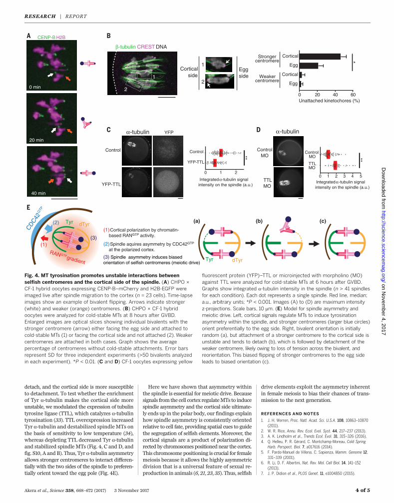
detach, and the cortical side is more susceptibleto detachment. To test whether the enrichmentof Tyr a-tubulin makes the cortical side moreunstable, we modulated the expression of tubulintyrosine ligase (TTL), which catalyzes a-tubulintyrosination (33). TTL overexpression increasedTyr a-tubulin and destabilized spindle MTs onthe basis of sensitivity to low temperature (34),whereas depleting TTL decreased Tyr a-tubulinand stabilized spindle MTs (Fig. 4, C and D, andfig. S10, A and B). Thus, Tyr a-tubulin asymmetryallows stronger centromeres to interact differen-tially with the two sides of the spindle to preferen-tially orient toward the egg pole (Fig. 4E).
Here we have shown that asymmetry withinthe spindle is essential for meiotic drive. Becausesignals from the cell cortex regulateMTs to inducespindle asymmetry and the cortical side ultimate-ly ends up in the polar body, our findings explainhow spindle asymmetry is consistently orientedrelative to cell fate, providing spatial cues to guidethe segregation of selfish elements. Moreover, thecortical signals are a product of polarization di-rected by chromosomes positioned near the cortex.This chromosome positioning is crucial for femalemeiosis because it allows the highly asymmetricdivision that is a universal feature of sexual re-production in animals (6, 21, 23, 35). Thus, selfish
drive elements exploit the asymmetry inherentin female meiosis to bias their chances of trans-mission to the next generation.
REFERENCES AND NOTES
1. J. H. Werren, Proc. Natl. Acad. Sci. U.S.A. 108, 10863–10870(2011).
2. W. R. Rice, Annu. Rev. Ecol. Evol. Syst. 44, 217–237 (2013).3. A. K. Lindholm et al., Trends Ecol. Evol. 31, 315–326 (2016).4. Q. Helleu, P. R. Gérard, C. Montchamp-Moreau, Cold Spring
Harb. Perspect. Biol. 7, a017616 (2014).5. F. Pardo-Manuel de Villena, C. Sapienza, Mamm. Genome 12,
331–339 (2001).6. R. Li, D. F. Albertini, Nat. Rev. Mol. Cell Biol. 14, 141–152
(2013).7. J. P. Didion et al., PLOS Genet. 11, e1004850 (2015).
Akera et al., Science 358, 668–672 (2017) 3 November 2017 4 of 5
(1) Cortical polarization by chromatin-based RANGTP activity.
(2) Spindle aquires asymmetry by CDC42GTP
at the polarized cortex.
(3) Spindle asymmetry induces biased orientation of selfish centromeres (meiotic drive)
CD
C42
GTP
β-tubulin CREST DNA
Corticalside
Eggside
Egg
CorticalStrongercentromere
Weakercentromere
Unattached kinetochores (%)
Integrated α-tubulin signal intensity on the spindle (a.u.)
Tyr dTyr
CENP-B H2B
Control
YFP-TTL
α-tubulin YFP
(1)
(2)
(3)
RANGTP gradient
Tyr dTyr
1
22
1Egg
Cortical
α-tubulin
ControlMO
TTLMO
Integrated α-tubulin signal intensity on the spindle (a.u.)
0 1 2
YFP-TTL
Control **
0 1 2 3 4 5
TTLMO
ControlMO
(a) (b) (c)
0 min
20 min
40 min
0 20 40 60
***
Fig. 4. MT tyrosination promotes unstable interactions betweenselfish centromeres and the cortical side of the spindle. (A) CHPO ×CF-1 hybrid oocytes expressing CENP-B–mCherry and H2B-EGFP wereimaged live after spindle migration to the cortex (n = 23 cells). Time-lapseimages show an example of bivalent flipping. Arrows indicate stronger(white) and weaker (orange) centromeres. (B) CHPO × CF-1 hybridoocytes were analyzed for cold-stable MTs at 8 hours after GVBD.Enlarged images are optical slices showing individual bivalents with thestronger centromere (arrow) either facing the egg side and attached tocold-stable MTs (1) or facing the cortical side and not attached (2).Weakercentromeres are attached in both cases. Graph shows the averagepercentage of centromeres without cold-stable attachments. Error barsrepresent SD for three independent experiments (>50 bivalents analyzedin each experiment). *P < 0.01. (C and D) CF-1 oocytes expressing yellow
fluorescent protein (YFP)–TTL or microinjected with morpholino (MO)against TTL were analyzed for cold-stable MTs at 6 hours after GVBD.Graphs show integrated a-tubulin intensity in the spindle (n > 41 spindlesfor each condition). Each dot represents a single spindle. Red line, median;a.u., arbitrary units; *P < 0.001. Images (A) to (D) are maximum intensityz-projections. Scale bars, 10 mm. (E) Model for spindle asymmetry andmeiotic drive. Left, cortical signals regulate MTs to induce tyrosinationasymmetry within the spindle, and stronger centromeres (larger blue circles)orient preferentially to the egg side. Right, bivalent orientation is initiallyrandom (a), but attachment of a stronger centromere to the cortical side isunstable and tends to detach (b), which is followed by detachment of theweaker centromere, likely owing to loss of tension across the bivalent, andreorientation. This biased flipping of stronger centromeres to the egg sideleads to biased orientation (c).
RESEARCH | REPORTon N
ovember 4, 2017
http://science.sciencem
ag.org/D
ownloaded from
8. L. Fishman, A. Saunders, Science 322, 1559–1562(2008).
9. F. Pardo-Manuel de Villena, C. Sapienza, Genetics 159,1179–1189 (2001).
10. A. Iwata-Otsubo et al., Curr. Biol. 27, 2365–2373.e8 (2017).11. G. M. Hewitt, Chromosoma 56, 381–391 (1976).12. L. Chmátal et al., Curr. Biol. 24, 2295–2300 (2014).13. P. Robison et al., Science 352, aaf0659 (2016).14. M. Barisic et al., Science 348, 799–803 (2015).15. C. Janke, J. Cell Biol. 206, 461–472 (2014).16. J. Azoury, K. W. Lee, V. Georget, P. Hikal, M.-H. Verlhac,
Development 138, 2903–2908 (2011).17. M. Almonacid, M.-É. Terret, M.-H. Verlhac, J. Cell Sci. 127,
477–483 (2014).18. M. Schuh, J. Ellenberg, Curr. Biol. 18, 1986–1992 (2008).19. K. Yi et al., J. Cell Biol. 200, 567–576 (2013).20. J. Azoury et al., Curr. Biol. 18, 1514–1519 (2008).21. M. Deng, P. Suraneni, R. M. Schultz, R. Li, Dev. Cell 12,
301–308 (2007).22. B. Dehapiot, G. Halet, Cell Cycle 12, 1672–1678 (2013).23. G. Halet, J. Carroll, Dev. Cell 12, 309–317 (2007).
24. B. Dehapiot, V. Carrière, J. Carroll, G. Halet, Dev. Biol. 377,202–212 (2013).
25. I. M. Ahearn, K. Haigis, D. Bar-Sagi, M. R. Philips, Nat. Rev. Mol.Cell Biol. 13, 39–51 (2011).
26. K. Yi et al., Nat. Cell Biol. 13, 1252–1258 (2011).27. E. R. Ballister, C. Aonbangkhen, A. M. Mayo, M. A. Lampson,
D. M. Chenoweth, Nat. Commun. 5, 5475 (2014).28. H. Zhang et al., Nat. Chem. Biol. 13, 1096–1101 (2017).29. A. K. Gillingham, S. Munro, EMBO Rep. 1, 524–529 (2000).30. T. S. Kitajima, M. Ohsugi, J. Ellenberg, Cell 146, 568–581 (2011).31. L. Chmátal, K. Yang, R. M. Schultz, M. A. Lampson, Curr. Biol.
25, 1835–1841 (2015).32. C. L. Rieder, Chromosoma 84, 145–158 (1981).33. A. E. Prota et al., J. Cell Biol. 200, 259–270 (2013).34. S. Inoué, H. Sato, J. Gen. Physiol. 50, 259–292 (1967).35. R. Gorelick, J. Carpinone, L. J. Derraugh, Biol. J. Linn. Soc.
London 120, 1–21 (2016).
ACKNOWLEDGMENTS
We thank B. E. Black and M. T. Levine for comments on themanuscript, G. Halet for the CDC42 and RAC constructs and
discussions, R. Li for the RAN constructs, and B. L. Prosser forthe dTyr a-tubulin and TTL antibodies. The research wassupported by NIH grants GM107086 (M.A.L. and R.M.S.) andGM122475 (M.A.L.); by Institut Curie, CNRS, ANR-10-LBX-0038part of the IDEX Idex PSL ANR-10-IDEX-0001-02 PSL, and by thegrant INCA_6517 (C.J.); and by a Japan Society for the Promotionof Science postdoctoral fellowship for research abroad and aresearch fellowship from Uehara Memorial Foundation (T.A.). Datadescribed can be found in the main figures and supplementarymaterials. The authors declare no conflict of interests.
SUPPLEMENTARY MATERIALS
www.sciencemag.org/content/358/6363/668/suppl/DC1Materials and MethodsFigs. S1 to S10References (36–38)
17 February 2017; resubmitted 10 August 2017Accepted 20 September 201710.1126/science.aan0092
Akera et al., Science 358, 668–672 (2017) 3 November 2017 5 of 5
RESEARCH | REPORTon N
ovember 4, 2017
http://science.sciencem
ag.org/D
ownloaded from