Embed Size (px)
Citation preview
Aus dem Frauen-, Kinder- und Perinatalzentrum
Universitätsklinikum Benjamin Franklin der Freien Universität Berlin
Geschäftsführender Direktor Prof. Dr. W. Kühn
Abteilung Frauenklinik und Poliklinik
Kommissarischer Abteilungsleiter Prof. Dr. W. Kühn
Metabolism of Arachidonic acid and formation of novel
3-Hydroxyoxylipins by Candida albicans and interaction
of Hela cells-Candida albicans as a model for
vulvovaginal candidiasis: redundancy of signaling
pathways for activation of COX-2
Inaugural-Dissertation
in fulfilment for the degree of
Doctor rerum medicarum
Faculty of Human Medicine
Freie Universität Berlin
Submitted by: Rupal Deva,
From : Nashik, India.
Examiner: Dr. Dr. Dr. h. c. S. Nigam, EO Professor (RSA)
Second Examiner:
Printed by permission of the Faculty of Human Medicine, Freien Universität,
Berlin
Graduated on:
ACKNOWLEDGEMENTS
I would like to express sincere gratitude to my supervisor Dr.Dr.Dr.h.c. Santosh Nigam EO
professor (RSA), Head of the Eicosanoid Research, for giving me opportunity to do Ph.D
on this very interesting topic in his renowned group. I am very grateful to him for his
continuous interest and support, as well as for putting excellent working facilities to my
disposal.
My sincere gratitude goes also to other lab colleagues:
P. Shankaranarayanan, who was soon a close friend, for introducing me in the world of
biochemical and molecular biological techniques. He combined collegiality and tutorship
in an excellent and unusual manner.
Roberto Ciccoli, for so friendly carrying many responsibilities and always being helpful in
all analytical matters related to mass spectrometry and HPLC.
Prof. Tankred Schewe, a research scientist in Dr. Nigam´s lab till July 2000, for his
guidance and helpful discussions.
Dr. Igor Ivanov, guest scientist from Lomonossow Inst., Moscow, for providing synthetic
3-HETE.
Hannah Rüter and Johannes Wolf and other lab people for making a nice and friendly work
environment.
I also wish to thank all those who were involved and collaborated in so many ways to these
studies:
Dr. Sankar Ghosh, Yale University, New Haven, CT for supplying IKK DN plasmid, Dr.
Eberle, Dermatology Dept., UKBF for the use of instruments in his department, Dr. Inka
Leo-Rossberg, Poliklinik UKBF and Prof. Hahn, Microbiology Dept. for providing patient
samples for isolation of C. albicans, Prof. J.L.F. Kock, Dept. of Microbiology, Univ.
Bloemfontein, South Africa for carrying out experiments on fungi.
ii
Also, I wish to especially acknowledge Mrs. Fangerau, Gyn. Endocrinology, for
ultracentrifugation, Mrs. Kühnert and Mrs Buck, Gyn. Morphology Dept., for
morphological stainings.
I wish to express my deep appreciation to Mrs. Nigam for her kindness and hospitality
during my stay in Berlin.
Finally, I wish to express my gratitude to my parents, sister, brother and many friends, who
are not mentioned above, for their trust and support.
For financial support I am grateful to Schering AG (Ni-89921), VW Foundation (Ni
1/77637), Boehringer-Ingelheim (Ni-IV01) and UKBF (FSP-Entzündung und Infektion).
Without their support this dissertation would not have been possible.
iii
SUMMARY
Candida albicans is a pathogenic yeast responsible for causing infection in patients under
immunosuppressive therapy, and is responsible for recurrent vulvovaginal candidiasis.
Although, number of C. albicans derived intrinsic factors and the host factors have been
found responsible for virulence and favouring the disease process, there is no antimycotic
drug which proves out to be completely fungicidal, suppressing recurrent infections. This
work deals with the study of Candida albicans-host cell interaction, to give an insight of
the disease process, which could lead to new therapeutic interventions. Earlier studies on
D. uninucleata revealed presence of a biologically active 3-HETE derived from
exogenously fed Arachidonic acid. It was found, that it is not only important for its own
life cycle, but also shows biological activity in human cells. We tried to find if a similar
mechanism occurs in pathogenic fungi. It was found that although C. albicans does not
contain arachidonic acid, it is able to grow on arachidonic acid as a sole carbon source as
efficiently as it can utilise linoleic acid. Metabolism of arachidonic acid by C. albicans
occurs both by mitochondrial dependent and independent pathways as shown by inhibitor
studies. Arachidonic acid was shown to block the alternative oxidase pathway of energy
generation in Candida albicans, and was efficiently metabolised to other fatty acids,
carbohydrates, and proteins. Arachidonic acid metabolism by Candida albicans produced a
novel compound 3,18-diHETE, which was closely related to previously discovered 3-
HETE by Dr. S. Nigam’s group. It was also observed that 3,18-diHETE was mainly
associated with the cells bearing germ-tube and hyphael forms. This production was found
to be aspirin sensitive. We found that C. albicans grown in aspirin showed diminished
immunofluorescence with anti-3-OH-oxylipins antibody, indicating decreased amounts of
3,18 diHETE. Apart from this, there was diminished germ-tube formation, which is
essential for infection, in C. albicans in presence of aspirin. Aspirin suppressed not only
cell growth, but it also reduced the adhesion of C. albicans to the host cells. These
observations prompted us to explore a therapeutic role for aspirin. When given along with
clotrimazole, aspirin was found to reduce minimum inhibitory concentration (MIC) of
clotrimazole against C. albicans. The action of aspirin was directed towards inhibiting 3-
hydroxylation mechanism in C. albicans and of clotrimazole towards 18-hydroxylation
mechanism and ergosterol synthesis, which occurs via cytochrome p450. This
demonstrates a novel approach to potentiate the action of antimycotic drug clotrimazole.
Moreover, aspirin is a cyclooxygenase-2 inhibitor, which inhibits formation of PGE2, and
iv
thus the infectivity of C. albicans. Our results thus showed that aspirin may be a suitable
drug for the therapy of recurrent Candidiasis.
It is known that host factors are also responsible for the establishment of infection. HDL,
which is lowered in sepsis patients, and therefore substituted, was found to further increase
virulence by inducing germtube formation in C. albicans.
The first step of infection includes adhesion of pathogen to host cells. C. albicans which
adheres to host cells rapidly is found to be more virulent, and is able to proceeds further to
establish the infection process. Our results demonstrate that aspirin is able to decrease the
adhesion of C. albicans to HeLa cell, thus decreasing its virulence.
Upon infection with C. albicans signal transduction pathways are triggered in host cells
(HeLa cells). We found upregulation of COX-2 in HeLa cells upon infection with C.
albicans as well as increase in PGE2 production. Not only C. albicans but also 3-HETE,
upregulated COX-2, showing that 3-HETE was important compound which mediated cell
signaling during infection. Using specific chemical inhibitors to various signal transduction
pathways, it was established that Protein Kinase C (PKC) and p38 MAP kinase pathway
were primarily involved in the COX-2 upregulation. In other systems, involving cytokines
and LPS, control of COX-2 transcription involved ERK1/2 and JNK MAP kinase
pathways. Further studies were performed to evaluate the exact sequence of activation of
kinases involved. GF 203190X (PKC inhibitor) prevented the phosphorylation of p38
MAP kinase, while other inhibitors failed to have any effect. Thus, PKC acts as an
upstream factor phosphorylating p38 MAP kinase. C. albicans induced p38 MAP kinase
was also observed to be involved in modulating cytoskeletol changes host cell. This effect
involved the activation, of phosphorylated HSP27, a factor involved in the modulation of
actin, which was abrogated by p38 MAP kinase inhibitor SB 202190.We find that
inhibition of free radicals, NAC inhibits COX-2 and PGE2 upregulation suggesting
involvement of reactive oxygen species and NFκB during the infection of HeLa cells with
C. albicans.
The control of transcription occur by the various transcription factors binding to the
concerned gene promoter. NFκB is one of the important transcription factors implicated in
the COX-2 transcription. The role of NFκB was studied using a NFκB-dependent reporter
plasmid and a dominant negative plasmid for IκB (prevents the release of active NFκB).
v
NFκB-dependent transcription was triggered upon infection with C albicans. However,
this could be only partially abrogated by the IκB dominant negative plasmid, moreover
PI3-kinase pathway is involved in COX-2 upregulation via NFκB pathway. Thus C.
albicans brings about redundant signalling for the upregulation and control of COX-2
transcription.
Extensive programmed cell death (apoptosis) was observed after 24 h in Hela cells infected
with C. albicans as shown by genomic DNA laddering and TUNEL assay. After 6 h
postinfection, upregulation of caspase-3 activity was observed. Caspase-3 is an effector
caspase which is involved in the cleavage of cellular proteins during apoptosis. Further
more, using DNA microarray technology, we showed that C. albicans upregulated PI-3-
kinase in HeLa cells. PI-3-kinase is a prominent mediator of anti-apoptotic functions via
AKT and p65 NFκB. Its inhibition by wortmannin increased the caspase-3 activity in
infected cells. IκB dominant negative transfected cells showed no caspase-3 activity upon
infection. This result was surprising as p65 NFκB, whose release is prevented by the
dominant negative plasmid, is an anti-apoptotic molecule. This paradoxical reaction was
clarified by the observation of truncated forms of p65 NFκB in C. albicans infected HeLa
cells. Carboxy terminal truncation of p65 NFκB by caspase-3 renders the molecule
inactive thereby preventing its anti-apoptotic function. The pro-apoptotic subunits of
NFκB, p50 and c-REL, however, were upregulated in C. albicans infected cells. Thus, the
anti-apoptotic pathway, from PI-3-kinase via AKT to p65 NFκB is rendered ineffective by
the cleavage of p65 NFκB by caspase-3.
vi
CONTENTS
Page
ACKNOWLEDGEMENTS i
SUMMARY ii
CONTENTS v
LIST OF FIGURES xi
LIST OF TABLES xiii
ABBREVIATIONS xiv
1 INTRODUCTION
1.1. Candida and vulvovaginal infection 1
1.2. Morphogenesis in Candida albicans 2
1.2.1. Regulation of morphogenesis at molecular level 4
1.3.1. Therapy 4
1.3.2. Mechanism of action of antifungal drugs 5
1.3.3. Current antimycotics 7
1.4.1. Role of lipids and Fatty acid metabolism in C. albicans 8
1.4.2. Lipid composition in C. albicans 9
1.4.3. Lipid and carbohydrate metabolism in Candida albicans 9
1.5. Pathways for energy generation in Candida albicans 10
1.6. Host response to the infection 13
1.7. Cyclooxygenases (Prostaglandin synthases) 14
1.8. COX inhibitors 15
1.9. 3-Hydroxy-oxylipins 16
1.10. Adhesion 18
1.11.1. MAP kinase signaling pathway 18
1.11.2 Specificity in MAPK activation and function 19
1.12. ERK1/2 and MKK1/2 Pathway 19
1.13.1. Stress activated protein kinases and Stress-activated
MAP Kinase Kinases 20
1.14. Nuclear factor-κB 20
1.15. Apoptosis 22

vii
2 AIMS AND OBJECTIVES OF STUDY 24
3 EXPERIMENTAL PROCEDURES
3.1. Materials 25
3.2.1. Organisms and cell culture 28
3.3. Isolation and identification of vaginal C. albicans from patients 28
3.4. Basal culture conditions for C. albicans 28
3.5. Preparation of Blastospores 29
3.6. Preparation of hyphal form 29
3.7. Determination of cell growth for the study of
metabolism of different fatty acids as carbon sources 29
3.8. Metabolism of [1-14C]-arachidonic acid 29
3.9. Distribution of radioactivity derived form arachidonic acid
into different biomolecules 30
3.10. Alkaline hydrolysis of lipids 31
3.11. Preparation of methyl esters of lipids 31
3.12. Preparation of lipid methyl esters and silyl derivatives
of hydroxylipids 31
3.13. GC-MS analysis 32
3.14. Immunofluorescence microscopy of 3(R)-hydroxy-oxylipins 32
3.15. Measurement of cell respiration 32
3.16. Effect of HDL on germ tube formation in C. albicans
3.17. Synergistic effect of aspirin on the minimum
inhibitory concentration of Candida albicans 33
3.18. Effect of aspirin on germ tube formation and adhesion
in C. albicans 33
3.19. Effect of Candida albicans on the release of arachidonic
acid from HeLa cells 34
3.20. Release of Arachidonic acid by Candida albicans cell extract 34
3.21. Phospholipase A2 assay 34
3.22. Infection of HeLa cells with C. albicans. 35
3.23. SDS-PAGE and Western blotting 35
viii
3.24. Preparation of Nuclear protein 36
3.25. Analysis of PI-3-kinase pathway during infection of
HeLa cells with C. albicans 37
3.26. Immunoprecipitation 37
3.27. Determination of PGE2 37
3.28. Isolation of RNA and identification of COX-2 by RT-PCR 38
3.29. Effect of C. albicans on actin cytoskeleton 38
3.30.1 Labeling of C. albicans cells with 5-CFDA 39
3.30.2 Infection and assay for phagocytosis 39
3.31.1. cDNA array 39
3.32. COX-2 Promoter construct 40
3.32.1 Designing of primers 40
3.32.2 PCR amplification 40
3.32.3 Cloning into Luciferase Reporter Vector (pLUC) 40
3.32.4 Transformation and screening of clone 41
3.33. Transfections 41
3.33.1. Transient transfection of hela cells with cox-2, pNfκΒp-luc
promoter and effect of C. albicans on the promoter activity 41
3.33.2. Transfection of HeLa cells with NF-kB dominant negative
plasmids 42
3.34. Tunel assay for apoptosis 42
3.35. DNA Ladder assay for apoptosis 42
3.36. Caspase-3 assay 43
4 RESULTS
I. Arachidonic acid metabolism by C. albicans
4.1 C. albicans is able to metabolize exogenous arachidonic acid. 44
4.2 Metabolism of arachidonic acid by C. albicans occurs
ix
independent of mitochondrial pathway 44
4.3 C. albicans is able to grow on arachidonic acid or
linoleic acid as sole carbon source 45
4.4 Arachidonic acid selectively inhibited the alternative oxidase
of C. albicans 47
4.5 Analysis of the distribution of radioactivity from
arachidonic acid by C. albicans 48
4.6 Analysis of radioactivity derived from arachidonic acid
in the carbohydrate fraction of C. albicans 49
4.7 Analysis of lipid fraction 50
4.8 Carbon derived from arachidonic acid, gets
incorporated into monoacylglycerols in C. albicans 52
4.9 Presence of alternative carbon source does not significantly
alter the pattern of products formed form AA 55
4.10 Identification of 3,18-diHETE as arachidonic acid metabolite
of C. albicans 56
4.11 Location of Compound 56
4.12 Utilisation of 3-HETE as a carbon source by C. albicans 57
4.13 The cell extract of Candida albicans is able to release
arachidonic acid from Hela cells 58
4.14 Candida albicans shows Phosholipase activity 59
4.15 Different morphological forms found in Candida albicans 60
4.16 Evidence for the presence of 3(R)-hydroxy-oxylipins in
C. albicans in situ 60
4.17 Effect of Aspirin on cell growth of C. albicans 62
4.18 Effect of Aspirin on germ-tube formation of C .albicans. 62
4.19 Effect of aspirin on the minium inhibitory ( MIC) of
clotrimazole on the vaginal isolates of C. albicans 64
4.20 Aspirin inhibits the percent adhesion of C. albicans to HeLa cells 65
4.21. High density lipoprotein ( HDL) induces formation
of germ-tube in C. albicans 66
4.22. C. albicans upregulates selectively COX-2 in HeLa cells 67
4.23. C. albicans does not upregulate COX-1 in HeLa cells 68
4.24. 3-HETE is capable of inducing COX-2 in HeLa cells 68
x
4.25. C. albicans upregulates PGE2 in HeLa cells 69
4.26. PI-3-Kinase is upregulated upon infection with C. albicans. 70
4.27. C. albicans upregulates PI-3-kinase in HeLa cells
(Western analysis) 70
4.28. C. albicans upregulates COX-2 via p38 MAP kinase pathway 71
4.29. The Upregulation of PGE2 is mediated via
p38 MAP kinase and PKC pathway 72
4.30. ERK1/ERK2 pathway is not involved in the upregulation
of PGE2 by C. albicans. 73
4.31. Upregulation of COX-2 in HeLa cells by C. albicans is
mediated by reactive oxygen species. 74
4.32. C. albicans induced upregulation of PGE2 is inhibited by PDTC 75
4.33. Western blot analysis of C. albicans mediated upregulation
of p38 MAP kinase in HeLa cells. 76
4.34. C. albicans does not upregulate JNK pathway in HeLa cells 76
4.35. C. albicans induces phosphorylation of p38 MAPK
via protein kinase C (PKC) 77
4.36. C. albicans triggers activation of NFκB 78
4.37. C. albicans induced NFκB expression is dependent
upon p38 MAPK, PKC and PI-3-kinase pathways. 79
4.38. Upregulation of COX-2 by C. albicans occurs via NFκB 82
4.39. C. albicans induces phosphorylation of HSP27 83
4.39. C. albicans triggers p38 MAPK induced cytoskeletal
changes in HeLa cells 84
4.41. C. albicans induces apoptosis in HeLa cells 85
4.42. C. albicans induces apoptosis via caspase-3 activity 85
4.43. Inhibition of PI-3-kinase by wortmannin increases the
C. albicans induced caspase-3 activity in HeLa cells 87
4.44. Inhibition of NF-κB by dominant negative of IKK reduces
C. albicans induced upregulation of caspase-3 89
4.45. C. albicans induces cleavage of p65 in HeLa cells
which is inhibited by wortmannin and the caspase inhibitor 89
4.46. C. albicans induces nuclear translocation of Rel C and p50 91

xi
5 DISCUSSION 91
6 REFERENCE 102
PUBLICATIONS
CURRICULUM VITAE

xii
LIST OF FIGURES
Figure 1. Clearance of [1-14C]-AA by C. albicans 44
Figure 2. Effect of respiratory inhibitors on the metabolism of
arachidonic acid by C. albicans. 45
Figure 3. Arachidonic acid or linoleic acid retains cell growth of
Candida albicans when present as a sole carbon source. 46
Figure 4. Action of arachidonic acid on cell respiration of C. albicans. 47
Figure. 5. Autoradiography of the TLC of lipids formed from
[1-14C]-AA in C. albicans. 51
Figure. 6. Immunofluoresence with fraction A 52
Figure. 7. HPLC profile of arachidonic acid metabolites formed by
Candida albicans. 53
Figure. 8. Electron impact mass spectrum of the arachidonic acid
metabolite produced by Candida albicans. 54
Figure. 9. TLC chromatogram of lipids from the supernatant of
C. albicans fed with C14 arachidonic acid 57
Figure 10. Effect of 3-HETE on cell growth of C. albicans 57
Figure 11. Arachidonic acid release by C. albicans. 58
Figure 12. Phospholipase activity in C. albicans. 59
Figure 13. Different morphological forms of C. albicans 60
Figure 14. Light and Immunofluorescence microscopy of C. albicans for
detection of 3-hydroxyoxylipins. 61
Figure 15. Aspirin diminishes the percentage of germ tubes formed
by C. albicans 63
Figure 16. Light microscopie of demonstrating the effect of aspirin
on C. albicans. 63
Figure 17. Combined effect of clotrimazole and aspirin on four different
vaginal isolates of C. albicans. 64
Figure 18. Aspirin inhibits adhesion of C. albicans to HeLa cells 65
Figure 19. HDL induces germ-tube formation in C. albicans 67
Figure 20. Upregulation of COX-2 in HeLa cells by C. albicans 68
Figure 21. C. albicans does not upregulate COX-1 protein 68
xiii
Figure 22. Upregulation of COX-2 in HeLa cells by 3-HETE 69
Figure 23. C. albicans upregulates PGE2 upon infection with C. albicans 69
Figure 24. Analysis of upregulation of PI-3-kinase RNA by Micro array 70
Figure 25. Upregulation of PI-3-kinase by C. albicans in HeLa cells 71
Figure 26. COX-2 is upregulated in HeLa cells via p38 MAPK pathway
and PKC pathway and is independent of ERK1/2 pathway 72
Figure 27. Involvement of p38 MAPK and PKC in C. albicans induced PGE2
upregulation 73
Figure 28. Involvement of Reactive Oxygen Intermediares during
C. albicans infection 74
Figure 29. C. albicans-induced upregulation of PGE2 is inhibited by PDTC 75
Figure 30. C. albicans upregulates p38 MAP kinase in Hela cells 76
Figure 31. Upregulation of COX-2 in HeLa cells does not occur via
JNK pathway. 76
Figure 32. C. albicans-induced upregulation of COX-2 is not inhibited by
PI-3-kinase inhibitor wortmannin 77
Figure 33. C. albicans induces phosphorylation of p38 MAPK in HeLa
cells by PKC pathway 78
Figure 34. C. albicans induces upregulation of NFκB in HeLa cells 79
Figure 35. Candida albicans-induced upregulation of NFκB is inhibited by
SB 202190, GF 203190X. 80
Figure 36. Candida albicans induced upregulation of NFκB is inhibited
by wortmannin 81
Figure 37. Involvement of NFκB in the upregulation of C. albicans
induced COX-2 82
Figure 38. C. albicans induces phosphorylation of HSP27. 83
Figure 39. C. albicans triggers p38MAPK induced cytoskeletal changes
in HeLa cells 84
Figure 40. C. albicans induces DNA fragmentation in HeLa cells 85
Figure 41. TUNEL assay 86
Figure 42. C. albicans induces caspase-3-mediated apoptosis. 87
Figure 43. Inhibition of PI-3-kinase upregulate C. albicans induced apoptosis88
Figure 44. Inhibition of NF-κB by dominant negative of IKK reduces
C. albicans-induced upregulation of caspase-3 89

xiv
Figure 45. C. albicans induces cleavage of p65 in HeLa cells 90
Figure 46. C. albicans induces nuclear translocation of cRel and p50 90
LIST OF TABLES
Table 1. Distribution of radioactivity in various fractions following
metabolism of [1-14C]-AA by C. albicans 49
Table 2. Distribution of radioactivity among major carbohydrates after
TLC separation of the aqueous non-protein fraction following
metabolism of [1-14C]-AA by C. albicans. 50
Table 3. Characteristics of lipophilic arachidonic acid metabolites
of C. albicans 51
Table 4. Effect of salicylic acid on the incorporation of radioactivity
from [1-14C]-AA into lipid metabolites other than free AA 53
Table 5. Radioactivity distribution from arachidonic acid in presence
and absence of glucose. 54
Table 6. Effect of aspirin on cell growth of C. albicans grown in
DMEM at 37°C for 48h. 62
xv
ABBREVIATIONS
3R-HETE - 3R-Hydroxy-5,8,11,14-eicosatetraenoic Acid
3R,18-diHETE - 3R,18-Dihydroxy-5,8,11,14-Eicosatetraenoic Acid
5-CFDA - 5-Carboxy Fluorescein diacetate
AA - Arachidonic acid
APS - Ammonium Persulphate
BSA - Bovine Serum Albumin
COX - Cyclooxygenase
DABCO - 1,4-DiAzaBiCyclo (2,2,2)-Octane
DMEM - Dulbecco`s Modified Eagles Medium
DTT - Di Thio Threitol
EDTA - Ethylenediaminetetraacetic Acid
ELISA - Enzyme Linked Immuno-Sorbent Assay
FCS - Fetal Calf Serum
GC-MS - Gas chromatography – Mass spectrometry
HDL - High density lipoprotein
HPLC - High pressure liquid chromatography
LA - Linoleic Acid
MOI -Multiplicity Of Infection
PAPC - 1-palmitoyl-2-arachidonoyl phosphatidylcholine
PBS - Phosphate Buffered Saline (Phosphate Buffer)
PCR - Polymerase Chain Reaction
PMN - Polymorphonuclear Leukocytes
RT-PCR - Reverse Transcription – Polymerase Chain Reaction
SSC - Sodium Chloride, sodium citrate
SHAM - Salicylhydroxamic acid
TEMED - N,N,N’,N’-Tetramethylethylene-diamine
TLC - Thin Layer Chromatography
Tris - Tris(hydroxymethyl)aminomethane
UV - Ultraviolet
1
INTRODUCTION
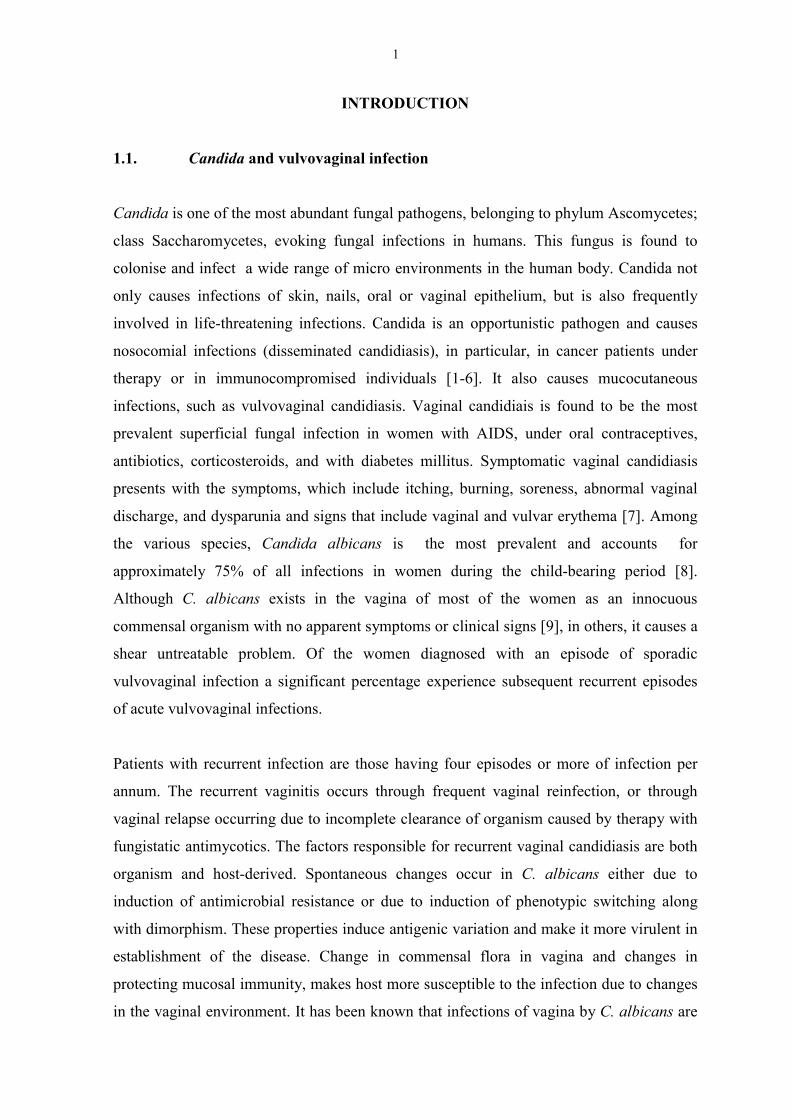
1.1. Candida and vulvovaginal infection
Candida is one of the most abundant fungal pathogens, belonging to phylum Ascomycetes;
class Saccharomycetes, evoking fungal infections in humans. This fungus is found to
colonise and infect a wide range of micro environments in the human body. Candida not
only causes infections of skin, nails, oral or vaginal epithelium, but is also frequently
involved in life-threatening infections. Candida is an opportunistic pathogen and causes
nosocomial infections (disseminated candidiasis), in particular, in cancer patients under
therapy or in immunocompromised individuals [1-6]. It also causes mucocutaneous
infections, such as vulvovaginal candidiasis. Vaginal candidiais is found to be the most
prevalent superficial fungal infection in women with AIDS, under oral contraceptives,
antibiotics, corticosteroids, and with diabetes millitus. Symptomatic vaginal candidiasis
presents with the symptoms, which include itching, burning, soreness, abnormal vaginal
discharge, and dysparunia and signs that include vaginal and vulvar erythema [7]. Among
the various species, Candida albicans is the most prevalent and accounts for
approximately 75% of all infections in women during the child-bearing period [8].
Although C. albicans exists in the vagina of most of the women as an innocuous
commensal organism with no apparent symptoms or clinical signs [9], in others, it causes a
shear untreatable problem. Of the women diagnosed with an episode of sporadic
vulvovaginal infection a significant percentage experience subsequent recurrent episodes
of acute vulvovaginal infections.
Patients with recurrent infection are those having four episodes or more of infection per
annum. The recurrent vaginitis occurs through frequent vaginal reinfection, or through
vaginal relapse occurring due to incomplete clearance of organism caused by therapy with
fungistatic antimycotics. The factors responsible for recurrent vaginal candidiasis are both
organism and host-derived. Spontaneous changes occur in C. albicans either due to
induction of antimicrobial resistance or due to induction of phenotypic switching along
with dimorphism. These properties induce antigenic variation and make it more virulent in
establishment of the disease. Change in commensal flora in vagina and changes in
protecting mucosal immunity, makes host more susceptible to the infection due to changes
in the vaginal environment. It has been known that infections of vagina by C. albicans are
2
dependent on the presence of reproductive hormones. Estrogen replacement therapy has
been shown to enhance the susceptibility to infection. Fidel et al. demonstrated that
estrogen plays an important role in the infection by enhancing on one hand the avidity of
C. albicans to vaginal cells and on the other reducing the ability of vaginal cells to inhibit
the growth of C. albicans [10]. Host defence mechanism to counteract Candida is mainly
mediated by cells like PMNs, macrophage and natural killer cells. Other factors such as
prostaglandins were found to be involved in candida infection. It was found that that
mononuclear cells from the patients suffering with recurrent vaginal candidiasis produce
higher PGE2, compared to control women [11], Moreover, Kustimur demonstrated
involvement of PGE2 and leukotrine C4 in the kidney damage induced by C. albicans [12].
These reports suggest an important role for host mediated response during infection.
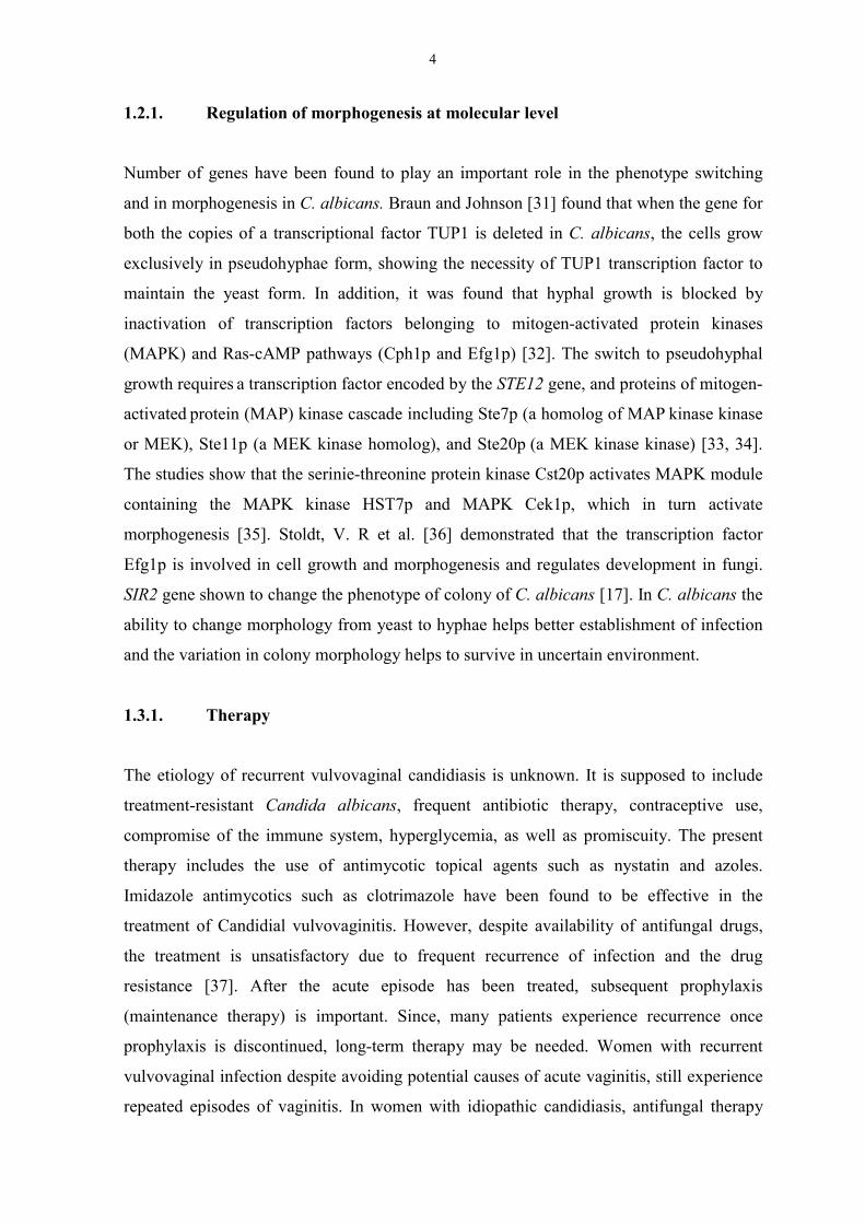
1.2. Morphogenesis in Candida albicans
Morphogenetic changes in C. albicans are important virulence factors [8,13]. C. albicans is
pleiomorphic, and undergoes reversible morphogenetic transitions between budding yeast
form (blastospores), pseudohyphal, and hyphal forms [14, 15]. Besides the morphological
heterogeneity at the cellular level, this pathogen exhibits variable colony morphologies,
like “white” and “opaque” colonies [16, 17]. The pseudohyphae form ranges from
relatively short to extended cells, while the hyphal forms possess constrictions at the septa
[18, 19]. Hyphae are found during the early stage of tissue colonisation, where as yeast
cells are found to be associated with diseased and necrotic tissues [8]. The formation of
hyphae enhances adherence and tissue invasion. The asymptomatic carriers carry C.
albicans in a small number and predominantly in a blastospore form rather then the hyphal
forms. The ability to switch between these forms is thought to be important for Candida's
virulence [20] and enables the pathogen to colonise different loci of the host, such as oral
and vaginal tracts, and to invade the parenchyma of inner organs via the blood stream [21].
The enormous range of environmental factors have been shown to play a role in the
phenotype switching. The factors which induce formation of hyphae include temperature
>37°C, pH >6.5, high CO2 to O2 ratio, low inoculum (<106 blastoconidia per ml), presence
of chemical factors like N-acetyl-D-glucosamine, glucose+glutamine, glucose+glycine,
proline, low zinc concentration, dextrin and particular formulations of growth media
include amino acid/salt medium, neopeptone-starch medium, TC199, and MEM medium.
The preferred morphological phenotype thus can switch in response to some external
3
stimuli or stress conditions. No single environmental factor has yet been considered as the
sole factor responsible for morphogenesis. The earlier studies showed that there are
number of host derived factors in vivo like prostaglandin E2 (PGE2), gamma interferon [22,
11], estrogen [23], progesterone [24, 25], which are responsible for morphogenetic
transformation in C. albicans. However, the exact mechanisms by which morphogenesis is
regulated by host-derived mediators are far from clear [15] and is still a controversial and a
stimulating area in fungal research. There are several aspects that support the role of
Candida dimorphism, in virulence, adhesion, invasion and escape from phagocytic cells
among others. Chaffin, et al. [26] showed that the different composition of the fungal cell
wall in the yeast and the hyphal forms play a role in adherence to different cell surfaces.
Moreover, the switch from to hyphal form of growth is important for the invasion of
epithelial cells [27] and may provide a mechanism for draining blastospores into a major
systemic bloodstream. Dimorphism also provides a pathway to efficiently escape from
professional phagocytic cells, since the extrusion of hyphae damages the cellular
membrane. The association between dimorphism and virulence has also been controversial.
It was found that isolation of mutants deficient in their ability to switch on or switch off
the dimorphic transition required different random mutagenic treatments. These mutants
also differed considerably in there pathogenicity [28, 29, 30]. It was found that those
mutants were less virulent than wild-type strains, however their phenotypes were very
pleiotropic in nature. This demonstrated that several other phenotypic traits apart from
morphogenetic change.
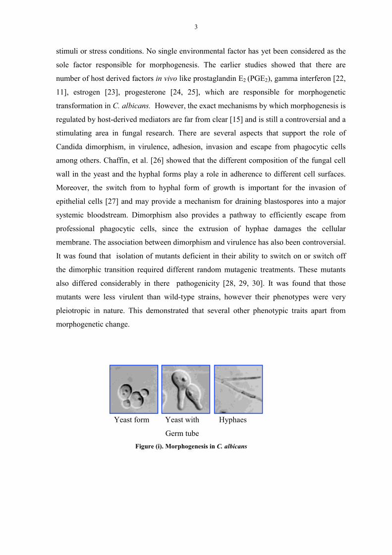
Yeast form Yeast with Hyphaes
Germ tube Figure (i). Morphogenesis in C. albicans
4
1.2.1. Regulation of morphogenesis at molecular level
Number of genes have been found to play an important role in the phenotype switching
and in morphogenesis in C. albicans. Braun and Johnson [31] found that when the gene for
both the copies of a transcriptional factor TUP1 is deleted in C. albicans, the cells grow
exclusively in pseudohyphae form, showing the necessity of TUP1 transcription factor to
maintain the yeast form. In addition, it was found that hyphal growth is blocked by
inactivation of transcription factors belonging to mitogen-activated protein kinases
(MAPK) and Ras-cAMP pathways (Cph1p and Efg1p) [32]. The switch to pseudohyphal
growth requires a transcription factor encoded by the STE12 gene, and proteins of mitogen-
activated protein (MAP) kinase cascade including Ste7p (a homolog of MAP kinase kinase
or MEK), Ste11p (a MEK kinase homolog), and Ste20p (a MEK kinase kinase) [33, 34].
The studies show that the serinie-threonine protein kinase Cst20p activates MAPK module
containing the MAPK kinase HST7p and MAPK Cek1p, which in turn activate
morphogenesis [35]. Stoldt, V. R et al. [36] demonstrated that the transcription factor
Efg1p is involved in cell growth and morphogenesis and regulates development in fungi.
SIR2 gene shown to change the phenotype of colony of C. albicans [17]. In C. albicans the
ability to change morphology from yeast to hyphae helps better establishment of infection
and the variation in colony morphology helps to survive in uncertain environment.
1.3.1. Therapy
The etiology of recurrent vulvovaginal candidiasis is unknown. It is supposed to include
treatment-resistant Candida albicans, frequent antibiotic therapy, contraceptive use,
compromise of the immune system, hyperglycemia, as well as promiscuity. The present
therapy includes the use of antimycotic topical agents such as nystatin and azoles.
Imidazole antimycotics such as clotrimazole have been found to be effective in the
treatment of Candidial vulvovaginitis. However, despite availability of antifungal drugs,
the treatment is unsatisfactory due to frequent recurrence of infection and the drug
resistance [37]. After the acute episode has been treated, subsequent prophylaxis
(maintenance therapy) is important. Since, many patients experience recurrence once
prophylaxis is discontinued, long-term therapy may be needed. Women with recurrent
vulvovaginal infection despite avoiding potential causes of acute vaginitis, still experience
repeated episodes of vaginitis. In women with idiopathic candidiasis, antifungal therapy
5
was found to be highly effective for individual attack. However, it failed to prevent future
recurrence, and episodes of vaginitis have been seen to appear as early few days to 3
months after the cessation of therapy [38]. Species differences were also observed in the
effectiveness of antifungal therapy, Polak [39] and Armstrong [40] demonstrated a
significantly higher degree of success with C. tropicalis as compared with C. albicans.
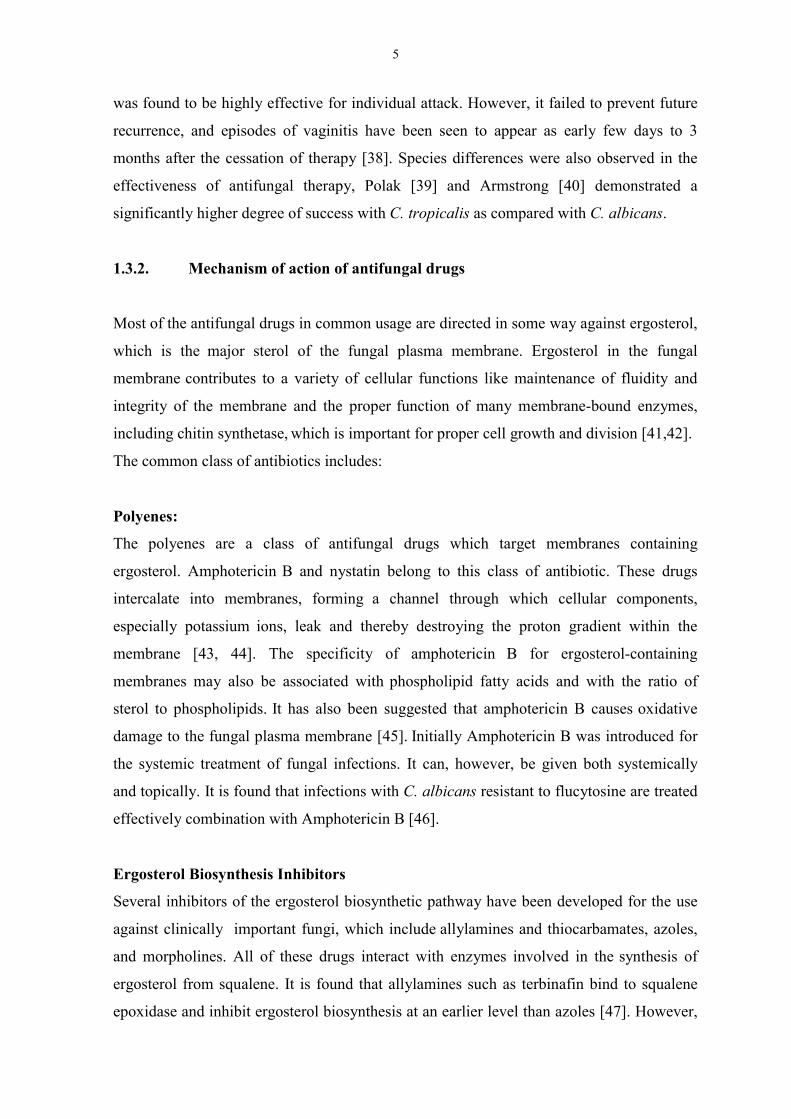
1.3.2. Mechanism of action of antifungal drugs
Most of the antifungal drugs in common usage are directed in some way against ergosterol,
which is the major sterol of the fungal plasma membrane. Ergosterol in the fungal
membrane contributes to a variety of cellular functions like maintenance of fluidity and
integrity of the membrane and the proper function of many membrane-bound enzymes,
including chitin synthetase, which is important for proper cell growth and division [41,42].
The common class of antibiotics includes:
Polyenes:
The polyenes are a class of antifungal drugs which target membranes containing
ergosterol. Amphotericin B and nystatin belong to this class of antibiotic. These drugs
intercalate into membranes, forming a channel through which cellular components,
especially potassium ions, leak and thereby destroying the proton gradient within the
membrane [43, 44]. The specificity of amphotericin B for ergosterol-containing
membranes may also be associated with phospholipid fatty acids and with the ratio of
sterol to phospholipids. It has also been suggested that amphotericin B causes oxidative
damage to the fungal plasma membrane [45]. Initially Amphotericin B was introduced for
the systemic treatment of fungal infections. It can, however, be given both systemically
and topically. It is found that infections with C. albicans resistant to flucytosine are treated
effectively combination with Amphotericin B [46].
Ergosterol Biosynthesis Inhibitors
Several inhibitors of the ergosterol biosynthetic pathway have been developed for the use
against clinically important fungi, which include allylamines and thiocarbamates, azoles,
and morpholines. All of these drugs interact with enzymes involved in the synthesis of
ergosterol from squalene. It is found that allylamines such as terbinafin bind to squalene
epoxidase and inhibit ergosterol biosynthesis at an earlier level than azoles [47]. However,
6
the mode of action of these compounds is found to be more fungicidal rather then
fungistatic. The allylamines (i.e., naftifine and terbinafine) and thiocarbamates (i.e.,
tolnaftate and tolciclate) inhibit the conversion of squalene to 2,3-oxidosqualene by the
enzyme squalene epoxidase [48,49]. The azoles antibiotics, such as, ketoconazole,
miconazole, fluconazole, itraconazole, voriconazole, are directed against lanosterol
demethylase in the ergosterol pathway. This enzyme is a cytochrome P-450 enzyme
containing a heme moiety in its active site [50, 51]. The morpholines, fenpropimorph and
amorolfine, inhibit two enzymes in the ergosterol biosynthetic pathway, C-14 sterol
reductase and C-8 sterol isomerase.
5-Flucytosine
5-Flucytosine (5-FC) has an entirely distinct mode of action from the azoles. 5-FC is taken
up into the cell, and deaminated into 5-fluorouracil. 5-FU is apparentely converted by
cellular pyrimidine-processing enzymes into 5-fluoro-dUMP (FdUMP). FdUMP is a
specific inhibitor of thymidylate synthetase, an essential enzyme for DNA synthesis, 5-
fluoro-UTP (FUTP) is incorporated into RNA, thus disrupting protein synthesis. 5-FC is
fungus specific drugs, since mammalian cells have little or no cytosine deaminase [43,45].
7
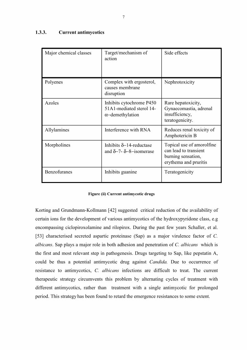
1.3.3. Current antimycotics
Figure (ii) Current antimycotic drugs
Korting and Grundmann-Kollmann [42] suggested critical reduction of the availability of
certain ions for the development of various antimycotics of the hydroxypyridone class, e.g
encompassing ciclopiroxolamine and rilopirox. During the past few years Schaller, et al.
[53] characterised secreted aspartic proteinase (Sap) as a major virulence factor of C.
albicans. Sap plays a major role in both adhesion and penetration of C. albicans which is
the first and most relevant step in pathogenesis. Drugs targeting to Sap, like pepstatin A,
could be thus a potential antimycotic drug against Candida. Due to occurrence of
resistance to antimycotics, C. albicans infections are difficult to treat. The current
therapeutic strategy circumvents this problem by alternating cycles of treatment with
different antimycotics, rather than treatment with a single antimycotic for prolonged
period. This strategy has been found to retard the emergence resistances to some extent.
Rare hepatoxicity,Gynaecomastia, adrenalinsufficiency,teratogenicity.
Inhibits cytochrome P45051A1-mediated sterol 14-α−demethylation
Azoles
Topical use of amorolfinecan lead to transientburning sensation,erythema and pruritis
Inhibits δ−14-reductaseand δ−7- δ−8−isomerase
Morpholines
TeratogenicityInhibits guanineBenzofuranes
Reduces renal toxicity ofAmphotericin B
Interference with RNAAllylamines
NephrotoxicityComplex with ergosterol,causes membranedisruption
Polyenes
Side effectsTarget/mechanism ofaction
Major chemical classes
8
1.4.1. Role of lipids and Fatty acid metabolism in C. albicans
Lipids contribute toward important structural and functional molecules in C. albicans.
Lipids constitute about 3.8-4.3 % of the dry weight of the fungal cells. Very little is known
about the precise role of fatty acids for the growth and virulance of C. albicans. Fatty acids
have been shown to modulate the fluidity and structure of plasma and other organelle
membranes depending upon the composition of the phopsholipid head groups. Singh, et al
[54] showed that alteration in lipid composition causes changes in the transport of amino
acids like lysine, proline, glutamic acid and glycine. Number of cellular processes like
cellular permeability, enzymatic activity, morphogenesis, and cell cycle are also influenced
by fatty acids. Adherence, and virulence, which are important pathogenic factors, are found
to dependent upon the lipid composition of C. albicans [55, 56, 57]. Goyal. and Khuller
[58] showed variations in the lipid composition of yeast and mycelia forms in Candida
albicans. They found that total lipid, phospholipid and sterol contents of log phase
mycelial cells were significantly higher than in yeast cells. Moreover, the saturated fatty
acid content is also much higher in the yeast forms. In general, yeasts are deficient in
polyunsaturated fatty acids, but Candida species are rare exceptions as they contain often
linoleic (18:2) and linolenic (18:3) acid.
9
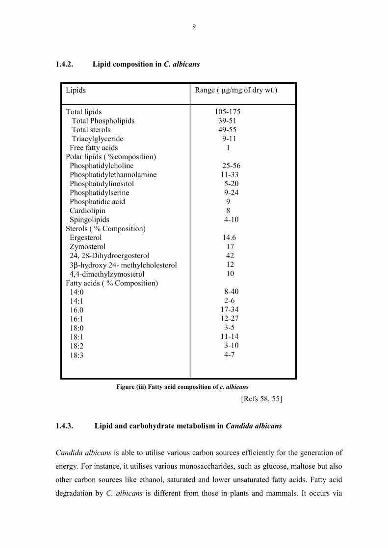
1.4.2. Lipid composition in C. albicans
Figure (iii) Fatty acid composition of c. albicans [Refs 58, 55]
1.4.3. Lipid and carbohydrate metabolism in Candida albicans
Candida albicans is able to utilise various carbon sources efficiently for the generation of
energy. For instance, it utilises various monosaccharides, such as glucose, maltose but also
other carbon sources like ethanol, saturated and lower unsaturated fatty acids. Fatty acid
degradation by C. albicans is different from those in plants and mammals. It occurs via
105-175 39-51 49-55 9-11 1
25-56 11-33 5-20 9-24 9 8 4-10
14.6 17 42 12 10
8-40 2-6 17-34 12-27 3-5 11-14 3-10 4-7
Total lipids Total Phospholipids Total sterols Triacylglyceride Free fatty acidsPolar lipids ( %composition) Phosphatidylcholine Phosphatidylethannolamine Phosphatidylinositol Phosphatidylserine Phosphatidic acid Cardiolipin SpingolipidsSterols ( % Composition) Ergesterol Zymosterol 24, 28-Dihydroergosterol 3β-hydroxy 24- methylcholesterol 4,4-dimethylzymosterolFatty acids ( % Composition) 14:0 14:1 16.0 16:1 18:0 18:1 18:2 18:3
Range ( µg/mg of dry wt.)Lipids
10
different mechanisms: alpha oxidation leading to the formation of CO2, beta oxidation
leading to acetyl CoA, which is returned to citrate cycle [60, 61, 62], or omega-oxidation
carried out by P450 cytochrome leading to the formation of fatty acids with a hydroxyl
group as well as to the formation of dicarboxylic acid. Generally fatty acid in Candida
species are degraded by peroxisomal beta-oxidation system. Beta-oxidation is a cyclic
oxidation system of fatty acids in beta position. In general, initially fatty acyl CoA
thioester undergo enzymatic dehydrogenation by acyl-CoA dehydrogenase to form trans-
enoyl coA. The double bond of trans-enoyl coA is hydrated to form 3-hydroxyacyl-CoA by
enzyme enoyl-CoA hydratase which is further dehydrogenated to form 3-ketoacyl-CoA. 3-
ketoacyl-CoA further undergoes cleavage by thiolase by interaction with a molecule of
free acetyl-CoA resulting into fatty acid shorter by two carbon atoms. In yeast β−oxidation
occurs in peroxisomes via series of reactions by a multienzyme complex MFE-2. The gene
encoding the multifunctional protein of peroxisomal beta-oxidation was first discovered in
Saccharomyces cerevisiae [59]. However, it is found that, non peroxisomal oxidation
enzymes are induced when grown on alkanes [64]. This reaction occurs in endoplasmic
reticulum and involes cytochrome P-450 monooxygenase [65]. This mechanism of fatty
acid beta-oxidation involves FAD and FMN redox system, which catalyse insertion of one
oxygen atom into the fatty acid.
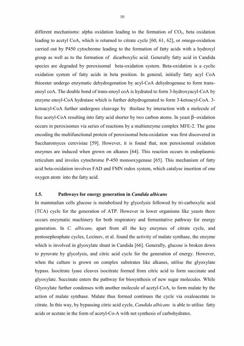
1.5. Pathways for energy generation in Candida albicans
In mammalian cells glucose is metabolised by glycolysis followed by tri-carboxylic acid
(TCA) cycle for the generation of ATP. However in lower organisms like yeasts there
occurs enzymatic machinery for both respiratory and fermentative pathway for energy
generation. In C. albicans, apart from all the key enzymes of citrate cycle, and
pentosephosphate cycles, Lozinov, et al. found the activity of malate synthase, the enzyme
which is involved in glyoxylate shunt in Candida [66]. Generally, glucose is broken down
to pyruvate by glycolysis, and citric acid cycle for the generation of energy. However,
when the culture is grown on complex substrates like alkanes, utilise the glyoxylate
bypass. Isocitrate lyase cleaves isocitrate formed from citric acid to form succinate and
glyoxylate. Succinate enters the pathway for biosynthesis of new sugar molecules. While
Glyoxylate further condenses with another molecule of acetyl-CoA, to form malate by the
action of malate synthase. Malate thus formed continues the cycle via oxaloacetate to
citrate. In this way, by bypassing citric acid cycle, Candida albicans is able to utilise fatty
acids or acetate in the form of acetyl-Co-A with net synthesis of carbohydrates.
11
.
Figure (iv). The Glyoxylate cycle. The reaction shown ar
utilise the breakdown product of fatty acid, Acetyl CoA,
For energy generation, C. albicans possesses the c
biochemical reduction-oxidation reactions that eff
series of carriers. An electron transport chain, also
the final stage of aerobic respiration. In the mitoch
the Krebs cycle, transfer their electrons through
ubiquinone and a series of cytochromes that u
reactions, accepting electrons and then donating t
process known as electron flow. Cytochrome oxi
ions with oxygen to form water. This process is c
possesses cyanide and antimycin A resista
salicylhydroxamic acid. Like components of
Acetyl-CoA
α−ketogSuccinyl CoA
Succinate
Fumarate
Malate
Oxaloacetate
Glyoxylat
I
Malatesynthase
Fatty acid metabolism
Gluconeogenesis
Glycolysis
Citrate
e catalysed by the en
for the biosynthesis o
lassical respiratory
ects the transfer o
known as the res
ondria, NADH or
a chain of carrier
ndergo reversible
hem to the next c
dase combines ele
oupled to the form
nt pathway an
the normal elect
Cis-aconitate
e
Oxalo
lutarate
e
socitrate lyase
Isocitrat
Glucose
zymes which efficiently
f sugar molecules.
chain A sequence of
f electrons through a
piratory chain, forms
FADH2, generated by
molecules, including
reduction-oxidation
arrier in the chain, a
ctrons and hydrogen
ation of ATP. It also
d is inhibited by
ron transport chain,
sucinate
12
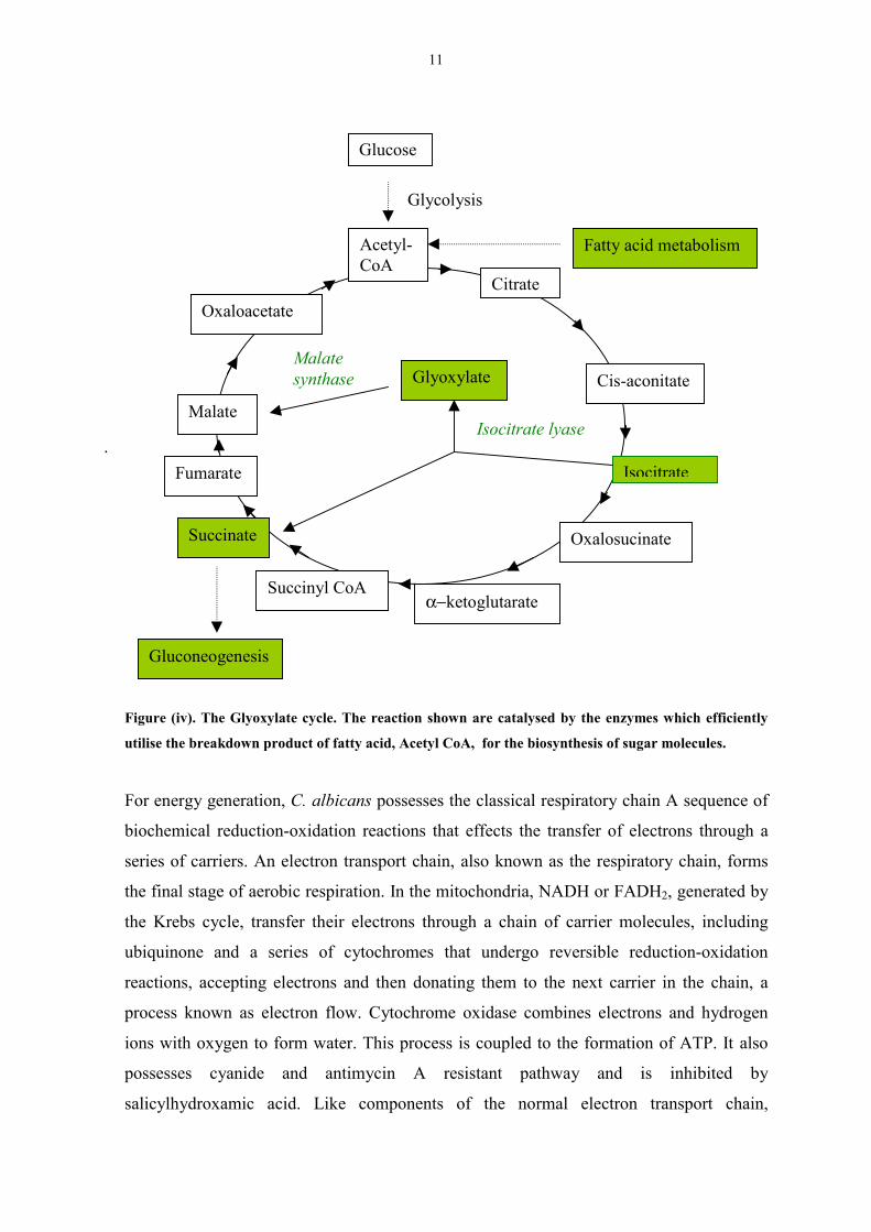
alternative oxidase, which reside in the mitochondrial inner membrane, transfers electrons
directly to oxygen and in doing so bypasses the regular transportation through respiratory
chain. The electron transfer reactions occur in mitochondria by transfer of electron from
NADH to oxygen through various substrates and are coupled with ATP generation. The
electrons are collected from many different cycles and transported to energy generation
system through the action of NAD-linked dehydrogenases. These electrons are transferred
form one substrate to another due to difference in oxido-reduction potentials which include
complexes of cytochrome b and c1, iron-sulphur proteins, and a complex of NADH
dehydrogenase. Various inhibitors like antimycin A, rotenone and cyanide act by blocking
the transfer of electrons through the respiratory chain.
Figure (v). Schematic representation of electron transport chain
C II ComplexFAD, FeS
CI ComplexFMN
UQAlternative Oxidation
CIII
Cyt C
C IV Complex
O2
Antimycin A
SHAM
Rotenone
Succinate NAD
13
1.6. Host response to the infection
Infection by C. albicans is known to be accompanied by strong inflammatory reactions of
host cells which are mediated via mannose and β-glucan receptors [67, 68, 69]. Castro et
al. [70] studied that upon stimulation by C. albicans macrophages and human monocytes,
release sizeable of arachidonic acid and are subsequently converted to lipoxygenase- and
cyclooxygenase-derived eicosanoids. It was also demonstrated that upon infection, C.
albicans induces monocytes to liberate proinflammatory cytokines like interleukin-1 beta
(IL-1β), tumor necrosis factor-alpha (TNFα), interleukin-6 (IL-6) and interleukin-8 (IL-8)
[71]. In human endothelial cells subjected to in vitro invasion of C. albicans, stimulation of
arachidonic acid metabolism leading to secretion of PGI2 has been reported [72, 73].
Moreover, Kustimur [12] demonstrated that in infected mouse kidney a shift of the
arachidonic acid metabolism to a preferential formation of LTC4 via lipoxygenase pathway
occurred. These examples accentuate a prominent role of host cell-derived arachidonic acid
in the inflammatory events induced by C. albicans during infection. Although arachidonic
acid does not occur in the lipids of C. albicans [55, 58], its release from host cells to a
significant extent renders it to a potential exogenous modulator of cell growth and
morphogenesis in C. albicans.
1.7. Cyclooxygenases (Prostaglandin synthases)
Prostglandins (PG) are shortlived substances derived from arachidonic acid that act as local
hormones. Prostaglandins play an important in normal physiology by maintaining
hemostatis acting as both vasodilatory agents and vasoconstrictors, renal function by
altering both sodium excretion and water clearance and control of gastric functions. Using
inhibitors of prostaglandin synthesis, it has also been shown that prostaglandins are
involved in platelet aggregation (prostacylins and thromboxanes), pain, fever and
respiratory functions such as, bronchoconstriction and bronchodilation. The role of
prostaglandins in reproductive physiology is amongst the most important of its functions.
Prostaglandins play a direct role in ovulation, lutenization, fertilization, fetal development
and parturition as demonstrated in human and animal studies. [review 81]. In pathological
conditions PGs are involved in diseases like arthritis, cancer, and inflammation mediating
swelling and pain [review 76].
14
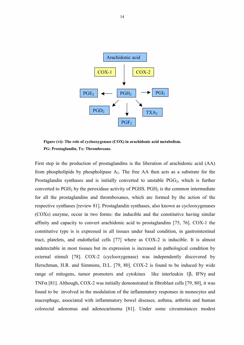
Figure (vi): The role of cyclooxygen
PG: Prostaglandin, Tx: Thrombox
First step in the production of pro
from phospholipids by phospholip
Prostaglandin synthases and is in
converted to PGH2 by the peroxida
for all the prostaglandins and th
respective synthases [review 81]. P
(COXs) enzyme, occur in two fo
affinity and capacity to convert ar
constitutive type is is expressed i
tract, platelets, and endothelial c
undetectable in most tissues but it
external stimuli [78]. COX-2 (
Herschman, H.R. and Simmons, D
range of mitogens, tumor promo
TNFα [81]. Although, COX-2 was
found to be involved in the modu
macrophage, associated with infla
colorectal adenomas and adeno
Arachidonic acid
ase (COX) in ar
ane.
staglandins is
ase A2. The f
itially convert
se activity of P
romboxanes, w
rostaglandin sy
rms: the induc
achidonic acid
n all tissues u
ells [77] wher
s expression is
cyclooxygenas
.L. [79, 80]. C
ters and cyto
initially demon
lation of the in
mmatory bowe
carinoma [81]
COX-2
COX-1achidonic a
the libera
ree AA th
ed to uns
GHS. PG
hich are
nthases, a
ible and t
to prosta
nder basa
e as COX
increase
e) was
OX-2 is
kines li
strated in
flammato
l diseases
. Under
PGI2
PGE2 PGH2PGD2
TXA2PGF2
cid metabolism.
tion of arachidonic acid (AA)
en acts as a substrate for the
table PGG2, which is further
H2 is the common intermediate
formed by the action of the
lso known as cyclooxygenases
he constitutive having similar
glandins [75, 76]. COX-1 the
l condition, in gastrointestinal
-2 is inducible. It is almost
d in pathological condition by
independently discovered by
found to be induced by wide
ke interleukin 1β, IFNγ and
fibroblast cells [79, 80], it was
ry responses in monocytes and
, asthma, arthritis and human
some circumstances modest

15
elevations of COX-1 could occur. For example, phorbol easter treatment can induce a
modest elevation COX-1 along with substantial increase in COX-2 in bronchial epithelial
cells [82, 83]. These data suggest that basal levels of COX-1 may differ as cell move
through differentiation pathways, and may be altered as a consequence of molecular
responses to cytokines and other agents modulating cell phenotype.
Upon stimulation, signals are initiated via both tyrosine kinase receptors and
transmembrane receptors and activate a number of distinct signal transduction pathways
mediated by protein kinase A, protein kinase C, JAK-STAT signalling mechanism etc. All
these signaling pathways converge on the regulatory region of the COX-2 gene and
influence the expression of COX-2 gene. Cloning and sequencing of the murine and human
COX-2 promoter region demonstrated presence of binding sites for AP-2, SRE and
ΝFκB, SP-1 transcription factors along with NFIL-6, CRE and E-Box sequences [84, 85].
There seems to exist a number of redundant pathways utilizing NFκB, ERK-2, p38 and
JNK MAP kinase pathways in COX-2 upregualtion by endotoxins in monocytes [86]. In
colon cancer cells, the expression of COX-2 was found to be regulated by p38 MAP kinase
pathway, but not by ERK pathway was not found to be involved [87]. In murine astrocytes,
Protein kinase C along with the MAP kinases upregulated COX-2 expression upon
stimulation with IL-1beta [88]. Each cell system and type of stimulus appears to have a
definite pathway for the expression of COX-2.
1.8. COX inhibitors
Non steroidal anti-inflammatery (NSAIDs) inhibit cyclooxygenase enzymes and thus are
used as anti-inflammatory, analgesic, and anti-pyretic drugs. The pharmacological
differences between COX-1 and COX-2 have been extensively explored and used for
development of new drugs. Depending upon the ability of drugs to inhibit COX-2 and
COX-1, these are classified into nonselective, selective and highly selective drugs. Aspirin,
indomethacin, ibuprofen and diclofenec are nonselective COX inhibitors that inhibit both
COX-2 and to an extent COX-1. Aspirin is a very potent inhibitor of COX-2 and is also
able to inhibit at a low dosage COX-1 in platelets, is thus effective in cardiovascular
medicine [89]. Treatment of patients with these drugs is found to reduce gastric
perforations, ulcers, and gastrointestinal bleeding. Agents show great preference for COX-
2 but are relatively ineffective towards COX-1. Animal studies show that these selective
inhibitors are non-ulcerogenic and non nephrotoxic. Both acryl methyl sulfonyl DuP 697,
16
and NS398 inactivate COX-2 by binding to the enzyme in a noncovalent fashion that
causes COX-2 induces a slow structural transition that results in its selective inactivation
[91].
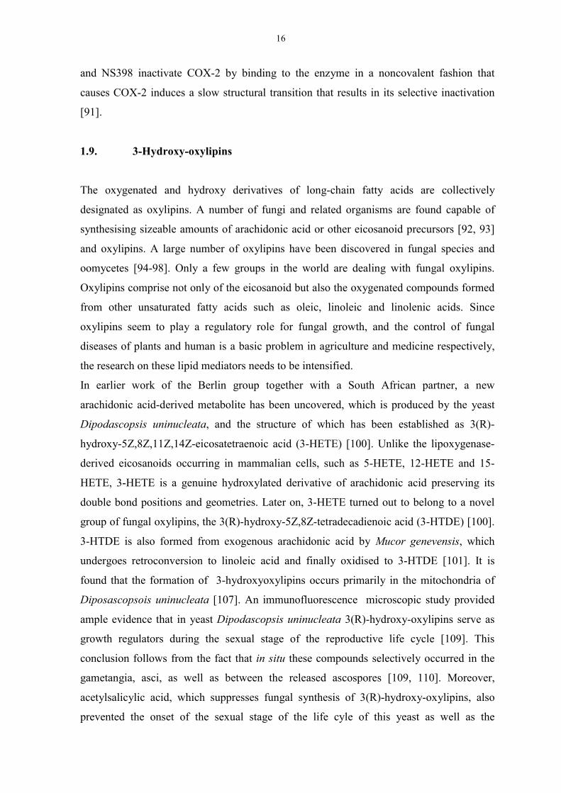
1.9. 3-Hydroxy-oxylipins
The oxygenated and hydroxy derivatives of long-chain fatty acids are collectively
designated as oxylipins. A number of fungi and related organisms are found capable of
synthesising sizeable amounts of arachidonic acid or other eicosanoid precursors [92, 93]
and oxylipins. A large number of oxylipins have been discovered in fungal species and
oomycetes [94-98]. Only a few groups in the world are dealing with fungal oxylipins.
Oxylipins comprise not only of the eicosanoid but also the oxygenated compounds formed
from other unsaturated fatty acids such as oleic, linoleic and linolenic acids. Since
oxylipins seem to play a regulatory role for fungal growth, and the control of fungal
diseases of plants and human is a basic problem in agriculture and medicine respectively,
the research on these lipid mediators needs to be intensified.
In earlier work of the Berlin group together with a South African partner, a new
arachidonic acid-derived metabolite has been uncovered, which is produced by the yeast
Dipodascopsis uninucleata, and the structure of which has been established as 3(R)-
hydroxy-5Z,8Z,11Z,14Z-eicosatetraenoic acid (3-HETE) [100]. Unlike the lipoxygenase-
derived eicosanoids occurring in mammalian cells, such as 5-HETE, 12-HETE and 15-
HETE, 3-HETE is a genuine hydroxylated derivative of arachidonic acid preserving its
double bond positions and geometries. Later on, 3-HETE turned out to belong to a novel
group of fungal oxylipins, the 3(R)-hydroxy-5Z,8Z-tetradecadienoic acid (3-HTDE) [100].
3-HTDE is also formed from exogenous arachidonic acid by Mucor genevensis, which
undergoes retroconversion to linoleic acid and finally oxidised to 3-HTDE [101]. It is
found that the formation of 3-hydroxyoxylipins occurs primarily in the mitochondria of
Diposascopsois uninucleata [107]. An immunofluorescence microscopic study provided
ample evidence that in yeast Dipodascopsis uninucleata 3(R)-hydroxy-oxylipins serve as
growth regulators during the sexual stage of the reproductive life cycle [109]. This
conclusion follows from the fact that in situ these compounds selectively occurred in the
gametangia, asci, as well as between the released ascospores [109, 110]. Moreover,
acetylsalicylic acid, which suppresses fungal synthesis of 3(R)-hydroxy-oxylipins, also
prevented the onset of the sexual stage of the life cyle of this yeast as well as the

17
concomitant release and the subsequent self-assembly of ascospores in orderly clusters
[110]. Regulatory functions in the life cycle of fungi have also been proposed for other
oxylipins [105].
In Berlin, also preliminary evidence was obtained for a putative role of 3(R)-hydroxy-
oxylipins in fungal diseases of humans. Using a polyclonal antibody raised against 3-
HETE a novel arachidonic acid metabolite was detected in the fungal pathogen Candida
albicans and identified as 3,18-dihydroxy-5Z,8Z,11Z,14Z-eicosatetraenoicacid (3,18-
diHETE) [102]. This observation merits particular attention owing to the fact that 3-HETE
was shown to cause chemotaxis and to modulate cell signaling of human neutrophils [104].
Neutrophils and other phagocytosing cells constitute the main defense system of the
organism against fungal pathogens. Therefore it is reasonable to assume that 3(R)-
hydroxy-oxylipins may play a role in the host-pathogen interaction during the infection
process.
Oxylipins play an important role in vegetative growth and sexual reproduction of yeast
[100, 108, 109]. Dr. Nigam’s group in Berlin and South Africa, screened a few hundreds
strains of yeast and have demonstrated that 3(R) hydroxy-oxylipins are specifically
observed in sexual reproductive stage of life cycle of D. uninucleata.
3-hydroxy oxylipins have also been investigated for potential biological activity in
mammalian cells. 3-HETE has diverse spectrum of biological activities. 3-HETE has been
observed to be a proinflammatory mediator Moreover, 3-HETE was found to be a strong
chemotactic agent, the potency of which is comparable to LTB4 or fMet-Leu-Phe. 3-
HETE, however does not exert chemokinesis and exocytosis. It augments the release of
arachidonic acid and platelet-activating factor (PAF) via. activation of phospholipase A2.
Cell signaling by 3-HETE seems to imply a G-protein-dependent process [112].
1.10. Adhesion
Attachment of microorganism to the human tissue is primary step for the establishment of
infection and the development of disease. Adhesion of Candida albicans to the host tissue
is considered and virulance factors in the development of disease and is one of the [113,
114]. The nature of the component which mediates adhesion is controversial. Some
findings indicate that mannan, mannanproteins or polysaccharides are responsible for the
adhesion. Other findings show aspartyl proteases and phospholipases relevant for
adherence and invasion of host structures by pathogenic yeasts [115]. It is believed that the
type of disease and the host response depend in part on the invasiveness of the strain of C.
18
albicans that is causing disease [56, 116]. Thus, phosphatidylinositol 3-kinase of Candida
albicans has been demonstrated to affect the adhesion of C. albicans to the host tissue. The
mutants depleted of PI-3 kinase gene have been shown to have decreased adherence and
thus less pathogenic [117].
1.11.1 MAP kinase signaling pathway
Mitogen-activated protein kinases (MAP kinases) are enzymes which transmit signals from
cell surface receptors to critical regulatory targets in the cells. These are activated by
various environmental signals, such as growth factors and cytokines, and physical stress
and play an important role in cell survival and adaptation to the new environment. These
are also found to be involved in cell growth, and in the co-ordination of cell functions. The
basic arrangement of the cascade includes a G-protein working upstream of a core module
consisting of three kinases: a MAPK kinase kinase (MAPKKK) that phosphorylates and
activates a MAPK kinase (MAPKK), which in turn activates MAPK, this set-up provides
not only the signal amplification, but, more importantly, additional regulatory interfaces
that allow the kinetics, duration and amplitude of the activity to be tuned precisely.
The distinct classes of MAPKs, which have been identified include p42-p44 extracellular
signal-regulated kinases (ERK MAPKs), Stress Activated Protein Kinases (SAPK) which
include NH2-terminal jun kinases (p46-p54 JNKs), and p38 MAPKs α, β , γ and δ. These
kinases are induced by growth factors and cytokines. Each MAPKs are induced by
different stimuli, and trigger specific biological response, whereby the specificity is
maintained throughout the whole cascade. Each MAPK is specifically recognised and
phosphorylated by map kinase kinase (MKKs). The MKK protein kinase has specificity
towards Ser/Thr and Tyr residues on exogenous substrates. MKK themselves get activated
by phosphorylation at Ser/Thr residues by various MAP kinase kinase kinases (MEKKs),
which include Raf family members, c-Mos, MEK kinase (MEKKs), and multilineage
protein kinase (MLKs). Thus, the diversity in the activation of MAPK pathway resides
upstream of MKK, and the transmission of signal occurs via sequential protein kinases
regulated by dual phosphorylation [118]. The overall sequence identity among JNK/SAPK,
p38 MAPK, and ERKs is 40-45%, and all three enzymes have a common mechanism of
activation through phosphorylation of Thr-X-Tyr motif [119].
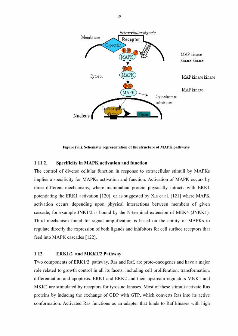
19
Figure (vii). Schematic representation of the structure of MAPK pathways
1.11.2. Specificity in MAPK activation and function
The control of diverse cellular function in response to extracellular stimuli by MAPKs
implies a specificity for MAPKs activation and function. Activation of MAPK occurs by
three different mechanisms, where mammalian protein physically intracts with ERK1
potentiating the ERK1 activation [120], or as suggested by Xia et al. [121] where MAPK
activation occurs depending upon physical interactions between members of given
cascade, for example JNK1/2 is bound by the N-terminal extension of MEK4 (JNKK1).
Third mechanism found for signal amplification is based on the ability of MAPKs to
regulate directly the expression of both ligands and inhibitors for cell surface receptors that
feed into MAPK cascades [122].
1.12. ERK1/2 and MKK1/2 Pathway
Two components of ERK1/2 pathway, Ras and Raf, are proto-oncogenes and have a major
role related to growth control in all its facets, including cell proliferation, transformation,
differentiation and apoptosis. ERK1 and ERK2 and their upstream regulators MKK1 and
MKK2 are stimulated by receptors for tyrosine kinases. Most of these stimuli activate Ras
proteins by inducing the exchange of GDP with GTP, which converts Ras into its active
conformation. Activated Ras functions as an adapter that binds to Raf kinases with high
20
affinity and causes their translocation to the cell membrane, where Raf activation takes
place [123]. Mammals possess three Raf proteins: Raf-1, A-Raf and B-Raf. All three Raf
isoforms share Ras as a common upstream activator and MEK as the only commonly
accepted downstream substrate [124]. MEK is activated by phosphorylation of two serine
residues in the activation loop. Although other kinases such as MEKK-1 (MEK kinase-1),
mos or Tpl-2 can phosphorylate the same serines. It is predominantly done by most cell
types by Raf kinases. Raf can activate both MEK-1 and MEK-2 (also called MKK-1 and
MKK-2) with similar efficacy in vitro. MEK in turn activates ERK-1 and ERK-2 (also
called p44 and p42 MAPK) via phosphorylation of a -Thr-Glu-Tyr- motif in the activation
loop. Although biochemical and transfection experiments suggest that ERK-1 and ERK-2
are functionally equivalent, it is still unclear as to why two ERK genes exist.
1.13.1. Stress activated protein kinases and Stress-activated MAP Kinase Kinases
The stress activated protein kinases include NH2-terminal c-jun kinase (JNK) and p38
MAP kinase, which are ubiquitously expressed, and get activated in response to cellular
stress, inflammation growth factors and lipopolysaccharides. Four different MKKs have
been identified, which phosphorylate MAP kinases. Derijard et al. [125] and Raingeaurd et
al. [126], found that MKK3 selectively phosphorylates p38 MAPK, while MKK6 which is
closely related to MKK3, also phosphorylates p38 MAPK, although with a higher basal
activity [127]. MKK4 and MKK7 are the regulators of JNK Stress activated protein
kinases, while MKK1 and MKK2 phosphorylate ERK at N-terminus. The activation of
these enzyme occurs by a dual phosphorylation at serine and threonine residues
[128].There are two classes of Map kinase kinase kinases which include: MEK Kinase
Kinase (MEKK) and Mixed Lineage Kinases (MLKs). Out of nine MKKs, JNK/SAPK
pathways are regulated through MKK4. In vitro MEKK2 phosphorylates MKK1 and 4
while MEKK3 phosphorylates MKKs 1, 3, and 4 [129, 130].
1.14. Nuclear factor-κκκκB
Upon infection or stress cells, try to adjust to the environment by bringing about the
changes in the pattern of gene expression. Sixteen years ago Sen & Baltimore [131], first
described NF-κB as a B-cell nuclear factor that bound a site in the immunoglobulin
enhancer. These changes are controlled by the transcriptional factors, which are
translocated from cytoplasm into the nucleus, and bind to there cognate site to activate or
repress the transcription. Nuclear factor-κB NFκB is one such transcription factor. For a
21
long time the transcription factor nuclear factor κB (NF-κB) has attracted attention because
of its unique activation pathway and its physiological importance as a key regulatory
molecule of the immune response, cell proliferation and apoptosis [132,133,134].
Endogenous activation of NF-κB is a cellular defence mechanism that protects cells by
inducing survival genes, such as xIAP and BCLxL [133, 134]. There is as well the evidence
for the independent upregulation of NFkB in apptosis [132]. NF-κB is a dimeric protein
composed of various combinations of the five different DNA-binding subunits: NF-κB1
(p50 and its precursor p105), NF-κB2 (p52 and its precursor p100), c-Rel, RelB and p65
(RelA) although the most frequently observed form of NF-κB is a p50¯p65 heterodimer.
All NF-κB family members have a conserved N-terminal Rel-homology domain (RHD),
which is responsible for dimerization, DNA binding and interaction with ΙκΒs (inhibitors
of NF-κB) [135]. Direct phosphorylation of NF-kappaB itself is essential for its
transcriptional activity [136]. The precursor proteins p105 and p100 can be processed by
the proteasome to generate p50 and p52, respectively. Recently the three-dimensional
structures of NF-κB·ΙκΒ ternary complexes (composed of the RHDs of p50 and p65 and
the repeat core of ΙκΒα) have been solved [137, 138]. In most cell types, NF-κB is
maintained in an inactive form in the cytoplasm by association with ΙκΒs. Physical and
chemical stresses, viruses, bacteria and pro-inflammatory cytokines like interleukins and
tumour necrosis factor (TNF) activate NF-κB by inducing the rapid phosphorylation of
ΙκΒ and its subsequent ubiquitination and proteolytic degradation. Released NF-κB then
translocates to the nucleus, binds to its cognate DNA element and activates transcription of
numerous target genes. The inducible phosphorylation of ΙκΒ is mediated by recently
identified ΙκΒ kinases (IKKα, β and ε). The catalytic subunits, IKKα and IKKβ, and the
regulatory IKKγ/NEMO (NF-κB essential modulator) subunit, form the prototypic core IB-
kinase complex (IKC). Importantly, this complex serves as an intracellular point of
convergence for distinct signals that ultimately activate NF-κB.
Although NF-κB is generally considered to be cytoplasmic in most cell types until
stimulation with an inducer. Many cell types appear to have moderate levels of the p50
(NF-κB1) homodimer in nucleus. A role for this factor in constitutive-type transcription is
not very clear, but it has been shown to serve to induce transcription of transcriptional
repressor proteins [135, 137].
22
However, there is accumulating evidence that NF-kB is also subject to an IkB-independent
level of regulation, as implicated by the earlier finding that the p38 mitogen-activated
protein (MAP) kinase inhibitor SB203580 does not interfere with induced nuclear
translocation and DNA binding of NF-κB, but significantly inhibits NF-κB-dependent
gene expression. Modulation of transcription factor function by regulatory
phosphorylations of the DNA-binding subunits is also observed in other inducible
transcription factors, such as activating protein 1 (AP-1) and cyclic AMP-responsive-
element binding protein (CREB).
1.15. Apoptosis
Apoptosis is a term which describes programmed cell death in vertebrates. The control of
cell death could help physicians control ageing process and in diseases like ischemic heart
disease and brain diseases [139]. The induction of apoptosis could help fight autoimmune
disease and cancer [140]. Apoptosis involves number of proteins like Bcl-2 family,
caspases, and mitochondrial proteins like cytochrome C. Bcl-2 protein blocks apoptosis
and acts as survival agent [141], which is triggered by stumulus like growth factor
deprivation and, irradiation. It is found that Bcl-xl, Bcl-2 and Bax can form channels in
lipid bilayers [142, 143] and thus control the movement of other regulators of apoptotis
that reside inside mitochondria. These proteins exerts pro or anti-apoptotic function by
regulating the release of mitochondria to cytosol initiating cell death, however the exact
mechanism of control of cytochrome C by the proteins of Bcl-2 family is not very clear
[144]. A similar mechanism is observed with Bcl-xL which interacts with human CED-4
homologue Apaf-1 and suppresses its pro-apoptotic activity [145,146]. Proteases involved
in apoptosis are collectively termed as caspases. Caspases are found to be involved in both
the initial signaling events and downstream protolytic cleavages that characterise the
apoptotic phenomenon [147]. These enzymes mediate cell death by cleavage of proteins
are the effectors of apoptosis [148]. Apoptosis is marked with disruption of mitochondria
and is a feature of apoptotic cell death. Disruption of mitochondria leads to the release of
cytochrome c, from the intermembrane space after disruption of outer membrane [149].
Cytochrome c brings about activation of caspases.Caspase-9 is processed into an active
enzyme when it combines with Apaf-1 in presence of cytochromec and either ATP or
dATP [150]. However all apoptotic pathways are not cytochrome c dependent [151]. TNF-
family death receptors directly induce apoptosis by by activating caspases without
involving mitochondria [152]. But since caspases trigger changes in mitochondrial

23
permeablity, leading to release of cytochrome c, it indirectly involves mitochondria for
apoptosis. Mechanism of induction of cell death also involves, cell surface death receptors
[153]. Fas, the receptor for Fas ligand (FasL) is a member of TNF (tumor necrosis factor)
family receptor. Binding of FasL to Fas induces apoptosis of Fas bearing cell [154].The
Fas mediated pathway is important in T-cell selection and perephiral clonal deletion. Trail
is found to be the member of TNF ligand family, and is shown to induce apoptosis.
One enigmatic molecule is NF-kB, which is a DNA-binding dimer of nuclear factor κB
(NF-κB) is retained in the cytoplasm by interaction with the inhibitor of NF-κB protein
(IκB) [156]. It has been shown to be an important transcription factor playing a role in and
apoptosis [157]. Levkau et al demonstrated that caspase-3 brings about cleavage of p65 at
COOH terminus and thus inactivating NFκB by generating truncated p65 which can still
bind to the DNA, thus potentially acting as a dominant negative inhibitor by competing
with the intact p65 [132]. There is ample data suggesting the therapeutic role of the study
of cell death. It has been shown that the gene transfer in tumor cells initiate apoptosis,
which could be the basis of therapy [158].
24
2. AIMS AND OBJECTIVE OF STUDY
The production of 3-HETE, an eicosanoid first reported by Dr. Nigam’s group in
collaboration with Dr. Kock’s group in South Africa from spore bearing fungus D.
uninucleata. fed with exogenous arachidonic acid was the beginning of a novel feild of
research, so called 3-hydroxyoxylipins. Most striking aspect of synthesis from D.
uninucleata was that it had to be supplied with AA exogenously, since most of the fungi do
not contain fatty acid higher then LA (18:2) and its formation was aspirin sensitive. While
screening of 3-OH-oxylipins in numerous fungi using antibody raised against 3-HETE,
which recognised only only fatty acids which contain hydroxyl group at position C-3, Dr.
Nigam’s group demonstrated the presence of 3-OH oxylipins in C. albicans, which was
later identified by us (section 4. Results) as 3,18-diHETE. The inflamed host tissue in
vulvovaginal candidiasis has been shown to release huge amount of AA. We therefore
hypothesized that this AA can be taken up by C. albicans and transformed to 3-OH-
oxylipins, which in turn may have effects on one side related to morphogenesis of the
attaching pathogen and on the other side host muscle cells to leading to
immunomodulatory activation. With this concept of host–pathogen interaction
Objectives were:
1. to find whether there occurs release of arachidonic acid by the host cells during
Candida infection, taking HeLa cells as model
2. to study the metabolism of arachidonic acid by Candida albicans and its ability to
convert arachidonic acid into 3-hydroxyoxylipins or related compound
3. to study the role of 3-hydroxyoxylipins on the growth and morphology of C. albicans.
4. to investigate the biological effects of 3-hydroxyoxylipins on HeLa cells
5. to study the upregulation of Candida albicans-induced eicosanoid production
6. to study the mechanism of signal transduction in HeLa cells mediated by C. albicans
7. to use microarray analysis to find the pathways triggered by C. albicans
25
3. EXPERIMENTAL PROCEDURES
3.1. Materials Fatty acids and Eicosanoids
Arachidonic acid, linoleic acid and PAPC- Sigma (Germany).
Inhibitors
Antimycin A- Sigma, SHAM- sigma, Rotenone SB212190, GF203190X, PD98059 –
Calbiochem (Germany); NS398 – Cayman (USA), LY294002, Wortmannin- Biomol
(Germany), Pyrrolidine Dithiocarbamate (PDTC) – Sigma (Germany).
Culture reagents
FCS, DMEM, Trypsin/EDTA, Streptomycin/Penicillin – Seromed (Germany);
CHROMagar – Mast diagnostica, (Germany), Yeast Nitrogen Base, Yeast extract, Peptone,
Malt Extract, Agar, Tryptone – Difco (USA).
Separation media
Agarose – Roth (Germany); Silica gel-60 TLC plates – Merck (Germany); Rotiphorese
acrylamide Gel 30 – ROTH (Germany); C8 reverse phase HPLC column, C18 RPLC
column – VDS Optilab (Germany).
Kits
RNeasy Mini Kit and QIA shredder - Qiagen (Germany); PGE2 ELISA - Cayman
Chemical Company (U.S.A); Protein Determination kit – Biorad (USA), ECL Detection kit
– Santa Cruz (California, USA); Atlas Microarray kit – Clontech (USA), Luciferase
Reporter Assay – Promega (Germany), PolyFect Transfection Kit- Qiagen (Germany).
Antibodies
Actin, phosphoserine – Sigma (Germany), COX-2 – Cayman (USA); phosphotyrosine –
ICN (USA) and Upstate, Germany; JNK, p38 MAP Kinase, p42/44 Kinase, PI-3 Kinase,
HSP-27, COX-1, BCL-2, anti-mouse, anti-rabbit, anti-goat secondary antibodies
conjugated with horse raddish peroxidase Anti mouse-FITC – Santa Cruz, (Heidelberg,
Germany)
26
RT-PCR reagents
Expand reverse transcriptase – Roche Biochemicals (Germany), Taq DNA Polymerase –
AB Technologies (Germany)
Radiochemicals14C-Arachidonic acid (55 mCi/mmol) - Amersham (England).
PrimersAll primers were purchased from TIB Biomol (Germany).
All organic solvents used in the experiments analysed by HPLC and GC-MS were of
LiChrosolv or SupraSolv quality, all other reagents were of analytical grade.
Other reagents
5-CFDA- Calbiochem, Aspirin, Clotrimazole, High density lipoprotein (HDL), estrogen,
progesterone, Gram staining reagents – Sigma, Scintillation fluid- dithiothreitol,
bromophenol blue, glycerol, Ponceau were from Sigma, nitrocellulose membrane
(Schleicher & Schuell), Luminol reagent-Amarsham, Protein A-Agarose- dianova,
Germany), Reporter lysis buffer -Promega)
Enzymes
Proteinase K- Roche Biochemicals, Germany T4 DNA ligase-Gene Craft, Germany
Electrophoresis material
Agarose –ICN Biochemicals, Germany, Silica gel-60 TLC plates – Merck (Germany),
Rotiphorese acrylamide Gel 30 – ROTH (Germany).
Solvents
All organic solvents used for TLC were of analytical grade and for HPLC and GC-MS
analysis were of LiChrosolv or SupraSolv quality.
MEDIA COMPOSITIONS: All chemicals from GIBCO BRL.
27
Universal medium for yeast: YEAST EXTRACT 0.3%
Malt extract 0.3%
Peptone 0.5%
Glucose 1%
Agar 1.5%
pH 5.6
Sabourauds glucose broth :
Peptone 1%
Glucose 4%
pH 7.4
Inorganic salt medium:KH2PO4 0.3%
(NH4)2SO4 0.025%
CaCl2 0.025%
MgSO4 0.025%
Biotin 0.01%
LB broth:Tryptone 1%
Yeast extract 0.5%
NaCl 1%
pH 6.5
Tissue Culture Medium
DMEM 90ml
FCS 10 ml.
Penicillin
CHROMR agar - Mast diagnostica, Reinfeld, Germany
28
3.2. Organisms and cell culture
Candida albicans 1386 (a clinical isolate from bronchomycosis) was obtained from
“Deutsche Sammlung von Mikroorganismen und Zellkulturen GmbH” (DSMZ), and a
culture of Candida albicans, isolated and identified as C. albicans from the vaginal swab
of a 34 year old women infected with recurrent severe vaginal candidiasis was used for all
our studies. The cultures were maintained on universal medium for yeast containing 0.3%
yeast extract, 0.3% malt extract, 0.5% peptone, and 1% glucose in 1.5% agar, pH 5.6. The
chemicals used for cell cultures were purchased from Difco Laboratories. HeLa cells were
used as model, grown in DMEM supplemented with 10 % FCS and incubated at 37°C with
5% CO2. The cells were split regularly, with a split ratio of 1:3 or 1:4, and passaged every
2-5 days. The cells were not grown over 100 % confluency. For most of the experiments
cells were used just when they reached confluency. For microscopic examination the cells
were grown on coverslip to 60-70 % confluency.
3.3. Isolation and identification of vaginal C. albicans from patients
Swabs from the vagina of the patients suffering form Candidiasis were collected in a tube
containing 1% peptone and were immediately used for the isolation. The swab was swirled
in the tube and the suspension was spun at 2000 X g for 20 min. The supernatant was
discarded and the pellet was suspended aseptically in 50 µl sterile PBS. The suspension
thus made was streaked on the Sabouraud agar plate. The colonies formed were examined
microscopically for the yeast forms and the probable colonies of C. albicans were
restreaked on CHROMagarR. Green colonies, which could possibly be C. albicans, as
recommended by the company, were picked up to check the capability for germ tube
formation at 37°C in human serum within 4-6 hr. A loopful of culture from the isolate was
inoculated in medium containing 1% peptone either with 1% glucose, sucrose, lactose or
maltose in presence of phenol red as indicator to see the fermentation capability of the
isolate and to confirm the isolate as C albicans biochemically.
3.4. Basal culture conditions for C. albicans
C. albicans was suspended in Sabouraud glucose broth containing 1% peptone giving an
optical density of 0.165 at 490 nm corresponding to a protein concentration of
approximately 0.05 mg/ml (Lowry method, Biorad kit), and grown in a rotary shaker at
29
100 rpm for 36 h at 37°C. The cells were separated from the medium by centrifugation at
3000 × g for 5 min, washed three times with phosphate-buffered saline (PBS), and finally
resuspended in 1 ml fresh medium of proper composition for the respective experiments.
3.5. Preparation of Blastospores
The blastospores were used for inoculation into the medium for the study of
morphogenesis or for inducing infection to HeLa cells. Blastospores were prepared by
inoculating a single colony from the plate in Sabouraud glucose broth pH 5.6, and
incubated for at 27°C for 48 hr followed by centrifugation at 2000 X g for 10 min.
3.6. Preparation of hyphal form
C. albicans was induced to form hyphae by inoculation in a tube containing 10 ml
Dulbecco’s Minimal Essential Medium (DMEM) in 15 mM HEPES buffer and incubation
at 37°C for 48 hr. The cells were centrifuged at 3000 X g and the supernatant was replaced
with the fresh medium and used for the experiments.
3.7. Determination of cell growth for the study of metabolism of different fatty
acids as carbon source
Cells (103) were inoculated in 1 ml of carbon source free inorganic salt medium (ISM)
containing 0.3% KH2PO4, 0.3% (NH4)2SO4, 0.025% CaCl2, 0.025% MgSO4, and 0.001%
biotin. To medium, various concentrations of arachidonic (1, 3, 10, 30 µM) or linoleic
acids (1, 3, 10, 30 µM) were added followed by incubation at 37°C for 24 h under shaking.
The cell number was determined by serial dilution of the culture and plating them out on
Sabouraud agar plates. The number of colony forming units (CFU) was counted after 24 h.
To study whether 3-HETE (1, 10 µM) stimulates the cell growth, similar experiments were
carried out on the microtitre plate containing inorganic salt medium and 3-HETE as a sole
carbon source. Medium (100µl) was inoculated with 103 C. albicans.
3.8. Metabolism of [1-14C]-arachidonic acid
C. albicans was grown in 1 ml Sabouraud broth made in 100mM phosphate buffer pH 7.4.
The cells were washed by centrifugation and suspended in 1.0 ml Sabouraud broth
30
containing 1% peptone without glucose followed by addition of 10 µM [1-14C]-arachidonic
acid (specific radioactivity = 50 µCi/mmol) and incubation at 37°C for 2, 4 and 6 h. Cells
and supernatant were separated by centrifugation and were subjected to Bligh and Dyer
[159] extraction at pH 3, with equal amount of chloroform: methanol (9:1) mixture. The
lipid extracted was dried under inert nitrogen atmosphere and were subjected to thin layer
chromatography (TLC) in solvent n-hexane: diethyl ether: acetic acid (50:50:1). The
distribution of radioactivity was quantified by radioscanning (automatic TLC linear
analyser, Berthold, Berlin, Germany) and scraping off of the labelled spots from the plates.
Radioactivity was measured by addition of liquid scintillation cocktail (Packard) in a liquid
scintillation counter (Beckman). The TLC Plates were also subjected to autoradiography
using X-ray films.
3.9. Distribution of radioactivity derived form arachidonic acid into different
biomolecules
C. albicans was grown for 36 h at 37°C in buffered Sabouraud broth. The cell suspension
was transferred to the main compartment of a Warburg vessel, the inset of which contained
0.1 ml 5 M KOH. The closed Warburg vessel was shaken at 105 rpm for 5 h at 37°C and
various components were separated and analysed for the presence of radioactivity. For
determination of labelled CO2 formation, the liquid of the inset was directly transferred to
scintillation counting cocktail and measured in scintillation counter. The liquid of the main
compartment was subjected to Bligh-Dyer extraction. The organic phase was evaporated to
dryness under nitrogen and dissolved in a small volume of chloroform. The lipid extract
was separated by thin-layer chromatography on pre-coated silica gel plates (Merck,
Darmstadt, Germany) using the solvent systems n-hexane–diethylether–glacial acetic acid
60 : 40 : 1 or 50 : 50 : 1 (by vol.). For separation of phospholipids and glycolipids the
solvent system chloroform–methanol–water 65 : 25 : 4 (by vol.) was applied. The aqueous
phase of the Bligh-Dyer extraction was subjected to precipitation of proteins by 10%
trichloroacetic acid (TCA). After centrifugation of the denatured proteins, the supernatant
fluid was extracted with diethylether to remove TCA and used for analysis of
carbohydrates, mono-, di- and oligosaccharides. Other low-molecular compounds were
separated from the polysaccharides (mainly glucans and mannans) by extensive dialysis.
The carbohydrates were concentrated under vacuum and separated by TLC on boric acid-
impregnated silica gel plates using the solvent n-butanol–glacial acetic acid–diethylether–
31
water 9 : 6 : 3 : 1 (by vol.) and visualised with either 4-methoxybenzaldehyde–H2SO4 or
N-(1-naphthyl)-ethylenediamine–H2SO4 reagents. Authentic trehalose, glucose and
mannose were run as standards. The distribution of radioactivity was estimated in
identically as described above for the lipids.
3.10. Alkaline hydrolysis of lipids
The total lipid extracts, obtained by the Bligh-Dyer method or single lipid fractions,
separated by TLC, scraped off and re-extracted with methanol, were hydrolysed with 1 ml
5 M KOH–methanol 4 : 1 for 45 min at 60°C under inert nitrogen atmosphere. The mixture
was then acidified to pH 3 and extracted with ethyl acetate. After drying the residue was
analysed by TLC as described above.
3.11. Preparation of methyl esters of lipids
The diazomethane apparatus was kept on ice. Two ml of diethyl ether was poured into the
bottom of the outer glass tube, and 133 mg of N-methyl-N’-nitro-N-nitrosoguanidine
(sigma) in the inner flask. The reaction was started by addition of 1ml 10 M KOH
dropwise through rubber septum. The diazomethane evolved got dissolved in diethyl ether
within 15 min. This solution was stored at –20° C and used for methylation.
3.12. Preparation of lipid methyl esters and silyl derivatives of hydroxylipids
The fatty acid to be methylated was dried under nitrogen and methylation was carried out
by addition of 50µl of methanol, and 300µl of etherial diazomethane solution, and
incuabated in dark for 15 min at room temperature. Esterification was confirmed by a
significantly higher migration distance of the corresponding lipid fraction in TLC than the
parent compound. Arachidonic acid was used as positive control to check the end of the
reaction. After the completion of the reaction, the solvent was evaporated and silylation
was carried out using 30 µl BSTFA, 60°C for 5 min. The methyl-silyl derivative was used
for GCMS analysis.

32
3.13. GC-MS analysis
The GC-MS system comprised a Varian Saturn 4D MS-MS equipment with a Supelco
SPB-5 fused silica capillary column (30m × 0.25 mm inner diameter; 0.25 µm coating
thickness) and a SPI injector. Helium was used as a carrier gas at a head pressure of 1 ×
105 Pa. Other operating conditions were: ion trap 220oC, electron impact energy 70 eV and
the multiplier voltage 2.0 kV. GC temperature parameters were: injector temperature
programmed from 80o C to 270o C at 300o C × min–1; initial column temperature, 130oC;
initial time, 1 min; ramp rate, 6oC × min–1; final temperature, 250oC; final time, 15 min;
injection volume 1 µl.
3.14. Immunofluorescence microscopy of 3(R)-hydroxy-oxylipins
Antibody raised against 3-HETE was prepared as described previously [109] and used for
the detection of 3(R)-hydroxy-oxylipins by means of immunofluorescence microscopy
both in situ and in isolated lipid fractions of C. albicans in a similar way as described
earlier for the studies with Dipodascopsis uninucleata. The cell smear was made on the
lysine coated slides (DAKO) using cytospin at 200 rpm for 5 min. The cells were fixed by
inserting the slides in ice cold methanol for 5 min followed by immersion in ice cold
acetone for 30 sec. Cells were then treated with 30µl 3-OH antibody (1:200 diluted) for 30
min. in a humidified chamber at room temperature followed by three washes with PBS
containing 0.05 % BSA. The slides were incubated in dark at room temperature with
secondary antibody anti-rabbit-FITC for 30 min and washed three times with PBS
containing 0.05% BSA, two times with PBS and once with saline. The coverslips were
mounted in 0.21 M 1,4-diazobicyclo-[2.2.2]octane (DABCO) solution and visualised by
immunofluorerescent microscopy.
3.15. Measurement of cell respiration
Oxygen uptake was recorded at 37°C with an Oxygen Meter Model 781 (Strathkelvin
Instruments, Glasgow, UK) equipped with a micro Clark electrode 1302 in a thermostat-
jacketed chamber of 400 µl. For the measurements cells were suspended in ISM (as
described earlier) up to a concentration of 107 cells/ml and kept at room temperature while
shaking throughout to avoid anaerobiosis before starting measurement. Cells (106) were
33
added to the chamber before inserting the Clark electrode and compounds dissolved in 2-
methoxyethanol or methanol were injected by a micro-syringe.
3.16. Effect of HDL on germ tube formation in C. albicans
The morphogenesis of C. albicans was studied in the charcoal stripped serum as discribed
by White and Larsen [160]. The serum was prepared from the blood of healthy donor and
was treated with 10% activated charcoal at 37°C for 1 hr followed by centrifugation at
50,000 g for 30 min to remove charcoal. The experiment was performed in a 96-well plate.
serum (100 µl) was added to each well together with HDL at concentrations of 10, 30,
100, 300 µM along with 106 Candida albicans blastospores and incubated for 4 h. Percent
germ tube formation was determined using Neubauer’s chamber.
3.17. Synergistic effect of aspirin on the minimum inhibitory concentration of
Candida albicans
To determine the effect of aspirin on the minimum inhibitory concentration (MIC), each
vaginal strain of Candida albicans was isolated using the double dilution method. The
Candida albicans cells were grown in DMEM in 15mM HEPES (Gibco BRL).
Clotrimazole was taken from the stock of 100 µg/ml using double dilution technique up to
0.75 µg/ml, followed by a serial dilution to obtain the final concentration of 1, 2, 3, 4, 6
µg/ml in 10 ml DMEM. To study the effect of aspirin on the inhibition of different isolates,
along with clotrimazole 1mM aspirin was added. To each tube containing 10 ml of DMEM
with and with out aspirin and clotrimazole, 106 Candida cells were added and incubated for
36 hr. The minimum concentration required for complete inhibition of Candida albicans
was considered as minimum inhibitory concentration ( MIC).
3.18. Effect of aspirin on germ tube formation in C. albicans and adhesion of C.
albicans to HeLa cells
To study the effect of aspirin on germ tube formation, C. albicans was grown with and
without aspirin in Sabouraud broth at 28°C for 36 hr. The yeast forms were then incubated
in DMEM medium at 37°C for 6 hr. Cells were then microscopically observed and counted
in Neubauer’s chamber.

34
To study the effect of aspirin on adhesion of C. albicans to HeLa cells, HeLa cells were
grown on coverslips in DMEM containing 10% FCS. Before starting the experiment, cells
were washed with PBS, and fresh DMEM containing 1% FCS was added with or without
1mM aspirin in the wells containing coverslips. C. albicans of MOI 200 were added to it,
and incubated for 1h. at 37°C in a humidified chamber with 5% CO2. Cells were washed
several times with PBS. For better visualisation, coverslips were treated with 1% periodic
acid for 10 min and with Schiffs reagent for 10 min, and counterstained with haematoxylin
for 10 mins. Number of cells per field were counted, with total 5 fields per coverslip.
3.19. Effect of Candida albicans on the release of arachidonic acid from HeLa
cells
HeLa cells were grown in DMEM supplemented with 10 % FCS up to 70% confluence in
6 well plate. Confluent monolayer HeLa cells were labeled with 1 µCi [1-14C]-AA for 18 h
in DMEM supplemented with 1% FCS. Cells were washed and inoculated with C. albicans
with MOI 5 for 6 h. After centrifugation at 2000 X g, the supernatant was collected and an
aliquot was counted in scintillation counter (Beckman) for the release of radioactive
arachidonic acid.
3.20. Release of Arachidonic acid by Candida albicans cell extract
HeLa cells were grown in 75 cm2 flask and were labelled with 1 µCi [1-14C]-AA for 18 h.
Cells were washed three times with PBS, and lysed with lysis buffer (1.2M sorbitol, 10
mg/ml lyticase ) at 37°C for 30 min. C. albicans cells were washed with PBS and broken
mechanically using alumina. To the HeLa cell lysate, 0.04 mg of C. albicans protein was
added and incubated for 1 h. After incubation Bligh and Dyer extraction of lipids was
performed. The Lipids were seperated by TLC using the solvent diethyl ether: n-hexane:
acetic acid 50 : 50 : 1 and analysed by radio TLC (automatic TLC linear analyser,
Berthold, Berlin, Germany) for the liberated labelled arachidonic acid.
3.21. Phospholipase A2 assay
Pure substrates, 1-Palmitoyl-2-arachidonoyl phosphatidyl Choline (PAPC) and PAPC-OH
prepared as described previously by Chaitidis et al. [161]. Each substrate at a concentration
35
of 25 µM, was dissolved in the assay buffer containing 10mM tris-HCl, pH 8, 180 mM
NaCl, and 5mM CaCl2 by sonication for one minute. C. albicans cells were broken down
mechanically using alumina, and spun at 5000 X g for 10 min to remove the cell debris. 50
µg protein of Candida albicans lysate was added to the assay buffer and incubated for
one hr. Following extraction with ethyl acetate, fatty acids were analysed by HPLC using
solvent methanol: acetonitrile: water 90:6:4 with 20 mM choline chloride on a reverse
phase Nucleosil 100-5 C8 column (Macherary and Nagel, Germany) by detection at 210nm
for (AA), and 235nm for (15-HETE) using a diode-array detector (SPD-M10A, Shimazu,
Japan).
3.22. Infection of HeLa cells with C. albicans.
C. albicans 1386 was grown as blastospores at 30°C for 36 h. Cells were centrifuged at
3000 X g and washed three times with PBS and suspended in DMEM in such a way that
the MOI was 5. HeLa Cells (3 x 10⁶) were taken as a monolayer in a 25 mm2 flask and
washed with PBS. Blastospores suspended in the DMEM were added to the HeLa cells
and incubated for 6, 12, or 24 hr. The cells were washed three times with PBS and lysed
using ice cold RIPA buffer containing PBS, 1% nonidet P-40 (NP-40), 0.5% sodium
deoxycholate, 1% SDS, 10 mM sodium orthovanadate, and 2mM, PMSF. The buffer lysed
only HeLa cells leaving C. albicans unlysed. For the inhibitor studies, a stock solution
was prepared and diluted appropriately. Preincubation with Hela cells was carried out for
one hour with SB202109 at concentrations of 0.01-10 µM for inhibition of p38 MAPK,
with GF203190X 10 µM, for inhibition of PKC, with PTDC 0.1-10µM, for the inhibition
of free radicals, and NFκB, and with PD98059, 10µM for the inhibition of ERK1/2. COX-
2 inhibitors NS398 and Indomethacin were used at a final concentration of 10 µM while
dexamethasone at a final concentration of 1 µM was used. After preincubation, cells were
infected with C. albicans for 6 hr. Cells were then lysed using RIPA buffer at 4°C and
spun at 12000 X g The supernatant was collected and PMSF (2mM) sodium orthovanadate
(10 mM) 20µg/ml, leupeptin, 2µM pepstatin A 20 µg/ml of aprotinin were added.
3.23. SDS-PAGE and Western blotting
The protein was resolved on 10 or 12% SDS-Polyacralymide gel electrophoresis (PAGE)
[162]. For this, protein was denatured by heating at 100°C for 10 min in SDS loading
36
buffer containing 100 mM Tris-Cl ( pH 6.8), 200 mM dithiothreitol, 4% SDS, 0.2 %
bromophenol blue, 20 % glycerol ( all chemicals from Sigma). Gel (12 %) were made by
addition of 3.3 ml water, 4 ml 30% acrylamide solution containing 0.8% bisacrylamide
(Roth, GmbH.), 2.5 ml Tris-HCl pH 8.8 (Sigma), 100 µl 10 % SDS, 100 µl 10 %
ammonium persulphate (APS), and polymerized using 25 µl N, N, N´,N´-tertamethyl
ethylenediamine (TEMED). The electrophoresis was peformed on a mini gel appratus
(Biorad Inc.) at 20 mA in SDS PAGE buffer (0.1% SDS (Sigma), 25 mM Trizma Base,
(Sigma), 250 mM glycine (Sigma), and was transferred on to nitrocellulose membrane
(Schleicher & Schuell, Germany) in Tris-glycine buffer (39mM glycine, 48 mM Tris base,
0.037% SDS and 20% methanol) at 0.65 mA per cm2 for 1h. The membrane were then
stained in 0.1 Ponceau S. (Sigma) in 1% acetic acid (MERCK, Darmstadt), and washed
with water to visualise the protein transfer on the membrane. The western blot was blocked
overnight in 5% fat free milk powder in 0.5% tween-20, incubated with primary antibody
COX-1 (Santa Cruz), COX-2 (Santa Cruz), p38 (Santa Cruz), ERK1, p42/44 (Santa Cruz)
for 1-2 h, and rinsed with water 2 times followed by PBS containing 0.5% PBS for 15 min.
The blots were then incubated with the appropriate anti-rabbit-HRP (Santa Cruz, Germany)
or anti mouse-HRP (Santa Cruz) secondary antibody (1:2000 or 1:3000) for 45 min. After
incubations with primary or secondary antibody, blots were washed three times, 15 min
each using PBS containing 0.5% Tween 20. The blots were developed and visualised by
Luminol reagent (Amersham, Braunschweig, germany).
3.24. Preparation of Nuclear protein
HeLa cells were infected with C. albicans for 6 h as described earlier. Nuclear protein was
prepared as described by Dignam et al [163]. After the incubation, the cells were washed
with ice cold PBS, scraped and pelleted at 1000 X g. The cells were resuspended in 400µl
of hypotonic buffer (10mM HEPES, pH 7.9, 1.5 mM MgCl2, 10mM KCl, 1mM PMSF).
The cells were lysed with addition of 100µl 2.5% NP40 on ice for 15 min. Nuclei were
pelleted for 30 sec at 2000 X g and resuspended in 50 µl extraction buffer containing
20mM HEPES, pH 7.9, 0.48M NaCl, 1.5 mM MgCl2, 0.2 mM EDTA, 25% glycerol and
incubated on ice for 30 min. Following centrifugation for 10 min at 12000 X g, the
supernatant was mixed with 50 µl of 20mM HEPES, pH 7.9, 0.1 mM KCl, 0.2 mM EDTA
and 20% glycerol for further use.
37
3.25. Analysis of PI-3 kinase pathway during infection of HeLa cells with C.
albicans
HeLa cells were grown as a monolayer in 25 cm2 flask incubated with C. albicans for 6 hr
in DMEM without FCS. Flasks were preincubated for 1 hr with PI-3 kinase Inhibitor
wortmannin (100nM) (Biomol) and LY294002 (50 µM) (Biomol) before infection with C.
albicans. After incubation cells were lysed using RIPA buffer and the protein was resolved
on 8% SDS-PAGE. The Protein was transferred on the nitrocellulose membrane and
probed with anti-PI-3 kinase antibody (Biomol, Germany), and secondary antimouse-HRP
(Santa Cruz, Heidelberg). Proteins were visualised by Luminol reagent (Amersham,
Germany).
3.26. Immunoprecipitation
Cells were infected with C. albicans as described earlier. After incubations, cells were
washed with PBS and lysed with 1 ml ice cold RIPA buffer. The cellular debris was
pelleted by centrifugation at 10,000 X g for 10 minutes at 4° C. Subsequently, PMSF
(2mM), sodium orthovanadate (10 mM ) 20µg/ml, leupeptin, 2µM pepstatin A and 20
µg/ml of aprotinin were addd. To 50µg of cellular protein, 2 µg of primary antibody
against phosphotyrosine (Upstate biotechnology, USA and ICN, Germany) was added and
incubated for 1 h at 4°C, followed by addition of 20 µl of Protein A-Agarose (Dianova,
Germany) 4°C on a rocker platform or rotating device for 1 hour. Immunoprecipitates were
collected by centrifugation at 1,000 X g for 5 minutes at 4°C. The aspirate was carefully
removed and discarded. The pellets were washed thrice with RIPA buffer and after the
final wash the protein was lysed in SDS buffer and analysed by western blotting using
antibody against p38 MAPK and p42/44 ERK kinases (Santa Cruz, Germany).
3.27. Determination of PGE2
HeLa cells were grown as a confluent monolayer in 6 well plate. Blastospores of C.
albicans were added in such a way that multiplicity of infection (MOI) was 5. To study
effect of inhibitors of MAPK, ERK1/2, PKC, free radical scavengers were preincubated
for 30 min. Arachidonic acid (1µM) was added as a positive control and incubated for 4h.
The reaction was stopped by keeping the culture on ice. The plate was centrifuged at 2000
X g for 10 min and the supernatant was stored at –20°C till analysis of PGE2 by ELISA

38
(Cayman). Three separate experiments were performed with three determinanations for
each set up.
3.28. Isolation of RNA and identification of COX-2 by RT-PCR.
HeLa cells were incubated with or without C. albicans with MOI of 5 for 6, 12 and 24 hr at
37°C in a CO2 incubator. Cells were lysed using RTL buffer and the total RNA was
extracted using RNAeasy extraction kit (Qiagen). RNA (5 µg) was denatured at 65 0C for
10 min and immediately cooled on ice. The reverse transcription was performed in
presence of 10 mM DTT, 1 mM dNTP, 2.5 µM (dT)18 primer, 2.5 U RNAsin. The reaction
was performed at 42 0C for 60 minutes. PCR amplification of 1 µl cDNA was carried out
using forward primer 5’ GTC ACA AGA TGG CAA AAT GCT G 3’ and reverse 5’ TAA
GAT AAC ACT GCA GTG GCT C 3’ in 1.5 mM MgCl2, 200µM dNTPs and 1 unit of Taq
polymerase ( AB Technologies, Berlin, Germany) on Unoblock thermo cycler ( Biometra,
Göttingen, Germany). Initially the DNA was denatured at 94°C for 3 min, and 30 cycles of
94°C for one min, 52°C for one min and, 72°C for one min were carried out, followed by a
final extention for 7 min. The PCR products were separated on 1% agarose gel and the
DNA was stained using ethidium bromide.
3.29. Effect of C. albicans on actin cytoskeleton
The HeLa cells were grown on coverslips till 60% of them were confluent. Coverslips
were then subjected to infection with C. albicans in DMEM with MOI of 5 in presence and
absence of 1mM aspirin to study effect of SB 202109 on C. albicans induced changes in
actin cytoskeleton. Cells were incubated with 10µM SB 202109 for 1h prior to infection
with C. albicans. After 6 h of incubation at 37°C in humid 5% CO2 atmosphere, cells were
fixed using 4% paraformaldehyde for 20 min. The cells were then washed with PBS two
times and incubated with 1:50 diluted monoclonal anti-actin antibody (Sigma Che Co.) in
a humid chamber for 1 hr. The coverslips were washed three times with PBS containing
0.05 % BSA, followed by incubation with 1:2000 diluted secondary anti mouse-FITC
antibody. After washing coverslips were mounted in 30 µl of DABCO and viewed under
fluorescence microscope for changes in actin.
39
3.30. Effect of C. albicans on actin cytoskeleton
3.30.1 Labeling of C. albicans cells with 5-CFDA : C. albicans was grown in
Sabouraud broth at 28°C for 24 h. Cells were washed by centrifugation and suspended in
25µM 5- Carboxy fluorescein (5-CFDA) in PBS pH 6.5 and incubated at 28°C for 10 h.
Cells were then washed, centrifuged and resuspended in DMEM for further experiment.
3.30.2. Infection and assay for phagocytosis: HeLa cells were grown on heat
sterilised coverslips in DMEM supplimented with 10 % FCS, till 60 % confluency was
achieved.. The cells were washed 3 times. C. albicans at the MOI of 5 was added to the
HeLa cells and was incubated for 6 h. The cells attached to the surface were removed by
washing the cells with PBS without Ca++ and Mg++ with 50mM EDTA three times
followed by three washes with PBS. The coverslips were visualised by
immunofluorescence microscopy.
3.31. cDNA array
RNA was prepared from the cells using Qiagen RNeasy kit. The total RNA was then
converted to the corresponding cDNA and labeled with Atlas Pure total RNA labeling
system (Clontech, USA) using [α-32P]dATP. Approximately 5 µg of total RNA was
denatured at 65 0C along with CDS primer mix for 10 minutes. The primers were allowed
to bind to the RNA by incubating the reaction mix at 50 0C for 2 minutes. After the
completion of the 2 minute incubation, a master mix containing dNTPs (A labelling mix),
[α-32P]dATP (3,000 Ci/mmol), DTT and MMLV reverse transcriptase were added to the
reaction mixture and the production of the labeled cDNA was performed at 50 0C for 25
minutes. The reaction was stopped using the termination mix provided with the kit. The
labeled cDNA was purified from the unincorporated nucleotides using NucleoSpin
columns (Macherey-Nagel, Germany). The labeling efficiency was determined by liquid
scintillation counting. Probes with labeling efficiency of 5 x 10⁸ cpm /µg were used for the
hybridization.
The atlas array membrane was hybridized to the cDNA as per instructions of manufacturer.
The membranes were pre-hybridized for 1 hour at 60 0C. In the meantime the cDNA probe
was denatured at 100 0C for 5 minutes and snap cooled on ice. The denatured cDNA probe
was added to the hybridization mix and hybridization with the membrane was performed at
60 0C for 16 h. Membranes were washed in the following order :
40
2x Sodium Chloride, sodium citrate (SSC) for 5 minutes at room temperature.
2x SSC + 0.1 % SDS at 60 0C for 15 minutes, 3 times.
0.5x SSC + 0.1 %SDS at 60 0C for 10 minutes.
The blot was then washed with 2x SSC (0.03M sodium citrate, 0.3M NaCl) for 5 minutes
at room temperature and sealed in a plastic bag. The membranes were them exposed to
Hyperfilm (Amersham, UK) at -700C. The spot intensities were quantified using TINA
program and were normalised with the housekeeping gene spot intensities in the same
membrane. The genes corresponding to the spots on the membrane were identified by
matching the X-ray film with the provided template.
3.32. COX-2 Promoter construct
3.32.1 Designing of primers. Primers were designed for the promoter for Human
prostaglandin H synthase (Cox-2) [85, 159] with minor modification in insertion of
cleavage sites. The primers used were
GGGGTACCACATTTAGCGTCCCTGCA (Kpn I site) (forward)
GGAAGCTTCGGACGTGCTCCT (HindIII). (reverse)
Kph1 and HindIII sites were inserted as underlined. The two sites were chosen for the
insertion since they could be cloned directly into pGL3 Luciferase Reporter Vector.
3.32.2. PCR amplification
PCR amplification of the promoter region of Cox-2: PCR amplification was carried out
using the primers as described above. The PCR was performed using 0.5 µl of genomic
DNA in 1.5 mM MgCl2, 200µM dNTPs and 1unit of Taq polymerase (AB Technologies,
Berlin, Germany) on Unoblock thermo cycler (Biometra, Göttingen, Germany). Initially
the DNA was denatured at 94°C for 3 min, and 35 cycles of 94°C for one min, 54°C for
one min and, 72°C for one min, followed by a final extension for 7min. The PCR product
were separated on 1% agarose gel, and the DNA was stained using ethidium bromide. The
1 Kb fragment was cut from the gel and the DNA was extracted from the gel using
Nucleospin extract kit from Macherey Nagel (Germany).
3.32.3. Cloning into Luciferase Reporter Vector (pLUC)
The DNA was eluted and digested along with the vector pLUC, with KpnI (Gene Craft,
Germany) in 50 mM Tris-HCL (pH 8.0), 10 mM MgCl2 for 1 h followed by digestion with
HindIII (Gibco BRL) in the reaction buffer 50 mM Tris-HCL (pH 8.0), 10 mM MgCl2, 50
41
mM NaCl for 1 h and precipitated using 1/10th volume of 3M sodium acetate, and 0.65
volumes of isopropanol. The DNA was precipitated by centrifugation at 4°C at 12000 x g
for 20 min, washed with 80% ethanol and air dried. The DNA was resuspended in water
and ligation was performed in a ligase buffer containing 1mM ATP and 10 Weiss units of
T4 DNA ligase (Gene Craft, Germany) at 14°C for 16 h.
3.32.4. Transformation and screening of clone
The ligated plasmid DNA was subjected totransformation into chemically competent E.
coli DH5α (Gibco BRL), and plated on LB plates containing ampicillin 50µg/ml and was
incubated at 37°C for 24 hr. Positive colonies were picked up and cultured into fresh LB
broth for plasmid extraction. For plasmid extraction, 3 ml of culture was grown overnight,
centrifuged for 1 min. and suspended in 100 µl distilled water. Cells were lysed by boiling
for 2 min. in 100 µl lysis buffer (1% SDS, 10mM EDTA, 0.1N NaOH), followed by
addition of 50 µl of 1M MgCl2, and 50µl 5M potassium acetate, mixed and kept on ice for
another 2 min. Cells were then centrifuged for 2 min at 12000 X g. The supernatant was
collected and the DNA was precipitated with 600 µl Isopropanol at 4°C. The pellet was
washed with 70% ethanol, dried and resuspended in 50 µl Tris-EDTA containing 20µg/ml
of RNAse A.
3.33. Transfections
3.33.1. Transient transfection of HeLa cells with COX-2, pNFκΒκΒκΒκΒp-Luc promoter
and effect of C. albicans on the promoter activity
The day before transfection, 8X10⁵ HeLa cells were grown per well in 6 well plate, in
DMEM supplemented with 10% FCS, penicillin (100 units/ml) and streptomycin (100
units/ml). Cells were incubated at 37°C in a humid 5% CO2 incubator till the cells were
about 60% confluent. Plasmid construct at a concentration of 1.5 µg dissolved in TE
buffer, pH 8.0, was mixed by vortexing with DMEM without FCS to a volume of 100 µl.
To this, 12 µl of PolyFect transfection reagent (QIAGEN) was mixed and vortexed for 10
sec. Samples were incubated for 10 min at room temperature for complex formation. While
complex formation took place, the growth medium was gently aspirated from the wells and
washed with PBS before addition of 1.5 ml DMEM containing 10% FCS. Six hundred µl
of DMEM with 10 % FCS was then mixed with the complex and added to the cells in each
42
well. Cells were then incubated at 37°C and 5% CO2 for 24-48 h for the gene expression.
To investigate effect of C. albicans on HeLa cells, HeLa cells were infected with C.
albicans (MOI=5) for 2 hr. Cells were then washed and lysed with reporter lysis buffer
(Promega) The promoter activity was measured in a liquid scintillation counter.
3.33.2. Transfection of HeLa cells with NF-kB dominent negative plasmids
HeLa cells were grown per well in 6 well plate in DMEM supplemented with 10% FCS till
60% confluency was achieved. For transfection of pNFκB-Luc plasmid (CLONTECH) or
with NFkB dominent negative DNA (kindly provided by Ghosh, S., Yale University,
Connecticut, USA), plasmid at a concentration of 1.5µg was transfected into the cells using
Polyfect reagent according manufacturer’s instructions (QIAGEN). Cells were then
incubated at 37° C and 5% CO2 for 24 h. The cells were then infected with C. albicans
with a multiplicity of infection (MOI) of 5 in 1% DMEM for a period of 6h. Luciferase
activity was measured using luciferase substrate (Promega) to measure the upregulation of
NF-κB activation by C. albicans. For investigating the effect of IKK dominant negative
plasmid on COX-2, a ratio of 6:1 of dominant negative and reporter plasmids, was used to
get a final concentration of 1.5µg of DNA, for each transfection.
3.34. TUNEL assay for apoptosis
HeLa cells were grown on coverslips and were infected with C. albicans for 6h with a
MOI of 5 in DMEM containing 1% FCS. Cells were fixed using 4% Paraformaldehyde for
20 min at RT, washed with PBS, and permeabilized using 0.5% saponin in PBS at RT for
30 minutes. Coverslips were washed once and were incubated for 2 minutes in 1x
cacodylate buffer containing 100 mM cacodylate, pH 6.8, 0.1 mM DTTg 150 U/ml TdT
(terminal deoxynuclueotide transferase) and 0.5 µM dUTP-Biotin in a humidified chamber
at 37°C for 30 min. Coverslips were washed 3 times with PBS and blocked by incubation
in 2% BSA in PBS for 10 min at room temperature. The coverslips were then incubated
with streptavidin-HRP conjugate in blocking buffer for 20 min at RT, followed by PBS
wash and staining with TrueBlue (KPL labs) for 10 minutes at RT. After another wash
cells were observed under microscope.
43
3.35. DNA Ladder assay for apoptosis
HeLa cells (1.5x10⁷) were incubated with C. albicans for 6h. with a MOI of 5. Cells were
then lysed by addition of 500µl hypotonic lysing buffer containing 10mM Tris, 1mM
ethylenediaminetetraacetate ( EDTA), 2%Triton X-100. pH 7.4) for 10 min. C. albicans
was removed by centrifugation at 12,000 X g for 20 min. The supernatant was treated with
RNAse (20µg/ml) at 50°C, followed by the treatment with proteinase K (100µg/ml) at
37°C. DNA was extracted using phenol-chloroform-isoamyl alcohol (50:49:1) three times
followed by a chloroform wash. DNA seperation was performed by electrophoresis in 3%
agarose gel. 1µM storosporin was added to HeLa cells as a positive control.
3.36. Caspase-3 assay
HeLa cells were incubated with C. albicans for 6h or for different time periods with a MOI
of 5 in DMEM containing 1% FCS. Cells were lysed using lysis buffer (50 mM HEPES,
pH 7.4, 100 mM NaCl, 0,1 % CHAPS, 1 mM DTT, 100 µM EDTA) for 10 min on ice.
The cells were then scraped and incubated in ice for 10 minutes, spun at 10,000 X g for 10
min. 100µg of the protein was used for the assay. The assay was performed using
microtitre spectrophotometric measurement at 405 nm by incubation of cell lysates with 2
mM of AcDEVD-pNA as a substratein an assay buffer containing 50 mM HEPES, pH 7.4,
100 mM NaCl, 0.1 % CHAPS, 10 mM DTT, 100 µM EDTA. 10 % glycerol at 37°C. To
see the effect of wortmannin, the cells were preincubated with 10µM of wortmannin for 1h
prior to infection.

44
4. RESULTS
4.1 C. albicans is able to metabolize exogenous arachidonic acid.
Rationale : On the basis of previous experiments with D. uninucleata, it was known that
fungi are capable of metabolizing exogenous arachidonic acid [99]. We, therefore,
implicated that upon infection HeLa cells produce arachidonic acid which could be used by
C. albicans as substrate for the metabolism.
Candida albicans was incubated with [1-14C]-AA and the utilisation was studied by
determining the reduction of [1-14C]-AA with time.
(a) (b)
Figure. 1. Clearance of [1-14C]-AA by C. albicans. The cells were spun down at 3000 ×××× g for 5 min and
resuspended in a Warburg vessel in 1.0 ml Sabouraud medium without glucose containing 10 µM [1-14C]-AA and incubated for the time indicated at 37°C under shaking. Thereafter, the lipids were
extracted according to Bligh and Dyer, and separated by TLC using the solvent system n-hexane–
diethylether–acetic acid 60 : 40 : 1 (by vol.). The AA fraction was scraped off and quantified by liquid
scintillation counting. The values are indicated as mean ±±±± SD of three experiments.
As shown in Fig. 1a and 1b, the labelled fatty acid was cleared from the medium in a time-
dependent manner. The reduction of arachidonic acid to a large extent was found within 6
h. of incubation with Candida albicans.
4.2. Metabolism of arachidonic acid by C. albicans occurs independent of
mitochondrial pathway.
Rationale: Since C. albicans possesses different modes of energy generation, via
mitochondria, via peroxisomes and by alternative oxidase pathway [63,165,166], and it
was found to metabolize arachidonic acid (see above), the experiment was carried out to
find the biochemical pathways involved in the metabolism of arachidonic acid.
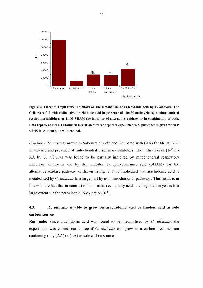
45
Figure 2. Effect of respiratory inhibitors on the metabolism of arachidonic acid by C. albicans. The
Cells were fed with radioactive arachidonic acid in presence of 10µM antimycin A, a mitochondrial
respiration inhibitor, or 1mM SHAM the inhibitor of alternative oxidase, or in combination of both.
Data represent mean ±±±± Standard Deviation of three separate experiments. Significance is given when P
< 0.05 in comparision with control.
Candida albicans was grown in Sabouraud broth and incubated with (AA) for 6h. at 37°C
in absence and presence of mitochondial respiratory inhibitors. The utilisation of [1-14C]-
AA by C. albicans was found to be partially inhibited by mitochondrial respiratory
inhibitors antimycin and by the inhibitor Salicylhydroxamic acid (SHAM) for the
alternative oxidase pathway as shown in Fig. 2. It is implicated that arachidonic acid is
metabolised by C. albicans to a large part by non-mitochondrial pathways. This result is in
line with the fact that in contrast to mammalian cells, fatty acids are degraded in yeasts to a
large extent via the peroxisomal β-oxidation [63].
4.3. C. albicans is able to grow on arachidonic acid or linoleic acid as sole
carbon source
Rationale: Since arachidonic acid was found to be metabolised by C. albicans, the
experiment was carried out to see if C. albicans can grow in a carbon free medium
containing only (AA) or (LA) as sole carbon source.
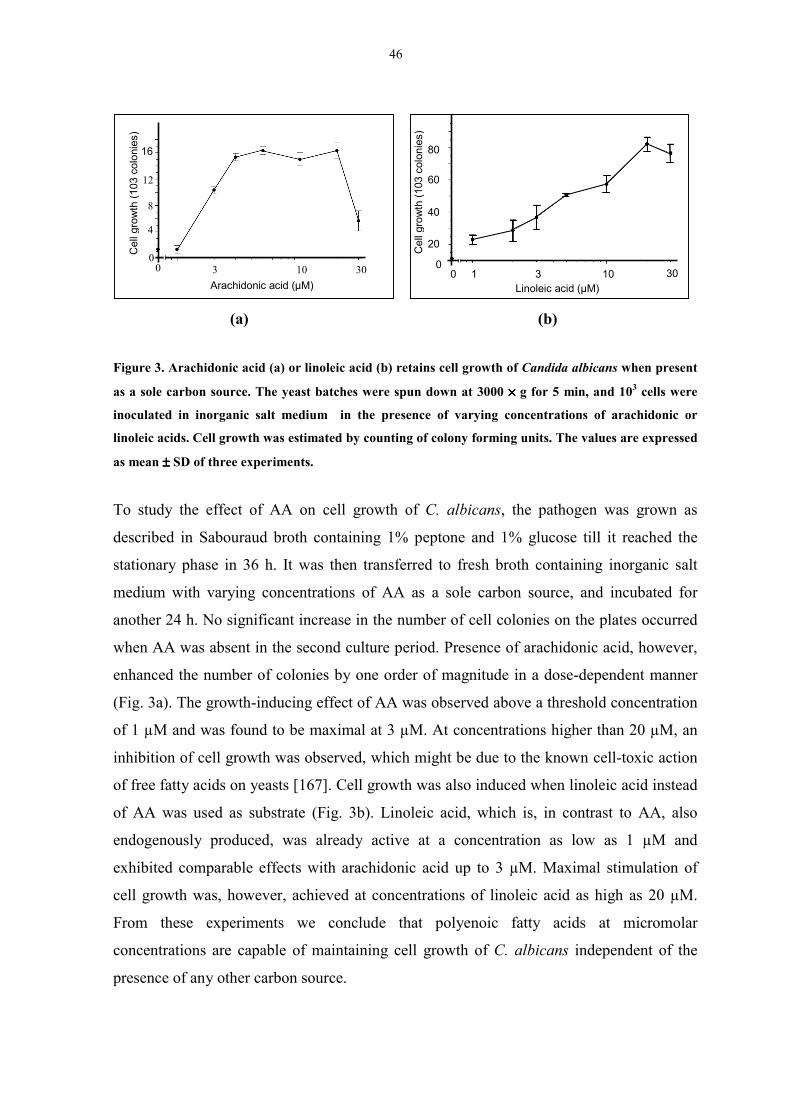
46
(a) (b)
Figure 3. Arachidonic acid (a) or linoleic acid (b) retains cell growth of Candida albicans when present
as a sole carbon source. The yeast batches were spun down at 3000 ×××× g for 5 min, and 103 cells were
inoculated in inorganic salt medium in the presence of varying concentrations of arachidonic or
linoleic acids. Cell growth was estimated by counting of colony forming units. The values are expressed
as mean ±±±± SD of three experiments.
To study the effect of AA on cell growth of C. albicans, the pathogen was grown as
described in Sabouraud broth containing 1% peptone and 1% glucose till it reached the
stationary phase in 36 h. It was then transferred to fresh broth containing inorganic salt
medium with varying concentrations of AA as a sole carbon source, and incubated for
another 24 h. No significant increase in the number of cell colonies on the plates occurred
when AA was absent in the second culture period. Presence of arachidonic acid, however,
enhanced the number of colonies by one order of magnitude in a dose-dependent manner
(Fig. 3a). The growth-inducing effect of AA was observed above a threshold concentration
of 1 µM and was found to be maximal at 3 µM. At concentrations higher than 20 µM, an
inhibition of cell growth was observed, which might be due to the known cell-toxic action
of free fatty acids on yeasts [167]. Cell growth was also induced when linoleic acid instead
of AA was used as substrate (Fig. 3b). Linoleic acid, which is, in contrast to AA, also
endogenously produced, was already active at a concentration as low as 1 µM and
exhibited comparable effects with arachidonic acid up to 3 µM. Maximal stimulation of
cell growth was, however, achieved at concentrations of linoleic acid as high as 20 µM.
From these experiments we conclude that polyenoic fatty acids at micromolar
concentrations are capable of maintaining cell growth of C. albicans independent of the
presence of any other carbon source.
Linoleic acid (µM)
00 1 3 10 30
20
40
80
60
4
8
16
12
00 3 10 30
Arachidonic acid (µM)
Cel
l gro
wth
(103
col
onie
s)
Cel
l gro
wth
(103
col
onie
s)
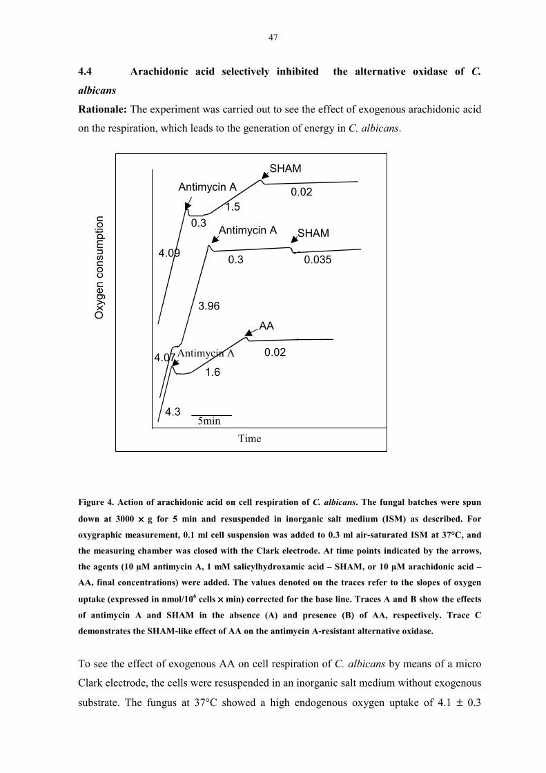
47
4.4 Arachidonic acid selectively inhibited the alternative oxidase of C.
albicans
Rationale: The experiment was carried out to see the effect of exogenous arachidonic acid
on the respiration, which leads to the generation of energy in C. albicans.
Figure 4. Action of arachidonic acid on cell respiration of C. albicans. The fungal batches were spun
down at 3000 ×××× g for 5 min and resuspended in inorganic salt medium (ISM) as described. For
oxygraphic measurement, 0.1 ml cell suspension was added to 0.3 ml air-saturated ISM at 37°C, and
the measuring chamber was closed with the Clark electrode. At time points indicated by the arrows,
the agents (10 µM antimycin A, 1 mM salicylhydroxamic acid – SHAM, or 10 µM arachidonic acid –
AA, final concentrations) were added. The values denoted on the traces refer to the slopes of oxygen
uptake (expressed in nmol/106 cells ×××× min) corrected for the base line. Traces A and B show the effects
of antimycin A and SHAM in the absence (A) and presence (B) of AA, respectively. Trace C
demonstrates the SHAM-like effect of AA on the antimycin A-resistant alternative oxidase.
To see the effect of exogenous AA on cell respiration of C. albicans by means of a micro
Clark electrode, the cells were resuspended in an inorganic salt medium without exogenous
substrate. The fungus at 37°C showed a high endogenous oxygen uptake of 4.1 ± 0.3
Oxy
gen
cons
umpt
ion
Time5min
4.3
1.6
0.024.07
3.96
0.3 0.0354.09
0.31.5
0.02
Antimycin A
Antimycin A
AA
SHAM
SHAMAntimycin A

48
nmoles/106 cells × min (n = 3) which was linear for more than 10 min in the oxygraphic
measuring chamber, and was retained for several hours when stored at room temperature
under gentle shaking. AA, at concentrations up to 10 µM, did not affect this oxygen uptake
which suggests that the mobilisation of substrates from endogenous sources is adequate for
the full capacity of the respiratory system of the fungus. Addition of antimycin A, a
specific inhibitor of the main pathway of the mitochondrial electron transfer chain, caused
an instantaneous stop of oxygen uptake, which was partially reactivated after a short lag
period (Fig. 4). This induction of antimycin A-resistant respiration in C. albicans has been
reported earlier and is attributed to the ”alternative oxidase” (AOX) pathway [168], which
by-passes the cytochrome chain of mitochondria and is inhibited by salicylhydroxamic
acid (SHAM). It was observed that the antimycin A-resistant respiration of C. albicans was
completely inhibited not only by 1 mM SHAM but also by 10 µM AA (Fig. 4). This
inhibitory effect of AA was detectable at 2 µM and was complete at 5 µM . It occurred
independent of whether AA was added before or after antimycin A (traces B and C in Fig.
4). Together with the lack of any effect on cell respiration in the absence of antimycin A,
we conclude that AA selectively inhibits the alternative oxidase.
4.5. Analysis of the distribution of radioactivity from arachidonic acid by C.
albicans
Rationale: The distribution of arachidonic acid which is metabolised by C. albicans was
determined to study the metabolism pattern and the biochemical pathways involved during
the metabolism of AA.
As shown in the table 1, the distribution of radioactivity between various compound
classes formed from [1-14C]-AA was carried out, For this purpose, the reaction samples
were fractionated by Bligh-Dyer extraction for lipids followed by trichloroacetic acid
precipitation in the aqueous phase to separate proteins from other polar compounds. In
addition, the formation of labeled carbon dioxide was measured. The data are compiled in
Table 1. Notably, only one-seventh of the radioactivity metabolised was found in CO2,
whereas two-thirds occurred in the fraction containing water-soluble compounds other than
proteins.
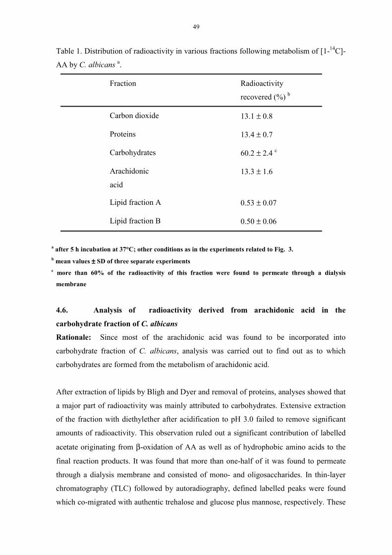
49
Table 1. Distribution of radioactivity in various fractions following metabolism of [1-14C]-
AA by C. albicans a.
Fraction Radioactivity
recovered (%) b
Carbon dioxide 13.1 ± 0.8
Proteins 13.4 ± 0.7
Carbohydrates 60.2 ± 2.4 c
Arachidonic
acid
13.3 ± 1.6
Lipid fraction A 0.53 ± 0.07
Lipid fraction B 0.50 ± 0.06
a after 5 h incubation at 37°C; other conditions as in the experiments related to Fig. 3.b mean values ±±±± SD of three separate experimentsc more than 60% of the radioactivity of this fraction were found to permeate through a dialysis
membrane
4.6. Analysis of radioactivity derived from arachidonic acid in the
carbohydrate fraction of C. albicans
Rationale: Since most of the arachidonic acid was found to be incorporated into
carbohydrate fraction of C. albicans, analysis was carried out to find out as to which
carbohydrates are formed from the metabolism of arachidonic acid.
After extraction of lipids by Bligh and Dyer and removal of proteins, analyses showed that
a major part of radioactivity was mainly attributed to carbohydrates. Extensive extraction
of the fraction with diethylether after acidification to pH 3.0 failed to remove significant
amounts of radioactivity. This observation ruled out a significant contribution of labelled
acetate originating from β-oxidation of AA as well as of hydrophobic amino acids to the
final reaction products. It was found that more than one-half of it was found to permeate
through a dialysis membrane and consisted of mono- and oligosaccharides. In thin-layer
chromatography (TLC) followed by autoradiography, defined labelled peaks were found
which co-migrated with authentic trehalose and glucose plus mannose, respectively. These
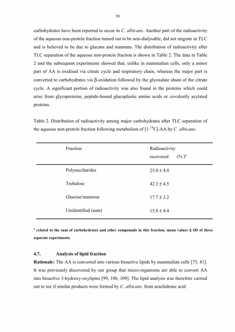
50
carbohydrates have been reported to occur in C. albicans. Another part of the radioactivity
of the aqueous non-protein fraction turned out to be non-dialysable, did not migrate in TLC
and is believed to be due to glucans and mannans. The distribution of radioactivity after
TLC separation of the aqueous non-protein fraction is shown in Table 2. The data in Table
2 and the subsequent experiments showed that, unlike in mammalian cells, only a minor
part of AA is oxidised via citrate cycle and respiratory chain, whereas the major part is
converted to carbohydrates via β-oxidation followed by the glyoxalate shunt of the citrate
cycle. A significant portion of radioactivity was also found in the proteins which could
arise from glycoproteins, peptide-bound glucoplastic amino acids or covalently acylated
proteins.
Table 2. Distribution of radioactivity among major carbohydrates after TLC separation of
the aqueous non-protein fraction following metabolism of [1-14C]-AA by C. albicans.
Fraction Radioactivity
recovered (% )a
Polysaccharides 25.0 ± 4.0
Trehalose 42.3 ± 4.5
Glucose/mannose 17.7 ± 3.2
Unidentified (sum) 15.0 ± 4.4
a related to the sum of carbohydrates and other compounds in this fraction; mean values ±±±± SD of three
separate experiments.
4.7. Analysis of lipid fraction
Rationale: The AA is converted into various bioactive lipids by mammalian cells [75, 81].
It was previously discovered by our group that micro-organisms are able to convert AA
into bioactive 3-hydroxy-oxylipins [99, 100, 109]. The lipid analysis was therefore carried
out to see if similar products were formed by C. albicans from arachidonic acid

51
A small but significant amount of radioactivity recovered (approximately 1%) was formed
in lipids other than non-metabolised AA. Separation of the lipids in various TLC solvent
systems revealed that there was no incorporation of AA into either triacylglycerols or
phospholipids. However, two other labeled fractions could be detected by autoradiography
(Fig.5).
Figure 5. Autoradiography of the TLC of lipids formed from [1-14C]-AA in C. albicans. The cells were
treated in an identical manner as in the experiments in Fig. 3, except that the solvent system n-hexane–
diethylether–acetic acid 50 : 50 : 1 (by vol.) was used. Representative separations are shown for the
metabolism of AA in the presence (lane 1) and absence (lane 2) of 1 mM salicylic acid. For
characteristics of the fractions A, B and C, see Table 3.
These fractions were characterised with respect to their stability against alkaline hydrolysis
and to their ability to react with diazomethane in order to test for the presence of a free
carboxylic group. Moreover, the fractions were examined for immunoreactivity with an
antibody raised against 3-HETE that revealed group-specificity against 3(R)-hydroxy fatty
acids of different structures [109]. The data are summarised in Table 3. It was found that
the non migrating fraction A showed a strong immunofluoresence with anti 3-OH antibody
as shown in Fig. 6. while other fractions did not show the strong immunoreactivity.
Fraction B was found to be cleaved upon alkaline hydrolysis and did not react with
Arachidonic acid
Fraction BFraction C( C1and C2)
Fraction A
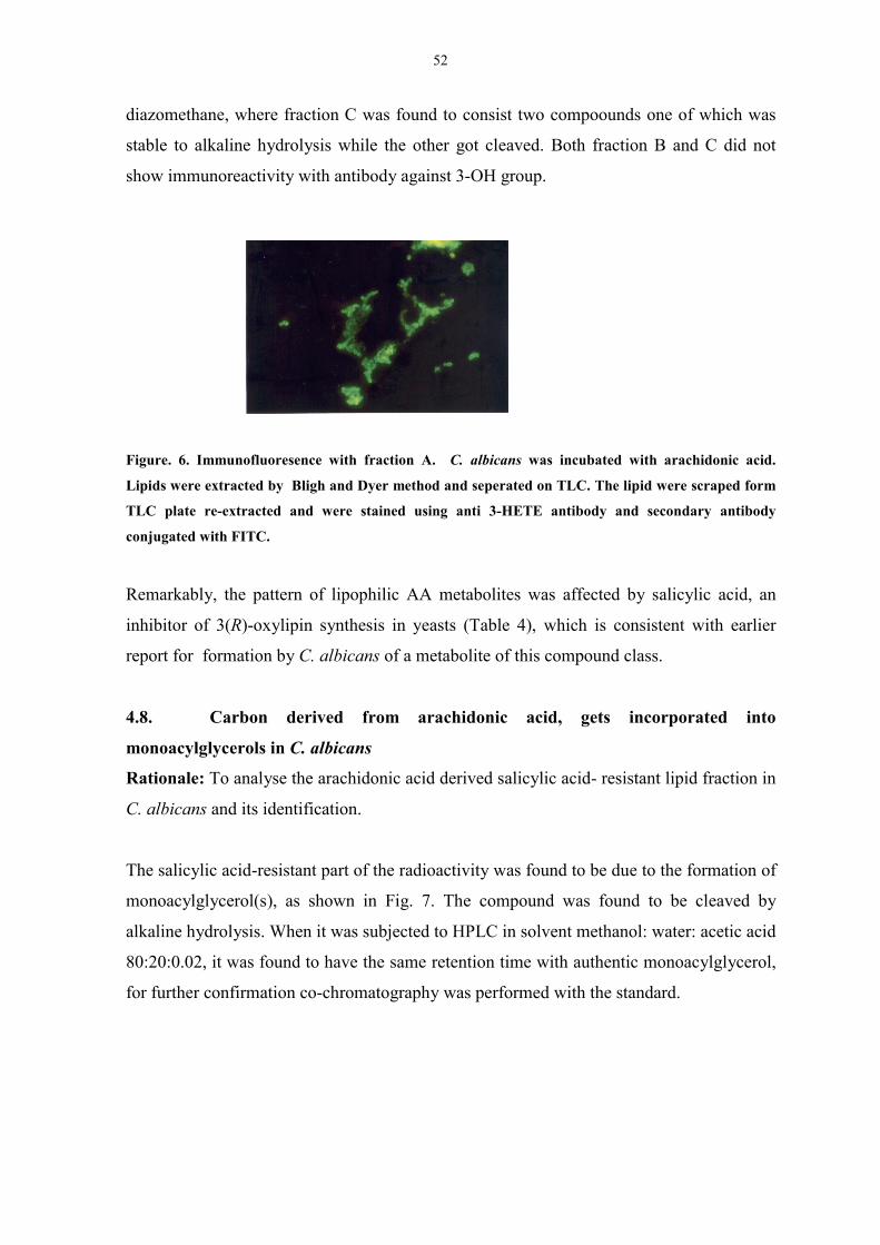
52
diazomethane, where fraction C was found to consist two compoounds one of which was
stable to alkaline hydrolysis while the other got cleaved. Both fraction B and C did not
show immunoreactivity with antibody against 3-OH group.
Figure. 6. Immunofluoresence with fraction A. C. albicans was incubated with arachidonic acid.
Lipids were extracted by Bligh and Dyer method and seperated on TLC. The lipid were scraped form
TLC plate re-extracted and were stained using anti 3-HETE antibody and secondary antibody
conjugated with FITC.
Remarkably, the pattern of lipophilic AA metabolites was affected by salicylic acid, an
inhibitor of 3(R)-oxylipin synthesis in yeasts (Table 4), which is consistent with earlier
report for formation by C. albicans of a metabolite of this compound class.
4.8. Carbon derived from arachidonic acid, gets incorporated into
monoacylglycerols in C. albicans
Rationale: To analyse the arachidonic acid derived salicylic acid- resistant lipid fraction in
C. albicans and its identification.
The salicylic acid-resistant part of the radioactivity was found to be due to the formation of
monoacylglycerol(s), as shown in Fig. 7. The compound was found to be cleaved by
alkaline hydrolysis. When it was subjected to HPLC in solvent methanol: water: acetic acid
80:20:0.02, it was found to have the same retention time with authentic monoacylglycerol,
for further confirmation co-chromatography was performed with the standard.
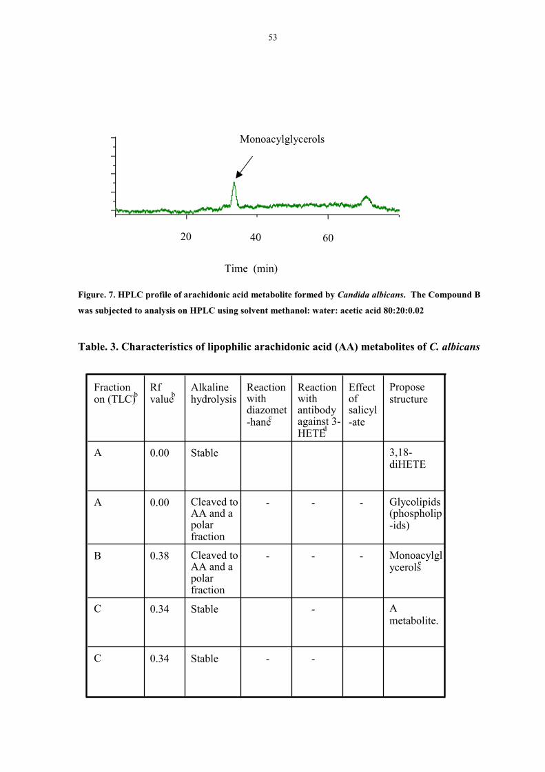
53
Figure. 7. HPLC profile of arachidonic acid metabolite formed by Candida albicans. The Compound B
was subjected to analysis on HPLC using solvent methanol: water: acetic acid 80:20:0.02
Table. 3. Characteristics of lipophilic arachidonic acid (AA) metabolites of C. albicans
20 40 60
Monoacylglycerols
Time (min)
Ametabolite.
-Stable0.34C
3,18-diHETE
Stable0.00A
- -Stable0.34C
Monoacylglycerolse
- - -Cleaved toAA and apolarfraction
0.38B
Glycolipids(phospholip-ids)
- - -Cleaved toAA and apolarfraction
0.00A
Proposestructure
Effectofsalicyl-ate
Reactionwithantibodyagainst 3-HETEd
Reactionwithdiazomet-hanec
Alkalinehydrolysis
Rfvalueb
Fractionon (TLC)b
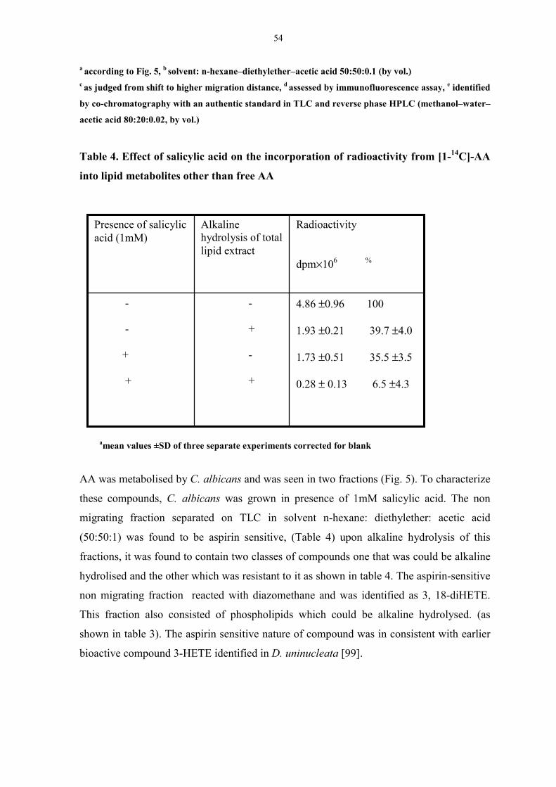
54
a according to Fig. 5, b solvent: n-hexane–diethylether–acetic acid 50:50:0.1 (by vol.)c as judged from shift to higher migration distance, d assessed by immunofluorescence assay, e identified
by co-chromatography with an authentic standard in TLC and reverse phase HPLC (methanol–water–
acetic acid 80:20:0.02, by vol.)
Table 4. Effect of salicylic acid on the incorporation of radioactivity from [1-14C]-AA
into lipid metabolites other than free AA
amean values ±SD of three separate experiments corrected for blank
AA was metabolised by C. albicans and was seen in two fractions (Fig. 5). To characterize
these compounds, C. albicans was grown in presence of 1mM salicylic acid. The non
migrating fraction separated on TLC in solvent n-hexane: diethylether: acetic acid
(50:50:1) was found to be aspirin sensitive, (Table 4) upon alkaline hydrolysis of this
fractions, it was found to contain two classes of compounds one that was could be alkaline
hydrolised and the other which was resistant to it as shown in table 4. The aspirin-sensitive
non migrating fraction reacted with diazomethane and was identified as 3, 18-diHETE.
This fraction also consisted of phospholipids which could be alkaline hydrolysed. (as
shown in table 3). The aspirin sensitive nature of compound was in consistent with earlier
bioactive compound 3-HETE identified in D. uninucleata [99].
4.86 ±0.96 100
1.93 ±0.21 39.7 ±4.0
1.73 ±0.51 35.5 ±3.5
0.28 ± 0.13 6.5 ±4.3
-
+
-
+
-
-
+
+
Radioactivity
dpm×106 %
Alkalinehydrolysis of totallipid extract
Presence of salicylicacid (1mM)
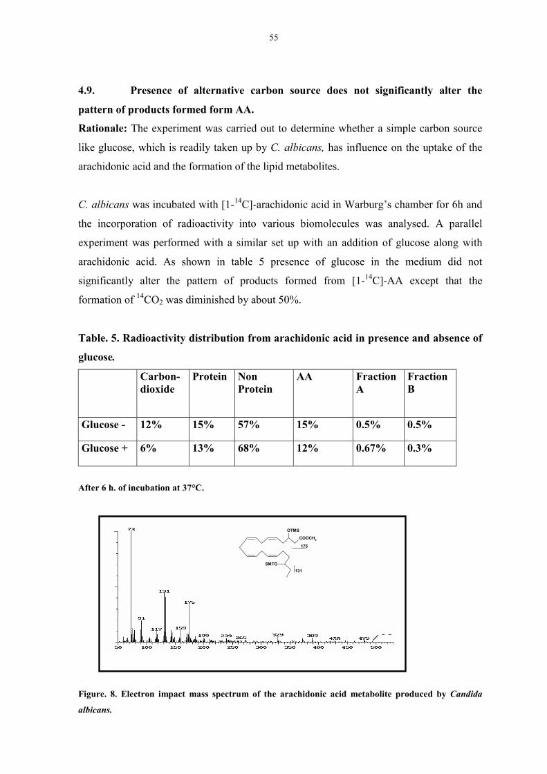
55
4.9. Presence of alternative carbon source does not significantly alter the
pattern of products formed form AA.
Rationale: The experiment was carried out to determine whether a simple carbon source
like glucose, which is readily taken up by C. albicans, has influence on the uptake of the
arachidonic acid and the formation of the lipid metabolites.
C. albicans was incubated with [1-14C]-arachidonic acid in Warburg’s chamber for 6h and
the incorporation of radioactivity into various biomolecules was analysed. A parallel
experiment was performed with a similar set up with an addition of glucose along with
arachidonic acid. As shown in table 5 presence of glucose in the medium did not
significantly alter the pattern of products formed from [1-14C]-AA except that the
formation of 14CO2 was diminished by about 50%.
Table. 5. Radioactivity distribution from arachidonic acid in presence and absence of
glucose.
Carbon-dioxide
Protein NonProtein
AA FractionA
FractionB
Glucose - 12% 15% 57% 15% 0.5% 0.5%
Glucose + 6% 13% 68% 12% 0.67% 0.3%
After 6 h. of incubation at 37°C.
Figure. 8. Electron impact mass spectrum of the arachidonic acid metabolite produced by Candida
albicans.
1
M M
COOCH3
OTMS
SMTO
175
131
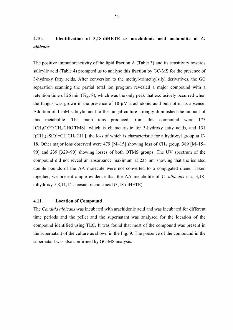
56
4.10. Identification of 3,18-diHETE as arachidonic acid metabolite of C.
albicans
The positive immunoreactivity of the lipid fraction A (Table 3) and its sensitivity towards
salicylic acid (Table 4) prompted us to analyse this fraction by GC-MS for the presence of
3-hydroxy fatty acids. After conversion to the methyl-trimethylsilyl derivatives, the GC
separation scanning the partial total ion program revealed a major compound with a
retention time of 26 min (Fig. 8), which was the only peak that exclusively occurred when
the fungus was grown in the presence of 10 µM arachidonic acid but not in its absence.
Addition of 1 mM salicylic acid to the fungal culture strongly diminished the amount of
this metabolite. The main ions produced from this compound were 175
[CH3O.CO.CH2.CHO.TMS], which is characteristic for 3-hydroxy fatty acids, and 131
[(CH3)3.SiO+=CH.CH2
.CH3], the loss of which is characteristic for a hydroxyl group at C-
18. Other major ions observed were 479 [M–15] showing loss of CH3 group, 389 [M–15–
90] and 239 [329–90] showing losses of both OTMS groups. The UV spectrum of the
compound did not reveal an absorbance maximum at 235 nm showing that the isolated
double bounds of the AA molecule were not converted to a conjugated diene. Taken
together, we present ample evidence that the AA metabolite of C. albicans is a 3,18-
dihydroxy-5,8,11,14-eicosatetraenoic acid (3,18-diHETE).
4.11. Location of Compound
The Candida albicans was incubated with arachidonic acid and was incubated for different
time periods and the pellet and the supernatant was analysed for the location of the
compound identified using TLC. It was found that most of the compound was present in
the supernatant of the culture as shown in the Fig. 9. The presence of the compound in the
supernatant was also confirmed by GC-MS analysis.

57
Figure 9. TLC chromatogram of lipids from the supernatant of C. albicans fed with C14 arachidonic
acid. C. albicans was fed with arachidonic acid, and incubated for 5h. Cells were separated from
supernatant.ipids were extracted using Bligh and Dyers method and separated by thin layer
chromatography using solvent n-hexane: methanol: water (50:50:1).
4.12. Utilisation of 3-HETE as a carbon source by C. albicans
Rationale: 3-HETE was observed to be associated with the life cycle of D. uninucleata,
[109] therefore, it was of interest to determine whether 3-HETE, which is similar to 3, 18-
diHETE, is able to influence and stimulate the growth of C. albicans.
Figure 10. Effect of 3-HETE on cell growth of C. albicans. Cells were grown in the presence of 3µM
and 10 µM 3-HETE as a sole carbon source and incubated for 48 h. at 37°C. The number of cells were
counted using Neubauer’s chamber. Data represent means ±±±± Standard Deviation of three experiments
P < 0.05 compared to the control.
To see the effect of 3-HETE on C. albicans, it was incubated with 3-HETE at the
concentration of 3 µM and 10 µM. It was found that 3-HETE was able to stimulate the
0
2
4
6
8
10
12
14
16
18
Control 1µM 3-HETE 10µM 3-HETE
No
of C
olon
ies o
f C. a
lbic
ans X
103
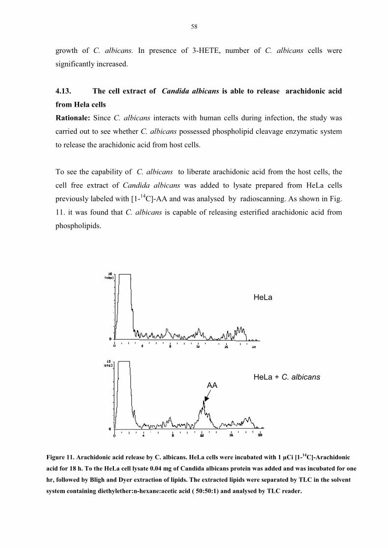
58
growth of C. albicans. In presence of 3-HETE, number of C. albicans cells were
significantly increased.
4.13. The cell extract of Candida albicans is able to release arachidonic acid
from Hela cells
Rationale: Since C. albicans interacts with human cells during infection, the study was
carried out to see whether C. albicans possessed phospholipid cleavage enzymatic system
to release the arachidonic acid from host cells.
To see the capability of C. albicans to liberate arachidonic acid from the host cells, the
cell free extract of Candida albicans was added to lysate prepared from HeLa cells
previously labeled with [1-14C]-AA and was analysed by radioscanning. As shown in Fig.
11. it was found that C. albicans is capable of releasing esterified arachidonic acid from
phospholipids.
Figure 11. Arachidonic acid release by C. albicans. HeLa cells were incubated with 1 µCi [1-14C]-Arachidonic
acid for 18 h. To the HeLa cell lysate 0.04 mg of Candida albicans protein was added and was incubated for one
hr, followed by Bligh and Dyer extraction of lipids. The extracted lipids were separated by TLC in the solvent
system containing diethylether:n-hexane:acetic acid ( 50:50:1) and analysed by TLC reader.
HeLa
HeLa + C. albicansAA
59
4.14. Candida albicans shows Phosholipase activity
Rationale: The experiment was carried to see whether C. albicans possess phospholipase
A2 activity to release arachidonic acid in a significant amount from HeLa cells.
To determine the phospholipase A2 activity, 25µg protein lysate of C. albicans was
incubated with pure substrates PAPC and PAPC-OH which are specific for PLA2 activity
for one hr. After Bligh and Dyer extraction lipids were analysed by HPLC for AA and 15-
HETEs. It was found as shown in Fig. 12 a and b that Candida albicans is able to release
AA from phospholipids. Both 1-Palmitoyl-2-arachidonoyl phosphatidylcholine (PAPC),
and PAPC-OH released arachidonic acid and 15-HETE, respectively, demonstrating
phospholipase in C. albicans.
Figure 12. Phospholipase activity in C. albicans. Cell free extract of C. albicans was incubated with 1-
Palmitoyl-2-arachidonoyl phosphatidyl Choline (PAPC) and PAPC-OH and incubated for 1h. After
incubation the lipids were extracted by Bligh and Dyer and was analysed by Reverse phase HPLC in
solvent methanol: acetonitrile: water 90:6:4 with 20 mM choline chloride.
0 5 10 15 20 25 30 35
Time (min)
AA
PAPC
15-HETE
Time ( min)
0 2 4 6 8
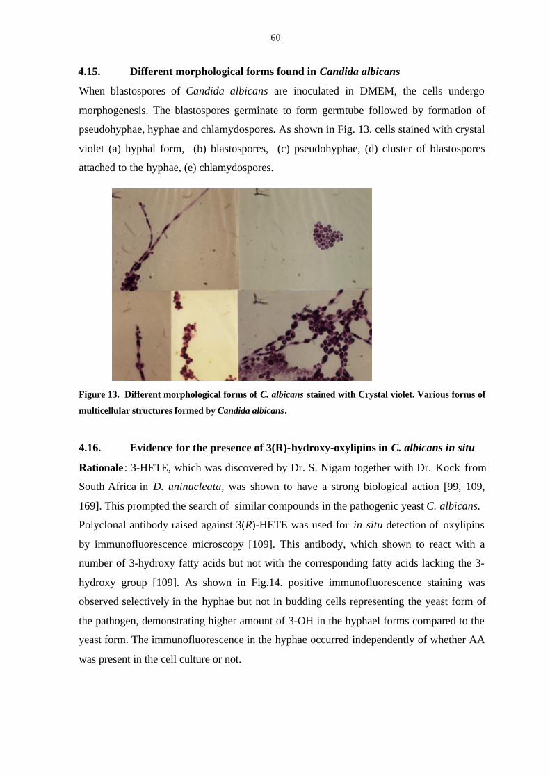
60
4.15. Different morphological forms found in Candida albicans
When blastospores of Candida albicans are inoculated in DMEM, the cells undergo
morphogenesis. The blastospores germinate to form germtube followed by formation of
pseudohyphae, hyphae and chlamydospores. As shown in Fig. 13. cells stained with crystal
violet (a) hyphal form, (b) blastospores, (c) pseudohyphae, (d) cluster of blastospores
attached to the hyphae, (e) chlamydospores.
Figure 13. Different morphological forms of C. albicans stained with Crystal violet. Various forms of
multicellular structures formed by Candida albicans.
4.16. Evidence for the presence of 3(R)-hydroxy-oxylipins in C. albicans in situ
Rationale : 3-HETE, which was discovered by Dr. S. Nigam together with Dr. Kock from
South Africa in D. uninucleata, was shown to have a strong biological action [99, 109,
169]. This prompted the search of similar compounds in the pathogenic yeast C. albicans.
Polyclonal antibody raised against 3(R)-HETE was used for in situ detection of oxylipins
by immunofluorescence microscopy [109]. This antibody, which shown to react with a
number of 3-hydroxy fatty acids but not with the corresponding fatty acids lacking the 3-
hydroxy group [109]. As shown in Fig.14. positive immunofluorescence staining was
observed selectively in the hyphae but not in budding cells representing the yeast form of
the pathogen, demonstrating higher amount of 3-OH in the hyphael forms compared to the
yeast form. The immunofluorescence in the hyphae occurred independently of whether AA
was present in the cell culture or not.
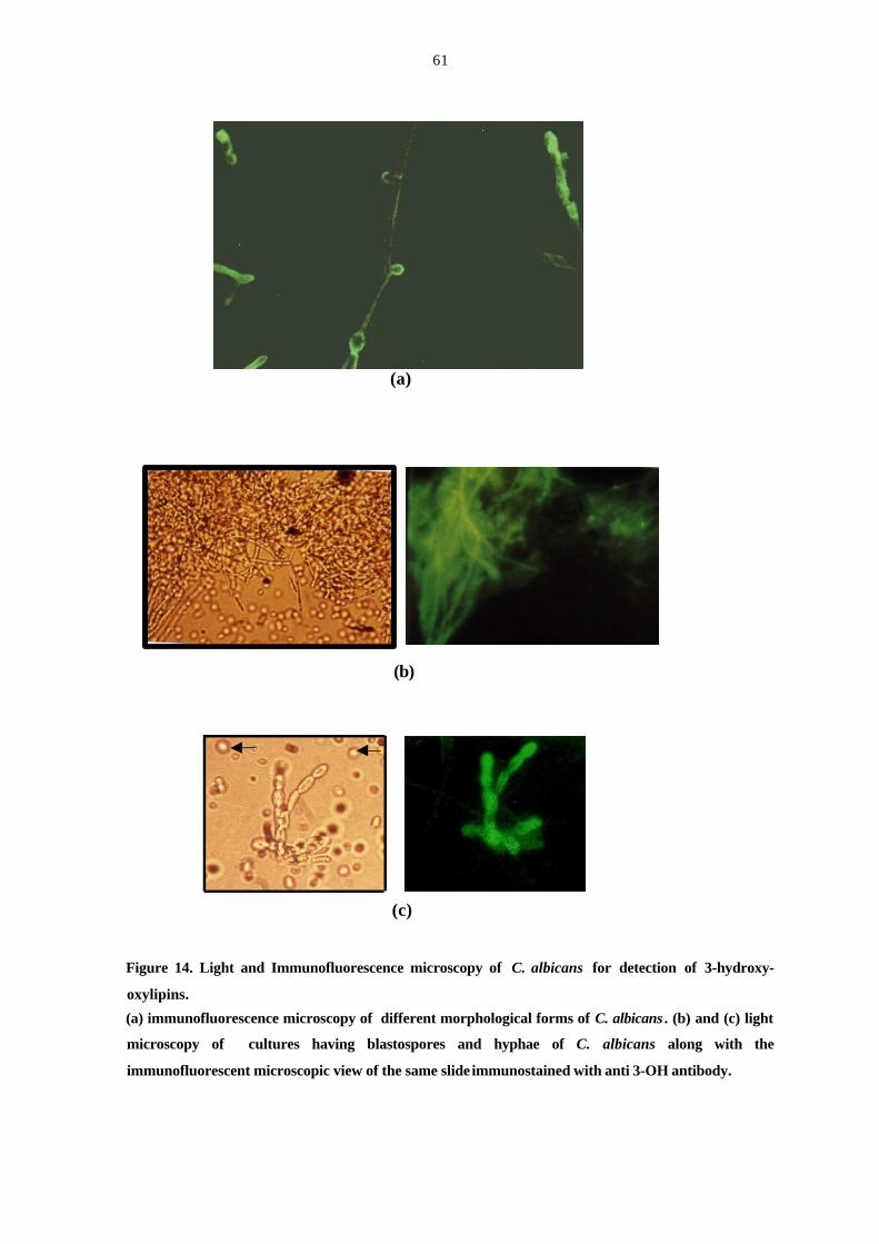
61
Figure 14. Light and Immunofluorescence microscopy of C. albicans for detection of 3-hydroxy-
oxylipins.
(a) immunofluorescence microscopy of different morphological forms of C. albicans . (b) and (c) light
microscopy of cultures having blastospores and hyphae of C. albicans along with the
immunofluorescent microscopic view of the same slide immunostained with anti 3-OH antibody.
(a)
(b)
(c)
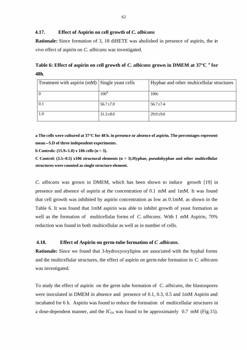
62
4.17. Effect of Aspirin on cell growth of C. albicans
Rationale: Since formation of 3, 18 diHETE was abolished in presence of aspirin, the in
vivo effect of aspirin on C. albicans was investigated.
Table 6: Effect of aspirin on cell growth of C. albicans grown in DMEM at 37°C a for
48h.
Treatment with aspirin (mM) Single yeast cells Hyphae and other multicellular structures
0 100b 100c
0.1 56.7±7.0 56.7±7.4
1.0 31.3±8.0 29.0±9.6
a The cells were cultured at 37°C for 48 h. in presence or absence of aspirin. The percentages represent
mean ±±S.D of three independent experiments.
b Controls: (15.9±±1.0) x 106 cells (n = 3).
C Control: (3.5±±0.5) x106 structural elements (n = 3).Hyphae, pseudohyphae and other multicellular
structures were counted as single structure element.
C. albicans was grown in DMEM, which has been shown to induce growth [19] in
presence and absence of aspirin at the concentration of 0.1 mM and 1mM. It was found
that cell growth was inhibited by aspirin concentration as low as 0.1mM. as shown in the
Table 6. It was found that 1mM aspirin was able to inhibit growth of yeast formation as
well as the formation of multicellular forms of C. albicans. With 1 mM Aspirin, 70%
reduction was found in both multicellular as well as in number of cells.
4.18. Effect of Aspirin on germ-tube formation of C .albicans.
Rationale: Since we found that 3-hydroxyoxylipins are associated with the hyphal forms
and the multicellular structures, the effect of aspirin on germ-tube formation in C. albicans
was investigated.
To study the effect of aspirin on the germ tube formation of C. albicans, the blastospores
were inoculated in DMEM in absence and presence of 0.1, 0.3, 0.5 and 1mM Aspirin and
incubated for 6 h. Aspirin was found to reduce the formation of multicellular structures in
a dose-dependent manner, and the IC50 was found to be approximately 0.7 mM (Fig.15).

63
Moreover, the formation of germ tube was found to be reduced by 15% on incubation with
1 mM aspirin as shown in Fig 15a.
Figure 15. Aspirin diminishes the percentage of germ tubes formed by C. albicans after incubation for
6 h. in DMEM in absence and presence of various concentration of Aspirin. Number of cell containing
germ tubes were counted using neubauers chamber. The values expressed is the mean ± S.D of three
independent experiments.
(a) (b)
Figure 16. Light microscopie of demonstration of the effect of aspirin on C. albicans. Cells were
incubated in DMEM for 6h in presence (a) and absence of 1mM (b) aspirin. (400 X magnification).
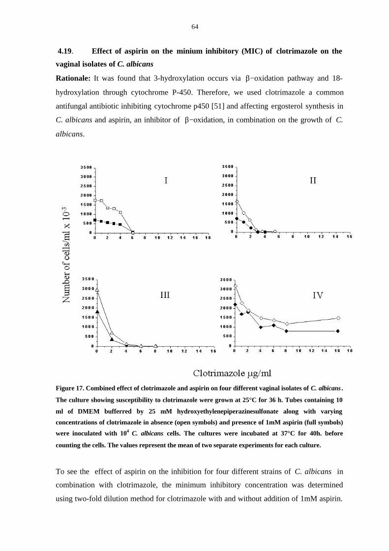
64
4.19. Effect of aspirin on the minium inhibitory (MIC) of clotrimazole on the
vaginal isolates of C. albicans
Rationale: It was found that 3-hydroxylation occurs via β−oxidation pathway and 18-
hydroxylation through cytochrome P-450. Therefore, we used clotrimazole a common
antifungal antibiotic inhibiting cytochrome p450 [51] and affecting ergosterol synthesis in
C. albicans and aspirin, an inhibitor of β−oxidation, in combination on the growth of C.
albicans.
Figure 17. Combined effect of clotrimazole and aspirin on four different vaginal isolates of C. albicans .
The culture showing susceptibility to clotrimazole were grown at 25°C for 36 h. Tubes containing 10
ml of DMEM bufferred by 25 mM hydroxyethylenepiperazinesulfonate along with varying
concentrations of clotrimazole in absence (open symbols) and presence of 1mM aspirin (full symbols)
were inoculated with 104 C. albicans cells. The cultures were incubated at 37°C for 40h. before
counting the cells. The values represent the mean of two separate experiments for each culture.
To see the effect of aspirin on the inhibition for four different strains of C. albicans in
combination with clotrimazole, the minimum inhibitory concentration was determined
using two-fold dilution method for clotrimazole with and without addition of 1mM aspirin.
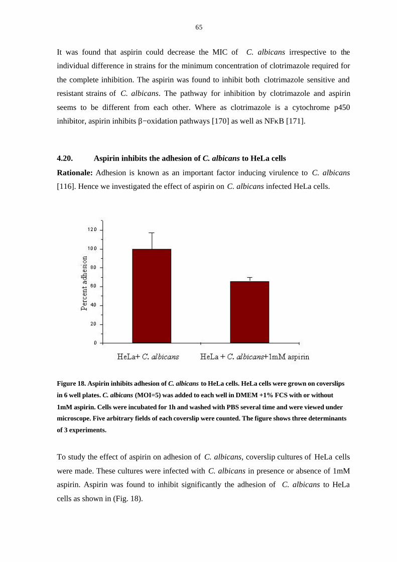
65
It was found that aspirin could decrease the MIC of C. albicans irrespective to the
individual difference in strains for the minimum concentration of clotrimazole required for
the complete inhibition. The aspirin was found to inhibit both clotrimazole sensitive and
resistant strains of C. albicans. The pathway for inhibition by clotrimazole and aspirin
seems to be different from each other. Where as clotrimazole is a cytochrome p450
inhibitor, aspirin inhibits β−oxidation pathways [170] as well as NFκB [171].
4.20. Aspirin inhibits the adhesion of C. albicans to HeLa cells
Rationale: Adhesion is known as an important factor inducing virulence to C. albicans
[116]. Hence we investigated the effect of aspirin on C. albicans infected HeLa cells.
Figure 18. Aspirin inhibits adhesion of C. albicans to HeLa cells. HeLa cells were grown on coverslips
in 6 well plates. C. albicans (MOI=5) was added to each well in DMEM +1% FCS with or without
1mM aspirin. Cells were incubated for 1h and washed with PBS several time and were viewed under
microscope. Five arbitrary fields of each coverslip were counted. The figure shows three determinants
of 3 experiments.
To study the effect of aspirin on adhesion of C. albicans, coverslip cultures of HeLa cells
were made. These cultures were infected with C. albicans in presence or absence of 1mM
aspirin. Aspirin was found to inhibit significantly the adhesion of C. albicans to HeLa
cells as shown in (Fig. 18).
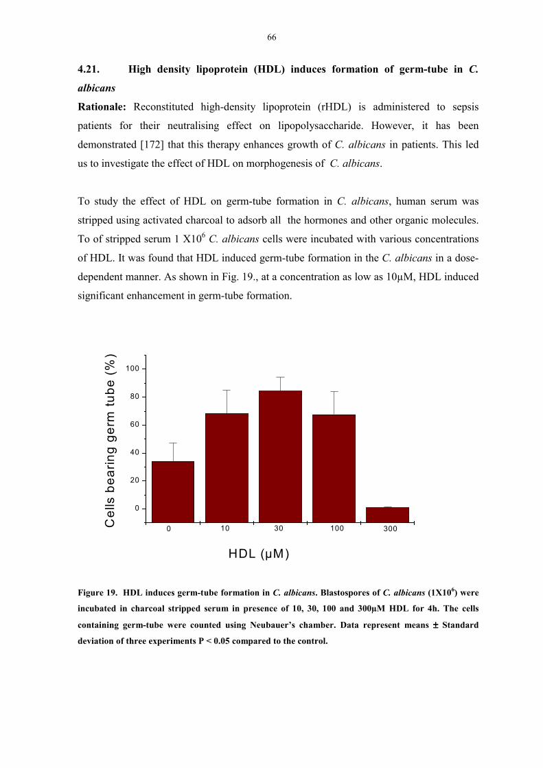
66
4.21. High density lipoprotein (HDL) induces formation of germ-tube in C.
albicans
Rationale: Reconstituted high-density lipoprotein (rHDL) is administered to sepsis
patients for their neutralising effect on lipopolysaccharide. However, it has been
demonstrated [172] that this therapy enhances growth of C. albicans in patients. This led
us to investigate the effect of HDL on morphogenesis of C. albicans.
To study the effect of HDL on germ-tube formation in C. albicans, human serum was
stripped using activated charcoal to adsorb all the hormones and other organic molecules.
To of stripped serum 1 X10⁶ C. albicans cells were incubated with various concentrations
of HDL. It was found that HDL induced germ-tube formation in the C. albicans in a dose-
dependent manner. As shown in Fig. 19., at a concentration as low as 10µM, HDL induced
significant enhancement in germ-tube formation.
Figure 19. HDL induces germ-tube formation in C. albicans. Blastospores of C. albicans (1X10⁶) were
incubated in charcoal stripped serum in presence of 10, 30, 100 and 300µM HDL for 4h. The cells
containing germ-tube were counted using Neubauer’s chamber. Data represent means ±±±± Standard
deviation of three experiments P < 0.05 compared to the control.
0
20
40
60
80
100
30010030100Cel
ls b
earin
g ge
rm tu
be (%
)
HDL (µM)
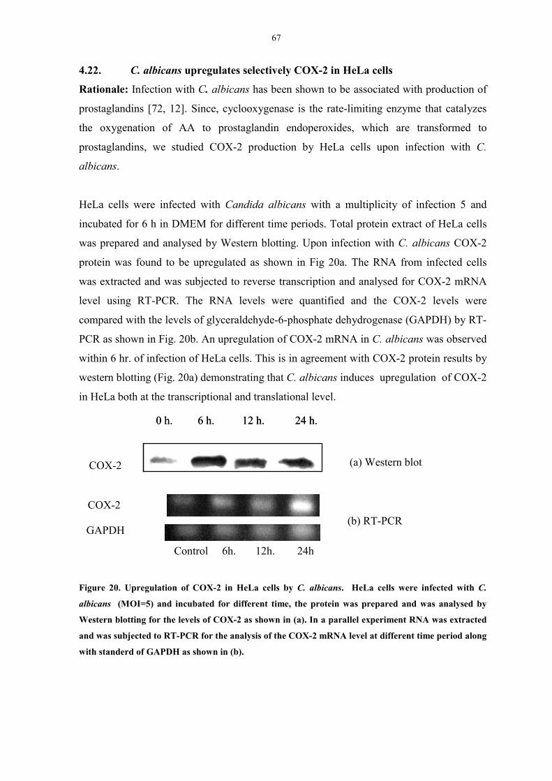
67
4.22. C. albicans upregulates selectively COX-2 in HeLa cells
Rationale: Infection with C. albicans has been shown to be associated with production of
prostaglandins [72, 12]. Since, cyclooxygenase is the rate-limiting enzyme that catalyzes
the oxygenation of AA to prostaglandin endoperoxides, which are transformed to
prostaglandins, we studied COX-2 production by HeLa cells upon infection with C.
albicans.
HeLa cells were infected with Candida albicans with a multiplicity of infection 5 and
incubated for 6 h in DMEM for different time periods. Total protein extract of HeLa cells
was prepared and analysed by Western blotting. Upon infection with C. albicans COX-2
protein was found to be upregulated as shown in Fig 20a. The RNA from infected cells
was extracted and was subjected to reverse transcription and analysed for COX-2 mRNA
level using RT-PCR. The RNA levels were quantified and the COX-2 levels were
compared with the levels of glyceraldehyde-6-phosphate dehydrogenase (GAPDH) by RT-
PCR as shown in Fig. 20b. An upregulation of COX-2 mRNA in C. albicans was observed
within 6 hr. of infection of HeLa cells. This is in agreement with COX-2 protein results by
western blotting (Fig. 20a) demonstrating that C. albicans induces upregulation of COX-2
in HeLa both at the transcriptional and translational level.
Figure 20. Upregulation of COX-2 in HeLa cells by C. albicans. HeLa cells were infected with C.
albicans (MOI=5) and incubated for different time, the protein was prepared and was analysed by
Western blotting for the levels of COX-2 as shown in (a). In a parallel experiment RNA was extracted
and was subjected to RT-PCR for the analysis of the COX-2 mRNA level at different time period along
with standerd of GAPDH as shown in (b).
COX-2
0 h. 6 h. 12 h. 24 h.
(a) Western blot
0 h. 6 h. 12 h. 24 h.
Control 6h. 12h. 24h
COX-2
GAPDH(b) RT-PCR
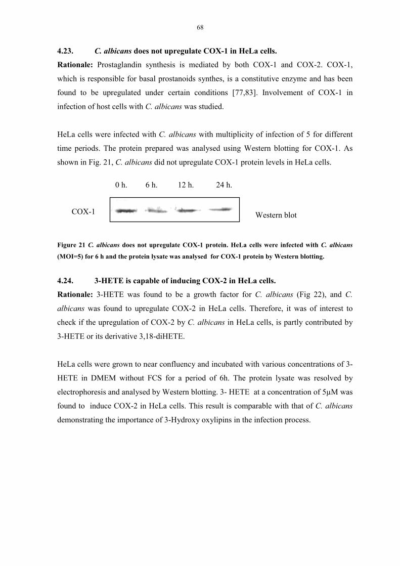
68
4.23. C. albicans does not upregulate COX-1 in HeLa cells.
Rationale: Prostaglandin synthesis is mediated by both COX-1 and COX-2. COX-1,
which is responsible for basal prostanoids synthes, is a constitutive enzyme and has been
found to be upregulated under certain conditions [77,83]. Involvement of COX-1 in
infection of host cells with C. albicans was studied.
HeLa cells were infected with C. albicans with multiplicity of infection of 5 for different
time periods. The protein prepared was analysed using Western blotting for COX-1. As
shown in Fig. 21, C. albicans did not upregulate COX-1 protein levels in HeLa cells.
Figure 21 C. albicans does not upregulate COX-1 protein. HeLa cells were infected with C. albicans
(MOI=5) for 6 h and the protein lysate was analysed for COX-1 protein by Western blotting.
4.24. 3-HETE is capable of inducing COX-2 in HeLa cells.
Rationale: 3-HETE was found to be a growth factor for C. albicans (Fig 22), and C.
albicans was found to upregulate COX-2 in HeLa cells. Therefore, it was of interest to
check if the upregulation of COX-2 by C. albicans in HeLa cells, is partly contributed by
3-HETE or its derivative 3,18-diHETE.
HeLa cells were grown to near confluency and incubated with various concentrations of 3-
HETE in DMEM without FCS for a period of 6h. The protein lysate was resolved by
electrophoresis and analysed by Western blotting. 3- HETE at a concentration of 5µM was
found to induce COX-2 in HeLa cells. This result is comparable with that of C. albicans
demonstrating the importance of 3-Hydroxy oxylipins in the infection process.
0 h. 6 h. 12 h. 24 h.
COX-1 Western blot
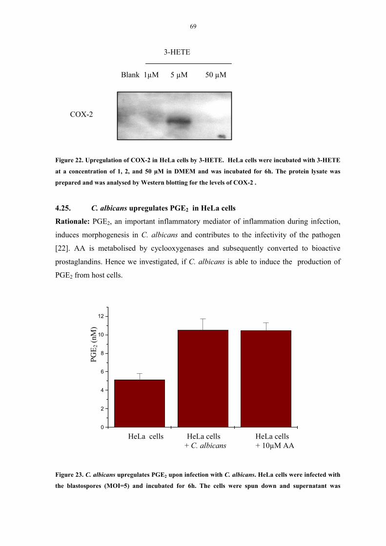
69
Figure 22. Upregulation of COX-2 in HeLa cells by 3-HETE. HeLa cells were incubated with 3-HETE
at a concentration of 1, 2, and 50 µM in DMEM and was incubated for 6h. The protein lysate was
prepared and was analysed by Western blotting for the levels of COX-2 .
4.25. C. albicans upregulates PGE2 in HeLa cells
Rationale: PGE2, an important inflammatory mediator of inflammation during infection,
induces morphogenesis in C. albicans and contributes to the infectivity of the pathogen
[22]. AA is metabolised by cyclooxygenases and subsequently converted to bioactive
prostaglandins. Hence we investigated, if C. albicans is able to induce the production of
PGE2 from host cells.
Figure 23. C. albicans upregulates PGE2 upon infection with C. albicans. HeLa cells were infected with
the blastospores (MOI=5) and incubated for 6h. The cells were spun down and supernatant was
Blank 1µM 5 µM 50 µM
3-HETE
COX-2
0
2
4
6
8
10
12
PGE 2
(nM
)
HeLa cells HeLa cells HeLa cells + C. albicans + 10µM AA
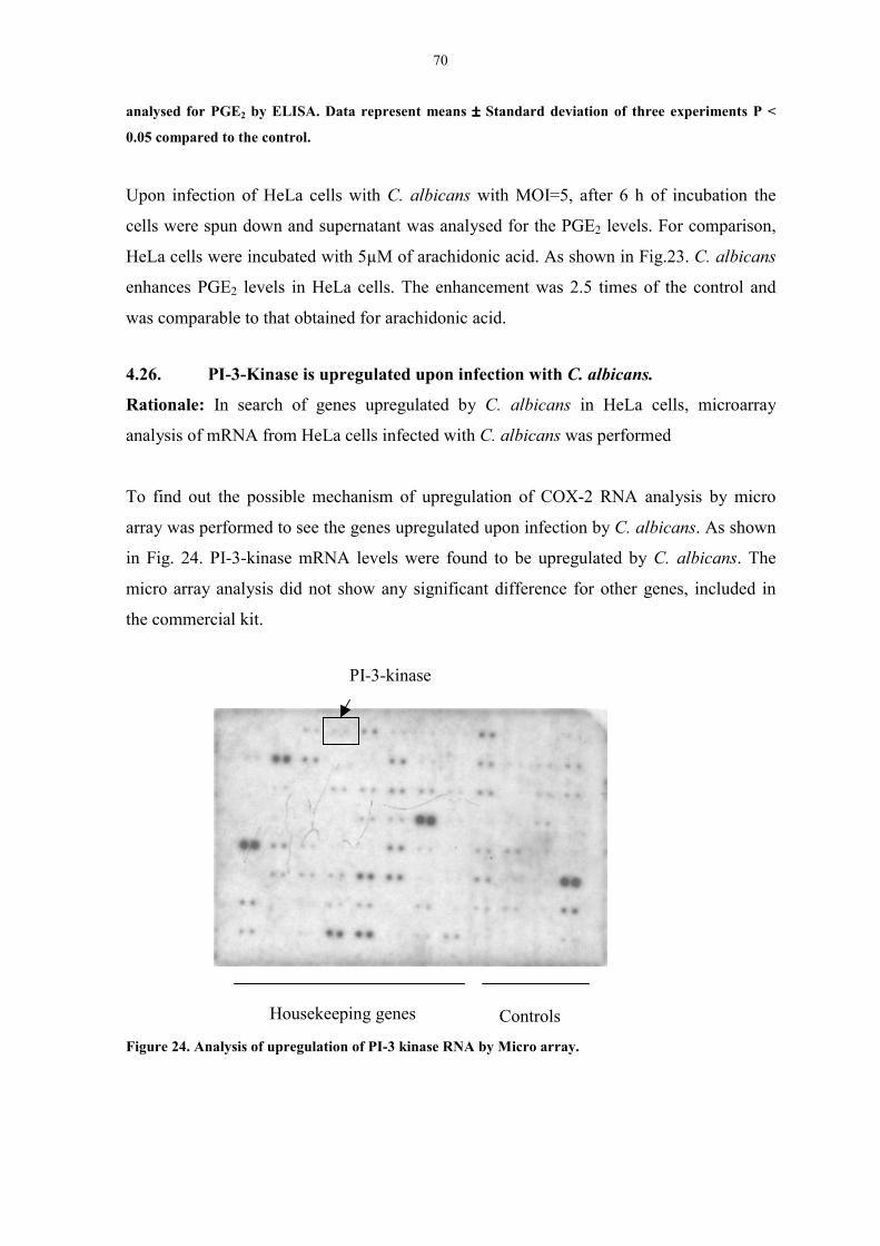
70
analysed for PGE2 by ELISA. Data represent means ±±±± Standard deviation of three experiments P <
0.05 compared to the control.
Upon infection of HeLa cells with C. albicans with MOI=5, after 6 h of incubation the
cells were spun down and supernatant was analysed for the PGE2 levels. For comparison,
HeLa cells were incubated with 5µM of arachidonic acid. As shown in Fig.23. C. albicans
enhances PGE2 levels in HeLa cells. The enhancement was 2.5 times of the control and
was comparable to that obtained for arachidonic acid.
4.26. PI-3-Kinase is upregulated upon infection with C. albicans.
Rationale: In search of genes upregulated by C. albicans in HeLa cells, microarray
analysis of mRNA from HeLa cells infected with C. albicans was performed
To find out the possible mechanism of upregulation of COX-2 RNA analysis by micro
array was performed to see the genes upregulated upon infection by C. albicans. As shown
in Fig. 24. PI-3-kinase mRNA levels were found to be upregulated by C. albicans. The
micro array analysis did not show any significant difference for other genes, included in
the commercial kit.
Figure 24. Analysis of upregulation of PI-3 kinase RNA by Micro array.
PI-3-kinase
Housekeeping genes Controls
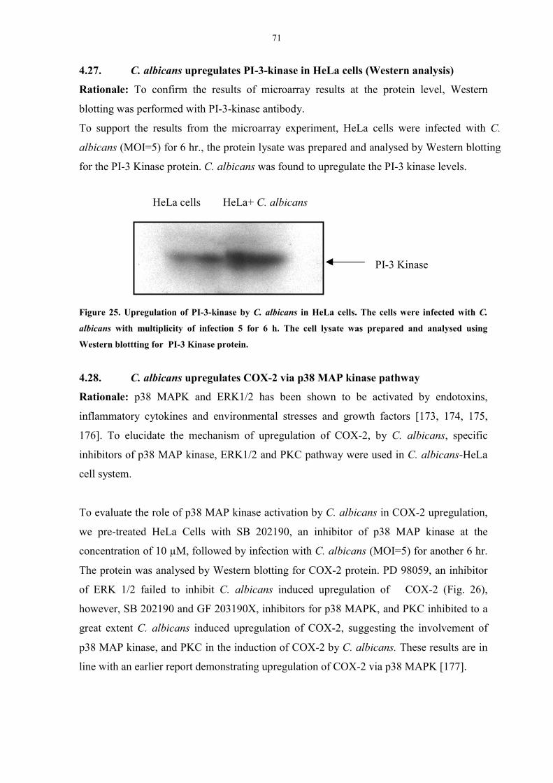
71
4.27. C. albicans upregulates PI-3-kinase in HeLa cells (Western analysis)
Rationale: To confirm the results of microarray results at the protein level, Western
blotting was performed with PI-3-kinase antibody.
To support the results from the microarray experiment, HeLa cells were infected with C.
albicans (MOI=5) for 6 hr., the protein lysate was prepared and analysed by Western blotting
for the PI-3 Kinase protein. C. albicans was found to upregulate the PI-3 kinase levels.
Figure 25. Upregulation of PI-3-kinase by C. albicans in HeLa cells. The cells were infected with C.
albicans with multiplicity of infection 5 for 6 h. The cell lysate was prepared and analysed using
Western blottting for PI-3 Kinase protein.
4.28. C. albicans upregulates COX-2 via p38 MAP kinase pathway
Rationale: p38 MAPK and ERK1/2 has been shown to be activated by endotoxins,
inflammatory cytokines and environmental stresses and growth factors [173, 174, 175,
176]. To elucidate the mechanism of upregulation of COX-2, by C. albicans, specific
inhibitors of p38 MAP kinase, ERK1/2 and PKC pathway were used in C. albicans-HeLa
cell system.
To evaluate the role of p38 MAP kinase activation by C. albicans in COX-2 upregulation,
we pre-treated HeLa Cells with SB 202190, an inhibitor of p38 MAP kinase at the
concentration of 10 µM, followed by infection with C. albicans (MOI=5) for another 6 hr.
The protein was analysed by Western blotting for COX-2 protein. PD 98059, an inhibitor
of ERK 1/2 failed to inhibit C. albicans induced upregulation of COX-2 (Fig. 26),
however, SB 202190 and GF 203190X, inhibitors for p38 MAPK, and PKC inhibited to a
great extent C. albicans induced upregulation of COX-2, suggesting the involvement of
p38 MAP kinase, and PKC in the induction of COX-2 by C. albicans. These results are in
line with an earlier report demonstrating upregulation of COX-2 via p38 MAPK [177].
PI-3 Kinase
HeLa cells HeLa+ C. albicans
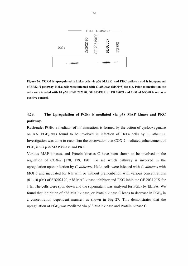
72
Figure 26. COX-2 is upregulated in HeLa cells via p38 MAPK and PKC pathway and is independent
of ERK1/2 pathway. HeLa cells were infected with C. albicans (MOI=5) for 6 h. Prior to incubation the
cells were treated with 10 µM of SB 202190, GF 203190X or PD 98059 and 1µM of NS398 taken as a
positive control.
4.29. The Upregulation of PGE2 is mediated via p38 MAP kinase and PKC
pathway.
Rationale: PGE2, a mediator of inflammation, is formed by the action of cyclooxygenase
on AA. PGE2 was found to be involved in infection of HeLa cells by C. albicans.
Investigation was done to reconfirm the observation that COX-2 mediated enhancement of
PGE2 is via p38 MAP kinase and PKC.
Various MAP kinases, and Protein kinases C have been shown to be involved in the
regulation of COX-2 [178, 179, 180]. To see which pathway is involved in the
upregulation upon infection by C. albicans, HeLa cells were infected with C. albicans with
MOI 5 and incubated for 6 h with or without preincubation with various concentrations
(0.1-10 µM) of SB202190, p38 MAP kinase inhibitor and PKC inhibitor GF 203190X for
1 h.. The cells were spun down and the supernatant was analysed for PGE2 by ELISA. We
found that inhibition of p38 MAP kinase, or Protein kinase C leads to decrease in PGE2 in
a concentration dependent manner, as shown in Fig 27. This demonstrates that the
upregulation of PGE2 was mediated via p38 MAP kinase and Protein Kinase C.

73
0
1
2
3
4
5
6
SB 202190 (µM)GF203190X (µM)
10µM
PD
9805
91010.11010.1
C. a
lbic
ans
HeL
a ce
lls
PG
E2 (
nM)
Figure 27. Involvement of p38 MAPK and PKC in C. albicans induced PGE2 upregulation. HeLa cells
were infected with C. albicans (MOI=5) for 6 h. with or with out preincubation of different inhibitors
for 1h. The cells were spun down and supernatant was analysed for PGE2 by ELISA. Data represent
means ±±±± Standard deviation of three experiments P < 0.05 compared to the control.
4.30. ERK1/ERK2 pathway is not involved in the upregulation of PGE2 by C.
albicans.
Rationale: ERK1/2 MAP kinase has also been observed to influence the expression of
COX-2 and PGE2 [174]. ERK1/2 pathway was studied for its involvement in the signal
transduction by C. albicans.
To find whether ERK1/ERK2 were involved in C. albicans induced upregulation of PGE2,
the cells were infected in presence of specific ERK1/2 MAP kinase inhibitor PD 98059.
After incubation cells were centrifuged and the supernatant was analysed for PGE2. It was
found that the inhibitor was unable to inhibit Candida albicans induced PGE2 production
(Fig. 27). This observation rules out the involvement of this particular pathway in the
Candida albicans induced upregulation of COX-2 and PGE2.
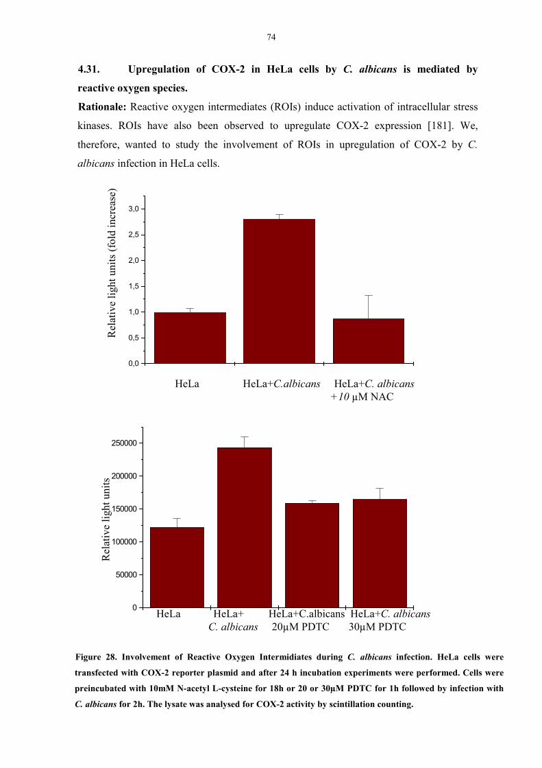
74
4.31. Upregulation of COX-2 in HeLa cells by C. albicans is mediated by
reactive oxygen species.
Rationale: Reactive oxygen intermediates (ROIs) induce activation of intracellular stress
kinases. ROIs have also been observed to upregulate COX-2 expression [181]. We,
therefore, wanted to study the involvement of ROIs in upregulation of COX-2 by C.
albicans infection in HeLa cells.
Figure 28. Involvement of Reactive Oxygen Intermidiates during C. albicans infection. HeLa cells were
transfected with COX-2 reporter plasmid and after 24 h incubation experiments were performed. Cells were
preincubated with 10mM N-acetyl L-cysteine for 18h or 20 or 30µM PDTC for 1h followed by infection with
C. albicans for 2h. The lysate was analysed for COX-2 activity by scintillation counting.
0,0
0,5
1,0
1,5
2,0
2,5
3,0
0
50000
100000
150000
200000
250000
Rel
ativ
e lig
ht u
nits
(fol
d in
crea
se)
HeLa HeLa+C.albicans HeLa+C. albicans +10 µM NAC
HeLa HeLa+ HeLa+C.albicans HeLa+C. albicans C. albicans 20µM PDTC 30µM PDTC
Rel
ativ
e lig
ht u
nits
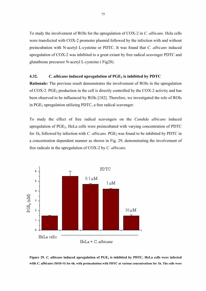
75
To study the involvement of ROIs for the upregulation of COX-2 in C. albicans. Hela cells
were transfected with COX-2 promoter plasmid followed by the infection with and without
preincubation with N-acetyl L-cysteine or PDTC. It was found that C. albicans induced
upregulation of COX-2 was inhibited to a great extant by free radical scavenger PDTC and
glutathione precursor N-acetyl L-cysteine ( Fig28).
4.32. C. albicans induced upregulation of PGE2 is inhibited by PDTC
Rationale: The previous result demonstrates the involvement of ROIs in the upregulation
of COX-2. PGE2 production in the cell is directly controlled by the COX-2 activity and has
been observed to be influenced by ROIs [182]. Therefore, we investigated the role of ROIs
in PGE2 upregulation utilizing PDTC, a free radical scavenger.
To study the effect of free radical scavengers on the Candida albicans induced
upregulation of PGE2, HeLa cells were preincubated with varying concentration of PDTC
for 1h, followed by infection with C. albicans. PGE2 was found to be inhibited by PDTC in
a concentration dependent manner as shown in Fig. 29, demonstrating the involvement of
free radicals in the upregulation of COX-2 by C. albicans.
Figure 29. C. albicans induced upregulation of PGE2 is inhibited by PDTC. HeLa cells were infected
with C. albicans (MOI=5) for 6h. with preincubation with PDTC at various concentrations for 1h. The cells were
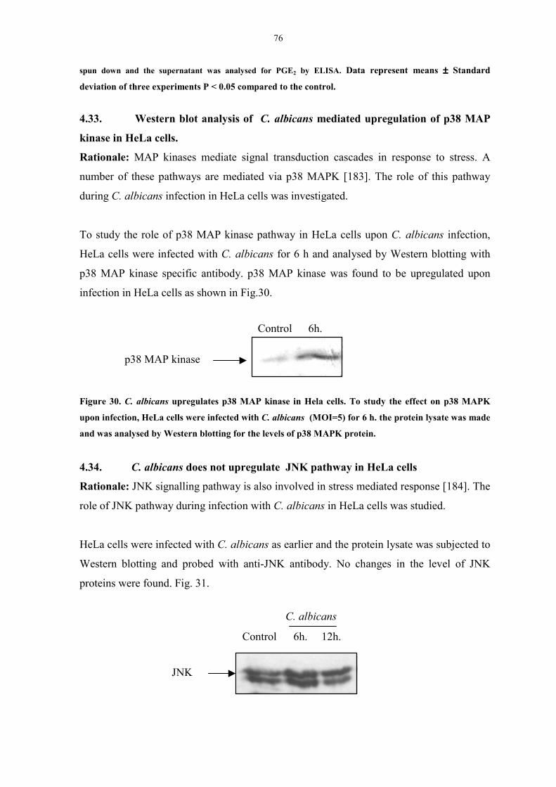
76
spun down and the supernatant was analysed for PGE2 by ELISA. Data represent means ±±±± Standard
deviation of three experiments P < 0.05 compared to the control.
4.33. Western blot analysis of C. albicans mediated upregulation of p38 MAP
kinase in HeLa cells.
Rationale: MAP kinases mediate signal transduction cascades in response to stress. A
number of these pathways are mediated via p38 MAPK [183]. The role of this pathway
during C. albicans infection in HeLa cells was investigated.
To study the role of p38 MAP kinase pathway in HeLa cells upon C. albicans infection,
HeLa cells were infected with C. albicans for 6 h and analysed by Western blotting with
p38 MAP kinase specific antibody. p38 MAP kinase was found to be upregulated upon
infection in HeLa cells as shown in Fig.30.
Figure 30. C. albicans upregulates p38 MAP kinase in Hela cells. To study the effect on p38 MAPK
upon infection, HeLa cells were infected with C. albicans (MOI=5) for 6 h. the protein lysate was made
and was analysed by Western blotting for the levels of p38 MAPK protein.
4.34. C. albicans does not upregulate JNK pathway in HeLa cells
Rationale: JNK signalling pathway is also involved in stress mediated response [184]. The
role of JNK pathway during infection with C. albicans in HeLa cells was studied.
HeLa cells were infected with C. albicans as earlier and the protein lysate was subjected to
Western blotting and probed with anti-JNK antibody. No changes in the level of JNK
proteins were found. Fig. 31.
p38 MAP kinase
Control 6h.
C. albicans
Control 6h. 12h.
JNK
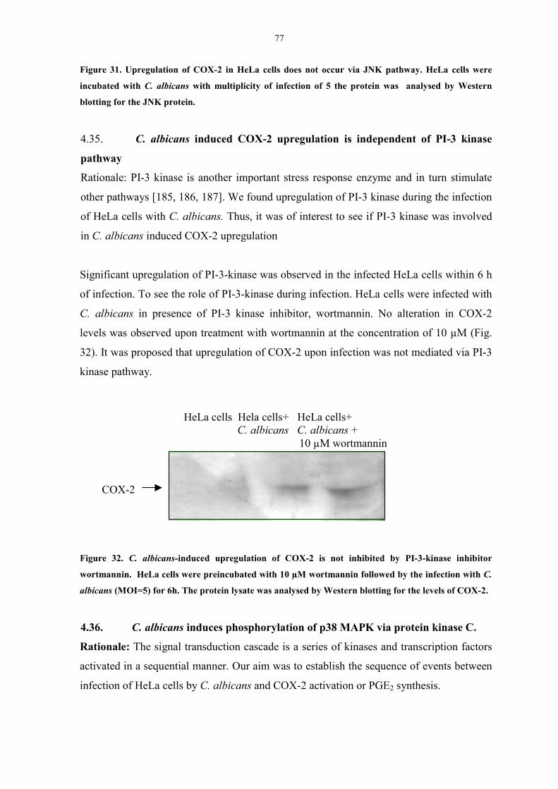
77
Figure 31. Upregulation of COX-2 in HeLa cells does not occur via JNK pathway. HeLa cells were
incubated with C. albicans with multiplicity of infection of 5 the protein was analysed by Western
blotting for the JNK protein.
4.35. C. albicans induced COX-2 upregulation is independent of PI-3 kinase
pathway
Rationale: PI-3 kinase is another important stress response enzyme and in turn stimulate
other pathways [185, 186, 187]. We found upregulation of PI-3 kinase during the infection
of HeLa cells with C. albicans. Thus, it was of interest to see if PI-3 kinase was involved
in C. albicans induced COX-2 upregulation
Significant upregulation of PI-3-kinase was observed in the infected HeLa cells within 6 h
of infection. To see the role of PI-3-kinase during infection. HeLa cells were infected with
C. albicans in presence of PI-3 kinase inhibitor, wortmannin. No alteration in COX-2
levels was observed upon treatment with wortmannin at the concentration of 10 µM (Fig.
32). It was proposed that upregulation of COX-2 upon infection was not mediated via PI-3
kinase pathway.
Figure 32. C. albicans-induced upregulation of COX-2 is not inhibited by PI-3-kinase inhibitor
wortmannin. HeLa cells were preincubated with 10 µM wortmannin followed by the infection with C.
albicans (MOI=5) for 6h. The protein lysate was analysed by Western blotting for the levels of COX-2.
4.36. C. albicans induces phosphorylation of p38 MAPK via protein kinase C.
Rationale: The signal transduction cascade is a series of kinases and transcription factors
activated in a sequential manner. Our aim was to establish the sequence of events between
infection of HeLa cells by C. albicans and COX-2 activation or PGE2 synthesis.
COX-2
HeLa cells Hela cells+ HeLa cells+ C. albicans C. albicans + 10 µM wortmannin
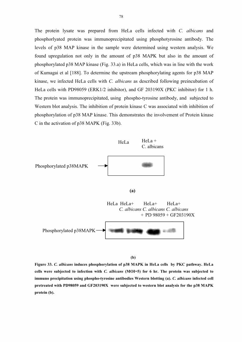
78
The protein lysate was prepared from HeLa cells infected with C. albicans and
phosphorlyated protein was immunoprecipitated using phosphotyrosine antibody. The
levels of p38 MAP kinase in the sample were determined using western analysis. We
found upregulation not only in the amount of p38 MAPK but also in the amount of
phosphorylated p38 MAP kinase (Fig. 33.a) in HeLa cells, which was in line with the work
of Kumagai et al [188]. To determine the upstream phosphorylating agents for p38 MAP
kinase, we infected HeLa cells with C. albicans as described following preincubation of
HeLa cells with PD98059 (ERK1/2 inhibitor), and GF 203190X (PKC inhibitor) for 1 h.
The protein was immunoprecipitated, using phospho-tyrosine antibody, and subjected to
Western blot analysis. The inhibition of protein kinase C was associated with inhibition of
phosphorylation of p38 MAP kinase. This demonstrates the involvement of Protein kinase
C in the activation of p38 MAPK (Fig. 33b).
(a)
(b)
(b)
Figure 33. C. albicans induces phosphorylation of p38 MAPK in HeLa cells by PKC pathway. HeLa
cells were subjected to infection with C. albicans (MOI=5) for 6 hr. The protein was subjected to
immuno precipitation using phospho-tyrosine antibodies Western blotting (a). C. albicans infected cell
pretreated with PD98059 and GF203190X were subjected to western blot analysis for the p38 MAPK
protein (b).
HeLa HeLa +C. albicans
Phosphorylated p38MAPK
HeLa HeLa+ HeLa+ HeLa+ C. albicans C. albicans C. albicans
+ PD 98059 + GF203190X
Phosphorylated p38MAPK
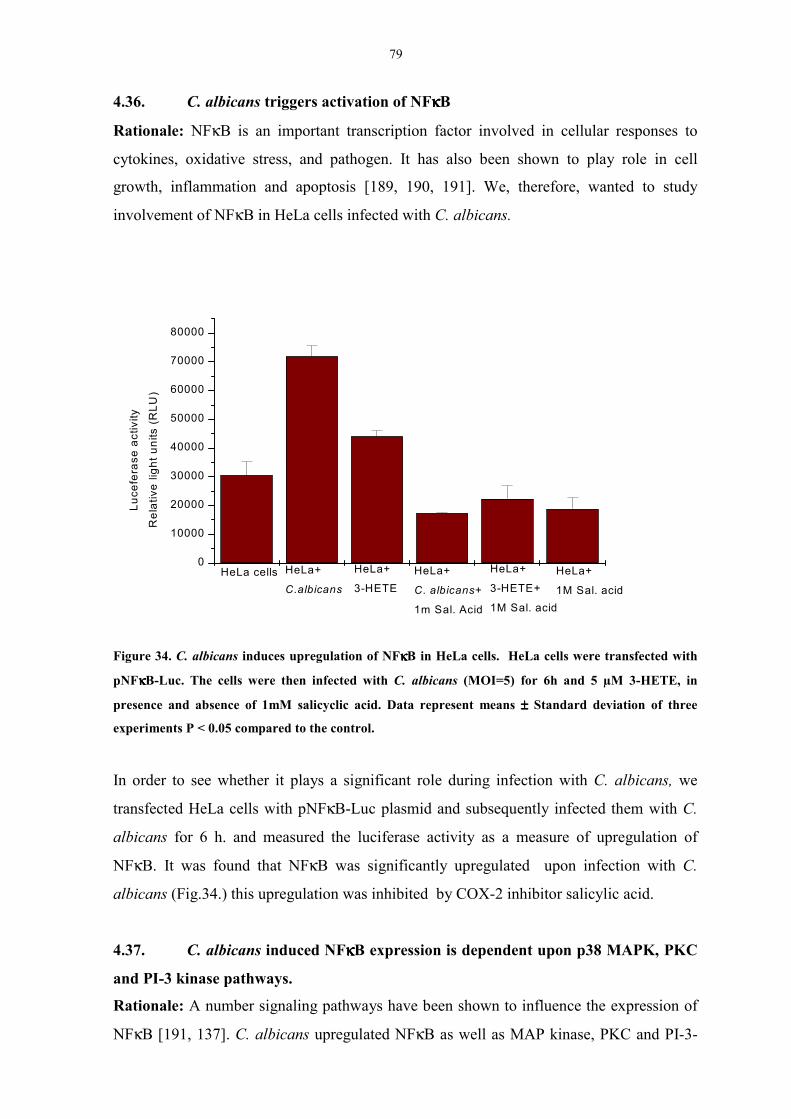
79
4.36. C. albicans triggers activation of NFκκκκB
Rationale: NFκB is an important transcription factor involved in cellular responses to
cytokines, oxidative stress, and pathogen. It has also been shown to play role in cell
growth, inflammation and apoptosis [189, 190, 191]. We, therefore, wanted to study
involvement of NFκB in HeLa cells infected with C. albicans.
Figure 34. C. albicans induces upregulation of NFκκκκB in HeLa cells. HeLa cells were transfected with
pNFκκκκB-Luc. The cells were then infected with C. albicans (MOI=5) for 6h and 5 µM 3-HETE, in
presence and absence of 1mM salicyclic acid. Data represent means ±±±± Standard deviation of three
experiments P < 0.05 compared to the control.
In order to see whether it plays a significant role during infection with C. albicans, we
transfected HeLa cells with pNFκB-Luc plasmid and subsequently infected them with C.
albicans for 6 h. and measured the luciferase activity as a measure of upregulation of
NFκB. It was found that NFκB was significantly upregulated upon infection with C.
albicans (Fig.34.) this upregulation was inhibited by COX-2 inhibitor salicylic acid.
4.37. C. albicans induced NFκκκκB expression is dependent upon p38 MAPK, PKC
and PI-3 kinase pathways.
Rationale: A number signaling pathways have been shown to influence the expression of
NFκB [191, 137]. C. albicans upregulated NFκB as well as MAP kinase, PKC and PI-3-
0
10000
20000
30000
40000
50000
60000
70000
80000
HeLa+
1M Sal. acid
HeLa+
3-HETE+
1M Sal. acid
HeLa+
C. albicans+
1m Sal. Acid
HeLa+
3-HETEHeLa+
C.albicansHeLa cells
Luce
fera
se a
ctiv
ity
Rel
ativ
e lig
ht u
nits
(R
LU)

80
kinase pathways inHeLa cells. It was, therefore, of interest to investigate the role of these
pathways in NFκB upregulation.
pNFκB-Luc transfected HeLa cells were infected with C. albicans in presence of p38
MAPK and PKC inhibitors and the luciferase activity was measured. It was found, that the
C. albicans induced NFκB expression was inhibited by p38 MAPK and PKC inhibitors as
shown in Fig 36. This indicates the involvement of these pathways in NFκB upregulation.
We also found that PI-3 kinase pathway, which is upregulated by C. albicans, is involved
in regulation of NFκB (Fig.35). PI-3 kinase has been reported in literature to be involved
in NFκB mediated anti apoptotic function.
Figure 35. Candida albicans induced upregulation of NFκκκκB is inhibited by SB 202190, GF 203190X.
HeLa cells transfected with pNFκΒκΒκΒκΒ-luc followed by infection with C. albicans (MOI=5) for 2 h. Data
represent means ±±±± Standard deviation of three experiments P < 0.05 compared to the control.
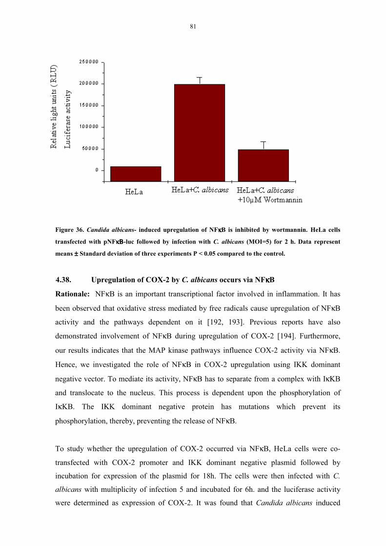
81
Figure 36. Candida albicans- induced upregulation of NFκκκκB is inhibited by wortmannin. HeLa cells
transfected with pNFκΒκΒκΒκΒ-luc followed by infection with C. albicans (MOI=5) for 2 h. Data represent
means ±±±± Standard deviation of three experiments P < 0.05 compared to the control.
4.38. Upregulation of COX-2 by C. albicans occurs via NFκκκκB
Rationale: NFκB is an important transcriptional factor involved in inflammation. It has
been observed that oxidative stress mediated by free radicals cause upregulation of NFκB
activity and the pathways dependent on it [192, 193]. Previous reports have also
demonstrated involvement of NFκB during upregulation of COX-2 [194]. Furthermore,
our results indicates that the MAP kinase pathways influence COX-2 activity via NFκB.
Hence, we investigated the role of NFκB in COX-2 upregulation using IKK dominant
negative vector. To mediate its activity, NFκB has to separate from a complex with IκKB
and translocate to the nucleus. This process is dependent upon the phosphorylation of
IκKB. The IΚK dominant negative protein has mutations which prevent its
phosphorylation, thereby, preventing the release of NFκB.
To study whether the upregulation of COX-2 occurred via NFκB, HeLa cells were co-
transfected with COX-2 promoter and IKK dominant negative plasmid followed by
incubation for expression of the plasmid for 18h. The cells were then infected with C.
albicans with multiplicity of infection 5 and incubated for 6h. and the luciferase activity
were determined as expression of COX-2. It was found that Candida albicans induced
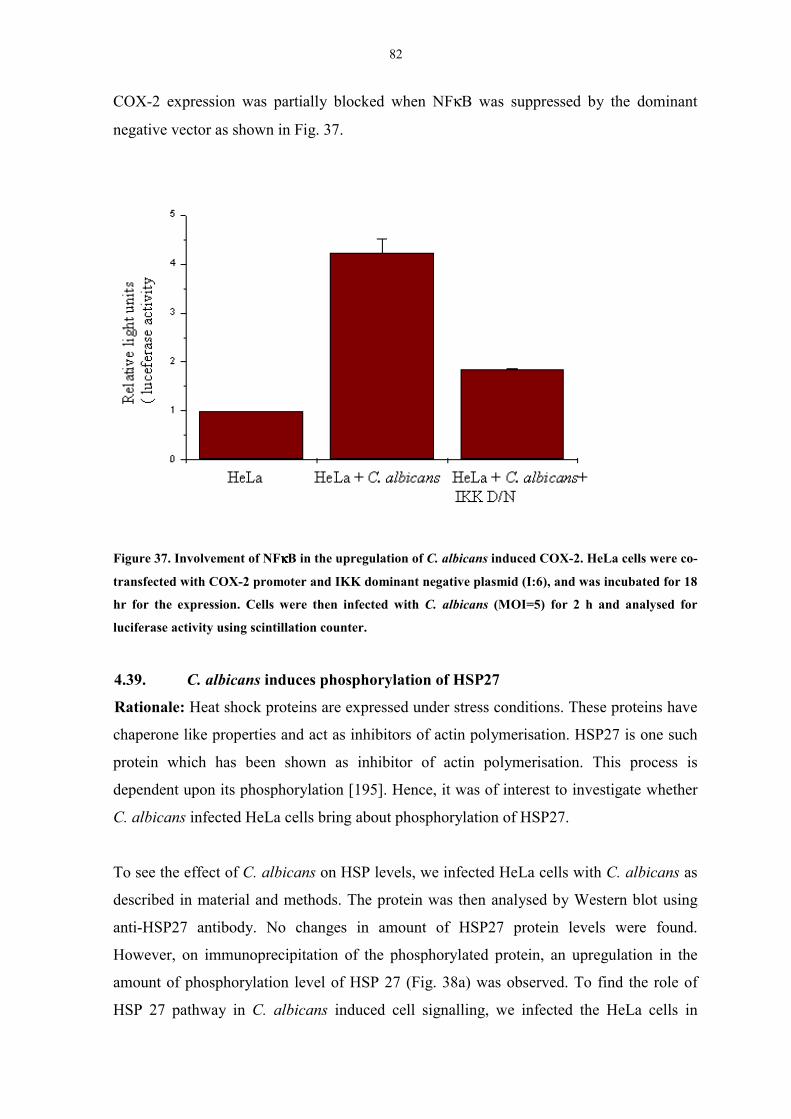
82
COX-2 expression was partially blocked when NFκB was suppressed by the dominant
negative vector as shown in Fig. 37.
Figure 37. Involvement of NFκκκκB in the upregulation of C. albicans induced COX-2. HeLa cells were co-
transfected with COX-2 promoter and IKK dominant negative plasmid (I:6), and was incubated for 18
hr for the expression. Cells were then infected with C. albicans (MOI=5) for 2 h and analysed for
luciferase activity using scintillation counter.
4.39. C. albicans induces phosphorylation of HSP27
Rationale: Heat shock proteins are expressed under stress conditions. These proteins have
chaperone like properties and act as inhibitors of actin polymerisation. HSP27 is one such
protein which has been shown as inhibitor of actin polymerisation. This process is
dependent upon its phosphorylation [195]. Hence, it was of interest to investigate whether
C. albicans infected HeLa cells bring about phosphorylation of HSP27.
To see the effect of C. albicans on HSP levels, we infected HeLa cells with C. albicans as
described in material and methods. The protein was then analysed by Western blot using
anti-HSP27 antibody. No changes in amount of HSP27 protein levels were found.
However, on immunoprecipitation of the phosphorylated protein, an upregulation in the
amount of phosphorylation level of HSP 27 (Fig. 38a) was observed. To find the role of
HSP 27 pathway in C. albicans induced cell signalling, we infected the HeLa cells in
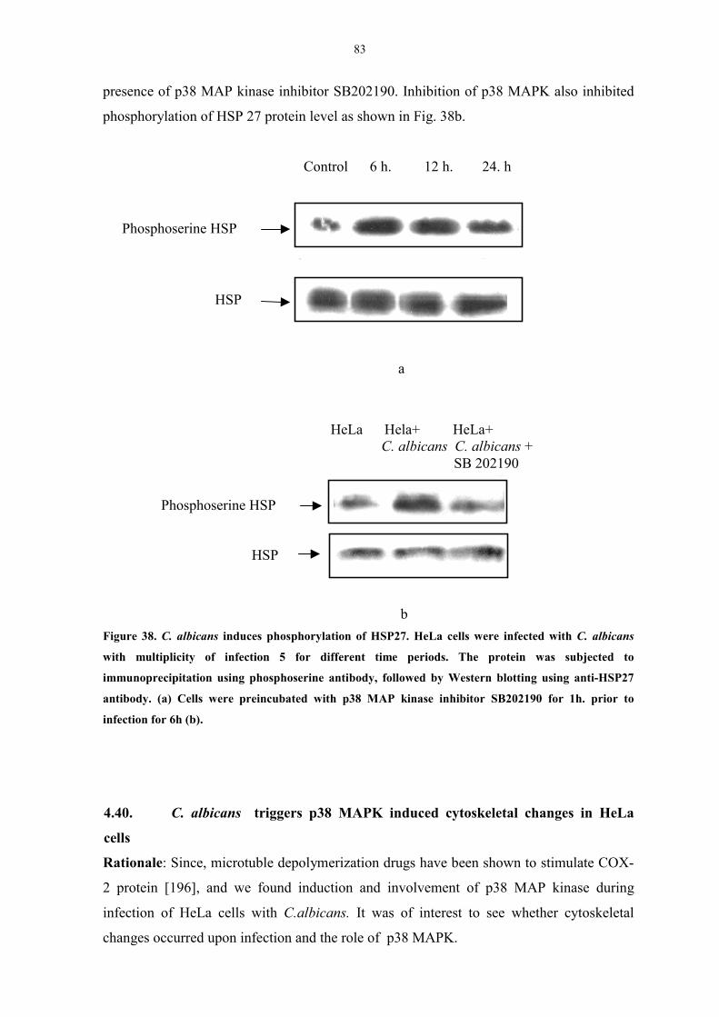
83
presence of p38 MAP kinase inhibitor SB202190. Inhibition of p38 MAPK also inhibited
phosphorylation of HSP 27 protein level as shown in Fig. 38b.
Figure 38. C. albicans induces phosphorylation of HSP27. HeLa cells were infected with C. albicans
with multiplicity of infection 5 for different time periods. The protein was subjected to
immunoprecipitation using phosphoserine antibody, followed by Western blotting using anti-HSP27
antibody. (a) Cells were preincubated with p38 MAP kinase inhibitor SB202190 for 1h. prior to
infection for 6h (b).
4.40. C. albicans triggers p38 MAPK induced cytoskeletal changes in HeLa
cells
Rationale: Since, microtuble depolymerization drugs have been shown to stimulate COX-
2 protein [196], and we found induction and involvement of p38 MAP kinase during
infection of HeLa cells with C.albicans. It was of interest to see whether cytoskeletal
changes occurred upon infection and the role of p38 MAPK.
Phosphoserine HSP
Control 6 h. 12 h. 24. h
Phosphoserine HSP
HeLa Hela+ HeLa+ C. albicans C. albicans + SB 202190
HSP
HSP
a
b

84
Figure 39. C. albicans triggers p38 MAPK induced cytoskeletal changes in HeLa cells. HeLa cells were grown
on coverslips in DMEM containing 10% FCS. Coverslips were infected with C. albicans (b, c) for 6 h. in
presence (c) or absence (a) of 20µM SB 202190. Coverslips after incubation were washed with PBS, fixed
using 4% paraformaldehyde for 15 min. The coverslips were washed and incubated with anti-actin antibody
for 30 min in a humid chamber followed by secondary anti rabbit-FITC conjugate antibody. The coverslips
were visualised under immunofluorescent microscope.
To study the effect of C. albicans on the cytoskeleton, HeLa cells were grown on
coverslips and infected with C. albicans for 6 h followed by immunostaining with anti-
(a)
(b)
(c)
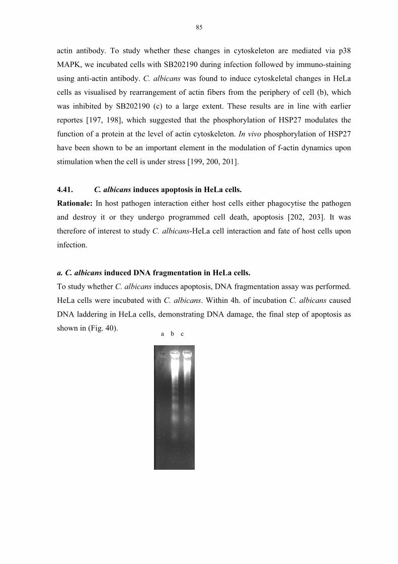
85
actin antibody. To study whether these changes in cytoskeleton are mediated via p38
MAPK, we incubated cells with SB202190 during infection followed by immuno-staining
using anti-actin antibody. C. albicans was found to induce cytoskeletal changes in HeLa
cells as visualised by rearrangement of actin fibers from the periphery of cell (b), which
was inhibited by SB202190 (c) to a large extent. These results are in line with earlier
reportes [197, 198], which suggested that the phosphorylation of HSP27 modulates the
function of a protein at the level of actin cytoskeleton. In vivo phosphorylation of HSP27
have been shown to be an important element in the modulation of f-actin dynamics upon
stimulation when the cell is under stress [199, 200, 201].
4.41. C. albicans induces apoptosis in HeLa cells.
Rationale: In host pathogen interaction either host cells either phagocytise the pathogen
and destroy it or they undergo programmed cell death, apoptosis [202, 203]. It was
therefore of interest to study C. albicans-HeLa cell interaction and fate of host cells upon
infection.
a. C. albicans induced DNA fragmentation in HeLa cells.
To study whether C. albicans induces apoptosis, DNA fragmentation assay was performed.
HeLa cells were incubated with C. albicans. Within 4h. of incubation C. albicans caused
DNA laddering in HeLa cells, demonstrating DNA damage, the final step of apoptosis as
shown in (Fig. 40). a b c
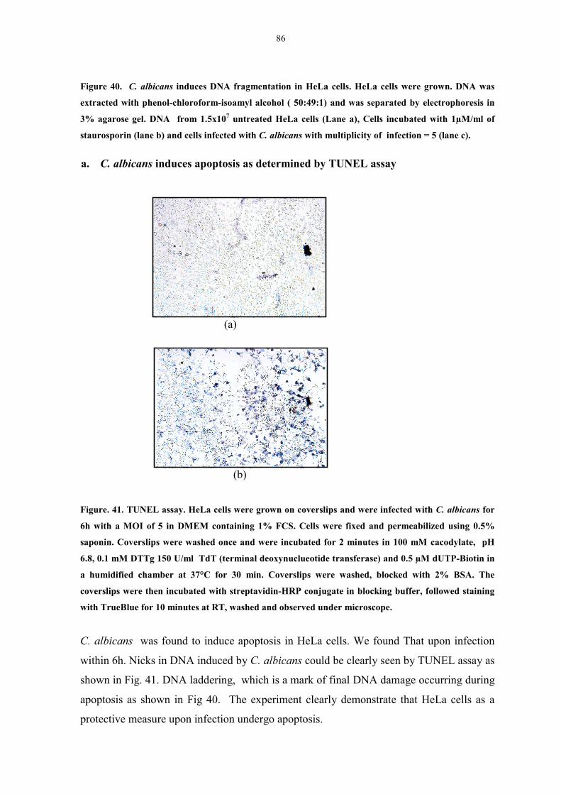
86
Figure 40. C. albicans induces DNA fragmentation in HeLa cells. HeLa cells were grown. DNA was
extracted with phenol-chloroform-isoamyl alcohol ( 50:49:1) and was separated by electrophoresis in
3% agarose gel. DNA from 1.5x10⁷ untreated HeLa cells (Lane a), Cells incubated with 1µM/ml of
staurosporin (lane b) and cells infected with C. albicans with multiplicity of infection = 5 (lane c).
a. C. albicans induces apoptosis as determined by TUNEL assay
Figure. 41. TUNEL assay. HeLa cells were grown on coverslips and were infected with C. albicans for
6h with a MOI of 5 in DMEM containing 1% FCS. Cells were fixed and permeabilized using 0.5%
saponin. Coverslips were washed once and were incubated for 2 minutes in 100 mM cacodylate, pH
6.8, 0.1 mM DTTg 150 U/ml TdT (terminal deoxynuclueotide transferase) and 0.5 µM dUTP-Biotin in
a humidified chamber at 37°C for 30 min. Coverslips were washed, blocked with 2% BSA. The
coverslips were then incubated with streptavidin-HRP conjugate in blocking buffer, followed staining
with TrueBlue for 10 minutes at RT, washed and observed under microscope.
C. albicans was found to induce apoptosis in HeLa cells. We found That upon infection
within 6h. Nicks in DNA induced by C. albicans could be clearly seen by TUNEL assay as
shown in Fig. 41. DNA laddering, which is a mark of final DNA damage occurring during
apoptosis as shown in Fig 40. The experiment clearly demonstrate that HeLa cells as a
protective measure upon infection undergo apoptosis.
(a)
(b)
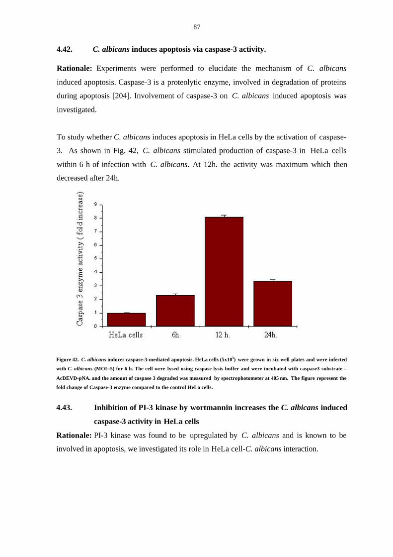
87
4.42. C. albicans induces apoptosis via caspase-3 activity.
Rationale: Experiments were performed to elucidate the mechanism of C. albicans
induced apoptosis. Caspase-3 is a proteolytic enzyme, involved in degradation of proteins
during apoptosis [204]. Involvement of caspase-3 on C. albicans induced apoptosis was
investigated.
To study whether C. albicans induces apoptosis in HeLa cells by the activation of caspase-
3. As shown in Fig. 42, C. albicans stimulated production of caspase-3 in HeLa cells
within 6 h of infection with C. albicans. At 12h. the activity was maximum which then
decreased after 24h.
Figure 42. C. albicans induces caspase-3-mediated apoptosis. HeLa cells (5x10⁵) were grown in six well plates and were infected
with C. albicans (MOI=5) for 6 h. The cell were lysed using caspase lysis buffer and were incubated with caspase3 substrate –
AcDEVD-pNA. and the amount of caspase 3 degraded was measured by spectrophotometer at 405 nm. The figure represent the
fold change of Caspase-3 enzyme compared to the control HeLa cells.
4.43. Inhibition of PI-3 kinase by wortmannin increases the C. albicans induced
caspase-3 activity in HeLa cells
Rationale: PI-3 kinase was found to be upregulated by C. albicans and is known to be
involved in apoptosis, we investigated its role in HeLa cell-C. albicans interaction.
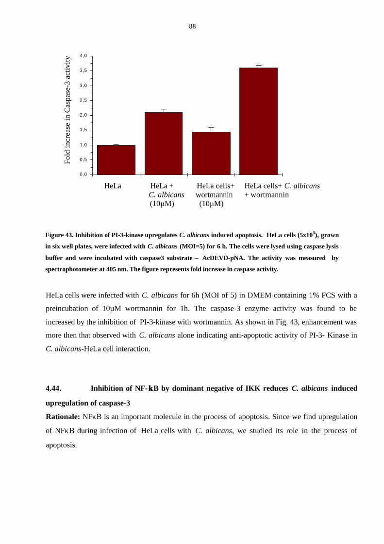
88
Figure 43. Inhibition of PI-3-kinase upregulates C. albicans induced apoptosis. HeLa cells (5x10⁵), grown
in six well plates, were infected with C. albicans (MOI=5) for 6 h. The cells were lysed using caspase lysis
buffer and were incubated with caspase3 substrate – AcDEVD-pNA. The activity was measured by
spectrophotometer at 405 nm. The figure represents fold increase in caspase activity.
HeLa cells were infected with C. albicans for 6h (MOI of 5) in DMEM containing 1% FCS with a
preincubation of 10µM wortmannin for 1h. The caspase-3 enzyme activity was found to be
increased by the inhibition of PI-3-kinase with wortmannin. As shown in Fig. 43, enhancement was
more then that observed with C. albicans alone indicating anti-apoptotic activity of PI-3- Kinase in
C. albicans-HeLa cell interaction.
4.44. Inhibition of NF- κκB by dominant negative of IKK reduces C. albicans induced
upregulation of caspase-3
Rationale: NFκB is an important molecule in the process of apoptosis. Since we find upregulation
of NFκB during infection of HeLa cells with C. albicans, we studied its role in the process of
apoptosis.
Fol
d in
crea
se in
Cas
pase
-3 a
ctiv
ity
0,0
0,5
1,0
1,5
2,0
2,5
3,0
3,5
4,0
HeLa HeLa + HeLa cells+ HeLa cells+ C. albicans C. albicans wortmannin + wortmannin
(10µM) (10µM)

89
Figure 44. Inhibition of NF-κκB by dominant negative of IKK reduces C. albicans-induced upregulation
of caspase-3. HeLa cells were transfected with IKK dominant negative, the cells were kept for 24 h for
expression followed by infection with C. albicans (MOI=5) for 2 h. The cells were lysed and assayed for
for caspase activity.
Prior to the infection, HeLa cells were transfected with Inhibitor kappa kinase dominant
negative (IKK D/N) plasmid. 24 hr later the cells were infected with C. albicans with a
MOI of 5 in DMEM containing 1% FCS. Caspase-3 activity was assayed, by monitoring
the degradation of caspase-3 substrate – AcDEVDpNA, in HeLa cell lysate. As shown in
Fig. 43, inhibition of NFκB, was coupled with decreased caspase-3 activity, demonstrating
the prominent role of NFκB in apoptosis.
4.45. C. albicans induces cleavage of p65 in HeLa cells
Rationale: NFκB is generally attributed with an anti-apoptotic role, though there are
reports of its pro-apoptotic activity [205, 206]. Since, in our system NFκB shows a
proapoptotic activity, it was of interest to elucidate this pathway. Truncated p65 molecule
has been shown to reduce anti-apoptotic activity of NFκB and thus induce apoptosis in the
cell. We wanted to study whether C. albicans induces apoptosis by this mechanism.
Fol
d in
crea
se in
Cas
pase
-3 a
ctiv
ity
0,0
0 ,5
1 ,0
1 ,5
2 ,0
2 ,5
HeLa cells HeLa + HeLa + HeLa+ IKK D/N C. albicans IKK D/N + C. albicans
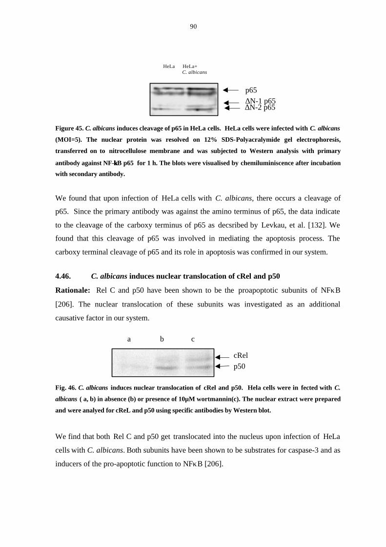
90
Figure 45. C. albicans induces cleavage of p65 in HeLa cells. HeLa cells were infected with C. albicans
(MOI=5). The nuclear protein was resolved on 12% SDS-Polyacralymide gel electrophoresis,
transferred on to nitrocellulose membrane and was subjected to Western analysis with primary
antibody against NF-κκB p65 for 1 h. The blots were visualised by chemiluminiscence after incubation
with secondary antibody.
We found that upon infection of HeLa cells with C. albicans, there occurs a cleavage of
p65. Since the primary antibody was against the amino terminus of p65, the data indicate
to the cleavage of the carboxy terminus of p65 as decsribed by Levkau, et al. [132]. We
found that this cleavage of p65 was involved in mediating the apoptosis process. The
carboxy terminal cleavage of p65 and its role in apoptosis was confirmed in our system.
4.46. C. albicans induces nuclear translocation of cRel and p50
Rationale: Rel C and p50 have been shown to be the proapoptotic subunits of NFκB
[206]. The nuclear translocation of these subunits was investigated as an additional
causative factor in our system.
Fig. 46. C. albicans induces nuclear translocation of cRel and p50. Hela cells were in fected with C.
albicans ( a, b) in absence (b) or presence of 10µM wortmannin(c). The nuclear extract were prepared
and were analyed for cReL and p50 using specific antibodies by Western blot.
We find that both Rel C and p50 get translocated into the nucleus upon infection of HeLa
cells with C. albicans. Both subunits have been shown to be substrates for caspase-3 and as
inducers of the pro-apoptotic function to NFκB [206].
p65
∆N-1 p65∆N-2 p65
HeLa HeLa+ C. albicans
cRel
p50
a b c
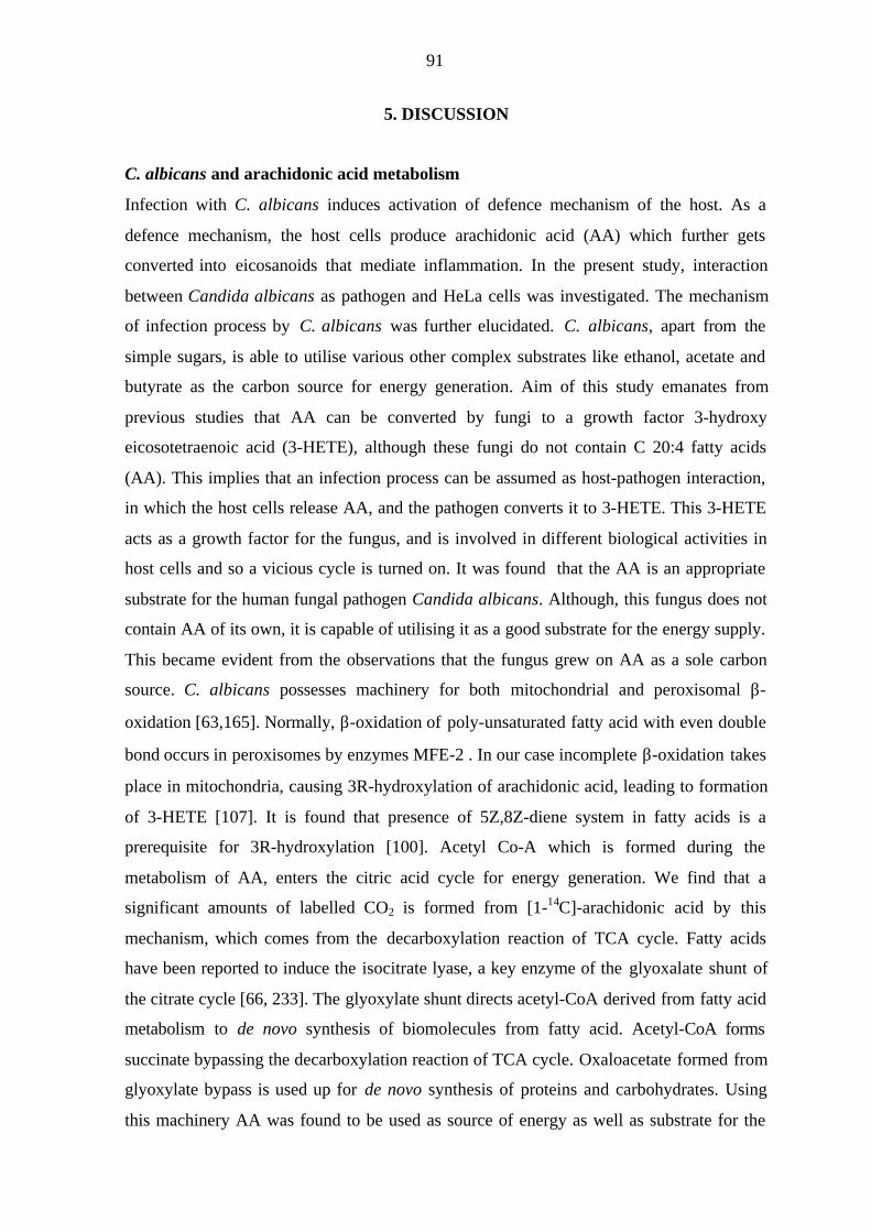
91
5. DISCUSSION
C. albicans and arachidonic acid metabolism
Infection with C. albicans induces activation of defence mechanism of the host. As a
defence mechanism, the host cells produce arachidonic acid (AA) which further gets
converted into eicosanoids that mediate inflammation. In the present study, interaction
between Candida albicans as pathogen and HeLa cells was investigated. The mechanism
of infection process by C. albicans was further elucidated. C. albicans, apart from the
simple sugars, is able to utilise various other complex substrates like ethanol, acetate and
butyrate as the carbon source for energy generation. Aim of this study emanates from
previous studies that AA can be converted by fungi to a growth factor 3-hydroxy
eicosotetraenoic acid (3-HETE), although these fungi do not contain C 20:4 fatty acids
(AA). This implies that an infection process can be assumed as host-pathogen interaction,
in which the host cells release AA, and the pathogen converts it to 3-HETE. This 3-HETE
acts as a growth factor for the fungus, and is involved in different biological activities in
host cells and so a vicious cycle is turned on. It was found that the AA is an appropriate
substrate for the human fungal pathogen Candida albicans. Although, this fungus does not
contain AA of its own, it is capable of utilising it as a good substrate for the energy supply.
This became evident from the observations that the fungus grew on AA as a sole carbon
source. C. albicans possesses machinery for both mitochondrial and peroxisomal β-
oxidation [63,165]. Normally, β-oxidation of poly-unsaturated fatty acid with even double
bond occurs in peroxisomes by enzymes MFE-2 . In our case incomplete β-oxidation takes
place in mitochondria, causing 3R-hydroxylation of arachidonic acid, leading to formation
of 3-HETE [107]. It is found that presence of 5Z,8Z-diene system in fatty acids is a
prerequisite for 3R-hydroxylation [100]. Acetyl Co-A which is formed during the
metabolism of AA, enters the citric acid cycle for energy generation. We find that a
significant amounts of labelled CO2 is formed from [1-14C]-arachidonic acid by this
mechanism, which comes from the decarboxylation reaction of TCA cycle. Fatty acids
have been reported to induce the isocitrate lyase, a key enzyme of the glyoxalate shunt of
the citrate cycle [66, 233]. The glyoxylate shunt directs acetyl-CoA derived from fatty acid
metabolism to de novo synthesis of biomolecules from fatty acid. Acetyl-CoA forms
succinate bypassing the decarboxylation reaction of TCA cycle. Oxaloacetate formed from
glyoxylate bypass is used up for de novo synthesis of proteins and carbohydrates. Using
this machinery AA was found to be used as source of energy as well as substrate for the
92
formation of building blocks for carbohydrate biosyntheses. As high as two-thirds of the
radioactivity from arachidonic acid metabolism was found in the carbohydrates of C.
albicans.
Hydrogen atoms derived during the metabolism of arachidonic acid, enter the electron
transport chain through a series of carriers including cytochromes, iron-sulfur proteins and
flavin linked proteins, bringing about oxidative phosphorylation and ultimately to
molecular oxygen. Antimycin A inhibits the flow of transport of electron from cytochrome
b and c1, thus inhibiting respiration. Unlike human cells, C. albicans is reported to possess
alternative terminal oxidase [207]. The gene of this enzyme in the alternative oxidase
pathway has been recently identified [168]. This pathway differs from the main respiratory
pathway in that the oxygen uptake is accompanied by heat production instead of ATP
formation. Alternative pathway branches off from the main respiratory chain at the site of
cytochrome b, and thus is insensitive to antimycin A or cyanide [234]. AA was found to
have a salicylhydroxamic acid-like action in the inhibition of alternative oxidase of the
mitochondrial electron transfer system pathway. This demonstrats that arachidonic acid
metabolites which have biological importance in the life cycle of C. albicans and in
pathogenicity in humans are formed with the supression of alternative mode of energy
generation. In higher plants, the expression of alternative oxidase has been reported to be
stimulated both in post-germinative development of cotyledons [208]. It is also involved in
infection-mediated necrosis [209]. It is tempting to speculate that in C. albicans the
alternative oxidase is involved in the alteration of morphogenesis. Thus, its selective
inhibition by arachidonic acid could play a role in the infection process.
The observation that C. albicans grows on arachidonic acid is in line with the facts that
certain fungi of the genus Candida are capable of growing readily on both hydrocarbons of
variable chain length [66, 210] and oleic acid [211]. Hydrocarbons are first oxidised to the
corresponding monocarboxylic acid and then in some species to the α, ω-dicarboxylic
acid. The further degradation proceeds via activation by an acyl CoA synthetase,
peroxisomal β-oxidation and citrate/glyoxalate cycle. Fatty acids have been reported to
induce the isocitrate lyase, a key enzyme of the glyoxalate shunt of the citrate cycle which
is involved in the effective conversion of arachidonic acid to carbohydrates [214]. Thus, C.
albicans is able to survive in the glucose deficient environment using glyoxylate shunt
upon internalisation by host cells [212].
93
Upon feeding C. albicans with AA, a novel 3-hydroxyl eicosanoid was found to be
formed. This compound was first detected by its immunoreactivity towards an antibody
specific to 3-OH group. It was then identified as by GS-MS analysis of the lipid extract as
3,18-dihydroxy-5,8,11,14-eicosatetraenoic acid. Its formation was strongly suppressed by
salicylate that also inhibits the formation of 3(R)-HETE in Dipodascopsis uninucleata
suggesting the occurrence of a common biosynthetic pathway to 3(R)-hydroxy-oxylipins
in fungi.
In a recent study (Ciccoli, R et al. submitted for publication), we found the formation of
3,18-DiHETE in D. uninucleata through mitochondrial-dependent and independent
pathways. In D uninucleata, 3R-HETE was formed exclusively from AA. 3-hydroxylation
occurs in mitochondria by enoyl-hydratase. The R-configurationof 3(R)-OH fatty acids
prevent their β-oxidation at the level of 3-OH fatty acid, 3(S) configuration is a suitable
substrate for β-oxidation. In C. albicans 3-hydroxylation is followed by 18-hydroxylation.
Although these organisms do not have arachidonic acid, they are able to metabolise
endogenous fatty acids with the formation of bioactive oxylipins like LA derived 3-HTDE.
Even though the fungus was grown in the absence of arachidonic acid, using a group-
specific antibody against 3(R)-hydroxy fatty acids, the presence of such compounds could
be detected in situ in the hyphae of C. albicans. Again, the immunofluorescence was
strongly suppressed, when the fungus was grown in the presence of acetylsalicylic acid or
salicylate. Since C. albicans does not contain arachidonic acid, the endogenous 3(R)-
hydroxy-oxylipin(s), which are important in the life cycle of this fungus, must be different
from 3,18-DiHETE. It may be speculated that the endogenous immunoreactive compounds
are metabolites of linoleic acid, which is one of the major fatty acids in the lipids of C.
albicans [55, 58], Moreover, a linoleic acid-derived 3(R)-hydroxy-oxylipin was identified
as 3-HTDE [100] in Dipodascopsis uninucleata. However, if exogenous AA is available to
Candida albicans, 3,18-DiHETE is formed as an additional metabolite upon interaction
with mammalian cells. It remains to be established whether the formation of 3,18-DiHETE
affects the pattern of endogenous 3(R)-hydroxy-oxylipins and whether the various
oxylipins reveal different biological activities. The possible biological role of the oxylipins
produced by Candida species is unknown. In a number of other fungi oxylipins of
94
different structures are proposed to play a regulatory role in the reproductive cycle of the
respective fungi.
Aspirin was found to suppress the growth of C. albicans and the formation of hyphal and
other multicellular structures due to inhibition of the biosynthesis of 3(R)-
hydroxyoxylipins by the pathogen. Oxylipins are found to have many biological effects in
fungi. In D. uninucleata, 3-HETE was found to be involved in the sexual phase [109].
Although C. albicans does not exhibit classical sexual cycle of fungi with distinct sexual
forms, it possess distinct differences in the morphological forms in its life cycle. It was
found that 3-hydroxy oxylipins is found selectively in hyphal stage of C. albicans and thus
it seems to have an important role in the morphogenesis. The mode of action of azole
fungicides is via inhibition of cytochrome p450 and ergosterol biosynthesis [51] hence
inhibits 18-hydroxylation mechanism. Aspirin, which is a β-oxidation inhibitor was found
to inhibit the 3-hydroxylation mechanism. For this reason combination of clotrimazole and
aspirin to inhibit 18-hydroxylation and 3-hydroxylation of 3,18-DiHETE was found to be
more effective. The widespread use of azole antifungal drugs has led to the development
of drug resistance in C. albicans, creating a major problem in the treatment of yeast
infections. The ability to morphologically switch from yeast cells (blastospores) to
filamentous forms (hyphae) is an important virulence factor which contributes to the
dissemination of Candida in host tissues and which promotes infection and invasion. The
filamentous forms of the fungus appear to be more infectious than the yeast forms,
however both types of morphogenetic forms are apparently necessary for colonisation and
invasion of the pathogen under different environmental conditions within the host
organism. Approaching both hyphal and yeast forms for the treatment could be the ideal
treatment since different forms are involved in spread and progress of infection. We found
that the aspirin, which inhibited formation of 3-hydroxyoxylipins, also inhibited formation
of filamentous forms as well as the growth of cell growth. Thus, pharmacological
intervention by aspirin in the fatty acid metabolism along with classical therapy would be a
good approach for the treatment of infection. The exact mechanism of inhibition of 3-
hydroxy oxylipins by aspirin is not yet known. It has been suggested that aspirin may
inhibit beta-oxidation. Previous studies in fibroblast cells demonstrated that the inhibition
of beta- oxidation occurs due to structural similarities between aspirin and 3-hydroxyacyl-
CoA and 3-ketoacyl-CoA, which are substrates for 3-hydroxyacyl-CoA-dehydrogenase
[170].
95
The first step of infection includes adhesion of pathogen to host. C. albicans which
adheres rapidly is found to be more virulent, and is able to proceed further to establish the
infection process. The ability of Candida albicans to respond to diverse environments
capacity for multiplication inside the host and resistance to environmental influences, is
critical for its success as a pathogen. We find that aspirin is able to decrease the adhesion
of C. albicans to HeLa cells. This ability along with the ability to inhibit candidal forms
and 3hydroxyoxylipins, demonstrate its potentiality for the chemotherapy against C.
albicans infection.
Host Pathogen Interaction
Host factors affecting the pathogenicity of Candida albicans
C. albicans is able to release arachidonic acid from HeLa cells upon infection which is
subsequently converted into eicosanoids, such as prostaglandin E2. COX-1 is the
constitutive form expressed at basal level in most of the tissues while COX-2 is induced by
a wide range of mitogens, tumor promoters and cytokines [79, 80]. The primary
observation has been the ability of Candida albicans to upregulate the production of COX-
2 enzyme in HeLa cells and the production of PGE2. Cyclooxygenase enzymes convert
arachidonic acid to PGG2, which is further converted to PGH2 by the peroxidase activity of
prostaglandin H synthase. The upregulation of COX-2 was clearly evident within 6 hours
of infection. This corresponds to the concurrent increase in the levels of PGE2. Selective
neutralisation of PGE2 production has been used to block pain and inflammation.
Cyclooxygenase inhibitors are the most widely used class of painkillers and anti-
inflammatory agents. In Candida albicans, PGE2 enhances the germ-tube formation.
Germ-tube formation has been characterised as a sign of pathogenicity of C. albicans [22].
This activity of PGE2 seems to inadvertently promote the infectivity of C. albicans by
inducing germ-tube formation which is necessary for penetration into the tissue. The data
demonstrates that C. albicans selectively induces COX-2 gene and protein synthesis but
not COX-1.
We also observed that 3-HETE, at a concentration as low as 5 µM is able to upregulate
COX-2 in HeLa cells, indicating that it is an important compound responsible for C.

96
albicans-induced upregulation of COX-2. It has been also observed that 3-OH oxylipins
are associated with the hyhyphal forms in Candida albicans [170]. It has been observed
that the host cells, especially monocytes and HeLa cells, engulf Candida albicans upon
infection (Rosseau S et al and present study). This internalisation process results in the
production of a novel arachidonic acid metabolite 3,18-DiHETE, which is believed to be
responsible for the process of inflammation.
Signal transduction cascade in COX-2 upregulation
Induction of COX-2 expression and prostaglandin formation has previously been
demonstrated to be associated with activation of the MAPK signaling pathway in various
cell systems. For example, Xie et al. provided evidence for the role of JNK and c-Jun in
mediating v-Src-induced COX-2 gene expression [214]. p38 MAP kinase has been
implicated in interleukin-1-stimulated COX-2-dependent prostaglandin production in
fibroblasts, and human umbilical vein endothelial cells [177, 215]. Other signaling
elements of the prostaglandin pathway have also been linked with activation of MAPKs.
For example, both ERK ½ and p38MAP kinase mediate biosynthetic activation of
cytosolic phospholipase A2 [211]. PKC and p38 MAP kinase are primarily
serine/threonine kinases and their activation is itself dependent upon the phosphorylation
by other kinases. ERK1/2 is another kinase shown to be involved the COX-2 upregulation.
Investigation of MAPK signaling pathways for the C. albicans induced activation of COX-
2 showed the stimulation of p38 MAPK pathway without involvement of ERK1/2 and
JNK pathways. Moreover, it was found that p38 MAPK was rapidly phosphorylated by
PKC, as GF203190X, a PKC inhibitor, abrogated p38MAPK protein and COX-2
activation. Previous studies have shown the involvement of PKC and p38 MAPK in the
production of COX-2 [88].
However, inhibition of this kinase by PD98059 did not modulate the expression of COX-2,
or the amount of PGE2 as seen in other systems. These results suggest key roles for PKC as
well as for p38 MAP kinase cascades in the biosynthesis of PGE2, likely by upregulating
the induction of cyclo-oxygenase-2 in C. albicans infected HeLa cells. PI-3 kinase was
also activated by Candida albicans infection, although inhibition by wortmannin failed to

97
suppress the COX-2 upregulation. This observation suggests a different role for PI-3-
kinase upon C. albicans mediated infection of HeLa cells.
Activation of NF-κB and MAP kinases as signalling events play a pivotal role in cytokine,
LPS, or stress induced gene expression. One of the important DNA binding proteins in
many stress inducible responses is NF-κB. Proteins of the NF-κB family are important
transcription factors retained in the cytoplasm through heterodimerization with one of
several types of IκB proteins. Stress inducing factors trigger the phosphorylation and
degradation of IκB, resulting in the release of NF-κB to enter the nucleus, where it
activates gene transcription [191, 217]. Following stimulation with agonists, IκB is
phosphorylated at the serine residue by inhibitory kappa β kinase (IKK) complex, and
subsequently degraded. NFκB then translocates to the nucleus, where it docks to DNA at
binding sites within promoter region of target genes [218]. In addition to regulating
cellular responses to cytokines, oxidative stress and pathogens, NFκB plays role in
inflammation [217], apoptosis [219] and tumor cell proliferation. The involvement of this
pathway was investigated in C. albicans infected HeLa cells. We also investigated the role
of the other members of the COX-2 cascade on the NF-κB activation. Remarkably high
levels of NF-κB promoter binding activity are detected in HeLa cells during infection with
C. albicans in HeLa cells. Sodium salicylate, an inhibitor of COX-2 activity, abolilshed
the NFκB activity. The inhibition was even beyond the basal level of NFκB activity.
Seemingly, the effect on cell cycle is specific to cell type [221].
C. albicans induced activation of NF-κB was inhibited upon inhibition of p38 MAPK with
SB202190, PKC with GF203190X and PI-3 kinase with wortmannin. PI-3 kinase pathway
was found to be involved in the upregulation of NF-κB activity. Inhibitor-mediated
repression of COX-2 by IKK dominant negative demonstrated only partially inhibition of
COX-2 upregulation and showed some residual promoter activity. This indicated that
factors other than NFκB may be involved in COX-2 promoter activation. Possible
candidate could be epidermal growth factor receptor (EGFR), an inducer of COX-2 [222],
and Vascular endothelial growth factor (VEGF), a potent angiogenic factor [223].
Increased synthesis of PGE2 may also stimulate IL-8 gene, another angiogenic factor
reported from human colonic epethelial cells [224]. Thus, many pathways may
simultaneously exist for the upregulation of COX-2. We find the involvement of free
98
radicals in COX-2 and PGE2 upregulation during the infection of HeLa cells with C.
albicans, which demonstrate involvement of reactive oxygen species and NFκB. Aspirin is
an inhibitor of cyclooxygenase and is able to inhibit NFκB [171]. Thus, it can act via dual
pathways in combating C. albicans induced infection. Specific inhibition by free radical
scavenger PDTC, and NAC leads to specific inhibition of COX-2 promoter activity and
PGE2 synthesis, providing a functional link between C. albicans-stimulated production of
Reactive Oxygen Intermediates (ROIs) and COX-2 activation.
HeLa cells and smooth muscle cells are important sources of xanthine oxidase [225,226]
which might generate reactive oxygen species (ROI). ROIs in turn cause cellular
inflammatory response by activation of intracellular stress kinases and subsequent tissue
injury [227]. We find that NAC, a precursor of glutathione, and PDTC, a free radical
scavenger, strongly decreased the COX-2 activity. These findings are consistent with the
earlier reports in which oxidative stress has been assigned a key role in the activation of
COX-2 [181]. Oxidative stress has been linked with intracellular MAP kinase signaling
pathways and that are associated with COX-2 gene expression. Using various inhibitors of
MAPK signalling pathway, as well as transient transfection with IKK dominant negative
plasmid, we could demonstrate that C. albicans influences COX-2 pathway via NFκB.
PGE2 produced by C. albicans-induced HeLa cells induces activation of PI-3 kinase which
is either through G protein coupled cytoplasmic PGE2 receptors or through PPAR γ
receptors [228]. The presence of PPAR γ receptors as well as COX-2 on nuclear envelop
pinpoint the feasibility that prostaglandins, analogous to 15-deoxy-PGJ2 may exert their
cell protective role through these receptors [232]. 3-HETE which is a β-oxidation product
of arachidonic acid produced by C. albicans, was found to be an excellent ligand for
PPARγ , which increased PPAR gamma promoter activity in transient transfected cells. In
similar experiments, albeit transfected with PPAR gamma dominant negative plasmid, 3-
HETE failed to enhance the promoter activity (unpublished results). Therefore, it can not
be ruled out that the action of C. albicans on HeLa cells is mediated by 3-HETE or its
derivative 3,18-DiHETE produced by C. albicans
Extensive programmed cell death (apoptosis) was observed after 24 h in Hela cells
infected with C. albicans as shown by genomic DNA laddering and TUNEL assay. After 6
h. postinfection, upregulation of caspase-3 activity was observed. Caspase-3 is an effector
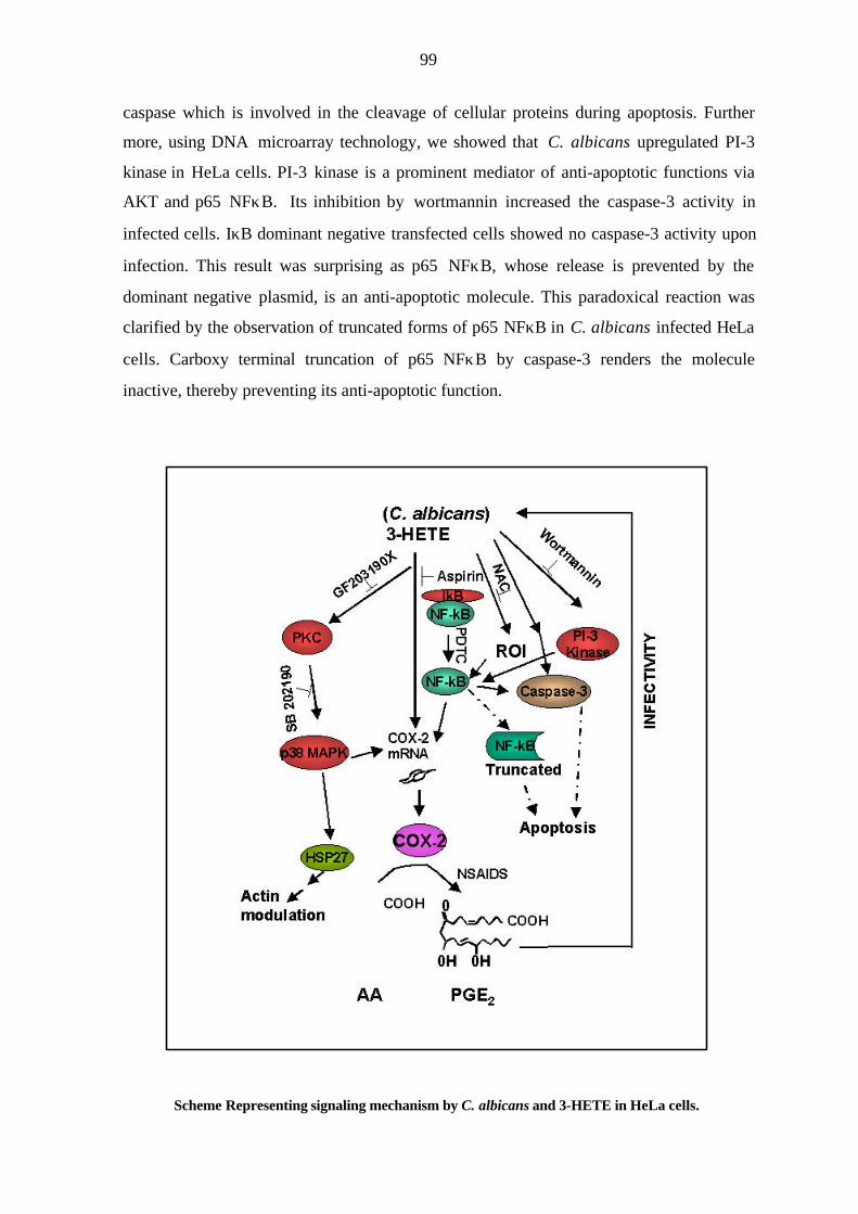
99
caspase which is involved in the cleavage of cellular proteins during apoptosis. Further
more, using DNA microarray technology, we showed that C. albicans upregulated PI-3
kinase in HeLa cells. PI-3 kinase is a prominent mediator of anti-apoptotic functions via
AKT and p65 NFκB. Its inhibition by wortmannin increased the caspase-3 activity in
infected cells. IκB dominant negative transfected cells showed no caspase-3 activity upon
infection. This result was surprising as p65 NFκB, whose release is prevented by the
dominant negative plasmid, is an anti-apoptotic molecule. This paradoxical reaction was
clarified by the observation of truncated forms of p65 NFκB in C. albicans infected HeLa
cells. Carboxy terminal truncation of p65 NFκB by caspase-3 renders the molecule
inactive, thereby preventing its anti-apoptotic function.
Scheme Representing signaling mechanism by C. albicans and 3-HETE in HeLa cells.
100
The pro-apoptotic subunits of NFκB, p50 and c-REL, were found to be nuclear
translocated in C. albicans infected cells. Wortmannin mediated inhibition of PI-3-kinase
strongly inhibited the activation of NF-κB leading to speculation on multiple roles of
NFκB dependent pathways involved in cell signaling.
p38 MAP kinase is not only involved in the upregulation of COX-2, but it is also involved
in the induction of cytoskeletal changes in HeLa cells [201]. Previous studies have shown
[197, 198, 199] the involvement of Heat Shock Protein 27 (HSP27) and p38 MAP kinase
in the modulation of actin cytoskeleton. Heat Shock Proteins, a ubiquitous family of highly
conserved proteins, function to stabilize cellular proteins during a variety of conditions
such as heat shock, infection, and inflammation. Phosphorylation of HSP27, primarily
chaperones, by p38 MAP kinase has been demonstrated as an important element in the
modulation of f-actin dynamics in cellular stress conditions. C. albicans induces serine
phosphorylation of HSP27 in HeLa cells. The changes in actin modulation are inhibited by
p38 MAP kinase inhibitor, indicating another function of p38 MAP kinase, other than the
upregulation of COX-2. Whether this modification in the shape of the cell has an influence
on the progression of C. albicans infection has to be further investigated.
101
REFERENCES
1. Maksymiuk, A.W., S. Thongprasert, R. Hopfer, M. Luna, V. Fainstein , G.P. Bodey.
1984. Systemic candidiasis in cancer patients. Am J Med. 77:20–27.
2. Meunier F. 1987. Prevention of mycoses in immunocompromised patients. Rev Infect
Dis. 9:408–416.
3. Antrum J. 1996. Meeting the challenge of systemic fungal infections in cancer:
nursing implications. Eur J Haematol Suppl 57:7–11.
4. Lew, M. A. 1989. Diagnosis of systemic Candida infections. Annu Rev Med. 40:87–
97.
5. Bow E. J. 1998. Invasive fungal infections in patients receiving intensive cytotoxic
therapy for cancer. Br J Haematol. 101(Suppl. 1):1–4.
6. De Pauw, D.E., J.F. Meis. 1998. Progress in fighting systemic fungal infections in
haematological neoplasia. Support Care Cancer. 6:31–38.
7. Sobel, J. D. 1990. Vaginal infections in adult women . Sex. Transm. Dis. 74:1573-
1601.
8. Odds F. C. 1988. Genital candidosis. In: Odds FC, ed. Candida and candidosis; a
review and bibliography. 2nd ed. London: Baillière Tindall. 124–135.
9. Lindler, G. E. M., F. H. F. Plantema, J. R. A. Hoogkamp-Korstange.1978.
Quantative studies of the vaginal flora of healthy women and of obstetric and
gynaecologic patients. Journal of med. Microbiol. 11:233-241.
10. Fidel, P. L. Jr, J. Cutright, C. Steele. 2000 Effects of reproductive hormones on
experimental vaginal candidiasis. Infect Immun.68:651-657.
11. Witkin, S. S., A. Kalo-Klein, L. Galland, M. Teich, W. J. Ledger. 1991. Effect of
Candida albicans plus histamine on prostaglandin E2 production by peripheral blood
mononuclear cells from healthy women and women with recurrent candidal vaginitis. J
Infect Dis. 164(2):396-399
102
12. Kustimur S. 1992 The role of eicosanoids in the kidney damage induced by Candida
albicans. Prostaglandins Leukot Essential Fatty Acids. 47:83–84.
13. Fidel, P. L., J. D. Jr, Sobel, 1996. Immunopathogenesis of recurrent vulvovaginal
candidiasis. Clin Microbiol Rev. 9:335-348.
14. Datta A, K. Ganesan, and K. Natarajan. 1989. Current trends in Candida albicans
research. Adv Microb Physiol. 30:53–88.
15. Brown A. J. P. and N. A. R. Gow. 1999. Regulatory networks controlling Candida
albicans morphogenesis. Trends Microbiol. 7:333–338.
16. Soll, D. R. 1992. High-frequency switching in Candida albicans. Clin Microbiol
Rev. 5:183–203.
17. Pérez-Martin, J., Uría, J. A., A. D. Johnson. 1999. Phenotyping switching in Candida
albicans is controlled by a SIR2 gene. EMBO J. 18:2580–2592.
18. Merson-Davies, L. A. and F. C. Odds. 1989. A morphology index for
characterization of cell shape in Candida albicans. J Gen Microbiol. 135:3143-52 .
19. Odds F. C. 1985. Morphogenesis in Candida albicans. Crit. Rev. Microbiol. 12:45-
93.
20. Cutler, J. E. 1991. Putative virulence factors of Candida albicans. Annu. Rev.
Microbiol. 45:187–218.
21. Mühlschlegel, F., W. Fonzi, L. Hoyer, P. Payne, F. M. Poulet, J. P. Latge, J. Calera
A. Beauvais, S. Paris, M. Monod, J. Sturtevant, M. Ghannoum, Y.Nozawa, R. Calderone,
1998. Molecular mechanisms of virulence in fungus-host interactions for Aspergillus
fumigatus and Candida albicans. Med Mycol. 36(Suppl. 1):238–248.
22. Kalo-Klein, A., and S. S. Witkin. 1990. Prostaglandin E2 enhances and gamma
interferon inhibits germ tube formation in Candida albicans. Infect Immun. 58:260-262.
23. White, S. and Larsen, B. (1997) Candida albicans morphogenesis is influlenced by
estrogen. Cell Mol Life Sci. 53:744-791
103
24. Kinsman, O. S., K. Pitblado, C. J. Coulson. 1988. Effect of mammalian steroid
hormones and luteinizing hormone on the germination of Candida albicans and
implications for vaginal candidosis. Mycoses.31:617-626.
25. Zhao X, P. J. Malloy, C. M. Ardies, D. Feldman. 1995. Oestrogen-binding protein in
Candida albicans: antibody development and cellular localization by electron
immunocytochemistry. Microbiology 141:2685-2692.
26. Chaffin W.L., J. L. Lopez-Ribot, M. Casanova, D. Gozalbo, J. P. Martinez. 1998.
Cell wall and secreted proteins of Candida albicans: identification, function, and
expression. Microbiol. Mol. Biol. Rev. 62:130¯180.
27. Zink, S., T. Nass, P. Rosen, J. F. Ernst. 1996. Migration of the fungal pathogen
Candida albicans across endothelial monolayers. Infect. Immun. 64:5085¯5091.
28. Hubbard, M. J., D. Markie, R. Poulter. 1986. Isolation and morphological
characterization of a mycelial mutant of Candida albicans. J. Bacteriol. 165:61¯65.
29. Gil, C., R. Pomés, C. Nombela1990, Isolation and characterization of Candida
albicans morphological mutants derepressed for the formation of filamentous hypha-type
structures. J. Bacteriol. 172:2384¯2391.
30. Diez-Orejas R., G. Molero, I. Ríos, A. Vázquez, C. Gil, C. Nombela, M. Sánchez-
Pérez. 1999. Low virulence of a morphological Candida albicans mutant. FEMS
Microbiol. Lett. 176:311¯319.
31. Braun, B. R. and A. D. Johnson,. 1997. Control of filament formation in Candida
albicans by the transcriptional repressor TUP1. Science 277:105-109.
32. Lo, H. J., J. R. Kohler, B. DiDomenico, D. Loebenberg, A. Cacciapuoti, G. R. Fink.
1997. Nonfilamentous C. albicans mutants are avirulent. Cell. 90:939-949
33. Liu, H., C. A. Styles, G. R. Fink 1993. Elements of the yeast pheromone response
pathway required for filamentous growth of diploids. Science. 262:1741-4
34. Roberts R. L., G. R. Fink 1994. Elements of a single MAP kinase cascade in
Saccharomyces cerevisiae mediate two developmental programs in the same cell type:
mating and invasive growth. Genes Dev. 8:2974-2985
104
35. Csank, C., K. Schroppel, E, Leberer. D. Harcus, O. Mohamed, S. Meloche, D. Y.
Thomas, M. Whiteway. 1998. Roles of the Candida albicans mitogen-activated protein
kinase homolog, Cek1p, in hyphal development and systemic candidiasis. Infect Immun.
66:2713-2721.
36. Stoldt, V. R., A Sonneborn, C. E. Leuker, J. F. Ernst. 1997. Efg1p, an essential
regulator of morphogenesis of the human pathogen Candida albicans, is a member of a
conserved class of bHLH proteins regulating morphogenetic processes in fungi. 1: EMBO
J. 16:1982-1991
37. Klepser, M. E. 2001. Antifungal resistance among Candida species.
Pharmacotherapy 8:124S-132S
38. Fleury, F. J. 1981. Adult vaginitis. Clin Obstet. Gynecol. 24 :407-438.
39. Polak A. 1989. Combination therapy for systemic mycosis. Infection. 17(4):203-209
40. Armstrong, D. 1993.Treatment of opportunistic fungal infections. Clin Infect Dis
16:1-7.
41. Joseph-Horne, T., D. Hollomon, R. S. T. Loeffler and S. L. Kelly. 1995. Altered
P450 activity associated with direct selection for fungal azole resistance. FEBS Lett. 374:
174¯178.
42. Vanden Bossche, H., G. Willemsens, P. Marichal. 1987. Anti-Candida drugs the
biochemical basis for their activity. Crit. Rev. Microbiol. 15:57-72
43. Vanden Bossche, H., P. Marichal, F. C. Odds. 1994. Molecular mechanisms of drug
resistance in fungi. Trends Microbiol. 2:393-400
44. Viviani, M. A., S. de Marie, J. R. Graybill, H. Yamaguchi, E. Anaissie, D.
Caillot.1998. New approaches to antifungal chemotherapy. Med. Mycol. 36:194¯206.
45. Vanden Bossche, H., D. W. Warnock, B. Dupont, D. Kerridge, G. S. Sen, L.
Improvisi, P. Marichal, F. C. Odds, F. Provost, O. Ronin. 1994. Mechanisms and clinical
impact of antifungal drug resistance. J. Med. Vet. Mycol. 32:189-202
46. Vanden Bossche, H. V., F. Dromer, I. Improvisi, M. Lozano-Chiu, J. H. Rex and D.
Sanglard. 1998. Antifungal drug resistance in pathogenic fungi. Med. Mycol. 36:119¯128.
105
47. Georgopapadakou, N. H. and A. Bertasso. 1992. Effects of squalene epoxidase
inhibitors on Candida albicans. Antimicrob. Agents Chemother. 36:1779¯1781.
48. Favre, B., and N. S. Ryder. 1996. Characterization of squalene epoxidase activity
from the dermatophyte Trichophyton rubrum and its inhibition by terbinafine and other
antimycotic agents. Antimicrob. Agents Chemother. 40:443-447.
49. Ryder, N. S. 1992. Terbinafine: mode of action and properties of the squalene
epoxidase inhibition. Br. J. Dermatol. 39:2-7.
50. Hitchcock, C. A. 1991. Cytochrome P-450-dependent 14 alpha-sterol demethylase of
Candida albicans and its interaction with azole antifungals. Biochem. Soc. Trans. 19:782-
787.
51. Vanden Bossche, H. 1991. Ergosterol biosynthesis inhibitors, p. 239-257. In R.
Prasad (ed.), Candida albicans. Springer-Verlag KG, Berlin, Germany.
52. Korting, H. C. and M. Grundmann-Kollmann. 1997. The hydroxypyridones: a class
of antimycotics of its own. Mycoses. 40:243¯247.
53. Schaller, M., H. C. Korting, W. Schaefer, J. Bastert, W. C. Chen and B. Hube. 1999.
Secreted aspartic proteinase (Sap) activity contributes to tissue damage in a model of
human oral candidosis. Mol. Microbiol. 34:169¯180.
54. Singh, M, A. Jayakumar, R. Prasad. 1978. The effect of altered lipid composition on
the transport of various amino acids in Candida albicans. Arch Biochem Biophys 191:680–
686.
55. Mishra, P., J. Bolard, R. Prasad. 1992. Emerging role of lipids of Candida albicans, a
pathogenic dimorphic yeast. Biochimica et Biophysica Acta.1127:1-14.
56. Barrett-Bee K, Y. Hayes, R. G. Wilson, Ryley J. F. 1985. A comparison of
phospholipase activity, cellular adherence and pathogenicity of yeasts. J Gen Microbiol
131:1217-1221.
57. Calderone R. A., R. L. Cihlar, D. D. Lee, K. Hoberg, W. M. Scheld. 1985. Yeast
adhesion in the pathogenesis of endocarditis due to Candida albicans: studies with
adherence-negative mutants. J Infect Dis. 152:710-715.
106
58. Goyal, S, and G. K. Khuller.1994. Structural and functional role of lipids in yeast
and mycelial forms of Candida albicans. Lipids. 29:793-7
59. Hiltunen, J. K., B. Wenzel, A. Beyer, R. Erdmann, A. Fossa, W. H. Kunau. 1992.
Peroxisomal multifunctional beta-oxidation protein of Saccharomyces cerevisiae.
Molecular analysis of the fox2 gene and gene product. J Biol Chem. 267:6646-6653.
60. Kawamoto, S., C. Nozaki, A. Tanaka, S. Fukui 1978. Fatty acid beta-oxidation
system in microbodies of n-alkane-grown Candida tropicalis. Eur. J. Biochem., 83:609-
613.
61. Ueda, M., K. Yamanoi, T. Morikawa, H. Okada, and A. Tanaka 1985. Peroxisomal
localisation of enzymes related to fatty acid beta-oxidation in an n-alkane-grown yeast,
Candida tropicalis. Agric. Biol. Chem. 49:1821-1828.
62. Tanaka, A., M. Osumi, S. Fukui. 1982. Peroxisomes of alkane-grown yeasts:
fundamental and practical aspects. Ann. N.Y. Acad: Sci. 386: 183-199.
63. Kunau, W. H., C. Kionka, A. Ledebur, M. Mateblowski, M. Moreno de la Garza, U.
Schultz-Borchard, R. Thieringer, M. Veenhuis. 1987. Beta-oxidation systems in eukaryotic
microorganisms in Preoxisomes in biology and medicine. 128-140. Fahimi, H. D. and Seis,
H., Eds., Springer-Varlag, Berlin.
64. Fulco, A. J. 1991. P450BM-3 and other inducible bacterial P450 cytochromes:
biochemistry and regulation. Annu Rev Pharmacol Toxicol. 31:177-203.
65. Meussdoerffer, F. 1987. Omega-hydroxylations. Fat Sci.Technology.579-581.
66. Lozinov, A. B., L. M. Glazunova, I. T. Ermakova. 1976. The activity of citrate
glyoxylate and pentosephosphate cycle enzymes during yeast growth on hexadecane and
glucose. Mikrobiologiia. 45:33-40.
67. Janusz, M. J., Austen K. F., Czop J. K. 1988. Phagocytosis of heat-killed
blastospores of Candida albicans by human monocyte β-glucan receptors.
Immunology.65:181–185.
68. Kitz, D. J, P. D. Stahl, J. R Little. 1992. The effect of a mannose-binding protein on
macrophage interactions with Candida albicans. Cell Mol Biol. 38:407–412.
107
69. Kanbe, T. H., M. A. Jutila, J. E. Cutler. 1993. Evidence that mannans from Candida
albicans are responsible for the adherence of yeast forms to spleen and lymph nodes. Infect
Immun. 61:2578–2583.
70. Castro, M., N. V. C. Ralston, I. Timothy, M. M. S. Rohrbach A. H. Limper. 1994.
Candida albicans stimulates arachidonic acid liberation from alveolar macrophages
through α-Mannan and β-Glucan cell wall components. Infection and Immunity. 62:3138-
3145.
71. Castro, M., J. A. Bjoraker, M. S. Rohrbach, A. H. Limper. 1996. Candida albicans
induces the release of inflammatory mediators from human peripheral blood monocytes.
Inflammation. 20:107–122.
72. Filler, S. G, B. O. Ibe, P. M. Luckett, J. U. Raj, J. E. Jr. Edwards. 1991 Candida
albicans stimulates endothelial cell eicosanoid production. J Infect Dis.164:928–935.
73. Filler, S. G., B. O. Ibe, A. S. Ibrahim, M. A. Ghannoum, J. U. Raj, J. E. Edwards J.
Jr. 1994. Mechanisms by which Candida albicans induces endothelial cell prostaglandin
synthesis. Infection and Immunity. 62:1064-1069.
74. DeWitt, D. L. 1991. Prostaglandin endoperoxide synthase: regulation of enzyme
expression. Biochim Biophys Acta. 1083:121-134.
75. Smith, W. L and L. J. Marnett. 1991. Prostaglandin endoperoxide synthase: structure
and catalysis. Biochim Biophys Acta. 1083:1-17.
76. Needleman P, and P. C. Isakson. 1997. The discovery and function of COX-2. J
Rheumatol. 24 Suppl 49:6-8.
77. Crofford, L. J. 1997. COX-1 and COX-2 tissue expression: implications and
predictions. J Rheumatol. Suppl 49:15-19.
78. Vane, J. R., J. A. Mitchell, I. Appleton, A. Tomlinson, D. A. Bishop-Bailey, J.
Croxtall, D. Willoughby.1994. Inducible isoforms of cyclooxygenase and nitric oxide
synthase in inflammation. Proc Natl Acad Sci USA 91: 2046-2050
108
79. Xie, W. L., J. G. Chipman, D. L. Robertson, R. L. Erikson, D. L. Simmons. 1991.
Expression of a mitogen-responsive gene encoding prostaglandin synthase is regulated by
mRNA splicing. Proc Natl Acad Sci U S A. 88:2692-2696.
80. Kubuju D. A., B. S. Fletcher, B. C. Barnum, R. W. Lim, H. R. Herschman. 1991.
TIS10, a phorbol ester tumor promoter-inducible mRNA from Swiss 3T3 cells, encodes a
novel prostaglandin synthase/cyclooxygenase homologue. J Biol Chem. 266:12866-12872.
81. Herschman HR. 1996. Prostaglandin synthase 2. Biochim Biophys Acta. 1299:125-
140.
82. Hamasaki, Y., J. Kitzler, R. Hardman, P. Nettesheim, T. E. Eling. 1993. Phorbol
ester and epidermal growth factor enhance the expression of two inducible prostaglandin H
synthase genes in rat tracheal epithelial cells. Arch Biochem Biophys. 304(1):226-234.
83. Smith, C. J., J. D. Morrow, L. J. Roberts 2nd, L. J. Marnett. 1993. Differentiation of
monocytoid THP-1 cells with phorbol ester induces expression of prostaglandin
endoperoxide synthase-1 (COX-1). Biochem Biophys Res Commun. 192:787-793.
84. Xie, W., J. R. Merrill, W. S. Bradshaw, D. L. Simmons. 1993. Structural
determination and promoter analysis of the chicken mitogen-inducible prostaglandin G/H
synthase gene and genetic mapping of the murine homolog. Arch Biochem Biophys
300:247-252.
85. Tazawa, R, X. M. Xu, K. K Wu, L. H. Wang. 1994. Characterization of the genomic
structure, chromosomal location and promoter of human prostaglandin H synthase-2 gene.
Biochem Biophys Res Commun. 203:190-199.
86. Mestre, J. R., P. J. Mackrell, D. E. Rivadeneira, P. P. Stapleton, T. Tanabe, J. M.
Daly. 2001. Redundancy in the signaling pathways and promoter elements regulating
cyclooxygenase-2 gene expression in endotoxin-treated macrophage/monocytic cells.
J Biol Chem. 276:3977-3982.
87. Arbabi, S. M. R. Rosengart, I. Garcia, S. Jelacic, R. V. Maier. 2001. Epithelial
cyclooxygenase-2 expression: a model for pathogenesis of colon cancer. J Surg Res 7:60-
64.
109
88. Molina-Holgado E, S. Ortiz, F. Molina-Holgado, C. Guaza. 2000. Induction of COX-
2 and PGE2 biosynthesis by IL-1beta is mediated by PKC and mitogen-activated protein
kinases in murine astrocytes. Br J Pharmacol. 131:152-159.
89. Mitchell, J. A., P. Akarasereenont, C. Thiemermann, R. J. Flower and J. R. Vane.
1993. Selectivity of non-steroid anti-inflammatory drugs as inhibitors of constitutive and
inducible cyclooxygenase. Proc Natl Acad Sci USA 90: 11693-11697
90. Vane, J.R, and Botting R. M. 1996. in improved Non-steroid Ant-inflammatory
Drugs-COX-2. Enzyme inhibitors ( Vane J. and Botting, R., eds) Kluwer Academic press.:
1-27.
91. Copeland, R. A., J. M. Williams, J. Giannaras, S. Nurnberg, M. Covington, D. Pinto,
S. Pick, J. M. Trzaskos. 1994. Mechanism of selective inhibition of the inducible isoform
of prostaglandin G/H synthase. Proc Natl Acad Sci U S A 91:11202-11206.
92. Shinmen, Y, S. Shimizu, K. Akimoto, K. Kawashima, H. Yamada. 1989 Production
of arachidonic acid by Mortierella fungi. Appl Microbiol Biotechnol.31:11–6.
93. Gandhi, S. R, and J. D. Weete. 1991. Production of the polyunsaturated fatty acids
arachidonic acid and eicosapentaenoic acid by the fungus Pythium ultimum. J Gen
Microbiol. 137:1825–1830.
94. Hamberg, M., C. A. Herman, R. P. Herman. 1986. Novel biological transformations
of 15-Ls-hydroperoxy-5,8,11,13-eicosatetraenoicacid. Biochim Biophys Acta. 877(3):447-
457.
95. Herman, R. P., M. Hamberg. 1987.Properties of the soluble arachidonic acid 15-
lipoxygenase and 15-hydroperoxide isomerase from the oomycete Saprolegnia parasitica.
Prostaglandins. 34(1):129-139.
96. Su, C., I. D. Brodowsky, E. H. Oliw. Studies on linoleic acid 8R-dioxygenase and
hydroperoxide isomerase of the fungus Gaeumannomyces graminis. Lipids.30(1):43-50.
97. Deva, R., R. Ciccoli, L. Kock, S. Nigam. 2001. Involvement of aspirin-sensitive
oxylipins in vulvovaginal candidiasis. FEMS Microbiol Lett. 198(1):37-43.
110
98. Mantle, P. G, L. J. Morris, S. W. Hall. 1969. Fatty acid composition of sphacelial and
sclerotial growth forms of Claviceps purpurea in relation to the production of ergoline
alkaloids in culture. Trans. Brit. Mycol. Soc.53:411-447.
99. Van Dyk M. S., J. L. F. Kock, D. J. Coetzee, O. P. H. Augustyn, S. Nigam. 1991.
Isolation of a novel arachidonic acid metabolite 3-hydroxy-5,8,11,14-eicosatetraenoic acid
(3-HETE) from the yeast Dipodascopsis uninucleata. FEBS Lett. 283:195–198.
100. Venter, P. J. L. F. Kock, D. Jansen van Vuuren, A. Botha, D. J. Coetzee, B. J. Botes,
R. K. Bhatt, R. K. Falck, T. Schewe, S. Nigam. 1997. Production of 3R-hydroxy-polyenoic
fatty acids by the yeast Dipodascopsis uninucleata. Lipids. 32:1277–83.
101. Pohl, C. H., A. Botha, J. L. F. Kock, D. J. Coetzee, P. J. Botes, T. Schewe, S. Nigam.
1998. Oxylipin formation in fungi: biotransformation of arachidonic acid to 3-hydroxy-5,8-
tetradecadienoic acid by Mucor genevensis. Biochem Biophys Res Commun. 253:703–706.
102. Deva R, Ciccoli R, Schewe T, Kock JL, Nigam S. 2000. Arachidonic acid stimulates
cell growth and forms a novel oxygenated metabolite in Candida albicans. Biochim
Biophys Acta. 1486:299-311.
103. Kock, J. L., T. Strauss, C. H. Pohl, D. P. Smith, P. J. Botes, E. E. Pretorius, Tepeny,
T. Sebolai, A. Botha, S. Nigam. 2001. Bioprospecting for novel oxylipins in fungi: the
presence of 3-hydroxy oxylipins in Pilobolus. Antonie Van Leeuwenhoek. 80:93-99.
104. Nigam, S, P. Venter, R.K. Bhatt. 2001. 3(R)-HETE, a novel potent chemotactic
eicosanoid of fungal origin stimulates cell signalling in human neutrophils. in preparation.
105. Brodowsky, I. D. and E. H. Oliw. 1992. Metabolism of 18:2(n - 6), 18:3(n - 3),
20:4(n - 6) and 20:5(n - 3) by the fungus Gaeumannomyces graminis: identification of
metabolites formed by 8-hydroxylation and by w2 and w3 oxygenation. Biochim. Biophy.
Acta. 1124:59-65.
106. Nigam S., T. Schewe, J. L. F. Kock .1999. (3R)-Hydroxy-oxylipins – a novel family
of oxygenated polyenoic fatty acids of fungal origin. In: Honn KV, Nigam S, Marnett L. E.
Dennis, eds. Eicosanoids and other bioactive lipids in cancer, inflammmation and related
diseases, Vol 5, San Diego, CA: Plenum Publ. Comp.
111
107. Fox, S. R., M. Hamberg, J. Friend, C. Ratledge. 2000. Evidence for the
mitochondrial biosynthesis of 3R-hydroxy-5Z,8Z,11Z,14Z-eicosatetraenoic acid in the
yeast Dipodascopsis uninucleata. Lipids. 35:1205-1214.
108. Jensen, E. C., C. Ogg, K.W. Nickerson. 1992. Lipoxygenase inhibitors shift the
yeast/micelium dimorphism in Ceratocystis ulmi. Appl. Environ. Microbiol. 58:2505-2508.
109. Kock J. L. F, P. Venter, D. Linke, T. Schewe, Nigam S. 1998. Biological dynamics
and distribution of 3-hydroxy fatty acids in the yeast Dipodascopsis uninucleata as
investigated by immunofluorescence microscopy. Evidence for a putative regulatory role
in the sexual reproductive cycle. FEBS Lett. 427:345–348.
110. Kock, J. L., P. W. van Wyk, P. Venter, D. J. Coetzee, D. P. Smith, B. C. Viljoen, S.
Nigam. 1999. An acetylsalicylic acid-sensitive aggregation phenomenon in Dipodascopsis
uninucleata. Antonie Van Leeuwenhoek. 75:261-266.
111. Kock J.L.F., P. Venter, D.P. Smith, P.W. Van Wyk, P.J. Botes, D.J. Coetzee, C.H.
Pohl, A. Botha, K.H. Riedel, S. Nigam. 2000. A novel oxylipin-associated ‘ghosting’
phenimenon in yeast flocculation. Antonie van Leeuwenhoek. 77:401-406.
112. Nigam S., T. Schewe, J. L. F. Kock. 1999. (3R)-hydroxy-oxylipins--a novel family
of oxygenated polyenoic fatty acids of fungal origin. Adv Exp Med Biol. 469:663-668.
113. Calderone, R. A., and Fonzi W. A. 2001. Virulence factors of Candida albicans.
Trends Microbiol. 9:327 335.
114. Bailey, A., E. Wadsworth, R. Calderone. 1995. Adherence of Candida albicans to
human buccal epithelial cells: host-induced protein synthesis and signaling events. Infect
Immun. 63:569 572.
115. Muller F.M., J. Morschhauser, G. Kohler, T. A. Oelschlaeger, W. Ziebuhr, J. Hacker.
1999. Adherence and invasion-two pathogenicity factors in bacterial and fungal pathogens]
Mycoses. 42 Suppl 1:39-42.
116. Bernhardt, J., D. Herman, M. Sheridan, R. Calderone. 2001. Adherence and invasion
studies of Candida albicans strains, using in vitro models of esophageal candidiasis. J
Infect Dis. 184:1170-1175.
112
117. Bruckmann, A., W. Künkel, A. Härtl, R. Wetzker, R. Eck. 2000. A
phosphatidylinositol 3-kinase of Candida albicans influences adhesion, filamentous
growth and virulence Microbiology. 146, 2755-2764.
118. Huang, C.Y. and J.E. Ferrell Jr. 1996. Ultrasensitivity in the mitogen-activated
protein kinase cascade. Proc Natl Acad Sci. 93:10078-10083.
119. Gupta, S., T. Barrett, A. J. Whitmarsh, J. Cavanagh, H. K. Sluss, B. Derijard, R. J.
Davis. 1996. Selective interaction of JNK protein kinase isoforms with transcription
factors EMBO J. 15:2760-70.
120. Schaeffer, H. J., A. D. Catling, S. T. Eblen, L. S. Collier, A. Krauss, M. J. Weber.
1998. MP1: A MEK Binding Partner That Enhances Enzymatic Activation of the MAP
Kinase Cascade. Science. 281:1668-1671.
121. Xia Y, Z. Wu, B. Su, B. Murray, M. Karin. 1998. JNKK1 organizes a MAP kinase
module through specific and sequential interactions with upstream and downstream
components mediated by its amino-terminal extension. Genes Dev. 12:3369-3381.
122. Wasserman, J. D, and M. Freeman.1998. An autoregulatory cascade of EGF
receptor signaling patterns the Drosophila egg. Cell. 95:355-364.
123. Moodie, S. A. and Wolfman, A. (1994). The 3Rs of life: Ras, Raf and growth
regulation. Trends Genet. 10: 44–48.
124. Schaeffer, H. J. and Weber, M. J. (1999) Mitogen-activated protein kinases: specific
messages from ubiquitous messengers. Mol. Cell. Biol. 19:2435–2444.
125. Derijard, B., J. Raingeaud, T. Barrett, I. H. Wu, J. Han, R. J. Ulevitch, R. J. Davis.
1995. Independent human MAP-kinase signal transduction pathways defined by MEK and
MKK isoforms. Science. 267:682-685.
126. Raingeaud J, A. J. Whitmarsh, T. Barrett, B. Derijard, R. J. Davis. 1996. MKK3- and
MKK6-regulated gene expression is mediated by the p38 mitogen-activated protein kinase
signal transduction pathway. Mol Cell Biol. 16:1247-1255.
127. Han, J., J. D. Lee, Y. Jiang, Z. Li, L. Feng, R. J. Ulevitch. 1996 Characterization of
the structure and function of a novel MAP kinase kinase (MKK6). J Biol Chem. 271:2886-
2891.
113
128. Zanke B. W., Rubie E. A., Winnett E., Chan J., Randall S., Parsons M., Boudreau K.,
McInnis M., Yan M., Templeton D. J., Woodgett J. R. 1996. Mammalian mitogen-
activated protein kinase pathways are regulated through formation of specific kinase-
activator complexes. J Biol Chem. 271:29876-29881.
129. Ellinger-Ziegelbauer, H., K. Brown, K. Kelly, U. Siebenlist. 1997.Direct Activation
of the Stress-activated Protein Kinase (SAPK) and Extracellular Signal-regulated Protein
Kinase (ERK) Pathways by an Inducible Mitogen-activated Protein Kinase/ERK Kinase
Kinase 3 (MEKK) Derivative. J Biol Chem 272:2668-2674.
130. Blank, J. L., P. Gerwins, E. M. Elliott, S. Sather, G. L. Johnson. 1996. Molecular
Cloning of Mitogen-activated Protein/ERK Kinase Kinases (MEKK) 2 and 3 . J. Biol.
Chem. 271: 5361-5368.
131. Sen, R. and D. Baltimore. 1986. Multiple nuclear factors interact with the
immunoglobulin enhancer sequence. Cell. 46:705-716.
132. Levkau, B., M. Scatena, C. M. Giachelli, R. Ross, E.W. Raines.1999. Apoptosis
overrides survival signals through a caspase-mediated dominant-negative NF- B loop. Nat
Cell Biol. 1:227–233.
133. Stehlik, C., R. de Martin, I. Kumabashiri, J.A. Schmid, B.R. Binder, J. Lipp. 1998.
Nuclear factor (NF)- B-regulated X-chromosome-linked IAP gene expression protects
endothelial cells from tumor necrosis factor -induced apoptosis. J Exp Med.188:211–216.
134. Chen C., L. C. Edelstein, C. Gelinas. 2000.The Rel/NF- B family directly activates
expression of the apoptosis inhibitor Bcl-x(L). Mol Cell Biol. 20:2687–2695.
135. Baeuerle, P. and D. Baltimore. 1988. I B: a specific inhibitor of the NF- B
transcription factor. Science. 242:540–546.
136. Ghosh, S. 1999. Regulation of inducible gene expression by the transcription factor
NF-κB. Immunol. Res. 19:183¯189.
137. Siebenlist, U., G. Franzoso, K. Brown. 1994. Structure, regulation and function of
NF-κB. Annu. Rev. Cell Biol. 10:405–455

114
138. Liou, H. C., D. Baltimore. 1993. Regulation of the NF- B/Rel transcription factor
and I B inhibitor system. Curr. Opin. Cell Biol. 5:477–877.
139. Vaux, D. L, S. J. Korsmeyer. 1999. Cell death in development. Cell. 96:245-254.
140. Jacobson, M. D., M. Weil, M. C. Raff. 1997. Programmed cell death in animal
development. Cell. 88:347-354.
141. Reed, J. C., M. Cuddy, T. Slabiak, C. M. Croce, P. C. Nowell. 1988. Oncogenic
potential of bcl-2 demonstrated by gene transfer. Nature. 336:259-261.
142. Minn, A. J., P. Velez, S. L. Schendel, H. Liang, S. W. Muchmore, S. W. Fesik, M.
Fill, C. B. Thompson.1997. Bcl-x(L) forms an ion channel in synthetic lipid membranes.
Nature. 385(6614):353-357.
143. Antonsson, B., F. Conti, A. Ciavatta, S. Montessuit, S. Lewis, I. Martinou, L.
Bernasconi, A. Bernard, J. J. Mermod, G. Mazzei, K. Maundrell, F. Gambale, R. Sadoul, J.
C. Martinou. 1997. Inhibition of Bax channel-forming activity by Bcl-2. Science. 277:370-
372.
144. Martinou, J. C., S. Desagher, B. Antonsson. 2000. Cytochrome c release from
mitochondria: all or nothing. Nat Cell Biol. 2:E41-3.
145. Pan, G., K. O'Rourke, V. M. Dixit. 1998. Caspase-9, Bcl-XL, and Apaf-1 form a
ternary complex. J Biol Chem. 273:5841-5845.
146. Hu, Y., M. A. Benedict, D. Wu, N. Inohara, G. Nunez. 1998. Bcl-XL interacts with
Apaf-1 and inhibits Apaf-1-dependent caspase-9 activation. Proc Natl Acad Sci USA.
95:4386-4391.
147. Wolf, B.B., M. Schuler, F. Echeverri. D. R. Green. 1999. Caspase-3 is the primary
activator of apoptotic DNA fragmentation via DNA fragmentation factor-45/inhibitor of
caspase-activated DNase inactivation. J Biol Chem. 274:30651-30656.
148. Salvesen, G. S. and V. M. Dixit. 1997. Caspases: intracellular signaling by
proteolysis. Cell. 91:443-446.
115
149. Vander Heiden M. G., N. S. Chandel, E. K. Williamson, P.T. Schumacker,C. B.
Thompson. 1997. Bcl-xL regulates the membrane potential and volume homeostasis of
mitochondria. Cell. 91:627-637.
150. Li, P., D. Nijhawan, I. Budihardjo, S. M. Srinivasula, M. Ahmad, E. S. Alnemri, X.
Wang. Cytochrome c and dATP-dependent formation of Apaf-1/caspase-9 complex
initiates an apoptotic protease cascade. Cell. 1997. 91:479-489.
151. Li, K., Y. Li, J. M. Shelton, J. A. Richardson, E. Spencer, Z. J. Chen, X. Wang, R.
S. Williams. 2000. Cytochrome c deficiency causes embryonic lethality and attenuates
stress-induced apoptosis. Cell. 101:389-399.
152. Reed, J. C. 1997. Cytochrome c- cant live with it- cant live without it. Cell. 91:559-
562.
153. Wallach, D, M. Boldin, E. Varfolomeev, R. Beyaert, P. Vandenabeele, W. Fiers.
1997. Cell death induction by receptors of the TNF family: towards a molecular
understanding. FEBS Lett. 410:96-106.
154. Baker, S. J. and E. P. Reddy. 1998. Modulation of life and death by the TNF receptor
superfamily. Oncogene. 17:3261-3270.
155. Coffer, P. J., J. Jin, J. R. Woodgett. 1998. Protein kinase B (c-Akt): a multifunctional
mediator of phosphatidylinositol 3-kinase activation. Biochem J. 335:1-13
156. Ghosh, S., M. J. May, Kopp, E. B. 1998. Nf-kB and Rel proteins: evolutionarily
conserved mediators of immune responses. Annu. Rev. Immunol. 16:225-260.
157. Baichwal, V. R. and Baeuerle, P. A. 1997. Activate NF-kB or die? Curr. Biol. 19:
2690-2698.
158. Baselga, J. 1999. New horizons: Gene therapy for cancer. Anti-cancer
Drugs.10(suppl 1): S39-42.
159. Bligh, E. G., and W. J. Dyer. 1959. A rapid method of total lipid extraction and
purification. Can J Biochem Physiol 37:911–917.
160. White S, Larsen B. 1997. Candida albicans morphogenesis is influenced by
estrogen. Cell Mol Life Sci. 53(9):744-749.
116
161. Chaitidis, P., T. Schewe, M. Sutherland, H. Kuhn, S. Nigam 1998. 15-
Lipoxygenation of phospholipids may precede the sn-2 cleavage by phospholipases A2:
reaction specificities of secretory and cytosolic phospholipases A2 towards native and 15-
lipoxygenated arachidonoyl phospholipids. FEBS Lett. 434:437-441.
162. Laemmli, U. K. 1970. Cleavage of structural proteins during the assembly of the
head of bacteriophage T4. Nature. 227:680-685.
163. Dignam, J. D., R. M. Lebovitz, R. G. Roeder. 1983. Accurate transcription initiation
by RNA polymerase II in a soluble extract from isolated mammalian nuclei. Nucleic Acids
Res. 11:1475-1489.
164. Kock, J. L. F, D. Janssen van Vuuren, A. Botha, M. S. van Dyk, D. J. Coetzee, P. J.
Botes, N. Shaw, J. Friend, C. Ratledge, A. D. Roberts, S. Nigam. 1997. The production of
biologically active 3-hydroxy-5,8,11,14-eicosatetraenoic acid (3-HETE) and linoleic acid
metabolites by Dipodascopsis. System Appl Microbiol 20:39–49.
165. Koshkin, V., M. L. Greenberg. 2000. Oxidative phosphorylation in cardiolipin-
lacking yeast mitochondria. Biochem J. 347:687-691.
166. Aoki S. and S. Ito-Kuwa. 1984. The appearance and characterization of cyanide-
resistant respiration in the fungus Candida albicans. Microbiol Immunol 28:393–406.
167. Hunková, Z. and A. Fencl. 1978. Toxic effects of fatty acids on yeast cells: possible
mechanisms of action. Biotechnol Bioeng.20:1235–1247.
168. Huh. W. K, S. O. Kanng. 1999. Molecular cloning and functional expression of
alternative oxidase from Candida albicans. J Bacteriol .181:4098-4102.
170. Deschamps, D., C. Fisch, B. Fromenty, A. Berson, C. Degott, D. Pessayre. 1991.
Inhibition by salicylic acid of the activation and thus oxidation of long chain fatty acids.
Possible role in the development of Reye's syndrome. J Pharmacol Exp Ther .259(2):894-
904.
171. Kopp, E., and S. Ghosh. 1995. Inhibition of NF-kappa B by sodium salicylate and
aspirin. Science. 270:2017-2019.

117
172. Netea M. G., J. H. Curfs, P. N. Demacker, J. F. Meis, J. W. Van der Meer, B. J.
Kullberg. 1999. Infusion of lipoproteins into volunteers enhances the growth of Candida
albicans. Clin Infect Dis. 5:1148-1151
173. Ibata-Ombetta, S., T. Jouault, P. A. Trinel, D. Poulain. 2001. Role of extracellular
signal-regulated protein kinase cascade in macrophage killing of Candida albicans. J
Leukoc Biol. 70:149-154.
174. Tang, P., C. L. Sutherland, M. R. Gold, B. B. Finlay. 1998. Listeria monocytogenes
invasion of epithelial cells requires the MEK-1/ERK-2 mitogen-activated protein kinase
pathway. Infect Immun. 66:1106-1112.
175. Park, J. W., Y. J. Choi, S. I. Suh, T. K. Kwon. 2001. Involvement of ERK and
protein tyrosine phosphatase signaling pathways in EGCG-induced cyclooxygenase-2
expression in Raw 264.7 cells. Biochem Biophys Res Commun. 286(4):721-725.
176. Subbaramaiah, K., W. J. Chung, A. J. Dannenberg. 1998. Ceramide regulates the
transcription of cyclooxygenase-2. Evidence for involvement of extracellular signal-
regulated kinase/c-Jun N-terminal kinase and p38 mitogen-activated protein kinase
pathways. J Biol Chem. 273(49):32943-32949.
177. Guan, Z., S. Y. Buckman, A. P. Pentland, D. J. Templeton, A. R. Morrison. 1998.
Induction of Cyclooxygenase-2 by the Activated MEKK1 SEK1/MKK4 p38 Mitogen-
activated Protein Kinase Pathway. J. Biol. Chem. 273, 12901-12908.
178. Wang, H. Q., M. P. Kim, H. F. Tiano, R. Langenbach, R. C. Smart. 2001. Protein
kinase C-alpha coordinately regulates cytosolic phospholipase A(2) activity and the
expression of cyclooxygenase-2 through different mechanisms in mouse keratinocytes.
Mol Pharmacol. 59:860-866.
179. Lin, C. H., S. Y. Sheu, H. M. Lee, Y. S. Ho, W. S. Lee, W. C. Ko, J. R. Sheu. 2000.
Involvement of protein kinase C-gamma in IL-1beta-induced cyclooxygenase-2 expression
in human pulmonary epithelial cells. Mol Pharmacol. 57:36-43.
180. Akarasereenont, P., K. Techatraisak, A. Thaworn, S. Chotewuttakorn. 2000. The
induction of cyclooxygenase-2 by 17beta-estradiol in endothelial cells is mediated through
protein kinase C Inflamm Res. 49:460-465.
118
181. Feng, L., Y. Xia, G. E. Garcia, D. Hwang, C. B. Wilson. 1995. Involvement of
reactive oxygen intermediates in cyclooxygenase-2 expression induced by interleukin-1,
tumor necrosis factor-alpha, and lipopolysaccharide. J Clin Invest. 95:1669-1675.
182. Yan, Z., K. Subbaramaiah, T. Camilli, F. Zhang, T. Tanabe, T. A. McCaffrey, A. J.
Dannenberg, B. B. Weksler. 2000. Benzo[a]pyrene induces the transcription of
cyclooxygenase-2 in vascular smooth muscle cells. Evidence for the involvement of
extracellular signal-regulated kinase and NF-kappaB. J Biol Chem. 275:4949-4955.
183. Nebreda, A. R., A. Porras A. 2000. p38 MAP kinases: beyond the stress response.
Trends Biochem Sci. 25:257-260.
184. Harper, S. J., P. LoGrasso. 2001. Signalling for survival and death in neurones: the
role of stress-activated kinases, JNK and p38. Cell Signal.13:299-310.
185. Berk, B. C., J. I. Abe, W. Min, J. Surapisitchat, C. Yan. 2001. Endothelial
atheroprotective and anti-inflammatory mechanisms. Ann N Y Acad Sci. 947:93-111.
186. Okkenhaug, K., B. Vanhaesebroeck. 2001. New responsibilities for the PI3K
regulatory subunit p85 alpha. Sci STKE .16:PE1.
187. Klippel, A., M. A. Escobedo, M. S. Wachowicz, G. Apell, T. W. Brown, M. A.
Giedlin, W. M. Kavanaugh, L. T. Williams. 1998. Activation of phosphatidylinositol 3-
kinase is sufficient for cell cycle entry and promotes cellular changes characteristic of
oncogenic transformation. Mol Cell Biol 18:5699-5711.
188. Kumagai, T., Y. Nakamura, T. Osawa, K. Uchida. 2002. Role of p38 mitogen-
activated protein kinase in the 4-hydroxy-2-nonenal-induced cyclooxygenase-2 expression.
Arch Biochem Biophys. 397(2):240-245.
189. Andresen, L., J. Eugen-Olsen, A. Perner, J. Rask-Madsen. 1999. NF-kappaB in IBD:
does binding to DNA imply transcriptional activity? Gastroenterology. 116(6):1504-5105.
190. Barnes, P. J., M. Karin. 1997. Nuclear factor-kappaB: a pivotal transcription factor in
chronic inflammatory diseases. N Engl J Med. 336(15):1066-1071.
191. Baeuerle, P. A., and D. Baltimore, 1996. NF-kappa B: ten years after. Cell. 87:13-20.
119
192. Zhou, L. Z., A. P. Johnson, T. A. Rando. 2001. NF kappa B and AP-1 mediate
transcriptional responses to oxidative stress in skeletal muscle cells. Free Radic Biol Med.
31:1405-1416.
193. Marshall, H. E., K. Merchant, J. S. Stamler. 2000. Nitrosation and oxidation in the
regulation of gene expression. FASEB J. 14:1889-1900.
194. Chen, C. C., Y. T. Sun, J. J. Chen, K. T. Chiu. TNF-alpha-induced cyclooxygenase-
2 expression in human lung epithelial cells: involvement of the phospholipase C-gamma 2,
protein kinase C-alpha, tyrosine kinase, NF-kappa B-inducing kinase, and I-kappa B kinase
1/2 pathway. J Immunol.165:2719-2728.
195. Hino, M., K. Kurogi, M. A. Okubo, M. Murata-Hori, H. Hosoya. 2000. Small heat
shock protein 27 (HSP27) associates with tubulin/microtubules in HeLa cells. Biochem
Biophys Res Commun. 271:164-169.
196. Subbaramaiah, K., J. C. Hart, L. Norton, A. J. Dannenberg. 2000. Microtubule-
interfering agents stimulate the transcription of cyclooxygenase-2. Evidence for
involvement of ERK1/2 AND p38 mitogen-activated protein kinase pathways. J Biol
Chem.275:14838-14845.
197. Kato, K., K. Hasegawa, S. Goto, Y. Inaguma. 1994. Dissociation as a result of
phosphorylation of an aggregated form of the small stress protein, hsp27. J Biol Chem.
269:11274-11278.
198. Lavoie, J. N., H. Lambert, E. Hickey, L. A. Weber, J. Landry. 1995. Modulation of
cellular thermoresistance and actin filament stability accompanies phosphorylation-
induced changes in the oligomeric structure of heat shock protein 27. Mol Cell Biol
15:505-516.
199. Lavoie, J. N., E. Hickey, L. A. Weber, J. Landry. 1993. Modulation of actin
microfilament dynamics and fluid phase pinocytosis by phosphorylation of heat shock
protein 27. J Biol Chem. 268:24210-24214.
200. Huot, J., F. Houle, D. R. Spitz, J. Landry. 1996. HSP27 phosphorylation-mediated
resistance against actin fragmentation and cell death induced by oxidative stress. Cancer
Res. 56:273-279.

120
201. Guay, J., H. Lambert, G. Gingras-Breton, J. N. Lavoie, J. Huot, J. Landry. 1997.
Regulation of actin filament dynamics by p38 map kinase-mediated phosphorylation of
heat shock protein 27. J Cell Sci. 110:357-368.
202. Grassme, H., V. Jendrossek, E. Gulbins. 2001. Molecular mechanisms of bacteria
induced apoptosis. Apoptosis. 6:441-445.
203. Abul-Milh, M., Y. Wu, B. Lau, C. A. Lingwood, D. B. Foster. 2001. Induction of
epithelial cell death including apoptosis by enteropathogenic Escherichia coli expressing
bundle-forming pili. Infect Immun. 69:7356-7364.
204. Wang, X. 2001. The expanding role of mitochondria in apoptosis. Genes Dev.
5:2922-2933.
205. Rong, R, Q. He, Y. Liu, M. S. Sheikh, Y. Huang. 2002.TC21 mediates
transformation and cell survival via activation of phosphatidylinositol 3-kinase/Akt and
NF-kappaB signaling pathway. Oncogene. 21:1062-1070.
206. Ravi, R., G. C. Bedi, L. W. Engstrom, Q. Zeng, B. Mookerjee, C. Gelinas, E. J.
Fuchs, A. Bedi. 2001 Regulation of death receptor expression and TRAIL/Apo2L-induced
apoptosis by NF-kappaB. Nat Cell Biol. 3:409-416.
207. Shepherd M. G., Chin C. M., Sullivan P. A. 1978. The alternative respiratory
pathway of Candida albicans. Arch. Microbiol. 116:61-67.
208. Lyr. H, T. Schewe. 1975. On the mechanism of the cyanide-insensitive alternative
pathway of respiration in fungi and higher plants and the nature of the alternative terminal
oxidase. Acta biol. med. ger. 34:1631-1641.
209. McCabe, T. C, P., M. Finnegan, A. Harvey Millar, D. A. Day, J. Whelan. 1998
Differential expression of alternative oxidase genes in soybean cotyledons during
postgerminative development. Plant Physiol.118:675–682.
210. Simons, H., F. F. Millenaar, L. Mulder, L. C. van Loon, H. Lambers. 1999.
Enhanced expression and activation of the alternative oxidase during infection of
Arabidopsis with Pseudomonas syringae pv tomato. Plant Physiol.120:525–38.
121
211. Picataggio, S., T. Rohrer, K. Deanda, D. Lanning, R. Reynolds, J. Mielenz, L.D.
Eirich. 1992. Metabolic engineering of Candida tropicalis for the production of long-chain
dicarboxylic acids. Biotechnology. 10:894–898.
212. Dommes, P., V. Dommes, W. H. Kunau. 1983. Beta-Oxidation in Candida
tropicalis. Partial purification and biological function of an inducible 2,4-dienoyl
coenzyme A reductase. J Biol Chem. 258:10846-10852.
213. D.A. Whitworth, C. Ratledge. J. 1975. An analysis of intermediary metabolism and
its control in a fat-synthesizing yeast (Candida 107) growing on glucose or alkanes. J Gen
Microbiol. Gen. Microbiol. 88:275–288.
214. Lorenz, M.C. and Fink, G.R. 2001. The glyoxylate cycle is required for fungal
virulence. Nature. 412:83-6
215. Ha, K.C. and T.C. White. 1999. Effect of azole antifungal drugs on the transition
from yeast cells to hyphae in susceptible and resistant isolates of the pathogenic yeast
Candida albicans. Antimicrob. Agents Chemother. 43:763¯768.
216. Xie, W., and H. R Herschman. 1995. v-src Induces Prostaglandin Synthase 2 Gene
Expression by Activation of the c-Jun N-terminal Kinase and the c-Jun Transcription
Factor. J. Biol. Chem. 270: 27622-27628
217. Bogoyevitch, M. A., P. E. Glennon, M. B. Andersson, A. Clerk, A. Lazou, C. J.
Marshall, P. J. Parker, P. H. Sugden. 1994. Endothelin-1 and fibroblast growth factors
stimulate the mitogen- activated protein kinase signaling cascade in cardiac myocytes. The
potential role of the cascade in the integration of two signaling pathways leading to
myocyte hypertrophy J. Biol. Chem. 269:1110-1119.
218. Rosseau, S. A. Guenther, W. Seeger, J. Lohmeyer. 1997. Phagocytosis of viable
Candida albicans by alveolar macrophages: lack of opsonin function of surfactant protein
A. J infect Dis. 175(2):421-428.
219. Barnes, P. J., and M. Karin, 1997. Nuclear Factor-B — A Pivotal Transcription
Factor in Chronic Inflammatory Diseases N. Engl. J. Med. 336:1066-1071.

122
220. Kaltschmidt B, Kaltschmidt C, Hofmann TG, Hehner SP, Droge W, Schmitz ML.
2000. The pro- or anti-apoptotic function of NF-kappaB is determined by the nature of the
apoptotic stimulus. Eur J Biochem.267(12):3828-3835.
221. Yu Y, and K. Chadee. 1998. Prostaglandin E2 stimulates IL-8 gene expression in
human colonic epithelial cells by a posttranscriptional mechanism. J Immunol. 161:3746-
3752.
222. Coffey, R. J., C. J. Hawkey, L. Damstrup, R. Graves-Deal, V. C. Daniel, P. J.
Dempsey, R. Chinery, S. C. Kirkland, R. N. DuBois, T. L. Jetton, J. D. Morrow. 1997.
Epidermal growth factor receptor activation induces nuclear targeting of cyclooxygenase-
2, basolateral release of prostaglandins, and mitogenesis in polarizing colon cancer cells.
Proc Natl Acad Sci U S A. 94:657-662.
223. Stark, L.A., F.V. Din, R.M. Zwacka, M.G. Dunlop. 2001. Aspirin-induced activation
of the NF-kappaB signaling pathway: a novel mechanism for aspirin-mediated apoptosis in
colon cancer cells. FASEB J. 15:1273-1275.
224. Akaike, T., M. Ando, T. Oda, T. Doi, S. Ijiri, S. Araki, H. Maeda. 1990. Dependence
on O2- generation by xanthine oxidase of pathogenesis of influenza virus infection in mice.
J Clin Invest. 85:739-745.
225. Burdon, R. H., and Gill V. 1993. Cellularly generated active oxygen species and
HeLa cell proliferation. Free Radic Res Commun. 19:203-213.
226. Yang, T., D. E Michele, J. Park, A. M. Smart, Z. Lin, F. C. Brosius 3rd, J. B.
Schnermann, J. P. Briggs. 1999. Expression of peroxisomal proliferator-activated receptors
and retinoid X receptors in the kidney. Am J Physiol. 277:F966-973.
227. Forman, B. M., J. Chen, R. M Evans. 1996. The peroxisome proliferator-activated
receptors: ligands and activators. Ann N Y Acad Sci. 804:266-275.
228. Yang, C. H., A. Murti, S. R. Pfeffer, J. G. Kim, D. B. Donner, L. M. Pfeffer. 2001.
Interferon alpha /beta promotes cell survival by activating nuclear factor kappa B through
phosphatidylinositol 3-kinase and Akt. J Biol Chem. 276:13756-13761.
229. Jeay, S., G. E. Sonenshein, P. A. Kelly, M. C. Postel-Vinay, E. Baixeras. 2001.
Growth hormone exerts antiapoptotic and proliferative effects through two different

123
pathways involving nuclear factor-kappaB and phosphatidylinositol 3-kinase.
Endocrinology. 142:147-156.
230. Xu XM, Sansores-Garcia L, Chen XM, Matijevic-Aleksic N, Du M, Wu KK. 1999.
Suppression of inducible cyclooxygenase 2 gene transcription by aspirin and sodium
salicylate. Proc Natl Acad Sci U S A. 96:5292-5297.
The work leading to the submission of this thesis was carried out under the supervision of
Dr. Dr. Dr. h.c. Santosh Nigam, at eicosanoid research division, UKBF, Berlin from Jan
1999 to feb 2002. parts of thesis have been published in following papers:
Rupal Deva , Roberto Ciccoli, Tankard Schewe, Johan L.F. Kock, Santosh Nigam (2000).
Arachidonic acid stimulates cell growth and forms a novel oxygenated metabolite in Candida
albicans. Biochimica et Biophysica Acta 1486:299-311.
Rupal Deva, Roberto Ciccoli, Johan L.F. Kock, Santosh Nigam.2001. Involvement of Aspirin-
Sensitive Oxylipins in Vulvovaginal Candidiasis. FEMS Microbiology Letters. 198:37-43.
Rupal Deva, P. Shankaranarayanan, Santosh Nigam. Candida albicans induces
transcriptional activation of cyclooxygenase-2 in HeLa cells. Involvement of p38 Mitogen-
activated protein kinase (p38 MAPK), Nuclear Factor-κB and heat shock protein
27(HSP27). JBC. submitted.
I have presented parts of this thesis as following posters:
Santosh Nigam , Pierre Venter, Rupal Deva, Helmar Waiczies, M Khysar Pasha, Johan
L.F. Kock, Tankerd Schewe, Dandre Smith, Studies on 3-Hydroxy oxylipins, A group of
novel eicosanoids produced by fungi. Presented at the 6th International conference on
Eicosanoid and other bioactive lipids in Cancer, Inflammation and Related Diseases,
September 1999, Boston MA, abstract No. 67.
Rupal Deva, Roberto Ciccoli, Lodewyk Kock, Tankerd Schewe and Santosh Nigam
Metabolism of arachidonic acid and formation of oxylipins in fungi. Poster ( no. 18)
presented at International symposium on non mammalian eicosanoids and bioactive lipids,
May, 2000, Berlin Germany.
Santosh Nigam Rupal Deva, Roberto Ciccoli, Inka Leo-Rossberg, Dandre P. Smith,
Lodewyk Kock, Tankard Schewe. Aspirin-sensitive oxylipins in Candida albicans.
Implications for treatment of vulvovaginal candidiasis. Poster (no. 16) presented at
International symposium on non mammalian eicosanoids and bioactive lipids, May, 2000,
Berlin Germany.

Roberto Ciccoli, Rupal Deva, Tankard Schewe and Santosh Nigam. On the mechanism of
formation of 3R-hydroxy –oxylipins in fungi. Poster (no.19) presented at International symposium
on non mammalian eicosanoids and bioactive lipids, May, 2000, Berlin Germany.
Deva, R. Ciccoli, R, Leo-Rossberg, I., Schewe, T, Nigam, S. Role of 3-Hydroxyoxylipins in the
Morphogenesis of Candida albicans: Is Aspirin as an Inhibitor of 3-Hydroxyoxylipins a
Therapeutic against Vulvovaginal Candidiasis? Poster (no. 43) presented at 34. Wissenschaftliche
Tagung der Deutschesprachigen Mykologischen Gesellschafte e.V, Myk‘ 2000, September 2000,
Berlin.

CURRICULUM VITAE
Name: Rupal Deva
Date of birth 23-09-1975
Place of birth Agra, India
Marital status Single
Nationality Indian
1980 – 1990 Senior Secondary Examination (R.J.E.M.H. School Pune Board,
India).
1990-1992 Higher Secondary Certificate examination
(R.Y.K College Pune Board, India)
1992-1995 Bachelor of Science, Microbiology (hon.) With zoology and
chemistry. R.Y.K College Pune University, India.
1995-1997 Master of Science, Microbiology.
Pune University, India.
1999-2002 Working towards Ph.D degree under supervision of Dr. Dr.
Dr. h. c S. Nigam, Eicosanoiod and lipid research division,
Department of Gynecology, University Medical centre
Benjamin Franklin, Free Univeristy, Berlin.