Embed Size (px)
Citation preview
Methyl deficient diet aggravates experimental colitis in rats
Min Chen a, b, #, Laurent Peyrin-Biroulet a, #, Amandine George a, Florence Coste a,Aude Bressenot a, Carine Bossenmeyer-Pourie a, Jean-Marc Alberto a, Bing Xia b,
Bernard Namour a, Jean-Louis Guéant a, *
a Inserm U954, Medical faculty and CHU of Nancy, Nancy-Université, Nancy, Franceb Department of Gastroenterology, Zhongnan Hospital, Hubei Provincial Center for Clinical Study of Intestinal Diseases,
the Key Laboratory of Allergy and Immune-related Diseases, Wuhan University School of Medicine, Wuhan, China
Received: June 7, 2010; Accepted: December 13, 2010
Abstract
Inflammatory bowel diseases (IBD) result from complex interactions between environmental and genetic factors. Low blood levels ofvitamin B12 and folate and genetic variants of related target enzymes are associated with IBD risk, in population studies. To investigatethe underlying mechanisms, we evaluated the effects of a methyl-deficient diet (MDD, folate, vitamin B12 and choline) in an experimen-tal model of colitis induced by dextran sodium sulphate (DSS), in rat pups from dams subjected to the MDD during gestation and lac-tation. Four groups were considered (n � 12–16 per group): C DSS� (control/DSS�), D DSS� (deficient/DSS�), C DSS�
(control/DSS�) and D DSS� (deficient/DSS�). Changes in apoptosis, oxidant stress and pro-inflammatory pathways were studied withincolonic mucosa. In rat pups, the MDD produced a decreased plasma concentration of vitamin B12 and folate and an increased homo-cysteine (7.8 � 0.9 versus 22.6 � 1.2 �mol/l, P � 0.001). The DSS-induced colitis was dramatically more severe in the D DSS� groupcompared with each other group, with no change in superoxide dismutase and glutathione peroxidase activity, but decreased expres-sion of caspase-3 and Bax, and increased Bcl-2 levels. The mRNA levels of tumour necrosis factor (TNF)-� and protein levels of p38,cytosolic phospolipase A2 and cyclooxygenase 2 were significantly increased in the D DSS� pups and were accompanied by a decreasein the protein level of tissue inhibitor of metalloproteinases (TIMP)3, a negative regulator of TNF-�. MDD may cause an overexpressionof pro-inflammatory pathways, indicating an aggravating effect of folate and/or vitamin B12 deficiency in experimental IBD. These find-ings suggest paying attention to vitamin B12 and folate deficits, frequently reported in IBD patients.
Keywords: choline • inflammation • inflammatory bowel diseases • folate • methyl donors • vitamin B12
J. Cell. Mol. Med. Vol 15, No 11, 2011 pp. 2486-2497
© 2011 The AuthorsJournal of Cellular and Molecular Medicine © 2011 Foundation for Cellular and Molecular Medicine/Blackwell Publishing Ltd
doi:10.1111/j.1582-4934.2010.01252.x
Introduction
The inflammatory bowel diseases (IBD), encompassingCrohn’s disease (CD) and ulcerative colitis, are chronic inflam-matory disorders of the gastrointestinal tract. Current conceptsof IBD pathogenesis suggest a complex interplay betweengenetic, nutritional and environmental factors, and gut micro-biota [1, 2]. Over the past decade, we, and others havereported mild to moderate hyperhomocysteinemia in theplasma of patients with IBD [3, 4]. Markedly elevated concen-
trations of homocysteine were also found in the colonicmucosa of IBD patients [5, 6]. Hyperhomocysteinaemia shouldonly be considered a surrogate marker for methyl donor defi-ciency. Homocysteine plasma level is mainly influenced bynutritional status in folate, vitamin B12, vitamin B6 and bygenetic polymorphisms of key enzymes of its metabolism [4].Vitamin B12 and methylenetetrahydrofolate reductase 677TTgenotype may be the main determinants of hyperhomocys-teinemia in CD patients [3].
Methyl donor deficiency (folate and/or vitamin B12 deficiency)leads to increased homocysteine in tissues and might promotemucosal inflammation by activating a wide range of pathwaysrelated with inflammation, cellular and oxidative stress and apop-tosis [4]. Homocysteine can induce the production of severalinflammatory cytokines and chemokines, including the monocytechemoattractant protein-1 (MCP-1, also termed CCL2) and inter-leukin (IL)-8 [5, 7]. Homocysteine-induced expression of these
#These authors contributed equally.*Correspondence to: Prof. Jean-Louis GUEANT, Inserm U-954, Nutrition-Genetics-Environment, Faculté de Médecine, 9 avenue de la Forêt de Haye, B.P. 184, 54500, Nancy-Vandœuvre, France.Tel.: �(33) 3 83 68 39 92Fax: �(33) 3 83 68 32 79E-mail: [email protected]
J. Cell. Mol. Med. Vol 15, No 11, 2011
2487© 2011 The AuthorsJournal of Cellular and Molecular Medicine © 2011 Foundation for Cellular and Molecular Medicine/Blackwell Publishing Ltd
pro-inflammatory molecules requires NF-B (nuclear factor-kappaB) activation, a crucial transcription factor involved in mediatingdownstream inflammatory process [8]. Elevated homocysteinelevels were also shown to enhance superoxide dismutase (SOD)activity, a well-established marker of oxidative stress [9, 10]. Werecently showed that the level of SOD was significantly correlatedwith Crohn’s Disease Activity Index (DAI) in CD patients carryingmethionine synthase reductase MTRR AA genotype [3]. Methyldonor deficiency affects the cell organization and function in thegastric mucosa of rat [11]. Apoptosis of T-cells has a key role inthe pathophysiology of IBD [12]. A disturbed ratio between pro-apoptotic and anti-apoptotic pathways was shown to mediateapoptosis resistance in patients with CD [13]. By modulatingapoptotic pathways [14], methyl donor status might contribute tointestinal homeostasis [4].
Despite the association of nutritional and genetic determinantsof homocysteine with IBD, the effects of methyl donor deficiencyon intestinal homeostasis remain poorly investigated from amolecular standpoint. Therefore, the aim of our study was toexamine the impact of a methyl-deficient diet (MDD; diet deprivedof folate, vitamin B12 and choline) on colonic lesions induced bydextran sodium sulphate (DSS) in rats and to identify mediators ofinflammation, apoptosis and oxidative stress potentially contribut-ing to colonic inflammation.
Materials and methods
Animals, diet and induction of colitis
Animal experiments were performed in accredited establishments(Inserm U 954) according to governmental guidelines N86/609/CEE.Wistar rats (Charles River, l’Arbresle, France) were constantly maintainedunder standard laboratory conditions on a 12 hr light–dark cycle (lightson at 6:00 a.m.) with food and water available ad libitum, as describedrecently [11]. One month before pregnancy, adult females were fed witheither standard food (n � 2) (Maintenance diet M20, Scientific AnimalFood and Engineering, Villemoisson-sur-Orge, France) or a diet withoutvitamins B12, folate and choline (n � 3) (Special Diet Service, Saint-Gratien, France), as previously described [11]. The assigned diet wasconstantly maintained until the weaning of offspring, i.e. postnatal day 21and the pups were fed with the same diet as their mother until the killing.Colitis was induced by administration of 5% DSS (molecular weight36,000–50,000, MP Biomedicals, Strasbourg, France) dissolved in water.After 23 days of age, 14 pups of the control group (n � 26) and 16 pupsof the deficient group (n � 29) were treated with DSS in the drinkingwater for 4 days. Age-matched control rats of the two groups wereoffered tap water. The pups were killed at 26 days of age (the fourth dayof DSS treatment). A total of 55 pups (sex ratio females versus males �0.96) were divided into four groups: (1) rats fed with standard diet, nottreated with DSS and used as controls (noted C DSS�, n � 12); (2) ratsfed with deficient diet and treated with DSS (noted D DSS�, n � 13); (3)rats fed with standard diet, treated with DSS and used as controls (CDSS�, n � 14) and (4) rats fed with deficient diet and treated with DSS(D DSS�, n � 16).
Disease activity index
Daily weight, physical condition, stool consistency, water/food consump-tion and the presence of gross and occult blood in excreta and at the anuswere determined. The colitis score was calculated by assigning scores ofthese parameters resulting in the DAI. DAI was used to evaluate grade andextent of intestinal inflammation based on a previously published gradingsystem [15]. The score ranges from 0 to 4 (total score), which representsthe sum of scores for weight loss, stool consistency and rectal bleedingdivided by three. DAI has been shown to be well correlated with tissuedamage scores and with specific measurements of inflammation, such asmyeloperoxidase activity index [16].
Colon tissue and blood samples
For subsequent studies, pups were killed at 26 days of age by exposure toexcess halothane. Intracardiac blood samples were drawn for the measure-ment of plasma concentrations of vitamin B12, folate and Hcy. The colonwas quickly removed, open longitudinally and gently washed in PBS1X (2.7mmol/l KCl, 140 mmol/l NaCl, 6.8 mmol/l Na2HPO4•2H2O, 1.5 mmol/lKH2PO4, pH 7.4). Subsequently, the colon tissue were either fixed informaldehyde, embedded in paraffin or snap-frozen in liquid nitrogen andstored at �80C until usage.
Determination of methyl donor and oxidative stress status
Levels of vitamin B12 and folate were determined in plasma by radio-dilu-tion isotope assay (simulTRAC-SNB; ICN Pharmaceuticals, Costa Mesa,CA, USA) and homocysteine by the fluorescence polarization immunoas-say (FPIA, IMx analyser, Abbott Laboratories, Rungis, France) [11]. TheCu/Zn and Mn SOD activities were assessed using the Ransod kit (Randox,Oceanside, CA, USA), while the glutathione peroxidase (GPX) activity wascarried out on a Kone Pro automate (Kone, Evry, France) using the Pagliaet al. method [17]. We measured the activity of myeloperoxidase in colonsamples, using the method previously described by Xia and Zweier [18].
Quantification of mRNA of tumour necrosis factor (TNF)-� by Q-RT-PCR
Total RNA was purified from nitrogen frozen colon samples of control (n � 5) and MDD (n � 5) pups with the RNeasy Lipid Tissue kit followingthe recommendation of Qiagen (Courtaboeuf, France), which includes treat-ment with DNase. To check for possible DNA contamination of the RNA sam-ples, reactions were also performed in the absence of Omniscript RT enzyme(Qiagen). The PCR was performed with the Quantitect SYBR Green PCR kitfrom Qiagen and the Light Cycler instrument from Roche Diagnostics(Manheim, Germany). Specific amplification of TNF-� mRNA was performedwith as primers, forward: 5�ATG GGC TCC CTC TCA TCA GT 3�, reverse: 5�
GCT TGG TGG TTT GCT ACG AC 3�. Quantization was performed with ribo-somal protein S29 (RPS29) as internal standard with the following primers:forward, 5�ATG GGT CTA CAG CAG CTC TA 3�; reverse: 5�GCC CGT ATT TACGGA TCA GA 3�. Real-time PCR was carried out using the DNA binding dyeSYBR Green I for the detection of PCR products. Temperature cycling for
2488 © 2011 The AuthorsJournal of Cellular and Molecular Medicine © 2011 Foundation for Cellular and Molecular Medicine/Blackwell Publishing Ltd
TNF-� run proceeded such as: 15 min. at 95C to activate the enzyme, fol-lowed by 50 cycles consisting of: 90C for 10 sec., 58C for 15 sec. and 72Cfor 15 sec. Temperature cycling for RPS29 run proceeded as: 15 min. at 95Cto activate the enzyme, followed by 50 cycles consisting of: 94C for 10 sec.,55C for 20 sec. and 72C for 15 sec. Then melting curves analyses were per-formed by increasing temperature from 65 to 95C. Results were expressedas arbitrary units (AU) by calculating the ratio of crossing points of amplifi-cation curves of TNF-� mRNA and internal standard, respectively, using theRelQuant software (Roche Diagnostics).
Immunoblot analysis
Soluble extracts (30 �g/lane) were separated by 5–10% SDS-PAGE inreducing conditions and transferred to Immobilon membranes (MilliporeCorp., Bedford, MA, USA). Blots were blocked in 5% non-fat milk in TBST(10 mM Tris-HCl, 150 mM NaCl, pH 7.6, containing 0.1% Tween 20) for 1 hr at 37C and then incubated with primary antibodies overnight at 4C.Membrane were extensively washed with TBST and then incubated for 1 hrat room temperature with horseradish peroxidase-conjugated secondaryantibody. After future washings, blots were developed using enhancedchemiluminescence (ECL; Amersham Biosciences, Amersham, UK). Bandswere revealed and quantified by densitometry using the ImageQuant 5.1program. Primary antibodies for caspase-3, p38 and cytosolic phospoli-pase A2 (cPLA2) were obtained from Cell Signaling Technology (Beverly,MA, USA); antibodies for cyclooxygenases (COX1 and COX2), and 5-lipooxygenase (5-LOX) were obtained from Cayman (Tallinn, Estonia),TIMP3, Bcl-2, Bax and actin were obtained from Santa Cruz (Santa Cruz,CA, USA), TNF-� convertase [TACE/ADAM (a disintegrin and metallopepti-dase domain)17] from Chemicon (Temecula, CA, USA). Appropriate sec-ondary antibodies conjugated to HRP were used for detection with ECL orECL PLUS reagent (Amersham Biosciences).
Immunohistochemistry and immunofluorescence microscopy
Colonic sample were fixed in 4% (m/v) formaldehyde pH 7.4 for 24 hrs andwere routinely processed and embedded in paraffin. Paraffin blocks wereused to generate 5-�m-thick haematoxylin- and eosin-stained sections.Experimenters assigned the histological scores in blind condition of sam-ple identity. Colonic epithelial damage was assigned scores as follows: 0 �normal; 1 � hyperproliferation, irregular crypts and goblet cell loss; 2 �mild to moderate crypt loss (10–50%); 3 � severe crypt loss (50–90%);4 � complete crypt loss, surface epithelium intact; 5 � small- to medium-sized ulcer (�10 crypt widths) and 6 � large ulcer (�10 crypt widths).Infiltration with inflammatory cells was assigned scores separately formucosa (0 � normal, 1 � mild, 2 � modest, 3 � severe), submucosa (0 � normal, 1 � mild to modest, 2 � severe) and muscle/serosa (0 �normal, 1 � moderate to severe). Scores for epithelial damage and inflam-matory cell infiltration resulted in a total scoring range of 0–12, accordingto Katakura et al. [19].
To identify the expression of cPLA2, cryosections of rat colon werefixed in acetone for immunofluorescence microscopy. After washing inPBS1X, non-specific binding was blocked with 10% bovine serum albuminfor 1 hr prior to the addition of the cPLA2 antibody (1:200), and incubatedovernight at 4C in a humidified chamber. After repeated washings inPBS1X, the sections were incubated with a fluorescein isothiocyanate-conjugated antimouse secondary antibody for 1 hr 1/1000, Alexa Fluor
488, Cell Signaling Technology). The sections were washed again in PBS1Xand mounted in Vectashield media (Vector Labs, Burlingame, CA, USA).
Sections were processed for peroxidase immunostaining using theDako Laboratories, (Trappes, France) system following the manufacturer’srecommendations. Immunohistochemistry was performed on formalin-fixed, paraffin-embedded tissue sections using the streptavidin-biotin-peroxydase method in a Dakocytomation AutoStainer (Glostrup,Denmark). Sections were first deparaffinized and rehydrated. Antigenretrieval was performed by incubating the slides in Tris-citrate buffer pH 6.0 for 20 min. at 97C (PT Link, Dakocytomation). Endogenousperoxydase activity was blocked by incubation in 3% hydrogen peroxidefor 10 min. Polyclonal rabbit anti-myeloperoxydase (dilution: 1/4000;Dakocytomation) and monoclonal mouse anti-E-Cadherin (dilution: 1/120,clone 4A2C7; Invitrogen, Carlsbad, CA, USA) were incubated on slides for30 min. at temperature room. Biotinylated secondary antibodies were apolyclonal swine anti-rabbit (Dakocytomation) or polyclonal goat anti-mouse (Dakocytomation) antibodies.
Sections were incubated with 3,3-diaminobenzidine substrate (DakoLaboratories) for 1 min. before the reaction was stopped in distilled water, andcounterstained with haematoxylin. Withdrawal of the primary antibody andreplacement with a non-specific antibody were used as negative controls.
Statistical analysis
Data were prospectively collected and analysed with SAS software (SASInstitute, Berkley, CA, USA). Continuous variables were reported as means� S.D. Raw data were compared by using one-way ANOVA with Fisher’s test.A P-value � 0.05 was considered to indicate statistical significance.
Results
Confirmation of methyl donor deficiency
Levels of vitamin B12, folate and homocysteine were measured inblood samples of rats to evaluate the influence of MDD on theseparameters. As expected, at 26 days of age, compared with thestandard diet, the deficient diet significantly decreased the plasmaconcentration of both vitamin B12 (919 � 54 versus 489 � 27pmol/l, respectively, P � 0.01) and folate (99 � 38 versus 13 �2 nmol/l, respectively, P � 0.01) and was accompanied by anincrease in the plasma concentration of homocysteine (7.8 � 0.9versus 22.6 � 1.2 �mol/l, respectively, P � 0.01). Of note, treat-ment of rats with DSS did not influence folate, vitamin B12 andhomocysteine plasma levels.
Effects of methyl donor deficiency during DSS-induced colitis
Treatment with DSS induced a colitis characterized by inflamma-tory cell infiltrations, as previously described [20, 21]. Of note,DSS intake was similar in C DSS� and D DSS� rats. The DAI pro-vides a well-characterized scoring system to quantify disease
J. Cell. Mol. Med. Vol 15, No 11, 2011
2489© 2011 The AuthorsJournal of Cellular and Molecular Medicine © 2011 Foundation for Cellular and Molecular Medicine/Blackwell Publishing Ltd
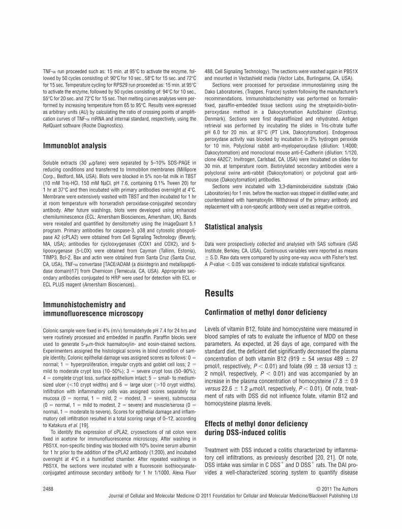
severity that is correlated with histological lesions [21]. Maximumseverity of colonic inflammation was reached at 6 days after initi-ation of DSS treatment. As expected, the DAI was higher in the CDSS� group than in the C DSS� group (P � 0.05). Importantly,methyl donor deficient diet further aggravated the severity of coli-tis induced by DSS, as reflected by the dramatically higher DAI inthe D DSS� group compared with the C DSS� group (P � 0.01,Fig. 1). There was no difference in terms of DAI when comparingthe D DSS� group with the C DSS� group. The total score forepithelial damage and inflammatory cell infiltration was estimatedto 0.0 � 0.0, 0.0 � 0.0, 2.0 � 0.5 and 5.7 � 0.6 for the C DSS�,D DSS�, C DSS� and D DSS� groups, respectively, showing adramatic increase of the score in the deficient rats exposed toDSS, compared to the other groups (P � 0.001).
Oxidative stress markers
In animals drinking tap water, levels of both Cu/Zn SOD and MnSOD activities were significantly higher in the colon of rat pups ofthe D/DSS� group compared to the C DSS� group, suggesting thatmethyl donor deficiency promotes oxidative stress under physiolog-ical conditions (Table 1). Methyl donor deficiency did not influenceSOD activity after the rats were treated with DSS. There was no dif-ference in GPX activity between the four groups of rats (Table 1).
Apoptosis
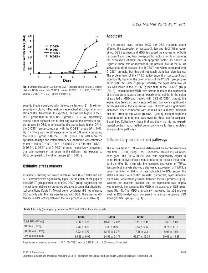
At the protein level, neither MDD nor DSS treatment aloneaffected the expression of caspase-3, Bax and Bcl2. When com-bined, DSS treatment and MDD decreased the expression of bothcaspase-3 and Bax, two pro-apoptotic factors, while increasingthe expression of Bcl2, an anti-apoptotic factor. As shown in Figure 2, there was an increase in the protein level of the 17 kDactive subunit of caspase-3 in D DSS� rats when compared withC DSS� animals, but this did not reach statistical significance.The protein level of the 17 kD active subunit of caspase-3 wassignificantly higher in the colon of rats of the C/DSS� group com-pared with the D/DSS� group. Similarly, the expression level ofBax was lower in the D/DSS� group than in the C/DSS� group(Fig. 2), indicating that MDD may further decrease the expressionof pro-apoptotic factors during experimental colitis. In the colonof rats fed a MDD and treated with DSS (D DSS� group), theexpression levels of both caspase-3 and Bax were significantlydecreased while the expression level of Bcl2 was significantlyincreased when compared with animals fed a methyl-deficientdiet but drinking tap water (D DSS� group), even though themagnitude of the difference was lower for Bcl2 than for caspase-3 and Bax. Collectively, these findings show that during experi-mental colitis in rats, methyl donor deficiency further stimulatesanti-apoptotic pathways.
Inflammatory mediators and pathways
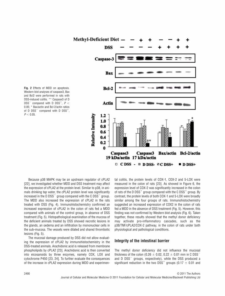
The mRNA level of TNF-� was determined by semi-quantitativereal time RT-PCR, using PR29 (Ribosomal protein 29) as refer-ence gene. The TNF-� mRNA level was significantly higher incolon from methyl-deficient rats compared to the rats fed a stan-dard diet (Fig. 3). In line with the increased expression of TNF-�,Western blot analysis showed a decreased expression of TIMP3, apotent inhibitor of TNF-� in rats subjected to DSS and/or theMDD, compared with control animals. By contrast, expression lev-els of TACE were broadly similar between the four groups (Fig. 3).Western blot analysis revealed that the expression level of p38was markedly increased by the MDD in the absence of DSS treat-ment (Fig. 4). The MDD dramatically increased the p38 proteinlevel in DSS-treated rats, compared to animals receiving DSSalone (C/DSS� group) (Fig. 4).
Fig. 1 Effects of MDD on DAI during DSS�-induced colitis in rats. Deficientdiet and DSS-treated rats *C DSS� versus C DSS�, P � 0.05; **D DSS�
versus C DSS�, P � 0.01, ANOVA, Fisher test.
Table 1 Activity (per mg or g protein) of SOD and GPX in the colon of rats
C/DSS� D/DSS� C/DSS� D/DSS�
Total SOD (UI/mg) 7.98 � 1.85 13.46 � 7.01* 8.11 � 2.41 7.65 � 1.08SOD Mn (UI/mg) 0.76 � 0.23 1.05 � 0.27* 0.53 � 0.12 0.74 � 0.17SOD Cu/Zn (UI/mg) 7.22 � 1.73 12.42 � 6.77* 7.58 � 2.3 6.91 � 1.01GPX (µmol/min/g) 64.86 � 6.95 84.55 � 31.77 66.97 � 15.33 69.03 � 14.88
Results are expressed as mean � S.D. *D DSS� versus C DSS�, P � 0.05, ANOVA, Fisher test.
2490 © 2011 The AuthorsJournal of Cellular and Molecular Medicine © 2011 Foundation for Cellular and Molecular Medicine/Blackwell Publishing Ltd
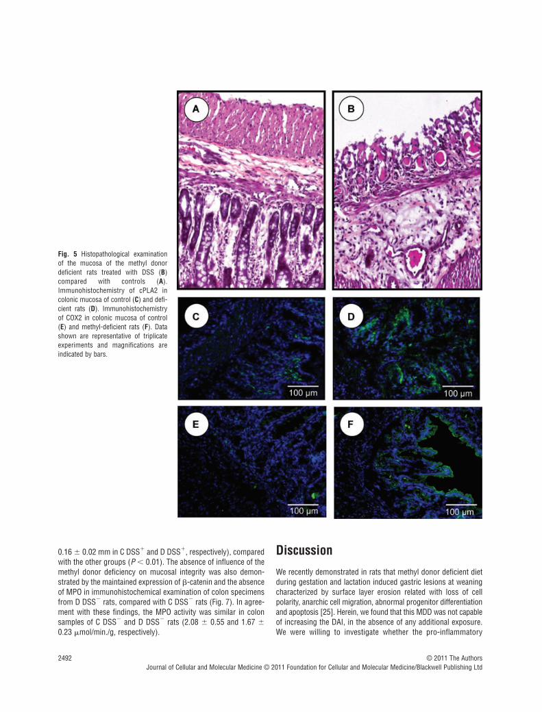
Because p38 MAPK may be an upstream regulator of cPLA2[22], we investigated whether MDD and DSS treatment may affectthe expression of cPLA2 at the protein level. Similar to p38, in ani-mals drinking tap water, the cPLA2 protein level was significantlyincreased in the D DSS� group compared with the C DSS� group.The MDD also increased the expression of cPLA2 in the ratstreated with DSS (Fig. 4). Immunohistochemistry confirmed anincreased expression of cPLA2 in the colon of rats fed a MDDcompared with animals of the control group, in absence of DSStreatment (Fig. 5). Histopathological examination of the mucosa ofthe deficient animals treated by DSS showed necrotic lesions inthe glands, an oedema and an infiltration by mononuclear cells inthe sub-mucosa. The vessels were dilated and shared thromboticlesions (Fig. 5).
The mucosal damage produced by DSS did not allow evaluat-ing the expression of cPLA2 by immunohistochemistry in theDSS-treated animals. Arachidonic acid is released from membranephospholipids by cPLA2 [23]. Arachidonic acid is then convertedinto eicosanoids by three enzymes, namely COX, LOX andcytochrome P450 [23, 24]. To further evaluate the consequencesof the increase in cPLA2 expression during MDD and experimen-
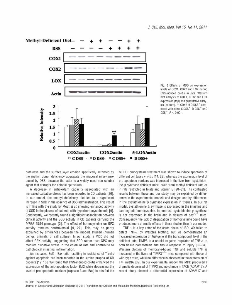
tal colitis, the protein levels of COX-1, COX-2 and 5-LOX weremeasured in the colon of rats [23]. As showed in Figure 6, theexpression level of COX-2 was significantly increased in the colonof rats of the D DSS� group compared with the C DSS� group. Bycontrast, the protein levels of both COX-1 and 5-LOX were broadlysimilar among the four groups of rats. Immunohistochemistrysuggested an increased expression of COX2 in the colon of ratsfed a MDD in the absence of DSS treatment (Fig. 5). However, thisfinding was not confirmed by Western blot analysis (Fig. 6). Takentogether, these results showed that the methyl donor deficiencymay activate pro-inflammatory cascades, such as thep38/TNF/cPLA2/COX-2 pathway, in the colon of rats under bothphysiological and pathological conditions.
Integrity of the intestinal barrier
The methyl donor deficiency did not influence the mucosalthickness of the colon (0.26 � 0.02, 0.22 � 0.01 mm in C DSS�
and D DSS� groups, respectively), while the DSS produced asignificant reduction in the two DSS� groups (0.17 � 0.01 and
Fig. 2 Effects of MDD on apoptosis.Western blot analyses of caspase3, Baxand Bcl2 were performed in rats withDSS-induced colitis. ** Caspase3 of DDSS� compared with D DSS�, P �
0.05; * Bax/actin and Bcl-2/actin ratiosof D DSS� compared with D DSS�, P � 0.05.
J. Cell. Mol. Med. Vol 15, No 11, 2011
2491© 2011 The AuthorsJournal of Cellular and Molecular Medicine © 2011 Foundation for Cellular and Molecular Medicine/Blackwell Publishing Ltd
Fig. 3 Effects of MDD on expressionlevels of TNF, TIMP3 and TACE in DSS-induced colitis in rats. Western blotanalysis of TIMP3 and TACE expression(top and bottom right) and mRNA levelof TNF determined by semi-quantitativereal time RT-PCR, using PR29 as refer-ence gene (bottom left). *TNF of CDSS� compared with D DSS�, P �
0.05; **TIMP3 of C DSS� comparedwith either D DSS�, C DSS� or DDSS�, P � 0.01.
Fig. 4 Effects of MDD on expressionlevels of p38 and cPLA2 during DSS-induced colitis in rats. Western blotanalysis of p38 and cPLA2 expression(top) and quantitative analysis (bot-tom). *p38 and cPLA2 of C DSS� com-pared with D DSS�, P � 0.05; **p38 and cPLA2 of D DSS� comparedwith either C DSS� or D DSS�, P �
0.01.
2492 © 2011 The AuthorsJournal of Cellular and Molecular Medicine © 2011 Foundation for Cellular and Molecular Medicine/Blackwell Publishing Ltd
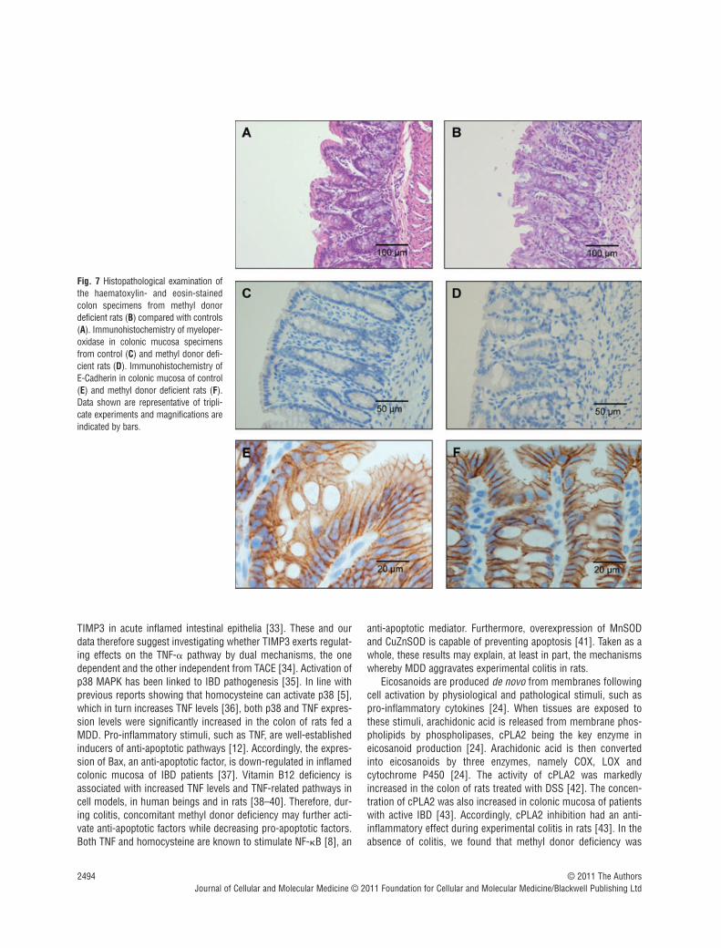
0.16 � 0.02 mm in C DSS� and D DSS�, respectively), comparedwith the other groups (P � 0.01). The absence of influence of themethyl donor deficiency on mucosal integrity was also demon-strated by the maintained expression of -catenin and the absenceof MPO in immunohistochemical examination of colon specimensfrom D DSS� rats, compared with C DSS� rats (Fig. 7). In agree-ment with these findings, the MPO activity was similar in colonsamples of C DSS� and D DSS� rats (2.08 � 0.55 and 1.67 �0.23 �mol/min./g, respectively).
Discussion
We recently demonstrated in rats that methyl donor deficient dietduring gestation and lactation induced gastric lesions at weaningcharacterized by surface layer erosion related with loss of cellpolarity, anarchic cell migration, abnormal progenitor differentiationand apoptosis [25]. Herein, we found that this MDD was not capableof increasing the DAI, in the absence of any additional exposure.We were willing to investigate whether the pro-inflammatory
Fig. 5 Histopathological examination of the mucosa of the methyl donordeficient rats treated with DSS (B)compared with controls (A).Immunohistochemistry of cPLA2 incolonic mucosa of control (C) and defi-cient rats (D). Immunohistochemistryof COX2 in colonic mucosa of control(E) and methyl-deficient rats (F). Datashown are representative of triplicateexperiments and magnifications areindicated by bars.
J. Cell. Mol. Med. Vol 15, No 11, 2011
2493© 2011 The AuthorsJournal of Cellular and Molecular Medicine © 2011 Foundation for Cellular and Molecular Medicine/Blackwell Publishing Ltd
pathways and the surface layer erosion specifically activated bythe methyl donor deficiency aggravate the mucosal injury pro-duced by DSS, because the latter is a widely used non solubleagent that disrupts the colonic epithelium.
A decrease in antioxidant capacity associated with anincreased oxidative stress has been reported in CD patients [26].In our model, the methyl deficiency diet led to a significantincrease in SOD in the absence of DSS administration. This resultis in line with the study by Moat et al. showing enhanced activityof SOD in the plasma of patients with hyperhomocysteinemia [9].Consistently, we recently found a significant association betweenclinical activity and the SOD activity in CD patients carrying theMTRR 66AA genotype [3]. The effect of homocysteine on GPXactivity remains controversial [9, 27]. This may be partlyexplained by differences between the models studied (humanbeings, animals, or cell culture). In our study, a MDD did notaffect GPX activity, suggesting that SOD rather than GPX maymediate oxidative stress in the colon of rats and contribute topathological intestinal inflammation.
An increased Bcl2 : Bax ratio resulting in resistance of T cellsagainst apoptosis has been reported in the lamina propria of CDpatients [12, 13]. We found that DSS-induced colitis enhanced theexpression of the anti-apoptotic factor Bcl2 while decreasing thelevel of pro-apoptotic markers (capsase-3 and Bax) in rats fed the
MDD. Homocysteine treatment was shown to induce apoptosis ofdifferent cell types in vitro [14, 28], whereas the expression level ofpro-apoptotic markers was increased in the liver from cystathion-ine synthase-deficient mice, brain from methyl-deficient rats orin rats restricted in folate and vitamin E [28–31]. The contrastedresults between these and our study may be explained by differ-ences in the experimental models and designs and by differencesin the cystathionine synthase expression in tissues. In our ratmodel, cystathionine synthase is expressed in the intestine andcan degrade homocysteine. In contrast, cystathionine synthaseis not expressed in the brain and in tissues of cbs�/� mice.Consequently, the lack of degradation of homocysteine could haveproduced more dramatic effects in these studies than in our model.
TNF-� is a key actor of the acute phase of IBD. We failed todetect TNF-� by Western blotting, but we demonstrated anincreased expression of TNF gene at the transcriptional level in thedeficient rats. TIMP3 is a crucial negative regulator of TNF-� inboth tissue homeostasis and tissue response to injury [32–34].Western blotting of membrane-bound TNF and soluble TNF isincreased in the livers of TIMP3�/� mice compared with those ofwild-type mice, while no difference is observed in the expression ofTNF mRNA [32]. In our experimental model, the MDD produced adramatic decreased of TIMP3 and no change in TACE (ADAM17). Arecent study showed a differential expression of ADAM17 and
Fig. 6 Effects of MDD on expressionlevels of COX1, COX2 and LOX duringDSS-induced colitis in rats. Westernblot analysis of COX1, COX2 and LOXexpression (top) and quantitative analy-sis (bottom). ** COX2 of D DSS� com-pared with either C DSS�, D DSS� or CDSS�, P � 0.001.
2494 © 2011 The AuthorsJournal of Cellular and Molecular Medicine © 2011 Foundation for Cellular and Molecular Medicine/Blackwell Publishing Ltd
TIMP3 in acute inflamed intestinal epithelia [33]. These and ourdata therefore suggest investigating whether TIMP3 exerts regulat-ing effects on the TNF-� pathway by dual mechanisms, the onedependent and the other independent from TACE [34]. Activation ofp38 MAPK has been linked to IBD pathogenesis [35]. In line withprevious reports showing that homocysteine can activate p38 [5],which in turn increases TNF levels [36], both p38 and TNF expres-sion levels were significantly increased in the colon of rats fed aMDD. Pro-inflammatory stimuli, such as TNF, are well-establishedinducers of anti-apoptotic pathways [12]. Accordingly, the expres-sion of Bax, an anti-apoptotic factor, is down-regulated in inflamedcolonic mucosa of IBD patients [37]. Vitamin B12 deficiency isassociated with increased TNF levels and TNF-related pathways incell models, in human beings and in rats [38–40]. Therefore, dur-ing colitis, concomitant methyl donor deficiency may further acti-vate anti-apoptotic factors while decreasing pro-apoptotic factors.Both TNF and homocysteine are known to stimulate NF-B [8], an
anti-apoptotic mediator. Furthermore, overexpression of MnSODand CuZnSOD is capable of preventing apoptosis [41]. Taken as awhole, these results may explain, at least in part, the mechanismswhereby MDD aggravates experimental colitis in rats.
Eicosanoids are produced de novo from membranes followingcell activation by physiological and pathological stimuli, such aspro-inflammatory cytokines [24]. When tissues are exposed tothese stimuli, arachidonic acid is released from membrane phos-pholipids by phospholipases, cPLA2 being the key enzyme ineicosanoid production [24]. Arachidonic acid is then convertedinto eicosanoids by three enzymes, namely COX, LOX andcytochrome P450 [24]. The activity of cPLA2 was markedlyincreased in the colon of rats treated with DSS [42]. The concen-tration of cPLA2 was also increased in colonic mucosa of patientswith active IBD [43]. Accordingly, cPLA2 inhibition had an anti-inflammatory effect during experimental colitis in rats [43]. In theabsence of colitis, we found that methyl donor deficiency was
Fig. 7 Histopathological examination ofthe haematoxylin- and eosin-stainedcolon specimens from methyl donordeficient rats (B) compared with controls(A). Immunohistochemistry of myeloper-oxidase in colonic mucosa specimensfrom control (C) and methyl donor defi-cient rats (D). Immunohistochemistry ofE-Cadherin in colonic mucosa of control(E) and methyl donor deficient rats (F).Data shown are representative of tripli-cate experiments and magnifications areindicated by bars.
J. Cell. Mol. Med. Vol 15, No 11, 2011
2495© 2011 The AuthorsJournal of Cellular and Molecular Medicine © 2011 Foundation for Cellular and Molecular Medicine/Blackwell Publishing Ltd
associated with an increase in cPLA2 levels in the rat colon.Similar to apoptosis, the increase in p38 levels associated withMDD may contribute to high cPLA2 in these animals, as sug-gested by previous reports [21–23]. Therefore, a MDD may fur-ther enhance colonic lesions induced by DSS treatment in rats.
COX and LOX are two enzymes responsible for the synthesis ofeicosanoids, using arachidonic acid as substrate [24]. Two COXisoforms have been identified, COX-1, the constitutive form andCOX-2, the inducible form [24]. The expression of 5-LOX andCOX-1 was unchanged in the colon of IBD patients [44].Consistently, DSS treatment and/or methyl donor deficiency hadno impact on 5-LOX and COX-1 levels in our animal model. COX-2 is undetectable in normal ileum or colon of IBD patients and, butit is induced in apical epithelial cells of inflamed foci in IBDpatients [45]. Hendel et al. found a significant correlation betweenendoscopic activity and COX-2 mRNA levels [46]. COX-2expression level was dramatically increased in the colon ofmethyl-deficient rats treated with DSS. This elevation of COX-2concentration in the rat colon may occur through direct and indi-rect mechanisms. High TNF levels within the colonic mucosa ofrats may directly enhance COX-2 expression [47]. In addition, pro-inflammatory stimuli, such as p38 and TNF, may increase therelease of arachidonic acid from membrane phospholipids, whichin turns serves as substrate for LOX, COX-1 and COX-2 [24].Overall, combined elevation of cPLA2 and COX-2 may increase thesynthesis of prostaglandins and thromboxanes and contribute tomaintenance of mucosal inflammation in the colon of IBD patients.Interestingly, platelet incubation with homocysteine significantlyincreased thromboxane levels [48], further supporting a role formethyl donor deficiency in the development and/or maintenanceof colonic inflammation in rats. From a clinical point of view,beside their direct role in the severity of the disease, inflammationpathways are key players in the development of IBD-associatedcolon cancer [49]. The decreased expression of pro-apoptoticmarkers in colon mucosa of the deficient animals has also to beconsidered in regards to the controversy that exists between thefolate status and the risk for colon cancer [50–52]. The mucosalintegrity and the transepithelial migration of neutrophils are
interacting parameters to be considered in the mechanismsbeyond the relations between methyl donor deficiency, inflamma-tion and the severity of the mucosal injury produced by DSS. Theapical junctional complex is the main component of the intestinalbarrier; it consists of tight and adherens junctions that are com-promised in IBD, with disappearance of key proteins such asoccluding and E-cadherin [53] and infiltration of neutrophils [54].These mechanisms had a limited influence, if any, in our model,since we observed a maintained thickness and integrity of thecolon mucosa in the DSS� methyl-deficient rats, with no disap-pearance of E-cadherin expression and no transepithelial migra-tion of neutrophils.
In conclusion, a methyl deficiency diet aggravate experimentalcolitis in rats by promoting oxidative stress, decreasing cell apop-tosis and by activating inter-related pro-inflammatory mecha-nisms, including the TNF pathway, p38, cPLA2 and COX-2.However, the validity of these findings needs to be investigated byevaluating the clinical implications. In IBD patients, homocysteinelevels were positively correlated with activity, number of flares andduration of IBD [52]. Therefore, screening for folate and vitaminB12 deficiencies aimed at restoring a normal methyl donor statusmight improve the management of IBD patients, possibly modify-ing the clinical course of the disease. Further studies are nowrequired to assess the benefit/risk ratio for treating the patientswith B12 and/or folate deficit in human IBD.
Acknowledgement
Institutional grants were obtained from the region Lorraine and fromInserm, France.
Conflict of interest
The authors confirm that there are no conflicts of interest.
References
1. Xavier RJ, Podolsky DK. Unravelling thepathogenesis of inflammatory bowel dis-ease. Nature. 2007; 448: 427–34.
2. Peyrin-Biroulet L, Desreumaux P,Sandborn WJ, et al. Crohn’s disease:beyond antagonists of tumour necrosisfactor. Lancet. 2008; 372: 67–81.
3. Peyrin-Biroulet L, Gueant-Rodriguez RM,Chen M, et al. Association of MTRR66A�G polymorphism with superoxidedismutase and disease activity in patientswith Crohn’s disease. Am J Gastroenterol.2008; 103: 399–406.
4. Peyrin-Biroulet L, Rodriguez-Gueant RM,Chamaillard M, et al. Vascular and cellu-lar stress in inflammatory bowel disease:revisiting the role of homocysteine. Am JGastroenterol. 2007; 102: 1108–15.
5. Danese S, Sgambato A, Papa A, et al.Homocysteine triggers mucosal microvas-cular activation in inflammatory boweldisease. Am J Gastroenterol. 2005; 100:886–95.
6. Morgenstern I, Raijmakers MT, PetersWH, et al. Homocysteine, cysteine, andglutathione in human colonic mucosa:
elevated levels of homocysteine in patientswith inflammatory bowel disease. Dig DisSci. 2003; 48: 2083–90.
7. Poddar R, Sivasubramanian N, DiBelloPM, et al. Homocysteine induces expres-sion and secretion of monocyte chemoat-tractant protein-1 and interleukin-8 inhuman aortic endothelial cells: implica-tions for vascular disease. Circulation.2001; 103: 2717–23.
8. Collins T, Cybulsky MI. NF-kappaB: pivotalmediator or innocent bystander in athero-genesis? J Clin Invest. 2001; 107: 255–64.
2496 © 2011 The AuthorsJournal of Cellular and Molecular Medicine © 2011 Foundation for Cellular and Molecular Medicine/Blackwell Publishing Ltd
9. Moat SJ, Bonham JR, Cragg RA, et al.Elevated plasma homocysteine elicits anincrease in antioxidant enzyme activity.Free Radic Res. 2000; 32: 171–9.
10. Wang XL, Duarte N, Cai H, et al.Relationship between total plasma homo-cysteine, polymorphisms of homocysteinemetabolism related enzymes, risk factorsand coronary artery disease in theAustralian hospital-based population.Atherosclerosis. 1999; 146: 133–40.
11. Nishio E, Watanabe Y. Homocysteine as amodulator of platelet-derived growth fac-tor action in vascular smooth muscle cells:a possible role for hydrogen peroxide. Br JPharmacol. 1997; 122: 269–74.
12. Mudter J, Neurath MF. Apoptosis of Tcells and the control of inflammatorybowel disease: therapeutic implications.Gut. 2007; 56: 293–303.
13. Ina K, Itoh J, Fukushima K, et al.Resistance of Crohn’s disease T cells tomultiple apoptotic signals is associatedwith a Bcl-2/Bax mucosal imbalance. JImmunol. 1999; 163: 1081–90.
14. Zhang C, Cai Y, Adachi MT, et al.Homocysteine induces programmed celldeath in human vascular endothelial cellsthrough activation of the unfolded proteinresponse. J Biol Chem. 2001; 276:35867–74.
15. Stevceva L, Pavli P, Husband A, et al.Dextran sulphate sodium-induced colitis isameliorated in interleukin 4 deficient mice.Genes Immun. 2001; 2: 309–16.
16. Cooper HS, Murthy S, Kido K, et al.Dysplasia and cancer in the dextran sulfatesodium mouse colitis model. Relevance tocolitis-associated neoplasia in the human:a study of histopathology, B-catenin andp53 expression and the role of inflamma-tion. Carcinogenesis. 2000; 21: 757–68.
17. Paglia DE, Valentine WN. Studies on thequantitative and qualitative characteriza-tion of erythrocyte glutathione peroxidase.J Lab Clin Med. 1967; 70: 158–69.
18. Xia Y, Zweier JL. Measurement ofmyeloperoxidase in leukocyte-containingtissues. Anal Biochem. 1997; 245: 93–6.
19. Katakura K, Lee J, Rachmilewitz D, et al.Toll-like receptor 9-induced type I IFN pro-tects mice from experimental colitis. J ClinInvest. 2005; 115: 695–702.
20. Okayasu I, Hatakeyama S, Yamada M, et al. A novel method in the induction ofreliable experimental acute and chroniculcerative colitis in mice. Gastroenterology.1990; 98: 694–702.
21. Melgar S, Karlsson A, Michaelsson E.Acute colitis induced by dextran sulfate
sodium progresses to chronicity inC57BL/6 but not in BALB/c mice: correla-tion between symptoms and inflammation.Am J Physiol Gastrointest Liver Physiol.2005; 288: G1328–38.
22. Murthy SN, Cooper HS, Shim H, et al.Treatment of dextran sulfate sodium-inducedmurine colitis by intracolonic cyclosporin.Dig Dis Sci. 1993; 38: 1722–34.
23. Borsch-Haubold AG, Ghomashchi F,Pasquet S, et al. Phosphorylation ofcytosolic phospholipase A2 in platelets ismediated by multiple stress-activated pro-tein kinase pathways. Eur J Biochem.1999; 265: 195–203.
24. Harizi H, Corcuff JB, Gualde N.Arachidonic-acid-derived eicosanoids:roles in biology and immunopathology.Trends Mol Med. 2008; 14: 461–9.
25. Bossenmeyer-Pourié C, Blaise S, PouriéG, et al. Methyl donor deficiency affectsfetal programming of gastric ghrelin cellorganization and function in the rat. Am JPathol. 2010; 176: 270–7.
26. Maor I, Rainis T, Lanir A, et al. Oxidativestress, inflammation and neutrophilsuperoxide release in patients with Crohn’sdisease: distinction between active andnon-active disease. Dig Dis Sci. 2008; 53:2208–14.
27. Handy DE, Zhang Y, Loscalzo J.Homocysteine down-regulates cellular glu-tathione peroxidase (GPx1) by decreasingtranslation. J Biol Chem. 2005; 280:15518–25.
28. Mangiagalli A, Samuele A, ArmenteroMT, et al. Effects of homocysteine onapoptosis-related proteins and anti-oxidantsystems in isolated human lymphocytes.Eur J Biochem. 2004; 271: 1671–6.
29. Robert K, Nehme J, Bourdon E, et al.Cystathionine beta synthase deficiencypromotes oxidative stress, fibrosis, andsteatosis in mice liver. Gastroenterology.2005; 128: 1405–15.
30. Blaise S, Alberto JM, Nedelec E, et al.Mild neonatal hypoxia exacerbates theeffects of vitamin-deficient diet on homo-cysteine metabolism in rats. Pediatr Res.2005; 57: 777–82.
31. Vijayalakshhmi B, Sesikeran B,Udaykumar P, et al. Effects of vitaminrestriction and supplementation on ratintestinal epithelial cell apoptosis. FreeRadic Biol Med. 2005; 38: 1614–24.
32. Mohammed FF, Smookler DS, Taylor SE,et al. Abnormal TNF activity in Timp3�/�mice leads to chronic hepatic inflammationand failure of liver regeneration. Nat Genet.2004; 36: 969–77.
33. Cesaro A, Abakar-Mahamat A, Brest P, et al. Differential expression and regula-tion of ADAM17 and TIMP3 in acuteinflamed intestinal epithelia. Am J PhysiolGastrointest Liver Physiol. 2009; 296:G1332–43.
34. Black RA. TIMP3 checks inflammation.Nat Genet. 2004; 36: 934–5.
35. Waetzig GH, Seegert D, Rosenstiel P, et al. p38 mitogen-activated proteinkinase is activated and linked to TNF-alphasignaling in inflammatory bowel disease. J Immunol. 2002; 168: 5342–51.
36. Kyriakis JM, Avruch J. Mammalianmitogen-activated protein kinase signaltransduction pathways activated by stressand inflammation. Physiol Rev. 2001; 81:807–69.
37. Iimura M, Nakamura T, Shinozaki S, et al.Bax is downregulated in inflamed colonicmucosa of ulcerative colitis. Gut. 2000; 47:228–35.
38. Peracchi M, Bamonti Catena F, et al.Human cobalamin deficiency: alterations inserum tumour necrosis factor-alpha andepidermal growth factor. Eur J Haematol.2001; 67: 123–7.
39. Hsu-Battaglia SF, Akchiche N, Noel N, et al. Vitamin B12 deficiency reducesproliferation and promotes differentiationof neuroblastoma cells and upregulatesPP2A, proNGF and TACE. Proc Natl AcadSci USA. 2009; 106: 21930–5.
40. Scalabrino G. The multi-faceted basis ofvitamin B12 (cobalamin) neurotrophism inadult central nervous system: lessonslearned from its deficiency. ProgNeurobiol. 2009; 88: 203–20.
41. Zemlyak I, Brooke SM, Singh MH, et al.Effects of overexpression of antioxidantson the release of cytochrome c and apop-tosis-inducing factor in the model ofischemia. Neurosci Lett. 2009; 453:182–5.
42. Tomita Y, Jyoyama H, Kobayashi M, et al. Role of group IIA phospholipase A2in rat colitis induced by dextran sulfatesodium. Eur J Pharmacol. 2003; 472:147–58.
43. Minami T, Shinomura Y, Miyagawa J, et al. Immunohistochemical localization ofgroup II phospholipase A2 in colonicmucosa of patients with inflammatorybowel disease. Am J Gastroenterol. 1997;92: 289–92.
44. Krimsky M, Yedgar S, Aptekar L, et al.Amelioration of TNBS-induced coloninflammation in rats by phospholipase A2inhibitor. Am J Physiol Gastrointest LiverPhysiol. 2003; 285: G586–92.
J. Cell. Mol. Med. Vol 15, No 11, 2011
2497© 2011 The AuthorsJournal of Cellular and Molecular Medicine © 2011 Foundation for Cellular and Molecular Medicine/Blackwell Publishing Ltd
45. Singer II, Kawka DW, Schloemann S, et al. Cyclooxygenase 2 is induced incolonic epithelial cells in inflammatorybowel disease. Gastroenterology. 1998;115: 297–306.
46. Hendel J, Nielsen OH. Expression ofcyclooxygenase-2 mRNA in active inflam-matory bowel disease. Am J Gastroenterol.1997; 92: 1170–3.
47. Maier JA, Hla T, Maciag T.Cyclooxygenase is an immediate-earlygene induced by interleukin-1 in humanendothelial cells. J Biol Chem. 1990; 265:10805–8.
48. Leoncini G, Bruzzese D, Signorello MG.Activation of p38 MAPKinase/cPLA2 path-
way in homocysteine-treated platelets. J Thromb Haemost. 2006; 4: 209–16.
49. Fantini MC, Pallone F. Cytokines: fromgut inflammation to colorectal cancer. Curr Drug Targets. 2008; 9: 375–80.
50. Van Guelpen B, Hultdin J, Johansson I,et al. Low folate levels may protect againstcolorectal cancer. Gut. 2006; 55: 1461–6.
51. Jang H, Mason JB, Choi SW. Genetic andepigenetic interactions between folate andaging in carcinogenesis. J Nutr. 2005; 135:2967S–71S.
52. Crott JW, Liu Z, Keyes MK, et al.Moderate folate depletion modulates theexpression of selected genes involved incell cycle, intracellular signaling and folate
uptake in human colonic epithelial celllines. J Nutr Biochem. 2008; 19: 328–35.
52. Drzewoski J, Gasiorowska A, Malecka-Panas E, et al. Plasma total homocysteinein the active stage of ulcerative colitis. JGastroenterol Hepatol. 2006; 21: 739–43.
53. Bruewer M, Samarin S, Nusrat A.Inflammatory bowel disease and the apicaljunctional complex. Ann N Y Acad Sci.2006; 1072: 242–52.
54. Kucharzik T, Walsh SV, Chen J, et al.Neutrophil transmigration in inflammatorybowel disease is associated with differen-tial expression of epithelial intercellularjunction proteins. Am J Pathol. 2001; 159:2001–9.