Embed Size (px)
Citation preview
1
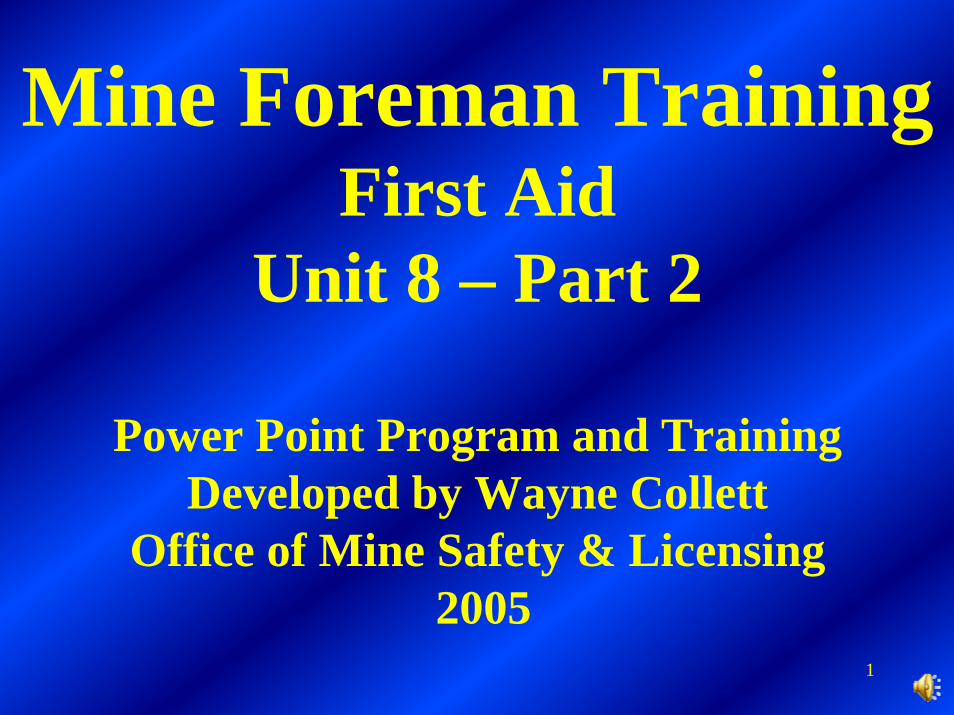
Mine Foreman TrainingFirst Aid
Unit 8 – Part 2
Power Point Program and TrainingDeveloped by Wayne Collett
Office of Mine Safety & Licensing2005
2
The following program was developed for Mine Emergency Technician (MET) training, which is the basis for this Mine Foreman first aid training. It provides a pictorial overview onbleeding/bandaging and splinting.
3
First Aid
Bleeding Controland
BandagingPrepared by Wayne Collett
Harlan District Instructor January 3, 2003
4
Your safety must be your
first consideration

5
Diseases can be transmitted through
body fluids
6
Body Substance Isolation (BSI) Precautions:
• Prior to examination or treatment of any patient you must take proper BSI precautions. During “Patient Assessment”, the MET will determine the necessity and priority for performing this skill.
7
Bleeding must be controlled!
Apply direct pressure to the bleeding wound when appropriate:Use the flat part of your fingertips to apply direct pressure to the point of bleeding. If the wound is large and gaping and finger-tip pressure is not controlling bleeding, you may need to use a sterile gauze and direct hand pressure.
8
Bleeding must be controlled!
Elevate the extremity:Elevate the bleeding extremity only if there is no major injury to the underlying muscle or bone. Continue to apply direct pressure at the same time.
9
Bleeding must be controlled!Reassess the wound:Inspect the dressing to determine whether or not bleeding hasstopped. If bleeding continues through the dressing, and the dressing appears to be soaked with blood, apply additional dressing to the wound site. However, do not remove the soaked dressing. Doing so might prevent clotting and allow additional contamination.
10
Bleeding must be controlled!If bleeding continues, apply arterial pressure:If bleeding in the extremities does not stop with direct pressure and elevation, use a pressure point (the place where an artery lies over a bone close to the surface of the body). For an arm, compress the brachial artery. For a leg compress the femoral artery. Use the flat part of your fingers or the palm of your hand. Do this while you maintain direct pressure and elevation of the wound site.
11
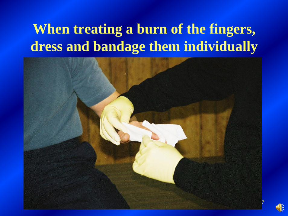
Bleeding must be controlled!
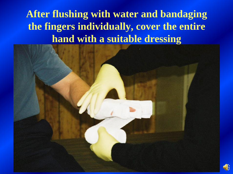
When bleeding is under control, dress and bandage properly:Do not remove the soaked dressing. Apply additional dressings if necessary and bandage in place.
12
Emergency care for an open wound:
• Expose wound - cut away any clothing so the entire wound is exposed.
• Clear wound surface - remove loose materials from wound area, but do not attempt to remove an impaled object. Leave it in the patient, because it could be controlling bleeding. Stabilize the object in place by applying bulky dressings.
13
Emergency care for an open wound:
Prevent further contamination - when possible use sterile or very clean materials. Avoid touching the dressing in the area that will come in contact with the wound. Grasp the dressing by the corner, taking it directly from its protective pack, and place it on the wound.
14
Emergency care for an open wound:
Bandage dressing in place after bleeding has been controlled - a bandage is any material that holds a dressing in place. In a “pinch”, any clean material can be used as a dressing or bandage. Assess pulse, sensation, and movement below the wound before and after bandaging.
15
Emergency care for an open wound:
• Keep patient lying still.• Reassure the patient.• Treat for shock.
16
Treat for shock.• Maintain the patient’s airway and breathing. If
possible, provide high flow oxygen via non-rebreather mask.
• Prevent any further blood loss.• Place the patient in the shock position. Have
the patient lie down and elevate the lower extremities about eight to 12 inches. If the patient has serious injuries to the pelvis, lower extremities, head, chest, abdomen, neck or spine, keep the patient supine. And…..

17
Treat for shock.
• Cover the patient with a blanket to help prevent the loss of body heat.
• Withhold all food and drink (patients in shock often vomit).
• Provide care for specific injuries as needed.
• Comfort, calm, and reassure your patient while waiting for transport.
18
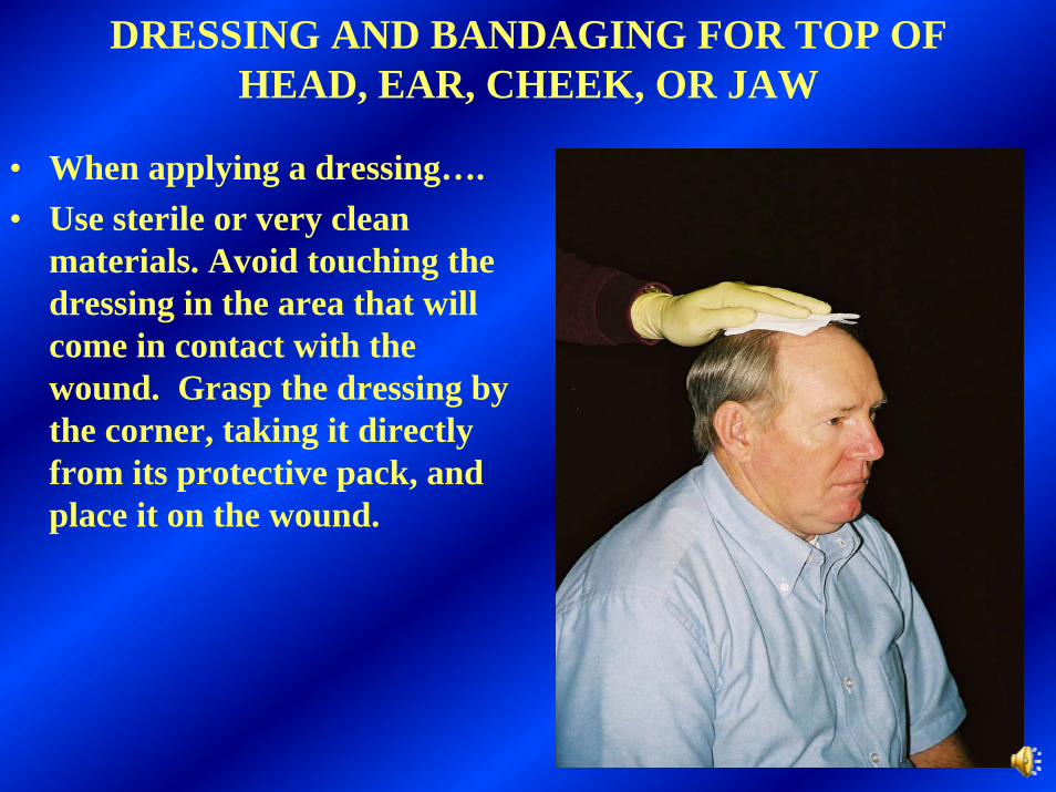
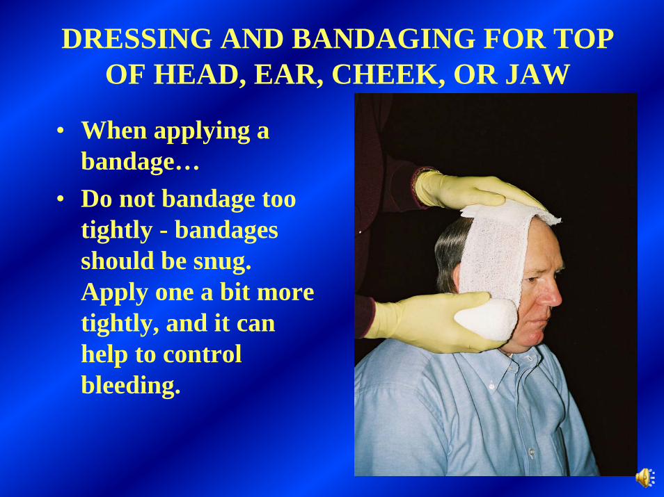
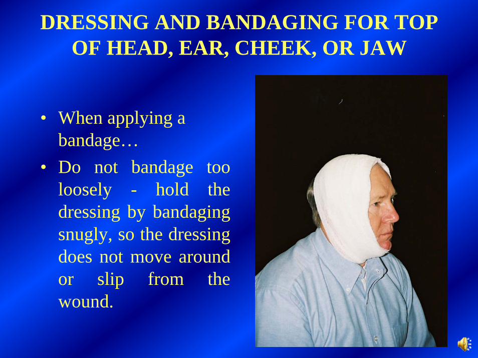
DRESSING AND BANDAGING FOR TOP OF HEAD, EAR, CHEEK, OR JAW
• When applying a dressing….• Use sterile or very clean
materials. Avoid touching the dressing in the area that will come in contact with the wound. Grasp the dressing by the corner, taking it directly from its protective pack, and place it on the wound.
19
DRESSING AND BANDAGING FOR TOP OF HEAD, EAR, CHEEK, OR JAW
• When applying a bandage…
• Do not bandage too tightly - bandages should be snug. Apply one a bit more tightly, and it can help to control bleeding.
20
DRESSING AND BANDAGING FOR TOP OF HEAD, EAR, CHEEK, OR JAW
• When applying a bandage…
• Do not bandage too loosely - hold the dressing by bandaging snugly, so the dressing does not move around or slip from the wound.
21
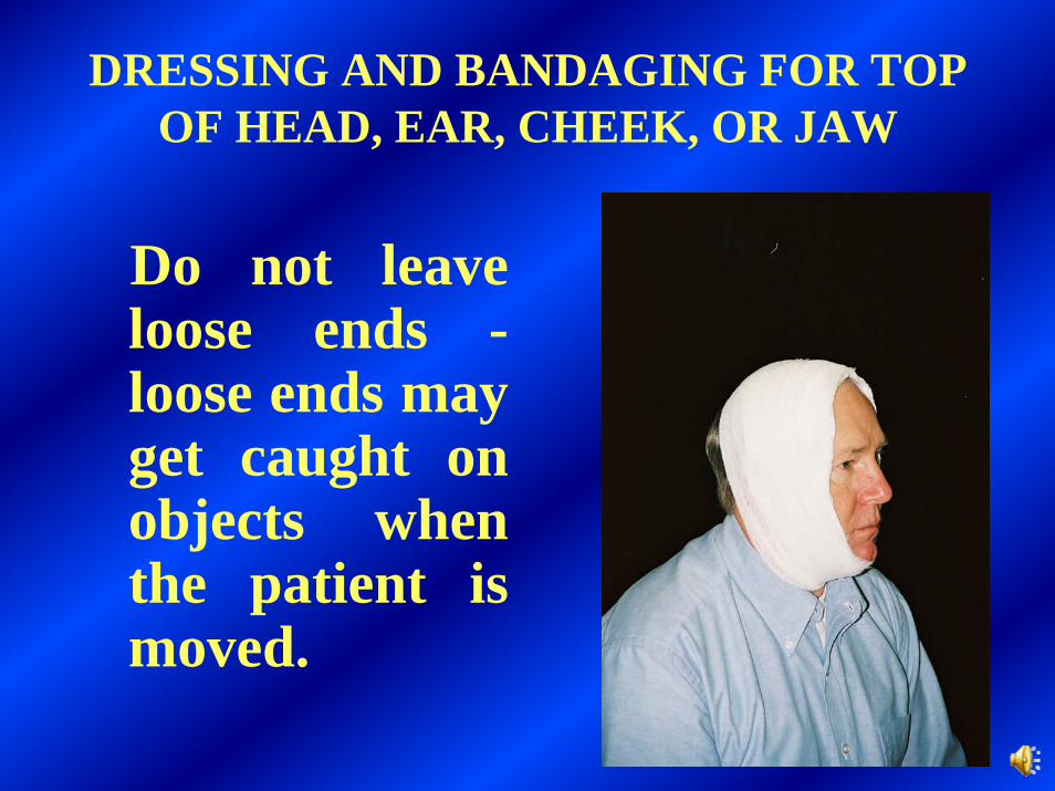
DRESSING AND BANDAGING FOR TOP OF HEAD, EAR, CHEEK, OR JAW
Do not leave loose ends -loose ends may get caught on objects when the patient is moved.
22
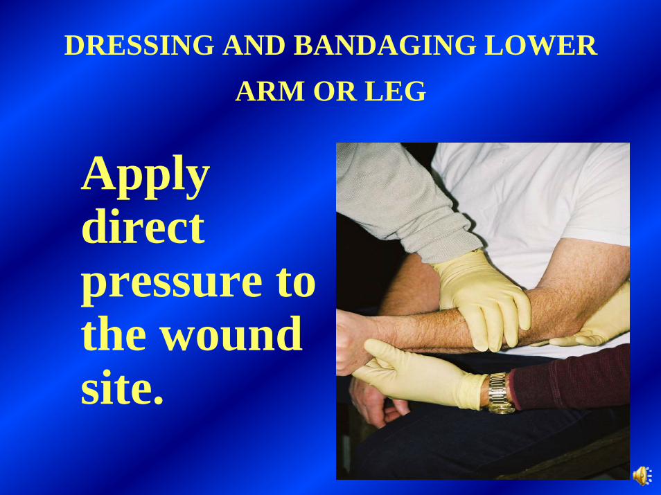
DRESSING AND BANDAGING LOWER ARM OR LEG
Apply direct pressure to the wound site.
23
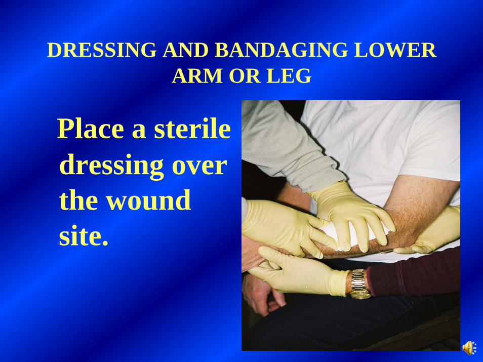
DRESSING AND BANDAGING LOWER ARM OR LEG
Place a sterile dressing over the wound site.
24
DRESSING AND BANDAGING LOWER ARM OR LEG
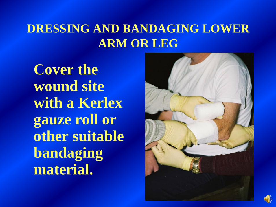
Cover the wound site with a Kerlexgauze roll or other suitable bandaging material.
25
DRESSING AND BANDAGING LOWER ARM OR LEG
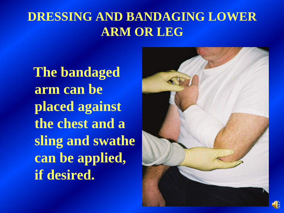
The bandaged arm can be placed against the chest and a sling and swathe can be applied, if desired.
26
Bandaging an impaled object in the eye
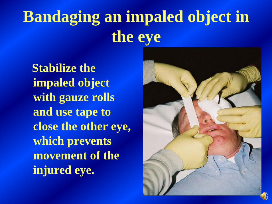
Stabilize the impaled object with gauze rolls and use tape to close the other eye, which prevents movement of the injured eye.
27
Bandaging an impaled object in the eye
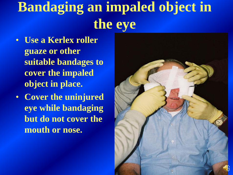
• Use a Kerlex roller guaze or other suitable bandages to cover the impaled object in place.
• Cover the uninjured eye while bandaging but do not cover the mouth or nose.
28
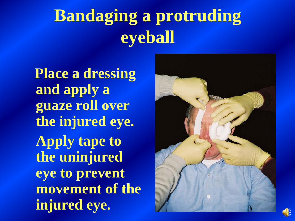
Bandaging a protruding eyeball
Place a dressing and apply a guaze roll over the injured eye.Apply tape to the uninjured eye to prevent movement of the injured eye.
29
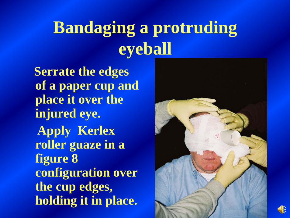
Bandaging a protruding eyeball
Serrate the edges of a paper cup and place it over the injured eye.Apply Kerlexroller guaze in a figure 8 configuration over the cup edges, holding it in place.
30
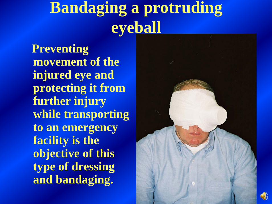
Bandaging a protruding eyeball
Preventing movement of the injured eye and protecting it from further injury while transporting to an emergency facility is the objective of this type of dressing and bandaging.

31
When treating a burn…
• Relieve the pain by flushing with lots of water.
• If possible, remove clothing and jewelry from the affected area.
• Do not attempt to clear debris from the burned area.
• Apply dressing and bandage.

32
Eye protection can prevent many injuries to the eyes. However, injuries to the eyes can occur and must be properly treated.

33
Chemical burns of the eye
• Brush off any remaining powder from the patient and flush eyes with plenty of water• It is best to have the patient lying down with the eyelids open, while flushing the eyes with plenty of water• Be sure the water flows across the eyes in large amounts

34
When treating a burn of the fingers, dress and bandage them individually

35
After flushing with water and bandaging the fingers individually, cover the entire
hand with a suitable dressing
36
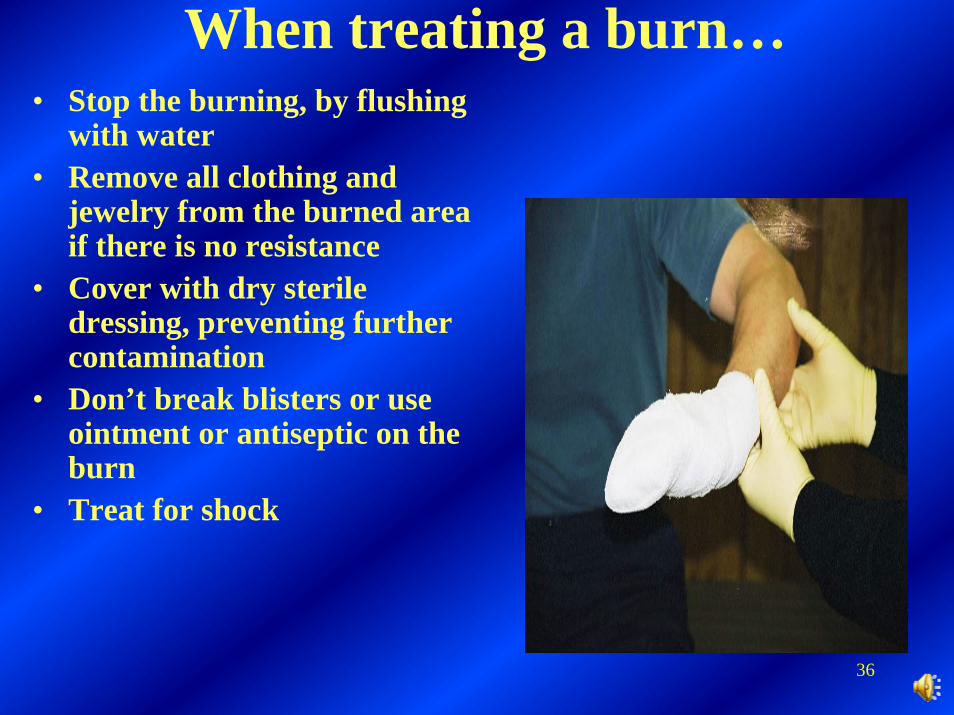
When treating a burn…• Stop the burning, by flushing
with water• Remove all clothing and
jewelry from the burned area if there is no resistance
• Cover with dry sterile dressing, preventing further contamination
• Don’t break blisters or use ointment or antiseptic on the burn
• Treat for shock
37
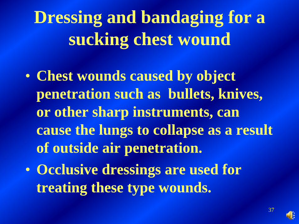
Dressing and bandaging for a sucking chest wound
• Chest wounds caused by object penetration such as bullets, knives, or other sharp instruments, can cause the lungs to collapse as a result of outside air penetration.
• Occlusive dressings are used for treating these type wounds.
38
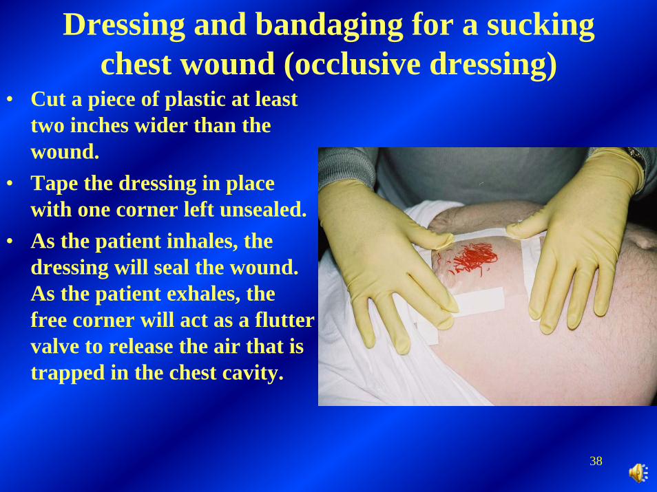
Dressing and bandaging for a sucking chest wound (occlusive dressing)
• Cut a piece of plastic at least two inches wider than the wound.
• Tape the dressing in place with one corner left unsealed.
• As the patient inhales, the dressing will seal the wound. As the patient exhales, the free corner will act as a flutter valve to release the air that is trapped in the chest cavity.
39
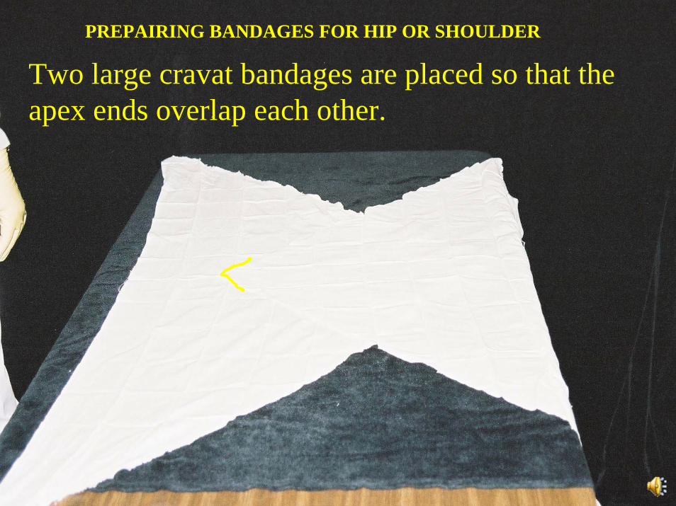
REPARING BANDAGES FOR HIP OR SHOULDER
P
PREPAIRING BANDAGES FOR HIP OR SHOULDER
Two large cravat bandages are placed so that the apex ends overlap each other.
40
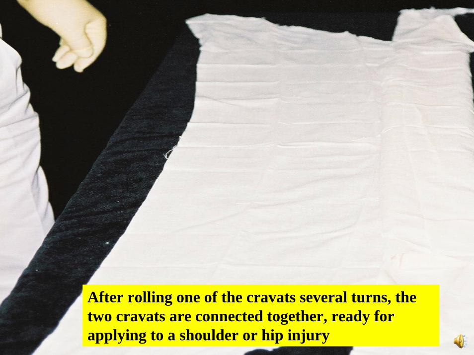
After rolling one of the cravats several turns, the two cravats are connected together, ready for applying to a shoulder or hip injury

41
Dressing and bandagingfor the Shoulder
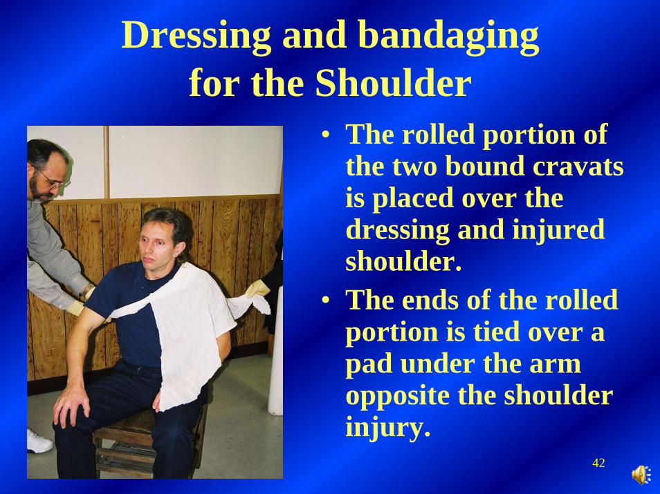
• The rolled portion of the two bound cravats is placed over the dressing and injured shoulder.
• The ends of the rolled portion are tied over a pad under the arm opposite the shoulder injury.
42
Dressing and bandagingfor the Shoulder
• The rolled portion of the two bound cravats is placed over the dressing and injured shoulder.
• The ends of the rolled portion is tied over a pad under the arm opposite the shoulder injury.
43
Dressing and bandagingfor the Shoulder
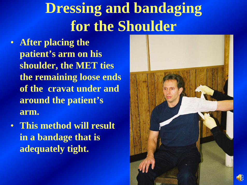
• After placing the patient’s arm on his shoulder, the MET ties the remaining loose ends of the cravat under and around the patient’s arm.
• This method will result in a bandage that is adequately tight.
44
Dressing and bandagingfor the Shoulder
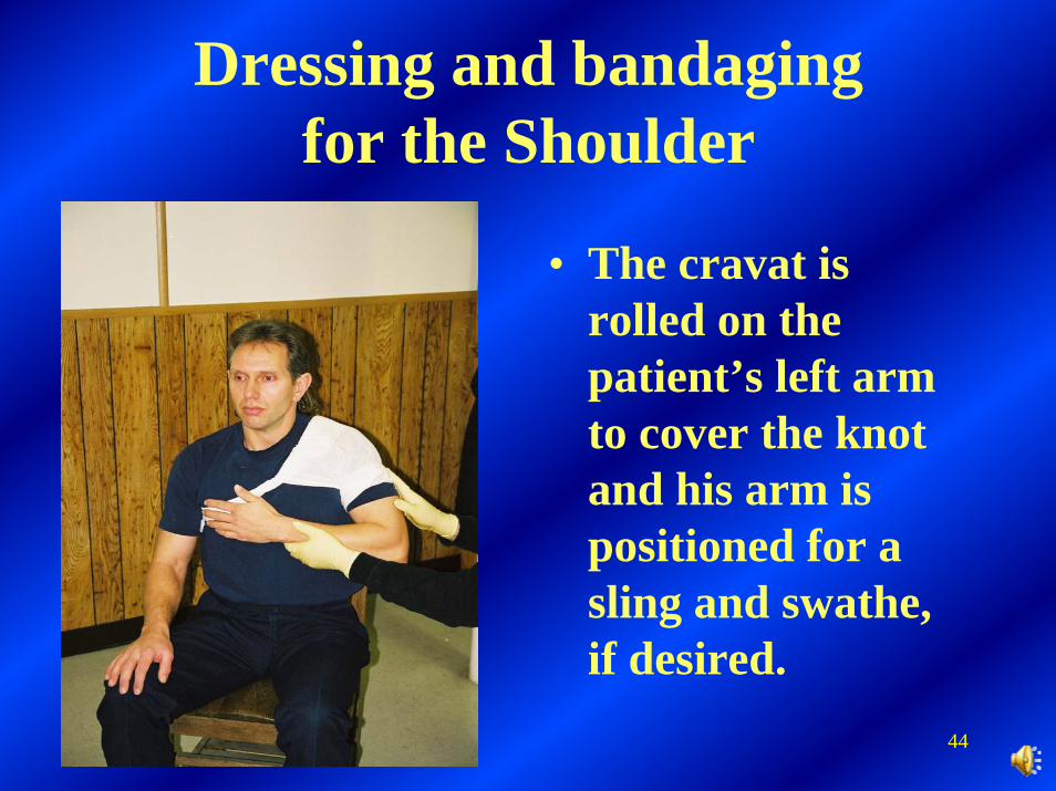
• The cravat is rolled on the patient’s left arm to cover the knot and his arm is positioned for a sling and swathe, if desired.
45
Dressing and bandagingfor the Hip
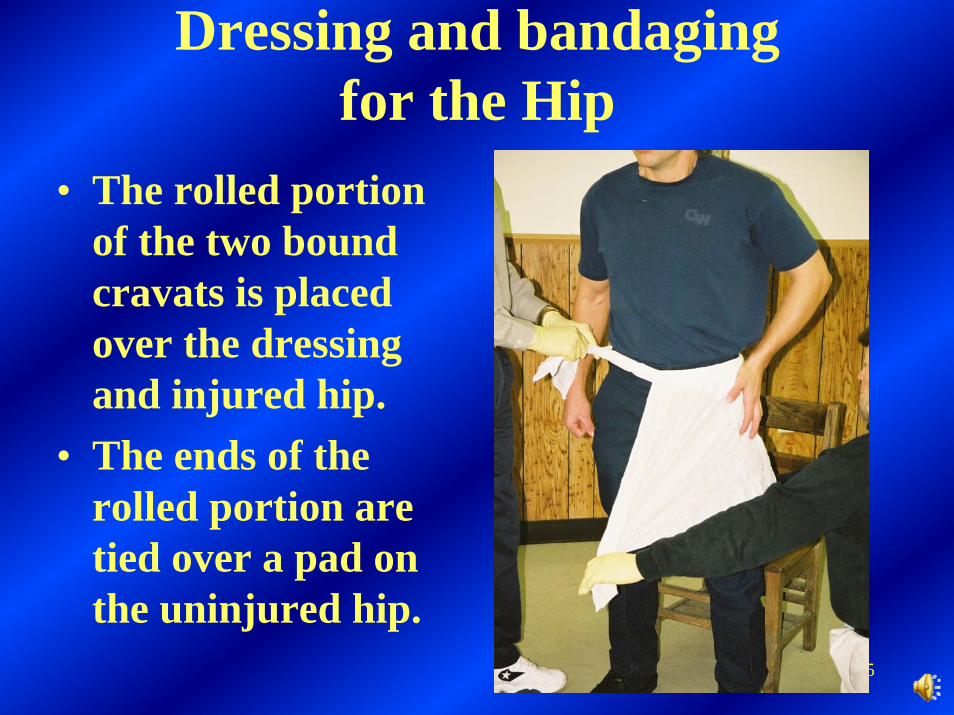
• The rolled portion of the two bound cravats is placed over the dressing and injured hip.
• The ends of the rolled portion are tied over a pad on the uninjured hip.
46
Dressing and bandagingfor the Hip
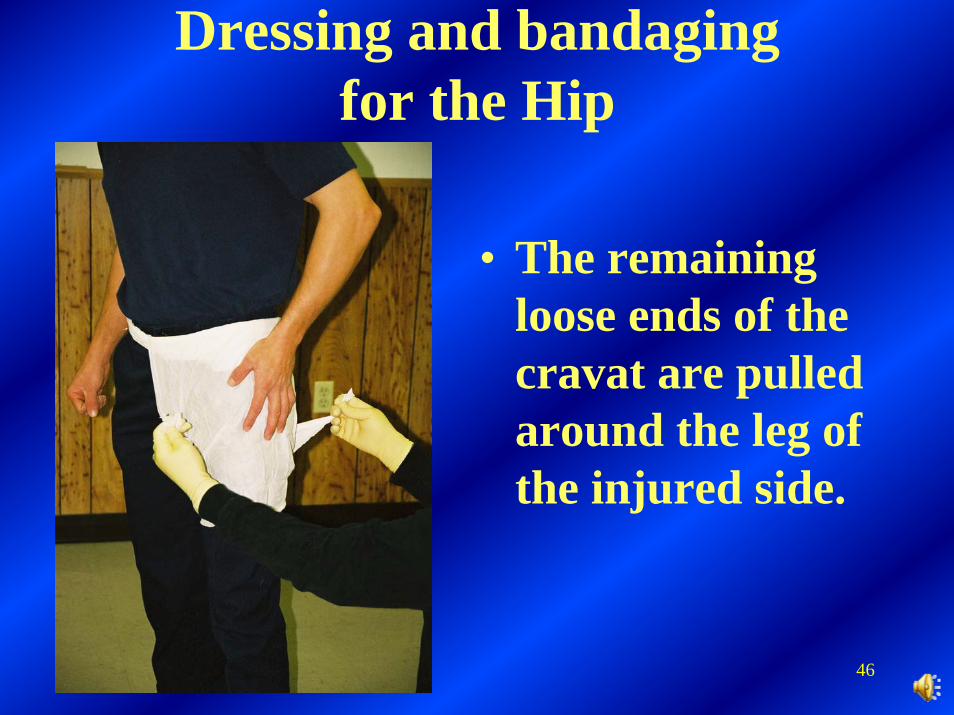
• The remaining loose ends of the cravat are pulled around the leg of the injured side.
47
Dressing and bandagingfor the Hip
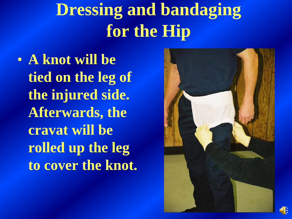
• A knot will be tied on the leg of the injured side. Afterwards, the cravat will be rolled up the leg to cover the knot.
48
Dressing and bandagingfor the Neck occlusive dressing
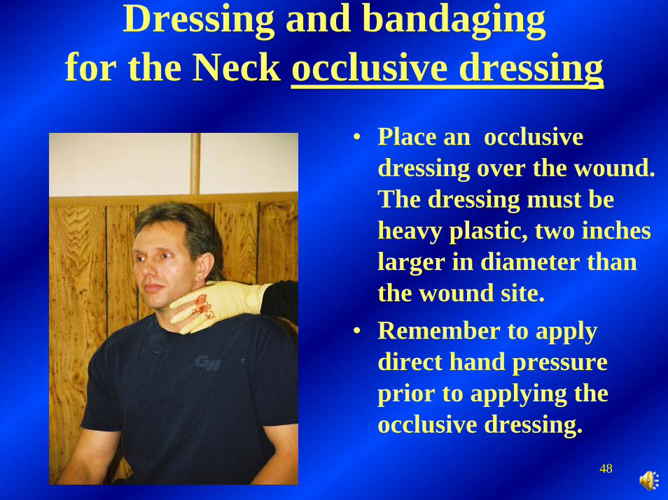
• Place an occlusive dressing over the wound. The dressing must be heavy plastic, two inches larger in diameter than the wound site.
• Remember to apply direct hand pressure prior to applying the occlusive dressing.
49
Dressing and bandagingfor the Neck occlusive dressing
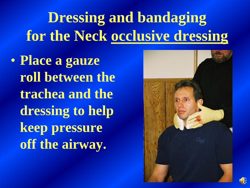
• Place a gauze roll between the trachea and the dressing to help keep pressure off the airway.
50
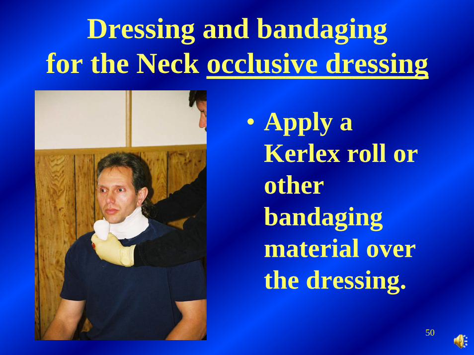
Dressing and bandagingfor the Neck occlusive dressing
• Apply a Kerlex roll or other bandaging material over the dressing.
51
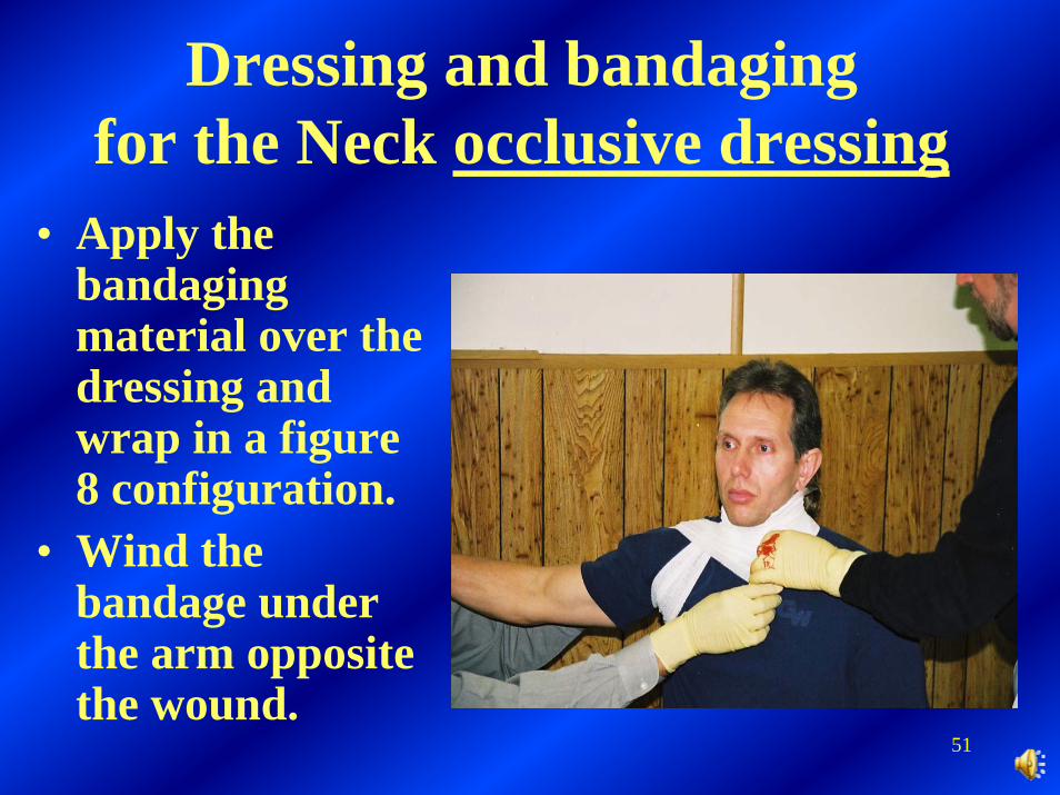
Dressing and bandagingfor the Neck occlusive dressing
• Apply the bandaging material over the dressing and wrap in a figure 8 configuration.
• Wind the bandage under the arm opposite the wound.
52
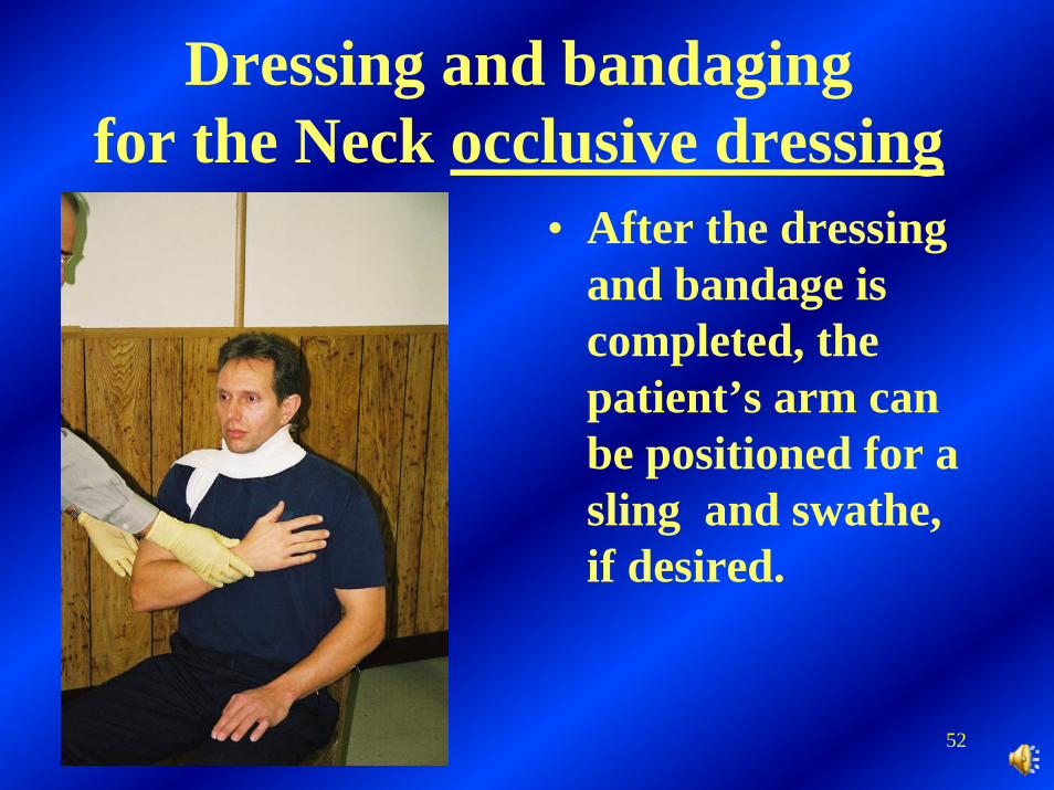
Dressing and bandagingfor the Neck occlusive dressing
• After the dressing and bandage is completed, the patient’s arm can be positioned for a sling and swathe, if desired.
53
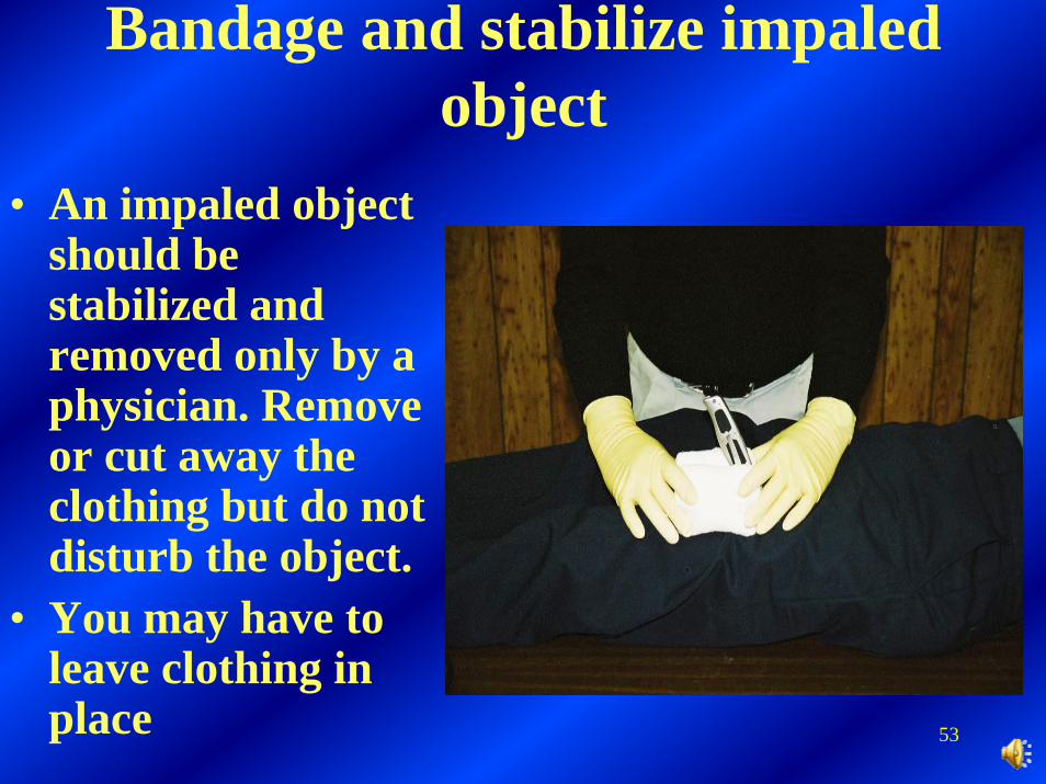
Bandage and stabilize impaled object
• An impaled object should be stabilized and removed only by a physician. Remove or cut away the clothing but do not disturb the object.
• You may have to leave clothing in place
54
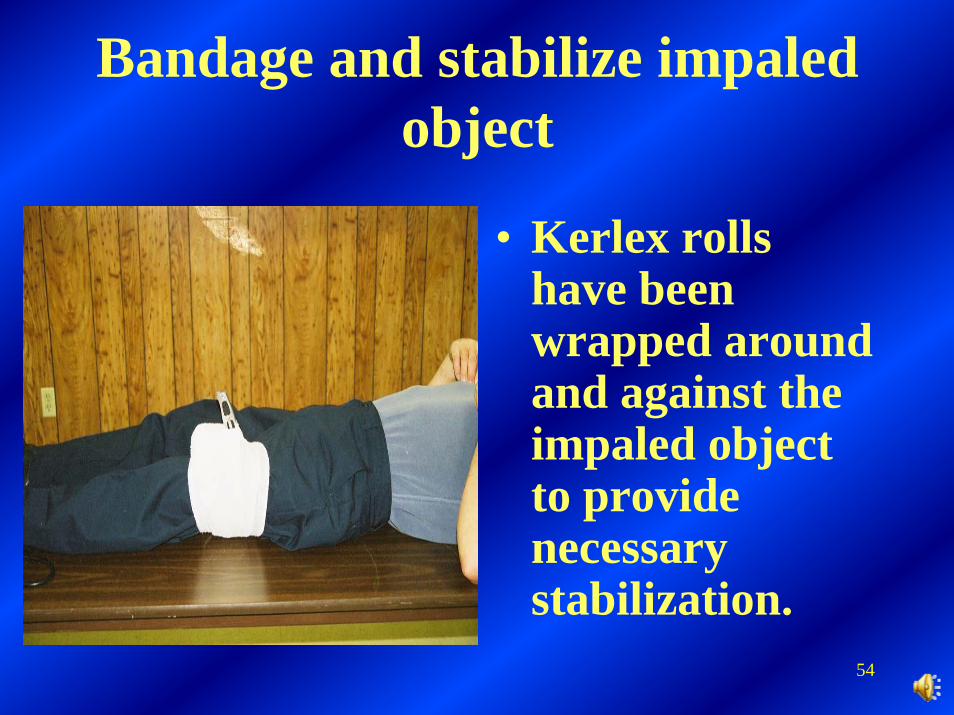
Bandage and stabilize impaled object
• Kerlex rolls have been wrapped around and against the impaled object to provide necessary stabilization.
55
First Aid
Splinting
Prepared by Wayne Collett Harlan District Instructor
January 3, 2003
56
In days past, first aid people were taught and had to know the difference between a fracture (a break in a bone) and a dislocation (displacement of a bone in a joint) or a sprain (stretching or tearing of ligaments) and a strain(pulling or tearing of muscles).
57
Signs and SymptomsThe signs and symptoms of muscle and bone injuries include the following:
• Pain and tenderness (An injury is said to be tender when touching it causes pain.)
• Swelling • Deformity • Bruising • Grating of bone ends (called crepitus)• Exposed bones• Loss or reduction of function

58
Treatment•Treat all life threats. Administer oxygen.•Place patient in position of comfort, unless spinal injury is suspected.•Stabilize injury above and below injury site•Don’t try to pull the bones to realign them•Cover open wounds with sterile dressings•Apply cold pack to injury site•Assess above and below injury site for pulse, sensation, and movement
59
Splinting immobilizes bones and joints, helps to prevent further muscle and bone injuries, and reduces bleeding and pain.
60
During patient assessment, the MET or the first aid person will determine the necessity and priority for performing this skill. (Performing this skill may require the assistance of additional people available at the scene and instructions must be provided by the MET or the first aid person in charge).
61
Assess and treat life threats first. Splinting an extremity should never take priority. If the patient has signs of shock or other life threats, prepare immediately for transport. Do not delay by trying to splint individual injuries.
62
Instead, guide the patient’s body into a neutral, in-line position and immobilize him from head to toe on a long back-board. The back-board will serve to “splint” the injured extremities until the patient can get to a hospital. When possible, splint an injured extremity before moving the patient.
63
Take and verbalize BSI precautions:
Prior to examination or treatment of any patient you must take proper BSI precautions. During “Patient Assessment”, the MET or first aid person will determine the necessity and priority for performing this skill.
64
NOTE: Remember to remove or cut away the patient’s clothing from the injury site and cover open wounds with sterile dressings.
65
Apply manual stabilization to the injured extremity:
Manually stabilize the extremity above and below the injury site. Place one gloved hand above and one gloved hand below the injury to keep it from moving. Do not release until the injured extremity is properly immobilized. Do not try to pull the bones to realign them and do not try to replace protruding bones.
66
Cover open wounds with sterile dressings and attempt to reduce pain by applying cold packs to injured sites. Make sure dressings or towels are placed between the injury and the cold pack.
67
Check for pulse and sensation
Assess below the injury site for the patient’s pulse, sensation, and movement before and after splinting. This can be done by checking the patient’s fingers and toes. This is one reason for leaving them exposed.

68
If there is a deformity, and if the extremity below the injury is cyanotic (bluish) or has no pulse, then align the extremity with gentle traction (pulling). However, if the injury site is at a joint, stop traction immediately if you feel any resistance at all.
69
Measure the splint and pad it appropriately. To immobilize long – bone injuries, apply the splint so that the joint above and below the injury site is immobilized too. To immobilize a joint, apply the splint so that the bones above and below it are immobilized.
SPLINTING
70
Types of SplintsSeveral types of splints are available, such as: air splints, rigid splints and improvised splints. Splints can be made from cardboard, wood, hard plastic, tongue depressors, pillows and blankets. A sling and swathe, made from two triangular bandages, works well to immobilize a shoulder injury.
71
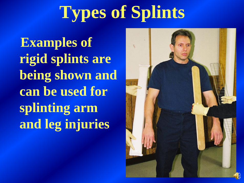
Types of SplintsExamples of rigid splints are being shown and can be used for splinting arm and leg injuries
72
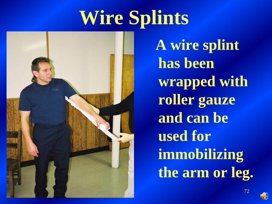
Wire SplintsA wire splint has been wrapped with roller gauze and can be used for immobilizing the arm or leg.
73
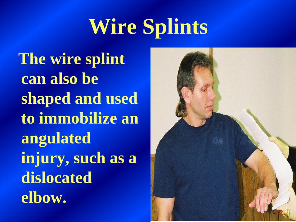
Wire SplintsThe wire splint can also be shaped and used to immobilize an angulated injury, such as a dislocated elbow.
74
Rigid splinting can be used for immobilizing the arm or leg.The suspected location of the injury will determine the length of the splint.Joint injuries require splinting above and below the injured site
75
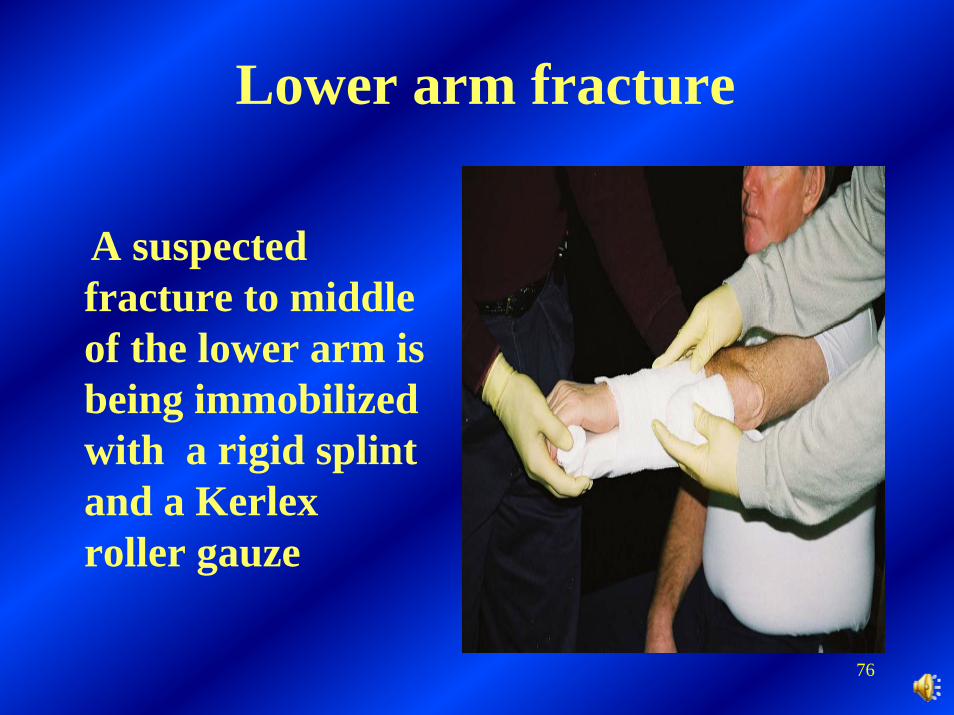
Lower arm fracture
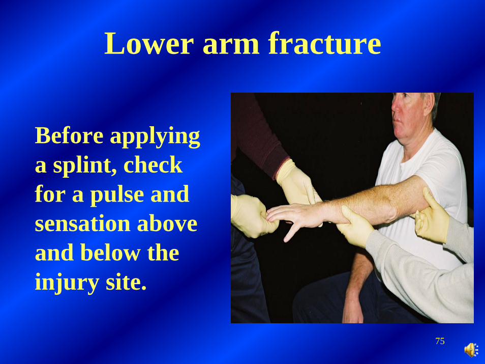
Before applying a splint, check for a pulse and sensation above and below the injury site.
76
Lower arm fracture
A suspected fracture to middle of the lower arm is being immobilized with a rigid splint and a Kerlexroller gauze
77
Lower arm fracture
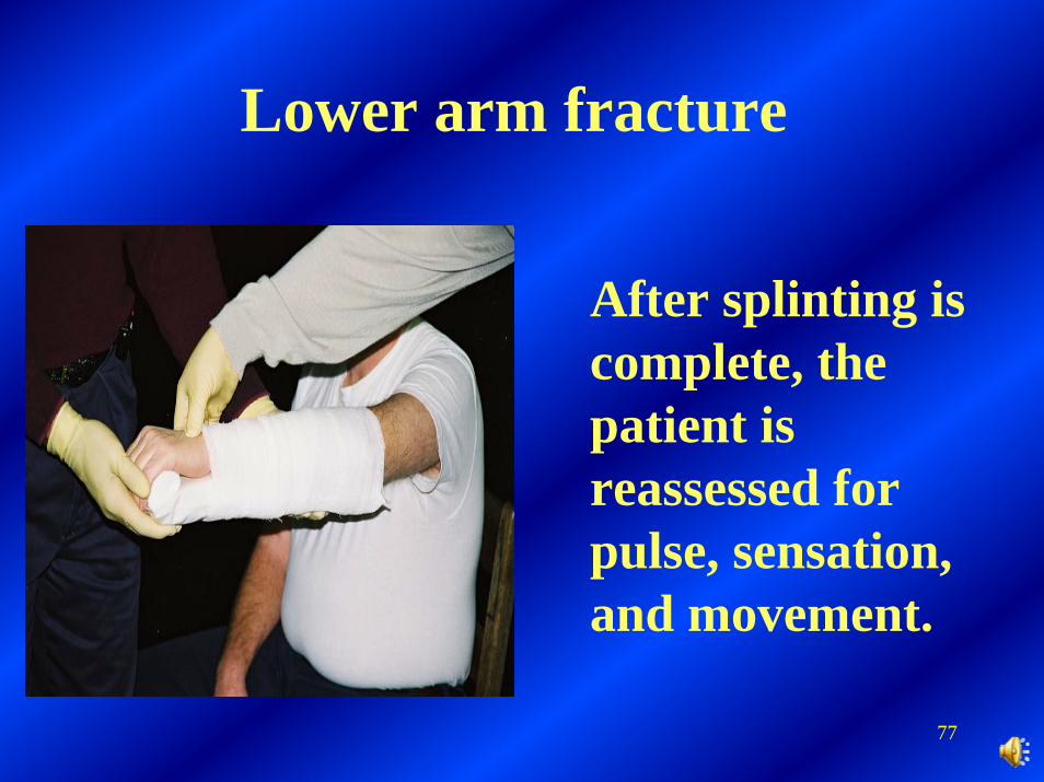
After splinting is complete, the patient is reassessed for pulse, sensation, and movement.

78
Lower arm fracture
The patient’s arm is placed against his chest, in preparation for a sling and swathe.
79
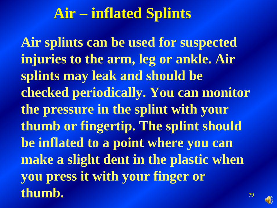
Air splints can be used for suspected injuries to the arm, leg or ankle. Air splints may leak and should be checked periodically. You can monitor the pressure in the splint with your thumb or fingertip. The splint should be inflated to a point where you can make a slight dent in the plastic when you press it with your finger or thumb.
Air – inflated Splints
80
Air – inflated Splints
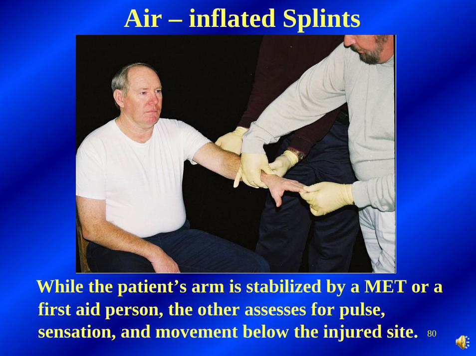
While the patient’s arm is stabilized by a MET or a first aid person, the other assesses for pulse, sensation, and movement below the injured site.
81
Air – inflated Splints
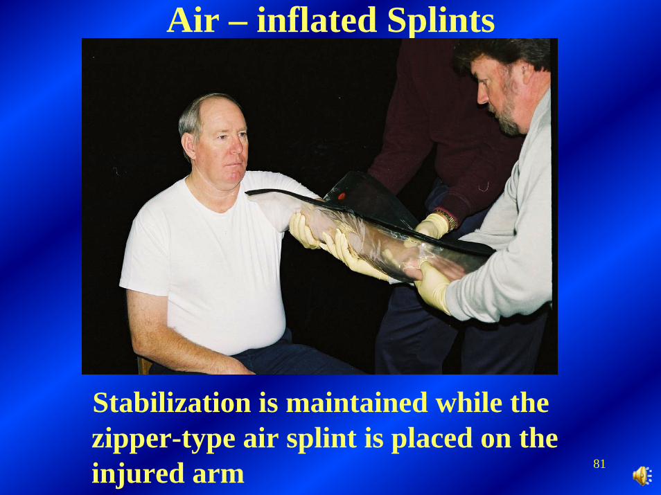
Stabilization is maintained while the zipper-type air splint is placed on the injured arm
82
Air – inflated Splints
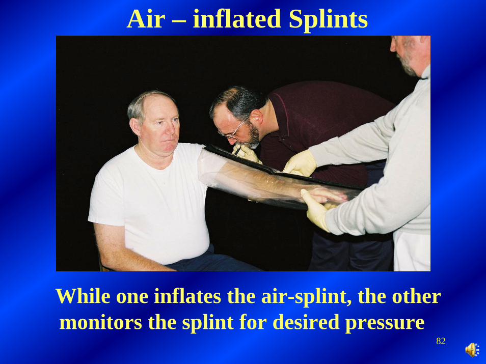
While one inflates the air-splint, the other monitors the splint for desired pressure
83
Air – inflated Splints
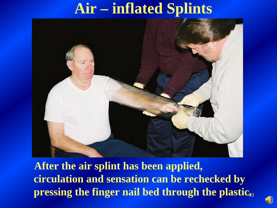
After the air splint has been applied, circulation and sensation can be rechecked by pressing the finger nail bed through the plastic.
84
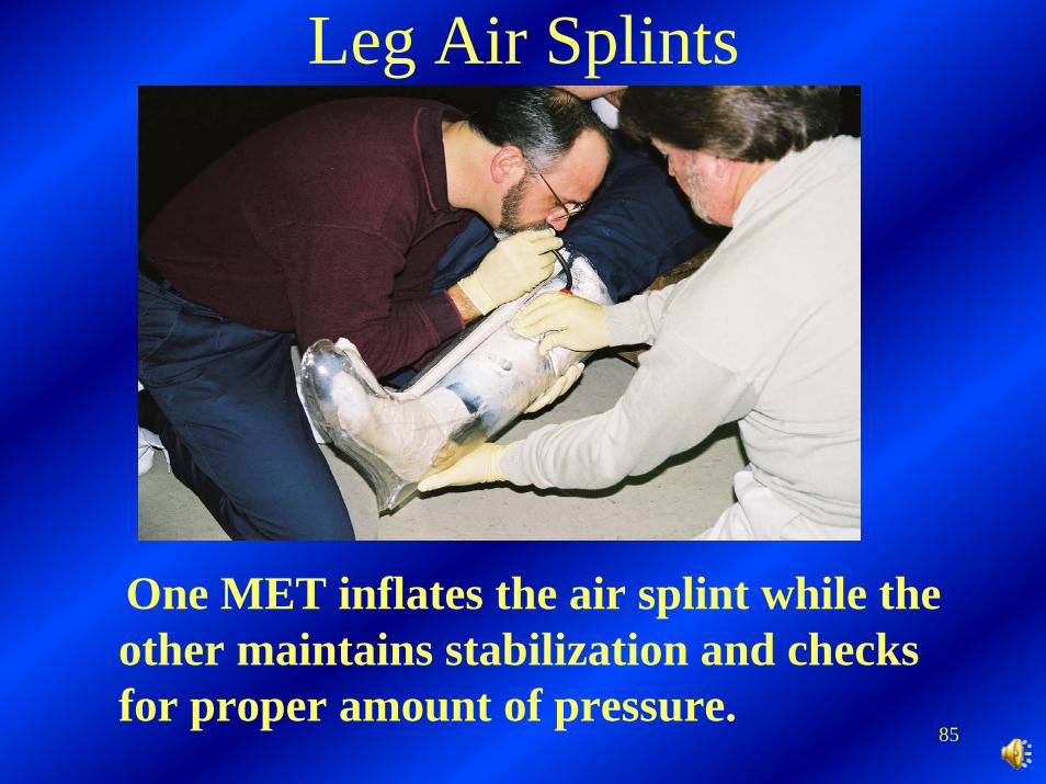
Leg Air Splints
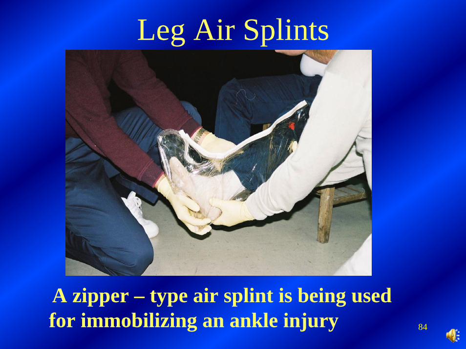
A zipper – type air splint is being used for immobilizing an ankle injury
85
Leg Air Splints
One MET inflates the air splint while the other maintains stabilization and checks for proper amount of pressure.
86
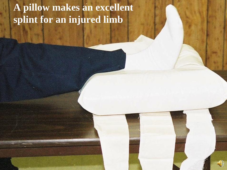
A pillow makes an excellentsplint for an injured limb
87
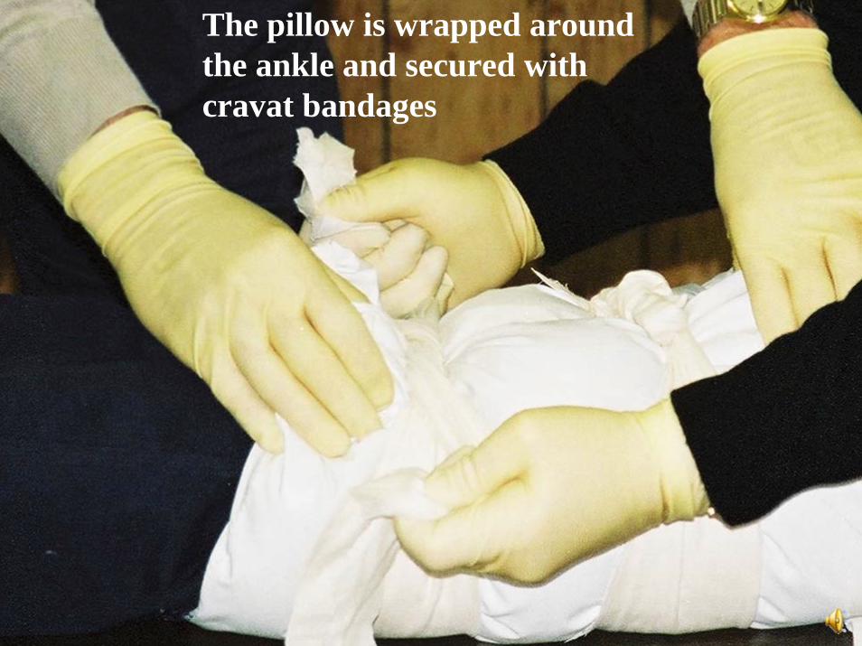
The pillow is wrapped around the ankle and secured with cravat bandages
88
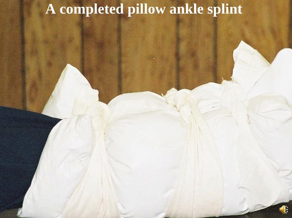
A completed pillow ankle splint
89
Sling and SwatheA sling is a triangular bandage used to support the shoulder and arm. Once the patient’s arm is placed in a sling, a swathe can be used to hold the arm against the patient’s chest. Commercial slings are available. Velcro straps can be used to form a swathe, but you can use whatever materials you have on hand, provided they will not cut into the patient.
90
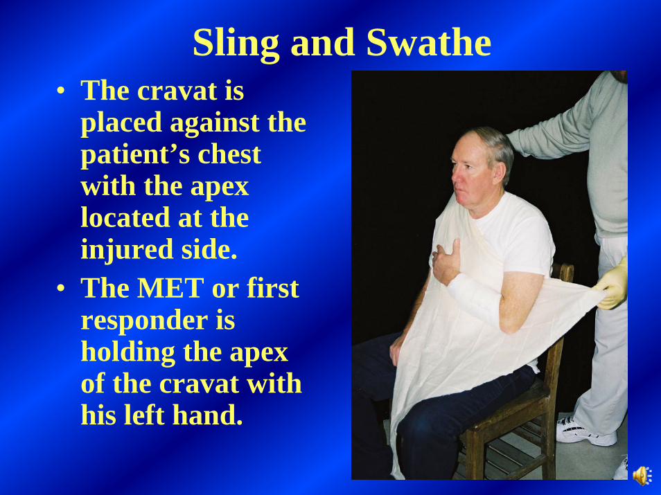
Sling and Swathe• The cravat is
placed against the patient’s chest with the apex located at the injured side.
• The MET or first responder is holding the apex of the cravat with his left hand.
91
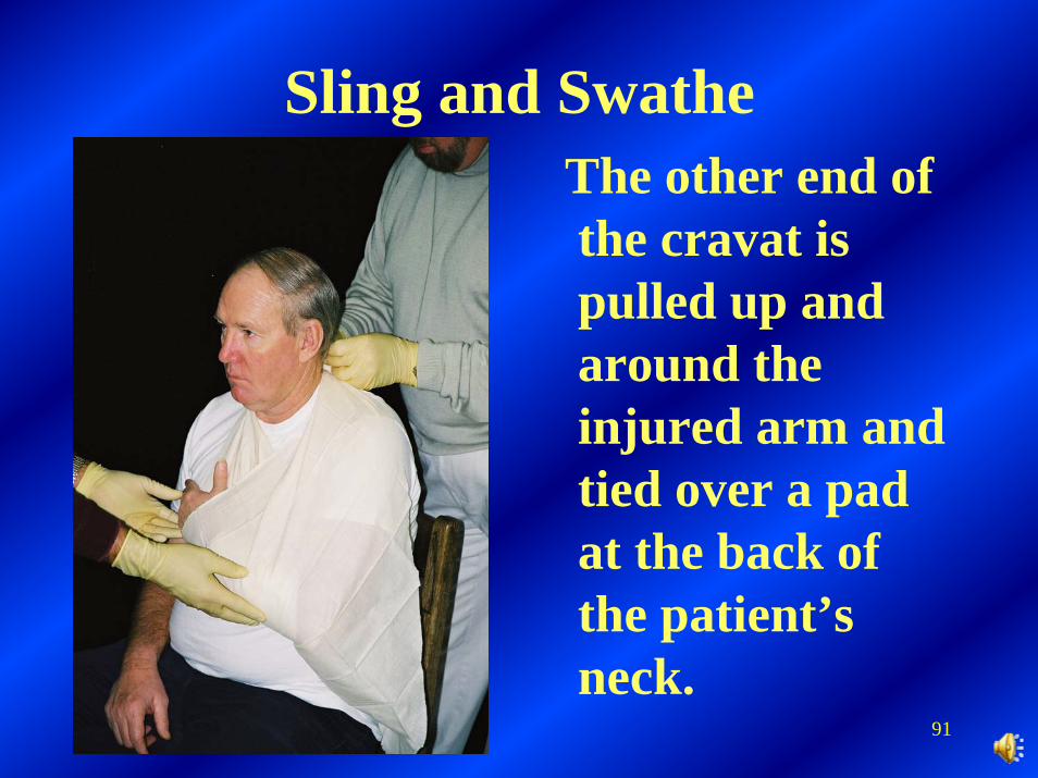
Sling and SwatheThe other end of the cravat is pulled up and around the injured arm and tied over a pad at the back of the patient’s neck.
92
Sling and Swathe
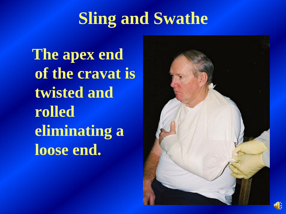
The apex end of the cravat is twisted and rolled eliminating a loose end.
93
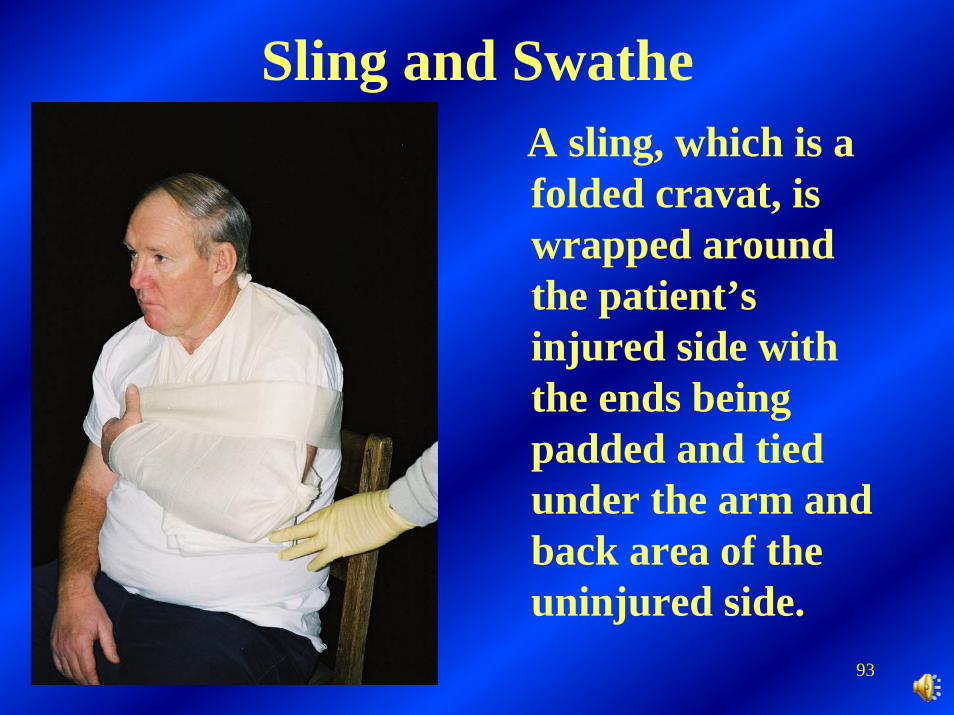
Sling and SwatheA sling, which is a folded cravat, is wrapped around the patient’s injured side with the ends being padded and tied under the arm and back area of the uninjured side.

94
Dislocation
A dislocation is a disruption or (coming apart) of a joint. In order for a joint to dislocate, the soft tissue of the joint capsule and and ligaments must be stretched beyond the normal range of motion and torn.
95
Dislocated Elbow
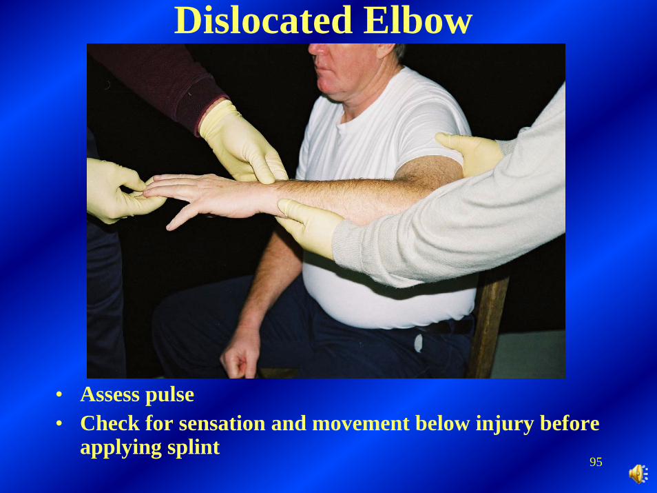
• Assess pulse• Check for sensation and movement below injury before
applying splint
96
Dislocated Elbow
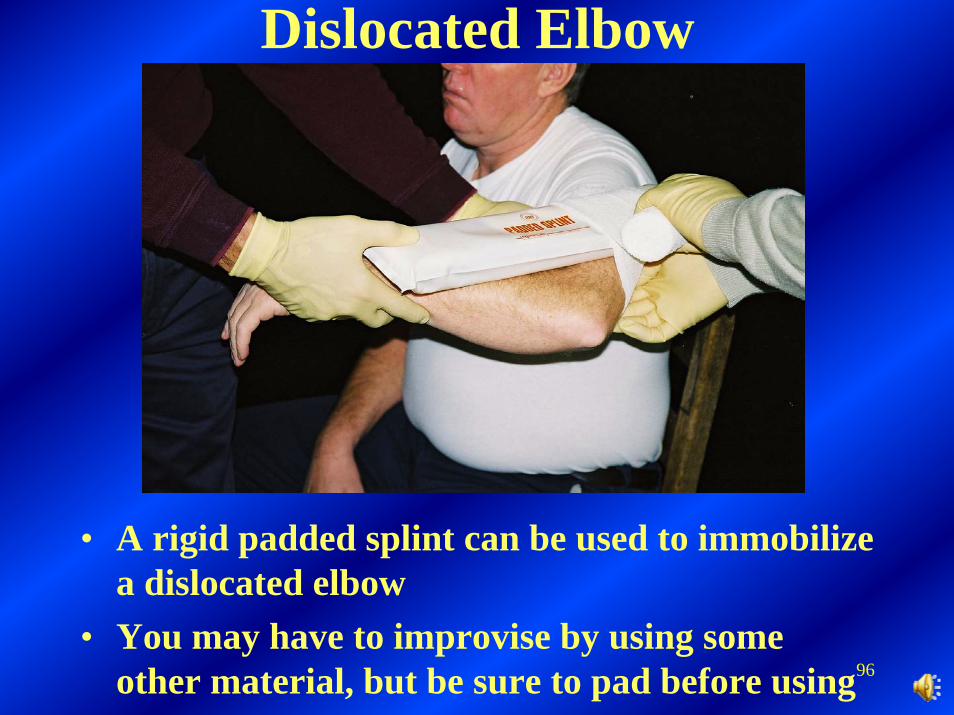
• A rigid padded splint can be used to immobilize a dislocated elbow
• You may have to improvise by using some other material, but be sure to pad before using
97
Dislocated Elbow
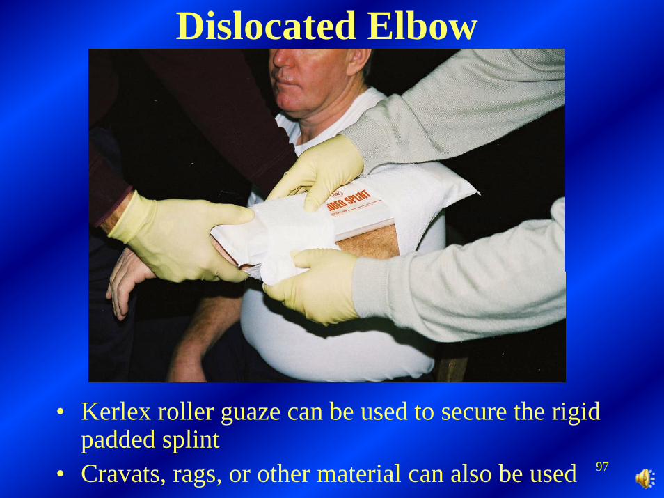
• Kerlex roller guaze can be used to secure the rigid padded splint
• Cravats, rags, or other material can also be used
98
Dislocated Elbow
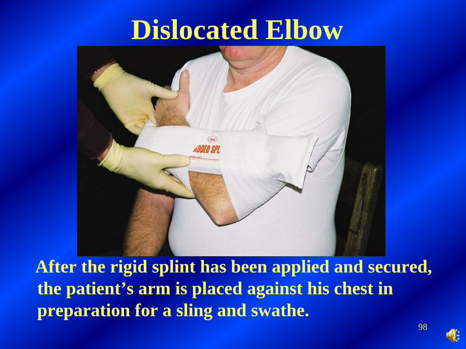
After the rigid splint has been applied and secured, the patient’s arm is placed against his chest in preparation for a sling and swathe.
99
Pelvic injuriesFractures of the pelvis may occur with falls, in motor vehicle collisions, or when a person is crushed by being squeezed between two objects. If pelvic fractures occur, there may be serious damage to internal organs, blood vessels, and nerves. Internal bleeding may be profuse and lead to shock. A force strong enough to fracture the pelvis can also cause injury to the spine.
100
• Complaint of pain in pelvis, hips, groin, or back may be the only indication. Usually, obvious deformity is associated with the pain.• Painful reaction when pressure is applied to pelvic area.• Patient complains that he cannot lift his legs when lying on his back. (Do not test for this, but do check for sensation).• The foot on the injured side may turn outward (lateral rotation). This may also indicate a hip fracture.• The patient has an unexplained pressure on the urinary bladder and the feeling of having to empty the bladder.
Signs and Symptoms – Pelvic Injuries

101
Fractured Pelvis
It may be difficult to tell a fractured pelvis from a fracture of the upper femur. Assume that it is a pelvic fracture, which can result in damage to internal organs, blood vessels, and nerves.
102
• Move the patient as little as possible. Any emergency move should be done so that the patient moves as a unit.• Never lift the patient with the pelvis unsupported and do not use a log roll to move a patient with a suspected pelvic fracture.• Assess pulse, sensation, and movement below the injury site.• Straighten the patient’s lower limbs if it is possible to do so without causing excessive pain or meeting resistance.
Patient Care for Pelvic Injury
103
• Prevent additional injury to the pelvis by stabilizing the lower limbs. Place a folded blanket between the patient’s legs from the groin to the feet, and bind them with wide cravats.• Assume there are spinal injuries. Immobilize the patient on a long back-board. When securing the patient, avoid placing the straps or ties over the pelvic area.• Care for shock and provide a high concentration of oxygen, if possible.• Monitor vital signs and transport the patient as soon as possible.
Patient Care for Pelvic Injury
104
Hip Injury
A hip dislocation occurs when the head of the femur is pulled or pushed from its pelvic socket. The injury, usually called a hip fracture, is actually a fracture of the uppermost portion of the femur. It is difficult to tell a hip dislocation from a fracture of the upper femur or pelvis. Conscious patients will complain of intense pain with either of these injuries.
105
Hip Injury – Signs and Symptoms
• Swelling and pain.• Anterior hip dislocation - The patient’s entire lower limb is rotated outward and the hip is usually flexed.
106
Hip Injury – Signs and Symptoms
• Posterior hip dislocation (most common) -The patient’s leg is rotated inward, the hip is flexed, and the knee is bent. The foot may hang loose (foot droop) and the patient is unable to flex the foot or lift the toes. Often there is a lack of sensation in the limb. These signs indicate possible damage caused by the dislocated femoral head, to the sciatic nerve, the major nerve that extends from the lower spine to the posterior thigh. This injury often occurs when a person’s knees strike the dashboard during a motor vehicle collision.Injured limb may appear shorter.
107
Patient Care for Hip Injury• Immobilize the limb with pillows or rolled blankets.• Move or lift the patient as a unit on to a long spine board.• Assess pulse, sensation, and movement below the injury site.• Secure the patient to the long back-board with straps or cravats.• Care for shock and provide a high concentration of oxygen, if possible.• Monitor vital signs and transport the patient as soon as possible.
108
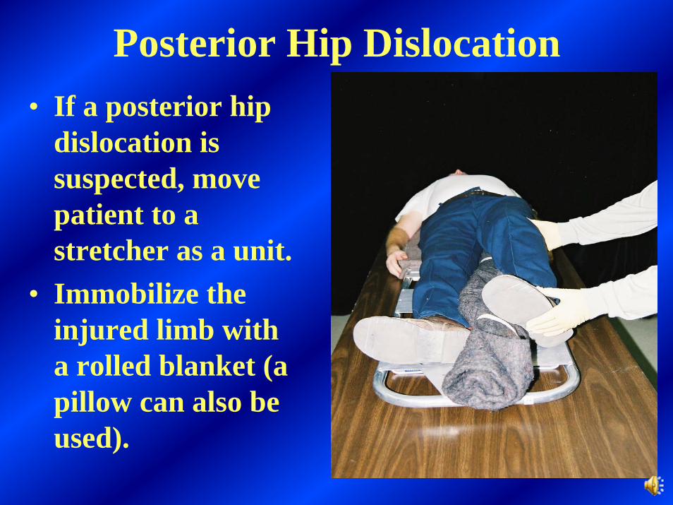
Posterior Hip Dislocation• If a posterior hip
dislocation is suspected, move patient to a stretcher as a unit.
• Immobilize the injured limb with a rolled blanket (a pillow can also be used).
109
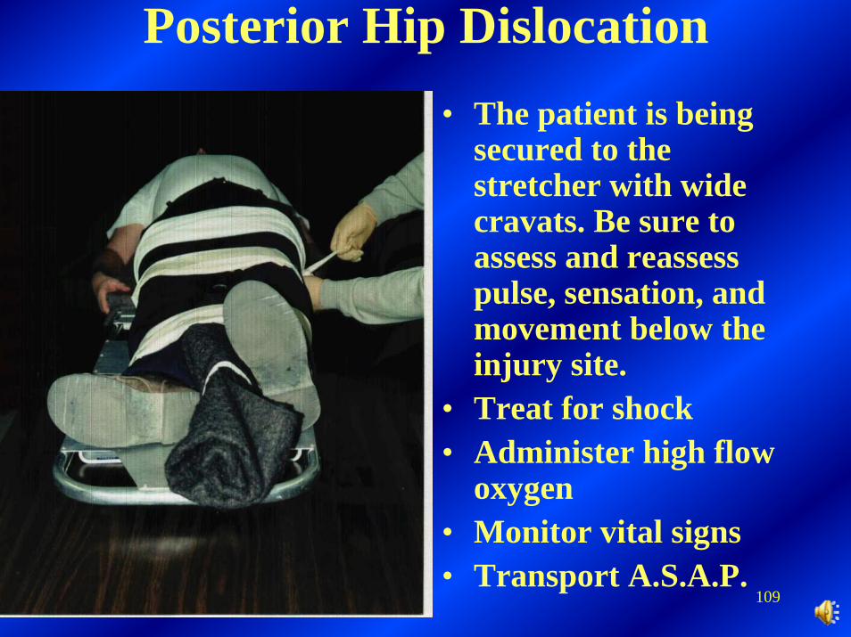
Posterior Hip Dislocation• The patient is being
secured to the stretcher with wide cravats. Be sure to assess and reassess pulse, sensation, and movement below the injury site.
• Treat for shock• Administer high flow
oxygen• Monitor vital signs• Transport A.S.A.P.
110
Transportation of the Injured
111
The responsibility of a first aid person in transporting an injured person is to ensure that the patient is transported in a manner that will: (1) prevent further injury; (2) not increase the severity of the original injuries; (3) subject the patient to no unnecessary pain or discomfort.

112
It becomes necessary to lift and/or move an injured or ill person when the patient is in immediate danger, when moving will prevent further injuries, and when it is necessary in preparing for transport.
113
When the injured or ill person must be moved immediately, an emergency move must be performed.
114
Conditions that require an emergency move are: (1) when there is immediate danger to the patient because of life-threatening hazards; (2) when the location of the patient blocks access to other injured persons who need life-saving care; (3) when the injured or ill person is in a location or position which makes life-saving care impossible to provide.
115
When performing an emergency move there is a possibility of causing further injury to the injured persons spine. Remember, however, that clearing a blocked airway and stopping hemorrhaging are life-saving measures that take precedence over potential spinal damage when an emergency move is being considered.
116
In an emergency, make every effort to protect the spine by pulling your patient in the direction of the long axis of his body.
117
Three emergency moves to use when patients are at ground level are the shirt drag, blanket drag, and shoulder drag.
118
In the shirt drag, pull the patient’s clothing in the neck and shoulder area. In the blanket drag, place the patient on a blanket and drag the blanket. For the shoulder drag, get behind the patient, put your hands under the armpits, and grasp the patient’s forearms.
119
Non-urgent moves are used to transport an injured person from a sitting or lying position to a stretcher.

120
Two non-urgent moves that can be used while administering first aid underground are the direct ground lift and the extremity lift.
121
The direct ground lift can be used when there are no suspected spinal injuries to the injured and when there are at least two rescuers to perform the lift.
122
The steps for performing the direct ground liftare: (1) two rescuers line up on one side of the injured person. Each kneels on one knee, preferably the same one; (2) place the injured person’s arms on his/her chest; (3) the rescuer at the head places one arm under the injured person’s neck and shoulder, cradling the head. The other arm is placed under the injured person’s lower back. The second rescuer places one arm under the injured person’s knees and one arm above the buttocks.
123
Direct ground lift continued:(4) on signal from the rescuer at the head, both lift the injured person to their knees; (5) on signal they both roll the injured toward their chests; (6) on signal the rescuers stand and move the injured person to the stretcher; (7) reverse the steps to lower the person to the ground level; if a third person is available, he should place both hands under the injured person’s waist.

124
The extremity lift is a two-rescuer lift that can be performed on injured persons who have no suspected spinal injuries.
125
The steps for performing the extremity lift are: (1) one rescuer kneels at the injured person’s head. The other kneels at the injured person’s knees. (2) the rescuer at the head works one hand under each of the injured’s shoulders and grasps the injured’s wrists; (3) the other rescuer slips one hand under each of the injured’s knees; (4) on signal, both rescuers move up to a crouching position; (5) on signal, both rescuers stand up and move the patient to the stretcher.
126
Stretchers that are available are: long backboards or spine boards, scoop stretchers, wheeled stretchers, and portable metal or canvas stretchers.

127
A stretcher should always be tested by a weight equal to or greater than that of the patient before the injured person is placed on it.
128
Oral Review
Burns, Splinting, and Transportation
Questions and Answers
129
How can you relieve the pain of a burn?

130
By flushing with lots of water.

131
How do you treat a burn?
132
When treating a burn…
• Relieve the pain by flushing with lots of water.
• If possible, remove clothing and jewelry from the affected area.
• Do not attempt to clear debris from the burned area.
• Apply dressing and bandage.
133
Eye protection can prevent many injuries to the eyes. However, injuries to the eyes can occur and must be properly treated.
134
How do you treat a chemical burn to the eye?
135
Chemical burns of the eye
• Brush off any remaining powder from the patient and flush eyes with plenty of water• It is best to have the patient lying down with the eyelids open, while flushing the eyes with plenty of water• Be sure the water flows across the eyes in large amounts
136
How do you treat a burn to the fingers?
137
When treating a burn of the fingers, dress and bandage them individually
138
After flushing with water and bandaging the fingers individually, cover the entire
hand with a suitable dressing
139
What is the general treatment for a burn?

140
When treating a burn…• Stop the burning, by flushing
with water• Remove all clothing and
jewelry from the burned area if there is no resistance
• Cover with dry sterile dressing, preventing further contamination
• Don’t break blisters or use ointment or antiseptic on the burn
• Treat for shock

141
What is a fracture?

142
A fracture is a break in a bone.

143
What is a dislocation?

144
A dislocation is a displacement of a bone in a joint.

145
What is a simple fracture?

146
A simple fracture is one in which the bone is broken but there is no wound extending from the ends of the bone out through the skin.

147
What is a compound fracture?

148
A compound fracture is one in which the bone is broken and the wound extends from the bone out through the skin.

149
What is a sprain?
150
A sprain is a stretching or tearing of ligaments.
151
What is a strain?
152
A strain is a pulling or tearing of the muscles.
153
Name the signs and symptoms of muscle and bone injuries?
154
Signs and Symptoms• Pain and tenderness (An injury is said to
be tender when touching it causes pain.) • Swelling • Deformity • Bruising • Grating of bone ends (called crepitus)• Exposed bones• Loss or reduction of function
155
What is the treatment for muscle and bone injuries?
156
Treatment•Treat all life threats. Administer oxygen.•Place patient in position of comfort, unless spinal injury is suspected.•Stabilize injury above and below injury site•Don’t try to pull the bones to realign them•Cover open wounds with sterile dressings•Apply cold pack to injury site•Assess above and below injury site for pulse, sensation, and movement
157
What must be checked above and below the injury site before and after splinting?
158
The patient’s pulse and sensation. (This can be done by checking the patient’s fingers and toes. This is one reason for leaving them exposed.)

159
If there is a deformity, and if the extremity below the injury is cyanotic (bluish) or has no pulse, then align the extremity with gentle traction (pulling). However, if the injury site is at a joint, stop traction immediately if you feel any resistance at all.
160
Measure the splint and pad it appropriately. To immobilize long bone injuries, apply the splint so that the joint _____ and _____ the injury site is immobilized too.
SPLINTING

161
above and below
162
To immobilize a joint, apply the splint so that the bones _____ and ______ it are immobilized.

163
above and below
164
Several types of splints can be used to immobilize bones and joints.
Name some.

165
Air splints, rigid splints and improvised splints. Splints can be made from cardboard, wood, hard plastic, tongue depressors, pillows and blankets.
166
A sling and swathe, made from two triangular bandages, works well to immobilize a shoulder injury.
167
What type of splint can be used and shaped to immobilize an angulated injury?
168
A wire splint.

169
Wire SplintsA wire splint has been wrapped with roller gauze and can be used for immobilizing the arm or leg.
170
What type of splint is located in the first aid box and can be used for suspected injuries to the arm, leg, or ankle?
171
Air splint
172
Air splints may leak and should be checked periodically. You can monitor the pressure in the splint with your thumb or fingertip. The splint should be inflated to a point where you can make a slight dent in the plastic when you press it with your finger or thumb.
173
It becomes necessary to lift or move an injured or ill person when the patient is in immediate danger, when moving will prevent further injuries, and when it is necessary in preparing for transport.
174
What is the responsibility of a first aid person in transporting an injured person?

175
(1) To prevent further injury; (2) Not to increase the severity of the original injuries; (3) Not to subject the patient to unnecessary pain or discomfort.
176
When the injured or ill person must be moved immediately, what _____________ move must be performed.
177
emergency

178
In an emergency, make every effort to protect the spine by pulling your patient in the direction of the long axis of his body.

179
What are the three emergency moves that can be used when patients are at ground level?
180
The shirt drag, blanket drag, and shoulder drag.

181
Should a stretcher always be tested by a weight equal to or greater than that of the patient before the injured person is placed on it?

182
Yes

183
End of Part 2 Unit 8