Embed Size (px)
Citation preview
Salt Tolerance in Cereals: MolecularMechanisms and Applications 5Allah Ditta
Abstract
Major abiotic stress that limits plant growth and agriculture productivity is
the soil salinity. In order to minimize the detrimental effects of salinity,
highly complex salt-responsive signaling and metabolic processes at the
cellular, organ, and whole-plant levels have been evolved in the plants.
Currently, it has become the need of the hour to understand the molecular
basis of salt stress signaling and tolerance mechanisms in cereals for
engineering and/or screening for more tolerance to salt stress. Valuable
information will be provided through investigation of the physiological
and molecular mechanisms of salt tolerance for effective engineering
strategies. Current advancement in proteomics has helped us in studying
the sophisticated molecular networks in plants. Reports of proteomics
studies about plant salt response and tolerance mechanisms, especially
that of cereals, have revealed the mechanisms that include changes in
photosynthesis, scavenging system of reactive oxygen species (ROS), ion
homeostasis, osmotic homeostasis, membrane transport, signaling trans-
duction, transcription, protein synthesis/turnover, cytoskeleton dynamics,
and cross talks with other stresses.
Introduction
Salinity, one of the most significant abiotic stres-
ses, not only limits the productivity and geograph-
ical distribution of plants but also causes ion
imbalance, hyperosmotic stress, and oxidative
damage in plants leading to molecular damage,
growth and yield reduction, and even plant
death (Wang et al. 2004). Infiltration and accumu-
lation of NaCl (Tuteja 2007) is the major cause
of salinization and can result in soil Na+ concen-
tration above 40 mM which can suppress the
growth of most crops (Wong et al. 2006). Salt
accumulation has been attributed to the natural
phenomena and human activities like irrigation.
About 1/5 of the earth’s arable land and 50% of
the irrigated one are under salinity (Mahajan and
Tuteja 2005; Munns and Tester 2008). Plants have
coped with this problem through various sophisti-
cated mechanisms that include selective ion
A. Ditta (*)
Institute of Soil & Environmental Sciences, University of
Agriculture, Faisalabad 38040, Pakistan
e-mail: [email protected]
G.R. Rout and A.B. Das (eds.), Molecular Stress Physiology of Plants,DOI 10.1007/978-81-322-0807-5_5, # Springer India 2013
133
uptake/exclusion, compartmentalization of toxic
ions, synthesis of compatible products, adjustment
of photosynthetic and energy metabolism, accu-
mulation of antioxidative enzymes, regulation of
hormones, and modification of cell structure.
Molecular and physiological aspects of plant salt
stress tolerance have been revealed through phys-
iological, molecular genetics, and functional
genomics studies. A few important genes respon-
sible for the above-stated mechanisms have been
cloned, and their involvement in plants’ response
and adaptation to salinity has been confirmed
(Tuteja 2007).
However, state-of-the-art transcriptomics
studies have helped us in the collection of
immense data on their expression at the mRNA
level (Chand and Kumar 2006; Diedhiou et al.
2009; Jha et al. 2009; Wong et al. 2005, 2006;
Zhang et al. 2001b, 2008; Zouari et al. 2007),
and these data present a global vision about
salt-responsive genes in different plants. But
mRNA levels do not usually correlate with the
expression levels of proteins due to posttran-
scriptional events and posttranslational modifica-
tions such as phosphorylation and glycosylation.
These proteins are more directly related to sig-
naling and metabolic processes under salt stress
conditions. So the need of the hour is to study the
salt tolerance mechanisms at the so-called pro-
tein level which is possible through the optimum
utilization of proteomic technologies.
The plant taxa have an extensive genetic
diversity for salt tolerance as it is distributed
over numerous genera (Flowers and Colmer
2008) making them either glycophytes (salt-
sensitive or hypersensitive plants) or halophytes
(native flora of saline environments). In some
halophytes, very special anatomical and morpho-
logical adaptations or avoidance mechanisms
have been employed (Flowers and Colmer
2008), but on the basis of these, we are unable
to introgress the responsible genes into crop
plants. During the last decade, it has been estab-
lished that most halophytes and glycophytes use
analogous tactical processes rather than similar
strategies to tolerate salinity (Hasegawa et al.
2000b). For example, cytotoxic ions (Na+ and
Cl�) are compartmentalized into the vacuole
and used as osmotic solutes under saline envir-
onments (Blumwald et al. 2000; Niu et al. 1995).
It follows that there is similarity among many
of the molecular entities that mediate ion homeo-
stasis and salt stress signaling in all plants
(Hasegawa et al. 2000b) as, for example, ion
homeostasis that facilitates plant salt tolerance
resembles that described for yeast (Bressan
et al. 1998; Serrano et al. 1999). According to
another finding of genomics, there is remarkable
colinearity (gene synteny and homology) of gene
sequences among different grass species, includ-
ing cultivated crops (Tang et al. 2008; Zahn et al.
2008), and strongly suggests their resemblance in
evolution and probably in functions. The facts
stated above have made it feasible to use a model
system for the dissection of the plant salt stress
response (Bressan et al. 1998; Hasegawa et al.
2000a; Sanders et al. 1999; Serrano et al. 1999;
Zhu 2000, 2001b). Since a salt-tolerant genetic
model is required for complete delineation that if
salt tolerance is affected most by form or func-
tion of genes or more by differences in the
expression of common genes either due to tran-
scriptional or posttranscriptional control (Zhu
et al. 2007). In this way, our understanding of
cellular salt tolerance mechanisms has been
greatly increased through research on the plant
genetic model, the Arabidopsis, a glycophyte.
It will also enable us to effectively apply the
genetic learning of one crop to another and also
will yield large spillover benefits from invest-
ment in such research, like possible eventual
interspecies gene transfers.
Till now, more than 2,171 salt-responsive pro-
teins/enzymes have been discovered in different
parts of 34 plant species like shoots, leaves,
roots, seedlings, radicles, hypocotyls, grains,
gametophytes, and unicells (Zhang et al. 2012).
Most of the research has been conducted on
plants like Arabidopsis thaliana (Jiang et al.
2007; Kim et al. 2007; Lee et al. 2004; Ndimba
et al. 2005; Pang et al. 2010) and Oryza sativa
(Abbasi and Komatsu 2004; Cheng et al. 2009;
Chitteti and Peng 2007; Dooki et al. 2006; Kim
et al. 2005; Li et al. 2010; Nohzadeh et al. 2007;
Parker et al. 2006; Ruan et al. 2011; Salekdeh
et al. 2002; Wen et al. 2010; Yan et al. 2005;
134 A. Ditta
Zhang et al. 2009), Triticum durum (Caruso et al.
2008), Triticum aestivum (Huo et al. 2004;
Jacoby et al. 2010; Peng et al. 2009; Wang
et al. 2008a), Hordeum vulgare (Rasoulnia et al.
2010; Sugimoto and Takeda 2009; Witzel et al.
2009, 2010), Zea mays (Zorb et al. 2004, 2009,
2010), Setaria italica (Veeranagamallaiah et al.
2008), Sorghum bicolor (Kumar et al. 2011), and
Agrostis stolonifera (Xu et al. 2010).
This chapter includes the reports of proteo-
mics studies about plant salt response and toler-
ance mechanisms, especially that of cereals, and
future perspectives in food security. These
mechanisms include changes in photosynthesis,
scavenging system of reactive oxygen species
(ROS), ion homeostasis, osmotic homeostasis,
membrane transport, signaling transduction,
transcription, protein synthesis/turnover, cyto-
skeleton dynamics, and cross talks with other
stresses.
Role of Cereals in Food Security
The word cereal has been derived from Ceres,the name of the Roman goddess of harvest and
agriculture. The common cereal crops include
rice, wheat, corn, barley, sorghum, millet, oats,
and rye. It also includes flours, meals, breads, and
alimentary pastes or pasta. Cereals, the poor
man’s meat, provide staple food in almost every
country and region as in the world as a whole;
about 95% of starchy staple food comes from
cereals and only 5% from root crops (mainly
cassava, potato, and yams, depending on cli-
mate). Currently, about 50% of the world’s crop-
land is under the cultivation of cereals. These
provide about two-thirds of all human calorie
intake, if we combine their direct intake (e.g.,
as cooked rice or bread) with their indirect con-
sumption, in the form of foods like meat and milk
(about 40% of all grain is currently fed to live-
stock).
Among cereals, wheat is the predominant
commodity consumed for food accounting 68%
of total cereal use. By 2020, total cereal con-
sumption is projected to reach nearly 746 metric
tons (Mt) (145 Mt wheat, 529 Mt rice, and the
rest maize, barley, etc.) with per capita food
consumption of around 66 kg per person per
annum, and 2% of world wheat is utilized for
biofuel production; ultimately the total consump-
tion is expected to increase from 68 to 75%
(Chand and Kumar 2006).
According to Chand and Kumar (2006), to
feed a population of eight billion by 2025, aver-
age world cereal yield of about 4 metric tons/ha
will be required (Evans 1998). In addition, it has
been found out the reasons for food insecurity
that include increasing demand from growing
population, climate change, and increased lin-
kages between energy and agricultural commod-
ities due to the growing demand for biofuels
(Chand and Kumar 2006). The emerging food
insecurity condition will prevail mostly in devel-
oping countries, especially that of Asia. How-
ever, more than 90% of rice and 43% of wheat
in the world is produced and consumed in Asia.
Detrimental Effects of Salinity onCereals
Salinity is one of the major environmental factors
that adversely affect the crop growth and devel-
opment processes like seed germination (Dash
and Panda 2001); seedling growth (Mahajan
and Tuteja 2005; Tuteja 2007; Wang et al.
2004; Wong et al. 2006); enzyme activity
(Seckin et al. 2009); deoxyribonucleic acid
(DNA), ribonucleic acid (RNA), and protein syn-
thesis (Anuradha and Rao 2001); mitosis (Tabur
and Demir 2010); vegetative growth (Hamed
et al. 2007; Panda and Khan 2009); and flowering
and fruit set (Zhu 2001a, b) and ultimately result
in diminished economic yield and also quality of
produce.
Since sensitivity or tolerance to salt stress is
different in all plant species (Ashraf and Harris
2004), so plants are classified into two groups,
namely, glycophytes or halophytes, on the basis
of their ability to grow on high-salt medium.
Most of the grain crops and vegetables are gly-
cophytes and cannot tolerate salt stress as high
salt concentrations decrease the osmotic poten-
tial of soil solution creating water stress in plants
5 Salt Tolerance in Cereals: Molecular Mechanisms and Applications 135
and ion toxicity (e.g., Na+, Cl�) since Na+ is notreadily sequestered into vacuoles as in halo-
phytes. Ultimately, it results in nutrient imbal-
ances and their deficiencies which can lead to
plant death as a result of growth arrest and
molecular damage (McCue and Hanson 1990).
In addition, salinity causes oxidative stress due to
the production of induced active oxygen species
(Heidari 2009; Munns and Tester 2008) which
disrupt the cellular metabolism through oxidative
damage to membrane lipid, proteins, and nucleic
acids (Mittler 2002).
Salinity Tolerance, Multigenic Trait
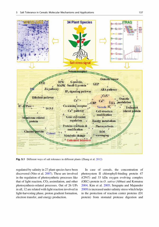
Salinity is a quantitative trait, and a large number
of salt-induced genes have been isolated which
are concomitantly up- and downregulated
(Bohnert et al. 1995). According to Meyer et al.
(1990), in M. crystallinum, more than a 100
genes are induced, and probably transcripts,
three times that number, are repressed in
response to salt stress. Salinity tolerance is a
mutagenic trait due to the fact that sublethal salt
stress conditions cause an osmotic effect that is
similar to that brought about by water deficit and
to some extent by cold as well as heat stresses
(Almoguera et al. 1988). So there is a high degree
of similarity between salt and dehydration stress
with respect to their physiological, biochemical,
molecular, and genetical effects (Cushman et al.
1992). The ways in which different plants confer
salt resistance are represented in the following
figure (Fig. 5.1):
There is a diverse expression pattern of salt-
responsive genes found in different plants (espe-
cially glycophytes and halophytes) under differ-
ent salinity conditions (e.g., salt concentration
and treatment time) (Nito et al. 2007). The
expression pattern of genes in cereals indicates
that most of the proteins related with photosyn-
thesis in O. sativa (Abbasi and Komatsu 2004;
Kim et al. 2005; Parker et al. 2006), T. durum
(Caruso et al. 2008), and T. aestivum (Huo et al.
2004; Peng et al. 2009) and proteins involved in
carbohydrate and energy metabolism in O. sativa
(Abbasi and Komatsu 2004; Chitteti and Peng
2007; Dooki et al. 2006; Kim et al. 2005; Li
et al. 2010; Nohzadeh et al. 2007; Parker et al.
2006; Ruan et al. 2011), T. aestivum (Peng et al.
2009; Wang et al. 2008a), H. vulgare (Rasoulnia
et al. 2010; Witzel et al. 2010), Z. mays (Zorb
et al. 2004, 2010), and S. bicolor (Kumar et al.
2011) are induced by salinity.
Most of the salt-responsive proteins are
involved in basic metabolic processes like photo-
synthesis, energy metabolism, ROS scavenging,
and ion homeostasis which make halophytes
highly efficient in photosynthetic and energy
metabolism, ion exclusion/compartmentaliza-
tion, compatible product synthesis, induction of
antioxidative enzymes and hormones, as well as
modification of cell structure. Also evolution of
different salt tolerance mechanisms have been
found during the study of the specific proteins
and/or their expression patterns in different
halophytes.
Molecular Mechanisms of SalinityTolerance in Cereals
Photosynthesis
In addition to osmotic, ionic, and nutrient imbal-
ances in plants, salinity also disturbs the plant
water uptake and biosynthesis of abscisic acid
(ABA) in leaves (Fricke et al. 2004) which
affects the stomatal conductance. This in turn
affects the photosynthetic electron transport and
the enzyme activities for carbon fixation during
dark reaction (Parida and Das 2005; Tuteja
2007). Several genes involved in photosynthesis
(encoding chlorophyll a-/b-binding proteins
(CAB), ribulose-1,5-bisphosphate carboxylase/
oxygenase (RuBisCO), and RuBisCO activase
(RCA)) have been isolated and characterized
through previous studies which are directly or
indirectly involved in salinity tolerance (Wong
et al. 2006; Zhang et al. 2001a, b, 2008).
Currently, through the advancement in the field
of proteomics, our understanding of the photosyn-
thetic processes underlying salinity response and
tolerance has been greatly enhanced. About 367
photosynthesis-related IDs, representing 26 UPs,
136 A. Ditta
regulated by salinity in 27 plant species have been
discovered (Nito et al. 2007). These are involved
in the regulation of photosynthetic processes like
that of light reaction, CO2 assimilation, and other
photosynthesis-related processes. Out of 26 UPs
in all, 12 are related with light reaction involved in
light-harvesting phase, proton gradient formation,
electron transfer, and energy production.
In case of cereals, the concentration of
photosystem II chlorophyll-binding protein 47
(CP47) and 33 kDa oxygen evolving complex
(OEC) protein in O. sativa (Abbasi and Komatsu
2004; Kim et al. 2005; Sengupta and Majumder
2009) is increased under salinity stresswhich helps
in the protection of reaction center proteins (D1
protein) from stomatal protease digestion and
Fig. 5.1 Different ways of salt tolerance in different plants (Zhang et al. 2012)
5 Salt Tolerance in Cereals: Molecular Mechanisms and Applications 137
ensures optimal functioning of photosystem II
(PSII) (Enami et al. 1997). Salinity stress affects
the abundance of cytochrome b6f complex
involved in the transfer of electrons from PSII
to photosystem I (PSI) in Z. mays (Zorb et al.
2009) and PSI reaction center protein inH. vulgare(Rasoulnia et al. 2010). The variation in the abun-
dance of cytochrome b6f complex disturbs the
electron transfer efficiency and transmembrane
electrochemical proton gradients and ultimately
affects ATP synthesis and NADPH formation.
Adjustment of ATP synthesis and thermal dissi-
pation take place in halophytes due to the fact that
multiple isoforms of chloroplast ATP synthases
(Bandehagh et al. 2011; Caruso et al. 2008; Chen
et al. 2011; Huo et al. 2004; Katz et al. 2007;
Kim et al. 2005; Li et al. 2011; Liska et al. 2004;
Pang et al. 2010; Parker et al. 2006; Sobhanian
et al. 2010a,b; Wang et al. 2008b; Yu et al. 2011;
Zorb et al. 2009) and ferredoxin NADP(H) oxidor-
eductases (FNR) (Bandehagh et al. 2011; Caruso
et al. 2008; Li et al. 2011; Liska et al. 2004; Pang
et al. 2010; Peng et al. 2009; Tanou et al. 2009;
Wakeel et al. 2011; Xu et al. 2010; Yu et al. 2011;
Zorb et al. 2009) are regulated by salinity.
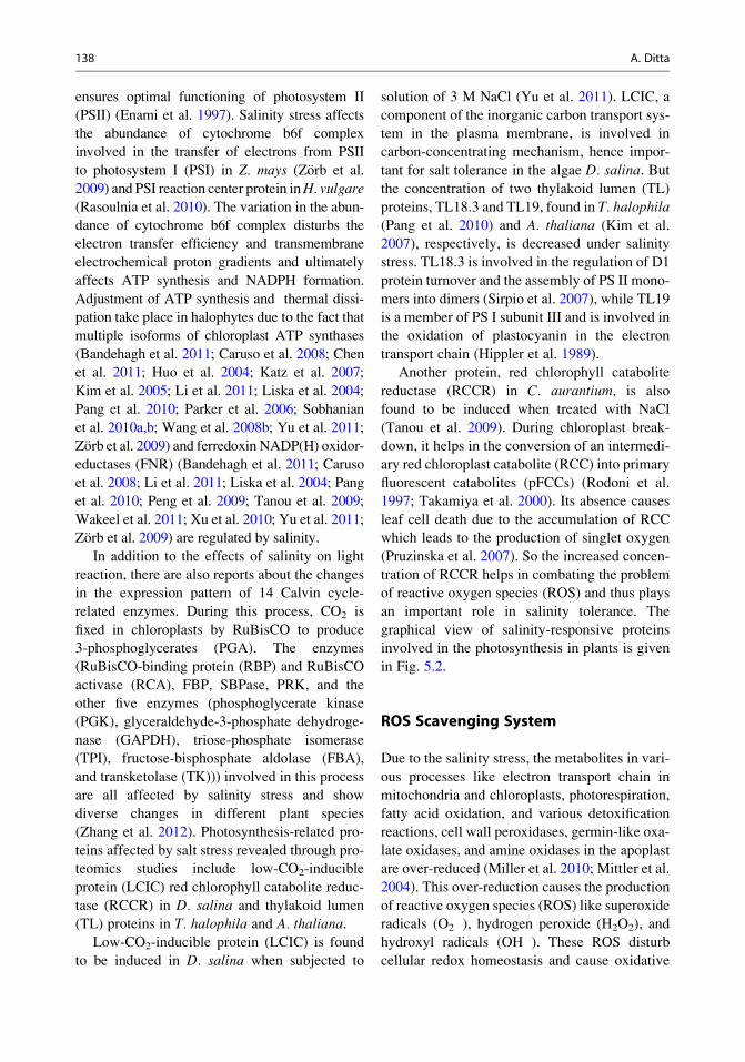
In addition to the effects of salinity on light
reaction, there are also reports about the changes
in the expression pattern of 14 Calvin cycle-
related enzymes. During this process, CO2 is
fixed in chloroplasts by RuBisCO to produce
3-phosphoglycerates (PGA). The enzymes
(RuBisCO-binding protein (RBP) and RuBisCO
activase (RCA), FBP, SBPase, PRK, and the
other five enzymes (phosphoglycerate kinase
(PGK), glyceraldehyde-3-phosphate dehydroge-
nase (GAPDH), triose-phosphate isomerase
(TPI), fructose-bisphosphate aldolase (FBA),
and transketolase (TK))) involved in this process
are all affected by salinity stress and show
diverse changes in different plant species
(Zhang et al. 2012). Photosynthesis-related pro-
teins affected by salt stress revealed through pro-
teomics studies include low-CO2-inducible
protein (LCIC) red chlorophyll catabolite reduc-
tase (RCCR) in D. salina and thylakoid lumen
(TL) proteins in T. halophila and A. thaliana.
Low-CO2-inducible protein (LCIC) is found
to be induced in D. salina when subjected to
solution of 3 M NaCl (Yu et al. 2011). LCIC, a
component of the inorganic carbon transport sys-
tem in the plasma membrane, is involved in
carbon-concentrating mechanism, hence impor-
tant for salt tolerance in the algae D. salina. But
the concentration of two thylakoid lumen (TL)
proteins, TL18.3 and TL19, found in T. halophila
(Pang et al. 2010) and A. thaliana (Kim et al.
2007), respectively, is decreased under salinity
stress. TL18.3 is involved in the regulation of D1
protein turnover and the assembly of PS II mono-
mers into dimers (Sirpio et al. 2007), while TL19
is a member of PS I subunit III and is involved in
the oxidation of plastocyanin in the electron
transport chain (Hippler et al. 1989).
Another protein, red chlorophyll catabolite
reductase (RCCR) in C. aurantium, is also
found to be induced when treated with NaCl
(Tanou et al. 2009). During chloroplast break-
down, it helps in the conversion of an intermedi-
ary red chloroplast catabolite (RCC) into primary
fluorescent catabolites (pFCCs) (Rodoni et al.
1997; Takamiya et al. 2000). Its absence causes
leaf cell death due to the accumulation of RCC
which leads to the production of singlet oxygen
(Pruzinska et al. 2007). So the increased concen-
tration of RCCR helps in combating the problem
of reactive oxygen species (ROS) and thus plays
an important role in salinity tolerance. The
graphical view of salinity-responsive proteins
involved in the photosynthesis in plants is given
in Fig. 5.2.
ROS Scavenging System
Due to the salinity stress, the metabolites in vari-
ous processes like electron transport chain in
mitochondria and chloroplasts, photorespiration,
fatty acid oxidation, and various detoxification
reactions, cell wall peroxidases, germin-like oxa-
late oxidases, and amine oxidases in the apoplast
are over-reduced (Miller et al. 2010; Mittler et al.
2004). This over-reduction causes the production
of reactive oxygen species (ROS) like superoxide
radicals (O2�), hydrogen peroxide (H2O2), and
hydroxyl radicals (OH�). These ROS disturb
cellular redox homeostasis and cause oxidative
138 A. Ditta
damage to many cellular components and
structures (Jithesh et al. 2006; Parida and Das
2005; Zhang et al. 2001a, b).
To cope with this problem, plants need to acti-
vate the ROS scavenging system for enhanced salt
tolerance (Zhang et al. 2001a, b). Through the
advancement in the field of proteomic, scientists
have discovered 184 protein IDs (representing 12
UPs) as ROS scavenging-related proteins, and
most of them (143 IDs) are induced by salinity
in 24 plant species (Nito et al. 2007). These pro-
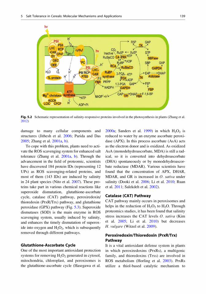
teins take part in various chemical reactions like
superoxide dismutation, glutathione-ascorbate
cycle, catalase (CAT) pathway, peroxiredoxin/
thioredoxin (PrxR/Trx) pathway, and glutathione
peroxidase (GPX) pathway (Fig. 5.3). Superoxide
dismutases (SOD) is the main enzyme in ROS
scavenging system, usually induced by salinity,
and enhances the timely dismutation of superox-
ide into oxygen and H2O2, which is subsequently
removed through different pathways.
Glutathione-Ascorbate CycleOne of the most important antioxidant protection
systems for removing H2O2 generated in cytosol,
mitochondria, chloroplast, and peroxisomes is
the glutathione-ascorbate cycle (Hasegawa et al.
2000a; Sanders et al. 1999) in which H2O2 is
reduced to water by an enzyme ascorbate peroxi-
dase (APX). In this process ascorbate (AsA) acts
as the electron donor and is oxidized. As oxidized
AsA (monodehydroascorbate, MDA) is still a rad-
ical, so it is converted into dehydroascorbate
(DHA) spontaneously or by monodehydroascor-
bate reductase (MDAR). Various scientists have
found that the concentration of APX, DHAR,
MDAR, and GR is increased in O. sativa under
salinity (Dooki et al. 2006; Li et al. 2010; Ruan
et al. 2011; Salekdeh et al. 2002).
Catalase (CAT) PathwayCAT pathway mainly occurs in peroxisomes and
helps in the reduction of H2O2 to H2O. Through
proteomics studies, it has been found that salinity
stress increases the CAT levels O. sativa (Kim
et al. 2005; Li et al. 2010) but decreases
H. vulgare (Witzel et al. 2009).
Peroxiredoxin/Thioredoxin (PrxR/Trx)PathwayIt is a vital antioxidant defense system in plants
in which peroxiredoxins (PrxRs), a multigenic
family, and thioredoxins (Trxs) are involved in
ROS metabolism (Horling et al. 2003). PrxRs
utilize a thiol-based catalytic mechanism to
Fig. 5.2 Schematic representation of salinity-responsive proteins involved in the photosynthesis in plants (Zhang et al.
2012)
5 Salt Tolerance in Cereals: Molecular Mechanisms and Applications 139
reduce H2O2 and are regenerated using Trxs as
electron donors (Dietz 2011). Advancement in the
field of proteomics has revealed that salinity
affects the two main proteins, that is, PrxRs
(Askari et al. 2006; Caruso et al. 2008; Chatto-
padhyay et al. 2011; Du et al. 2010; Kim et al.
2007; Ndimba et al. 2005; Pang et al. 2010; Peng
et al. 2009; Rasoulnia et al. 2010; Sobhanian et al.
2010b; Wang et al. 2008b, 2009; Yu et al. 2011;
Zorb et al. 2010) and Trxs (Du et al. 2010; Lee
et al. 2004; Nohzadeh et al. 2007; Sobhanian et al.
2010a; Wang et al. 2007), of this pathway. In Z.mays, the concentration of PrxRs is increased
(Zorb et al. 2010).
Glutathione Peroxidase (GPX) PathwayThis pathway is one of the major mechanisms of
ROS scavenging system (Yoshimura et al. 2004)
in which GPX can reduce H2O2 to the
corresponding hydroxyl compounds using GSH
and/or other reducing equivalents. The concen-
tration of GPXs is increased in S. europaea
(Li et al. 2011), while reduced in S. aegyptiaca(Askari et al. 2006) under salt stress conditions.
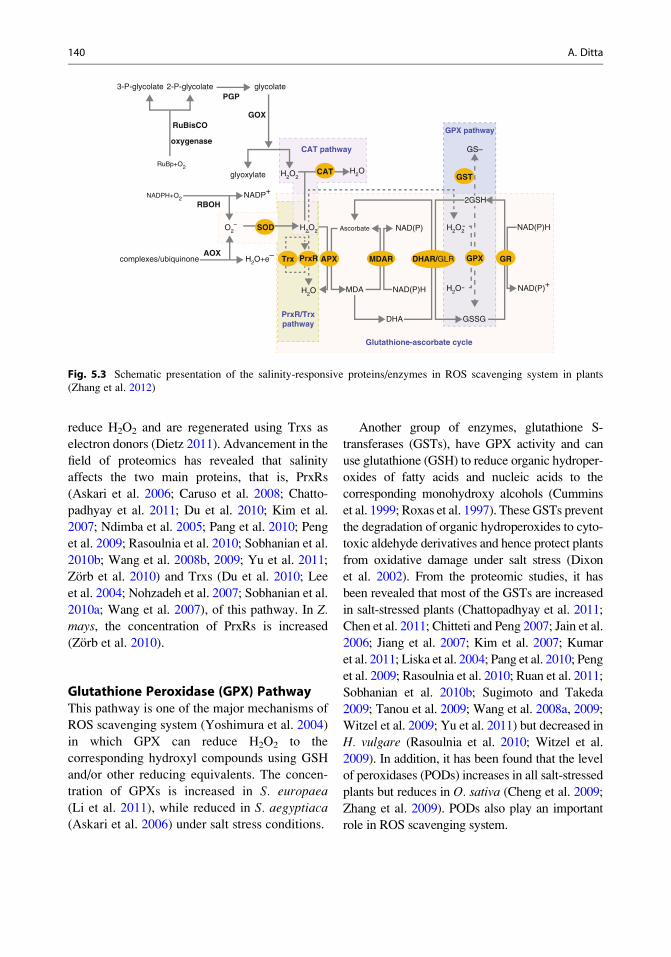
Another group of enzymes, glutathione S-
transferases (GSTs), have GPX activity and can
use glutathione (GSH) to reduce organic hydroper-
oxides of fatty acids and nucleic acids to the
corresponding monohydroxy alcohols (Cummins
et al. 1999; Roxas et al. 1997). These GSTs prevent
the degradation of organic hydroperoxides to cyto-
toxic aldehyde derivatives and hence protect plants
from oxidative damage under salt stress (Dixon
et al. 2002). From the proteomic studies, it has
been revealed that most of the GSTs are increased
in salt-stressed plants (Chattopadhyay et al. 2011;
Chen et al. 2011; Chitteti and Peng 2007; Jain et al.
2006; Jiang et al. 2007; Kim et al. 2007; Kumar
et al. 2011; Liska et al. 2004; Pang et al. 2010; Peng
et al. 2009; Rasoulnia et al. 2010; Ruan et al. 2011;
Sobhanian et al. 2010b; Sugimoto and Takeda
2009; Tanou et al. 2009; Wang et al. 2008a, 2009;
Witzel et al. 2009; Yu et al. 2011) but decreased in
H. vulgare (Rasoulnia et al. 2010; Witzel et al.
2009). In addition, it has been found that the level
of peroxidases (PODs) increases in all salt-stressed
plants but reduces in O. sativa (Cheng et al. 2009;
Zhang et al. 2009). PODs also play an important
role in ROS scavenging system.
3-P-glycolate 2-P-glycolate
RuBisCO
oxygenase
RuBp+O2
NADPH+O2
complexes/ubiquinoneAOX
PGPglycolate
GOX
glyoxylate
RBOHNADP+
H2O2
O2–
H2O+e–
H2O2 H2O2
H2O
H2OH2O
Ascorbate
MDA NAD(P)H
NAD(P)HNAD(P)
NAD(P)+
DHAPrxR/Trxpathway
CAT pathway
GSSG
Glutathione-ascorbate cycle
GPX pathway
GS–
2GSH
GPX
SOD
Trx
GST
PrxR
CAT
APX MDAR DHAR/GLR GR
Fig. 5.3 Schematic presentation of the salinity-responsive proteins/enzymes in ROS scavenging system in plants
(Zhang et al. 2012)
140 A. Ditta
Osmotic Homeostasis
As mentioned earlier, physiological water deficit
and osmotic stress are the main effects of salinity
in plants. In order to maintain the osmotic
homeostasis, plants tend to accumulate osmo-
lytes such as proline, soluble sugars, and glycine
betaine (GB). GB is a major osmolyte which not
only stabilizes the protein quaternary structure
and highly ordered membrane state but also
reduces lipid peroxidation during salinity stress
(Chen and Murata 2008; Chinnusamy et al. 2006;
Wang et al. 2004). There are a variety of proteins
which are involved in osmotic homeostasis. For
example, late embryogenesis abundant (LEA)
proteins function to protect the steady structure
of proteins, membranes, and cells (Chinnusamy
et al. 2006), and their expression is increased in
roots and hypocotyls of salt-treated O. sativa
(Li et al. 2010). In T. aestivum, cold-regulated
proteins and cold-responsive group-3 LEA-/
RAB-related COR proteins are induced under
stress conditions (Caruso et al. 2008). Moreover,
in O. sativa panicles, the level of an ABA-/salt-
responsive 40 kDa protein, Osr40c1s, is
increased in response to salt stress (Dooki et al.
2006). This protein consists of 151 amino acids
in a duplicated domain which have the ability to
form amphiphilic a-helical structures that asso-
ciate with membrane proteins for salt tolerance
(Moons et al. 1997).
Salt Stress Signal Transduction
Ionic signaling, osmotic signaling, detoxification
signaling, and signaling to coordinate cell divi-
sion and expansion are all included in salt stress
signaling (Zhu 2002). For salt tolerance in plants,
the signal transduction is a burning issue, and
several salt-responsive signaling pathways have
been predicted which include salt overly sensi-
tive (SOS) signaling pathway, ABA signaling
pathway, Ca2+ signal transduction pathway,
protein kinase pathway, phospholipid pathway,
ethylene signaling pathway, and jasmonate acid
(JA)-induced signaling pathway (Cao et al. 2008;
Darwish et al. 2009; Mahajan et al. 2008; Zhu
2001a, b, 2002).
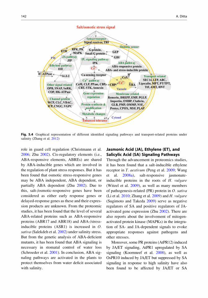
According to Tuteja (2007) and Zouari et al.
(2007), advancement in proteomic research has
identified about 85 IDs (24 UPs) as signal
transduction-related proteins in response to salt
stress (Fig. 5.4). The details of the signaling
pathways mentioned above are given as follows.
G-Protein-Coupled ReceptorsUnder salinity conditions, kinase-mediated pro-
tein phosphorylation and/or G-proteins are
involved in the transduction of stress signals
(e.g., ions, ROS, and ethylene), perceived by
their receptors/sensors, and helped in the regula-
tion of corresponding signaling and metabolic
pathways. From the current proteomic research,
it has been clear that there are two types of recep-
tors (the ethylene receptor (ETR) and a transform-
ing growth factor (TGF)-beta receptor-interacting
protein) which are induced in T. aestivum under
salinity stress (Peng et al. 2009). In addition, there
are also reports about the stimulation of some of
G-proteins/small G-proteins and three isoforms of
receptor protein kinase (RPK) identified from
T. aestivum (Peng et al. 2009) and O. sativa
(Dooki et al. 2006; Zhang et al. 2009) under saline
conditions. The above-stated facts suggest that
ethylene and ABA signaling pathways may be
involved in salt response (Cao et al. 2008). In
addition to the above-stated receptors, G-protein-
coupled receptors are dynamically regulated to
cope with salinity. It has been found through the
proteomic studies that the reduced levels of a
signal receiver and G-proteins/small G-proteins
in T. aestivum (Peng et al. 2009) and O. sativa
(Chitteti and Peng 2007), as well as two
abundance-changed guanine nucleotide exchange
proteins (GEP) are involved in small GTPase acti-
vation in O. sativa (Chitteti and Peng 2007).
Abscisic Acid (ABA) Signaling PathwayABA signaling pathway is an endogenous mes-
senger which helps in controlling plant water
status and osmotic stress tolerance through its
5 Salt Tolerance in Cereals: Molecular Mechanisms and Applications 141
role in guard cell regulation (Christmann et al.
2006; Zhu 2002). Cis-regulatory elements (i.e.,
ABA-responsive elements, ABREs) are shared
by ABA-inducible genes which are involved in
the regulation of plant stress responses. But it has
been found that osmotic stress-responsive genes
may be ABA independent, ABA dependent, or
partially ABA dependent (Zhu 2002). Due to
this, salt-/osmotic-responsive genes have been
considered as either early response genes or
delayed-response genes as these and their expres-
sion products are unknown. From the proteomic
studies, it has been found that the level of several
ABA-related proteins such as ABA-responsive
proteins (ABR17 and ABR18) and ABA-/stress-
inducible proteins (ASR1) is increased in O.sativa (Salekdeh et al. 2002) under salinity stress.
But from the genetic analysis of ABA-deficient
mutants, it has been found that ABA signaling is
necessary in stomatal control of water loss
(Schroeder et al. 2001). In conclusion, ABA sig-
naling pathways are activated in the plants to
protect themselves from water deficit associated
with salinity.
Jasmonic Acid (JA), Ethylene (ET), andSalicylic Acid (SA) Signaling PathwaysThrough the advancement in proteomics studies,
it has been found that a salt-inducible ethylene
receptor in T. aestivum (Peng et al. 2009; Wang
et al. 2008a), salt-responsive jasmonate-
inducible proteins in the roots of H. vulgare
(Witzel et al. 2009), as well as many members
of pathogenesis-related (PR) protein in O. sativa
(Li et al. 2010; Zhang et al. 2009) and H. vulgare
(Sugimoto and Takeda 2009) serve as negative
regulators of SA and positive regulators of JA-
activated gene expression (Zhu 2002). There are
also reports about the involvement of mitogen-
activated protein kinase (MAPKs) in the integra-
tion of SA- and JA-dependent signals to evoke
appropriate responses against pathogens and
other stresses.
Moreover, some PR proteins (AtPR12) induced
by JA/ET signaling, AtPR1 upregulated by SA
signaling (Koornneef et al. 2008), as well as
OsPR10 induced by JA/ET but suppressed by SA
signaling in response to high salinity have also
been found to be affected by JA/ET or SA
Fig. 5.4 Graphical representation of different identified signaling pathways and transport-related proteins under
salinity (Zhang et al. 2012)
142 A. Ditta
signaling. So it has been clear that powerful appli-
cations of proteomics have helped us in unraveling
the molecular mechanisms underlying hormone
signaling in salt tolerance.
Ca2+/Calmodulin (CaM) SignalingPathwayIt is calcium (Ca2+)-dependent signaling network
that has been reported to mediate Na+ homeosta-
sis and salt resistance in many crop plants
(Mahajan et al. 2008). For example, in Z. mays
chloroplast, a Na+ sensing element and Ca2+
sensing receptor are reported to be induced at
25 mM NaCl for 1 h but decreased after 4 h
(Zorb et al. 2009). Calcium-binding proteins
(CBPs) are regulated by salinity in O. sativa (Li
et al. 2010). Moreover, salinity regulates the
dynamics of calmodulin in O. sativa (Yan et al.
2005) and in Zea mays (Zorb et al. 2010) and thatof calcineurin-like phosphoesterase in T. aesti-
vum (Peng et al. 2009) and in O. sativa (Li et al.
2010). These changes modulate the levels of
intracellular Ca2+ and induce specific protein
kinase/phosphatase systems (Jiang et al. 2007).
So Ca2+ signaling network is closely related to
the activation of the SOS signal transduction
pathway, which is responsible for cellular Na+/
K+ homeostasis and the osmolytes accumulation
(Zhu 2002).
14-3-3 Proteins14-3-3 proteins are a group of proteins which are
commonly found and are multifunctional regula-
tors of many cellular signaling pathways. This
group of proteins interacts with a number of
signaling molecules like calcium-dependent pro-
tein kinase (CDPK) and mitogen-activated pro-
tein kinase (MAPK). By interacting with the C
terminus which is essential for the control of ion
transport and cytoplasmic pH, these proteins act
as positive regulators of plasma membrane (PM)
H+-ATPase (Palmgren 1998). These proteins are
also known to be involved in response to salinity
at multiple levels including regulating target
proteins with functions including signaling, tran-
scription activation, and defense and also work-
ing as components of transcription factor
complexes associated with ABA-induced gene
expression. Through the advancement in proteo-
mics, it has been found that in T. aestivum
(Wang et al. 2008a), O. sativa (Cheng et al.
2009; Nohzadeh et al. 2007), and Z. mays (Zhu
2003), many members of this group such as 14-3-
3 protein (gi13928452, and gi12229593) (Jain
et al. 2006; Zorb et al. 2010), 14-3-3-like protein
(gi1168189, and gi7267542) (Wang et al. 2008a),
GF14a(XP-48289), GF14b (gi50924768)
(Nohzadeh et al. 2007), and GF14 kappa isoform
(gi30698122) (Ndimba et al. 2005) are regulated
by salinity conditions. So we can conclude that
14-3-3 proteins regulate multiple pathways
involved in salt stress response.
Ion Homeostasis and Cross-MembraneTransport
Under salinity conditions, aqueous and ionic
thermodynamic equilibrium is altered through
high apoplastic levels of Na+ and Cl� which
results in hyperosmotic stress, ionic imbalance,
and toxicity. So to cope with this problem, plants
reestablish cellular ion homeostasis by regulating
ion uptake/exclusion and in vivo compartmental-
ization. Ion (e.g., K+ and Na+) homeostasis, a
fine-tuned process, mainly relies on the proton-
motive forces created by the action of H+-
ATPases, various ion channels, and transporters
(Gong et al. 2001). From the recent studies, it has
been found that for the regulation of the activity
of plasma membrane H+-ATPases, phototropin
and 14-3-3 proteins have to work cooperatively
as well as independently for opening and closing
of the ion channels (e.g., K+ channel) (Inoue
et al. 2005; van den Wijngaard et al. 2005).
Currently, various researchers through proteomic
studies have found that salinity stress signifi-
cantly affects H+-ATPase (Barkla et al. 2009;
Cheng et al. 2009; Du et al. 2010; Jiang et al.
2007; Katz et al. 2007; Manaa et al. 2011; Nat
et al. 2004; Ndimba et al. 2005; Pang et al. 2010;
Wang et al. 2008a, 2009; Yu et al. 2011; Zorb
et al. 2004), ATP-binding cassette (ABC) trans-
porter (Wang et al. 2008a, b, 2009), and other ion
channels and transporters (Li et al. 2011; Peng
et al. 2009; Sobhanian et al. 2010b; Wakeel et al.
5 Salt Tolerance in Cereals: Molecular Mechanisms and Applications 143
2011; Wang et al. 2008a, b, 2009; Zorb et al.
2010).
H+-ATPaseFor the maintenance of ion homeostasis in plant
cells, H+-ATPases are one of the most important
enzymes because most of the vacuolar H+-
ATPases in glycophytes like O. sativa (Cheng
et al. 2009), T. aestivum (Wang et al. 2008a),
and Z. mays (Zorb et al. 2004) are induced
under salinity stress. For Na+ transport by salt
overly sensitive (SOS1) which is essential for
salt tolerance, it has been that the level of H+-
ATPases is increased which provides a more
strong driving force for this transport (Kim
et al. 2007; Zhu 2001a, b, 2003). The proton
electrochemical gradient for vacuolar Na+/H+
antiporter to compartmentalize Na+ in the
vacuoles is generated through vacuolar H+-
ATPases, the major H+-pumps on the tonoplast
(Chinnusamy et al. 2005). For Na+ sequestration
and osmotic adjustment under salinity stress,
increased levels and/or activities of the vacuolar
H+-ATPases are found to be a cost-effective
strategy (Pang et al. 2010). Moreover, it has
also been found that mitochondrial-/chloroplast-
located H+-ATPases are also involved in ion
homeostasis (Huo et al. 2004; Kim et al. 2005;
Ndimba et al. 2005).
ABC Transporters and Other TransportersIn T. aestivum, it has been found that ABC trans-
porters (the in charge of transporting of stress-
related secondary metabolites, such as alkaloids,
terpenoids, polyphenols, and quinines (Yazaki
2006)) are induced under salinity stress (Wang
et al. 2008a; Peng et al. 2009). There are three
proteins which cause abundant changes in plants
under salinity and include ferritin (Chen et al.
2009; Parker et al. 2006; Wang et al. 2009), iron
deficiency-induced protein (IDI), and iron
deficiency-specific protein (IDS) (Witzel et al.
2009). The first protein, ferritin, through the
Fenton reaction, helps in the sequestration of
excess free irons and prevents formation of
hydroxyl radicals (Laohavisit et al. 2010; Parker
et al. 2006) and is reported to be induced in O.
sativa (Parker et al. 2006) under certain salinity
conditions. But in contrast, the level of other two
transporters, that is, IDIs and IDSs, is decreased
in H. vulgare under salinity stress (Witzel et al.
2009) which is beneficial in avoiding excessive
ion uptake.
Ion Channel ProteinsTo maintain ion homeostasis under salinity
stress, the level of different ion channels is
changed, that is, increases or decreases; for
example, the level of voltage-gated potassium
channels in T. aestivum (Peng et al. 2009) is
induced which is crucial for a balance of K+/
Na+ in the cells, but in case of a cyclic
nucleotide-gated ion channel (CNGC), the level
is reduced as a nonselective cation channel
(Wang et al. 2008a). The direct binding of cyclic
nucleotides (cAMP and cGMP) helps in the
opening of CNGC of which the activity is of little
voltage dependence, but Ca2+/calmodulin and
phosphorylation help in its modulation. Another
ion channel, annexin, revealed through proteo-
mic studies, is a Ca2+-permeable channel at
endomembrane and plasma membrane for the
formation of a ROS-stimulated passive Ca2+
transport pathway (Laohavisit et al. 2010), and
its level is reported to be induced in various
cereal crops like O. sativa (Li et al. 2010) and
T. aestivum (Peng et al. 2009). Its increased level
plays a vital role in osmotic adjustment and
subsequently cell expansion and exocytosis
(Faurobert et al. 2007; Lee et al. 2004). More-
over, it has been found that the level of voltage-
dependent anion channel protein (VDAC), a
barrel protein located at the outer mitochondrial
membrane and responsible for passage of small
molecules (<1,000 Da) into the intermembrane
space, is induced under salinity stress in Z. mays
(Sugimoto and Takeda 2009). Its dynamic
changes are found to influence the mitochondrial
respiration (Rostovtseva and Bezrukov 2008;
Rostovtseva et al. 2008).
Plasma Membrane and Other Membrane-Associated ProteinsTwenty four plasma membrane proteins are
reported to be found in rice which are induced
under salinity stress and regulate the development
144 A. Ditta
of plasma membrane polypeptides containing a
Glu-rich site at the C terminus, responsible for
calcium binding in Ca2+ signal transduction path-
way (Yuasa and Maeshima 2000). In addition, a
plant-specific PM/lipid-raft protein, remorin, helps
in maintaining the membrane skeletons (Bariola
et al. 2004) and thus contributes in the stabilization
of damaged PM under salinity stress (Cheng et al.
2009; Nohzadeh et al. 2007). Moreover, the level
of importin (a nuclear membrane transporter) and
an outer mitochondrial membrane porin in T. aes-
tivum (Peng et al. 2009; Wang et al. 2008a; Witzel
et al. 2009) is reduced under salinity stress.
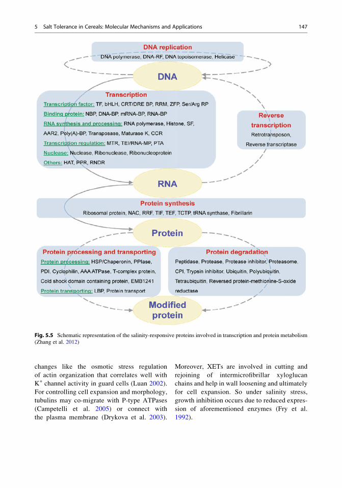
Transcription and Protein Fates
Changes in transcriptional regulatory networks
of cis-/trans-elements and transcription factors
can be triggered through aforementioned signal-
ing systems. Through the advancement in pro-
teomic research, it has been found that the levels
of transcription factors and transcription-related
proteins are regulated under salinity stress and
play a vital role in salt tolerance (Zhang et al.
2012). To cope with salt stress in T. aestivum
(Peng et al. 2009; Wang et al. 2008a), the salt-
induced transcription factor, basic transcription
factor 3 (BTF3), is an important regulatory com-
ponent and controls diverse processes. In addi-
tion, the increased levels of DNA polymerases in
Z. mays (Zorb et al. 2004) and DNA helicases in
T. aestivum (Wang et al. 2008a) enhance DNA
replication, unwinding, and transcription under
salinity. Furthermore, there are also reports about
the effects of salinity on some RNA processing-
and spicing-related proteins such as maturase K
(Chattopadhyay et al. 2011; Wang et al. 2009;
Zorb et al. 2010), nucleic acid- binding proteins
(Aghaei et al. 2008; Bandehagh et al. 2011; Car-
uso et al. 2008; Chen et al. 2009; Jain et al. 2006;
Kim et al. 2005; Pang et al. 2010; Tanou et al.
2009; Wang et al. 2008b; Witzel et al. 2009),
glycine-rich RNA-binding proteins (Askari
et al. 2006; Dooki et al. 2006; Jiang et al. 2007;
Manaa et al. 2011), and RNA splicing factors
(Wang et al. 2008a; Yan et al. 2005). In abiotic
stress adaptation, protein synthesis plays a very
important role, and proteomics studies have
found that many of its components including
different ribosomal proteins (Aghaei et al. 2008;
Bandehagh et al. 2011; Chattopadhyay et al.
2011; Chen et al. 2009; Chitteti and Peng
2007; Dani et al. 2005; Du et al. 2010; Jiang
et al. 2007; Kim et al. 2005; Ndimba et al.
2005; Pang et al. 2010; Peng et al. 2009;
Sobhanian et al. 2010a, b; Veeranagamallaiah
et al. 2008; Wang et al. 2008b; Zorb et al. 2004,
2010), translation initiation factors (Jiang et al.
2007; Ndimba et al. 2005; Pang et al. 2010; Parker
et al. 2006; Peng et al. 2009; Wang et al. 2008a, b,
2009; Yu et al. 2011), poly(A)-binding proteins
(Jiang et al. 2007; Witzel et al. 2009), translation
elongation factors (Chen et al. 2009; Liska et al.
2004; Ndimba et al. 2005; Pang et al. 2010; Peng
et al. 2009; Sobhanian et al. 2010b; Witzel et al.
2010), translationally controlled tumor proteins
(Nat et al. 2004; Pang et al. 2010; Sobhanian
et al. 2010b; Vincent et al. 2007; Witzel et al.
2010; Yu et al. 2011; Zorb et al. 2010), RNA
recognition motif (RRM)-containing proteins
(Pang et al. 2010; Yu et al. 2011), and tRNA
synthases (Ndimba et al. 2005; Pang et al. 2010;
Peng et al. 2009; Wen et al. 2010) are altered in
expression under salinity stress conditions.
Generally, salinity stress represses protein
synthesis (Tuteja 2007). However, the level of
some of the above proteins is increased which
shows that normal cellular processes are required
for the maintenance of protein synthesis activities
under salinity stress (Zhang et al. 2012). For main-
taining normal cellular functions under salinity
stress, correct protein folding and transport is
crucial. For this, heat shock proteins (HSPs) and
other molecular chaperons play a very important
role in protein structure stabilization and subcellular
localization (Vierling 1991). Salinity affects vari-
ous HSPs/chaperonins (Aghaei et al. 2008;
Chattopadhyay et al. 2011; Chen et al. 2009,
2011; Chitteti and Peng 2007; Dani et al. 2005;
Du et al. 2010; Geissler et al. 2010; Jain et al.
2006; Jiang et al. 2007; Katz et al. 2007; Kim
et al. 2005, 2007; Li et al. 2010; Liska et al. 2004;
Manaa et al. 2011; Nat et al. 2004; Ndimba et al.
2005; Pang et al. 2010; Peng et al. 2009; Razaviza-
deh et al. 2009; Sengupta and Majumder 2009;
5 Salt Tolerance in Cereals: Molecular Mechanisms and Applications 145
Sobhanian et al. 2010a, b; Tanou et al. 2009; Wang
et al. 2008a, b, 2009; Wen et al. 2010; Witzel et al.
2010; Xu et al. 2010; Yu et al. 2011; Zorb
et al. 2010), luminal-binding proteins (LBP) (Kim
et al. 2007; Liska et al. 2004; Pang et al. 2010;
Wang et al. 2008b, 2009), peptidyl-prolyl cis-trans
isomerases (Aghaei et al. 2008; Askari et al. 2006;
Chen et al. 2011; Tanou et al. 2009; Zorb et al.
2009), protein disulfide isomerases (PDI) (Chen
et al. 2011; Jiang et al. 2007; Nohzadeh et al.
2007; Pang et al. 2010; Yu et al 2011), T-complex
proteins (Pang et al. 2010;Wang et al. 2008a),AAA
ATPase superfamily proteins (Chen et al. 2011;
Wen et al. 2010; Yu et al 2011), and cold shock
domain-containing proteins (Huo et al. 2004; Peng
et al. 2009; Ruan et al. 2011) which are involved
in maintaining normal protein folding, repair,
and renaturation of the stress-damaged proteins
(Fig. 5.5). In addition, for the selective degradation
of proteins, plants use proteasome pathways. Some
members of these pathways such as ubiquitin/poly-
ubiquitin/tetraubiquitin (Du et al. 2010; Katz et al.
2007; Xu et al. 2010), SKP1 protein (Liska et al.
2004), proteasome components (Askari et al. 2006;
Chattopadhyay et al. 2011; Jiang et al. 2007; Kim
et al. 2005; Liska et al. 2004; Peng et al. 2009;
Sobhanian et al. 2010b; Tanou et al. 2009; Vincent
et al. 2007; Wang et al. 2008a, b, 2009), various
proteases (Katz et al. 2007; Liska et al. 2004;
Manaa et al. 2011; Pang et al. 2010; Peng et al.
2009; Veeranagamallaiah et al. 2008; Wang et al.
2008b, 2009; Xu et al. 2010; Yu et al. 2011;
Zorb et al. 2009;) and peptidases (Chitteti and
Peng 2007; Jiang et al. 2007; Li et al. 2010;
Liska et al. 2004; Manaa et al. 2011; Ndimba
et al. 2005; Pang et al. 2010; Wang et al. 2008b;
Yu et al. 2011), protease inhibitors (Aghaei et al.
2009; Chen et al. 2011; Peng et al. 2009; Sobhanian
et al. 2010a), and reversed protein-methionine-S-
oxide reductases (Pang et al. 2010) under salinity
stress exhibit tremendous changes. Degradation
of proteins not only is important in protein
turnover during ubiquitin-mediated degradation
of proteins but also helps in the regulation of
other cellular processes such as signal transduction
and transcription. So, these salt-responsive pro-
teins have a vital role to play in salinity tolerance.
Cytoskeleton and Cell Structure
For cell turgor maintenance, a rapid remodeling
of cytoskeleton for the adjustment of cell size
takes place during salinity stress (Pang et al.
2010; Li et al. 2011). Through the advancement
in the field of proteomics, it has been found that
basic cytoskeleton components such as actin
(Cheng et al. 2009; Jiang et al. 2007; Li et al.
2010; Tanou et al. 2009; Xu et al. 2010) and
tubulin (Jiang et al. 2007; Katz et al. 2007; Kim
et al. 2007; Liska et al. 2004; Pang et al. 2010;
Peng et al. 2009) and other cytoskeleton-related
proteins (some actin-binding proteins (ABPs))
(Yan et al. 2005), kinesin motor (Chitteti and
Peng 2007; Sobhanian et al. 2010a; Wang et al.
2009), myosin (Cheng et al. 2009; Peng et al.
2009; Wang et al. 2009), and xyloglucan endo-
transglycosylase (XET) hydrolases (Zorb et al.
2010) are changed under salinity stress. ABPs
such as actin-depolymerizing factors (ADFs)
(Sobhanian et al. 2010b), profilins (Askari
et al. 2006; Du et al. 2010; Wang et al. 2009),
and cyclase-associated proteins (CAPs)
(Ndimba et al. 2005) play key roles in the remo-
deling as these have the ability to bind with
actin cytoskeletons; for example, ADFs pro-
mote filamentous actin disassembly and thus
modulate the dynamic organization of actin
cytoskeletons. Profilin has the ability to join
with actin monomers and cause polymeriza-
tion/depolymerization of actin filaments to
maintain cell structure integrity, cell mobility,
tumor cell metastasis, and growth factor signal-
ing (Staiger et al. 1997). Similarly CAPs
which are multifunctional ABPs are involved
in various signal transduction pathways
involved in cell growth, development, vesicle
trafficking, and endocytosis. Under salinity
stress, it has been found that cytoskeleton
dynamics is associated with other physiological
146 A. Ditta
changes like the osmotic stress regulation
of actin organization that correlates well with
K+ channel activity in guard cells (Luan 2002).
For controlling cell expansion and morphology,
tubulins may co-migrate with P-type ATPases
(Campetelli et al. 2005) or connect with
the plasma membrane (Drykova et al. 2003).
Moreover, XETs are involved in cutting and
rejoining of intermicrofibrillar xyloglucan
chains and help in wall loosening and ultimately
for cell expansion. So under salinity stress,
growth inhibition occurs due to reduced expres-
sion of aforementioned enzymes (Fry et al.
1992).
Fig. 5.5 Schematic representation of the salinity-responsive proteins involved in transcription and protein metabolism
(Zhang et al. 2012)
5 Salt Tolerance in Cereals: Molecular Mechanisms and Applications 147
Cross-Tolerance to Multiple Stresses
Various signaling and metabolic pathways are
connected into networks because plants have
developed cross-tolerance mechanisms to cope
with different stresses at the same time (Tuteja
2007); for example, in salt tolerance, some biotic
stress-responsive proteins/genes play important
roles. Carbohydrate-binding proteins like lectins
are involved not only in defense against preda-
tors and pathogens (De Hoff et al. 2009) but also
in plant salt tolerance (Van Damme et al. 2004).
Researchers through proteomics studies have
found that salinity stress enhances not only the
levels of lectins in O. sativa (Chitteti and Peng
2007) and S. bicolor (Kumar et al. 2011) but also
that of cytoplasmic mannose-binding lectins
(Claes et al. 1990; Hirano et al. 2000; Zhang
et al. 2000) and salt stress-induced proteins (salt
proteins) (Chitteti and Peng 2007; Wen et al.
2010). The biotic stress-related proteins,
revealed through proteomics, include elicitor
peptides (Aghaei et al. 2009; Wang et al. 2009),
disease-related/resistance proteins (Chattopad-
hyay et al. 2011; Chitteti and Peng 2007; Nat
et al. 2004; Peng et al. 2009; Wang et al. 2008a,
2009), hypersensitive-induced response proteins
(Cheng et al. 2009; Nohzadeh et al. 2007),
pathogenesis-related proteins (Chattopadhyay
et al. 2011; Jain et al. 2006; Li et al. 2010;
Manaa et al. 2011; Pang et al. 2010; Sugimoto
and Takeda 2009; Vincent et al. 2007; Wang
et al. 2008b, 2009; Zhang et al. 2009), stress-
inducible proteins (Dooki et al. 2006; Wang
et al. 2008a; Witzel et al. 2009), and universal
stress protein family (Kumar et al. 2011; Li et al.
2010; Ndimba et al. 2005; Wang et al. 2008a),
and while that of abiotic stress-related proteins
include cold-regulated proteins (Wang et al.
2009), cold-responsive LEA-/RAB-related COR
proteins (Caruso et al. 2008), and copper homeo-
stasis factors (Jiang et al. 2007). Salt tolerance is
also contributed by the multifunctional glyoxa-
lase system. Under salinity stress, plants tend to
accumulate high amounts of methylglyoxal
(MG) which is a by-product of glycolysis mainly
from triose phosphate. MG has detrimental
effects on plants as it involved in the inhibition
of cell proliferation (Ray et al. 1994), degrada-
tion of proteins, and inactivation of antioxidant
defense system (Martins et al. 2001). MG is
detoxified through glyoxalase system consisting
of glyoxalase I (GlyI) and glyoxalase II (GlyII).
From the previous molecular studies involving
transgenic plants, it has found that overexpres-
sion of GlyI and GlyII enhances plant salt toler-
ance (Singla-Pareek et al. 2003, 2008; Yadav
et al. 2005). From the proteomics, it has been
found that the level of GlyI increases in O. sativa
(Chitteti and Peng 2007; Li et al. 2010) when
treated with 150 mM NaCl for 6 or 48 h (Jiang
et al. 2007) which implies that the glyoxalase
system is under dynamic regulation.
Conclusions and Perspectives
A sophisticated fine-tuned signaling and meta-
bolic network is there under plant salinity
response and tolerance. Significant discoveries
of salinity-responsive genes, proteins, and meta-
bolites in different cellular pathways important
for salt stress response and tolerance have been
made through previous morphological, physio-
logical, genetic, and genomic analyses. But a
large gap exists in the systematic understanding
of the molecular processes and networks, as this
understanding is still in the beginning. Modern
highly sophisticated and state-of-the-art proteo-
mics techniques have helped us in the acquisition
of more detailed quantitative information on the
temporal and spatial expression of proteins. Cur-
rently, overall 2,171 proteins have been identi-
fied in 34 plants, but the need of the hour is to fill
the gaps present between the research and actual
field conditions.
The dynamic nature of the proteins involved
under salinity stress further provides invaluable
information toward understanding of the under-
lying sophisticated cellular and molecular pro-
cesses like photosynthesis, energy metabolism,
ROS scavenging, ion/osmotic homeostasis,
148 A. Ditta
signaling transduction, transcription and transla-
tional regulation, and cytoskeleton dynamics.
However, a large gap still exists in our knowl-
edge of transmembrane ion transport and cellular
compartmentalization, sensors/receptors in sig-
naling transduction, molecules in long distance
signaling, and metabolites in energy supply, espe-
cially in case of cereals. Moreover, molecular
interactions and pathway cross talks should have
to be arranged, and future targets should be set on
the basis of the gaps found during these talks.
New state-of-the-art technologies like nanotech-
nology should be used in the understanding of
molecular mechanisms. Through nanotechnology,
enzyme biosensors (controlled-pore glass beads
with optical transducer element, polyurethane
foam with photothermal transducer element,
ion-selective membrane with either potentiomet-
ric or amperometric transducer element, and
screen-printed electrode with amperometric
transducer element) should be used for the
detection of very minute changes occurring during
the quantitative analysis of the salt tolerance
mechanisms. Also the advanced and sophisticated
proteomics approaches and technologies like mul-
tidimensional protein fractionation, isobaric tags
for relative and absolute quantitation (iTRAQ),
label-free quantification mass spectrometry, and
phosphoprotein and glycoprotein enrichment and
tagging will definitely help us in discovering low-
abundance proteins (e.g., transcriptional factors,
kinases, channels, and transporters) and novel
regulatory mechanisms (e.g., phosphorylation) in
salt stress signaling and metabolism pathways.
Integration of proteomics results with findings
from other large scale sources will definitely facil-
itate in the establishment of molecular networks
underlying salt stress response and tolerance in
cereals. After integration of the knowledge col-
lected, we will be able to predict the underlying
molecular mechanisms of salinity tolerance. It
will prove helpful toward the ultimate goal of
improving plant salt tolerance for enhanced yield
and bioenergy.
References
Abbasi FM, Komatsu S (2004) A proteomic approach to
analyze salt-responsive proteins in rice leaf sheath.
Proteomics 4:2072–2081
Aghaei K, Ehsanpour AA, Komatsu S (2008) Proteome
analysis of potato under salt stress. J Proteome Res
7:4858–4868
Aghaei K, Ehsanpour AA, Shah AH, Komatsu S (2009)
Proteome analysis of soybean hypocotyl and root
under salt stress. Amino Acids 36:91–98
Almoguera C, Shibata D, Forrester K, Matin J, Arnheim
N, Perucho M (1988) Most human carcinomas of the
exocrine pancreas contain mutant c-K-ras genes. Cell
53:549–554
Anuradha S, Rao SSR (2001) Effect of brassinosteroids
on salinity stress induced inhibition of seed germina-
tion and seedling growth of rice (Oryza sativa L.).
Plant Growth Regul 33:151–153
Ashraf M, Harris PJC (2004) Potential biochemical indi-
cators of salinity tolerance in plant. Plant Sci 166:3–16
Askari H, Edqvist J, Hajheidari M, Kafi M, Salekdeh GH
(2006) Effects of salinity levels on proteome of
Suaeda aegyptiaca leaves. Proteomics 6:2542–2554
Bandehagh A, Salekdeh GH, Toorchi M, Mohammadi A,
Komatsu S (2011) Comparative proteomic analysis of
canola leaves under salinity stress. Proteomics
11:1965–1975
Bariola PA, Retelska D, Stasiak A, Kammerer RA,
Fleming A, Hijri M, Frank S, Farmer EE (2004)
Remorins form a novel family of coiled coil-forming
oligomeric and filamentous proteins associated with
apical, vascular and embryonic tissues in plants. Plant
Mol Biol 55:579–594
Barkla BJ, Vera-Estrella R, Hernandez-Coronado M,
Pantoja O (2009) Quantitative proteomics of the tono-
plast reveals a role for glycolytic enzymes in salt
tolerance. Plant Cell 21:4044–4058
Blumwald E, Aharon GS, Apse MP (2000) Sodium trans-
port in plant cells. Biochemica et Biophysica Acta
1465:140–151
Bohnert HJ, Nelson DE, Jensen RG (1995) Adaptations to
environmental stresses. Plant Cell 7:1099–1111
Bressan RA, Hasegawa PM, Pardo JM (1998) Plants use
calcium to resolve salt stress. Trends Plant Sci
3:411–412
Campetelli AN, Previtali G, Arce CA, Barra HS, Casale
CH (2005) Activation of the plasma membrane H+-
ATPase of Saccharomyces cerevisiae by glucose is
mediated by dissociation of the H+-ATPase-acety-
lated tubulin complex. FEBS J 272:5742–5752
Cao YR, Chen SY, Zhang JS (2008) Ethylene signaling
regulates salt stress response: an overview. Plant Sig-
nal Behav 3:761–763
5 Salt Tolerance in Cereals: Molecular Mechanisms and Applications 149
Caruso G, Cavaliere C, Guarino C, Gubbiotti R, Foglia
P, Lagana A (2008) Identification of changes in Tri-ticum durum L. leaf proteome in response to salt
stress by two-dimensional electrophoresis and
MALDI-TOF mass spectrometry. Anal Bioanal
Chem 391:381–390
Chand R, Kumar P (2006) Country case study: India. In:
Thomas H (ed) Trade reforms and food security. Food
and Agriculture Organization of the United Nations,
Rome
Chattopadhyay A, Subba P, Pandey A, Bhushan D,
Kumar R, Datta A, Chakraborty S, Chakraborty N
(2011) Analysis of the grass pea proteome and identi-
fication of stress-responsive proteins upon exposure to
high salinity, low temperature, and abscisic acid treat-
ment. Phytochemistry 72:1293–1307
Chen TH, Murata N (2008) Glycinebetaine: an effective
protectant against abiotic stress in plants. Trends Plant
Sci 13:499–505
Chen SB, Gollop N, Heuer B (2009) Proteomic analysis
of salt stressed tomato (Solanum lycopersicum) seed-lings: effect of genotype and exogenous application of
glycinebetaine. J Exp Bot 60:2005–2019
Chen FG, Zhang S, Jiang H, Ma WJ, Korpelainen H, Li C
(2011) Comparative proteomics analysis of salt
response reveals sex-related photosynthetic inhibition
by salinity in Populus cathayana cuttings. J Proteome
Res 10:3944–3958
Cheng YW, Qi YC, Zhu Q, Chen X, Wang N, Zhao X,
Chen HY, Cui XJ, Xu LL, Zhang W (2009)
New changes in the plasma membrane-associated
proteome of rice roots under salt stress. Proteomics
9:3100–3114
Chinnusamy V, Jagendorf A, Zhu JK (2005) Understand-
ing and improving salt tolerance in plants: genetic and
metabolic engineering for value-added traits. Crop Sci
Soc Am 45:437–448
Chinnusamy V, Zhu J, Zhu JK (2006) Salt stress signaling
and mechanisms of plant salt tolerance. Genet Eng
27:141–177
Chitteti BR, Peng Z (2007) Proteome and phosphopro-
teome differential expression under salinity stress
in rice (Oryza sativa) roots. J Proteome Res
6:1718–1727
Christmann A, Moes D, Himmelbach A, Yang Y, Tang Y,
Grill E (2006) Integration of abscisic acid signalling
into plant responses. Plant Biol 8:314–325
Claes B, Dekeyser R, Villarroel R, Van den Bulcke M,
Bauw G, Van Montagu M, Caplan A (1990) Charac-
terization of a rice gene showing organ-specific
expression in response to salt stress and drought.
Plant Cell 2:19–27
Cummins I, Cole DJ, Edwards R (1999) A role for gluta-
thione transferases functioning as glutathione perox-
idases in resistance to multiple herbicides in black-
grass. Plant J 18:285–292
Cushman JC, Vermon DM, Bohnert HJ (1992) ABA and
the transcriptional control of CAM induction during
salt stress in the common ice plant. In: Verma DPS
(ed) Control of plant gene expression. CRC Press,
Boca Raton, pp 287–300
Dani V, Simon WJ, Duranti M, Croy RR (2005) Changes
in the tobacco leaf apoplast proteome in response to
salt stress. Proteomics 5:737–745
Darwish E, Testerink C, Khalil M, El-Shihy O,
Munnik T (2009) Phospholipid signaling responses
in salt-stressed rice leaves. Plant Cell Physiol
50:986–997
Dash M, Panda SK (2001) Salt stress induced changes in
growth and enzyme activities in germination Phaseo-lus mungo seeds. Biol Plant 44:587–589
De Hoff PL, Brill LM, Hirsch AM (2009) Plant lectins:
the ties that bind in root symbiosis and plant defense.
Mol Genet Genomics 282:1–15
Diedhiou CJ, Popova OV, Golldack D (2009) Transcript
profiling of the salt-tolerant Festuca rubra ssp. litor-
alis reveals a regulatory network controlling salt accli-
matization. J Plant Physiol 166:697–711
Dietz KJ (2011) Peroxiredoxins in plants and cyanobac-
teria. Antioxid Redox Signal 15:1129–1159
Dixon DP, Lapthorn A, Edwards R (2002) Plant glutathi-
one transferases. Genome Biol 3:No. REVIEWS3004
Dooki AD, Mayer-Posner FJ, Askari H, Zaiee AA,
Salekdeh GH (2006) Proteomic responses of rice
young panicles to salinity. Proteomics 6:6498–6507
Drykova D, Cenklova V, Sulimenko V, Volc J, Draber P,
Binarova P (2003) Plant gamma-tubulin interacts with
alpha beta-tubulin dimers and forms membrane-
associated complexes. Plant Cell 15:465–480
Du CX, Fan HF, Guo SR, Tezuka T, Li J (2010) Proteo-
mic analysis of cucumber seedling roots subjected to
salt stress. Phytochemistry 71:1450–1459
Enami I, Tohri A, Kamo M, Ohta H, Shen JR (1997)
Identification of domains on the 43 kDa chlorophyll-
carrying protein (CP43) that are shielded from tryptic
attack by binding of the extrinsic 33 kDa protein with
Photosystem II complex. Biochim Biophys Acta Bioe-
nerg 1320(1):17–26
Evans LT (1998) Feeding the ten billion: plants
and population growth, Cambridge, UK Cambridge
University Press
Faurobert M, Mihr C, Bertin N, Pawlowski T, Negroni L,
Sommerer N, Causse M (2007) Major proteome var-
iations associated with cherry tomato pericarp devel-
opment and ripening. Plant Physiol 143:1327–1346
Flowers TJ, Colmer TD (2008) Salinity tolerance in halo-
phytes. New Phytol 179:945–963
Fricke W, Akhiyarova G, Veselov D, Kudoyarova G
(2004) Rapid and tissue-specific changes in ABA
and in growth rate in response to salinity in barley
leaves. J Exp Bot 55:1115–1123
Fry SC, Smith RC, Renwick KF, Martin DJ, Hodge SK,
Matthews KJ (1992) Xyloglucan endotransglycosy-
lase, a new wall-loosening enzyme activity from
plants. Biochem J 282:821–828
Geissler N, Hussin S, Koyro HW (2010) Elevated atmo-
spheric CO2 concentration enhances salinity tolerance
in Aster tripolium L. Planta 231:583–594
150 A. Ditta
Gong ZZ, Koiwa H, Cushman MA, Ray A, Bufford D,
Kore-eda S, Matsumoto TK, Zhu JH, Cushman JC,
Bressan RA, Hasegawa PM (2001) Genes that are
uniquely stress regulated in salt overly sensitive
(SOS) mutants. Plant Physiol 126:363–375
Hamed K, Castagna A, Salem E, Ranieri A, Abdelly C
(2007) Sea Fennel (Crithmum martimum L.) under
salinity conditions: a comparison of leaf and root
antioxidant responses. Plant Growth Regul
53:185–194
Hasegawa PM, Bressan RA, Pardo JM (2000a) The dawn
of plant salt to tolerance genetics. Trends Plant Sci
5:317–319
Hasegawa PM, Bressan RA, Zhu J-K, Bohnert HJ
(2000b) Plant cellular and molecular responses to
high salinity. Annu Rev Plant Physiol Plant Mol Biol
51:463–499
Heidari M (2009) Variation in seed germination, seedling
growth, nucleic acids and biochemical component in
canola (Brassica nupus L.) under salinity stress. AsianJ Plants Sci 8:557–561
Hippler M, Ratajczak R, Haehnel W (1989) Identification
of the plastocyanin binding subunit of photosystem I.
FEBS Lett 250:280–284
Hirano K, Teraoka T, Yamanaka H, Harashima A,
Kunisaki A, Takahashi H, Hosokawa D (2000)
Novel mannose-binding rice lectin composed of
some isolectins and its relation to a stress-inducible
salT gene. Plant Cell Physiol 41:258–267
Horling F, Lamkemeyer P, Konig J, Finkemeier I,
Kandlbinder A, Baier M, Dietz KJ (2003) Divergent
light-, ascorbate-, and oxidative stress-dependent reg-
ulation of expression of the peroxiredoxin gene family
in Arabidopsis. Plant Physiol 131:317–325
Huo CM, Zhao BC, Ge RC, Shen YZ, Huang ZJ (2004)
Proteomic analysis of the salt tolerance mutant of
wheat under salt stress. Yi Chuan Xue Bao
31:1408–1414
Inoue S, Kinoshita T, Shimazaki K (2005) Possible
involvement of phototropins in leaf movement of kid-
ney bean in response to blue light. Plant Physiol
138:1994–2004
Jacoby RP, Millar AH, Taylor NL (2010) Wheat mito-
chondrial proteomes provide new links between anti-
oxidant defense and plant salinity tolerance.
J Proteome Res 9:6595–6604
Jain S, Srivastava S, Sarin NB, Kav NN (2006) Proteo-
mics reveals elevated levels of PR 10 proteins in
saline-tolerant peanut (Arachis hypogaea) calli. PlantPhysiol Biochem 44:253–259
Jha B, Agarwal PK, Reddy PS, Lal S, Sopory SK, Reddy
MK (2009) Identification of salt-induced genes from
Salicornia brachiata, an extreme halophyte through
expressed sequence tags analysis. Genes Genet Syst
84:111–120
Jiang Y, Yang B, Harris NS, Deyholos MK (2007) Com-
parative proteomic analysis of NaCl stress-responsive
proteins in Arabidopsis roots. J Exp Bot
58:3591–3607
Jithesh MN, Prashanth SR, Sivaprakash KR, Parida AK
(2006) Antioxidative response mechanisms in halo-
phytes: their role in stress defense. J Genet
85:237–254
Katz A, Waridel P, Shevchenko A, Pick U (2007) Salt-
induced changes in the plasma membrane proteome of
the halotolerant alga Dunaliella salina as revealed by
blue native gel electrophoresis and nano- LC-MS/MS
analysis. Mol Cell Proteomics 6:1459–1472
Kim DW, Rakwal R, Agrawal GK, Jung YH, Shibato J,
Jwa NS, Iwahashi Y, Iwahashi H, Kim DH, IeS S,
Usui K (2005) A hydroponic rice seedling culture
model system for investigating proteome of salt stress
in rice leaf. Electrophoresis 26:4521–4539
Kim YO, Pan S, Jung CH, Kang H (2007) A zinc finger
containing glycine-rich RNA-binding protein, atRZ-
1a, has a negative impact on seed germination and
seedling growth of Arabidopsis thaliana under salt or
drought stress conditions. Plant Cell Physiol
48:1170–1181
Koornneef A, Leon-Reyes A, Ritsema T, Verhage A, Den
Otter FC, Van Loon LC, Pieterse CM (2008) Kinetics
of salicylate mediated suppression of jasmonate sig-
naling reveal a role for redox modulation. Plant
Physiol 147:1358–1368
Kumar Swami A, Alam SI, Sengupta N, Sarin R (2011)
Differential proteomic analysis of salt stress response
in Sorghum bicolor leaves. Environ Exp Bot
71:321–328
Laohavisit A, Brown AT, Cicuta P, Davies JM (2010)
Annexins: components of the calcium and reactive oxy-
gen signaling network. Plant Physiol 152:1824–1829
Lee S, Lee EJ, Yang EJ, Lee JE, Park AR, Song WH,
Park OK (2004) Proteomic identification of annexins,
calcium-dependent membrane binding proteins that
mediate osmotic stress and abscisic acid signal trans-
duction in Arabidopsis. Plant Cell 16:1378–1391
Li XJ, Yang MF, Chen H, Qu LQ, Chen F, Shen SH
(2010) Abscisic acid pretreatment enhances salt toler-
ance of rice seedlings: proteomic evidence. Biochim
Biophys Acta 1804:929–940
Li W, Zhang CY, Lu QT, Wen XG, Lu CM (2011) The
combined effect of salt stress and heat shock on prote-
ome profiling in Suaeda salsa. J Plant Physiol
168:1743–1752
Liska AJ, Shevchenko A, Pick U, Katz A (2004)
Enhanced photosynthesis and redox energy produc-
tion contribute to salinity tolerance in Dunaliella as
revealed by homology-based proteomics. Plant
Physiol 136:2806–2817
Luan S (2002) Signalling drought in guard cells. Plant
Cell Environ 25:229–237
Mahajan S, Tuteja N (2005) Cold, salinity and drought
stresses: an overview. Arch Biochem Biophys
444:139–158
Mahajan S, Pandey GK, Tuteja N (2008) Calcium-
and salt-stress signaling in plants: shedding light
on SOS pathway. Arch Biochem Biophys
471:146–158
5 Salt Tolerance in Cereals: Molecular Mechanisms and Applications 151
Manaa A, Ben Ahmed H, Valot B, Bouchet JP, Aschi-
Smiti S, Causse M, Faurobert M (2011) Salt and
genotype impact on plant physiology and root prote-
ome variations in tomato. J Exp Bot 62:2797–2813
Martins AM, Cordeiro CA, Ponces Freire AM (2001)
In situ analysis of methylglyoxal metabolism in Sac-
charomyces cerevisiae. FEBS Lett 499:41–44
McCue KF, Hanson AD (1990) Drought and salt toler-
ance: towards understanding and application. Trends
Biotechnol 8:358–362
Meyer G, Schmitt JM, Bohnert HJ (1990) Direct screen-
ing of a small genome: estimation of the magnitude of
plant gene expression changes during adaptation to
high salt. Mol Gen Genet 224:347–356
Miller G, Suzuki N, Ciftci-Yilmaz S, Mittler R (2010)
Reactive oxygen species homeostasis and signaling
during drought and salinity stresses. Plant Cell Envi-
ron 33:453–467
Mittler R (2002) Oxidative stress, antioxidants and stress
tolerance. Trends Plant Sci 7:405–410
Mittler R, Vanderauwera S, Gollery M, Van Breusegem F
(2004) Reactive oxygen gene network of plants.
Trends Plant Sci 9:490–498
Moons A, Gielen J, Vandekerckhove J, Van der Straeten
D, Gheysen G, Van Montagu M (1997) An abscisic-
acid- and salt-stress responsive rice cDNA froma
novel plant gene family. Planta 202:443–454
Munns R, Tester M (2008) Mechanisms of salinity toler-
ance. Annu Rev Plant Biol 59:651–681
Nat NVK, Sanjeeva S, Laksiri G, Stanford FB
(2004) Proteome level changes in the roots of Pisumsativum in response to salinity. Ann Appl Biol
145:217–230
Ndimba BK, Chivasa S, Simon WJ, Slabas AR (2005)
Identification of Arabidopsis salt and osmotic stress
responsive proteins using two-dimensional difference
gel electrophoresis and mass spectrometry. Proteo-
mics 5:4185–4196
Nito K, Kamigaki A, Kondo M, Hayashi M, Nishimura M
(2007) Functional classification of Arabidopsis
peroxisome biogenesis factors proposed from
analyses of knockdown mutants. Plant Cell Physiol
48:763–774
Niu X, Bressan RA, Hasegawa PM, Pardo JM (1995) Ion
homeostasis in NaCl stress environments. Plant
Physiol 109:735–742
Nohzadeh MS, Habibi RM, Heidari M, Salekdeh GH
(2007) Proteomics reveals new salt responsive pro-
teins associated with rice plasma membrane. Biosci
Biotechnol Biochem 71:2144–2154
Palmgren MG (1998) Proton gradients and plant growth:
role of the plasma membrane H+-ATPase. Adv Bot
Res 28:1–70
Panda SK, Khan MH (2009) Growth, oxidative damage
and antioxidant responses in greengram (Vigna radi-ate L.) under short- term salinity stress and its recov-
ery. J Agron Crop Sci 195:442–454
Pang QY, Chen SX, Dai SJ, Chen YZ, Wang Y, Yan XF
(2010) Comparative proteomics of salt tolerance in
Arabidopsis thaliana and Thellungiella halophila.J Proteome Res 9:2584–2599
Parida AK, Das AB (2005) Salt tolerance and salinity
effects on plants: a review. Ecotoxicol Environ Saf
60:324–349
Parker R, Flowers TJ, Moore AL, Harpham NV (2006)
An accurate and reproducible method for proteome
profiling of the effects of salt stress in the rice leaf
lamina. J Exp Bot 57:1109–1118
Peng ZY, Wang MC, Li F, Lv HJ, Li CL, Xia GM (2009)
A proteomic study of the response to salinity and
drought stress in an introgression strain of bread
wheat. Mol Cell Proteomics 8:2676–2686
Pruzinska A, Anders I, Aubry S, Schenk N, Tapernoux-
Luthi E, Muller T, Krautler B, Hortensteiner S (2007)
In vivo participation of red chlorophyll catabolite
reductase in chlorophyll breakdown. Plant Cell
19:369–387
Rasoulnia A, Bihamta M, Peyghambari S, Alizadeh H,
Rahnama A (2011) Proteomic response of barley
leaves to salinity. Mol Biol Rep 38: 5055–5063
Ray S, Dutta S, Halder J, Ray M (1994) Inhibition of
electron flow through complex I of the mitochondrial
respiratory chain of Ehrlich ascites carcinoma cells by
methylglyoxal. Biochem J 303:69–72
Razavizadeh R, Ehsanpour AA, Ahsan N, Komatsu S
(2009) Proteome analysis of tobacco leaves under
salt stress. Peptides 30:1651–1659
Rodoni S, Vicentini F, Schellenberg M, Matile P, Horten-
steiner S (1997) Partial purification and characteriza-
tion of red chlorophyll catabolite reductase, a stroma
protein involved in chlorophyll breakdown. Plant
Physiol 115:677–682
Rostovtseva TK, Bezrukov SM (2008) VDAC regulation:
role of cytosolic proteins and mitochondrial lipids. J
Bioenerg Biomembr 40:163–170
Rostovtseva TK, Sheldon KL, Hassanzadeh E, Monge C,
Saks V, Bezrukov SM, Sackett DL (2008) Tubulin
binding blocks mitochondrial voltage-dependent
anion channel and regulates respiration. Proc Natl
Acad Sci USA 105:18746–18751
Roxas VP, Smith RK Jr, Allen ER, Allen RD (1997) Over-
expression of glutathione S-transferase/glutathione
peroxidase enhances the growth of transgenic tobacco
seedlings during stress. Nat Biotechnol 15:988–991
Ruan SL, Ma HS, Wang SH, Fu YP, Xin Y, Liu WZ,
Wang F, Tong JX, Wang SZ, Chen HZ (2011) Pro-
teomic identification of OsCYP2, a rice cyclophilin
that confers salt tolerance in rice (Oryza sativa L.)
seedlings when overexpressed. BMC Plant Biol 11:34
Salekdeh GH, Siopomgcp J, Wade LJ, Ghareyazie B,
Bennett J (2002) A proteomic approach to analyzingdrought- and salt responsiveness in rice. Field Crops
Res 76:199–219
Sanders D, Brownlee C, Harper JF (1999) Communicat-
ing with calcium. Plant Cell 11:691–706
Schroeder JI, Allen GJ, Hugouvieux V, Kwak JM, Waner
D (2001) Guard cell signal transduction. Annu Rev
Plant Physiol Plant Mol Biol 52:627–658
152 A. Ditta
Seckin B, Sekmen AH, Turkan I (2009) An enhancing
effect of exogenous mannitol on the antioxidant
enzyme activities in roots of wheat under salt stress.
J Plant Growth Regul 28:12–20
Sengupta S, Majumder AL (2009) Insight into the salt
tolerance factors of a wild halophytic rice, Porteresiacoarctata: a physiological and proteomic approach.
Planta 229:911–929
Serrano R, Culianz-Macia A, Moreno V (1999) Genetic
engineering of salt and drought tolerance with yeast
regulatory genes. Sci Hortic 78:261–269
Singla-Pareek SL, Reddy MK, Sopory SK (2003) Genetic
engineering of the glyoxalase pathway in tobacco
leads to enhanced salinity tolerance. Proc Natl Acad
Sci USA 100:14672–14677
Singla-Pareek SL, Yadav SK, Pareek A, Reddy MK,
Sopory SK (2008) Enhancing salt tolerance in a crop
plant by overexpression of glyoxalase II. Transgenic
Res 17:171–180
Sirpio S, Allahverdiyeva Y, Suorsa M, Paakkarinen V,
Vainonen J, Battchikova N, Aro EM (2007) TLP18.3,
a novel thylakoid lumen protein regulating photosys-
tem II repair cycle. Biochem J 406:415–425
Sobhanian H, Motamed N, Jazii FR, Nakamura T,
Komatsu S (2010a) Salt stress induced differential
proteome and metabolome response in the shoots of
Aeluropus lagopoides (Poaceae), a halophyte C4
plant. J Proteome Res 9:2882–2897
Sobhanian H, Razavizadeh R, Nanjo Y, Ehsanpour AA,
Jazii FR, Motamed N, Komatsu S (2010b) Proteome
analysis of soybean leaves, hypocotyls and roots under
salt stress. Proteome Sci 8:19
Staiger CJ, Gibbon BC, Kovar DR, Zonia LE (1997)
Profilin and actin-depolymerizing factor: modulators
of actin organization in plants. Trends Plant Sci
2:275–281
Sugimoto M, Takeda K (2009) Proteomic analysis of
specific proteins in the root of salt-tolerant barley.
Biosci Biotechnol Biochem 73:2762–2765
Tabur S, Demir K (2010) Role of some growth regulators
on cytogenetic activity of barley under salt stress.
Plant Growth Regul 60:99–104
Takamiya KI, Tsuchiya T, Ohta H (2000) Degradation
pathway-(s) of chlorophyll: what has gene cloning
revealed? Trends Plant Sci 5:426–431
Tang L, Kim MD, Yang KS, Kwon SY, Kim SH, Kim JS,
Yun DJ, Kwak SS, Lee HS (2008) Enhanced tolerance
of transgenic potato plants overexpressing nucleoside
diphosphate kinase 2 against multiple environmental
stresses. Transgenic Res. doi:10.1007/s11248-007-
9155-2
Tanou G, Job C, Rajjou L, Arc E, Belghazi M, Diamanti-
dis G, Molassiotis A, Job D (2009) Proteomics reveals
the overlapping roles of hydrogen peroxide and nitric
oxide in the acclimation of citrus plants to salinity.
Plant J 60:795–804
Tuteja N (2007) Mechanisms of high salinity tolerance in
plants. Methods Enzymol 428:419–438
Van Damme EJM, Barre A, Rouge P, Peumans WJ (2004)
Cytoplasmic/nuclear plant lectins: a new story. Trends
Plant Sci 9:484–489
van den Wijngaard PW, Sinnige MP, Roobeek I, Reumer
A, Schoonheim PJ, Mol JN, Wang M, De Boer AH
(2005) Abscisic acid and 14-3-3 proteins control K
channel activity in barley embryonic root. Plant J
41:43–55
Veeranagamallaiah G, Jyothsnakumari G, Thippeswamy
M, Chandra Obul Reddy P, Surabhi GK,
Sriranganayakulu G, Mahesh Y, Rajasekhar B,
Madhurarekha C, Sudhakar C (2008) Proteomic anal-
ysis of salt stress responses in foxtail millet (Setariaitalica L. cv. Prasad) seedling. Plant Sci 175:631–641
Vierling E (1991) The roles of heat shock proteins in
plants. Annu Rev Plant Physiol Plant Mol Biol
42:579–620
Vincent D, Ergul A, Bohlman MC, Tattersall EA, Tillett
RL, Wheatley MD, Woolsey R, Quilici DR, Joets J,
Schlauch K, Schooley DA, Cushman JC, Cramer GR
(2007) Proteomic analysis reveals differences between
Vitis vinifera L. cv. Chardonnay and cv. CabernetSauvignon and their responses to water deficit and
salinity. J Exp Bot 58:1873–1892
Wakeel A, Asif AR, Pitann B, Schubert S (2011) Prote-
ome analysis of sugar beet (Beta vulgaris L.) eluci-
dates constitutive adaptation during the first phase of
salt stress. J Plant Physiol 168:519–526
Wang ZL, Li PH, Fredricksen M, Gong ZM, Kim CS,
Zhang C, Bohnert HJ, Zhu JK, Bressan RA, Hasegawa
PM, Zhao YX, Zhang H (2004) Expressed sequence
tags from Thellungiella halophila, a new model to
study plant salt-tolerance. Plant Sci 166:609–616
Wang XC, Li XF, Deng X, Han HP, Shi WL, Li YX
(2007) A protein extraction method compatible with
proteomic analysis for the euhalophyte Salicorniaeuropaea. Electrophoresis 28:3976–3987
Wang MC, Peng ZY, Li CL, Li F, Liu C, Xia GM (2008a)
Proteomic analysis on a high salt tolerance introgres-
sion strain of Triticum aestivum/Thinopyrum ponti-cum. Proteomics 8:1470–1489
Wang XQ, Yang PF, Gao Q, Liu XL, Kuang TY, Shen
SH, He YK (2008b) Proteomic analysis of the
response to high-salinity stress in Physcomitrella
patens. Planta 228:167–177
Wang XC, Fan PX, Song HM, Chen XY, Li XF, Li YX
(2009) Comparative proteomic analysis of differen-
tially expressed proteins in shoots of Salicornia euro-paea under different salinity. J Proteome Res
8:3331–3345
Wen FP, Zhang ZH, Bai T, Xu Q, Pan YH (2010) Proteo-
mics reveals the effects of gibberellic acid (GA3) on
salt-stressed rice (Oryza sativa L.) shoots. Plant Sci
178:170–175
Witzel K, Weidner A, Surabhi GK, Borner A, Mock HP
(2009) Salt stress-induced alterations in the root pro-
teome of barley genotypes with contrasting response
towards salinity. J Exp Bot 60:3545–3557
5 Salt Tolerance in Cereals: Molecular Mechanisms and Applications 153
Witzel K, Weidner A, Surabhi GK, Varshney RK,
Kunze G, Buck-Sorlin GH, Borner A, Mock HP
(2010) Comparative analysis of the grain proteome
fraction in barley genotypes with contrasting salinity
tolerance during germination. Plant Cell Environ
33:211–222
Wong CE, Li Y, Whitty BR, Dıaz-Camino C, Akhter
SR, Brandle JE, Golding GB, Weretilnyk EA, Moffatt
BA, Griffith M (2005) Expressed sequence tags
from the Yukon ecotype of Thellungiella reveal
that gene expression in response to cold, drought and
salinity shows little overlap. Plant Mol Biol
58:561–574
Wong CE, Li Y, Labbe A, Guevara D, Nuin P, Whitty B,
Diaz C, Golding GB, Gray GR,Weretilnyk EA, Griffith
M, Moffatt BA (2006) Transcriptional profiling impli-
cates novel interactions between abiotic stress and hor-
monal responses in Thellungiella, a close relative of
Arabidopsis. Plant Physiol 140:1437–1450
Xu CP, Sibicky T, Huang BR (2010) Protein profile
analysis of salt responsive proteins in leaves and
roots in two cultivars of creeping bentgrass differing
in salinity tolerance. Plant Cell Rep 29:595–615
Yadav SK, Singla-Pareek SL, Ray M, Reddy MK,
Sopory SK (2005) Methylglyoxal levels in plants
under salinity stress are dependent on glyoxalase I
and glutathione. Biochem Biophys Res Commun
337:61–67
Yan SP, Tang ZC, Su WA, Sun WN (2005) Proteomic
analysis of salt stress-responsive proteins in rice root.
Proteomics 5:235–244
Yazaki K (2006) ABC transporters involved in the trans-
port of plant secondary metabolites. FEBS Lett
580:1183–1191
Yoshimura K, Miyao K, Gaber A, Takeda T, Kanaboshi
H, Miyasaka H, Shigeoka S (2004) Enhancement of
stress tolerance in transgenic tobacco plants overex-
pressing Chlamydomonas glutathione peroxidase in
chloroplasts or cytosol. Plant J 37:21–33
Yu JJ, Chen SX, Zhao Q, Wang T, Yang CP, Diaz C, Sun
GR, Dai SJ (2011) Physiological and proteomic anal-
ysis of salinity tolerance in Puccinellia tenuiflora. JProteome Res 10:3852–3870
Yuasa K, Maeshima M (2000) Purification, properties,
and molecular cloning of a novel Ca2+-binding protein
in radish vacuoles. Plant Physiol 124:1069–1078
Zahn DK, Moreira JA, Millar JG (2008) Identification,
synthesis and bioassays of a male specific aggregation
pheromone from the Harlequin bugMurgantia histrio-nica. J Chem Ecol 34:238–251
Zhang W, Peumans WJ, Barre A, Astoul CH, Rovira P,
Rouge P, Proost P, Truffa-Bachi P, Jalali AA,
Van Damme EJ (2000) Isolation and characterization
of a jacalin-related mannose-binding lectin from
salt-stressed rice (Oryza sativa) plants. Planta
210:970–978
Zhang HX, Hodson JN,Williams JP, Blumwald E (2001a)
Engineering salt-tolerant Brassica plants: characteri-
zation of yield and seed oil quality in transgenic plants
with increased vacuolar sodium accumulation. Proc
Natl Acad Sci USA 98:12832–12836
Zhang L, Ma XL, Zhang Q, Ma CL, Wang PP, Sun YF,
Zhao YX, Zhang H (2001b) Expressed sequence tags
from a NaCl treated Suaeda salsa cDNA library. Gene
267:193–200
Zhang YY, Lai JB, Sun SH, Li Y, Liu YY, Liang LM,
Chen MS, Xie Q (2008) Comparison analysis of tran-
scripts from the halophyte Thellungiella halophila. JIntegr Plant Biol 50:1327–1335
Zhang L, Tian LH, Zhao JF, Song Y, Zhang CJ, Guo Y
(2009) Identification of an apoplastic protein involved
in the initial phase of salt stress response in rice root
by two-dimensional electrophoresis. Plant Physiol
149:916–928
Zhang H, Han B, Wang T, Chen S, Li H, Zhang Y, Dai S
(2012) Mechanisms of plant salt response: insights
from proteomics. J Proteome Res 11:49–67
Zhu JK (2000) Genetic analysis of plant salt tolerance
using Arabidopsis. Plant Physiol 124:941–948
Zhu JK (2001a) Cell signaling under salt, water and cold
stresses. Curr Opin Plant Biol 4:401–406
Zhu JK (2001b) Plant salt tolerance. Trends Plant Sci
6:66–71
Zhu JK (2002) Salt and drought stress signal transduction
in plants. Annu Rev Plant Biol 53:247–273
Zhu JK (2003) Regulation of ion homeostasis under salt
stress. Curr Opin Plant Biol 6:441–445
Zhu CL, Zheng Y, Jia Y (2007) A theoretical study on
activation of transcription factor modulated by intra-
cellular Ca2+ oscillations. Biophys Chem 129:49–55
Zorb C, Schmitt S, Neeb A, Karl S, Linder M, Schubert S
(2004) The biochemical reaction of maize (Zea maysL.) to salt stress is characterized by a mitigation of
symptoms and not by a specific adaptation. Plant Sci
167:91–100
Zorb C, Herbst R, Forreiter C, Schubert S (2009) Short-
term effects of salt exposure on the maize chloroplast
protein pattern. Proteomics 9:4209–4220
Zorb C, Schmitt S, Muhling KH (2010) Proteomic
changes in maize roots after short-term adjust-
ment to saline growth conditions. Proteomics
10:4441–4449
Zouari N, Ben Saad R, Legavre T, Azaza J, Sabau X,
Jaoua M, Masmoudi K, Hassairi A (2007) Identifica-
tion and sequencing of ESTs from the halophyte grass
Aeluropus littoralis. Gene 404:61–69
154 A. Ditta


![Regulation of Freezing Tolerance and Flowering in · Regulation of Freezing Tolerance and Flowering in Temperate Cereals: The VRN-1 Connection1[W][OA] Taniya Dhillon2, Stephen P](https://img.pdfslide.net/doc/110x75/5f6692bf608910770e5a3d2e/regulation-of-freezing-tolerance-and-flowering-in-regulation-of-freezing-tolerance.jpg)