Embed Size (px)
Citation preview
Available online at www.sciencedirect.com
journal homepage: www.elsevier.com/locate/yexcr
E X P E R I M E N T A L C E L L R E S E A R C H 3 2 6 ( 2 0 1 4 ) 1 0 3 – 1 1 1
http://dx.doi.org/10.10014-4827/& 2014 P
nCorresponding auLaboratory, Avenue P
E-mail address: m
Research Article
Morphofunctional characterization of decellularizedvena cava as tissue engineering scaffolds
Matheus Bertanhaa,b,n, Andrei Morozc, Rodrigo G. Jaldina, Regina A.M. Silvab,Jaqueline C. Rinaldid, Márjorie A. Golime, Sérgio L. Felisbinod,Maria A.C. Dominguesf, Marcone L. Sobreiraa, Patricia P. Reisa, Elenice Deffuneb,g
aDepartment of Surgery and Orthopedics, Botucatu Medical School, São Paulo State University (UNESP), VascularLaboratory, Avenue Prof. Montenegro, S/N. Rubião Júnior, Botucatu 18618-970, SP, BrazilbBlood Transfusion Center, Cell Engineering Laboratory, Botucatu Medical School, São Paulo State University (UNESP),Botucatu, SP, BrazilcDepartment of Bioprocess and Biotechnology, School of Pharmaceutical Sciences, São Paulo State University (UNESP),Araraquara, SP, BrazildDepartment of Morphology, Extracellular Matrix Laboratory, Botucatu Biosciences Institute, São Paulo State University(UNESP), Botucatu, SP, BrazileBlood Transfusion Center, Flow Cytometry Laboratory, Botucatu Medical School, São Paulo State University (UNESP),Botucatu, SP, BrazilfDepartment of Pathology, Immunohistochemistry Laboratory, Botucatu Medical School, São Paulo State University(UNESP), Botucatu, SP, BrazilgDepartment of Urology, Botucatu Medical School, São Paulo State University (UNESP), Botucatu, SP, Brazil
a r t i c l e i n f o r m a t i o n
Article Chronology:
Received 16 April 2014Received in revised form24 May 2014Accepted 26 May 2014Available online 12 June 2014
Keywords:
Peripheral arterial diseaseBlood vesselsTissue engineeringBiomechanicsExtracellular matrix
016/j.yexcr.2014.05.023ublished by Elsevier Inc.
thor at: São Paulo State Urof. Montenegro, S/N. [email protected]
a b s t r a c t
Clinical experience for peripheral arterial disease treatment shows poor results when synthetic graftsare used to approach infrapopliteal arterial segments. However, tissue engineering may be an optionto yield surrogate biocompatible neovessels. Thus, biological decellularized scaffolds could providenatural tissue architecture to use in tissue engineering, when the absence of ideal autologous veinsreduces surgical options. The goal of this study was to evaluate different chemical induceddecellularization protocols of the inferior vena cava of rabbits. They were decellularized with Triton
X100 (TX100), sodium dodecyl sulfate (SDS) or sodium deoxycholate (DS). Afterwards, we assessedthe remaining extracellular matrix (ECM) integrity, residual toxicity and the biomechanical resistanceof the scaffolds. Our results showed that TX100 was not effective to remove the cells, while protocolsusing SDS 1% for 2 h and DS 2% for 1 h, efficiently removed the cells and were better characterized.These scaffolds preserved the original organization of ECM. In addition, the residual toxicityassessment did not reveal statistically significant changes while decellularized scaffolds retainedthe equivalent biomechanical properties when compared with the control. Our results concluded
niversity—UNESP, Botucatu Medical School, Department of Surgery and Orthopedics, Vascularião Júnior, 18618-970 Botucatu, SP, Brazil. Fax: þ55 1438801444.(M. Bertanha).

E X P E R I M E N T A L C E L L R E S E A R C H 3 2 6 ( 2 0 1 4 ) 1 0 3 – 1 1 1104
that protocols using SDS and DS were effective at obtaining decellularized scaffolds, which may beuseful for blood vessel tissue engineering.
& 2014 Published by Elsevier Inc.
Introduction
Cardiovascular disease is the leading cause of death in Westerncountries [1], associated with an increased incidence of peripheralarterial disease (PAD) [2]. Clinical experience in PAD treatmentsshows poor results when using synthetic grafts with a diameterless than 6 mm. This is especially true when it is intended to treatsmall caliber arteries such as those found in the lower limbs [3]and that have thus benefited from tissue engineering for thedevelopment of blood vessels using both synthetic and biologicalmaterials. The challenge of tissue engineering is to produce threedimensional (3D) biocompatible scaffolds: structures that provideseeded cells with a microenvironment mimicking natural in vivotissue architecture [4]. Given that the development of tissues andorgans using in vitro cellular engineering often suffers from ashortage of donor tissue, (which limits the potential of obtainingtissue engineered constructs), scaffolding technology is a promis-ing approach to overcome these limitations [5,6].Synthetic materials such as the commercially available polyglycolic
acid (PLG) and polylactic acid (PLA) may be used to obtain 3D tubularstructures, given that it presents some advantages; e.g., they do notinduce immunogenic reactions [7]. These PLG/PLA based scaffoldswere tested and proven efficient in providing mechanical support,good interaction with cells, and to have relative ease in handling.However, scaffolds from these synthetic materials merely providethree-dimensionality for cultured cells. They are artificially produced,and do not possess a natural 3D architecture [7]. An alternative is touse organs or tissues decellularized by different methods which doesnot promote the immunogenic reaction of allogeneic tissue trans-plants. Such methods have many advantages, such as the presence ofextracellular matrix (ECM) proteins, bioactivity, and natural 3D tissuearchitecture [8].A major concern around these bioengineered conduits for
clinical application is whether these 3D tubular structures sup-port the various mechanical forces resulting from blood flow, untilthe implanted cells reach a natural level of ECM deposition thatallow for the development of natural tissue properties and,therefore, mechanical resistance [9].Herein, we focus on the use of three different detergents in 12
decellularization protocols in order to obtain a decellularizedblood vessel scaffold. Furthermore, the characterization ofremaining ECM composition, its potential toxic residual effects,and its biomechanical properties were performed and analyzed.
Methods
Animal housing conditions and tissue harvesting
Fifty nonpregnant female adult rabbits (New Zealand) were usedin all experiments. All procedures were conducted respecting theEthical Guidelines for Animal Experimentation, after the approval
by the Brazilian College for Animal Experimentation (COBEAProcess no. 711). All experiments followed the US NationalInstitutes of Health or European Commission guidelines. Rabbitswere housed under controlled conditions and fed a standardpellet diet and water ad libitum, with the median age of animalsbeing 6 months with weight ranging between 2.5 and 3 kg.Animals were anesthetized with tiletamine hydrochloride/zola-zepam hydrochloride (20 mg/kg, i.m.) in association with 2%xylazine chloridate (4 mg/kg, i.m.). The areas of tissue harvestingwere previously shaved and disinfected with water-soluble iodinepolyvinyl pyrrolidone solution. All following procedures wereconducted under aseptic conditions, and the animals wereeuthanized using high doses of pentobarbital.
The infrarenal inferior vena cava was surgically removed andtheir lumens washed off with sterile saline solution supplemen-ted with unfractionated heparin (1000 UI/100 mL). These werethen stored at a sterile RPMI-1640 cell culture medium (Invitro-gen™, Carlsbad, USA) supplemented with 100 U/mL penicillin,100 mg/mL streptomycin, and 25 mg/mL amphotericin B (Invitro-gen™) at 4 1C. Adipose tissue samples were surgically removedfrom interscapular region, and immediately stored in RPMI cellculture medium supplemented as described above.
Adipose derived mesenchymal stem cell culture
Adipose derived mesenchymal stem cells (ADMSCs) were obtained aspreviously described through digestion with type I collagenase(Invitrogen™) [10]. Cell culture procedures were performed with aninitial cell count of 2�104 cells/cm2, obtained from five adipose tissuefragments (different donors). These cells were seeded and expandedin six-well culture plates (Techno Plastic Products™, Trasadingen,Switzerland) using Dulbecco's modified Eagle's medium nutrient F12mixture medium, supplemented with 10% fetal bovine serum (FBS),100 U/mL penicillin, 100 mg/mL streptomycin, 25 mg/mL amphoter-icin B (2 mmol/L L-glutamine; Invitrogen™), 1% (v/v) minimumessential medium (MEM) essential amino acids solution (Invitro-gen™), and 0.5% (v/v) of 10 mM MEM nonessential amino acidssolution (Invitrogen™). Upon reaching 80% confluence, the mono-layers were detached from culture wells with 0.25% trypsin/ethyle-nediaminetetraacetic acid (Invitrogen™) and seeded at 75 cm2 cultureflasks (Nunc™, Roskilde, Denmark). After two additional trypsinexposures, cells were cryopreserved with ice-cold FBS supplementedwith 10% dimethyl sulfoxide and stored in liquid nitrogen [11].
Scaffold decellularization
Sodium dodecyl sulfate (SDS), sodium deoxycholate (DS), andTriton X100 (TX100) were chosen as the decellularizing agents,based on previous literature reports [12–17]. Working concentra-tions were 1% and 2%, both during 1 h and 2 h of incubationat 160 rpm at 37 1C in a shaker (News Brunswick Scientific™,Nijmegen, The Netherlands). Following the decellularization of
E X P E R I M E N T A L C E L L R E S E A R C H 3 2 6 ( 2 0 1 4 ) 1 0 3 – 11 1 105
vein fragments, scaffolds were washed 5 times with sterile PBS,and stored in fresh PBS at 4 1C until further analysis.
Histological processing and analysis of decellularizationefficiency
Decellularized vein fragments were fixed in 10% buffered formalinduring 48 h. After washing five times in PBS, specimens weredehydrated in a series of ethanol solutions and embedded inparaffin. From these, 5 mM sections were obtained in a microtome,and placed at microscopy slides. These slides were then stainedwith hematoxilin and eosin (H&E) and mounted in Permouts
[18].General morphology and absence of cells were examined by
H&E staining, with tests performed in triplicate. For morphologi-cal assessment, 20 random fields at 400� magnification werecaptured with a mounted-microscope camera and objectivelyanalyzed by investigating the total remaining cell nuclei/wholecells and the ECM structure. To quantify the effectiveness inremoving cellular components, a scale was generated classifyingas optimal result the complete absence of or up to 3 nuclei.Unsatisfactory results were classified as the presence of 4–50nuclei, with unresponsive results being the presence of more than50 nuclei. For the same 20 random fields, the ECM was visuallyanalyzed for its integrity. The absence of ECM disruption areaswas considered an optimum result; however the presence of oneor more areas of ECM disruption was considered an unsatisfactory
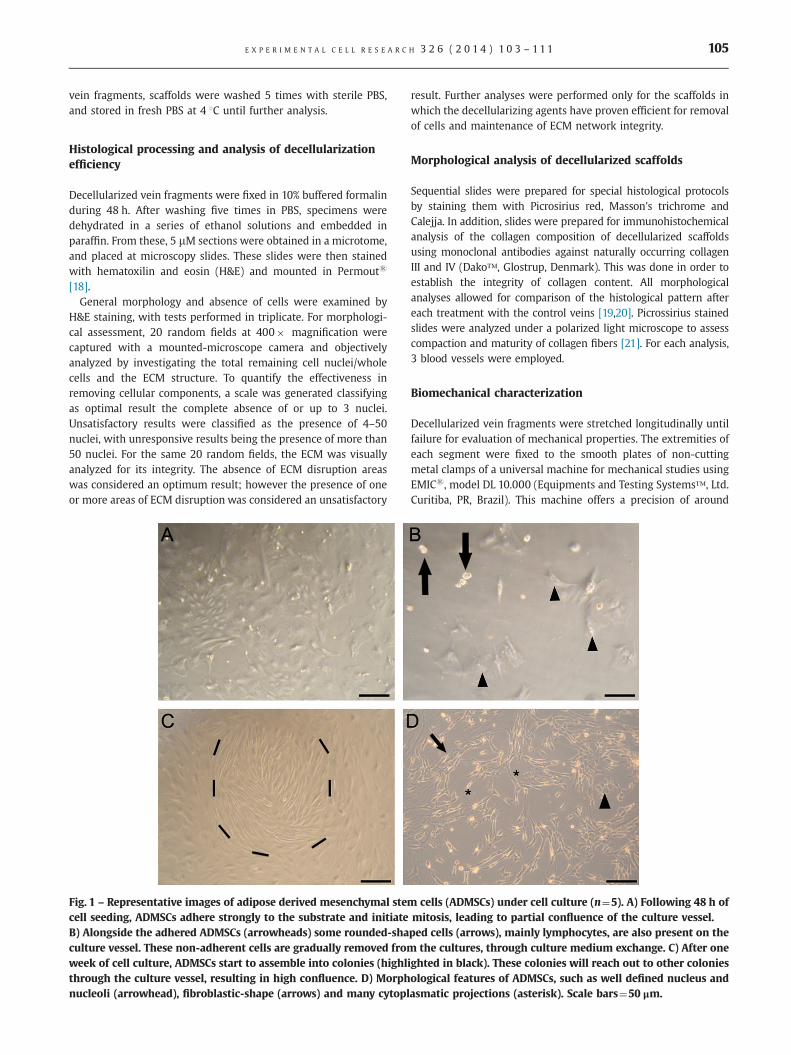
Fig. 1 – Representative images of adipose derived mesenchymal stemcell seeding, ADMSCs adhere strongly to the substrate and initiateB) Alongside the adhered ADMSCs (arrowheads) some rounded-shaculture vessel. These non-adherent cells are gradually removed fromweek of cell culture, ADMSCs start to assemble into colonies (highlthrough the culture vessel, resulting in high confluence. D) Morphnucleoli (arrowhead), fibroblastic-shape (arrows) and many cytopl
result. Further analyses were performed only for the scaffolds inwhich the decellularizing agents have proven efficient for removalof cells and maintenance of ECM network integrity.
Morphological analysis of decellularized scaffolds
Sequential slides were prepared for special histological protocolsby staining them with Picrosirius red, Masson's trichrome andCalejja. In addition, slides were prepared for immunohistochemicalanalysis of the collagen composition of decellularized scaffoldsusing monoclonal antibodies against naturally occurring collagenIII and IV (Dako™, Glostrup, Denmark). This was done in order toestablish the integrity of collagen content. All morphologicalanalyses allowed for comparison of the histological pattern aftereach treatment with the control veins [19,20]. Picrossirius stainedslides were analyzed under a polarized light microscope to assesscompaction and maturity of collagen fibers [21]. For each analysis,3 blood vessels were employed.
Biomechanical characterization
Decellularized vein fragments were stretched longitudinally untilfailure for evaluation of mechanical properties. The extremities ofeach segment were fixed to the smooth plates of non-cuttingmetal clamps of a universal machine for mechanical studies usingEMICs, model DL 10.000 (Equipments and Testing Systems™, Ltd.Curitiba, PR, Brazil). This machine offers a precision of around
cells (ADMSCs) under cell culture (n¼5). A) Following 48 h ofmitosis, leading to partial confluence of the culture vessel.ped cells (arrows), mainly lymphocytes, are also present on thethe cultures, through culture medium exchange. C) After one
ighted in black). These colonies will reach out to other coloniesological features of ADMSCs, such as well defined nucleus andasmatic projections (asterisk). Scale bars¼50 μm.
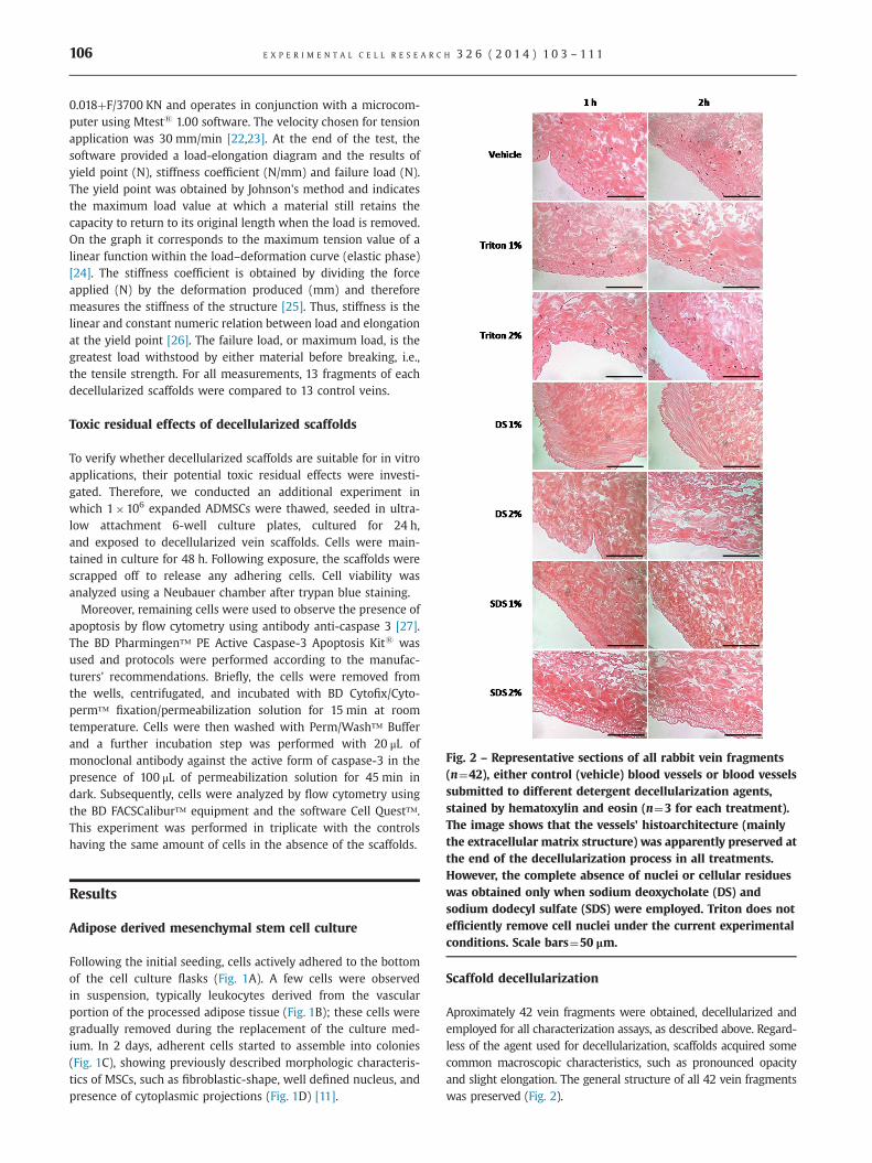
Fig. 2 – Representative sections of all rabbit vein fragments(n¼42), either control (vehicle) blood vessels or blood vesselssubmitted to different detergent decellularization agents,stained by hematoxylin and eosin (n¼3 for each treatment).The image shows that the vessels' histoarchitecture (mainlythe extracellular matrix structure) was apparently preserved at
E X P E R I M E N T A L C E L L R E S E A R C H 3 2 6 ( 2 0 1 4 ) 1 0 3 – 1 1 1106
0.018þF/3700 KN and operates in conjunction with a microcom-puter using Mtests 1.00 software. The velocity chosen for tensionapplication was 30 mm/min [22,23]. At the end of the test, thesoftware provided a load-elongation diagram and the results ofyield point (N), stiffness coefficient (N/mm) and failure load (N).The yield point was obtained by Johnson's method and indicatesthe maximum load value at which a material still retains thecapacity to return to its original length when the load is removed.On the graph it corresponds to the maximum tension value of alinear function within the load–deformation curve (elastic phase)[24]. The stiffness coefficient is obtained by dividing the forceapplied (N) by the deformation produced (mm) and thereforemeasures the stiffness of the structure [25]. Thus, stiffness is thelinear and constant numeric relation between load and elongationat the yield point [26]. The failure load, or maximum load, is thegreatest load withstood by either material before breaking, i.e.,the tensile strength. For all measurements, 13 fragments of eachdecellularized scaffolds were compared to 13 control veins.
Toxic residual effects of decellularized scaffolds
To verify whether decellularized scaffolds are suitable for in vitroapplications, their potential toxic residual effects were investi-gated. Therefore, we conducted an additional experiment inwhich 1�106 expanded ADMSCs were thawed, seeded in ultra-low attachment 6-well culture plates, cultured for 24 h,and exposed to decellularized vein scaffolds. Cells were main-tained in culture for 48 h. Following exposure, the scaffolds werescrapped off to release any adhering cells. Cell viability wasanalyzed using a Neubauer chamber after trypan blue staining.Moreover, remaining cells were used to observe the presence of
apoptosis by flow cytometry using antibody anti-caspase 3 [27].The BD Pharmingen™ PE Active Caspase-3 Apoptosis Kits wasused and protocols were performed according to the manufac-turers' recommendations. Briefly, the cells were removed fromthe wells, centrifugated, and incubated with BD Cytofix/Cyto-perm™ fixation/permeabilization solution for 15 min at roomtemperature. Cells were then washed with Perm/Wash™ Bufferand a further incubation step was performed with 20 μL ofmonoclonal antibody against the active form of caspase-3 in thepresence of 100 μL of permeabilization solution for 45 min indark. Subsequently, cells were analyzed by flow cytometry usingthe BD FACSCalibur™ equipment and the software Cell Quest™.This experiment was performed in triplicate with the controlshaving the same amount of cells in the absence of the scaffolds.
the end of the decellularization process in all treatments.However, the complete absence of nuclei or cellular residueswas obtained only when sodium deoxycholate (DS) andsodium dodecyl sulfate (SDS) were employed. Triton does notefficiently remove cell nuclei under the current experimentalconditions. Scale bars¼50 μm.
Results
Adipose derived mesenchymal stem cell culture
Following the initial seeding, cells actively adhered to the bottomof the cell culture flasks (Fig. 1A). A few cells were observedin suspension, typically leukocytes derived from the vascularportion of the processed adipose tissue (Fig. 1B); these cells weregradually removed during the replacement of the culture med-ium. In 2 days, adherent cells started to assemble into colonies(Fig. 1C), showing previously described morphologic characteris-tics of MSCs, such as fibroblastic-shape, well defined nucleus, andpresence of cytoplasmic projections (Fig. 1D) [11].
Scaffold decellularization
Aproximately 42 vein fragments were obtained, decellularized andemployed for all characterization assays, as described above. Regard-less of the agent used for decellularization, scaffolds acquired somecommon macroscopic characteristics, such as pronounced opacityand slight elongation. The general structure of all 42 vein fragmentswas preserved (Fig. 2).
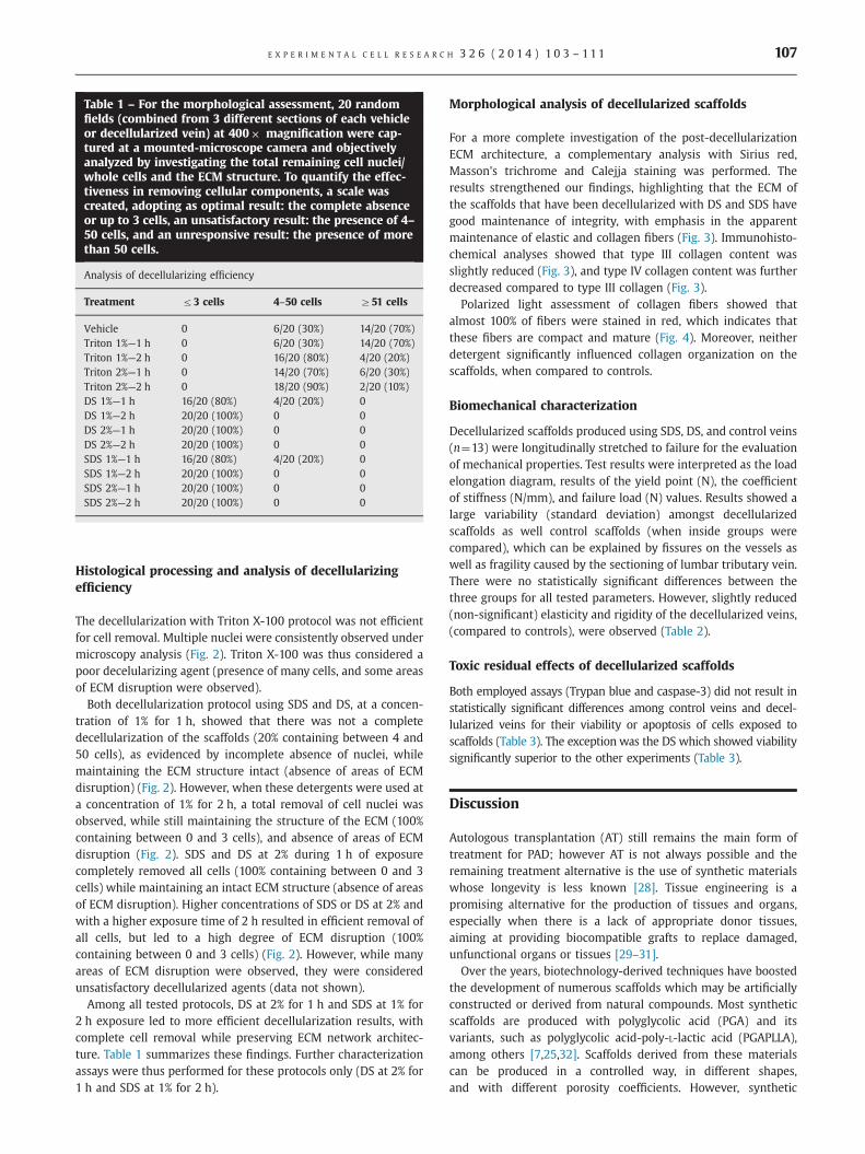
Table 1 – For the morphological assessment, 20 randomfields (combined from 3 different sections of each vehicleor decellularized vein) at 400� magnification were cap-tured at a mounted-microscope camera and objectivelyanalyzed by investigating the total remaining cell nuclei/whole cells and the ECM structure. To quantify the effec-tiveness in removing cellular components, a scale wascreated, adopting as optimal result: the complete absenceor up to 3 cells, an unsatisfactory result: the presence of 4–50 cells, and an unresponsive result: the presence of morethan 50 cells.
Analysis of decellularizing efficiency
Treatment r3 cells 4–50 cells Z51 cells
Vehicle 0 6/20 (30%) 14/20 (70%)Triton 1%—1 h 0 6/20 (30%) 14/20 (70%)Triton 1%—2 h 0 16/20 (80%) 4/20 (20%)Triton 2%—1 h 0 14/20 (70%) 6/20 (30%)Triton 2%—2 h 0 18/20 (90%) 2/20 (10%)DS 1%—1 h 16/20 (80%) 4/20 (20%) 0DS 1%—2 h 20/20 (100%) 0 0DS 2%—1 h 20/20 (100%) 0 0DS 2%—2 h 20/20 (100%) 0 0SDS 1%—1 h 16/20 (80%) 4/20 (20%) 0SDS 1%—2 h 20/20 (100%) 0 0SDS 2%—1 h 20/20 (100%) 0 0SDS 2%—2 h 20/20 (100%) 0 0
E X P E R I M E N T A L C E L L R E S E A R C H 3 2 6 ( 2 0 1 4 ) 1 0 3 – 11 1 107
Histological processing and analysis of decellularizingefficiency
The decellularization with Triton X-100 protocol was not efficientfor cell removal. Multiple nuclei were consistently observed undermicroscopy analysis (Fig. 2). Triton X-100 was thus considered apoor decelularizing agent (presence of many cells, and some areasof ECM disruption were observed).
Both decellularization protocol using SDS and DS, at a concen-tration of 1% for 1 h, showed that there was not a completedecellularization of the scaffolds (20% containing between 4 and50 cells), as evidenced by incomplete absence of nuclei, whilemaintaining the ECM structure intact (absence of areas of ECMdisruption) (Fig. 2). However, when these detergents were used ata concentration of 1% for 2 h, a total removal of cell nuclei wasobserved, while still maintaining the structure of the ECM (100%containing between 0 and 3 cells), and absence of areas of ECMdisruption (Fig. 2). SDS and DS at 2% during 1 h of exposurecompletely removed all cells (100% containing between 0 and 3cells) while maintaining an intact ECM structure (absence of areasof ECM disruption). Higher concentrations of SDS or DS at 2% andwith a higher exposure time of 2 h resulted in efficient removal ofall cells, but led to a high degree of ECM disruption (100%containing between 0 and 3 cells) (Fig. 2). However, while manyareas of ECM disruption were observed, they were consideredunsatisfactory decellularized agents (data not shown).
Among all tested protocols, DS at 2% for 1 h and SDS at 1% for2 h exposure led to more efficient decellularization results, withcomplete cell removal while preserving ECM network architec-ture. Table 1 summarizes these findings. Further characterizationassays were thus performed for these protocols only (DS at 2% for1 h and SDS at 1% for 2 h).
Morphological analysis of decellularized scaffolds
For a more complete investigation of the post-decellularizationECM architecture, a complementary analysis with Sirius red,Masson's trichrome and Calejja staining was performed. Theresults strengthened our findings, highlighting that the ECM ofthe scaffolds that have been decellularized with DS and SDS havegood maintenance of integrity, with emphasis in the apparentmaintenance of elastic and collagen fibers (Fig. 3). Immunohisto-chemical analyses showed that type III collagen content wasslightly reduced (Fig. 3), and type IV collagen content was furtherdecreased compared to type III collagen (Fig. 3).Polarized light assessment of collagen fibers showed that
almost 100% of fibers were stained in red, which indicates thatthese fibers are compact and mature (Fig. 4). Moreover, neitherdetergent significantly influenced collagen organization on thescaffolds, when compared to controls.
Biomechanical characterization
Decellularized scaffolds produced using SDS, DS, and control veins(n¼13) were longitudinally stretched to failure for the evaluationof mechanical properties. Test results were interpreted as the loadelongation diagram, results of the yield point (N), the coefficientof stiffness (N/mm), and failure load (N) values. Results showed alarge variability (standard deviation) amongst decellularizedscaffolds as well control scaffolds (when inside groups werecompared), which can be explained by fissures on the vessels aswell as fragility caused by the sectioning of lumbar tributary vein.There were no statistically significant differences between thethree groups for all tested parameters. However, slightly reduced(non-significant) elasticity and rigidity of the decellularized veins,(compared to controls), were observed (Table 2).
Toxic residual effects of decellularized scaffolds
Both employed assays (Trypan blue and caspase-3) did not result instatistically significant differences among control veins and decel-lularized veins for their viability or apoptosis of cells exposed toscaffolds (Table 3). The exception was the DS which showed viabilitysignificantly superior to the other experiments (Table 3).
Discussion
Autologous transplantation (AT) still remains the main form oftreatment for PAD; however AT is not always possible and theremaining treatment alternative is the use of synthetic materialswhose longevity is less known [28]. Tissue engineering is apromising alternative for the production of tissues and organs,especially when there is a lack of appropriate donor tissues,aiming at providing biocompatible grafts to replace damaged,unfunctional organs or tissues [29–31].Over the years, biotechnology-derived techniques have boosted
the development of numerous scaffolds which may be artificiallyconstructed or derived from natural compounds. Most syntheticscaffolds are produced with polyglycolic acid (PGA) and itsvariants, such as polyglycolic acid-poly-L-lactic acid (PGAPLLA),among others [7,25,32]. Scaffolds derived from these materialscan be produced in a controlled way, in different shapes,and with different porosity coefficients. However, synthetic
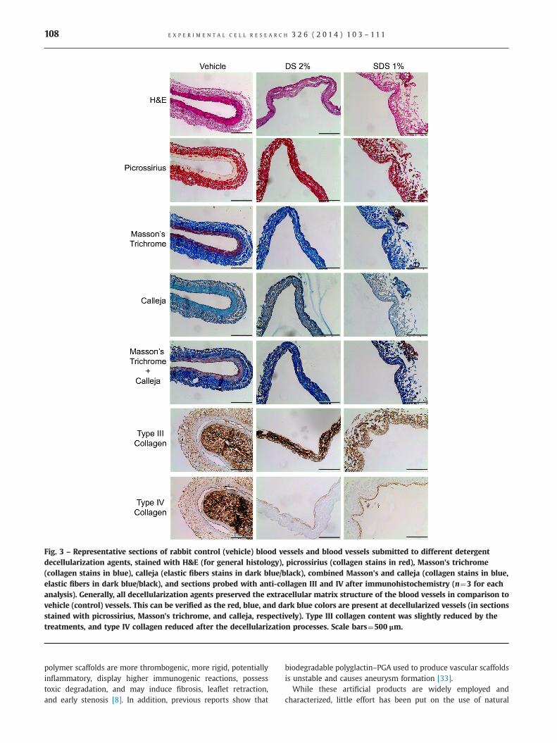
Fig. 3 – Representative sections of rabbit control (vehicle) blood vessels and blood vessels submitted to different detergentdecellularization agents, stained with H&E (for general histology), picrossirius (collagen stains in red), Masson's trichrome(collagen stains in blue), calleja (elastic fibers stains in dark blue/black), combined Masson's and calleja (collagen stains in blue,elastic fibers in dark blue/black), and sections probed with anti-collagen III and IV after immunohistochemistry (n¼3 for eachanalysis). Generally, all decellularization agents preserved the extracellular matrix structure of the blood vessels in comparison tovehicle (control) vessels. This can be verified as the red, blue, and dark blue colors are present at decellularized vessels (in sectionsstained with picrossirius, Masson's trichrome, and calleja, respectively). Type III collagen content was slightly reduced by thetreatments, and type IV collagen reduced after the decellularization processes. Scale bars¼500 μm.
E X P E R I M E N T A L C E L L R E S E A R C H 3 2 6 ( 2 0 1 4 ) 1 0 3 – 1 1 1108
polymer scaffolds are more thrombogenic, more rigid, potentiallyinflammatory, display higher immunogenic reactions, possesstoxic degradation, and may induce fibrosis, leaflet retraction,and early stenosis [8]. In addition, previous reports show that
biodegradable polyglactin–PGA used to produce vascular scaffoldsis unstable and causes aneurysm formation [33].
While these artificial products are widely employed andcharacterized, little effort has been put on the use of natural

Fig. 4 – Representative sections of rabbit control (vehicle) blood vessels and blood vessels submitted to DS 2% and SDS 1%decellularization agents, stained with picrossirius, under light (left column) and polarized (right column) microscopy (n¼3 foreach analysis). The image shows that the vessels' collagen content, stained in red with picrossirius, (on the left column), wascharacterized mainly as compact old collagen fibers, stained in dark red, as seen in the right column, under the polarized light.Apparently, sodium deoxycholate (DS) and sodium dodecyl sulfate (SDS), at these concentrations, are able to preserve the collagenarchitecture. This resembles the collagen architecture of the vehicle (control) vessels. Scale bars¼500 μm.
E X P E R I M E N T A L C E L L R E S E A R C H 3 2 6 ( 2 0 1 4 ) 1 0 3 – 11 1 109
biological compounds as the main source of scaffold. Biologicalscaffolds may circumvent the hurdles of synthetic scaffolds,as they are much more cell-compatible, such as the compo-nents of the ECM: collagen, elastin, fibronectin, hyaluronic acid,glycosaminoglycans (GAGs), and others, which benefits fromtheir bioactive, biocompatible, and similar mechanical propertiescompared to in vivo tissues [8,33,34]. One approach to obtainbiological 3D scaffolds is to employ the original tissue where thecell cultures shall be seeded in the moment of transplantation. Tothis purpose, however, in a heterologous approach, these scaffoldsneed to be decellularized to prevent immune rejections. Previousreports suggest that scaffolds derived from decellularized tissueshave a facilitating role in the differentiation and obtainment of theexpected tissue, as they contain the bioactive molecules and ligandsnecessary for stem cell maturation and diffusion of nutrients, as wellas the naturally occurring 3D tissue architecture [11,33].
Given that the most commonly used tissue graft substitute forcardiovascular surgery in humans is the great saphenous vein, itseemed logical to study, initially in animal models, the behavior ofdecellularized veins, given that it is feasible to set up banks ofdecellularized veins from cadaveric donors, aiming at, in the
future, the customization of blood vessel production. In thisreport, we have chosen the infrarenal inferior vena cava, due tosimilarities such as diameter, thickness, and histological structureto the great saphenous vein in humans [35].We first demonstrated that the production of decellularized veins
was feasible, depending on the detergent selected for this purpose.While Triton X100 does not completely remove cell nuclei, SDS andDS are more efficient for decellularization, being also able to preserveECM matrix architecture. Interestingly, other authors have reportedsuccessful decellularization using Triton X100; however, not onlywere they decellularizing other tissues, but they also employeddifferent concentrations and time of exposure that could account forthe discrepancies observed in the efficacy of cell removal by TritonX100 [4,35,36]. Given that for heterologous usage these veins mustbe cell-free, and Triton X100 did not completely remove cell nuclei,this detergent cannot be used to produce blood vessel scaffolds fortherapeutic purposes and was removed early from our study.Even though cell removal is a key point in the process of
decelularization for scaffold production, it also is important thatthe remaining ECM structure is maintained. Previous reports haveshown that ECM components such as collagen, elastin, fibronectin,
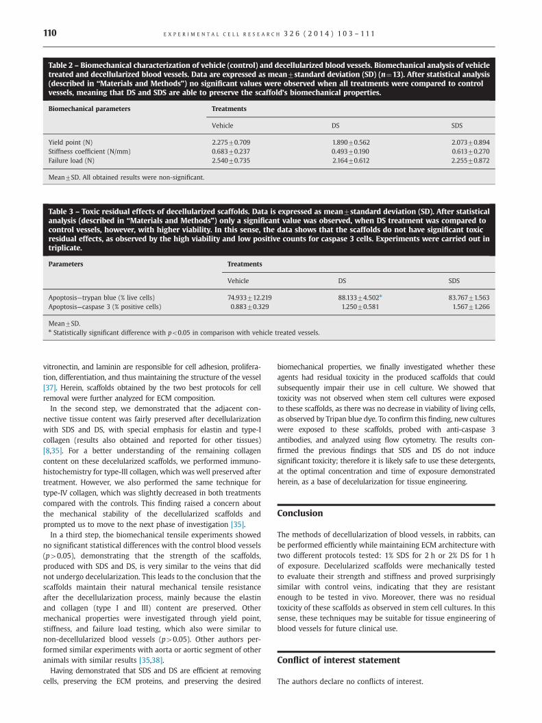
Table 2 – Biomechanical characterization of vehicle (control) and decellularized blood vessels. Biomechanical analysis of vehicletreated and decellularized blood vessels. Data are expressed as mean7standard deviation (SD) (n¼13). After statistical analysis(described in “Materials and Methods”) no significant values were observed when all treatments were compared to controlvessels, meaning that DS and SDS are able to preserve the scaffold's biomechanical properties.
Biomechanical parameters Treatments
Vehicle DS SDS
Yield point (N) 2.27570.709 1.89070.562 2.07370.894Stiffness coefficient (N/mm) 0.68370.237 0.49370.190 0.61370.270Failure load (N) 2.54070.735 2.16470.612 2.25570.872
Mean7SD. All obtained results were non-significant.
Table 3 – Toxic residual effects of decellularized scaffolds. Data is expressed as mean7standard deviation (SD). After statisticalanalysis (described in “Materials and Methods”) only a significant value was observed, when DS treatment was compared tocontrol vessels, however, with higher viability. In this sense, the data shows that the scaffolds do not have significant toxicresidual effects, as observed by the high viability and low positive counts for caspase 3 cells. Experiments were carried out intriplicate.
Parameters Treatments
Vehicle DS SDS
Apoptosis—trypan blue (% live cells) 74.933712.219 88.13374.502n 83.76771.563Apoptosis—caspase 3 (% positive cells) 0.88370.329 1.25070.581 1.56771.266
Mean7SD.n Statistically significant difference with po0.05 in comparison with vehicle treated vessels.
E X P E R I M E N T A L C E L L R E S E A R C H 3 2 6 ( 2 0 1 4 ) 1 0 3 – 1 1 1110
vitronectin, and laminin are responsible for cell adhesion, prolifera-tion, differentiation, and thus maintaining the structure of the vessel[37]. Herein, scaffolds obtained by the two best protocols for cellremoval were further analyzed for ECM composition.In the second step, we demonstrated that the adjacent con-
nective tissue content was fairly preserved after decellularizationwith SDS and DS, with special emphasis for elastin and type-Icollagen (results also obtained and reported for other tissues)[8,35]. For a better understanding of the remaining collagencontent on these decelularized scaffolds, we performed immuno-histochemistry for type-III collagen, which was well preserved aftertreatment. However, we also performed the same technique fortype-IV collagen, which was slightly decreased in both treatmentscompared with the controls. This finding raised a concern aboutthe mechanical stability of the decellularized scaffolds andprompted us to move to the next phase of investigation [35].In a third step, the biomechanical tensile experiments showed
no significant statistical differences with the control blood vessels(p40.05), demonstrating that the strength of the scaffolds,produced with SDS and DS, is very similar to the veins that didnot undergo decelularization. This leads to the conclusion that thescaffolds maintain their natural mechanical tensile resistanceafter the decellularization process, mainly because the elastinand collagen (type I and III) content are preserved. Othermechanical properties were investigated through yield point,stiffness, and failure load testing, which also were similar tonon-decellularized blood vessels (p40.05). Other authors per-formed similar experiments with aorta or aortic segment of otheranimals with similar results [35,38].Having demonstrated that SDS and DS are efficient at removing
cells, preserving the ECM proteins, and preserving the desired
biomechanical properties, we finally investigated whether theseagents had residual toxicity in the produced scaffolds that couldsubsequently impair their use in cell culture. We showed thattoxicity was not observed when stem cell cultures were exposedto these scaffolds, as there was no decrease in viability of living cells,as observed by Tripan blue dye. To confirm this finding, new cultureswere exposed to these scaffolds, probed with anti-caspase 3antibodies, and analyzed using flow cytometry. The results con-firmed the previous findings that SDS and DS do not inducesignificant toxicity; therefore it is likely safe to use these detergents,at the optimal concentration and time of exposure demonstratedherein, as a base of decelularization for tissue engineering.
Conclusion
The methods of decellularization of blood vessels, in rabbits, canbe performed efficiently while maintaining ECM architecture withtwo different protocols tested: 1% SDS for 2 h or 2% DS for 1 hof exposure. Decelularized scaffolds were mechanically testedto evaluate their strength and stiffness and proved surprisinglysimilar with control veins, indicating that they are resistantenough to be tested in vivo. Moreover, there was no residualtoxicity of these scaffolds as observed in stem cell cultures. In thissense, these techniques may be suitable for tissue engineering ofblood vessels for future clinical use.
Conflict of interest statement
The authors declare no conflicts of interest.

E X P E R I M E N T A L C E L L R E S E A R C H 3 2 6 ( 2 0 1 4 ) 1 0 3 – 11 1 111
Acknowledgments
This study was supported by the São Paulo Research Foundation(FAPESP) Grant number 2010/52549-8. The authors would like tostate their gratitude to Mr. Chris Gieseke at the University of Texasat San Antonio, for the excellent help in the English revision ofthis manuscript.
r e f e r e n c e s
[1] V.L. Roger, A.S. Go, D.M. Lloyd-Jones, R.J. Adams, J.D. Berry, T.M.Brown, M.R. Carnethon, S. Dai, G. de Simone, E.S. Ford, Heartdisease and stroke statistics—2011 update a report from theamerican heart association, Circulation 123 (2011) e18–e209.
[2] B.C. Isenberg, C. Williams, R.T. Tranquillo, Small-diameter artifi-cial arteries engineered in vitro, Circ. Res. 98 (2006) 25–35.
[3] O.E. Teebken, A. Haverich, Tissue engineering of small diametervascular grafts, Eur. J. Vasc. Endovasc. 23 (2002) 475–485.
[4] P.M. Crapo, T.W. Gilbert, S.F. Badylak, An overview of tissue andwhole organ decellularization processes, Biomaterials 32 (2011)3233–3243.
[5] H. Nagase, R. Visse, G. Murphy, Structure and function of matrixmetalloproteinases and timps, Cardiovasc. Res. 69 (2006) 562–573.
[6] P. Bornstein, E.H. Sage, Matricellular proteins: extracellular modu-lators of cell function, Curr. Opin. Cell Biol. 14 (2002) 608–616.
[7] M. Peck, D. Gebhart, N. Dusserre, T.N. McAllister, N. L’Heureux,The evolution of vascular tissue engineering and current state ofthe art, Cells Tissues Organs 195 (2011) 144–158.
[8] J.G. Nemeno-Guanzon, S. Lee, J.R. Berg, Y.H. Jo, J.E. Yeo, B.M. Nam,Y.-G. Koh, J.I. Lee, Trends in tissue engineering for blood vessels,J. Biomed. Biotechnol. 2012 (2012) 9563451.
[9] R.M. Nerem, Tissue engineering a blood vessel substitute: therole of biomechanics, Yonsei Med. J. 41 (2000) 735–739.
[10] A. de Mattos Carvalho, A.L.G. Alves, M.A. Golim, A. Moroz,C.A. Hussni, P.G.G. de Oliveira, E. Deffune, Isolation and immu-nophenotypic characterization of mesenchymal stem cellsderived from equine species adipose tissue, Vet. Immunol.Immunopathol. 132 (2009) 303–306.
[11] M. Bertanha, A. Moroz, R. Almeida, F.C. Alves, M.J. Acorci Valério,R. Moura, M.A.C. Domingues, M.L. Sobreira, E. Deffune, Tissue-engineered blood vessel substitute by reconstruction ofendothelium using mesenchymal stem cells induced by plateletgrowth factors, J. Vasc. Surg. S0741–5214 (13) (2013) 00973–00977 (pii).
[12] P.J. Schaner, N.D. Martin, T.N. Tulenko, I.M. Shapiro, N.A. Tarola,R.F. Leichter, R.A. Carabasi, P.J. DiMuzio, Decellularized vein as apotential scaffold for vascular tissue engineering, J. Vasc. Surg. 40(2004) 146–153.
[13] T.W. Gilbert, T.L. Sellaro, S.F. Badylak, Decellularization of tissuesand organs, Biomaterials 27 (2006) 3675–3683.
[14] B.D. Elder, S.V. Eleswarapu, K.A. Athanasiou, Extraction techni-ques for the decellularization of tissue engineered articularcartilage constructs, Biomaterials 30 (2009) 3749.
[15] S. Funamoto, K. Nam, T. Kimura, A. Murakoshi, Y. Hashimoto,K. Niwaya, S. Kitamura, T. Fujisato, A. Kishida, The use of high-hydrostatic pressure treatment to decellularize blood vessels,Biomaterials 31 (2010) 3590–3595.
[16] S.L. Dahl, J. Koh, V. Prabhakar, L.E. Niklason, Decellularized nativeand engineered arterial scaffolds for transplantation, Cell Trans-plant. 12 (2003) 659–666.
[17] E. Rieder, M.-T. Kasimir, G. Silberhumer, G. Seebacher, E. Wolner,P. Simon, G. Weigel, Decellularization protocols of porcine heartvalves differ importantly in efficiency of cell removal andsusceptibility of the matrix to recellularization with humanvascular cells, J. Thorac. Cardiovasc. Surg. 127 (2004) 399–405.
[18] A. Moroz, R.A.C. Bittencourt, R.P. Almeida, S.L. Felisbino,E. Deffune, Platelet lysate 3d scaffold supports mesenchymalstem cell chondrogenesis: an improved approach in cartilagetissue engineering, Platelets 24 (2012) 219–225.
[19] A. Patel, B. Fine, M. Sandig, K. Mequanint, Elastin biosynthesis:the missing link in tissue-engineered blood vessels, Cardiovasc.Res. 71 (2006) 40–49.
[20] C.B. Weinberg, E. Bell, A blood vessel model constructed fromcollagen and cultured vascular cells, Science 231 (1986) 397–400.
[21] L.C. Junqueira, G. Bignolas, R. Brentani, Picrosirius staining pluspolarization microscopy, a specific method for collagen detectionin tissue sections, Histochem. J. 11 (1979) 447–455.
[22] W. Yoshida, S. Müller, I. Carvalho, V. Fabris, L. Naresse, F. Maffei,Tensile strength and histological changes of abdominal aorta ofmalnourished rats, Vascular 3 (1995) 437–439.
[23] N.F. Cerqueira, W.B. Yoshida, S.S. Müller, J.L. Sequeira, de Rodri-gues A.C., C.R. Padovani, Morphological and biomechanical studyof abdominal aorta of rats submitted to experimental chronicalcoholism, Acta Cir. Bras. 20 (2005) 213–218.
[24] S. Lee, Y. Fung, M. Matsuda, H. Xue, D. Schneider, K. Han, Thedevelopment of mechanical strength of surgically anastomosedarteries sutured with dexon, J. Biomech. 18 (1985) 81–89.
[25] N. L’heureux, S. Pâquet, R. Labbé, L. Germain, F.A. Auger,A completely biological tissue-engineered human blood vessel,FASEB J. 12 (1998) 47–56.
[26] R.G. Jaldin, É. Castardelli, J.E. Perobelli, W.B. Yoshida, A. de CastroRodrigues, J.L. Sequeira, S.A.R. Paiva, Morphologic and biome-chanical changes of thoracic and abdominal aorta in a rat modelof cigarette smoke exposure, Ann. Vasc. Surg. 27 (2013) 791–800.
[27] M.D. Jacobson, M. Weil, M.C. Raff, Programmed cell death inanimal development, Cell 88 (1997) 347–354.
[28] L. Norgren, W.R. Hiatt, J.A. Dormandy, M.R. Nehler, K.A. Harris,F.G. Fowkes, Inter-society consensus for the management of per-ipheral arterial disease (tasc ii), J. Vasc. Surg. 45 (2007) S5–S67.
[29] A. Atala, Tissue engineering and regenerative medicine: conceptsfor clinical application, Rejuvenation Res. 7 (2004) 15–31.
[30] J.R. Fuchs, B.A. Nasseri, J.P. Vacanti, Tissue engineering: a 21stcentury solution to surgical reconstruction, Ann. Thorac. Surg. 72(2001) 577–591.
[31] M. Patruno, T. Martinello, Treatments of the injured tendon inveterinary medicine: from scaffolds to adult stem cells, Histol.Histopathol. 29 (2014) 417–422.
[32] G. Matsumura, N. Nitta, S. Matsuda, Y. Sakamoto, N. Isayama,K. Yamazaki, Y. Ikada, Long-term results of cell-free biodegrad-able scaffolds for in situ tissue-engineering vasculature: in acanine inferior vena cava model, PLoS One 7 (2012) e35760.
[33] H.L. Prichard, R.J. Manson, L. DiBernardo, L.E. Niklason,J.H. Lawson, S.L. Dahl, An early study on the mechanisms that allowtissue-engineered vascular grafts to resist intimal hyperplasia,J. Cardiovasc. Transl. Res. 4 (2011) 674–682.
[34] H. Naderi, M.M. Matin, A.R. Bahrami, Review paper: critical issuesin tissue engineering: biomaterials, cell sources, angiogenesis,and drug delivery systems, J. Biomater. Appl. 26 (2011) 383–417.
[35] A.F. Pellegata, M. Asnaghi, I. Stefani, A. Maestroni, S. Maestroni, T.Dominioni, S. Zonta, G. Zerbini, S. Mantero, Detergent-enzymaticdecellularization of swine blood vessels: insight on mechanicalproperties for vascular tissue engineering, Biomed. Res. Int. 2014(2014) 412838.
[36] J.S. Uzarski, A.B. Van De Walle, P.S. McFetridge, Preimplantationprocessing of ex vivo‐derived vascular biomaterials: effects on peri-pheral cell adhesion, J. Biomed. Mater. Res. A 101 (1) (2013) 123–131.
[37] J. Rouwkema, et al., The use of endothelial progenitor cells forprevascularized bone tissue engineering, Tissue Eng. 15 (8)(2009) 2015–2027.
[38] J.C. Fitzpatrick, P.M. Clark, F.M. Capaldi, Effect of decellularizationprotocol on the mechanical behavior of porcine descending aorta,Int. J. Biomater. (2010) pii: 620503. http://dx.doi.org/10.1155/2010/620503. Epub 2010 Jul 4.