Embed Size (px)
Citation preview
ПАРАЗИТОЛОГИЯ, 51, 1, 2017
УДК 576.895.121
MORPHOLOGY OF CESTODE WITH ATYPICAL MODE OF ATTACHMENT
© N. A. Pospekhova
Institute of Biological Problems of the North FEB RAS Magadan, 685000
E-mail: [email protected] Submitted 01.08.2016
Morphological features of Rauschitaenia ancora (Mamaev, 1959), a cestode with atypi-cal mode of attachment («anchoring» in the wall of the host intestine), are considered. At the center of the overgrown scolex there is a rostellum, size of which is close to that of the developed metacestode. Large suckers are composed mainly of loose parenchyma and fine radial muscle fibers. Fragments of host tissue are noted in the suckers' cavity. Surface of the scolex is covered with large microtriches, which are in contact with the host tissue ha-ving signs of degradation. Distal cytoplasm is filled with vesicles coming from tegumental cytons. Rostellar glands that are common to cyclophyllids are not found. Excretory canals contain fluid (closer to the surface) and numerous lipid droplets (deeper located canals).
Key words: Cestoda, ultrastructure, attachment organs, host-parasite relationships, exc-retory system, rostellar glands.
МОРФОЛОГИЯ ЦЕСТОДЫ С НЕТИПИЧНЫМ СПОСОБОМ ПРИКРЕПЛЕНИЯ
© Н. А. Поспехова
Институт биологических проблем Севера ДВО РАН Магадан, 685000
Поступила 01.08.2016
Рассматриваются особенности морфологии Rauschitaenia ancora (Mamaev, 1959), цестоды с нетипичным способом прикрепления («заякоривание» в стенке кишки хо-зяина). В центре разросшегося сколекса расположен хоботок, сохранивший размеры, близкие к таковым хоботка сформированной метацестоды. Крупные присоски состо-ят в основном из рыхлой паренхимы с включением радиальных мышечных волокон. В полости присосок отмечены фрагменты ткани хозяина. Поверхность сколекса по-крыта крупными микротрихиями, которые контактируют с тканями хозяина, имею-щими признаки деструкции. Дистальная цитоплазма заполнена везикулами, поступа-ющими из цитонов тегумента. Хоботковых желез, обычных для циклофиллидей, не
15
обнаружено. Экскреторные каналы содержат жидкость (поверхностные каналы) и многочисленные липидные капли (более глубокие каналы).
Ключевые слова: Cestoda, ультраструктура, органы прикрепления, паразито-хозя-инные взаимоотношения, экскреторная система, хоботковые железы.
The new genus of cestodes with a single species Rauschitaenia ancora (Ma-maev, 1959) was described within the family Dilepididae on the material from three common snipes Gallinago gallinago L., obtained in the Chaun lowland, Magadan Province (Bondarenko, Tomilovskaya, 1979). Representatives of this species differed by atypical mode of attachment to the host intestine. If the ma-jority of «higher» cestodes attach with the help of the rostellum and four suc-kers, besides, providing movement of the parasite, then the scolex and the ante-rior part of R. ancora strobila penetrate deeply into the intestinal wall almost perforating it. The scolex of the cestode increases almost by 10 times in compa-rison with the larva; a clearly visible capsule on the outer surface of the intestine is formed around the front part of the worm excluding any change of localiza-tion (Bondarenko, Tomilovskaya, 1979). Another structural peculiarity of R. ancora concerns the presence of multiple anastomosed excretory vessels, among which it is impossible to distinguish two pairs of main longitudinal ca-nals common for cestodes.
The atypical attachment mode which defines tissue localization of the front part of the cestode with the rest of strobila situated in the intestinal lumen was determined as «anchoring» (Dogel, 1947). Penetration and overgrowth of the scolex considerably increase contact area of the parasite with host tissues, cau-sing degradation of the mucosa and leading to the development of immune res-ponse of the host. Thus, the study of the morphology and ultrastructure of R. an-cora is of great interest for understanding of both biology peculiarities of cesto-des with atypical attachment mode and morphological aspects of relations between penetrated parasite and its host.
MATERIAL AND METHODS
Adult cestodes R. ancora from common snipes G. gallinago, taken with cap-sules and fragments of intestine wall, are served as the material. For convenien-ce some capsules were dissected, and scolices of cestodes were fixed separately. Fixation and preparation of the material for the light and electron microscopy were conducted according to the standard method with some modifications (Po-spekhova, Regel, 2015). Semi-thin sections obtained with the use of LKB Bromma 2088 and LKB Nova (Sweden) ultramicrotomes were stained with methylene blue according to Morgenstern (Morgenstern, 1969) and exami-ned under an Olympus CX41 microscope (Olympus Corporation, Japan) with an Olympus E-420 digital camera. Ultra-thin sections were examined un-der JEM-1011 and JEM-1400Plus transmission electron microscopes (JEOL, Japan).
16
RESULTS AND DISCUSSION
Light microscopy. In sections, the overgrown scolex taken from the capsule possesses loose net-like structure (fig. 1, a, b, see ins.). Muscular wall of the rostellum, rostellar sac and suckers, covers of the scolex and the neck look den-ser. Loose parenchyma of the scolex is formed mainly of widened processes, some part of the latter containing dense round bodies (apparently, lipid drop-lets), others have no visible content. Large suckers also have loose structure; they are mainly formed of multiple processes without visible content divided by radial musculature.
The rostellum is located in the center of the scolex and is deeply submerged in the bottom of the retraction channel (fig. 1, b). The size of the rostellum in the developed metacestode (fig. 1, c) is 0.12 X 0.04 mm; penetrated cestode po-ssesses slightly enlarged rostellum, 0.19—0.22 X 0.07—0.08 mm, that is disp-roportionately small comparing to the scolex of the penetrated cestode (1.7—1.8 X 1.2—1.4 mm) (Bondarenko, Tomilovskaya, 1979). Rostellum length of the penetrated cestode in our work does not exceed 0.1 mm (fig. 1, d), diameter of the scolex, 0.9 mm, and diameter of suckers, 0.4 mm.
The front part of the rostellum with closely fitting hooks protrudes into the cavity, which, apparently, corresponds to that of the rostellar sac, and under nor-mally developed rostellum forms its lateral surface when the rostellum is protru-ded. Walls of the cavity lack microtriches (we use here the term «microtrix» (pl. «microtriches») according to Chervy, 2009), which appear anteriad and co-ver the surface of the twisted retraction canal (fig. 1, d). Microtriches («spines», according to Bondarenko, Tomilovskaya, 1979) are large, varying from 3 to 5 p.m. Their fine structure was described earlier (Pospekhov, Pospekhova, 1993).
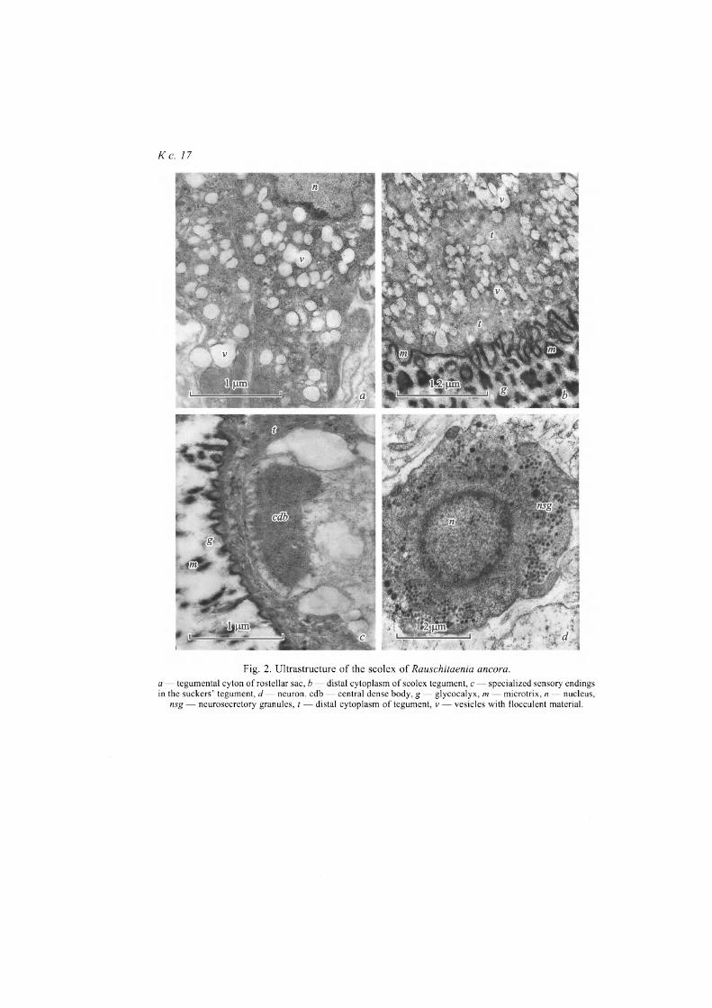
Electron microscopy. Muscular walls of the rostellum, that is up to 10 pm thick in its basal part, are formed of inner circular and outer longitudinal fibers. The widened base of the rostellum is crossed by retractor muscles of the hooks. Processes of muscle cells containing lipid drops and separate cells resembling tegumental cytons in the shape and density of the cytoplasm, or glandular cy-tons without secretory inclusions are found in spaces between hooks. The wall of the rostellar sac has insignificant thickness, about 4 pm, and is formed of an-nular fibers. The outer layer of longitudinal musculature, typical of other speci-es of the family Hymenolepididae, is not expressed here. No accumulations of glandular elements were noted in the rostellar sac. Tegumental cytons are large, with well-developed synthetic apparatus and numerous light vesicles, formed in the Golgi complex (fig. 2, a, see ins.). Cytons are located in the front part of the rostellar sac; their secretion (light vesicles with flaky content) runs into the te-gument of apical part of the scolex (fig. 2, b). Morphology of the scolex tegu-mental cytons is identical to that described in cytons of the rostellar sac.
Sensory endings, both with and without cilia, were found in scolex and neck covers, at that, specific endings with central dense body (fig. 2, c) were noted in suckers' tegument, those were also found in suckers' covers of other representa-tives of Cyclophyllidea (Pospekhov, Krasnoshchekov, 1992; Pospekhova, Po-spekhov, 1998; Pospekhova, Bondarenko, 2014). Authors suppose, those en-dings can register deformation degree of tegument in localization area of fixato-ry microtriches.
17

К ст. Н. А. Поспеховой, с. 17
Fig. 1. Semi-thin sections of the scolex of Rauschitaenia ancora without capsule (a, b, d) and sche-me of developed metacestode (after Bondarenko, Tomilovskaya, 1979, with courteous permission of
authors) (c). a — tangential section, b — longitudinal section through the middle of the scolex, d — region of the rostellum. h — hooks, hr — hook retractor, m — microtrix, r — rostellum, rc — retraction channel, rs — rostellar sac,
s — sucker.
К с. 17
Fig. 2. Ultrastructure of the scolex of Rauschitaenia ancora. a — tegumental cyton of rostellar sac, b — distal cytoplasm of scolex tegument, с — specialized sensory endings in the suckers' tegument, d — neuron, cdb — central dense body, g — glycocalyx, m — microtrix, n — nucleus,
nsg — neurosecretory granules, t — distal cytoplasm of tegument, v — vesicles with flocculent material.
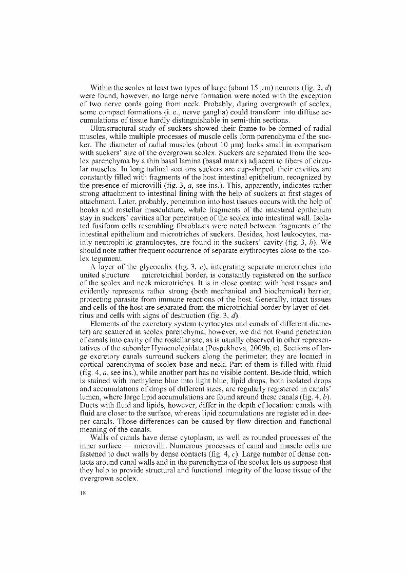
Within the scolex at least two types of large (about 15 p,m) neurons (fig. 2, d) were found, however, no large nerve formation were noted with the exception of two nerve cords going from neck. Probably, during overgrowth of scolex, some compact formations (i. e., nerve ganglia) could transform into diffuse ac-cumulations of tissue hardly distinguishable in semi-thin sections.
Ultrastructural study of suckers showed their frame to be formed of radial muscles, while multiple processes of muscle cells form parenchyma of the suc-ker. The diameter of radial muscles (about 10 p,m) looks small in comparison with suckers' size of the overgrown scolex. Suckers are separated from the sco-lex parenchyma by a thin basal lamina (basal matrix) adjacent to fibers of circu-lar muscles. In longitudinal sections suckers are cup-shaped, their cavities are constantly filled with fragments of the host intestinal epithelium, recognized by the presence of microvilli (fig. 3, a, see ins.). This, apparently, indicates rather strong attachment to intestinal lining with the help of suckers at first stages of attachment. Later, probably, penetration into host tissues occurs with the help of hooks and rostellar musculature, while fragments of the intestinal epithelium stay in suckers' cavities after penetration of the scolex into intestinal wall. Isola-ted fusiform cells resembling fibroblasts were noted between fragments of the intestinal epithelium and microtriches of suckers. Besides, host leukocytes, ma-inly neutrophilic granulocytes, are found in the suckers' cavity (fig. 3, b). We should note rather frequent occurrence of separate erythrocytes close to the sco-lex tegument.
A layer of the glycocalix (fig. 3, c), integrating separate microtriches into united structure — microtrichial border, is constantly registered on the surface of the scolex and neck microtriches. It is in close contact with host tissues and evidently represents rather strong (both mechanical and biochemical) barrier, protecting parasite from immune reactions of the host. Generally, intact tissues and cells of the host are separated from the microtrichial border by layer of det-ritus and cells with signs of destruction (fig. 3, d).
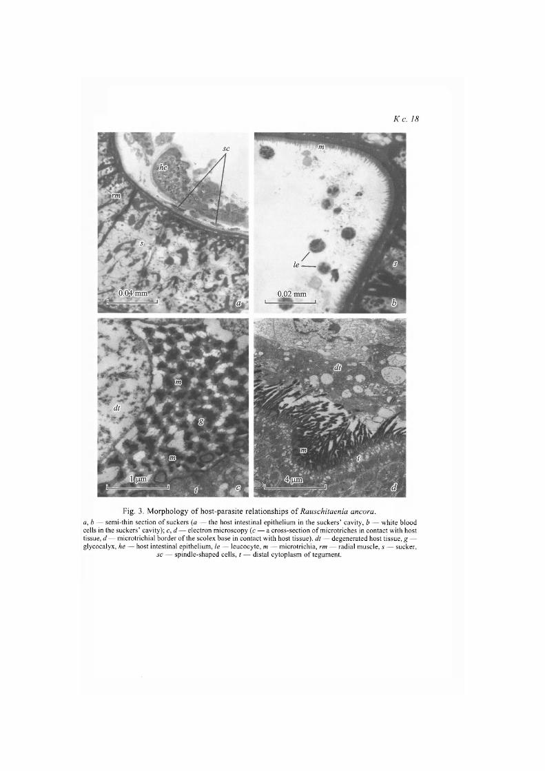
Elements of the excretory system (cyrtocytes and canals of different diame-ter) are scattered in scolex parenchyma, however, we did not found penetration of canals into cavity of the rostellar sac, as is usually observed in other represen-tatives of the suborder Hymenolepidata (Pospekhova, 2009b, c). Sections of lar-ge excretory canals surround suckers along the perimeter; they are located in cortical parenchyma of scolex base and neck. Part of them is filled with fluid (fig. 4, a, see ins.), while another part has no visible content. Beside fluid, which is stained with methylene blue into light blue, lipid drops, both isolated drops and accumulations of drops of different sizes, are regularly registered in canals' lumen, where large lipid accumulations are found around these canals (fig. 4, b). Ducts with fluid and lipids, however, differ in the depth of location: canals with fluid are closer to the surface, whereas lipid accumulations are registered in dee-per canals. Those differences can be caused by flow direction and functional meaning of the canals.
Walls of canals have dense cytoplasm, as well as rounded processes of the inner surface — microvilli. Numerous processes of canal and muscle cells are fastened to duct walls by dense contacts (fig. 4, c). Large number of dense con-tacts around canal walls and in the parenchyma of the scolex lets us suppose that they help to provide structural and functional integrity of the loose tissue of the overgrown scolex.
18
К с. 18
Fig. 3. Morphology of host-parasite relationships of Rauschitaenia ancora. a, b — semi-thin section of suckers (a — the host intestinal epithelium in the suckers' cavity, b — white blood cells in the suckers' cavity); c, d — electron microscopy (c — a cross-section of microtriches in contact with host tissue, d — microtrichial border of the scolex base in contact with host tissue), dt — degenerated host tissue, g — glycocalyx, he — host intestinal epithelium, le — leucocyte, m — microtrichia, rm — radial muscle, s — sucker,
sc — spindle-shaped cells, t — distal cytoplasm of tegument.
К с. 18
Fig. 4. Excretory system of Rauschitaenia ancora. a,b — tangential semi-thin sections of neck (a — surface canals with a liquid, b — deeper canals with lipid drop-lets); c, d — electron microscopy (с — canal wall with attached processes, d — muscle fibers close to canal wall), cl — canal lumen, em — extracellular matrix, / — lipid droplet, li — liquid, mf — muscle fibers, mi —
microvilli, sj — septate junction, tj — tight junction.
More or less expressed fibrous extracellular matrix with the immersed muscle fibers is located directly underneath the distal cytoplasm of the canal wall (fig. 4, d). Probably, muscle fibers provide indispensable tone of canal walls, or participate in their contraction transferring fluid and lipid drops. Areas of conf-luence of small collecting ducts with septate junctions are noted (fig. 4, c).
Among Cyclophyllidea, especially among Taeniidae, tissue parasitism is wi-dely distributed at larval stages, whereas adult cestodes rarely use host tissues as a habitat, e. g., for instance, Gastrotaenia dogieli (Gynezinskaja, 1944) from the gizzard of Anseriformes. During study of covers' morphology of G. dogieli (Davydov et al., 1990; Pospekhova, 2009с), the presence of developed glands in the rostellar apparatus and a thick layer of glycocalyx were noted; according to Davydov and Mikryakov (1998) the glands are similar to that performing barri-er and protecting functions in the cestode larvae in host tissues.
The study of the morphology of scolex tegument in R. ancora revealed four areas, characterized by different sets of cytoplasm inclusions and different mor-phology of microtriches: the area of suckers, the scolex behind suckers, neck, and strobila in intestinal lumen (Pospekhov, Pospekhova, 1993). It was noted, that even in cases when microtriches do not form a solid border, they bear a vi-sible layer of glycocalyx. Contact of microtriches with host cells was observed everywhere, frequently it was accompanied with integrity violation of the cy-toplasmic membrane of the latter. Erythrocytes, lymphocytes and fibroblasts, both intact and with signs of destruction, were noted in contact area. Also, evi-dences of the damage of the parasite tegument, from breaking of limiting mem-brane to complete absence of the surface syncytium over some areas, were seen.
Close contact between host tissues (capsule) and microtrichial border of R. ancora tegument was most frequently registered along the suckers' edge. Oc-casionally it was so strong, that dislocation of the parasite led to tearing of the layer of the tegument surface syncytium of the suckers from the basal plate ly-ing beneath. This phenomenon was observed in cestodes fixed together with the capsule, and apparently it is explained by the suckers muscle contraction at the time of fixation. The presence of erythrocytes close to parasite surface (both with and without capsule) indicates damages of intestinal tissue, which, probably, are performed by microtriches during cestode movements or during peristaltic motions of host intestine. The host, in its turn, actively attacks parasi-te; it is evidenced by partial destruction of the surface syncytium of the tegu-ment and adhesion of microtrichia to host tissues, resulting in separation of the surface syncytium from subjacent basal lamina.
According to literary data (Baer, 1940; Boertje, 1974) and our observations (Pospekhov, Pospekhova, 1993; Pospekhova, 2009а, б, с; Pospekhova, Bonda-renko, 2014), atypical localization and attachment mode of cestode can lead to morphological changes of attachment organs, and, in some cases, of the entire scolex. Apparently, processes of morphological transformations of attachment organs have adaptive character, and in the long ago formed parasite systems they lead to hypertrophy or, on the contrary, to the loss of some fixation organs. In this case the latter does not form anlage at the stage of scolexogenesis and is absent in the developed metacestode. Such situation is observed in Cloacotae-nia megalops (Nitzsch in Creplin, 1829), a parasite of Anseriformes, that atta-ches to cloacal wall of the host with the help of strong suckers. Completely de-veloped metacestode of this species has no rostellum, but possesses an accumu-
19

lation of glandular cells in its place (Gvozdev, Maksimova, 1978); later it develops into the gland of the rostellar sac, releasing secret into contact area bet-ween the parasite and the host (Pospekhova, 2009b).
As for visible absence of rostellar glands at R. ancora, which were noted practically for all representatives of higher cestodes, it can be connected with periodicity of secretory activity and fixation of material during decrease of sec-retion, when absence of specific secretion prevent distinguishing the regular cy-ton from the glandular one. There is, however, another probability: the rostellar glands of R. ancora stop their activity after penetration of the cestode into the intestinal wall, the overgrow of the scolex and the formation of the capsule, and their function is taken by regular tegumental cytons of the scolex, producing nu-merous vesicles coming into the tegument. Morphologically similar vesicles in contact area with host tissues were noted in two other representatives of the fa-mily Dilepididae: in the rostellar tegument of Dichoanotaenia clavigera (Krab-be, 1896) and in the front part of cestode scolex of Platyscolex ciliata (Fur-mann, 1913) (Pospekhova, 2009a). It should be mentioned, that P. ciliata has rudimentary rostellum with lack of armament even at the metacestode stage; in adults cestodes it does not function and is located at the bottom of the retraction channel (Krasnoshchekov, Pluzhnikov, 1981; Tomilovskaya, 1982). Thus, the rostellar apparatus of P. ciliata represents an intermediate variant between the rostellar apparatus of C. megalops (the rostellum is absent even at the metaces-tode stage) and R. ancora (the rostellum of proportional size is formed at the metacestode stage, and later it stops growing). Probably, these variants reflect different degree of parasite adaptation and different age of their parasite-host systems.
R e f e r e n c e s
B a e r J. G. 1940. Some avian tapeworms from Antigua. Parasitology. 32 : 174—197. B o e r t j e S . B. 1974. Life cycle and host-parasite relationships of Schistotaenia tenuicir-
rus (Cestoda Amabiliidae). Proceedings of Lousiana Academy of Sciences. 37 : 89— 103.
B o n d a r e n k o S. K., T o m i l o v s k a y a N. S. 1979. New dilepidid genus, Rauschitaenia n. g., and the life-cycle of R. ancora (Mamaev, 1959) n. comb., a parasite of snipes. In: So-nin M. D. (ed.) Ekologiya im morfologiya gel'mintov pozvonochnykh Chukotki. M.: Na-uka. 29—37. [In Russian].
C h e r v y L. 2009. Unified terminology for cestoda microtriches: a proposal from the Internati-onal Workshops on Cestode Systematics in 2002—2008. Folia Parasitologica. 56 (3): 199—230.
D a v y d o v V. G., M i k r y a k o v V. R. 1988. Adaptive structures of the body of some cestodes connected with defense of parasites from the host influence. Trudy GELAN USSR. 36 : 88—100. [In Russian].
D a v y d o v V. G., P o s p e k h o v a N . A., Y u r l o v a N . I. 1990.Ultrastructural organization of the scolex and strobila tegument in Gasrotaenia dogieli (Cestoda: Hymenolepididae). Parazitologiya. 24 (3) : 207—215. [In Russian].
D o g e l V. A. 1947. Kurs Obshchey Parasitologii. L., Uchpedgiz. 362 p. [In Russian]. G v o s d e v E . V., M a k s i m o v a A. P. 1978. Eucypris inflata, an intermediate host of avian
cestodes in the biocoenosis of the lake Tengiz. Parazitologiya. 12 (4) : 339—344. [In Russian].
K r a s n o s h c h e k o v G. P., P l u z h n i k o v L. T. 1981. Ultrastructure of the tegument of excys-ted larvae of Platyscolex ciliata (Cestoidea: Dilepididae). Parazitologiya. 15 (2) : 118—125. [In Russian].
20
M o r g e n s t e r n E. 1969. Vergleichende lichtoptische Untersuchungen im Rahmen elektronen-mikroskopischer Arbeiten an ultradünnen Schnitten. II. Färbemethoden. Mikroskopie. 25 : 250—260. [In Germany].
P o s p e k h o v V. V., K r a s n o s h c h e k o v G. P. 1992. A new type of sensory endings in the suckers of cestode. Parazitologiya. 26 (21): 168—170. [In Russian].
P o s p e k h o v V. V., P o s p e k h o v a N . A. 1993. The structure of the tegument of the cestode Rauschitaenia ancora (Cyclophyllidea: Dilepididae). Parazitologiya. 27 (2) : 155—160. [In Russian].
P o s p e k h o v a N . A. 2009a. Rostellar glands in two cestode of the family Dilepididae. Parazi-tologiya. 43 (1): 57—69. [In Russian].
P o s p e k h o v a N . A 2009b. Rostellar sac gland in Cloacotaenia megalops (Cestoda: Hymeno-lepididae). Invertebrate Zoology. 6 (1): 44—46. [In Russian].
P o s p e k h o v a N . A. 2009c. Rostellar apparatus in cestode Gastrotaenia dogieli (Cyclophylli-dea, Hymenolepididae). Vestnik zoologii. Supplement. 23 : 172—182. [In Russian].
P o s p e k h o v a N . A., B o n d a r e n k o S. K. 2014. Morpho-functional characteristic of the sco-lex of Wardium chaunense (Cestoda: Aploparaksidae) penetrated into host intestine. Pa-rasitology Research. 113 (1): 131—137.
P o s p e k h o v a N . A., P o s p e k h o v V. V. Ultrastructure of the scolex suckers in Diorchis ste-fanskii (Cestoda: Hymenolepididae). Parazitologiya. 32 (4) : 347—351. [In Russian].
P o s p e k h o v a N . A . , R e g e l K. V. 2015. Morphology and ultrastructure of two schistotaeniid cysticercoids (Cestoda: Cyclophyllidea) from the haemocoele of the dragonfly larvae. Parazitologiya. 49 (5) : 339—351.
T o m i l o v s k a y a N. S. 1982. Postembryonal development and intermediate hosts of dilepidids Dichoanotaenia gallinagilis (Davies, 1938) and Platysgolex ciliata (Fuhrmann, 1913) from birds of Chukotka. Parazitologiya. 16 (1) : 46—53. [In Russian].
21