Embed Size (px)
Citation preview

Morphometrics with
SPM12
John Ashburner
Wellcome Trust Centre for Neuroimaging,
12 Queen Square, London, UK.

What kind of differences are we looking for?
• Usually, we try to localise regions of difference. • Univariate models.
• Typically involves fitting a GLM
• Typically localising volumetric differences
• Some anatomical differences can not be localised. • Need multivariate models.
• Differences in terms of proportions among measurements.
• Where would the difference between male and female faces be localised?
• Need to select the best model of difference to use, before trying to fill in the details.
Overview
• Voxel-Based Morphometry
• Diffeomorphic Registration
• Tensor-Based Morphometry
• Longitudinal Registration

Voxel-Based Morphometry
• Produce a map of statistically significant differences
among populations of subjects.
• e.g. compare a patient group with a control group.
• or identify correlations with age, test-score etc.
• The data are pre-processed to sensitise the tests to
regional tissue volumes.
• Usually grey or white matter.
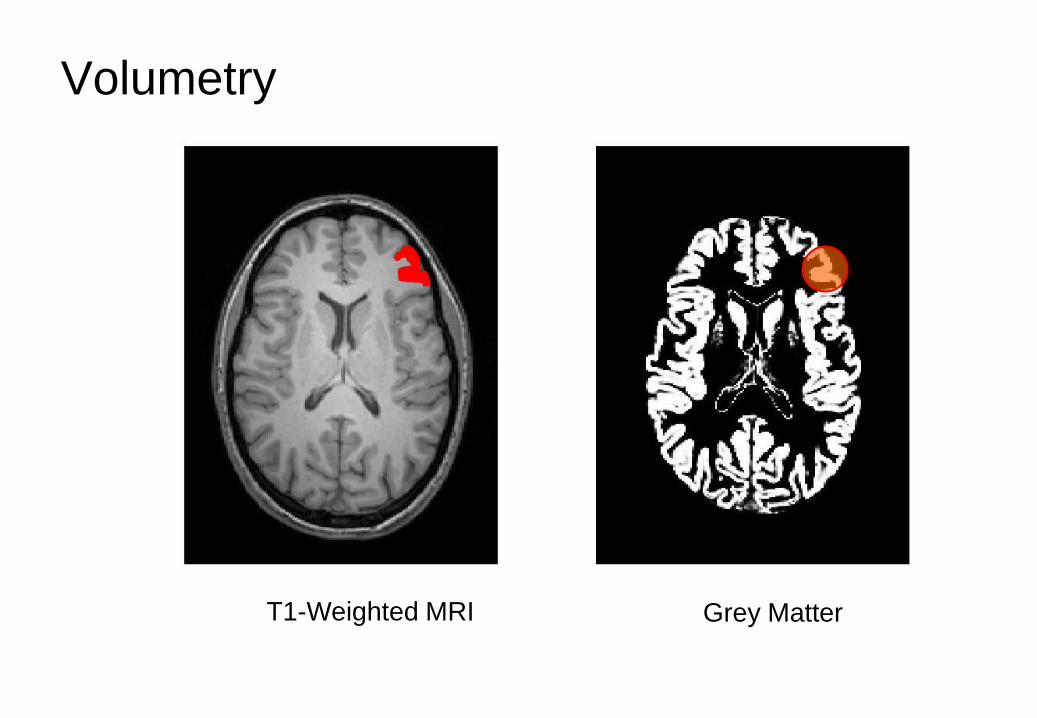
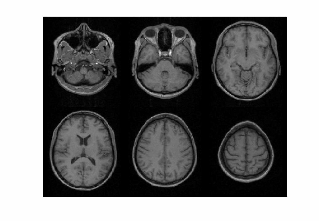
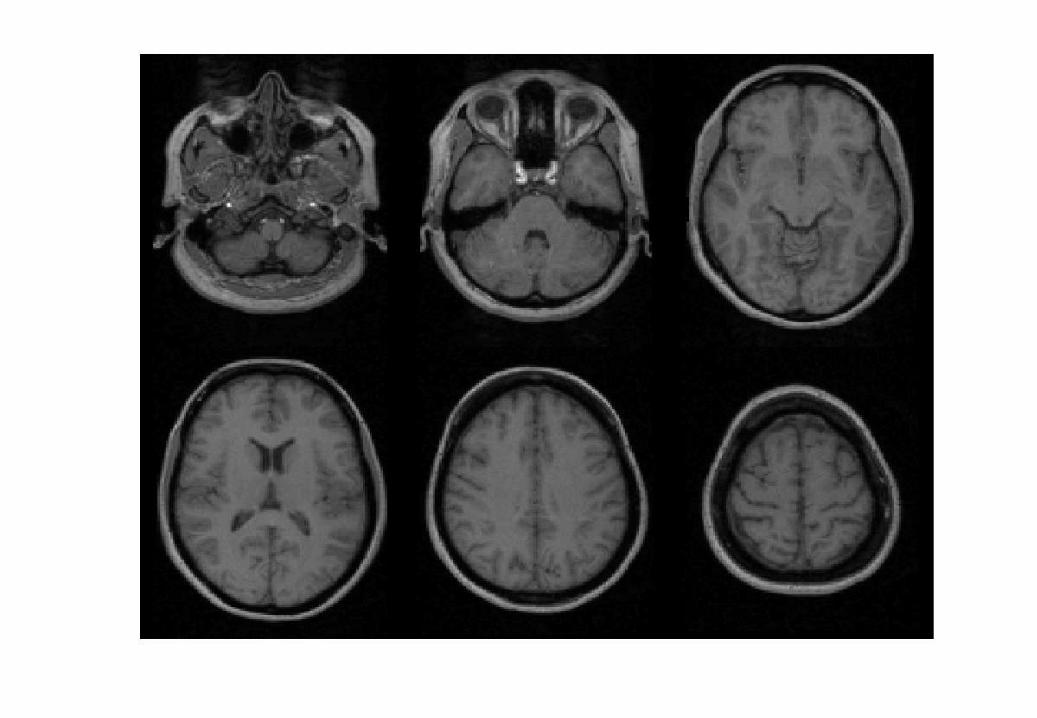
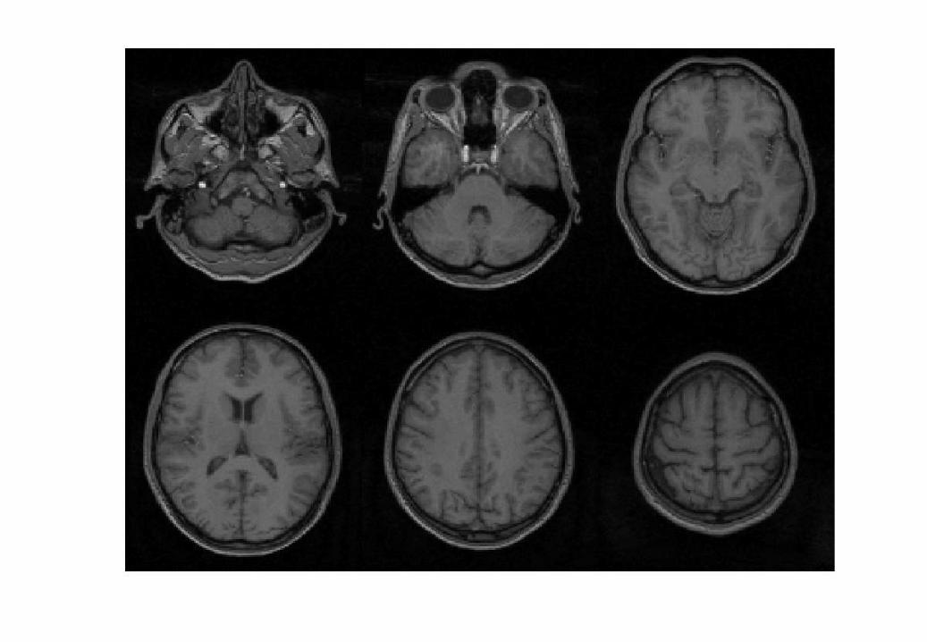
Volumetry
T1-Weighted MRI Grey Matter
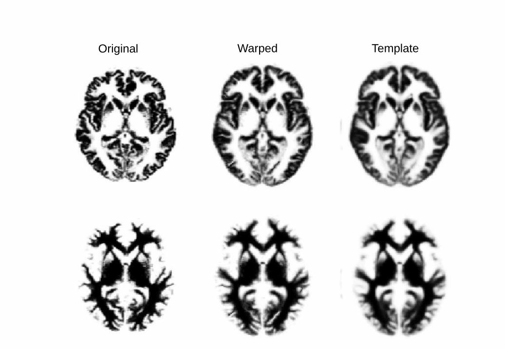
Original Warped Template

“Modulation” – change of variables.
Deformation Field Jacobians determinants
Encode relative volumes.
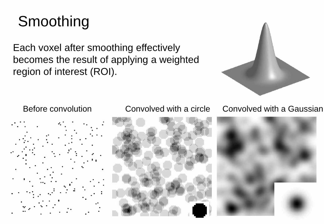
Smoothing
Before convolution Convolved with a circle Convolved with a Gaussian
Each voxel after smoothing effectively
becomes the result of applying a weighted
region of interest (ROI).

VBM Pre-processing
in SPM12 • Use Segment for
characterising intensity
distributions of tissue classes,
and writing out “imported”
images that Dartel can use.
• Run Dartel to estimate all the
deformations.
• Dartel warping to generate
smoothed, “modulated”,
warped grey matter.
• Statistics.

Some Explanations of the Differences
Thickening Thinning
Folding
Mis-classify
Mis-classify
Mis-register
Mis-register

Some References
• Ashburner & Friston. “Unified Segmentation”. NeuroImage
26:839-851, 2005.
• Ashburner. “A Fast Diffeomorphic Image Registration
Algorithm”. NeuroImage 38:95-113 (2007).
• Ashburner & Friston. “Computing Average Shaped Tissue
Probability Templates”. NeuroImage 45:333-341, 2009.
• Ashburner. “Computational Anatomy with the SPM software”.
Magnetic Resonance Imaging 27(8):1163-1174, 2009.

Overview
• Voxel-Based Morphometry
• Diffeomorphic Registration
• Tensor-Based Morphometry
• Longitudinal Registration
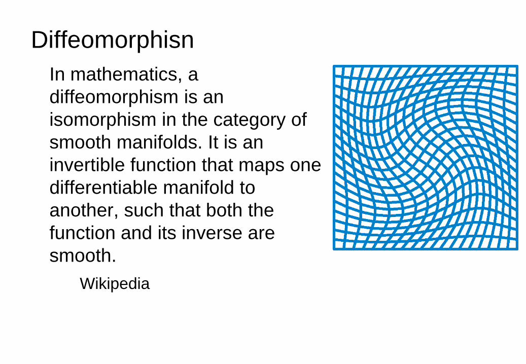
Diffeomorphisn
In mathematics, a
diffeomorphism is an
isomorphism in the category of
smooth manifolds. It is an
invertible function that maps one
differentiable manifold to
another, such that both the
function and its inverse are
smooth.
Wikipedia
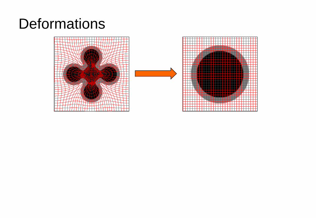
Deformations

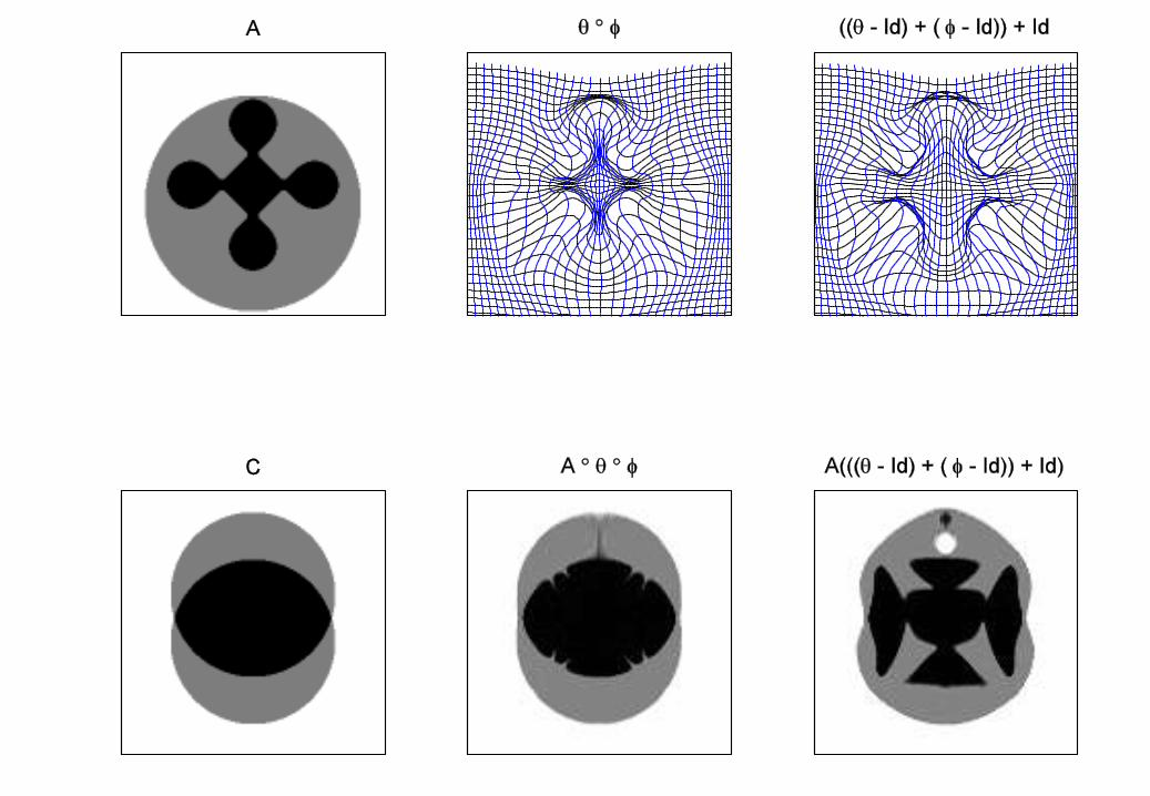
Composition

Small
Deformation
Approximation
The composition:
ϑ φ Would be approximated with:
Id +((ϑ-Id) + (φ-Id))
The inversion:
φ-1
Would be approximated with:
Id -(φ-Id)
Not good approximations for large deformations.

Diffeomorphic Image Registration
• Minimises two terms:
1. A measure of distance between images
2. A measure of the amount of distortion.
Because we can not simply add displacement
fields, large deformations are generated by
composing many small deformations.
The amount of distortion is computed by summing
up the distortion measures from the small
displacements.
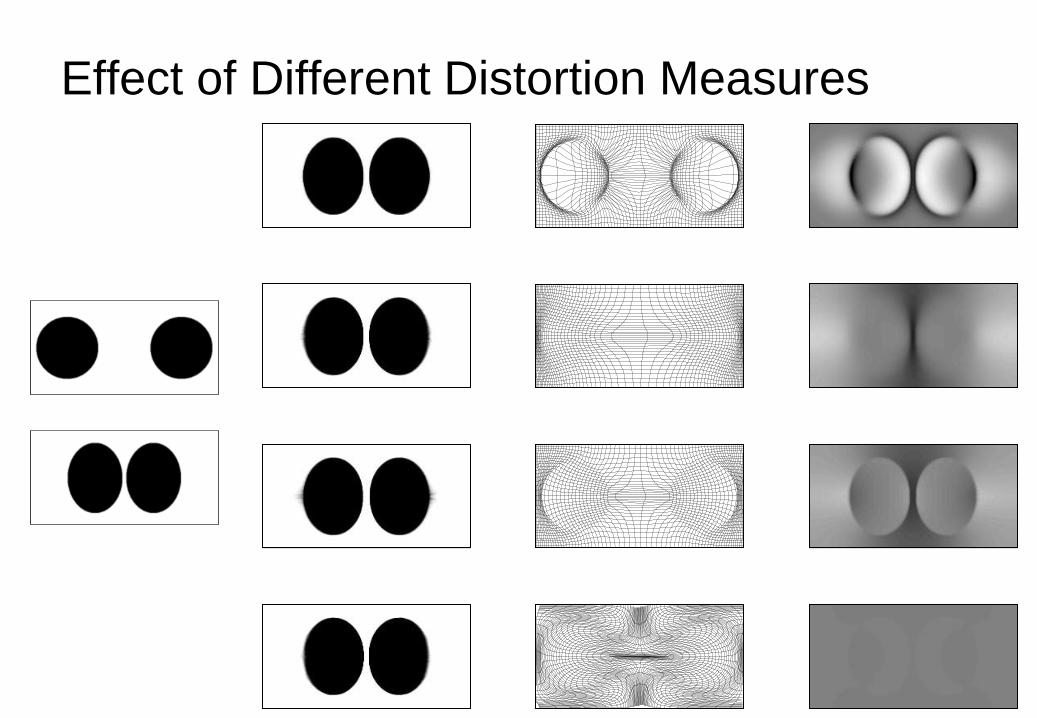
Effect of Different Distortion Measures
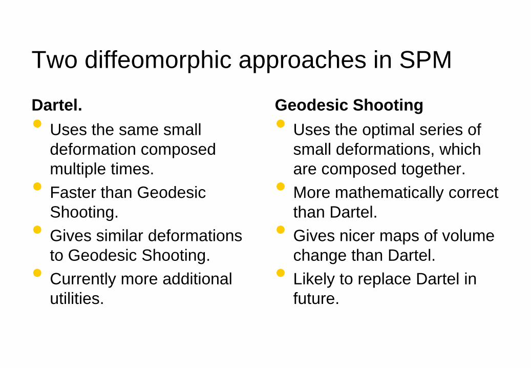
Two diffeomorphic approaches in SPM
Dartel.
• Uses the same small
deformation composed
multiple times.
• Faster than Geodesic
Shooting.
• Gives similar deformations
to Geodesic Shooting.
• Currently more additional
utilities.
Geodesic Shooting
• Uses the optimal series of
small deformations, which
are composed together.
• More mathematically correct
than Dartel.
• Gives nicer maps of volume
change than Dartel.
• Likely to replace Dartel in
future.
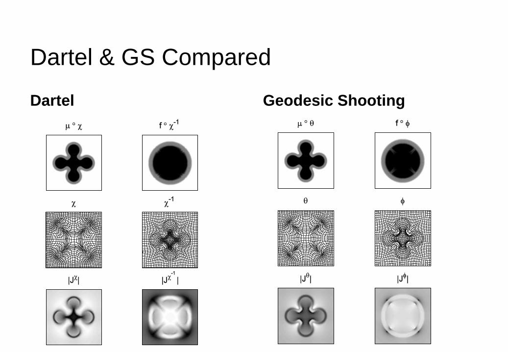
Dartel & GS Compared
Dartel Geodesic Shooting

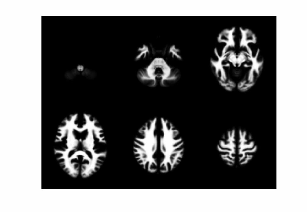
Simultaneous registration of GM to GM
and WM to WM
Grey matter
White matter
Grey matter
White matter
Grey matter
White matter
Grey matter
White matter
Grey matter
White matter
Template
Subject 1
Subject 2
Subject 3
Subject 4
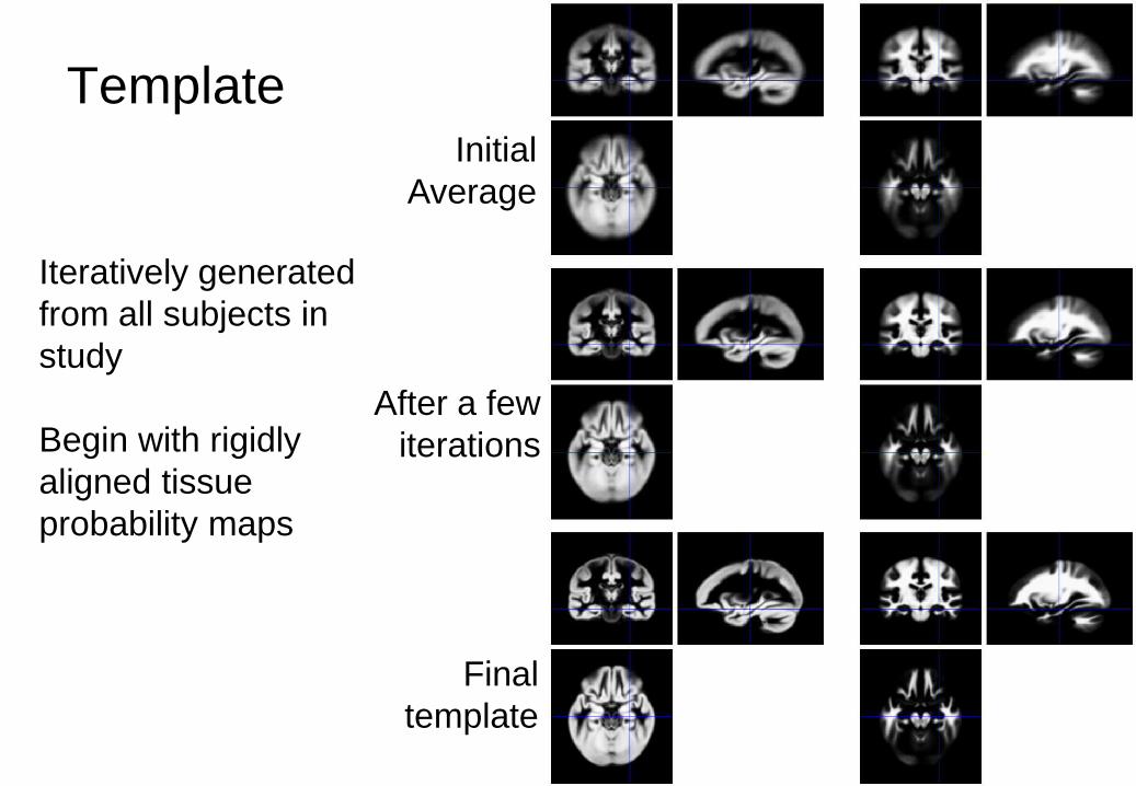
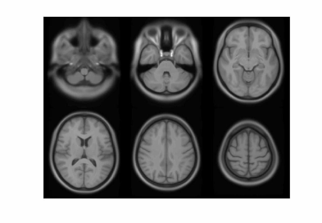
Template Initial
Average
After a few
iterations
Final
template
Iteratively generated
from all subjects in
study
Begin with rigidly
aligned tissue
probability maps
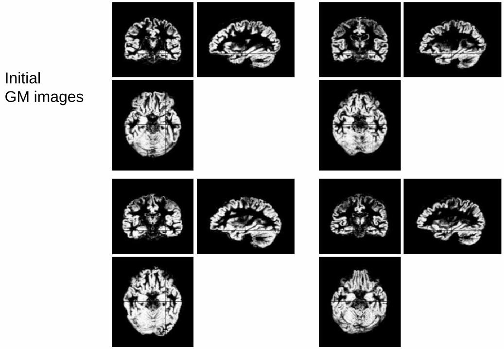
Initial
GM images
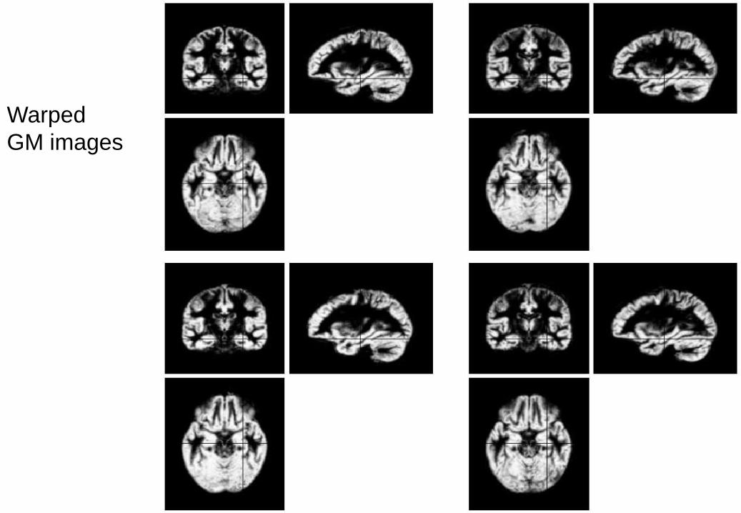
Warped
GM images


Evaluations of
nonlinear
registration
algorithms

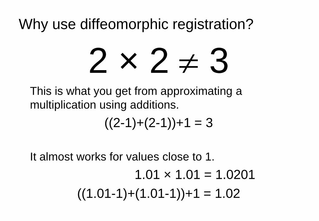
Why use diffeomorphic registration?
2 × 2 3 This is what you get from approximating a
multiplication using additions.
((2-1)+(2-1))+1 = 3
It almost works for values close to 1.
1.01 × 1.01 = 1.0201
((1.01-1)+(1.01-1))+1 = 1.02

Some References
• Ashburner. “A Fast Diffeomorphic Image Registration
Algorithm”. NeuroImage 38:95-113, 2007.
• Ashburner & Friston. “Computing Average Shaped Tissue
Probability Templates”. NeuroImage 45:333-341, 2009.
• Ashburner & Friston. “Diffeomorphic registration using
geodesic shooting and Gauss–Newton optimisation”.
NeuroImage 55(3):954-967, 2011.
• Klein, Andersson, Ardekani, Ashburner, Avants, Chiang,
Christensen, Collins, Gee, Hellier, Song, Jenkinson, Lepage,
Rueckert, Thompson, Vercauteren, Woods, Mann & Parsey.
“Evaluation of 14 nonlinear deformation algorithms applied to
human brain MRI registration”. NeuroImage 46:786-802, 2009.
Overview
• Voxel-Based Morphometry
• Diffeomorphic Registration
• Tensor-Based Morphometry
• Longitudinal Registration

Some 2D Shapes
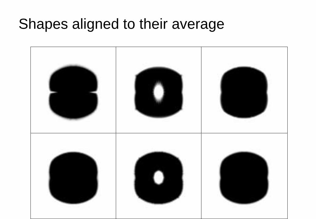
Shapes aligned to their average

These were the deformations for that
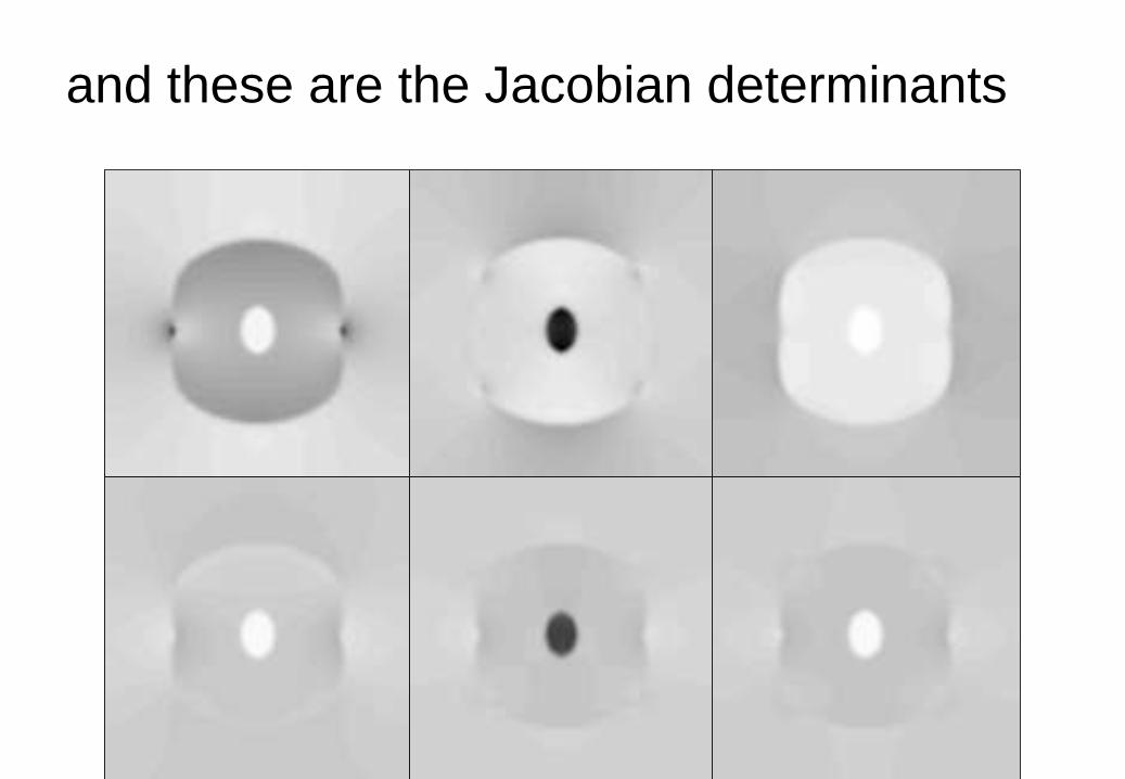
and these are the Jacobian determinants

Cross-Sectional Data
Used 550 T1w brain MRI from
IXI (Information eXtraction
from Images) dataset.
•http://www.brain-
development.org/
Data from three different
hospitals in London:
•Hammersmith Hospital
using a Philips 3T system
•Guy’s Hospital using a
Philips 1.5T system
•Institute of Psychiatry using
a GE 1.5T system5T system

Segmentation
Segmented into GM and WM.
Approximately aligned via rigid-body.
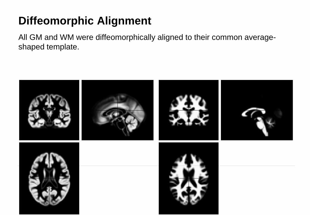
Diffeomorphic Alignment
All GM and WM were diffeomorphically aligned to their common average-
shaped template.

Divergence Maps
• Used maps of initial velocity
divergence.
• Similar to logarithms of Jacobian
determinants.
• Encode a sort of “growth rate”
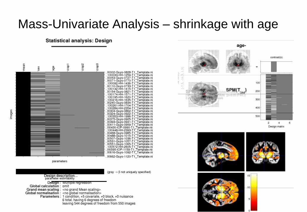
Mass-Univariate Analysis – shrinkage with age
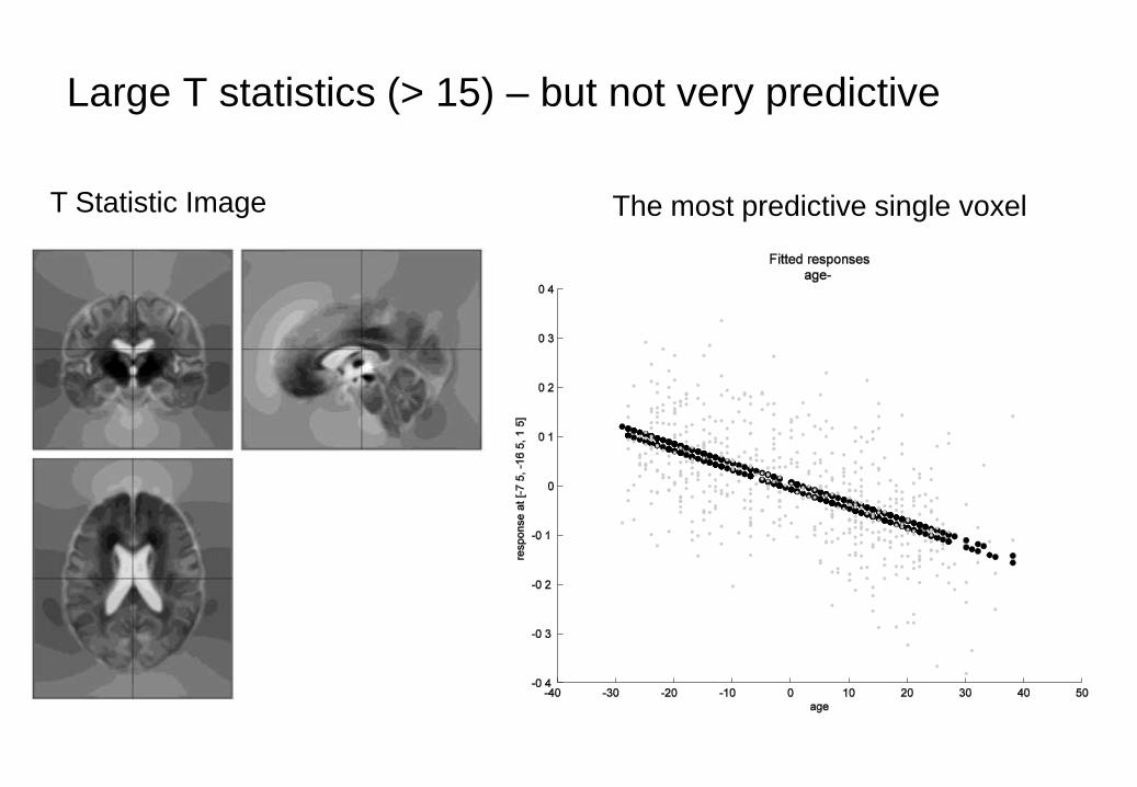
Large T statistics (> 15) – but not very predictive
T Statistic Image The most predictive single voxel

Some References
• Ashburner & Friston. “Unified Segmentation”. NeuroImage
26:839-851, 2005.
• Ashburner & Friston. “Computing Average Shaped Tissue
Probability Templates”. NeuroImage 45:333-341, 2009.
• Ashburner & Friston. “Diffeomorphic registration using
geodesic shooting and Gauss–Newton optimisation”.
NeuroImage 55(3):954-967, 2011.

Overview
• Voxel-Based Morphometry
• Diffeomorphic Registration
• Tensor-Based Morphometry
• Longitudinal Registration

Longitudinal Registration
• Unified model combines:
• Nonlinear diffeomorphic
registration.
• Rigid-body registration.
• Intensity inhomoheneity
correction.
• All made as mathematically
coherent as possible.
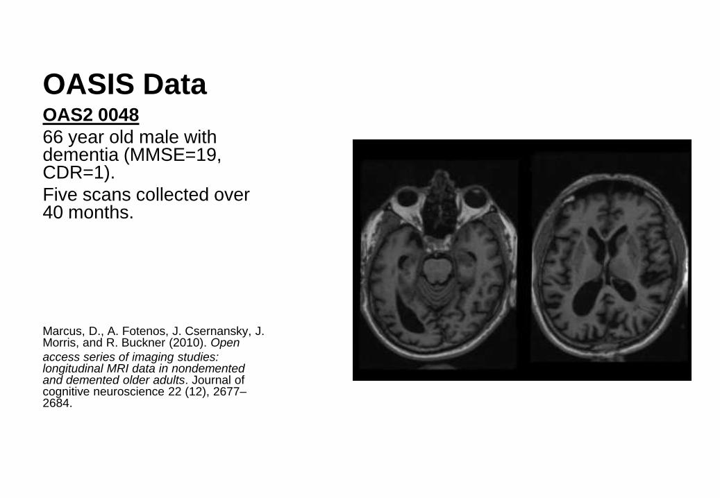
OASIS Data OAS2 0048
66 year old male with dementia (MMSE=19, CDR=1).
Five scans collected over 40 months.
Marcus, D., A. Fotenos, J. Csernansky, J. Morris, and R. Buckner (2010). Open
access series of imaging studies: longitudinal MRI data in nondemented and demented older adults. Journal of cognitive neuroscience 22 (12), 2677–2684.

OASIS Data OAS2 0048
66 year old male with
dementia (MMSE=19,
CDR=1).
Five scans collected over
40 months.
Difference between time
point and first scan.
OASIS Data OAS2 0048
66 year old male with
dementia (MMSE=19,
CDR=1).
Five scans collected over
40 months.
Expansion/contraction.
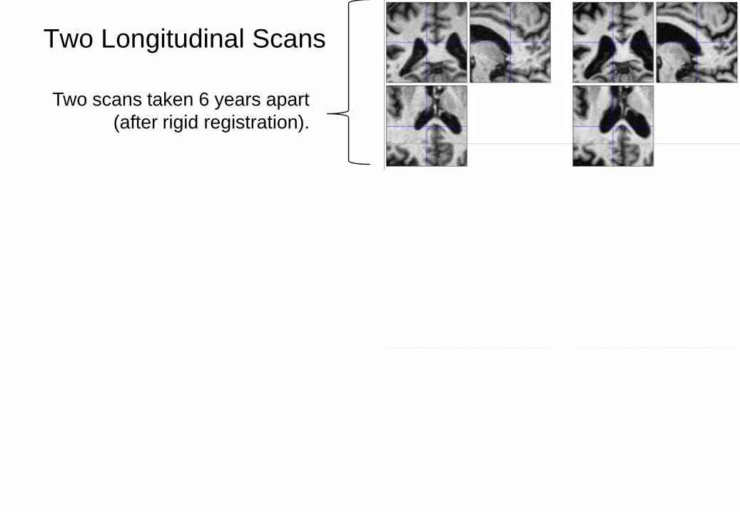
Two Longitudinal Scans
Two scans taken 6 years apart
(after rigid registration).
Average and difference images.
Shape average and map of
expansion/contraction
(after nonlinear registration)
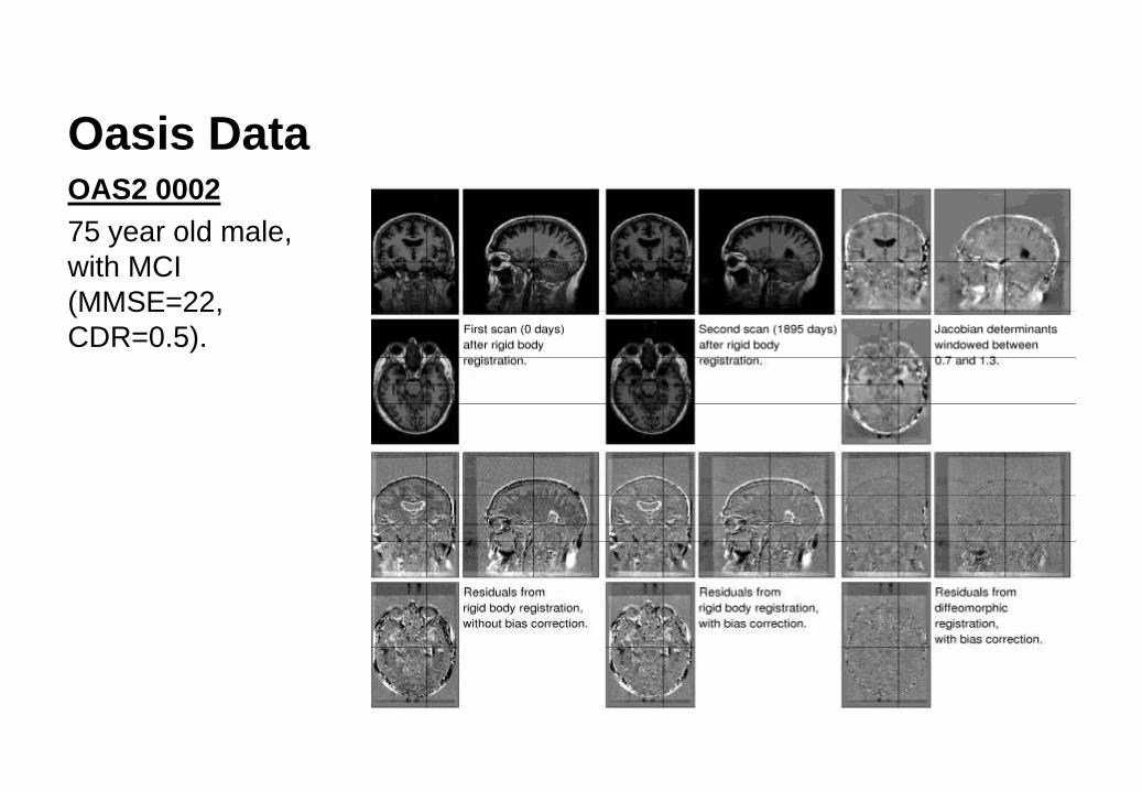
Oasis Data OAS2 0002
75 year old male,
with MCI
(MMSE=22,
CDR=0.5).
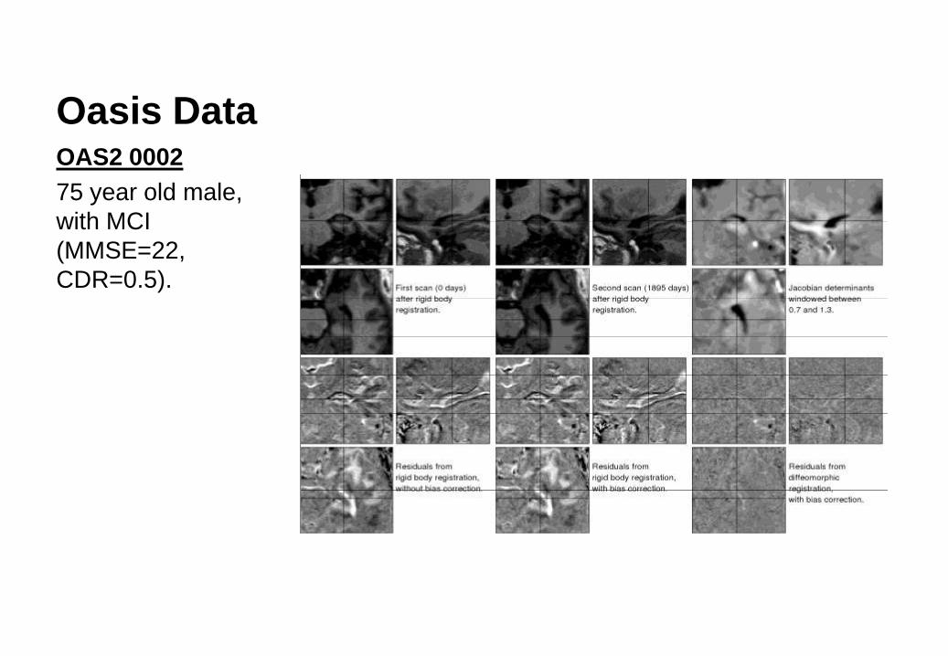
Oasis Data OAS2 0002
75 year old male,
with MCI
(MMSE=22,
CDR=0.5).

Oasis Data OAS2 0048
66 year old male, with MCI (MMSE=19, CDR=1).
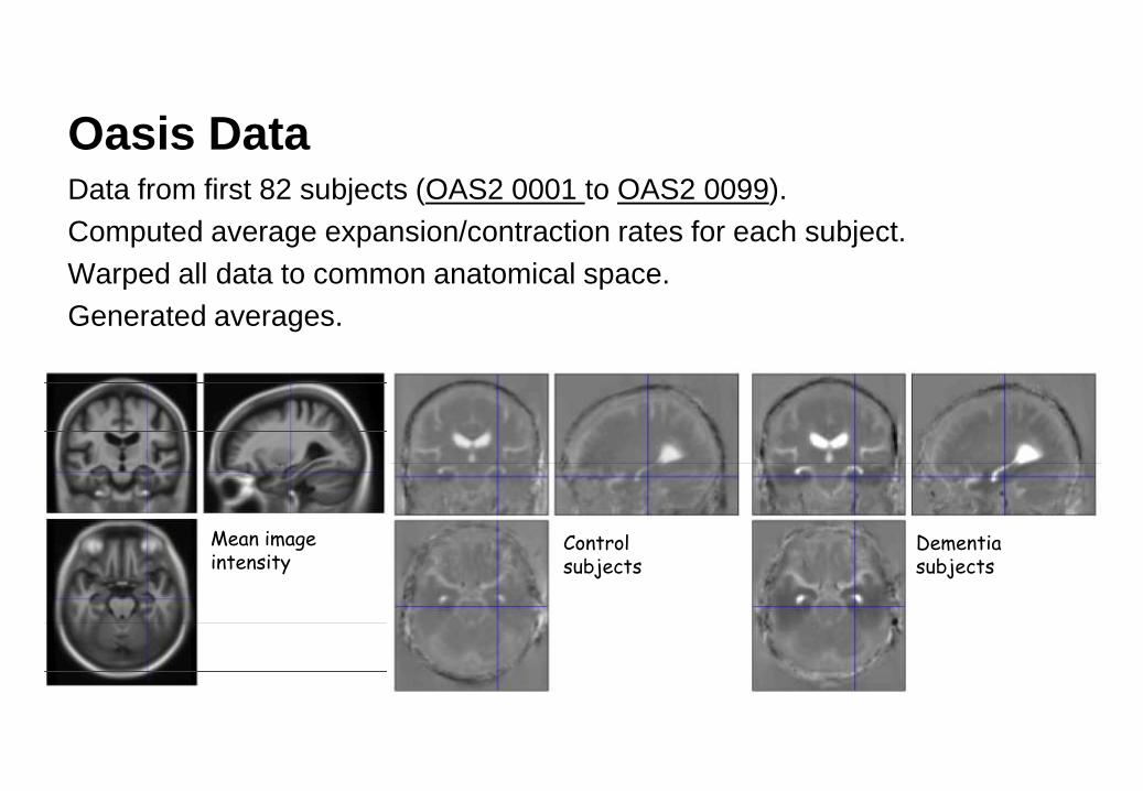
Oasis Data Data from first 82 subjects (OAS2 0001 to OAS2 0099).
Computed average expansion/contraction rates for each subject.
Warped all data to common anatomical space.
Generated averages.
Mean image intensity
Control subjects
Dementia subjects

References
• Ashburner & Ridgway (2013). Symmetric diffeomorphic modelling of longitudinal structural MRI. Frontiers in Brain Imaging Methods 6(197).