Embed Size (px)
Citation preview
International Journal of
Medical Science and Discovery
Open Access Scientific Journal
ISSN: 2148-6832
Lycia Press LONDON U.K.
www.medscidiscovery.com
MSD Medical Science & Discovery M
ay, 2
01
7, V
ol 4
, No
5
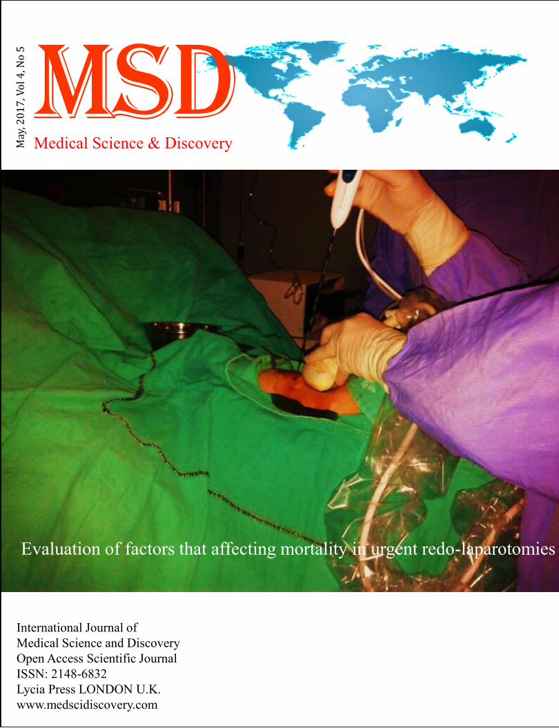
Evaluation of factors that affecting mortality in urgent redo-laparotomies


Medical Science and Discovery (http://www.medscidiscovery.com) is an international open access, peer-reviewed scientific
research journal that provides rapid publication of articles in all disciplines of human health, clinical and basic medical
science such as Biophysics, Biochemistry, Histology, Physiology, Genetics, Pathology, Toxicology, Anatomical Sciences,
Pharmacology, Embryology, Internal and Surgical Medicine.
The policy of top priority of MSD is to put forward and highlight medical innovations and inspiring patents.
MSD offers an exceptionally fast publication schedule including prompt peer-review by the experts in the field and immediate
publication upon acceptance. The editorial board aims at reviewing the submitted articles as fast as possible and promptly
including them in the forthcoming issues.
This journal is published under ethical publishing policy of international scientific Bioethics and publication rules.
MSD supports the Open Access Initiative. Abstracts and full texts (HTML and PDF format) of all articles published
by MSD are freely accessible to everyone immediately upon publication.
Medical Science and Discovery has scientific affiliation with Lycia Clinics London UK
Indexed Databases: NLM Catalog, Chemical Abstracts (CAS), Index Copernicus, Open Air, ULRICHS Database, Proquest,
Advanced Science Index, Turkish Citation Index, Tubitak Ulakbim, Research Bible, Scholar Google
Medical Science and Discovery is an international open access, peer-reviewed scientific research journal.
ISSN: 2148-6832 (Print) E-ISSN: 2148-6832 (Online)
Category: Multi Disciplinary Health Science Journal
Abbreviated key title: Med. Sci. Discov.
Frequency: Monthly
Review System: Double Blind Peer Review
Circulation: Globally, Online, Printed
Article Processing Charge (APC): US$ 100
Licensing: CC-BY-NC 4.0 International License Environmental
Editor-in-Chief: Assoc. Prof. Dr. Dr. Asghar Rajabzadeh, Anatomical Department, Lorestan University of Medical
Sciences, Khorramabad, Iran
Established: 30.04.2014
Web address: www.medscidiscovery.com; http://dergipark.ulakbim.gov.tr/msd
E-mail : editor [at] medscidiscovery.com
Phone : +44 020 3289 9294
Design and preparation of PDFs, Language editing, Web site design, Graphical design Services of international
Journal of Medical Science and Discovery has been contracted with Lycia Press LONDON, UK (as Publisher), by the
MSD Board of Directors
Publisher: Lycia Press Inc.
Address: 3rd Floor 86 - 90 Paul Street, EC2A 4NE, London, UK
Web address: www.lycians.com
Phone : +44 020 3289 9294
E-mail : office [at] lycians.com
E-mail : info [at] lycians.com
Editorial Board of Medical Science and Discovery
Honorary Editors
Prof. Dr.
Aziz Sancar
UNC, Faculty of Medicine, Dept. of Biochemistry-Biophysics, Chapel Hill, NC, USA
Prof. Dr. Giancarlo BAROLAT Barolat Institute, 1721 E 19th Ave #434, Denver, CO 80218, USA
Prof. Dr.
Joyce REARDON
UNC, Faculty of Medicine, Dept. of Biochemistry-Biophysics, Chapel Hill, NC, USA
Prof. Dr. Metin TULGAR Yuzuncu Yil University, School of Medicine, Dept. of Biophysics, Van, TR
Deputy Editors
Assoc. Prof.
Michael George KEMP
UNC, 120 Mason Farm Road, Campus Box 7260, Genetic Medicine Bldg Room 3010 Chapel
Hill, NC 27599 USA
Assoc. Prof. Zafer Akan (Founder) Lycia Press Inc., 3rd Floor 86 - 90 Paul Street, EC2A 4NE, London, UK
Internal Medicine
Asist. Prof. Dr. Ahmet YILMAZ Dicle University, Faculty of Medicine, Dept. of Family Medicine
Prof. Dr. Ali Rıza BILGE CBU, Faculty of Medicine, Dept. of cardiology, Manisa, TR
Assoc. Prof. Dr. Alparslan SAHİN Dicle University, Faculty of Medicine, Dept. of Eye
Prof. Dr. Ayşe YÜKSEL Arel University, Faculty of Medicine, Dept. of Public Health, Istanbul
Assoc. Prof. Dr. Bekir Serhat YILDIZ PAU, Faculty of Medicine, Dept. of Cardiology, Denizli, Turkey
Prof. Dr. Hatice Sınav USLU ISMU, Faculty of Medicine, Dept. of Nucleer Medicine, Istanbul, TR
Prof. Dr. Hikmet YILMAZ CBU, Faculty of Medicine, Dept. of Neurology, Manisa, TR
Prof. Dr. Hulya Ozdemir YYU Faculty of Medicine, Dept. of Pharmacology, Van
Assoc. Prof. Dr. Huseyin GUDUCUOGLU YYU Faculty of Medicine, Dept. of Microbiology, Van
Asist. Prof. Dr. Murat ÖZSARAÇ CBU, Faculty of Medicine, Dept. of Emergency Medicine
Prof. Dr. Muzaffer POLAT CBU, Faculty of Medicine,Dept. of Pediatric Neurology
Assist. Prof. Dr.
Nesrin CEYLAN
Ankara Children's Health, Training and Research Hospital, Department of Hematology
Oncology , Ankara, Turkey
Prof. Dr. Nobuo INOTSUME Hokkaido Pharmaceutical University, Clinical Pharmacology, Hokkaido AC, JAPAN
Assist Prof. Dr. Secil ILHAN YILMAZ Erciyes University, Genom and Stem Cell Research Center, Kayseri, TR
Prof. Dr. Talat ECEMIS CBU, Faculty of Medicine, Dept. of Microbiology, Manisa, TR
Surgical Medicine
Assoc. Prof. Dr. Abdullah BOYUK Dicle University, Faculty of Medicine, Dept. of General Surgery
Assist. Prof. Dr. Christopher Schmitt University of California, San Francisco Cardiovascular Res. Inst.
Prof. Dr. Çetin DİNÇEL Hacettepe University, Faculty of Medicine, Dept. of Urology
Prof. Dr. Cuneyt Temiz CBU, Faculty of Medicine, Dept. of Neurosurgery, Manisa
Prof. Dr. Gönül Tezcan KELEŞ CBU, Faculty of Medicine, Dept. of Anesthesiology and Rean.
Prof. Dr. M. Derya BALBAY Memorial Hospital, Dept. of Urooncology
Assoc. Prof. Dr. Mustafa USLU Duzce University, Faculty of Medicine, Dept. of Orthopedics, Bolu
Asist. Prof. Dr. Murat YILDIR BAU Faculty of Medicine, Dept. of General Surgery
Prof. Dr Nasuhi Engin AYDIN Katip Çelebi University, Faculty of Medicine, Dept. of Pathology
Assist. Prof. Dr. Pinar SOLMAZ HASDEMIR CBU, Faculty of Medicine, Dept. of Obstetrics and Gynecology, Manisa
Assoc. Prof. Dr. Tevfik GUNES PAU, Faculty of Medicine, Dept. of Cardiovascular Surgery, Denizli,
Assoc. Prof. Dr. Yusuf Izzettin ALIHANOGLU PAU, Faculty of Medicine, Dept. of Cardiology, Denizli

Editorial Board of Medical Science and Discovery
Basic Sciences
Dr. Alper Tunga ÖZDEMİR Manisa ME State Hospital Dept. of Medical Biochemistry
Prof. Dr. Alev Meltem ERCAN Istanbul University, Cerrahpasa Medical Faculty, Dept. of Biophysics, Istanbul
Assoc. Prof. Dr. Anzel BAHADIR Duzce University, Faculty of Medicine, Dept. of Biophysics, Bolu, TR
Assoc. Prof. Dr. Ayse Inhan GARIP Marmara University, Faculty of Medicine, Dept. of Biophysics
Assoc. Prof. Dr. Bahriye SİRAV Gazi University, Faculty of Medicine, Dept. of Biophysics
Prof. Dr. Beki KAN Acıbadem University, Faculty of Medicine, Dept. of Biophysics
Prof. Dr. Cevval ULMAN CBU, Faculty of Medicine, Dept. of Biochemistry, Manisa, TR
Assoc. Prof. Dr. Gokhan OTO YYU Faculty of Medicine, Dept. of Pharmacology, Van, TR
Prof. Dr. Halit DEMİR YYU Faculty of Science, Dept. of Biochemistry
Prof. Dr. Hasan YILMAZ YYU Faculty of Science, Dept. of Parasitology, Van, TR
Prof. Dr. M. Ali KORPINAR Istanbul University, Cerrahpasa Medical Faculty, Dept. of Biophysics, Istanbul
Prof. Dr. Mustafa ÖZBEK CBU, Faculty of Medicine, Dept. of Physiology
Prof. Dr. Nobuo Inotsume Hokkaido Pharmaceutical Unv., Clinical Pharmacology, Hokkaido AC, JAPAN
Asist. Prof. Dr. Özdemirhan Serçin Interdisciplinary Research Institute, Université Libre de Bruxelles, Belgium
Prof. Dr. Seda VATANSEVER CBU, Faculty of Medicine, Dept. of Histology and Embryology
Prof. Dr. Sevinç İNAN CBU, Faculty of Medicine, Dept. of Histology and Embryology
Asist. Prof. Dr.
Shoban GADDAMADI
Washington State University College of Pharmacy, Dept. of Experimental and Systems
Pharmacology, Spokane, WA, USA
Asist. Prof. Dr. Tahir CAKIR YYU Faculty of Medicine, Dept. of Nucleer Medicine Van, TR
Assoc. Prof. Dr. Tamer ZEREN CBU, Faculty of Medicine, Dept. of Biophysics
Prof. Dr. Tunaya KALKAN Istanbul University, Cerrahpasa Medical Faculty, Dept. of Biophysics, Istanbul
Assist Prof. Dr. Younes El Bouzekri EL IDRISSI Place Aboubakr, Imm 22, App 6, Bd Fal ould oumeir, Agdal Rabat
Assist Prof. Dr. Yusuf Kemal DEMIR Marmara University, Faculty of Pharmacy, Dept. of Pharmaceutical Tech. Istanbul TR
Statistical Editor
Prof. Dr. Sıddık KESKİN YYU Faculty of Medicine, Dept. of Medical Statistics, Van, TR
Language Editor
Asist. Prof. Dr. Hakan ERGİN Istanbul University, Dept. of Foreign Languages, Istanbul, TR
Editorial Office
General Coordinator Elena JALBA Office Lycia Press, London, UK
Typist-Compositor Gonul OZGOK
Office Lycia Press, London, UK
Typist-Compositor Bugra YOLDAS Office Lycia Press, London, UK

Instruction for Authors
• Important
• MSD is committed to deterring plagiarism, including self-plagiarism. Your manuscript will screen to compare for similarity
with published articles.
• For research studies using human or animal subjects, the trial's design, conduct and reporting of results must conform to Good
Clinical Practice guidelines (such as the Good Clinical Practice in Food and Drug Administration (FDA)-Regulated Clinical
Trials (USA) or the Medical Research Council Guidelines for Good Clinical Practice in Clinical Trials (UK)) and/or to the
World Medical Association (WMA) Declaration of Helsinki
• Dear Authors, please upload just these three files to the manuscript submission system
• Title Page Sample
• Manuscript Sample
• Copyright Transfer and Author Consent Form
• Please select Keywords from the MESH source
• (https://www.nlm.nih.gov/mesh/MBrowser.html)
• Manuscripts should be prepared in accordance with the "Uniform Requirements for Manuscripts Submission to Biomedical
Journals" proclaimed by the International Committee of Medical Journal Editors (www.icmje.org).
• MSD uses Vancouver reference style, please prepare articles due to Vancouver reference style rules.
• Manuscript Preparation Rules
• 1.Cover letter
• a- A statement that the manuscript has been read and approved by all the authors.
• b- That the requirements for authorship have been met for all the authors, based on the criteria stated by ICMJE.
• c- Approval of all the authors regarding the order in which their names have appeared.
• d- That each author confirms the manuscript represents honest work.
• e- The name, address, and telephone number of the corresponding author who is responsible for communicating with other
authors about revisions and final approval.
• f- The letter should give any additional information that may be helpful to the editor, such as the type or format of the article.
If the manuscript has been submitted previously to another journal or in another language, it is helpful to include the previous
editor's and reviewers' comments with the submitted manuscript, along with the authors' responses to those comments.
Submitting previous evaluatory review of another journal accelerates the review process.
• g- For accepted manuscripts, the authors are requested to fill and sign the journal's cover letter to express their consent for its
publication.
• h- To reproduce published material, to use illustrations or tables or report information about identifiable people, the author
should submit a copy of the permission with the manuscript to the journal.
• 2.Top Ethic Committee Approval
Inclusion of the approval letter from the relevant Ethics Committee or Institution's Review Board regarding the research
protocol and the rights of the subjects (if applicable to the study)
• 3.Top Consent Form
Attach a copy of the consent form to the letter, if applicable. Consent forms would be evaluated by the Ethics Committee and
then signed by the participant.
• 4.Top RCT or NCT Registration
Emailing the letter denoting registration of RCTs or NCTs in domestic or international databases (The trial's registration
number needs to be mentioned, too).
• 5. Manuscripts submitted in English, must be type written, double-spaced, on good quality A4 paper, or paper of similar
format. Authors are requested to reserve margins of at least 2.5cm all around the paper. Original drawings of photos, tables
and figures should be furnished together with the manuscripts.
• 6. Manuscripts should be kept to a minimum length and should be subdivided into labeled sections (Title page, Abstract,
Keywords, Introduction, Materials and Methods, Results, Discussion, Conclusion, Acknowledgement, and References).
• 7. A title page is to be provided and should include the title of the article, authors' names with full first name (with degrees),
authors' affiliation, suggested running title and corresponding author. The affiliation should comprise the department,
institution (usually university or company), city and state (or nation). The suggested running title should be less than 50
characters (including spaces) and should comprise the article title or an abbreviated version thereof. For office purposes, the
title page should include the name and complete mailing address, telephone and fax number, and email of the one author
designated to review proofs.
• 8. An abstract no longer than 250 words for reviews and research articles is to be provided as the second page. Abstract
should be structured as objective(s) (including purpose setting), materials and methods, results, and conclusion.

Instruction for Authors
• Case Report
• A case report is a case study, case report, or other description of a case that should contain 1500 - 2000 words with a
structured abstract of 200 words maximum. Case reports should comprise sections of Introduction, Case Presentation, and
Conclusions in Abstract and Introduction, Case Presentation, and Discussion in full text with not more than 2 tables or figures
and up to 20 references.
• Brief Report
• Brief Reports should contain 1000 - 2000 words with a structured abstract of 200 words maximum. Short reports should
comprise sections of Background, Objectives, Materials & Methods, Results and Discussion with not more than 2 tables or
figures and up to 20 references.
• Short Communication
• Short Communication, follow the instructions for original articles, except that the total word number of the main text
(excluding references, tables and figure legends) is limited to 2000 with no more than 2 figures and/or tables and no more
than 15 references. An abstract, not exceeding 150 words, should be presented at the beginning of the article.
• News
• News should contain 1000 - 2000 words with a structured abstract of 200 words maximum. News should comprise sections of
Background, Objectives, Materials & Methods, Results and Discussion with not more than 2 tables or figures and up to 20
references.
•
• Publication Policies
• Manuscripts, or the essence of their content, must be previously unpublished and should not be under simultaneous
consideration by another Journal. The authors should also declare if any similar work has been submitted to or published by
another Journal. By virtue of the submitted manuscript, the corresponding author acknowledges that all the co-authors have
seen and approved the final version of the manuscript. The corresponding author should provide all co-authors with
information regarding the manuscript, and obtain their approval before submitting any revisions. Manuscripts are only
accepted for publication on the understanding that the authors will permit editorial amendments, though proofs will always be
submitted to the corresponding author before being sent finally to press. Prior to the initial submission of a new manuscript,
please carefully consider that all authors’ names are included as no change to authors’ details will be permitted after the
acceptance. The decision to accept a contribution rests with the Editorial Board of the MSD.
• Manuscripts will be considered for publication in the form of original articles, Case report, short communications, Letter to
editor and review articles. The work should be original or a thorough by an authoritative person in a pertinent field.
• Peer review process
• All submissions will be reviewed anonymously by at least two independent referees. All manuscripts will be acknowledged
upon presenting to the Journal office, provided that all stated requirements are met. Authors are encouraged to suggest names
of three expert reviewers, but selection remains a prerogative of the Editor. The whole review process depends on receiving
referees comments and revising the manuscripts based on these comments to the author. On receipt of the revised article from
the author, and after final approving by referees, the letter of acceptance is issued to the author. Authors have the right to
communicate to the editor if they do not wish their manuscript to be reviewed by a particular reviewer because of potential
conflicts of interest. No article is rejected unless negative comments are received from at least two reviewers. MSD employs
double blind reviewing process, where both the referee and author remain anonymous throughout the process.
Instruction for Authors
• Ethical Rules and Rights
• Conflicts of interest
• Conflicts of interest arise when authors, reviewers, or editors have interests that are not fully apparent and that may influence
their judgments on what is published. They have been described as those which, when revealed later, would make a
reasonable reader feel misled or deceived. (The Committee on Publication Ethics (COPE) states in its Guidelines on Good
Publication Practice 2003).
• Authors should disclose, at the time of submission, information on financial conflicts of interest or other interests that may
influence the manuscript. Authors should declare sources of funding for the work undertaken.
• The Journal's Policy on Plagiarism
• Any practice of plagiarism will not be tolerated by the journal regarding submitted manuscripts. Non-identifiable quoted
segments of articles or close paraphrases from other author/s or even submitting the author's previously published work are
known as the act of plagiarism by this journal unless proper use of quotations or paraphrasing with decent citation or
referencing are in place. Heavy use of one or a couple of articles is discouraged, even if paraphrased fully. Advertent practice
of plagiarism will abort reviewing process or later submission to this journal. All submitted articles will evaluate
by iThenticate software belonged to cross check for stop any plagiarism and improve publication quality.
• Statement of Human and Animal Rights
• All submitted articles involving human experiments should be performed only in accordance with the ethical standards
provided by the responsible committee of the institution and in accordance with the Declaration of Helsinki (as revised in
Edinburgh 2000), available at http://www.wma.net/en/30publications/ 10policies/b3/index.html. Papers describing animal
experiments can be accepted for publication only if the experiment conforms the National Institute of Health Guide (National
Institute of Health Publications No. 80-23, Revised 1978) for the care and use of Laboratory Animals for experimental
procedure. Authors must provide a full description of their anesthetics and surgical procedures. All manuscripts reporting the
results of experimental investigations involving human subjects should include a statement confirming the informed consent
was obtained from each subject or subject's guardian.
• Humans: When reporting experiments on human subjects, authors should indicate whether the procedures followed were in
accordance with the ethical standards of the responsible committee on human experimentation (institutional and national) and
with the Helsinki Declaration of 1975, as revised in 2008 (5). If doubt exists whether the research was conducted in
accordance with the Helsinki Declaration, the authors must explain the rationale for their approach and demonstrate that the
institutional review body explicitly approved the doubtful aspects of the study.
• Animals: When reporting experiments on animals, authors should indicate whether the institutional and national guide for the
care and use of laboratory animals was followed.
• All animal or human subjects should be used after approval of the experimental protocol by a local ethics committee.
• Acknowledgements
• Contributors: In acknowledgement section, name people for their contributions or their permission to reproduce their
published material, to use their illustrations or provide information about them- try to fully name people who have helped
from the conception of the idea to adoption of the hypothesis, to finalization of the study, etc., earnestly. Statement of
financial support: Aside from the title page, state any financial or other relationships that might lead to a conflict of interest.
• Copyright
• After acceptance and publication; all ownership rights and Copyrights of the manuscript, passes to international journal of
Medical Science and Discovery. Please complete copyright form and send via email to editor. Download MSD Copyright
Transfer and Author Consent Form
•
This work is licensed under a Creative Commons Attribution-NonCommercial 4.0 International License.
• Copyright 2014: The Author(s); This is an open-access article distributed under the terms of the Creative Commons
Attribution License (http://creativecommons.org/licenses/by/4.0), which permits unrestricted use, distribution, and
reproduction in any medium, provided the original work is properly cited. All Rights reserved by international journal of
Medical Science and Discovery.
• Disposal of material
• Once published, all draft copies of the manuscript, correspondence and artwork will be held at least for 6 months before
disposal. Authors and Readers may find original PDF file of article on backup servers such as CLOKKS
(https://www.clockss.org/)
• Digital Object Identifier DOI
• Once a manuscript is accepted for publication it will be provided with a registered DOI number following the acceptance
decision. Manuscripts accepted for publication by the MSD will be published as ahead of print articles prior to the printing
date of their scheduled issue. Corresponding author will be provided with a PDF Proof by the publisher once the production
process of an accepted manuscript is over.

Instruction for Authors
• Article Processing Charge
• MSD is a non-profit Scientific Journal Platform; however, it uses professional services such as Language Editing, DOI,
domain and hosting, iThenticate Plagiarism or similarity Detection Software. All of these professional services are used for all
the article processes and an inevitable cost arises with this.
• Unfortunately, like most open journals, fees of the publication with MSD are charged to Authors. Payment is under the
responsibilities of corresponding Author(s). MSD does not charge any fee during the submission period. However, after the
peer-review process, a non-refundable charge (100 USD ) for each accepted manuscript must be paid by the author(s) via
MSD's official PayPal account. An invoice will be sent for each accepted manuscript to corresponding author(s).
• Following with completion of payment procedure, the galley proof and acceptance letter of article will be send to
authors for last check
• Preparation of articles in PDF and HTML format is covered by Lycia Press Inc. (press.lycians.com) and Article Processing
Charges paid to Lycia Press Inc. (press.lycians.com)
• MSD revenue sources and Sponsorships
• All costs arising from the publications are covered by the Sponsor Companies and Article Processing Charges. Sponsorship
request evaluates by the MSD Journal Management Board and the sponsor company logos will be included on the back page
of printed magazine and in the sponsor section of journal website
• *APC not includes Proofreading Services fee. Editor in Chief may direct the corresponding Author to Lycia Press, Language
Office for Proofreading Service www.lycians.com
•
• References
• Committee on Publication Ethics (COPE). (2011, March 7). Code of Conduct and Best-Practice Guidelines for Journal
Editors. Retrieved fromhttp://publicationethics.org/files/Code_of_conduct_for_journal_editors_Mar11.pdf
• World Association of Medical Editors (WAME). Principles of Transparency and Best Practice in Scholarly
Publishing. http://www.wame.org/about/principles-of-transparency-and-best-practice
Article Processing Charge (APC) Discount %
Regular 100 USD
for Editorial Board Members 70 USD 30%
for Affiliated Institution Members 80 USD 20%

Medical Science and Discovery, May 2017, Vol.4, No.5
Contents
Original Article
Evaluation of factors that affecting mortality in urgent redo-laparotomies 35-43
Erdal Uysal, Serkan Kadir Turel, Efe Sezgin


http://www.medscidiscovery.com
OPEN ACCESS JOURNAL ISSN: 2148-6832
MSD
Medical Science and Discovery 2017; 4(5):35-43
Original Article Doi: 10.17546/msd.315378
Received 22-05-2017 Accepted 28-05-2017 Available Online 31-05-2017
1 Sanko University, Medical Faculty, Dept. of General Surgery, Gaziantep, TR 2 Afyon State Hospital, Dept. of General Surgery, Afyon, TR
3 Izmir Institute of Technology, Engineering Faculty, Department of Food Engineering, Izmir, TR
* Corresponding Author: Erdal Uysal E-mail: [email protected] Phone: + 90342 211 65 69
Evaluation of factors that affecting mortality in urgent redo-laparotomies
Erdal Uysal 1*
, Serkan Kadir Turel2, Efe Sezgin
3
Introduction
Abdominal surgical interventions are applied for the
treatment of benign and malignant diseases under
urgent and elective conditions. Although laparascopic
abdominal interventions are widely preferred
currently, laparatomy has been used frequently as
well. The term redo-laparatomy or re-laparatomy is
used for the laparotomies performed within 60 days
following the primary surgery. Redo-laparatomies
may be performed urgently, electively, planned or
unplanned (1).
In our study, redo-laparatomies performed within 10
days following the initial surgery were described as
urgent redo-laparatomies. Urgent abdominal redo-
laparatomies (UARLs) have been conisdered as
complicated and last option operations. The rates of
mortality and morbidity of UARLs are quite high (2).
UARLs are generally performed due to the
complications observed after the initial operation. The
most common indications for UARLs include
anastomosis leaks, intra-abdominal abscess,
peritonitis, mechanical intestinal obstruction, intra-
abdominal hemorrhage, intestinal necrosis and intra-
abdominal organ injuries (2).
Patients indicated for UARLs generally have a critical
health situation. UARLs are still the most important
step of the treatment for these patients (1,3). The
mortality rates vary between 50 and 100% in cases
with peritonitis that cannot be taken under control,
namely those with spesis and multiple system organ
failure (4,5).
UARLs may be peformed as on-demand surgery and
planned. In planned UARLs, laparatomy is performed
with pre-determined time intervals and thus the
clinical situation is considered (6).
Both planned and on-demand surgeries are performed
in the clinical centers in our study. UARLs may be
performed once or multiple times according to clinical
need.
Abstract
Objective: The aim of this study was to investigate the factors that affect mortality in patients undergoing
Urgent abdominal redo-laparatomies (UARLs), and to analyze the common indications and
operative,demograhic, andclinical characteristics in UARLs.
Material and Methods: Our study was designed as a retrospective, observational and multi-centric study. A
total of 155 patients from two separate clinics undergoing urgent, unplanned redo-laparatomy were included in
the study. The data obtained from all clinics were collected and and the relations of the demographic, clinical
and operational factors with the mortalities of primary surgery and multiple UARLs were analyzed.
Results: Mortality was observed in 42 (27%) patients. The most frequent causes of mortality were sepsis and
multi-system organ failure in 23 (53.4%) patients. The relationships between the number of redo-laparotomy
presence of blood transfusion, older age,classification of surgery and mortality were significant (p<0.05).
Conclusion: Sepsis and multi-organ failure were the most frequent cause of mortality in our study as well, with
a rate of 53.4 %. No significant relationship was observed between mortality and initial surgery under
emergency conditions and presence of comorbidity. Redolaparatomies inevitable in some clinical situations.
Since multiple redolaparatomies are associated with mortality , it is necessary to avoid complications during
initial surgery to reduce UARLs. Major surgery operation UARLs in elderly people should be avoided. Finally,
the surgeon should also make the UARLs decision at the right time.
Key words: Urgent, redo-laparatomy, mortality
Uysal et al. http://dx.doi.org/10.17546/msd.315378
36 Medical Science and Discovery, 2017; 4(5):35-43
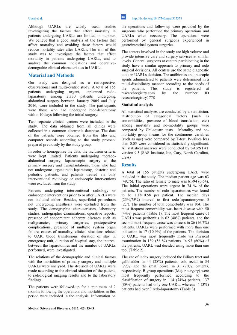
Although UARLs are widely used, studies
investigating the factors that affect mortality in
patients undergoing UARLs are limited in number.
We believe that a good analysis of the factors that
affect mortality and avoiding these factors would
reduce mortality rates after UARLs. The aim of this
study was to investigate the factors that affect
mortality in patients undergoing UARLs, and to
analyze the common indications and operative-
demograhic-clinical characteristics in UARLs.
Material and Methods
Our study was designed as a retrospective,
observational and multi-centric study. A total of 155
patients undergoing urgent, unplanned redo-
laparatomy among 2,830 patients undergoing
abdominal surgery between January 2005 and July
2016, were included in the study. The participants
were those who had undergone redo-laparatomy
within 10 days following the initial surgery.
Two separate clinical centers were included in the
study. The data obtained from all clinics were
collected in a common electronic database. The data
of the patients were obtained from the files and
computer records according to the study protocol
prepared previously by the study group.
In order to homogenize the data, the inclusion criteria
were kept limited. Patients undergoing thoraco-
abdominal surgery, laparascopic surgery as the
primary surgery and transplantations, those who had
not undergone urgent redo-laparatomy, obstetric and
pediatric patients, and patients treated via only
interventional radiology or endoscopic interventions
were excluded from the study.
Patients undergoing interventional radiology or
endoscopic interventions prior to or after UARLs were
not included either. Besides, superficial procedures
not undergoing anesthesia were excluded from the
study. The demographic characteristics, laboratory
studies, radiographic examinations, operative reports,
presence of concomitant adherent diseases such as
malignancies, primary surgeries, postoperative
complications, presence of multiple system organ
failure, causes of mortality, clinical situations related
to UAR, blood transfusions, duration of stay in
emergency unit, duration of hospital stay, the interval
between the laparotomies and the number of UARLs
performed, were investigated.
The relations of the demographic and clinical factors
with the mortalities of primary surgery and multiple
UARLs were analyzed. The decision of UARLs were
made according to the clinical situation of the patient,
to radiological imaging results and to the laboratory
findings.
The patients were followed-up for a minimum of 2
months following the operation, and mortalities in this
period were included in the analysis. Information on
the operations and follow-up were provided by the
surgeons who performed the primary operations and
UARLs when necessary. The operations were
performed by general surgeons experienced in
gastrointestinal system surgeries.
The centers involved in the study are high volume and
provide intensive care and surgery services at similar
levels. General surgeons at centers participating in the
study have a similar approach to primary and redo
surgical decisions. All centers use the same diagnostic
tools in UARLs decision. The antibiotics and inotropic
agents administered to patients were determined in a
multi-disciplinary manner according to the needs of
the patients. This study is registered at
researchregistry.com by the number ID
researchregistry1778
Statistical analysis
All statistical analyses are conducted by a statistician.
Distribution of categorical factors (such as
comorbidities, presence of blood transfusion, etc.)
among mortality and no-mortality groups were
compared by Chi-square tests. Mortality and no-
mortality group means for the continuous variables
(such as age) were compared by T-test. P-values less
than 0.05 were considered as statistically significant.
All statistical analyses were conducted by SAS/STAT
version 9.3 (SAS Institute, Inc, Cary, North Carolina,
USA)
Results
A total of 155 patients undergoing UARL were
included in the study. The median patient age was 63
(49,76). The ratio of female to male was 61/94 (0.64).
The initial operations were urgent in 74 % of the
patients. The number of redo-laparatomies was found
to be 1.18±0.58 per patient. The median days
(25%,75%) interval to first redo-laparatomywas 5
(2,7). The number of total comorbidity was 104. The
most frequent comorbidity was heart disease with 50
(44%) patients (Table 1). The most frequent cause of
UARLs was peritonitis in 62 (40%) patients, and the
second most frequent cause was abscess in 26 (16.7%)
patients. UARLs were performed with more than one
indication in 17 (10.9%) of the patients. The decision
of UARL was most frequently made via Physical
examination in 139 (56 %) patients. In 93 (60%) of
the patients, UARL wad decided using more than one
tool (Table 2).
The site of index surgery included the Biliary tract and
gallbladder in 44 (28%) patients, colo-rectal in 34
(22%) and the small bowel in 31 (20%) patients,
respectively. B group operations (Major surgery) were
most frequently performed according to the
classification of surgery in 114 (74%) patients. 137
(89%) patients had only one UARL, whereas 4 (3%)
patients had over 3 redo-laporatomy (Table 3)

Uysal et al. http://dx.doi.org/10.17546/msd.315378
37 Medical Science and Discovery, 2017; 4(5):35-43
Table 1: Patient Characteristics
Total Patients n=155 %
Sex (F/M) 61/94
Age 63 (49,76)
Emergency Operation 74
Number of redo-laparatomy 1.18±0.58
Median days Interval to first redo-laparatomy (25%,75%) 5 (2,7)
Median days Hospital stay (25%,75%) 12 (9,19)
Median days ICU stay (25%,75%) 9 (4,13)
Comorbidite n %
Heart disease 50 44
Pulmonary disease 13 11,4
Renal disease 14 12,3
Diabetes 19 17
Others 18 15,8
Total (n) 104
Causes of death
Sepsis and MOF 23 53,4
Intraabdominal hemorrhage 4 9,3
Respiratory failure and pneumonia 8 18,6
Cardiac 3 6,9
Unknown 4 11,6
Total (n) 42 M: male, F: female, ICU: intensive care unit, MOF: Multiorgan Failure, Comorbidite others: Autoimmune diseases, thyroid disease, hematological diseases, multiple comorbidite
Table 2: Indications of urgent Redo-laparotomy and decision tool
Patients (n=155) n %
Indications
Anastomotic leak 11 7
Intra-abdominal hemorrhage 21 13,5
Peritonitis 62 40
Abscess 26 16,7
Intestinal obstruction 21 13,5
Bowel necrosis 6 3,8
Abdominal dehiscence 7 4,5
Other 18 11,6
Total 172 110,6
Decision tool
Computed tomography/ultrasonography 85 34.2
Physical examination 139 56
Purulent discharge (wound) /drained content 16 6.4
Others 8 3.2
Total 248 100
Decision tool others: Multiple organ failure, positive blood culture, roentgenography, unexplained sepsis

Uysal et al. http://dx.doi.org/10.17546/msd.315378
38 Medical Science and Discovery, 2017; 4(5):35-43
Mortality was observed in 42 (27%) patients. The
most frequent causes of mortality were sepsis and
multi-system organ failure in 23 (53.4%) patients.
This was followed by Respiratory failure and
pneumonia in 8 (18.6%) patients. The relationship
between the number of redo-laparotomy and mortality
was found to be significant (p=0.04). Besides, a
significant correlation was observed for blood
transfusions, the number of redo-laparatomies, age,
site of index surgery and classification of surgery
(p<0.05).
No significant relationship was observed between
comorbidity, elective or emergency operations,
indication at redo-laparotomy and mortality (p>0.05).
No significant difference was observed between male
and female patients with respect to mortality (P=0.85)
(Table 4).
Primary abdominal closure was performed on 118
(76%) patients subsequent to presumed source control.
Secondary abdominal closure via mesh, and Bogota
bag were performed on 37 (24%) patients. 46 (29.6%)
patients recieved critical care support.
28 (18%) patients recieved ventilator support (over 48
hours). 30 (19.3%) patients recieved total paranteral
nutrition support. All patients received paranteral
antibiotics.
Table 3: Operational characteristics
Patients (n:155) n %
Site of index Surgery
Pancreas 3 2
Colo-rectal 34 22
Small Bowel 31 20
Biliary tract and gallbladder 44 28
Gastro-duedonal 17 11
Liver 8 5
Appendix 16 10
Spleen 2 1
Classification of surgery*
A 23 15
B 114 74
C 18 12
The number of redo-laparotomy
1 137 89
2 11 7
3 3 2
>3 4 3
Initial operation
Emergency 115 74
Electively 40 26
A group operation : Featured, major surgery and initiatives B group operation: Major surgery, C group
operation: Medium-sized operations. (*Turkish Ministry of Health Annex- 9 List, 2015, classification of
surgery list).

Uysal et al. http://dx.doi.org/10.17546/msd.315378
39 Medical Science and Discovery, 2017; 4(5):35-43
Table 4: Factors associated with mortality
Mortality
Yes (n:42) No (n:113) p
Female 16(38) 45 (40) 0.84
Comorbidite (n) 0.06
Heart disease 19 (50) 31 (41)
Pulmonary disease 7 (18) 6 (8)
Renal disease 4 (10) 10 (13)
Diabetes 4 (10) 15 (20)
Others 12 (30) 6 (8)
Emergency/ Electively operations 29/13 86/27 0.37
Presence of blood transfusion 36 (86) 53 (47) < .0001
The number of blood transfusion 0.29
1 Unit 7 (21) 9 (18)
2 Unit 17 (52) 31 (61)
3 Unit 6 (18) 4 (8)
4 Unit 2 (6) 1 (2)
>4 Unit 1 (3) 6 (12)
Classification of surgery* 0.0002
A 13 (31) 10 (9)
B 29 (69) 85 (75)
C 0 (0) 18 (16)
The number of redo-laparotomy 0.04
1 32 (76) 104 (92)
2 6 (14) 5 (4)
3 1 (2) 2 (2)
> 3 3 (7) 1 (0.9)
Site of index Surgery 0.0005
Pancreas 3 (7) 0 (0)
Colo-rectal 9 (21) 25 (22)
Small Bowel 11 (26) 20 (18)
Biliary tract and gallbladder 8 (19) 36 (32)
Gastro-duedonal 10 (24) 7 (6)
Liver 1 (2) 7 (6)
Appendix 0 (0) 16 (14)
Spleen 0 (0) 2 (2)
Indication at redo-laparotomy 0.54
Anastomotic leak 5 (12) 6 (5)
Intra-abdominal hemorrhage 6 (14) 15 (13)
Peritonitis 19 (45) 43 (35)
Abscess 5 (12) 21 (18)
Intestinal obstruction 6 (14) 15 (13)
Bowel necrosis 4 (9) 2 (2)
Abdominal dehiscence 2 (5) 5 (4)
Other 5 (12) 13 (12)
Age 71.5 (61.0/81.0) 60 (45,73) 0.002
Median days Interval to first redo-laparatomy 4 (2,6) 5 (2,7) 0.61
(25%,75%) A group operation: Featured, major surgery and initiatives, B group operation: Major surgery C group operation:
Medium-sized operations. (*Turkish Ministry of Health Annex- 9 List, 2015, classification of surgery list) The results
were given as mean±standard deviation. Site of index Surgery others: Abdominal Wall, multiple site

Uysal et al. http://dx.doi.org/10.17546/msd.315378
40 Medical Science and Discovery, 2017; 4(5):35-43
Discussion
UARLs are generally performed due to the
complications observed following the initial operation.
Anastamosis leaks, intra-abdominal abscess,
peritonitis, mechanical intestinal obstruction and intra-
abdominal hemorrhage are the main indications for
UARLs. The incidence of UARLs varies according to
the initial operations. The incidence of UARLs has
been reported as 1-4.4% in different studies (2,7,8).
In our study, the incidence of UARLs was 5%.
Different clinical centers report different orders for the
frequency of causes of UARLs. In the study of Unalp
et al., the most frequent causes of UARLs were
intestinal repair or anastamosis leaks in 51.85% of the
patients, followed by intra-abdominal hemorrhage in
18.51% and intra-abdominal abscess in 9.87%.
Koirala R et al. , have reported intra-abdominal
hemorrhage, intraabdominal abscess and collections as
the most frequent causes of UARLs in 34.2% and
29.6% patients, respectively (1,2). Koirala R et al.
have related the high rate of inta-abdominal
hemorrhage in their study to the high rate of hepatic
and pancreatic surgeries (2). In our study, UARLs
were most frequently performed due to peritonitis in
40 % of the patients, intra-abdominal hemorrhage in
13.5%, intra-abdominal abscess in 16.7 % and
mechanical intestinal obstruction in 13.5 %. Intra-
abdominal abscesses are common. Residual intra-
abdominal abscesses may be observed in
gastrointestinal perforations or following the surgical
treatment of acute appencitis. Intra-abdominal
abscesses may additionally be observed in
anastomosis leaks of the gastrointestinal system. The
majority of intra-abdominal abscesses may be drained
by interventional radiology. However, in presence of
diffuse intra-abdominal abscess, in situations where
generalized peritonitis accompanies the abscess and
where drainegae is technically impossible, surgical
drainage may be preferred. In our stuy, patients for
whom intra-abdominal abscess drainage could not be
performed by interventional radiology were evaluated.
Thus, the rate of intra-abdominal abscess-related
UARLs were found to be high. Intra-abdominal
hemorrhages form the first order in patients who had
undergone multiple UARLs in our study, with a rate
of 13.5%. The cause of increased intra-abdominal
hemorrhage in initial UARLs may be related to
insufficient hemostasis, impaired coagulation in
patients, insufficient amount of coagulation factors
and massive blood transfusion.
UARLs were most frequently performed following
biliary tract and gallbladder surgery in our study, with
a rate of 28%. The most frequent causes of UARLs in
biliary tract and gallbladder surgery were found to be
intra-abdominal hemorrhage and biliary leaks,
respectively. The high rate of initial biliary tract and
gallbladder surgery was believed to be related to the
increase in the number of UARLs. Furthermore,
studies report high rates of intra-abdominal
hemorrhage and anastomosis leaks following pancreas
surgery (9,10). Increased rates of UARLs may be
related to the increased rate of complications. The
most frequent causes of UARLs in patients
undergoing colo-rectal surgery were found to be
anastamosis leaks, intra-abdominal abescess and
peritonitis, respectively. In a study evaluating re-
laparatomies, the colon was reported to be the major
source of intra-abdominal infection leading to
peritonitis (11). Anastomosis leaks, intra-abdominal
abescess and peritonitis were mostly observed in
patients undergoing colorectal surgery in our study.
UARL was performed due to anastomosis leak in 7 %
of the patients in our study. The most frequent
anastomosis leaks were observed in colo-colic, colo-
rectal, oesophago-jejunostomy and pancreatico-
jejunostomy anastomoses, respectively. The last
anastomosis leak was observed in the anastomoses
performed between the small intestines. Anastomosis
leaks lead to generalized peritonitis, sepsis, fluid and
electrolyte loss, multi-system organ failure, and may
result in death. In some studies, high mortality rates
have been reported following UARLs performed due
to peritonitis. On the other hand, there are studies
reporting reduced mortality rates following planned
re-laparatomies since they provided effective
irrigation and drainage (11-14). Consideration of
UARLs has been suggested in the treatment of
uncontrolled intra-abdominal infection and multi-
system organ failure (15).
Mortality was mostly observed in relation to multi-
system organ failure and sepsis developing following
anastomosis leaks in our study. Despite multi-redo-
laparatomies performed on the patients, high mortality
rates were encountered. In the study of Sautner T et
al., re-laparatomies performed on patients with
abdominal sepsis were reported to increase the
inflammatory response, and the increased
inflammatory response was reported to increase the
mortality rates (12). In another study, re-laparatomy
was reported to change the multi-system organ failure
into an irreversible situation when the treatment to be
performed with re-laparatomy was not properly
selected (16). Purulant, fecal and biliary peritonitis
may continue at a rate of 9-41% despite redo-
laparatomies (17). The reason for the sepsis and
mortality that could not be controlled despite UARL
in our study may be the peritonitis, increased
inflammatory response and insufficient surgery in the
first UARL.
In addition to the sufficient surgical treatment in
UARL, the timing of the surgery is also important. A
delayed surgical intervention on the intra-abdominal
septic focus may lead to sepsis and multi-organ
failure. Therefore, early diagnosis and treatment will
reduce the mortality and morbidity (7,18). Mortality
rates may be reduced from 46% to 26.5% with early
diagnosis and treatment (19).

Uysal et al. http://dx.doi.org/10.17546/msd.315378
41 Medical Science and Discovery, 2017; 4(5):35-43
In our study, the Median interval to first redo-
laparatomy was 5 (2,7) days in patients without
mortality; it was 4 (2,6) days in patients with
mortality. The difference between the two groups was
not statistically significant. Despite the advances in
the surgical techniques and intensive care conditions,
the mortality rates after UARLs have been reported to
be as high as 61.5 % (1). In a study evaluating
UARLs, the mortality rate was found to be 33% (2).
It was reported to be 37.03% in the study of Unalp et
al. (1). In our study, the mortality rate was 27%. Our
findingis consistent with the findings in the literature.
Ching SS et al. have classified the risk of mortality in
UARLs as low, moderate and high. Accordingly,
wound separation was in the low risk group, and
anastamosis leaks were in the high risk group (9).
Unalp et al. have reported mesentery artery embolus,
intestinal perforation and anastomosis failure in the
high mortality risk group, and intra-abdominal
infection and abscess in the moderate and low risk
groups, respectively (1). Koirala R et al. have
reported intra-abdominal hemorrhages as diseases
with the highest mortality (2). In the same study, fecal
fistula without evidence of anastomotic failure was
reported to be the disease with the lowest mortality. In
our study, sepsis and multi-system organ failure was
the most common cause of mortality with a rate of
53.4 %. It was followed by respiratory failure and
pneumonia with a rate of 18.6 %, and respiratory
failure and intraabdominal hemorrhage at a rate of
9.3 %. However, no significant relationship was
observed between the indication for redo-laparotomy
and the mortality (p=0.54).
High mortality rates have been reported in UARLs
performed subsequent to gastrointestinal system
surgeries (20). Mortality rates were high among
patients undergoing gastrointestinal system surgeries.
Significant relationship was observed between the site
of initial surgery and the mortality in our study
(p=0.0005).
It has been reported that UARLs performed in
unexplained sepsis resistant to medical treatment
reduced the mortality. Holzheimer and Gathof have
demonstrated that re-laparatomy reduced the mortality
rates from 67% to 37.5% in persistent sepsis (21).
However, it is impossible to determine the septic foci
in all cases. Determining the septic focus is possible in
only 17% of the patients (22). In our study, UARL
was performed on 5 (3.2%) patients due to
unexplained resistant sepsis. The mortality rate in this
group was 20 %. This rate constituted 2.3 % of the
total mortality.
A significant relationship has been reported between
the number of redo-laparotomies and the mortality in
previous studies. It has been reported in the study of
Rygachev GP et al. that the mortality rates were
significantly higher in multiple re-laparatomies
compared to single re-laparatomies (23). In the study
of Koirala R et al. , the mortality rates were reported
to be 23.6% in single relaparatomies and 61.2% in
multiple re-laparatomies (1). Rygachev GP et al.
have found significant differences between single and
multiple redo-laparatomies with regard to mortality
rates (23). The patients may undergo multiple redo-
laparatomies due to an improper initial redo-
laparatomy. In our study, the mean number of UARLs
in patients with mortality was 1.18±0.58 per patient.
The difference between the two groups was
statistically significant (p=0.04). The presence of a
residual infection, insufficient treatment in the initial
UARL, insufficient managing of newly developed
complications and reduced patient reserve may be
responsible for the high mortality rates. The higher
incidence of multi-organ failures in elderly patients
supports this finding (24). Unalp et al. have reported
the etiology rather than the number of redo-
laparotomies as the resposible factor for increased
mortality (1).
Postoperative intra-abdominal hemorrhage is an
important cause of UARLs. Bleeding may originate
from a major vascular structure or a small vascular
structure. Intra-abdominal hemorrhage may originate
from the edges of the drain, the incision line, or from
the upper or lower gastrointestinal system. UARLs
may be necessary in large volume bleedings that
impair the hemodynamics of the patient. Disseminated
intravascular coagulopathy may also develop in
delayed bleedings that necessitate recurrent blood
transfusions. UARL may also be necessary for these
patients (1). The rate of hemorrhage in abdominal
surgeries has generally been reported to be between
0.9 and 4.7% (25). In another study, the rate of
postoperative intra-abominal hemorrhages was
reported to be between 3.3 and 19% in patients
undergoing UARL (26,27). The rate of hemorrhage
following abdominal surgical interventions was
reported to be 0.1% in the study of Kononov AG et al.
The reason for the low rate of intra-abdominal
hemorrhage in the study of Kononov AG et al. was
related to the early diagnosis and good preoperative
preparation of the patients with the risk of bleeding
(28). Rate of UARLs was found to be 1% in our study,
since intra-abdominal hemorrhage was observed in all
abdominal interventions. This rate is consistent with
the findings in the literature. Postoperative intra-
abdominal hemorrhages were related to improper and
insufficient hemostasis in the initial surgery with a
rate of 72.2% (28). In postoperative intra-abdominal
hemorrhages, delayed diagnosis-related mortality rate
was found to be 18.4 - 33.33% (7,29). In the study of
Koirala R et al., intra-abdominal hemorrhage-related
mortality rate was 42.4%. This high mortality rate
compared to the findings in the literature was
suggested to be due to high-volumed liver and
pancreas surgeries (28). The rate of intra-abdominal
hemorrhage related mortality in our study was 9.3 %,
and this was compatible with the findings in the
literature.

Uysal et al. http://dx.doi.org/10.17546/msd.315378
42 Medical Science and Discovery, 2017; 4(5):35-43
One of the important causes of UARLs is
postoperative mechanical intestinal obstructions. It has
been reported in different series that 5-60% of redo-
laparatomies were performed due to intestinal
obstructions (2). This rate was 5% in the study of
Unalp et al. and 6.7% in the study of Koirala R et al.
(1,2). Postoperative adhesions are the most important
causes of obstructions (30). Other causes include
intra-abdominal sepsis, abdominal dehiscence,
previous femoral or other inguinal hernias (31). The
frequency of mechanical intestinal obstructions
developing in the early postoperative period is less
than 1% (32). These patients may be recommended
surgical decompression, nasogastric decompression or
conservative approach (33). In our study, the rate of
UARL due to mechanical intestinal obstruction was
13.5%. This rate was consistent with the findings in
the literature. The conservative approach or UARL for
mechanical intestinal obstruction is still a debate.
There is no consensus on the timing of re-laparatomy
either. In the study of Unalp et al., the mean re-
dolaparatomy interval was 4 days, whereas it was 12.7
days in the study of Koirala R et al. (1,2). Median
interval to first redo-laparatomy was 5 (2,7) days in
our study. The longer interval time may be primarily
due the surgeons selecting a more conservative
approach. The mortality rates in mechanical intestinal
obstuction related redo-laparatomies was found to be
approximately 10% (34). The mortality rates in
patients undergoing redo-laparatomies due to
mechanical intestinal obstructions was found to be as
14.2 % in our study.
Different centers report different causes of mortality
following UARLs. In the study of Koirala R et al., the
most frequent causes of mortality were sepsis ve
multi-organ failure in 64% of the patients. In the study
of Oddeke VR, Haluk R and Wain MO, the sepsis and
multi-organ failure were reported to be the most
frequent cause of mortality as well (1,22,31). Sepsis
and multi-organ failure were the most frequent cause
of mortality in our study as well, with a rate of
53.4 %. This result was consistent with the findings in
the literature.
APACHE II reflects the Acute Physiology and
Chronic Health situation, and helps evaluate the
severity of the disease. There is a relationship between
high score and mortality. In the study of Pusajó JF et
al. on postoperative intra-abdominal sepsis requiring
re-operation, the APACHE II score was found to be
significantly higher in the mortality group (3).
Hinsdale JG et al. have reported that patients
undergoing re-laparatomy due to intra-abdominal
sepsis and multi-organ failure demonstrated
significantly higher mortality rates (8). Multi-organ
failure was also high in the mortality group in our
study. No significant relationship was found between
the initial surgery performed under emergency
conditions and presence of comormidity, and the
mortality in patients undergoing UARL. This finding
is consistent with the literature (1,2).
We found a significant correlation between the blood
transfusions and mortality in patients undergoing
UARL. Blood transfusion may lead to coagulation
disorders or DIC. The increased mortality in our study
may be directly related to the complications of blood
transfusion. On the other hand, it may be related to the
acidosis that develope as a result of hemorrhage, and
tissue hypoxia. Thus, blood transfusions may be
considered as an indirect indicator of a patient whos is
in a critical situation.
Study limitations
Although, there are a few limitations in our study, this
research was a multi-center study and had rather wide
series, more parameters could not be compared since
it was a retrospective study. In order to understand the
relationship between the surgeries and mortality, it
may be necessary to evaluate each surgery separately.
Initial UARLs, risk factors in UARLs performed for
the second, third and more than three times, and
indications at redo-laparotomy may be evaluated
separately. While many operations were performed,
the decision-making of UARLs may vary among the
two centers, even if the same diagnostic tool was used.
Factors affecting mortality following UARLs should
be further investigated in prospective studies including
larger series.
Conclusion
Sepsis and multi-organ failure were the most frequent
cause of mortality in our study with a rate of 53.4 %.
No significant relationship was observed between
mortality and initial surgery under emergency
conditions and presence of comorbidity.
Redolaparatomies are inevitable in some clinical
situations. Since multiple redolaparatomies are
associated with mortality, the first redolaparatomy is
very important. It is necessary to avoid complications
during initial surgery to reduce UARLs and mortality.
The surgeon should also make the UARLs decision at
the right time.
Conflict of Interest: The authors declare no potential
conflicts of interest with respect to the research,
authorship, and/or publication of this article.
Author’s Contributions: EU, SKT: Collecting of
patients data, Patient examination and operation,
writing and revision of article, EF: Statistical analysis
of findings.
Ethical issues: All Authors declare that Originality of
research/article etc... and ethical approval of research,
and responsibilities of research against local ethics
commission are under the Authors responsibilities.
The study was conducted due to defined rules by the
Local Ethics Commission guidelines and audits.

Uysal et al. http://dx.doi.org/10.17546/msd.315378
43 Medical Science and Discovery, 2017; 4(5):35-43
References 1. Koirala R, Mehta N, Varma V, Kapoor S, Kumaran V,
Nundy S. Urgent Redo-Laparotomies: Patterns and
Outcome-A Single Centre Experience. Indian J Surg 2015;77(3):195-9.
2. Haluk RU, Erdinc K, Haldun K, Ahmet B, Mustafa P,
Mehmet AO. Urgent abdominal re-explorations. World J Emerg Surg. 2006;1:10.
3. Pusajó JF, Bumaschny E, Doglio GR, et al. Postoperative
intra-abdominal sepsis requiring reoperation. Value of a predictive index. Arch Surg 1993;128(2):218-22.
4. Fry DE, Garrison RN, Neitsch RC, Calhoun K, Polk HC.
Determinants of death in patients with intra-abdominal abscess. Surgery1980; 88(4):517-523.
5. Christou NV, Barie PS, Dellinger EP, Waymack JP, Harlan
H. Surgical Infection Society Intra-abdominal Infection Study. Arch Surg 1993;128(2):193-199.
6. Butler JA, Huang J, Wilson SE. Repeated laparotomy for
postoperative intra-abdominal sepsis. An analysis of outcome predictors. Arch Surg 1987;122(6):702-6.
7. Ching SS, Muralikrishnan VP, Whiteley GS.
Relaparotomy: a fiveyear review of indications and outcome. Int J Clin Pract 2003; 57(4):333-337.
8. Hinsdale JG, Jaffe BM. Re-operation for intra-abdominal
sepsis. Indications and results in modern critical care setting. Ann Surg 1984;199(1):31-6.
9. Nakayama Y, Konishi M, Gotohda N, et al. Comparison of
postoperative early and late complications between pancreas-sparing duodenectomy and
pancreatoduodenectomy. Surg Today 2017;47(6):705-711.
10. Loveček M, Skalický P, Köcher M, et al. Postpancreatectomy haemorrhage (PPH), prevalence,
diagnosis and management. Rozhl Chir 2016;95(9):350-
357.
11. Teichmann W, Wittmann DH, Andreone PA. Scheduled
reoperations (etappenlavage) for diffuse peritonitis. Arch
Surg. 1986;121(2):147-52.
12. Sautner T, Gotzinger P, Redl-Wenzel EM, et al. Does
reoperation for abdominal sepsis enhance the inflammatory
host response? Arch Surg 1997;132(3):250-5.
13. Wittmann DH, Aprahamian C, Bergstein JM. Planned
relaparotomy: advanced diffuse peritonitis managed by
planned multiple laparotomies utilizing zippers, slide fastener, and Velcro analogue for temporary abdominal
closure. World J Surg. 1990;14(2):218-26.
14. Billing A, Frohlich D, Mialkowskyi O, Stokstad P, Schildberg FW. Treatment of peritonitis with staged
lavage: prognostic criteria and course of treatment.
Langenbecks Arch Chir 1992;377(5):305-13.
15. Nathens AB, Rotstein OD, Marshall JC. Tertiary
peritonitis: clinical features of a complex nosocomial infection. World J Surg 1998;22(2):158-63.
16. Marshall JC, Christou NV, Horn R, Meakins JL. The
microbiology of multiple organ failure. The proximal gastrointestinal tract as an occult reservoir of pathogens.
Arch Surg 1988;123(3):309-15.
17. Mulier S, Penninckx F, Verwaest C et al. Factors affecting
mortality in generalized postoperative peritonitis: multivariate analysis in 96 patients. World J Surg
2003;27(4):379-384.
18. Mulari K, Leppaniemi A. Severe secondary peritonitis following gastrointestinal tract perforation. Scand J Surg.
2004;93(3):204–208.
19. Zavernyi LG, Poida AI, Melik VM, et al. Prognosis in the outcome of relaparotomy. Klin Khir 1992;(8):12-16.
20. Oddeke VR, Cecilia WM, Kimberly RB. Comparison of
on-demand vs planned relaparotomy strategy in patients with severe peritonitis: a randomized trial. JAMA
2007;298(8):865–872.
21. Holzheimer RG, Gathof B. Re-operation for complicated secondary peritonitis – how to identify patients at risk for
persistent sepsis. Eur J Med Res 2003;8(3):125–134.
22. Hutchins RR, Gunning MP, Lucas DN, Allen-Mersh TG, Soni NC. Relaparotomy for suspected intraperitoneal
sepsis after abdominal surgery. World J Surg 2004;
28(2):137-141.
23. Rygachev GP, Nekhaev AN, Kerez PI, Kremen VE.
Relaparotomy in the treatment of generalized postoperative
Peritonitis. Khirurgiia 1997;(1):45–48.
24. Harbrecht PJ, Garrison RN, Fry DE. Early urgent
relaparotomy. Arch Surg 1984; 119(4):369-374.
25. Tasu JP, Vesselle G, Herpe G, et al. Postoperative abdominal bleeding. Diagn Interv Imaging 2015;96(7-
8):823-31.
26. Tera H, Aberg C. Relaparotomy. A ten-year series. Acta Chir Scand 1975;141(7):637-644.
27. Krivitskii D, Shuliarenko VA, Babin IA. Indications for
relaparotomy. Klin Khir 1990; (1):18-21.
28. Kononov AG, Sotnicenko BA, Makarov VI. Relaparotomy
for intra-abdominal hemorrhage. Acta Chir Iugosl
1990;37(1):65-73.
29. Mamchich VI, Shaprinskii VA, Palienko PK.
Intraabdominal hemorrhage after surgery on the abdominal
organs requiring relaparotomy. Klin Khir 1992;(8):31-34.
30. Leshchenko IG, Panov FI. Relaparotomy for postoperative
mechanical intestinal obstruction in abdominal injuries. Vestn Khir Im I I Grek 1991;146(4):88-91.
31. Wain MO, Sykes PA. Emergency abdominal re-exploration
in a district general hospital. Ann R Coll Surg Engl. 1987;69(4):169-74.
32. Zavernyi LG, Poida AI, Tarasov AA, Mel'nik VM, Nadeev
SS. Indications for relaparotomy in acute postoperative intestinal obstruction. Klin Khir 1992; (4):4-7.
33. Ellozy SH, Harris MT, Bauer JJ, Gorfine SR, Kreel I. Early
postoperative small-bowel obstruction: a prospective evaluation in 242 consecutive abdominal operations. Dis
Colon Rectum 2002;45(9):1214-1217.
34. QuatromoniJC, RosofiL, HallsJ, Yellin AE. Early postoperative small bowel obstruction. Ann Surg1980;
191(1):72-4.
Copyright © 2016 The Author(s); This is an open-access article distributed under the terms of the Creative Commons Attribution
License (http://creativecommons.org/licenses/by/4.0), which permits unrestricted use, distribution, and reproduction in any medium,
provided the original work is properly cited. All Rights reserved by international journal of Medical Science and Discovery.


www.lycians.com
International Journal of
Medical Science and Discovery
Open Access Scientific Journal
ISSN: 2148-6832
Lycia Press LONDON U.K.
www.medscidiscovery.com
MSD Medical Science & Discovery