Embed Size (px)
Citation preview

mTOR target NDRG1 confers MGMT-dependentresistance to alkylating chemotherapyMarkus Weilera,b,c,1, Jonas Blaesa,b,1, Stefan Puscha,d,e,1, Felix Sahma,d,e, Marcus Czabankaf, Sebastian Lugera,b,c,Lukas Bunsea,c,g, Gergely Soleckia,b, Viktoria Eichwalda,h, Manfred Jugolda,h, Sibylle Hodeckera,b,c, Matthias Osswalda,b,c,Christoph Meisneri, Thomas Hielschera,j, Petra Rübmanna,h, Philipp-Niklas Pfenninga,b, Michael Ronellenfitschk,Tore Kempfl, Martina Schnölzerl, Amir Abdollahia,m, Florian Langn, Martin Bendszuso, Andreas von Deimlinga,d,e,Frank Winklera,b,c, Michael Wellerp, Peter Vajkoczyf, Michael Plattena,c,g, and Wolfgang Wicka,b,c,2
aGerman Cancer Consortium, Clinical Cooperation Units bNeurooncology and dNeuropathology, gHelmholtz Group Experimental Neuroimmunology, hSmallAnimal Imaging Facility, jBiostatistics, and lFunctional Proteome Analysis, German Cancer Research Center, D-69120 Heidelberg, Germany; Departments ofcNeurooncology and mRadiation Oncology, National Center for Tumor Diseases, and Departments of eNeuropathology and oNeuroradiology, UniversityHospital Heidelberg, D-69120 Heidelberg, Germany; fNeurosurgery Clinic Charité, University Medicine Berlin, D-13353 Berlin, Germany; iDepartment ofMedical Biometry and nPhysiology, University of Tübingen, D-72076 Tübingen, Germany; kDr. Senckenberg Institute of Neurooncology, Goethe-UniversityHospital Frankfurt, D-60528 Frankfurt am Main, Germany; and pDepartment of Neurology, University Hospital Zurich, CH-8090 Zurich, Switzerland
Edited* by Webster K. Cavenee, Ludwig Institute for Cancer Research, University of California, San Diego, La Jolla, CA, and approved December 3, 2013(received for review July 31, 2013)
A hypoxic microenvironment induces resistance to alkylatingagents by activating targets in the mammalian target of rapamy-cin (mTOR) pathway. The molecular mechanisms involved in thismTOR-mediated hypoxia-induced chemoresistance, however, areunclear. Here we identify the mTOR target N-myc downstreamregulated gene 1 (NDRG1) as a key determinant of resistance to-ward alkylating chemotherapy, driven by hypoxia but also bytherapeutic measures such as irradiation, corticosteroids, andchronic exposure to alkylating agents via distinct molecularroutes involving hypoxia-inducible factor (HIF)-1alpha, p53, andthe mTOR complex 2 (mTORC2)/serum glucocorticoid-inducedprotein kinase 1 (SGK1) pathway. Resistance toward alkylatingchemotherapy but not radiotherapy was dependent on NDRG1expression and activity. In posttreatment tumor tissue of pa-tients with malignant gliomas, NDRG1 was induced and pre-dictive of poor response to alkylating chemotherapy. On a mo-lecular level, NDRG1 bound and stabilized methyltransferases,chiefly O6-methylguanine-DNA methyltransferase (MGMT), akey enzyme for resistance to alkylating agents in glioblastomapatients. In patients with glioblastoma, MGMT promoter meth-ylation in tumor tissue was not more predictive for response toalkylating chemotherapy in patients who received concomitantcorticosteroids.
Primary or acquired antitumor therapy resistance is one of themajor obstacles in oncology. For glioma, to date, this is
pivotal for the standard of care, radiotherapy, and temozolomide(TMZ) alkylating chemotherapy. The DNA repair protein O6-methylguanine-DNA methyltransferase (MGMT) plays a criticalrole in primary resistance to alkylating agents (1, 2). Serving asa central signaling hub integrating multiple intracellular andextracellular cues, the 289-kDa serine/threonine kinase mam-malian target of rapamycin (mTOR) is an attractive anticancertarget. Activation of the signaling network engaged by the proteininositol-3 kinase/AKT/mTOR axis frequently occurs by activationof receptor tyrosine kinases (RTK), chiefly the epidermal growthfactor receptor (EGFR) being the most commonly altered RTK inglioblastomas. However, mere inhibition of EGFR or mTOR hasbeen ineffective in glioblastomas.The hypoxic microenvironment has been proposed to serve as
germ center for more aggressive and therapy-resistant tumor cellphenotypes (3) especially preventing the efficacy of radiotherapy(4, 5). Hypoxia induces resistance to several anticancer agents inneurons (6) but also in glioma cells (7). In general, hypoxiacauses the accumulation of the transcription factor hypoxia-inducible factor (HIF)-1 leading to the expression of hypoxia-inducible genes such as those for vascular endothelial growthfactor (VEGF) and N-myc downstream regulated gene 1 (NDRG1)
(8). The NDRG family of proteins consists of four evolutionaryconserved members, NDRG1–4. The first member to be dis-covered and responsible for the family name was NDRG1 be-cause its expression is repressed by the protooncogenes MYCNand MYC (9). It has been hypothesized that NDRG1 expressionis inversely correlated with survival in glioblastomas (10), but themolecular and functional mechanisms involved in this associa-tion remain unclear.To identify critical pathways involved in the chemoresistance
of gliomas evoked by microenvironmental factors, particularlyhypoxia, we initiated an unbiased proteomics approach.
Materials and MethodsCell Culture, Reagents, Transfections, and Treatment Regimens. Details areprovided in SI Appendix, Methods.
Plasmid-Based Knockdown of NDRG1. To silence NDRG1 gene expression, twoshort-hairpin RNA (shRNA) sequences targeting different sites were clonedinto the pSUPER-puro vector (11, 12). The sequences are provided inSI Appendix.
Lentiviral Preparations. Lentiviral particles for the knockdown experimentswere produced by cotransfecting psPAX2, pMD2.G (both Addgene plasmid12259), and pLKO.1 constructs (TRC1; Sigma-Aldrich) in HEK293T cells usingTransIT LT1 (Mirus Bio). Details are given in SI Appendix.
Significance
N-myc downstream regulated gene 1 (NDRG1) is a central anddruggable molecular hub integrating diverse therapy-induced mi-croenvironmental factors to promote resistance toward alkylatingchemotherapy.We suggest that NDRG1-mediated chemoprotectionis achieved via binding and stabilizing methyltransferases, such asO6-methylguanine-DNA methyltransferase.
Author contributions: M. Weiler, J.B., M.B., A.v.D., F.W., P.V., M.P., and W.W. designedresearch; M. Weiler, J.B., S.P., F.S., M.C., S.L., L.B., G.S., V.E., M.J., S.H., M.O., P.R., P.-N.P.,M.R., A.A., andW.W. performed research; J.B., S.P., F.S., S.L., L.B., G.S., M.O., T.K., M.S., F.L.,and F.W. contributed new reagents/analytic tools; M. Weiler, J.B., S.P., F.S., M.C., S.L., L.B.,G.S., V.E., M.J., S.H., M.O., C.M., T.H., P.R., P.-N.P., M.R., T.K., M.S., A.A., F.L., M.B., A.v.D.,M. Weller, P.V., M.P., and W.W. analyzed data; and M. Weiler, J.B., G.S., V.E., M.J., C.M.,T.H., M. Weller, P.V., M.P., and W.W. wrote the paper.
The authors declare no conflict of interest.
*This Direct Submission article had a prearranged editor.1M. Weiler, J.B., and S.P. contributed equally to this work.2To whom correspondence should be addressed. E-mail: [email protected].
This article contains supporting information online at www.pnas.org/lookup/suppl/doi:10.1073/pnas.1314469111/-/DCSupplemental.
www.pnas.org/cgi/doi/10.1073/pnas.1314469111 PNAS | January 7, 2014 | vol. 111 | no. 1 | 409–414
MED
ICALSC
IENCE
S
Cloning of NDRG1 Variants. NDRG1 dephospho-variants were generated byusing site-directedmutagenesis PCR and changing the codons of the phospho-sitesThr and Ser to Val and Ala, respectively. Two different phosphorylation-deficient variants, NDRG1-T346V-T356V and NDRG1-T328V-S330A-T346V-T356V,were generated.
Immunoblot. Preparation of cell lysates and immunoblots were performed asdescribed before (12). Antibodies are given in SI Appendix, Table S8.
Proximity Ligation Assay. T98G and LN-229 cells (n = 2 × 10⁴) were confluentlygrown at O2 of 1% (vol/vol) on coverslips for 72 h. Fixation was done using30 min Cytofixx Pump Spray cell path and 30 min 4% (vol/vol) para-formaldehyde. For detection the Red Duolink In Situ Proximity LigationAssay (PLA) Kit was performed according to manufacturer´s instructions withanti-NDRG1 polyclonal rabbit (Sigma-Aldrich) and mouse anti-MGMT (LifeTechnologies) applied for 12 h. Mounting was done with VECTASHIELDHardSet Mounting Medium with DAPI.
Animal Experiments, Image Processing, and Histology. All animal work wasapproved by the governmental authorities (Regierungspräsidium Karlsruhe)and supervised by institutional animal protection officials in accordance withthe National Institutes of Health guidelines given in Guide for the Care andUse of Laboratory Animals. Details are provided in SI Appendix.
Clinical Data. All clinically related research in this manuscript is covered by theEthical Vote for the UKT-05 (13), NOA-04 (14), and NOA-08 (15) trials.
Statistical Analysis. Quantitative in vitro data are expressed as mean ± SD(SD), as indicated. All in vitro experiments reported here represent at leastthree independent replications performed in triplicate if not otherwisestated. Statistical significance was assessed by two-sided Student’s t test orANOVA (Microsoft Excel). Values of P < 0.05 were considered significant andasterisked without correction for multiple statistical tests. Mouse gliomavolumes were corrected for outliers using Grubbs’ test. Survival data wereplotted by the Kaplan–Meier method and analyzed by the log-rank test.SigmaPlot Software was used for all analyses.
ResultsHypoxia-Induced Alkylator but Not Radiotherapy Resistance inMalignant Gliomas Depends on NDRG1. To identify factors thatmediate hypoxia-induced alkylator resistance (SI Appendix, Fig.S1A), we screened human glioma cell lines for their response toalkylating chemotherapy in hypoxic conditions and subjectedLN-229 glioma cells to a proteome screen (SI Appendix, Fig.S1B). This screen revealed hypoxia-specific up-regulation ofseven and down-regulation of two proteins (SI Appendix, TableS1), of which up-regulated NDRG1 was further analyzed be-cause (i) its up-regulation in hypoxia was unequivocally con-firmed in all conducted assays (SI Appendix, Table S1), (ii) it hadbeen implicated as a target of hypoxia (16) and a prognosticfactor in other types of tumors (17), and (iii) it was found down-regulated in a transcriptome analysis following pharmaceuticalmTOR inhibition with RAD001 (at www.ebi.ac.uk/arrayexpressunder the accession number E-MEXP-3802). Hypoxia inducedNDRG1 in all tested glioma cell lines (Fig. 1A). This was specificfor NDRG1 because NDRG2–4 were not differentially regulated(Fig. 1B). In human glioma specimens, NDRG1 was associatedwith the degree of malignancy, and in glioblastomas it was prom-inently expressed in putatively hypoxic, perinecrotic areas (Fig. 1Cand SI Appendix, Fig. S2A).Knockdown of NDRG1 resulted in sensitization of established
glioma cells and naturally highly NDRG1-expressing T269 andT325 primary glioma cells to TMZ (Fig. 1D and SI Appendix, Fig.S3A), indicating that NDRG1 mediates hypoxia-induced resis-tance to alkylating agents. Conversely, NDRG1-overexpressingcells showed a reduction in the TMZ-induced G2/M arrest (Fig.1D and SI Appendix, Fig. S3B), which corresponded to a reductionin proliferation in vitro (Fig. 1E) and tumor growth in vivo (Fig.1F), whereas proliferation or clonogenicity of glioma cells exposedto radiotherapy at 2 or 4 Gy remained unaffected (SI Appendix,Fig. S3D). It is notable that NDRG1-overexpressing cells not
exposed to TMZ proliferated slower than the controls (Fig. 1Eand SI Appendix, Fig. S4 D and F).
NDRG1 Is Transcriptionally Activated by Radiotherapy and Phosphorylatedin the Course of TMZ Treatment. Next, we analyzed the influence oftherapeutic measures altering the tumor microenvironment onNDRG1 expression and activity. In vitro, irradiation (SI Appendix,Fig. S5A) but not TMZ induced NDRG1 mRNA and protein ex-pression. In contrast to hypoxia, irradiation-induced NDRG1 ex-pression was dependent on p53 expression (SI Appendix, Figs. S5Band S4 A and B) but was not impaired by HIF-1α or HIF-2α genesilencing (SI Appendix, Fig. S5C), indicating that hypoxia and irra-diation use diverse signaling pathways to induce chemoresistancevia NDRG1. Long-term exposure to TMZ led to an increased
Fig. 1. NDRG1 is a hypoxia-associated chemoresistance marker in glioma.(A) Immunoblot analyses for NDRG1 of lysates prepared from glioma cellsexposed to 1% O2 (H) or 21% O2 (N) for the indicated intervals. α-tubulinserved as a loading control. (B) qRT-PCR analysis of NDRG isoforms exposed to1% O2 (mean ± SD, n = 3, **P < 0.01, ***P < 0.005). (C) NDRG1 staining in WHO°II (n = 46), WHO °III (n = 57), andWHO °IV (n = 81) gliomas presented as numberof NDRG1+ cells per field (mean ± SD). Representative images of scatteredNDRG1+ cells (Left), increased numbers of NDRG1+ cells (Center), and peri-necrotic NDRG1+ cells (Right) are depicted by the specific red staining. (D) Cellcycle distributions and mean G2/M-arrest of TMZ-treated glioma cells relative toDMSO- (vehicle-) treated cells dependent on the NDRG1 status. TMZ concen-trations used were 10 μM for U87MG, 40 μM for T269, 300 μM for T325, and 300μM for T98G, and the medium was changed every 24 h with addition of freshTMZ. (Upper) Lentiviral knockdown in U87MG, T269, and T325 GIC. (Lower)NDRG1 overexpression in U87MG and T98G cells. (E ) Proliferation of TMZ/vehicle-treated U87MG cells overexpressing NDRG1 or control in RTCA. (F)MRI-determined tumor volumes of intracranially implanted U87MG gliomasoverexpressing NDRG1 or control vector. TMZ was given on days 10–15 asdescribed in Materials and Methods (*P < 0.05 versus control, t test, n = 6).
410 | www.pnas.org/cgi/doi/10.1073/pnas.1314469111 Weiler et al.
phosphorylation of NDRG1 at position T346 in surviving cells (SIAppendix, Fig. S5D). NDRG1 phosphorylation at T346 is associatedwith increased activity (18). Collectively, these data indicate thathypoxia and irradiation but not alkylating chemotherapy activateNDRG1 via distinct pathways resulting in resistance toward alky-lating chemotherapy (SI Appendix, Fig. S6).
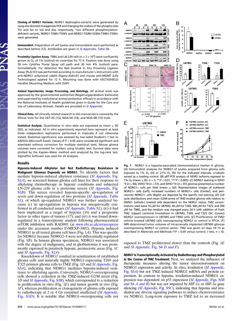
NDRG1 Is a Predictive Marker for Response to Alkylating Chemotherapy.Next, we interrogated patient tumor tissue to recapitulate the rel-evance of inducible NDRG1 for therapy resistance. NDRG1 is in-duced at tumor recurrence (Fig. 2 and SI Appendix, Fig. S2B). Asopposed to tumor tissue at diagnosis, NDRG1 expression in thetreated tissues was not predominantly seen in perinecrotic areasanymore, but NDRG1 was widely expressed in glioblastoma cellseven perivascularly opposed to the situation in the untreated tumors(Fig. 2B). NDRG1 expression in patients with low-grade gliomasprogressing without interim genotoxic treatment also increased(SI Appendix, Fig. S2B). High NDRG1 levels at recurrence suggesta poor response to alkylating chemotherapy, but not to the anti-angiogenic agent bevacizumab, in a small group of patients (SIAppendix, Table S2). A predictive role of NDRG1 for poor re-sponse to radiochemotherapy was suggested by post hoc NDRG1expression analyses, which revealed that progression-free survival(PFS) and overall survival (OS) of glioblastoma patients from the
UKT-05 trial (13) with moderate or high expression of NDRG1 wasreduced compared with patients with low NDRG1-expressingtumors (Fig. 2C). This was supported by an analysis of the Re-pository for Molecular Brain Neoplasia Data (REMBRANDT)database, which revealed that the OS of glioblastoma patients withintratumoral up-regulation of NDRG1 was reduced compared withpatients with intermediate or down-regulated expression of theNDRG1 transcript (Fig. 2D). To determine whether the prognosticimpact of NDRG1 is specifically related to alkylating chemother-apy, tissue samples of the NOA-04 trial comparing primary radio-therapy with primary alkylating chemotherapy (14) were analyzed.NDRG1 expression was associated with reduced PFS in TMZ-treated patients but not with radiotherapy in this not preplannedsubgroup analysis (Fig. 2E). Collectively, these data from severalstudy patient populations suggest that NDRG1 expression in gliomatissue is associated with a poor response specifically to alkylatingchemotherapy.
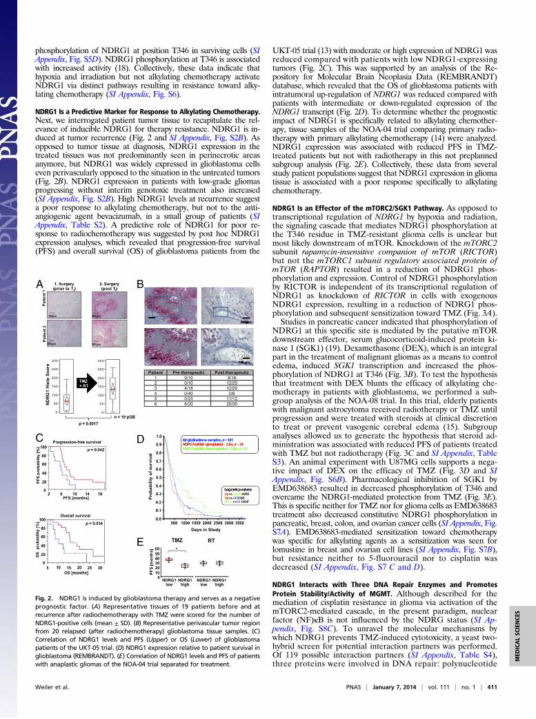
NDRG1 Is an Effector of the mTORC2/SGK1 Pathway. As opposed totranscriptional regulation of NDRG1 by hypoxia and radiation,the signaling cascade that mediates NDRG1 phosphorylation atthe T346 residue in TMZ-resistant glioma cells is unclear butmost likely downstream of mTOR. Knockdown of the mTORC2subunit rapamycin-insensitive companion of mTOR (RICTOR)but not the mTORC1 subunit regulatory associated protein ofmTOR (RAPTOR) resulted in a reduction of NDRG1 phos-phorylation and expression. Control of NDRG1 phosphorylationby RICTOR is independent of its transcriptional regulation ofNDRG1 as knockdown of RICTOR in cells with exogenousNDRG1 expression, resulting in a reduction of NDRG1 phos-phorylation and subsequent sensitization toward TMZ (Fig. 3A).Studies in pancreatic cancer indicated that phosphorylation of
NDRG1 at this specific site is mediated by the putative mTORdownstream effector, serum glucocorticoid-induced protein ki-nase 1 (SGK1) (19). Dexamethasone (DEX), which is an integralpart in the treatment of malignant gliomas as a means to controledema, induced SGK1 transcription and increased the phos-phorylation of NDRG1 at T346 (Fig. 3B). To test the hypothesisthat treatment with DEX blunts the efficacy of alkylating che-motherapy in patients with glioblastoma, we performed a sub-group analysis of the NOA-08 trial. In this trial, elderly patientswith malignant astrocytoma received radiotherapy or TMZ untilprogression and were treated with steroids at clinical discretionto treat or prevent vasogenic cerebral edema (15). Subgroupanalyses allowed us to generate the hypothesis that steroid ad-ministration was associated with reduced PFS of patients treatedwith TMZ but not radiotherapy (Fig. 3C and SI Appendix, TableS3). An animal experiment with U87MG cells supports a nega-tive impact of DEX on the efficacy of TMZ (Fig. 3D and SIAppendix, Fig. S6B). Pharmacological inhibition of SGK1 byEMD638683 resulted in decreased phosphorylation of T346 andovercame the NDRG1-mediated protection from TMZ (Fig. 3E).This is specific neither for TMZ nor for glioma cells as EMD638683treatment also decreased constitutive NDRG1 phosphorylation inpancreatic, breast, colon, and ovarian cancer cells (SI Appendix, Fig.S7A). EMD638683-mediated sensitization toward chemotherapywas specific for alkylating agents as a sensitization was seen forlomustine in breast and ovarian cell lines (SI Appendix, Fig. S7B),but resistance neither to 5-fluorouracil nor to cisplatin wasdecreased (SI Appendix, Fig. S7 C and D).
NDRG1 Interacts with Three DNA Repair Enzymes and PromotesProtein Stability/Activity of MGMT. Although described for themediation of cisplatin resistance in glioma via activation of themTORC2-mediated cascade, in the present paradigm, nuclearfactor (NF)κB is not influenced by the NDRG status (SI Ap-pendix, Fig. S8C). To unravel the molecular mechanisms bywhich NDRG1 prevents TMZ-induced cytotoxicity, a yeast two-hybrid screen for potential interaction partners was performed.Of 119 possible interaction partners (SI Appendix, Table S4),three proteins were involved in DNA repair: polynucleotide
Fig. 2. NDRG1 is induced by glioblastoma therapy and serves as a negativeprognostic factor. (A) Representative tissues of 19 patients before and atrecurrence after radiochemotherapy with TMZ were scored for the number ofNDRG1-positive cells (mean ± SD). (B) Representative perivascular tumor regionfrom 20 relapsed (after radiochemotherapy) glioblastoma tissue samples. (C)Correlation of NDRG1 levels and PFS (Upper) or OS (Lower) of glioblastomapatients of the UKT-05 trial. (D) NDRG1 expression relative to patient survival inglioblastoma (REMBRANDT). (E) Correlation of NDRG1 levels and PFS of patientswith anaplastic gliomas of the NOA-04 trial separated for treatment.
Weiler et al. PNAS | January 7, 2014 | vol. 111 | no. 1 | 411
MED
ICALSC
IENCE
S
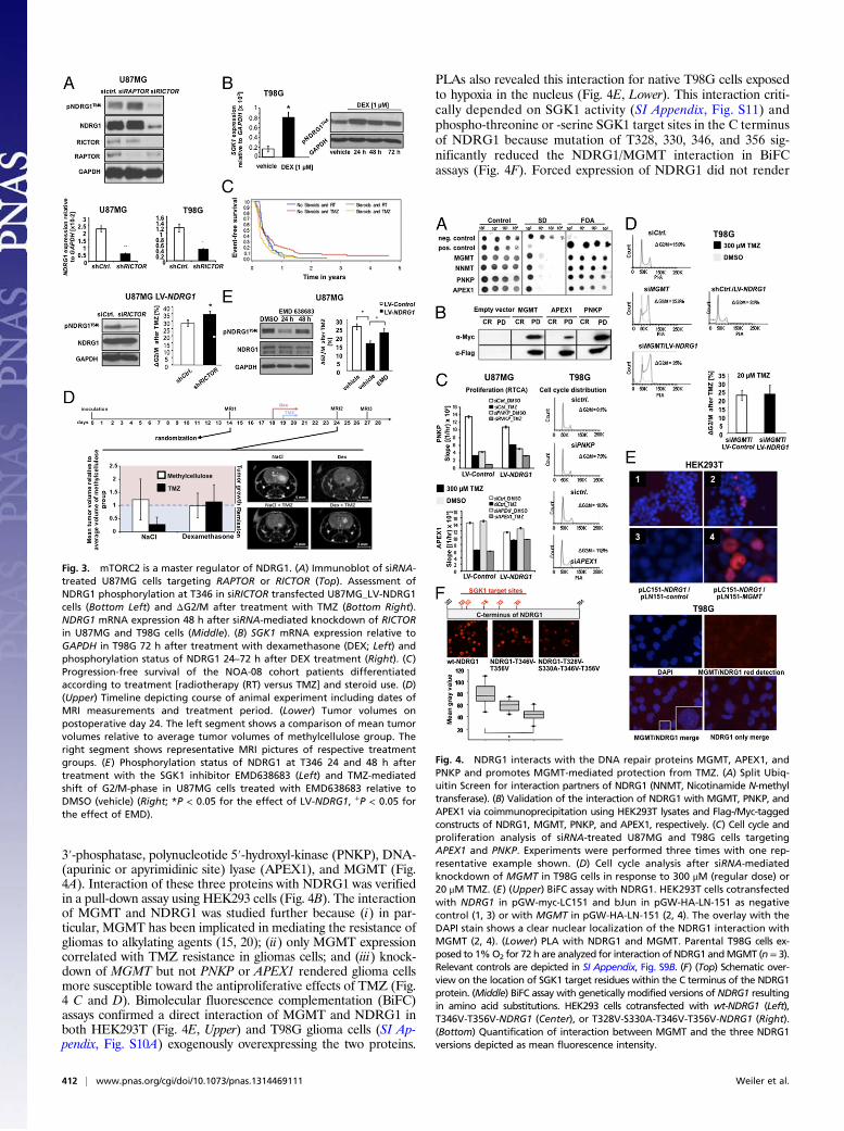
3′-phosphatase, polynucleotide 5′-hydroxyl-kinase (PNKP), DNA-(apurinic or apyrimidinic site) lyase (APEX1), and MGMT (Fig.4A). Interaction of these three proteins with NDRG1 was verifiedin a pull-down assay using HEK293 cells (Fig. 4B). The interactionof MGMT and NDRG1 was studied further because (i) in par-ticular, MGMT has been implicated in mediating the resistance ofgliomas to alkylating agents (15, 20); (ii) only MGMT expressioncorrelated with TMZ resistance in gliomas cells; and (iii) knock-down of MGMT but not PNKP or APEX1 rendered glioma cellsmore susceptible toward the antiproliferative effects of TMZ (Fig.4 C and D). Bimolecular fluorescence complementation (BiFC)assays confirmed a direct interaction of MGMT and NDRG1 inboth HEK293T (Fig. 4E, Upper) and T98G glioma cells (SI Ap-pendix, Fig. S10A) exogenously overexpressing the two proteins.
PLAs also revealed this interaction for native T98G cells exposedto hypoxia in the nucleus (Fig. 4E, Lower). This interaction criti-cally depended on SGK1 activity (SI Appendix, Fig. S11) andphospho-threonine or -serine SGK1 target sites in the C terminusof NDRG1 because mutation of T328, 330, 346, and 356 sig-nificantly reduced the NDRG1/MGMT interaction in BiFCassays (Fig. 4F). Forced expression of NDRG1 did not render
Fig. 3. mTORC2 is a master regulator of NDRG1. (A) Immunoblot of siRNA-treated U87MG cells targeting RAPTOR or RICTOR (Top). Assessment ofNDRG1 phosphorylation at T346 in siRICTOR transfected U87MG_LV-NDRG1cells (Bottom Left) and ΔG2/M after treatment with TMZ (Bottom Right).NDRG1 mRNA expression 48 h after siRNA-mediated knockdown of RICTORin U87MG and T98G cells (Middle). (B) SGK1 mRNA expression relative toGAPDH in T98G 72 h after treatment with dexamethasone (DEX; Left) andphosphorylation status of NDRG1 24–72 h after DEX treatment (Right). (C)Progression-free survival of the NOA-08 cohort patients differentiatedaccording to treatment [radiotherapy (RT) versus TMZ] and steroid use. (D)(Upper) Timeline depicting course of animal experiment including dates ofMRI measurements and treatment period. (Lower) Tumor volumes onpostoperative day 24. The left segment shows a comparison of mean tumorvolumes relative to average tumor volumes of methylcellulose group. Theright segment shows representative MRI pictures of respective treatmentgroups. (E) Phosphorylation status of NDRG1 at T346 24 and 48 h aftertreatment with the SGK1 inhibitor EMD638683 (Left) and TMZ-mediatedshift of G2/M-phase in U87MG cells treated with EMD638683 relative toDMSO (vehicle) (Right; *P < 0.05 for the effect of LV-NDRG1, +P < 0.05 forthe effect of EMD).
Fig. 4. NDRG1 interacts with the DNA repair proteins MGMT, APEX1, andPNKP and promotes MGMT-mediated protection from TMZ. (A) Split Ubiq-uitin Screen for interaction partners of NDRG1 (NNMT, Nicotinamide N-methyltransferase). (B) Validation of the interaction of NDRG1 with MGMT, PNKP, andAPEX1 via coimmunoprecipitation using HEK293T lysates and Flag-/Myc-taggedconstructs of NDRG1, MGMT, PNKP, and APEX1, respectively. (C) Cell cycle andproliferation analysis of siRNA-treated U87MG and T98G cells targetingAPEX1 and PNKP. Experiments were performed three times with one rep-resentative example shown. (D) Cell cycle analysis after siRNA-mediatedknockdown of MGMT in T98G cells in response to 300 μM (regular dose) or20 μM TMZ. (E) (Upper) BiFC assay with NDRG1. HEK293T cells cotransfectedwith NDRG1 in pGW-myc-LC151 and bJun in pGW-HA-LN-151 as negativecontrol (1, 3) or with MGMT in pGW-HA-LN-151 (2, 4). The overlay with theDAPI stain shows a clear nuclear localization of the NDRG1 interaction withMGMT (2, 4). (Lower) PLA with NDRG1 and MGMT. Parental T98G cells ex-posed to 1%O2 for 72 h are analyzed for interaction of NDRG1 andMGMT (n= 3).Relevant controls are depicted in SI Appendix, Fig. S9B. (F) (Top) Schematic over-view on the location of SGK1 target residues within the C terminus of the NDRG1protein. (Middle) BiFC assay with genetically modified versions of NDRG1 resultingin amino acid substitutions. HEK293 cells cotransfected with wt-NDRG1 (Left),T346V-T356V-NDRG1 (Center), or T328V-S330A-T346V-T356V-NDRG1 (Right).(Bottom) Quantification of interaction between MGMT and the three NDRG1versions depicted as mean fluorescence intensity.
412 | www.pnas.org/cgi/doi/10.1073/pnas.1314469111 Weiler et al.
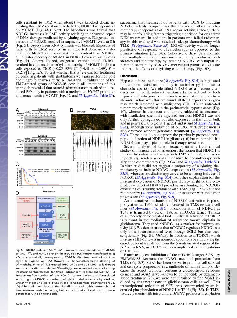
cells resistant to TMZ when MGMT was knocked down, in-dicating that TMZ resistance mediated by NDRG1 is dependenton MGMT (Fig. 4D). Next, the hypothesis was tested thatNDRG1 increases MGMT activity resulting in enhanced repairof DNA damage mediated by alkylating agents. Exogenous ex-pression of NDRG1 resulted in augmented MGMT levels at 8 h(Fig. 5A, Upper) when RNA synthesis was blocked. Exposure ofthese cells to TMZ resulted in an expected decrease via de-pletion of MGMT expression at 4 h independent from NDRG1but a faster recovery of MGMT in NDRG1-overexpressing cells(Fig. 5A, Lower). Indeed, exogenous expression of NDRG1resulted in enhanced demethylation activity of MGMT in gliomacells exposed to TMZ [−0.25, 95% CI (−0.41 to −0.09), P =0.0219] (Fig. 5B). To test whether this is relevant for treatmentoutcome in patients with glioblastoma we again performed posthoc subgroup analyses of the NOA-08 trial. Stratification of theTMZ-treated group of NOA-08 despite all limitations of thisapproach revealed that steroid administration resulted in a re-duced PFS only in patients with a methylated MGMT promoterand hence inactive MGMT (Fig. 5C and SI Appendix, Table S5),
suggesting that treatment of patients with DEX by inducingNDRG1 activity compromises the efficacy of alkylating che-motherapy independent of DNA repair activity, although theremay be confounding factors triggering a decision for or againstDEX treatment. In addition, in patients who failed radiother-apy in this trial and who received salvage chemotherapy withTMZ (SI Appendix, Table S5), MGMT activity was no longerpredictive of response to chemotherapy, as opposed to theprimary situation (Fig. 5C). Collectively, these data indicatethat multiple treatment measures including treatment withsteroids and radiotherapy by inducing NDRG1 can impair in-herent susceptibility of MGMT-methylated glioma cells to thetherapeutic effects of alkylating chemotherapy (Fig. 5D).
DiscussionHypoxia-induced resistance (SI Appendix, Fig. S1A) is implicatedin treatment resistance not only to radiotherapy but also tochemotherapy (5). We identified NDRG1 as a previously un-described clinically relevant resistance factor induced by bothhypoxia and iatrogenic stimuli such as irradiation and cortico-steroids. In line with this, we found NDRG1 expression in glio-mas, which increased with malignancy (Fig. 1C), in untreatedtumors mostly restricted to the perinecrotic, hypoxic areas (Fig.1C), whereas in the recurrent tumors, subjected to treatmentwith irradiation, chemotherapy, and steroids, NDRG1 was notonly further up-regulated but also expressed in the tumor bulkand in perivascular regions (Fig. 2 A and B and SI Appendix, Fig.S2A), although some induction of NDRG1 with progression isalso observed without genotoxic treatment (SI Appendix, Fig.S2B). These data do not support the previously proposed proa-poptotic function of NDRG1 in gliomas (16) but rather hint thatNDRG1 can play a pivotal role in therapy resistance.Several analyses of tumor tissue specimens from clinical
studies of malignant gliomas support the notion that NDRG1 isinduced by radiochemotherapy with TMZ (Fig. 2A) and, moreimportantly, renders gliomas insensitive to chemotherapy withalkylating chemotherapy (Fig. 2 C–E and SI Appendix, Table S2).In vitro studies did not suggest a propensity of alkylating che-motherapy to induce NDRG1 expression (SI Appendix, Fig.S5D), whereas irradiation appeared to be a strong inducer ofNDRG1 (SI Appendix, Fig. S5A). Another explanation for theincreased expression of NDRG1 posttherapy might be the cyto-protective effect of NDRG1 providing an advantage for NDRG1-expressing cells during treatment with TMZ (Fig. 1 D–F) but notradiotherapy (SI Appendix, Fig. S3C) or induction with the tumorprogression (SI Appendix, Fig. S2B).An alternative mechanism of NDRG1 activation is phos-
phorylation at T346, which is increased in TMZ-resistant celllines (SI Appendix, Fig. S8C). Phosphorylation of NDRG1 atT346 is triggered by SGK1 (19), an mTORC2 target. Tanakaet al. recently demonstrated that EGFRvIII-activated mTORC2is relevant in the mediation of resistance toward cisplatin inglioblastoma. They used pNDRG1 as a marker for pathway ac-tivity (21). We demonstrate that mTORC2 regulates NDRG1 notonly on a posttranslational level through SGK1 but also tran-scriptionally (Fig. 3A, Middle). In addition to mTORC1, whichincreases HIF-1α levels in normoxic conditions by stimulating thecap-dependent translation from the 5′-untranslated region of theHIF-1α mRNA, mTORC2 has been implicated in the regulationof HIF (22).Pharmacological inhibition of the mTORC2 target SGK1 by
EMD638683 overcame the NDRG1-mediated protection fromTMZ (Fig. 3D). SGK1 has been shown to promote cell survivaland cell cycle progression in a multitude of human tumors. Be-cause the SGK1 promoter contains a glucocorticoid responseelement and SGK1 is well-known to be inducible by dexameth-asone treatment (23), we were not surprised to find SGK1 in-duced by dexamethasone in glioblastoma cells as well. Thistranscriptional activation of SGK1 was accompanied by an in-creased phosphorylation of NDRG1 at T346 (Fig. 3B). In TMZ-treated patients with intratumoral MGMT promoter methylation
Fig. 5. NDRG1 stabilizes MGMT. (A) Time-dependent abundance of MGMT,pNDRG1T346, and NDRG1 proteins in T98G cells (Co, control transfected cells;ND, cells lentivirally overexpressing NDRG1) after treatment with actino-mycin D (Upper) or TMZ (Lower). (B) Immunofluorescent staining ofO6-methylguanine of TMZ-treated T98G LV-Co and LV-NDRG1 cells (Upper)and quantification of relative O6-methylguanine content depicted as log-transformed fluorescence for three independent replications (Lower). (C)Progression-free survival of the NOA-08 cohort patients differentiatedaccording to MGMT promoter methylation status (+, methylated; −,unmethylated) and steroid use in the temozolomide treatment group.(D ) Schematic overview of the signaling cascade with iatrogenic andmicroenvironmental activating factors (left side) and options for thera-peutic intervention (right side).
Weiler et al. PNAS | January 7, 2014 | vol. 111 | no. 1 | 413
MED
ICALSC
IENCE
S
from the NOA-08 cohort, cotreatment with steroids halved thePFS compared with TMZ treatment without steroid administra-tion (Fig. 5C and SI Appendix, Table S5). One alternative con-tributing factor is that larger tumors, which may require highersteroid doses, are more difficult to control. Further, patients withinactive MGMT would also suffer most from blood–brain barriernormalizing effects of corticosteroids.Our interpretation of the data led us to propose an interaction
of NDRG1 with factors involved in the execution or preventionof DNA damage. In our yeast two-hybrid screen, we see a proteininteraction of NDRG1 with the DNA repair enzymes APEX1,PNKP, and MGMT (2) (Fig. 4 A and B). In line with the obser-vation that only the expression of MGMT correlated with TMZresistance (SI Appendix, Fig. S8A), the interaction of NDRG1 withMGMT, but not with PNKP or APEX1, proved to be of functionalrelevance for the resistance phenotype in malignant glioma (Fig. 4C and D). Considering the observed augmentation of MGMTlevels under stress conditions in the presence of high NDRG1levels (Fig. 5A) and the colocalization at subcellular levels (Fig.4E), it is conceivable that NDRG1 stabilizes MGMT via a directprotein–protein interaction, thus fulfilling a chaperone-like func-tion. Nevertheless, MGMT alone cannot account for the observedNDRG1-dependent resistance phenotype because MGMT-nega-tive U87MG cells also become more resistant upon an elevatedNDRG1 expression level (Fig. 1D). Patients with intratumoralmethylation of the MGMT promoter and thus putatively noMGMT expression become more resistant in response to steroid
treatment (SI Appendix, Table S4, and Fig. 5C). These data suggestthat there may be additional mechanisms involved in the NDRG1-provoked resistance to alkylating chemotherapy in gliomas.In conclusion, we identified NDRG1 as a unique hypoxia-, ste-
roid-, and mTORC2/SGK1-regulated molecule in glioma that maybe developed as a predictive biomarker for response to treatmentwith TMZ in high-grade gliomas. Its TMZ-protective effect makesNDRG1 an attractive candidate for targeted therapy not only ingliomas but also in a variety of other cancer types, potentially viainhibition of SGK1. The preclinical data suggest multiple levels ofcell-intrinsic (mTORC1), microenvironmental (hypoxia), and iatro-genic (radiotherapy, dexamethasone) influences on this critical sig-naling pathway downstream of several growth factor receptors. ThemTORC2/SGK1/NDRG1 pathway may serve as a target for futurepreclinical and clinical research on therapy resistance (Fig. 5D).
ACKNOWLEDGMENTS. We thank Torsten Schmenger, Mona Friedrich, BSc,and Hans-Werner Pledl for assistance with some of the experiments. The HIF-knockdown cell lines were kindly provided by Prof. Dr. Till Acker (Justus LiebigUniversity Giessen), and EMD638683 was provided by Dr. Thomas Fuchs (MerckKGaA, Merck Serono). Support by the German Cancer Research Center (DKFZ)Light Microscopy Facility is gratefully acknowledged. This work was supportedwithin the Brain Tumor Network (BTN)plus, Subproject 6 by the Bundesmines-terium für Forschung und Technologie, the Förderverein für Gehirntumorfor-schung Karlsruhe e.V., the Charitable Hertie Foundation, and the DeutscheForschungsgemeinschaft [Sonderforschungsbereich (SFB) 773 and SFB 938 TPK]. J.B. is a doctoral student in the PhD Program of the DKFZ.
1. Sarkaria JN, et al. (2008) Mechanisms of chemoresistance to alkylating agents inmalignant glioma. Clin Cancer Res 14(10):2900–2908.
2. Dunn GP, et al. (2012) Emerging insights into the molecular and cellular basis ofglioblastoma. Genes Dev 26(8):756–784.
3. Amberger-Murphy V (2009) Hypoxia helps glioma to fight therapy. Curr Cancer DrugTargets 9(3):381–390.
4. Harris AL (2002) Hypoxia—A key regulatory factor in tumour growth. Nat Rev Cancer2(1):38–47.
5. Winkler F, et al. (2004) Kinetics of vascular normalization by VEGFR2 blockade gov-erns brain tumor response to radiation: Role of oxygenation, angiopoietin-1, andmatrix metalloproteinases. Cancer Cell 6(6):553–563.
6. Wick A, et al. (2002) Hypoxic neuroprotection requires sequential activation of vas-cular endothelial growth factor receptor and Akt. J Neurosci 22:6401–6407.
7. Henze AT, et al. (2010) Prolyl hydroxylases 2 and 3 act in gliomas as protective neg-ative feedback regulators of hypoxia-inducible factors. Cancer Res 70(1):357–366.
8. Salnikow K, Blagosklonny MV, Ryan H, Johnson R, Costa M (2000) Carcinogenic nickelinduces genes involved with hypoxic stress. Cancer Res 60(1):38–41.
9. Melotte V, et al. (2010) The N-myc downstream regulated gene (NDRG) family: diversefunctions, multiple applications. FASEB J 24(11):4153–4166.
10. Sun B, et al. (2009) Decreased expression of NDRG1 in glioma is related to tumorprogression and survival of patients. J Neurooncol 94(2):213–219.
11. Brummelkamp TR, Bernards R, Agami R (2002) Stable suppression of tumorigenicityby virus-mediated RNA interference. Cancer Cell 2(3):243–247.
12. Weiler M, et al. (2013) Suppression of proinvasive RGS4 by mTOR inhibition optimizesglioma treatment. Oncogene 32(9):1099–1109.
13. Weiler M, et al. (2010) Phase II trial of radiochemotherapy with daily concomitant andadjuvant intensified (one week on / one week off) TMZ plus indomethacin in newlydiagnosed glioblastoma: UKT-05. Int J Rad Biol Phys 77(3):670–676.
14. Wick W, et al. (2009) NOA-04 randomized phase III trial of sequential radiochemotherapyof anaplastic glioma with procarbazine, lomustine, and vincristine or temozolomide. J ClinOncol 27(35):5874–5880.
15. Wick W, et al.; NOA-08 Study Group of Neuro-oncology Working Group (NOA) ofGerman Cancer Society (2012) Temozolomide chemotherapy alone versus radio-therapy alone for malignant astrocytoma in the elderly: The NOA-08 randomised,phase 3 trial. Lancet Oncol 13(7):707–715.
16. Zhang P, Tchou-Wong KM, Costa M (2007) Egr-1 mediates hypoxia-inducible tran-scription of the NDRG1 gene through an overlapping Egr-1/Sp1 binding site in thepromoter. Cancer Res 67(19):9125–9133.
17. Azuma K, et al. (2012) NDRG1/Cap43/Drg-1 may predict tumor angiogenesis and pooroutcome in patients with lung cancer. J Thorac Oncol 7(5):779–789.
18. García-Martínez JM, Alessi DR (2008) mTOR complex 2 (mTORC2) controls hydro-phobic motif phosphorylation and activation of serum- and glucocorticoid-inducedprotein kinase 1 (SGK1). Biochem J 416(3):375–385.
19. Murakami Y, et al. (2010) Identification of sites subjected to serine/threonine phos-phorylation by SGK1 affecting N-myc downstream-regulated gene 1 (NDRG1)/Cap43-dependent suppression of angiogenic CXC chemokine expression in human pancreaticcancer cells. Biochem Biophys Res Commun 396(2):376–381.
20. Hegi ME, et al. (2005) MGMT gene silencing and benefit from temozolomide inglioblastoma. N Engl J Med 352(10):997–1003.
21. Tanaka K, et al. (2011) Oncogenic EGFR signaling activates an mTORC2-NF-κB path-way that promotes chemotherapy resistance. Cancer Discov 1(6):524–538.
22. Toschi A, Lee E, Gadir N, Ohh M, Foster DA (2008) Differential dependence of hyp-oxia-inducible factors 1 alpha and 2 alpha on mTORC1 and mTORC2. J Biol Chem283(50):34495–34499.
23. Mikosz CA, Brickley DR, Sharkey MS, Moran TW, Conzen SD (2001) Glucocorticoidreceptor-mediated protection from apoptosis is associated with induction of theserine/threonine survival kinase gene, sgk-1. J Biol Chem 276(20):16649–16654.
414 | www.pnas.org/cgi/doi/10.1073/pnas.1314469111 Weiler et al.