Embed Size (px)
Citation preview
Volume 11 Number 8 August 2009 pp. 804–811 804
Address allE-mail: rub1This worDepartmesupport of2This artic3These auReceived 3
CopyrightDOI 10.1
www.neoplasia.com
N-myc Downstream RegulatedGene 1 (NDRG1) Is Fused toERG in Prostate Cancer1,2
correspondence to: Mark A. Rubin, MD, Pathology and Laboratory Medicine,[email protected] was supported by National Institutes of Health (NIH)/National Cancer Instnt of Defense grant PC61474 (S.P.), and NIH/National Human Genome Rthe “Yale University Biomedical High Performance Computing Center” andle refers to supplementary materials, which are designated by Tables W1 to Wthors contributed equally to this work.April 2009; Revised 16 May 2009; Accepted 20 May 2009
© 2009 Neoplasia Press, Inc. All rights reserved 1522-8002/09/$25.00593/neo.09572
Dorothee Pflueger*,†,3, David S. Rickman*,3,Andrea Sboner‡,3, Sven Perner*,Christopher J. LaFargue*, Maria A. Svensson*,Benjamin J. Moss*, Naoki Kitabayashi*,Yihang Pan*, Alexandre de la Taille§,¶,Rainer Kuefer†, Ashutosh K. Tewari#,Francesca Demichelis*,**, Mark S. Chee††,Mark B. Gerstein‡,‡‡,§§ and Mark A. Rubin*
*Department of Pathology & Laboratory Medicine, WeillCornell Medical College, New York, NY, USA; †Departmentof Urology, University Hospital Ulm, Ulm, Germany;‡Department of Molecular Biophysics and Biochemistry, YaleUniversity, NewHaven, CT, USA; §INSERM, Unité 955, Créteil,France; ¶Department of Urology, CHU Mondor, Créteil,France; #Department of Urology, Weill Cornell MedicalCollege, New York, NY, USA; **Institute for ComputationalBiomedicine, Weill Cornell Medical College, New York,NY, USA; ††Prognosys Biosciences Inc., La Jolla, CA, USA;‡‡Program in Computational Biology and Bioinformatics,Yale University, New Haven, CT, USA; §§Department ofComputer Science, Yale University, New Haven, CT, USA
AbstractA step toward the molecular classification of prostate cancer was the discovery of recurrent erythroblast transformation–specific rearrangements, most commonly fusing the androgen-regulated TMPRSS2 promoter to ERG. The TMPRSS2-ERGfusion is observed in around 90% of tumors that overexpress the oncogene ERG. The goal of the current study was to com-plete the characterization of these ERG-overexpressing prostate cancers. Using fluorescence in situ hybridization and reversetranscription–polymerase chain reaction assays, we screened 101 prostate cancers, identifying 34 cases (34%) with theTMPRSS2-ERG fusion. Seven cases demonstrated ERG rearrangement by fluorescence in situ hybridization without the pres-ence of TMPRSS2-ERG fusion messenger RNA transcripts. Screening for known 5′ partners, we determined that three casesharbored the SLC45A3-ERG fusion. To discover novel 5′ partners in these ERG-overexpressing and ERG-rearranged cases, weused paired-end RNA sequencing. We first confirmed the utility of this approach by identifying the TMPRSS2-ERG fusion in aknown positive prostate cancer case and then discovered a novel fusion involving the androgen-inducible tumor suppressor,NDRG1 (N-myc downstream regulated gene 1), and ERG in two cases. Unlike TMPRSS2-ERG and SCL45A3-ERG fusions, theNDRG1-ERG fusion is predicted to encode a chimeric protein. Like TMPRSS2, SCL45A3 and NDRG1 are inducible not only byandrogen but also by estrogen. This study demonstrates that most ERG-overexpressing prostate cancers harbor hormonallyregulated TMPRSS2-ERG, SLC45A3-ERG, or NDRG1-ERG fusions. Broader implications of this study support the use of RNAsequencing to discover novel cancer translocations.
Neoplasia (2009) 11, 804–811
Abbreviations: RT-PCR, reverse transcription–polymerase chain reaction; FISH, fluorescence in situ hybridization; RNA-seq, RNA sequencing; (b/a), break-apart
Weill Cornell Medical College, 1300 York Ave, Room C 410-A, New York, NY 10021.itute grant R01 CA125612 (M.A.R. and F.D.), Heinrich Warner Foundation (D.P.),esearch Institute (NHGRI) grant R44HG004237 (M.S.C.). The authors thank theNIH grant no. RR19895 that funded the computer cluster instrumentation.4 and Figures W1 to W5 and are available online at www.neoplasia.com.
Neoplasia Vol. 11, No. 8, 2009 ERG Fusion Prostate Cancer Pflueger et al. 805
IntroductionMost prostate cancers detected through prostate-specific antigen (PSA)screening harbor an acquired recurrent chromosomal rearrangement[1]. The promoter region of the androgen-regulated transmembraneprotease, serine 2 (TMPRSS2) gene, is most often fused to the codingregion of members of the erythroblast transformation–specific (ETS)family of transcription factors, most commonly v-ets erythroblastosisvirus E26 oncogene homolog (avian) (ERG ). Other, less common,fusion events occur involving ETS family members (ETV1, ETV4,and ETV5) fused to TMPRSS2 or other 5′ partners that differ in theirprostate specificity and response to androgen (SLC45A3, HERV-K,C15orf21, HNRPA2B1, FLJ35294, DDX5, CANT1, and KLK2, re-viewed by Kumar-Sinha et al. [2] and more recently, ACSL3 [3]). More-over, variations in the structure of the gene fusions in prostate canceryielding different fusion transcript isoforms have been reported [4].Emerging data suggest that ETS-rearranged prostate cancer, similar
to other translocation tumors, represent a distinct molecular subclass ofprostate cancer based on studies demonstrating characteristic morpho-logic features [5], natural history [6,7], and specific genomic [8] andexpression profiles [9]. Herein, we report a comprehensive characteri-zation for ERG gene rearrangements in prostate cancer including theidentification of a novel hormone-regulated 5′ fusion partner usingpaired-end RNA sequencing (RNA-seq).
1http://genome.ucsc.edu.
Materials and Methods
Patient PopulationThe study is composed of 101 men with localized and locally ad-
vanced prostate cancer who underwent radical prostatectomy as amonotherapy. All prostate cancer cases were collected as part of institu-tional review board–approved research protocols.
Sample Processing for RNA AnalysesHematoxylin and eosin slides were prepared from formalin-fixed
paraffin-embedded material and evaluated for cancer extent and tumorgrade (Gleason score). Hematoxylin and eosin slides were preparedfrom the corresponding frozen tissue block and evaluated for the ex-tent of cancer involvement. To ensure for a high concentration of can-cer cells and minimized benign tissue, tumor isolation was performedby first selecting for high-density cancer foci (<10% stromal and othernontumor tissue contamination) and then taking 1.5-mm biopsy coresfrom the frozen tissue block for RNA extraction. Sections for fluo-rescence in situ hybridization (FISH) evaluation were taken from thefrozen tissue block used for molecular analysis. The cancer foci se-lected for RNA extraction were well characterized by FISH to evaluatethe ERG rearrangement status throughout the entire focus. We tookspecial care to extract the RNA from a single cancer focus to excludethe problem of heterogeneity when looking for putative fusion tran-scripts. RNA was isolated from frozen tissue using TRIzol LS reagent(Invitrogen, Carlsbad, CA) according to the manufacturer’s instruc-tions. After DNase treatment (Invitrogen), RNA concentration wasmeasured using a NanoDrop 8000 spectrophotometer (Thermo Scien-tific, Wilmington, DE). Quality was assessed using the Bioanalyzer2100 (Agilent Technologies, Inc, Santa Clara, CA). The qualitativedetection of fusion transcripts in the cases was performed using con-ventional reverse transcription–polymerase chain reaction (RT-PCR),agarose gel fractionation/purification, and subsequent complementaryDNA (cDNA) sequencing. For this, amplified DNA fragments cor-
responding to the expected sizes of fusion transcripts were gel-extractedusing the MinElute Gel Extraction Kit (Qiagen, Valencia, CA) andsequenced at the Life Sciences Core Laboratories Center’s DNA se-quencing facility of Cornell University (Ithaca, NY). QuantitativeERG and TMPRSS2-ERG RT-PCR was performed using QuantiTectSYBRGreen PCRKit (Qiagen). Each sample was run in duplicate. Theamount of each target gene relative to a control gene was determinedusing the comparative C t method (ABI Bulletin 2; Applied Biosys-tems, Foster City, CA). C t values for ERG were first normalized usingthe average C t values obtained for SART3 and TCFL1/VPS72 andthen calibrated using normalized C t values obtained from benign pros-tate. Protocols and primers are shown in Table W2.
Assessment of ERG, TMPRSS2, SLC45A3, and NDRG1Rearrangements Using Two-color FISH Assays
To assess for rearrangement of ERG, TMPRSS2, SLC45A3, andNDRG1, we used break-apart (b/a) FISH assays for each gene andfusion assays for SLC45A3-ERG or NDRG1-ERG on sections fromthe corresponding frozen tissue blocks. The centromeric probes forERG, TMPRSS2, SLC45A3, and NDRG1 were RP11-24A11, RP11-354C5, RP11-249H15, and RP11-185E14, respectively. The telomericprobes for ERG, TMPRSS2, SLC45A3, and NDRG1 were RP11-372O17, RP11-891L10, RP11-131E5, and RP11-1145H17, respec-tively. We used probes RP11-131E5 (SLC45A3), RP11-1145H17(NDRG1), and RP11-24A11 (ERG) for the SLC45A3-ERG andNDRG1-ERG fusion assays. Correct chromosomal probe localizationwas confirmed on normal lymphocyte metaphase preparations (seeFigure W4 for metaphase results for bacterial artificial chromosomes(BACs) targeting NDRG1 locus). For each sample, a minimum of100 nuclei were analyzed.
RNA-seq and Data AnalysisWe used the Illumina Genome Analyzer II for paired-end RNA-
sequencing. This provided a pair of approximately 30 to 36 base reads,from each end of a transcript fragment of relatively well-defined length(approximately 330 nucleotides). The paired reads were aligned inde-pendently to the human genome (hg18 assembly in the UCSC genomebrowser1: Homo_Sapiens March 2006) using “eland,” a short-readalignment tool included in the Genome Analyzer software suite. Foreach read, eland provides the coordinate(s) of the alignment to thereference genome, allowing for up to two mismatches in the sequence.We kept only the reads that are mapped uniquely to the genome,although they might have up to two mismatches. To search for noveltranslocations involving ERG, two strategies were applied. First, weselected for mapped paired reads that are more than 2 Mb apart. Thisallows us to identify translocations similar to TMPRSS2-ERG messen-ger RNA (mRNA). Indeed, the two genes are approximately 3 Mbapart. Second, paired reads mapping to different chromosomes werealso selected as potential candidates. Because we focused on novelERG partners, we selected for paired reads where one of the reads lieswithin ERG. This allowed us to identify several candidate fusion tran-scripts spanning all chromosomes. We finally selected the chromosomewith the highest number of reads and checked if those reads lie withina gene. This approach yielded numerous leads, which, because of thelow number of copies (from one to three reads), were considered back-ground. We also identified numerous examples of putative fusions
806 ERG Fusion Prostate Cancer Pflueger et al. Neoplasia Vol. 11, No. 8, 2009
that ambiguously mapped to multiple sites along the reference genomeprobably because of the small size of the paired reads (between 30 and36 bp). Sequences for NDRG1-ERG v1 and v2 have been submitted toGenBank (Accession Nos. FJ627786 and FJ627787, respectively).
Hormone Treatment of LNCaP CellsThe prostate cancer cell line LNCaP was obtained from ATCC
(Manassas, VA; catalog No. CRL-1740) and maintained accordingto the supplier’s instructions. For hormonal treatment, cells wereplated (500,000 cells/10 cm2) in the presence of complete growthmedium supplemented with 1% penicillin/streptomycin. Cells werestarved for 48 hours in charcoal-stripped medium (RPMI-1640 1×,5% charcoal-stripped FBS, 1% penicillin/streptomycin) and thentreated with R1881 (1 nM), 17β-estradiol (10 nM), diarylpropio-nitrile (DPN, 10 nM) or ethanol vehicle for 3, 12, and 24 hours.RNA was extracted using the TRIzol reagent (Invitrogen), subjectedto DNase treatment (DNA-free Kit; Applied Biosystems) accordingto the manufacturer’s instructions. To test for the specificity of andro-gen stimulation, cells were treated with 10 μM flutamide for 2 hoursand then treated with R1881 as described previously. TaqMan assays(Table W3) were used to quantify relative levels of SLC45A3, NDRG1,PSA (KLK3), and IGF1R.
Results
TMPRSS2-ERG and SLC45A3-ERG Account for 84% ofERG Overexpression in Prostate Cancer
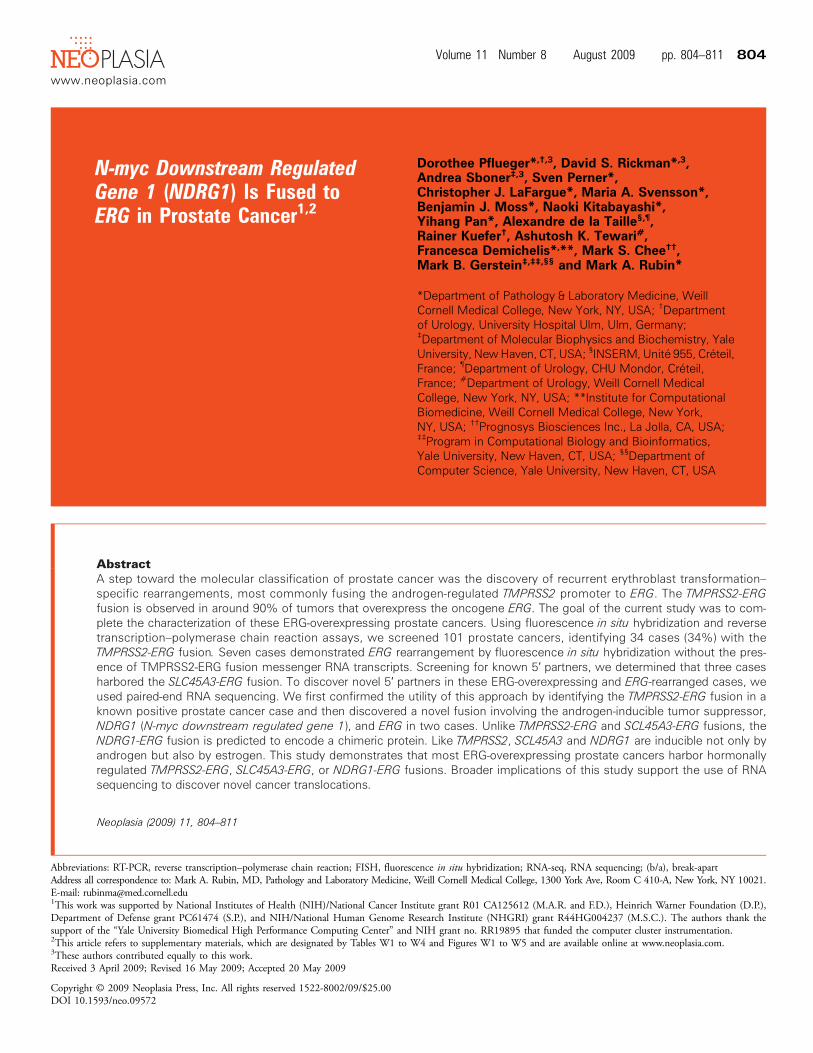
We screened prostate cancer cases from 101 men with localized andlocally advanced prostate cancer who underwent radical prostatectomyfor ERG gene rearrangement using a FISH b/a assay. In total, 44 caseswere positive for ERG rearrangement. Given the heterogeneity ofTMPRSS2-ERG mRNA expression level [4] in prostate cancer, wescreened for TMPRSS2-ERG mRNA variant expression using con-ventional RT-PCR and cDNA sequencing. Of the 44, 34 (77%) ex-pressed seven different variants of TMPRSS2-ERG mRNA describedby Wang et al. [4]. To determine the level of ERG mRNA over-expression, we performed quantitative PCR using cDNA from 29 cases(19 that were TMPRSS2-ERG mRNA-positive and 10 TMPRSS2-ERG mRNA-negative), 15 cases that did not show ERG rearrangementand 6 benign prostate tissue samples (Figure 1A). ERG mRNA wasoverexpressed up to 75 times (median, 27) in ERG-rearranged casescompared with baseline levels in benign prostate tissue and cases nega-tive for both ERG rearrangement and TMPRSS2-ERG mRNA. Con-trary to findings by Wang et al., TMPRSS2-ERG mRNA isoformexpression was not associated with ERG overexpression or with prostatecancer progression (Gleason score, pathologic stage, or surgical marginstatus; Table W1).
TMPRSS2-ERG mRNA was absent in 10 (23% of 44) ERG-rearranged cases, of which 7 expressed high ERG mRNA levels (5-38 times). To confirm the absence of TMPRSS2 rearrangement inthese cases, we performed FISH using a TMPRSS2 b/a assay. Weobserved TMPRSS2 rearrangement in 2 of 10 cases (60T and 51T)suggesting a novel TMPRSS2-ERG fusion that was not detectedusing standard RT-PCR approaches. Targeting the exon boundaryof exons 1 and 2 in TMPRSS2, we detected a TMPRSS2-ERGfusion transcript in sample 60T that lacks the 5′ end of TMPRSS2exon 1. This isoform (isoform VII) has been previously reported[4]. To screen for other possible fusion events with ERG, we per-
formed RT-PCR analysis targeting known ETS family fusion partners(SLC45A3, HERV-K, C15ORF21, HNRPA2B1, DDX5, CANT1,KLK2, and ACSL3). This screening revealed that exon 4 of ERG wasfused to exon 1 of SLC45A3 in three ERG mRNA-overexpressed cases(34T, 150B_M, and 145C_M; Figure 1B). This is consistent with therecent report from Han et al. [10]. The predicted open reading frame isidentical to what is encoded by the most commonTMPRSS2 (exon 1)–ERG (exon 4) mRNA transcript. We confirmed this fusion in situ usingan SLC45A3 b/a assay and SLC45A3-ERG fusion assay (Figure 1C).
Massively Parallel RNA-seq Discovers NDRG1-ERG FusionProstate Cancer
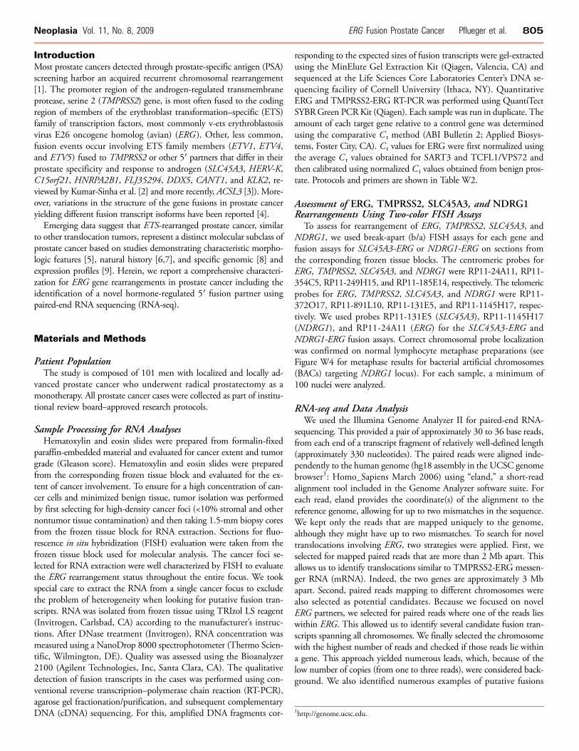
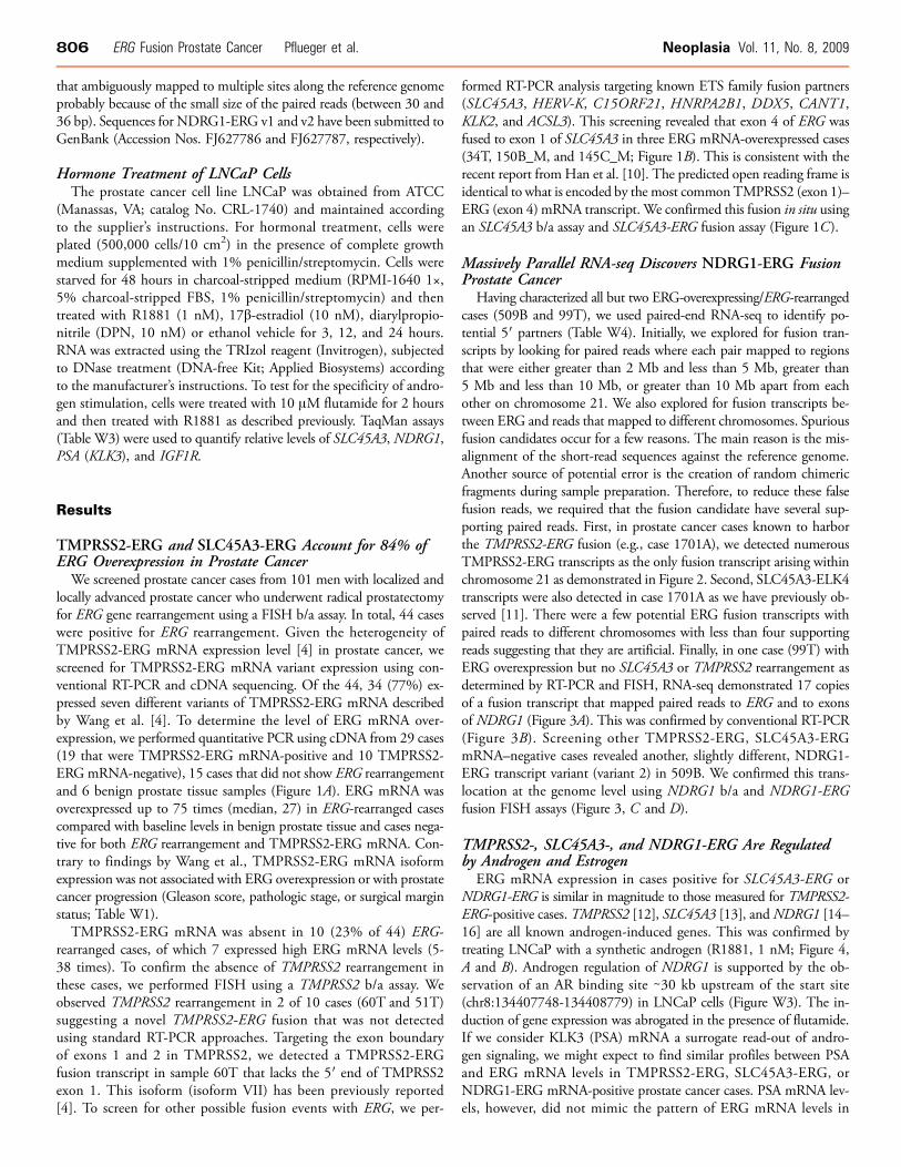
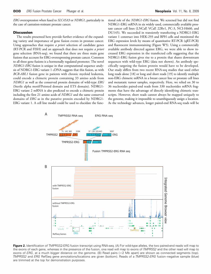
Having characterized all but two ERG-overexpressing/ERG-rearrangedcases (509B and 99T), we used paired-end RNA-seq to identify po-tential 5′ partners (Table W4). Initially, we explored for fusion tran-scripts by looking for paired reads where each pair mapped to regionsthat were either greater than 2 Mb and less than 5 Mb, greater than5 Mb and less than 10 Mb, or greater than 10 Mb apart from eachother on chromosome 21. We also explored for fusion transcripts be-tween ERG and reads that mapped to different chromosomes. Spuriousfusion candidates occur for a few reasons. The main reason is the mis-alignment of the short-read sequences against the reference genome.Another source of potential error is the creation of random chimericfragments during sample preparation. Therefore, to reduce these falsefusion reads, we required that the fusion candidate have several sup-porting paired reads. First, in prostate cancer cases known to harborthe TMPRSS2-ERG fusion (e.g., case 1701A), we detected numerousTMPRSS2-ERG transcripts as the only fusion transcript arising withinchromosome 21 as demonstrated in Figure 2. Second, SLC45A3-ELK4transcripts were also detected in case 1701A as we have previously ob-served [11]. There were a few potential ERG fusion transcripts withpaired reads to different chromosomes with less than four supportingreads suggesting that they are artificial. Finally, in one case (99T) withERG overexpression but no SLC45A3 or TMPRSS2 rearrangement asdetermined by RT-PCR and FISH, RNA-seq demonstrated 17 copiesof a fusion transcript that mapped paired reads to ERG and to exonsof NDRG1 (Figure 3A). This was confirmed by conventional RT-PCR(Figure 3B). Screening other TMPRSS2-ERG, SLC45A3-ERGmRNA–negative cases revealed another, slightly different, NDRG1-ERG transcript variant (variant 2) in 509B. We confirmed this trans-location at the genome level using NDRG1 b/a and NDRG1-ERGfusion FISH assays (Figure 3, C and D).
TMPRSS2-, SLC45A3-, and NDRG1-ERG Are Regulatedby Androgen and Estrogen
ERG mRNA expression in cases positive for SLC45A3-ERG orNDRG1-ERG is similar in magnitude to those measured for TMPRSS2-ERG-positive cases. TMPRSS2 [12], SLC45A3 [13], andNDRG1 [14–16] are all known androgen-induced genes. This was confirmed bytreating LNCaP with a synthetic androgen (R1881, 1 nM; Figure 4,A and B). Androgen regulation of NDRG1 is supported by the ob-servation of an AR binding site ∼30 kb upstream of the start site(chr8:134407748-134408779) in LNCaP cells (Figure W3). The in-duction of gene expression was abrogated in the presence of flutamide.If we consider KLK3 (PSA) mRNA a surrogate read-out of andro-gen signaling, we might expect to find similar profiles between PSAand ERG mRNA levels in TMPRSS2-ERG, SLC45A3-ERG, orNDRG1-ERG mRNA-positive prostate cancer cases. PSA mRNA lev-els, however, did not mimic the pattern of ERG mRNA levels in
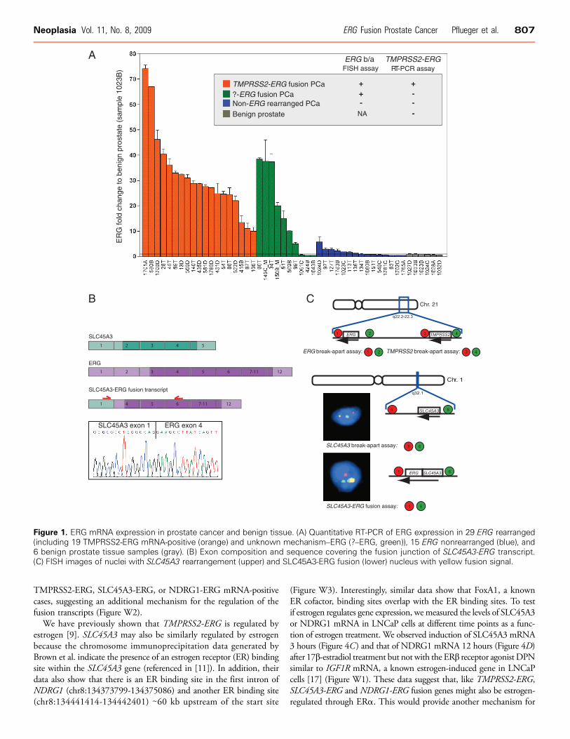
Figure 1. ERG mRNA expression in prostate cancer and benign tissue. (A) Quantitative RT-PCR of ERG expression in 29 ERG rearranged(including 19 TMPRSS2-ERG mRNA-positive (orange) and unknown mechanism–ERG (?–ERG, green)), 15 ERG nonrearranged (blue), and6 benign prostate tissue samples (gray). (B) Exon composition and sequence covering the fusion junction of SLC45A3-ERG transcript.(C) FISH images of nuclei with SLC45A3 rearrangement (upper) and SLC45A3-ERG fusion (lower) nucleus with yellow fusion signal.
Neoplasia Vol. 11, No. 8, 2009 ERG Fusion Prostate Cancer Pflueger et al. 807
TMPRSS2-ERG, SLC45A3-ERG, or NDRG1-ERG mRNA-positivecases, suggesting an additional mechanism for the regulation of thefusion transcripts (Figure W2).We have previously shown that TMPRSS2-ERG is regulated by
estrogen [9]. SLC45A3 may also be similarly regulated by estrogenbecause the chromosome immunoprecipitation data generated byBrown et al. indicate the presence of an estrogen receptor (ER) bindingsite within the SLC45A3 gene (referenced in [11]). In addition, theirdata also show that there is an ER binding site in the first intron ofNDRG1 (chr8:134373799-134375086) and another ER binding site(chr8:134441414-134442401) ∼60 kb upstream of the start site
(Figure W3). Interestingly, similar data show that FoxA1, a knownER cofactor, binding sites overlap with the ER binding sites. To testif estrogen regulates gene expression, we measured the levels of SLC45A3or NDRG1 mRNA in LNCaP cells at different time points as a func-tion of estrogen treatment. We observed induction of SLC45A3 mRNA3 hours (Figure 4C) and that of NDRG1 mRNA 12 hours (Figure 4D)after 17β-estradiol treatment but not with the ERβ receptor agonist DPNsimilar to IGF1R mRNA, a known estrogen-induced gene in LNCaPcells [17] (Figure W1). These data suggest that, like TMPRSS2-ERG,SLC45A3-ERG and NDRG1-ERG fusion genes might also be estrogen-regulated through ERα. This would provide another mechanism for
808 ERG Fusion Prostate Cancer Pflueger et al. Neoplasia Vol. 11, No. 8, 2009
ERG overexpression when fused to SLC45A3 orNDRG1, particularly inthe case of castration-resistant prostate cancer.
DiscussionThe results presented here provide further evidence of the expand-
ing variety and importance of gene fusion events in prostate cancer.Using approaches that require a priori selection of candidate genes(RT-PCR and FISH) and an approach that does not require a priorigene selection (RNA-seq), we found that there are three main genefusions that account for ERG-overexpressing prostate cancer. Commonto all three gene fusions is a hormonally regulated promoter. The novelNDRG1-ERG fusion is unique in that computational sequence analy-sis of NDRG1-ERG variant 1 cDNA suggests that this fusion, as withBCR-ABL1 fusion gene in patients with chronic myeloid leukemia,could encode a chimeric protein containing 33 amino acids fromNDRG1 as well as the conserved protein domains of wild-type ERG(Sterile alpha motif/Pointed domain and ETS domain). NDRG1-ERG variant 2 mRNA is also predicted to encode a chimeric proteinincluding the first 21 amino acids of NDRG1 and the same conserveddomains of ERG as in the putative protein encoded by NDRG1-ERG variant 1. A cell line model could be used to elucidate the func-
Figure 2. Identification of TMPRSS2-ERG fusion transcript using RNAthe exons of each gene, whereas in the presence of the fusion, oneexons of ERG, at a much bigger distance on the genome. (B) ReaTMPRSS2 and ERG RefSeq gene annotations/locations are given (bare trimmed at the top for demonstration purposes.
tional role of the NDRG1-ERG fusion. We screened but did not findNDRG1-ERG mRNA in six widely used, commercially available pros-tate cancer cell lines (LNCaP, VCaP, 22Rv1, PC-3, NCI-H660, andDU145). We succeeded in transiently transfecting a NDRG1-ERGvariant 1 construct into HEK-293 and BPH cells and monitored theERG expression levels by means of quantitative RT-PCR (qRT-PCR)and fluorescent immunostaining (Figure W5). Using a commerciallyavailable antibody directed against ERG, we were able to show in-creased ERG expression in the transfected cells suggesting that theNDRG1-ERG fusion gives rise to a protein that shares downstreamsequences with wild-type ERG (data not shown). An antibody spe-cifically targeting the fusion protein would have to be developed.Our study differs from two recent RNA-seq studies that used eitherlong reads alone [18] or long and short reads [19] to identify multiplenon-ERG chimeric mRNA in a breast cancer line or prostate cell linesand metastatic tumor samples, respectively. Here, we relied on 30 to36 nucleotides paired-end reads from 330 nucleotides mRNA frag-ments that have the advantage of directly identifying chimeric tran-scripts. However, short reads cannot always be mapped uniquely tothe genome, making it impossible to unambiguously assign a location.As the technology advances, longer paired-end RNA-seq reads will be
-seq. (A) For wild-type alleles, the two paired-end reads will map toread will map to exons of TMPRSS2 and the other read will map tod pairs (<2 Mb apart) are shown as connected segments (top).ottom). Reads of a TMPRSS2-ERG fusion–negative sample (blue)
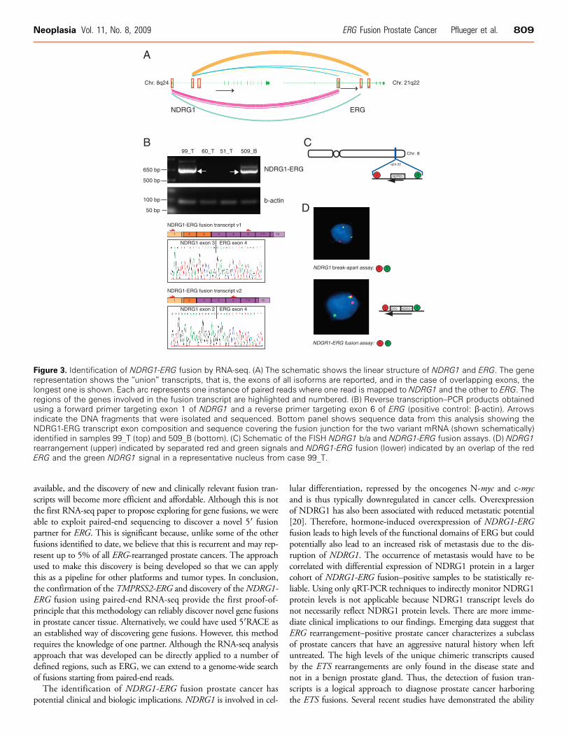
Figure 3. Identification of NDRG1-ERG fusion by RNA-seq. (A) The schematic shows the linear structure of NDRG1 and ERG. The generepresentation shows the “union” transcripts, that is, the exons of all isoforms are reported, and in the case of overlapping exons, thelongest one is shown. Each arc represents one instance of paired reads where one read is mapped to NDRG1 and the other to ERG. Theregions of the genes involved in the fusion transcript are highlighted and numbered. (B) Reverse transcription–PCR products obtainedusing a forward primer targeting exon 1 of NDRG1 and a reverse primer targeting exon 6 of ERG (positive control: β-actin). Arrowsindicate the DNA fragments that were isolated and sequenced. Bottom panel shows sequence data from this analysis showing theNDRG1-ERG transcript exon composition and sequence covering the fusion junction for the two variant mRNA (shown schematically)identified in samples 99_T (top) and 509_B (bottom). (C) Schematic of the FISH NDRG1 b/a and NDRG1-ERG fusion assays. (D) NDRG1rearrangement (upper) indicated by separated red and green signals and NDRG1-ERG fusion (lower) indicated by an overlap of the redERG and the green NDRG1 signal in a representative nucleus from case 99_T.
Neoplasia Vol. 11, No. 8, 2009 ERG Fusion Prostate Cancer Pflueger et al. 809
available, and the discovery of new and clinically relevant fusion tran-scripts will become more efficient and affordable. Although this is notthe first RNA-seq paper to propose exploring for gene fusions, we wereable to exploit paired-end sequencing to discover a novel 5′ fusionpartner for ERG. This is significant because, unlike some of the otherfusions identified to date, we believe that this is recurrent and may rep-resent up to 5% of all ERG-rearranged prostate cancers. The approachused to make this discovery is being developed so that we can applythis as a pipeline for other platforms and tumor types. In conclusion,the confirmation of the TMPRSS2-ERG and discovery of theNDRG1-ERG fusion using paired-end RNA-seq provide the first proof-of-principle that this methodology can reliably discover novel gene fusionsin prostate cancer tissue. Alternatively, we could have used 5′RACE asan established way of discovering gene fusions. However, this methodrequires the knowledge of one partner. Although the RNA-seq analysisapproach that was developed can be directly applied to a number ofdefined regions, such as ERG, we can extend to a genome-wide searchof fusions starting from paired-end reads.The identification of NDRG1-ERG fusion prostate cancer has
potential clinical and biologic implications. NDRG1 is involved in cel-
lular differentiation, repressed by the oncogenes N-myc and c-mycand is thus typically downregulated in cancer cells. Overexpressionof NDRG1 has also been associated with reduced metastatic potential[20]. Therefore, hormone-induced overexpression of NDRG1-ERGfusion leads to high levels of the functional domains of ERG but couldpotentially also lead to an increased risk of metastasis due to the dis-ruption of NDRG1. The occurrence of metastasis would have to becorrelated with differential expression of NDRG1 protein in a largercohort of NDRG1-ERG fusion–positive samples to be statistically re-liable. Using only qRT-PCR techniques to indirectly monitor NDRG1protein levels is not applicable because NDRG1 transcript levels donot necessarily reflect NDRG1 protein levels. There are more imme-diate clinical implications to our findings. Emerging data suggest thatERG rearrangement–positive prostate cancer characterizes a subclassof prostate cancers that have an aggressive natural history when leftuntreated. The high levels of the unique chimeric transcripts causedby the ETS rearrangements are only found in the disease state andnot in a benign prostate gland. Thus, the detection of fusion tran-scripts is a logical approach to diagnose prostate cancer harboringthe ETS fusions. Several recent studies have demonstrated the ability
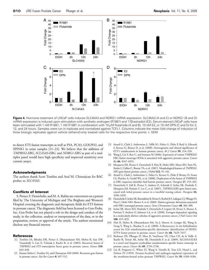
Figure 4. Hormone treatment of LNCaP cells induces SLC45A3 and NDRG1 mRNA expression. SLC45A3 (A and C) or NDRG1 (B and D)mRNA expression is induced upon stimulation with synthetic androgen (R1881) and 17β-estradiol (E2). Serum-starved LNCaP cells havebeen stimulated with 1 nM R1881, 1 nM R1881 in combination with 10 μM flutamide (A and B), 10 nM E2, or 10 nM DPN (C and D) for 3,12, and 24 hours. Samples were run in triplicate and normalized against TCFL1. Columns indicate the mean fold change of induction ofthree biologic replicates against vehicle (ethanol)-only treated cells for the respective time points ± SEM.
810 ERG Fusion Prostate Cancer Pflueger et al. Neoplasia Vol. 11, No. 8, 2009
to detect ETS fusion transcripts as well as PSA, PCA3, GOLPH2, andSPINK1 in urine samples [21–23]. We believe that the addition ofTMPRSS2-ERG, SCL45A3-ERG, and NDRG1-ERG as part of a mul-tiplex panel would have high specificity and improved sensitivity overcurrent assays.
AcknowledgmentsThe authors thank Scott Tomlins and Arul M. Chinnaiyan for BACprobes to SLC45A3.
Conflicts of InterestS. Perner, F. Demichelis, andM. A. Rubin are coinventors on a patent
filed by The University of Michigan and The Brigham and Women’sHospital covering the diagnostic and therapeutic fields for ETS fusionsin prostate cancer. The diagnostic field has been licensed to Gen-Probe,Inc. Gen-Probe has not played a role in the design and conduct of thestudy, in the collection, analysis or interpretation of the data, or in thepreparation, review, or approval of the article. The authors mentioneddisclose any financial interest.
References[1] Tomlins SA, Rhodes DR, Perner S, Dhanasekaran SM, Mehra R, Sun XW,
Varambally S, Cao X, Tchinda J, Kuefer R, et al. (2005). Recurrent fusion ofTMPRSS2 and ETS transcription factor genes in prostate cancer. Science 310,644–648.
[2] Kumar-Sinha C, Tomlins SA, and Chinnaiyan AM (2008). Recurrent gene fusionsin prostate cancer. Nat Rev Cancer 8, 497–511.
[3] Attard G, Clark J, Ambroisine L, Mills IG, Fisher G, Flohr P, Reid A, EdwardsS, Kovacs G, Berney D, et al. (2008). Heterogeneity and clinical significance ofETV1 translocations in human prostate cancer. Br J Cancer 99, 314–320.
[4] Wang J, Cai Y, Ren C, and Ittmann M (2006). Expression of variant TMPRSS2/ERG fusion messenger RNAs is associated with aggressive prostate cancer. CancerRes 66, 8347–8351.
[5] Mosquera JM, Perner S, Demichelis F, Kim R, Hofer MD, Mertz KD, Paris PL,Simko J, Collins C, Bismar TA, et al. (2007). Morphological features of TMPRSS2-ERG gene fusion prostate cancer. J Pathol 212, 91–101.
[6] Attard G, Clark J, Ambroisine L, Fisher G, Kovacs G, Flohr P, Berney D, FosterCS, Fletcher A, Gerald WL, et al. (2008). Duplication of the fusion of TMPRSS2to ERG sequences identifies fatal human prostate cancer. Oncogene 27, 253–263.
[7] Demichelis F, Fall K, Perner S, Andren O, Schmidt F, Setlur SR, Hoshida Y,Mosquera JM, Pawitan Y, Lee C, et al. (2007). TMPRSS2:ERG gene fusion asso-ciated with lethal prostate cancer in a watchful waiting cohort. Oncogene 26,4596–4599.
[8] Demichelis F, Setlur SR, BeroukhimR, Perner S, Korbel JO, LafargueCJ, PfluegerD,Pina C, Hofer MD, Sboner A, et al. (2009). Distinct genomic aberrations associatedwith ERG rearranged prostate cancer. Genes Chromosomes Cancer 48, 366–380.
[9] Setlur SR, Mertz KD, Hoshida Y, Demichelis F, Lupien M, Perner S, Sboner A,Pawitan Y, Andren O, Johnson LA, et al. (2008). Estrogen-dependent signalingin a molecularly distinct subclass of aggressive prostate cancer. J Natl Cancer Inst100, 815–825.
[10] Han B, Mehra R, Dhanasekaran SM, Yu J, Menon A, Lonigro RJ, Wang X,Gong Y, Wang L, Shankar S, et al. (2008). A fluorescence in situ hybridizationscreen for E26 transformation-specific aberrations: identification of DDX5-ETV4 fusion protein in prostate cancer. Cancer Res 68, 7629–7637.
[11] Rickman DS, Pflueger D, Moss B, VanDoren VE, Chen CX, de la Taille A,Kuefer R, Tewari AK, Setlur SR, Demichelis F, et al. (2009). SLC45A3-ELK4is a novel and frequent erythroblast transformation–specific fusion transcript inprostate cancer. Cancer Res 69, 2734–2738.
[12] Lin B, Ferguson C, White JT, Wang S, Vessella R, True LD, Hood L, andNelson PS (1999). Prostate-localized and androgen-regulated expression ofthe membrane-bound serine protease TMPRSS2. Cancer Res 59, 4180–4184.
Neoplasia Vol. 11, No. 8, 2009 ERG Fusion Prostate Cancer Pflueger et al. 811
[13] Xu J, Kalos M, Stolk JA, Zasloff EJ, Zhang X, Houghton RL, Filho AM,Nolasco M, Badaro R, and Reed SG (2001). Identification and characterizationof prostein, a novel prostate-specific protein. Cancer Res 61, 1563–1568.
[14] Lachat P, Shaw P, Gebhard S, van Belzen N, Chaubert P, and Bosman FT(2002). Expression of NDRG1, a differentiation-related gene, in human tissues.Histochem Cell Biol 118, 399–408.
[15] Segawa T, Nau ME, Xu LL, Chilukuri RN, Makarem M, Zhang W, Petrovics G,Sesterhenn IA, McLeod DG, Moul JW, et al. (2002). Androgen-induced expres-sion of endoplasmic reticulum (ER) stress response genes in prostate cancer cells.Oncogene 21, 8749–8758.
[16] Tu LC, Yan X, Hood L, and Lin B (2007). Proteomics analysis of the interac-tome of N-myc downstream regulated gene 1 and its interactions with the androgenresponse program in prostate cancer cells. Mol Cell Proteomics 6, 575–588.
[17] Pandini G, Genua M, Frasca F, Squatrito S, Vigneri R, and Belfiore A (2007).17beta-Estradiol up-regulates the insulin-like growth factor receptor through anongenotropic pathway in prostate cancer cells. Cancer Res 67, 8932–8941.
[18] Zhao Q, Caballero OL, Levy S, Stevenson BJ, Iseli C, de Souza SJ, Galante PA,Busam D, Leversha MA, Chadalavada K, et al. (2009). Transcriptome-guidedcharacterization of genomic rearrangements in a breast cancer cell line. Proc NatlAcad Sci USA 106, 1886–1891.
[19] Maher CA, Kumar-Sinha C, Cao X, Kalyana-Sundaram S, Han B, Jing X, Sam L,Barrette T, Palanisamy N, and Chinnaiyan AM (2009). Transcriptome sequencingto detect gene fusions in cancer. Nature 458, 97–101.
[20] Mostaghel EA, Page ST, Lin DW, Fazli L, Coleman IM, True LD, Knudsen B,Hess DL, Nelson CC, Matsumoto AM, et al. (2007). Intraprostatic androgensand androgen-regulated gene expression persist after testosterone suppression:therapeutic implications for castration-resistant prostate cancer. Cancer Res 67,5033–5041.
[21] Hessels D, Smit FP, Verhaegh GW,Witjes JA, Cornel EB, and Schalken JA (2007).Detection of TMPRSS2-ERG fusion transcripts and prostate cancer antigen 3 inurinary sediments may improve diagnosis of prostate cancer. Clin Cancer Res 13,5103–5108.
[22] Laxman B, Morris DS, Yu J, Siddiqui J, Cao J, Mehra R, Lonigro RJ, TsodikovA, Wei JT, Tomlins SA, et al. (2008). A first-generation multiplex biomarkeranalysis of urine for the early detection of prostate cancer. Cancer Res 68,645–649.
[23] Laxman B, Tomlins SA, Mehra R, Morris DS, Wang L, Helgeson BE, Shah RB,Rubin MA, Wei JT, and Chinnaiyan AM (2006). Noninvasive detection ofTMPRSS2:ERG fusion transcripts in the urine of men with prostate cancer.Neoplasia 8, 885–888.
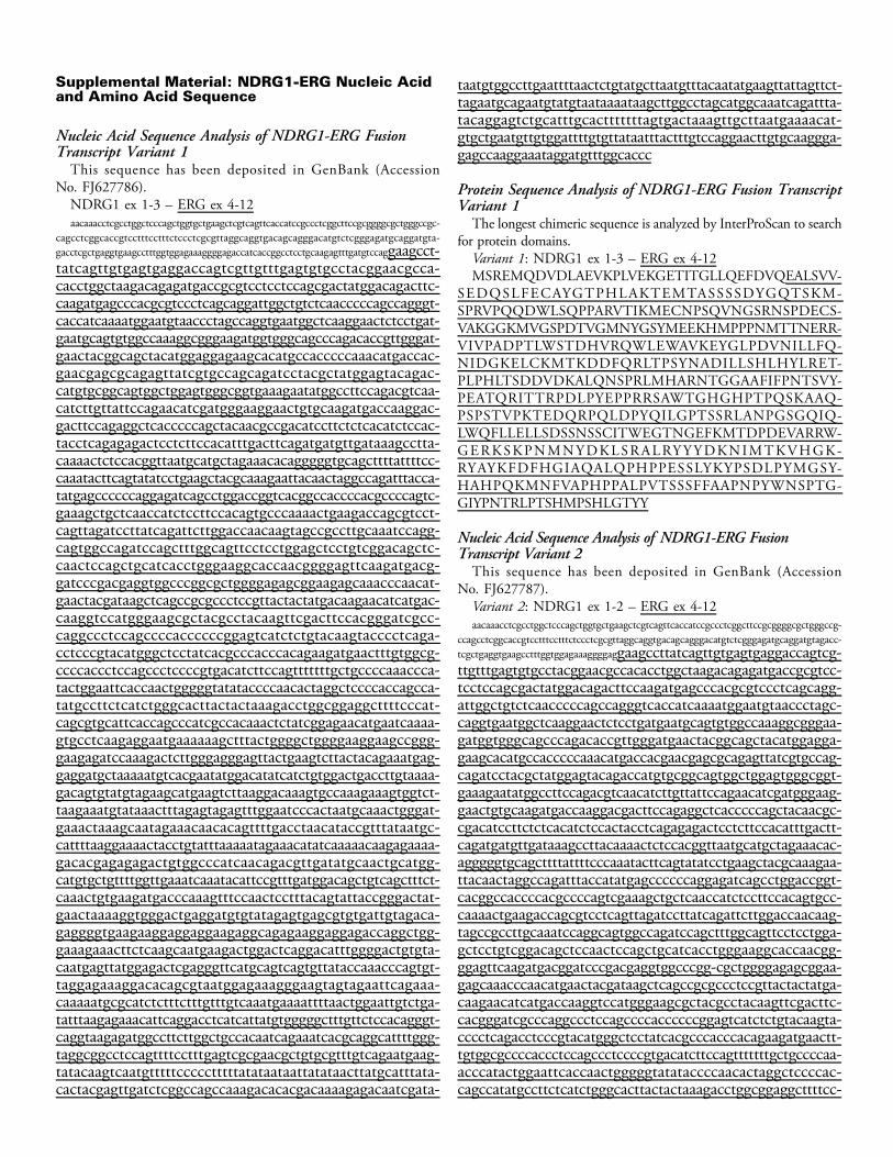
Supplemental Material: NDRG1-ERG Nucleic Acidand Amino Acid Sequence
Nucleic Acid Sequence Analysis of NDRG1-ERG FusionTranscript Variant 1
This sequence has been deposited in GenBank (AccessionNo. FJ627786).
NDRG1 ex 1-3 – ERG ex 4-12aacaaacctcgcctggctcccagctggtgctgaagctcgtcagttcaccatccgccctcggcttccgcggggcgctgggccgc-
cagcctcggcaccgtcctttcctttctccctcgcgttaggcaggtgacagcagggacatgtctcgggagatgcaggatgta-gacctcgctgaggtgaagcctttggtggagaaaggggagaccatcaccggcctcctgcaagagtttgatgtccaggaagcct-tatcagttgtgagtgaggaccagtcgttgtttgagtgtgcctacggaacgcca-cacctggctaagacagagatgaccgcgtcctcctccagcgactatggacagacttc-caagatgagcccacgcgtccctcagcaggattggctgtctcaacccccagccagggt-caccatcaaaatggaatgtaaccctagccaggtgaatggctcaaggaactctcctgat-gaatgcagtgtggccaaaggcgggaagatggtgggcagcccagacaccgttgggat-gaactacggcagctacatggaggagaagcacatgccacccccaaacatgaccac-gaacgagcgcagagttatcgtgccagcagatcctacgctatggagtacagac-catgtgcggcagtggctggagtgggcggtgaaagaatatggccttccagacgtcaa-catcttgttattccagaacatcgatgggaaggaactgtgcaagatgaccaaggac-gacttccagaggctcacccccagctacaacgccgacatccttctctcacatctccac-tacctcagagagactcctcttccacatttgacttcagatgatgttgataaagcctta-caaaactctccacggttaatgcatgctagaaacacagggggtgcagcttttattttcc-caaatacttcagtatatcctgaagctacgcaaagaattacaactaggccagatttacca-tatgagccccccaggagatcagcctggaccggtcacggccaccccacgccccagtc-gaaagctgctcaaccatctccttccacagtgcccaaaactgaagaccagcgtcct-cagttagatccttatcagattcttggaccaacaagtagccgccttgcaaatccagg-cagtggccagatccagctttggcagttcctcctggagctcctgtcggacagctc-caactccagctgcatcacctgggaaggcaccaacggggagttcaagatgacg-gatcccgacgaggtggcccggcgctggggagagcggaagagcaaacccaacat-gaactacgataagctcagccgcgccctccgttactactatgacaagaacatcatgac-caaggtccatgggaagcgctacgcctacaagttcgacttccacgggatcgcc-caggccctccagccccaccccccggagtcatctctgtacaagtacccctcaga-cctcccgtacatgggctcctatcacgcccacccacagaagatgaactttgtggcg-ccccaccctccagccctccccgtgacatcttccagtttttttgctgccccaaaccca-tactggaattcaccaactgggggtatataccccaacactaggctccccaccagcca-tatgccttctcatctgggcacttactactaaagacctggcggaggcttttcccat-cagcgtgcattcaccagcccatcgccacaaactctatcggagaacatgaatcaaaa-gtgcctcaagaggaatgaaaaaagctttactggggctggggaaggaagccggg-gaagagatccaaagactcttgggagggagttactgaagtcttactacagaaatgag-gaggatgctaaaaatgtcacgaatatggacatatcatctgtggactgaccttgtaaaa-gacagtgtatgtagaagcatgaagtcttaaggacaaagtgccaaagaaagtggtct-taagaaatgtataaactttagagtagagtttggaatcccactaatgcaaactgggat-gaaactaaagcaatagaaacaacacagttttgacctaacataccgtttataatgc-cattttaaggaaaactacctgtatttaaaaatagaaacatatcaaaaacaagagaaaa-gacacgagagagactgtggcccatcaacagacgttgatatgcaactgcatgg-catgtgctgttttggttgaaatcaaatacattccgtttgatggacagctgtcagctttct-caaactgtgaagatgacccaaagtttccaactcctttacagtattaccgggactat-gaactaaaaggtgggactgaggatgtgtatagagtgagcgtgtgattgtagaca-gaggggtgaagaaggaggaggaagaggcagagaaggaggagaccaggctgg-gaaagaaacttctcaagcaatgaagactggactcaggacatttggggactgtgta-caatgagttatggagactcgagggttcatgcagtcagtgttataccaaacccagtgt-taggagaaaggacacagcgtaatggagaaagggaagtagtagaattcagaaa-caaaaatgcgcatctctttctttgtttgtcaaatgaaaattttaactggaattgtctga-tatttaagagaaacattcaggacctcatcattatgtgggggctttgttctccacagggt-caggtaagagatggccttcttggctgccacaatcagaaatcacgcaggcattttggg-taggcggcctccagttttcctttgagtcgcgaacgctgtgcgtttgtcagaatgaag-tatacaagtcaatgtttttccccctttttatataataattatataacttatgcatttata-cactacgagttgatctcggccagccaaagacacacgacaaaagagacaatcgata-
taatgtggccttgaattttaactctgtatgcttaatgtttacaatatgaagttattagttct-tagaatgcagaatgtatgtaataaaataagcttggcctagcatggcaaatcagattta-tacaggagtctgcatttgcactttttttagtgactaaagttgcttaatgaaaacat-gtgctgaatgttgtggattttgtgttataatttactttgtccaggaacttgtgcaaggga-gagccaaggaaataggatgtttggcaccc
Protein Sequence Analysis of NDRG1-ERG Fusion TranscriptVariant 1
The longest chimeric sequence is analyzed by InterProScan to searchfor protein domains.
Variant 1: NDRG1 ex 1-3 – ERG ex 4-12MSREMQDVDLAEVKPLVEKGETITGLLQEFDVQEALSVV-
SEDQSLFECAYGTPHLAKTEMTASSSSDYGQTSKM-SPRVPQQDWLSQPPARVTIKMECNPSQVNGSRNSPDECS-VAKGGKMVGSPDTVGMNYGSYMEEKHMPPPNMTTNERR-VIVPADPTLWSTDHVRQWLEWAVKEYGLPDVNILLFQ-NIDGKELCKMTKDDFQRLTPSYNADILLSHLHYLRET-PLPHLTSDDVDKALQNSPRLMHARNTGGAAFIFPNTSVY-PEATQRITTRPDLPYEPPRRSAWTGHGHPTPQSKAAQ-PSPSTVPKTEDQRPQLDPYQILGPTSSRLANPGSGQIQ-LWQFLLELLSDSSNSSCITWEGTNGEFKMTDPDEVARRW-GERKSKPNMNYDKLSRALRYYYDKNIMTKVHGK-RYAYKFDFHGIAQALQPHPPESSLYKYPSDLPYMGSY-HAHPQKMNFVAPHPPALPVTSSSFFAAPNPYWNSPTG-GIYPNTRLPTSHMPSHLGTYY
Nucleic Acid Sequence Analysis of NDRG1-ERG FusionTranscript Variant 2
This sequence has been deposited in GenBank (AccessionNo. FJ627787).
Variant 2: NDRG1 ex 1-2 – ERG ex 4-12aacaaacctcgcctggctcccagctggtgctgaagctcgtcagttcaccatccgccctcggcttccgcggggcgctgggccg-
ccagcctcggcaccgtcctttcctttctccctcgcgttaggcaggtgacagcagggacatgtctcgggagatgcaggatgtagacc-tcgctgaggtgaagcctttggtggagaaaggggaggaagccttatcagttgtgagtgaggaccagtcg-ttgtttgagtgtgcctacggaacgccacacctggctaagacagagatgaccgcgtcc-tcctccagcgactatggacagacttccaagatgagcccacgcgtccctcagcagg-attggctgtctcaacccccagccagggtcaccatcaaaatggaatgtaaccctagc-caggtgaatggctcaaggaactctcctgatgaatgcagtgtggccaaaggcgggaa-gatggtgggcagcccagacaccgttgggatgaactacggcagctacatggagga-gaagcacatgccacccccaaacatgaccacgaacgagcgcagagttatcgtgccag-cagatcctacgctatggagtacagaccatgtgcggcagtggctggagtgggcggt-gaaagaatatggccttccagacgtcaacatcttgttattccagaacatcgatgggaag-gaactgtgcaagatgaccaaggacgacttccagaggctcacccccagctacaacgc-cgacatccttctctcacatctccactacctcagagagactcctcttccacatttgactt-cagatgatgttgataaagccttacaaaactctccacggttaatgcatgctagaaacac-agggggtgcagcttttattttcccaaatacttcagtatatcctgaagctacgcaaagaa-ttacaactaggccagatttaccatatgagccccccaggagatcagcctggaccggt-cacggccaccccacgccccagtcgaaagctgctcaaccatctccttccacagtgcc-caaaactgaagaccagcgtcctcagttagatccttatcagattcttggaccaacaag-tagccgccttgcaaatccaggcagtggccagatccagctttggcagttcctcctgga-gctcctgtcggacagctccaactccagctgcatcacctgggaaggcaccaacgg-ggagttcaagatgacggatcccgacgaggtggcccgg-cgctggggagagcggaa-gagcaaacccaacatgaactacgataagctcagccgcgccctccgttactactatga-caagaacatcatgaccaaggtccatgggaagcgctacgcctacaagttcgacttc-cacgggatcgcccaggccctccagccccaccccccggagtcatctctgtacaagta-cccctcagacctcccgtacatgggctcctatcacgcccacccacagaagatgaactt-tgtggcgccccaccctccagccctccccgtgacatcttccagtttttttgctgccccaa-acccatactggaattcaccaactgggggtatataccccaacactaggctccccac-cagccatatgccttctcatctgggcacttactactaaagacctggcggaggcttttcc-
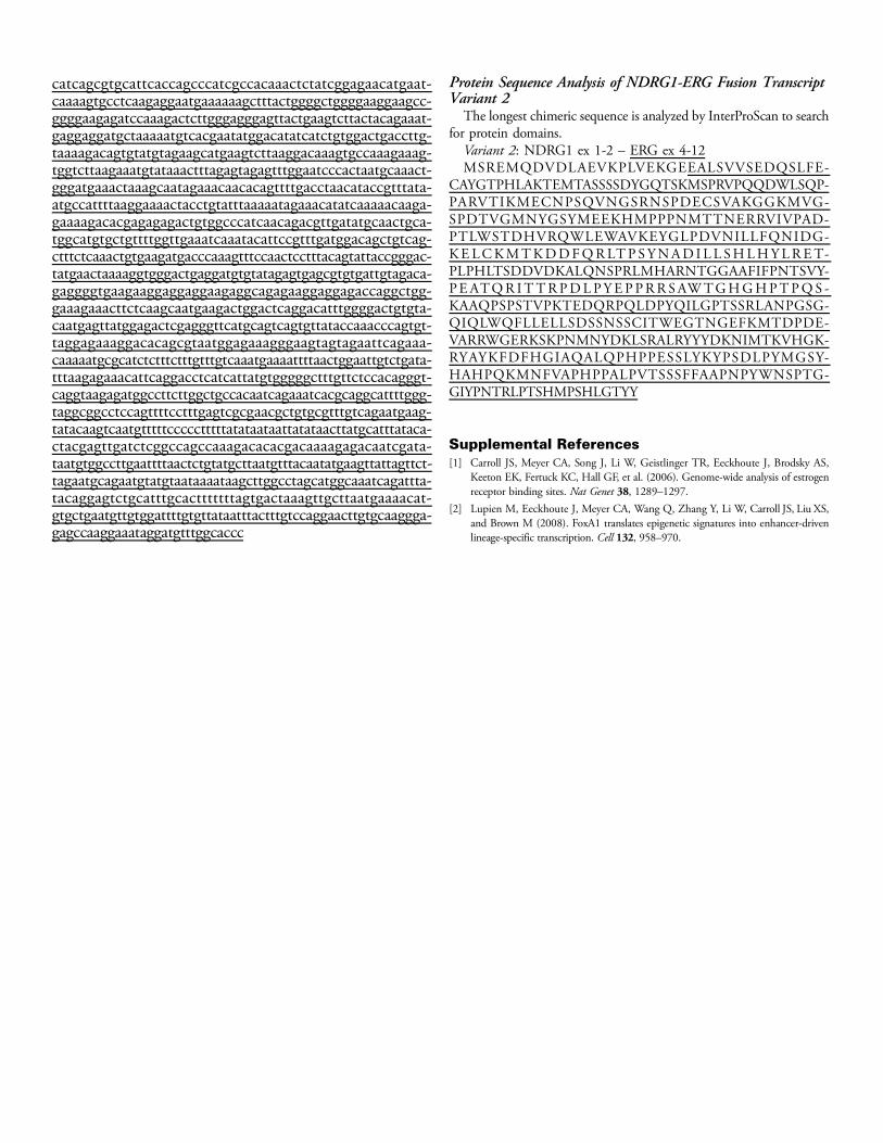
catcagcgtgcattcaccagcccatcgccacaaactctatcggagaacatgaat-caaaagtgcctcaagaggaatgaaaaaagctttactggggctggggaaggaagcc-ggggaagagatccaaagactcttgggagggagttactgaagtcttactacagaaat-gaggaggatgctaaaaatgtcacgaatatggacatatcatctgtggactgaccttg-taaaagacagtgtatgtagaagcatgaagtcttaaggacaaagtgccaaagaaag-tggtcttaagaaatgtataaactttagagtagagtttggaatcccactaatgcaaact-gggatgaaactaaagcaatagaaacaacacagttttgacctaacataccgtttata-atgccattttaaggaaaactacctgtatttaaaaatagaaacatatcaaaaacaaga-gaaaagacacgagagagactgtggcccatcaacagacgttgatatgcaactgca-tggcatgtgctgttttggttgaaatcaaatacattccgtttgatggacagctgtcag-ctttctcaaactgtgaagatgacccaaagtttccaactcctttacagtattaccgggac-tatgaactaaaaggtgggactgaggatgtgtatagagtgagcgtgtgattgtagaca-gaggggtgaagaaggaggaggaagaggcagagaaggaggagaccaggctgg-gaaagaaacttctcaagcaatgaagactggactcaggacatttggggactgtgta-caatgagttatggagactcgagggttcatgcagtcagtgttataccaaacccagtgt-taggagaaaggacacagcgtaatggagaaagggaagtagtagaattcagaaa-caaaaatgcgcatctctttctttgtttgtcaaatgaaaattttaactggaattgtctgata-tttaagagaaacattcaggacctcatcattatgtgggggctttgttctccacagggt-caggtaagagatggccttcttggctgccacaatcagaaatcacgcaggcattttggg-taggcggcctccagttttcctttgagtcgcgaacgctgtgcgtttgtcagaatgaag-tatacaagtcaatgtttttccccctttttatataataattatataacttatgcatttataca-ctacgagttgatctcggccagccaaagacacacgacaaaagagacaatcgata-taatgtggccttgaattttaactctgtatgcttaatgtttacaatatgaagttattagttct-tagaatgcagaatgtatgtaataaaataagcttggcctagcatggcaaatcagattta-tacaggagtctgcatttgcactttttttagtgactaaagttgcttaatgaaaacat-gtgctgaatgttgtggattttgtgttataatttactttgtccaggaacttgtgcaaggga-gagccaaggaaataggatgtttggcaccc
Protein Sequence Analysis of NDRG1-ERG Fusion TranscriptVariant 2
The longest chimeric sequence is analyzed by InterProScan to searchfor protein domains.
Variant 2: NDRG1 ex 1-2 – ERG ex 4-12MSREMQDVDLAEVKPLVEKGEEALSVVSEDQSLFE-
CAYGTPHLAKTEMTASSSSDYGQTSKMSPRVPQQDWLSQP-PARVTIKMECNPSQVNGSRNSPDECSVAKGGKMVG-SPDTVGMNYGSYMEEKHMPPPNMTTNERRVIVPAD-PTLWSTDHVRQWLEWAVKEYGLPDVNILLFQNIDG-KELCKMTKDDFQRLTP SYNAD I L L SHLHYLRET-PLPHLTSDDVDKALQNSPRLMHARNTGGAAFIFPNTSVY-PEATQR ITTRPDLPYEP PRR SAWTGHGHPTPQS -KAAQPSPSTVPKTEDQRPQLDPYQILGPTSSRLANPGSG-QIQLWQFLLELLSDSSNSSCITWEGTNGEFKMTDPDE-VARRWGERKSKPNMNYDKLSRALRYYYDKNIMTKVHGK-RYAYKFDFHGIAQALQPHPPESSLYKYPSDLPYMGSY-HAHPQKMNFVAPHPPALPVTSSSFFAAPNPYWNSPTG-GIYPNTRLPTSHMPSHLGTYY
Supplemental References[1] Carroll JS, Meyer CA, Song J, Li W, Geistlinger TR, Eeckhoute J, Brodsky AS,
Keeton EK, Fertuck KC, Hall GF, et al. (2006). Genome-wide analysis of estrogenreceptor binding sites. Nat Genet 38, 1289–1297.
[2] Lupien M, Eeckhoute J, Meyer CA, Wang Q, Zhang Y, Li W, Carroll JS, Liu XS,and Brown M (2008). FoxA1 translates epigenetic signatures into enhancer-drivenlineage-specific transcription. Cell 132, 958–970.
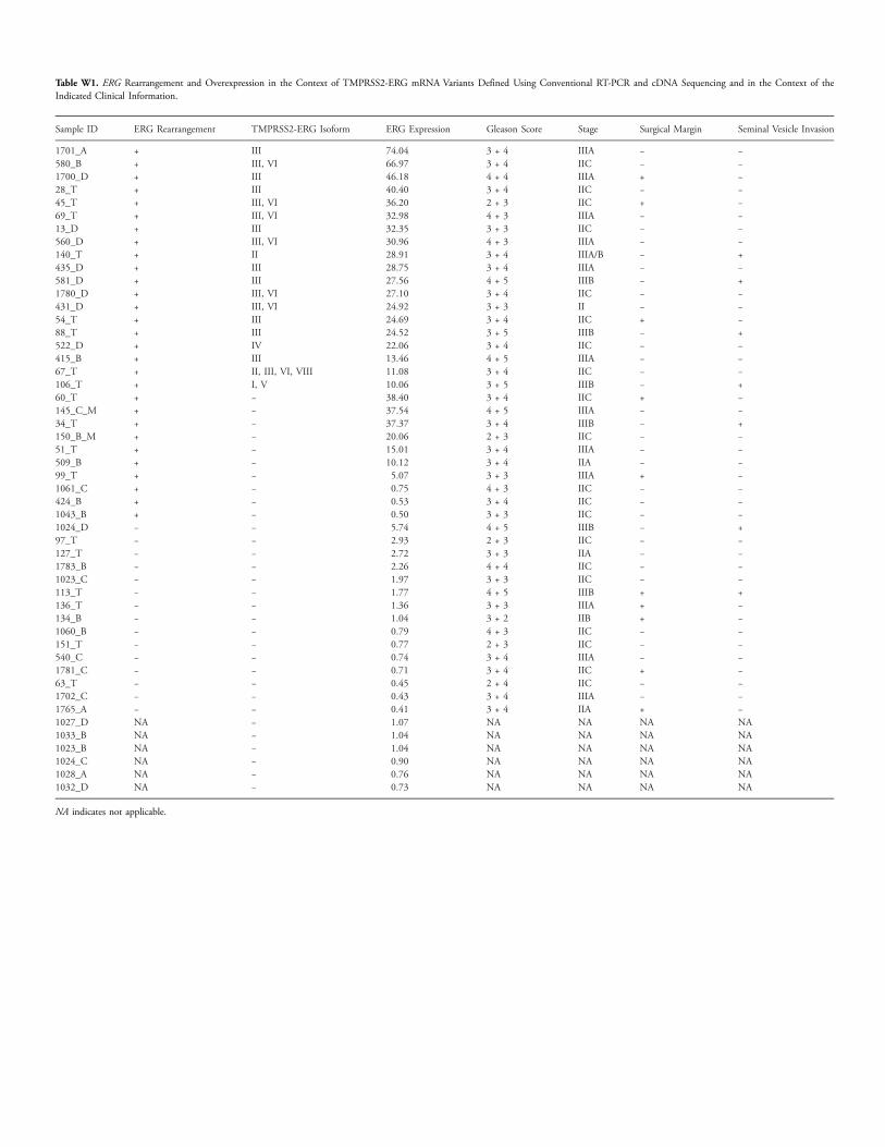
Table W1. ERG Rearrangement and Overexpression in the Context of TMPRSS2-ERG mRNA Variants Defined Using Conventional RT-PCR and cDNA Sequencing and in the Context of theIndicated Clinical Information.
Sample ID
ERG Rearrangement TMPRSS2-ERG Isoform ERG Expression Gleason Score Stage Surgical Margin Seminal Vesicle Invasion1701_A
+ III 74.04 3 + 4 IIIA − − 580_B + III, VI 66.97 3 + 4 IIC − − 1700_D + III 46.18 4 + 4 IIIA + − 28_T + III 40.40 3 + 4 IIC − − 45_T + III, VI 36.20 2 + 3 IIC + − 69_T + III, VI 32.98 4 + 3 IIIA − − 13_D + III 32.35 3 + 3 IIC − − 560_D + III, VI 30.96 4 + 3 IIIA − − 140_T + II 28.91 3 + 4 IIIA/B − + 435_D + III 28.75 3 + 4 IIIA − − 581_D + III 27.56 4 + 5 IIIB − + 1780_D + III, VI 27.10 3 + 4 IIC − − 431_D + III, VI 24.92 3 + 3 II − − 54_T + III 24.69 3 + 4 IIC + − 88_T + III 24.52 3 + 5 IIIB − + 522_D + IV 22.06 3 + 4 IIC − − 415_B + III 13.46 4 + 5 IIIA − − 67_T + II, III, VI, VIII 11.08 3 + 4 IIC − − 106_T + I, V 10.06 3 + 5 IIIB − + 60_T + − 38.40 3 + 4 IIC + − 145_C_M + − 37.54 4 + 5 IIIA − − 34_T + − 37.37 3 + 4 IIIB − + 150_B_M + − 20.06 2 + 3 IIC − − 51_T + − 15.01 3 + 4 IIIA − − 509_B + − 10.12 3 + 4 IIA − − 99_T + − 5.07 3 + 3 IIIA + − 1061_C + − 0.75 4 + 3 IIC − − 424_B + − 0.53 3 + 4 IIC − − 1043_B + − 0.50 3 + 3 IIC − − 1024_D − − 5.74 4 + 5 IIIB − + 97_T − − 2.93 2 + 3 IIC − − 127_T − − 2.72 3 + 3 IIA − − 1783_B − − 2.26 4 + 4 IIC − − 1023_C − − 1.97 3 + 3 IIC − − 113_T − − 1.77 4 + 5 IIIB + + 136_T − − 1.36 3 + 3 IIIA + − 134_B − − 1.04 3 + 2 IIB + − 1060_B − − 0.79 4 + 3 IIC − − 151_T − − 0.77 2 + 3 IIC − − 540_C − − 0.74 3 + 4 IIIA − − 1781_C − − 0.71 3 + 4 IIC + − 63_T − − 0.45 2 + 4 IIC − − 1702_C − − 0.43 3 + 4 IIIA − − 1765_A − − 0.41 3 + 4 IIA + − 1027_D NA − 1.07 NA NA NA NA 1033_B NA − 1.04 NA NA NA NA 1023_B NA − 1.04 NA NA NA NA 1024_C NA − 0.90 NA NA NA NA 1028_A NA − 0.76 NA NA NA NA 1032_D NA − 0.73 NA NA NA NANA indicates not applicable.
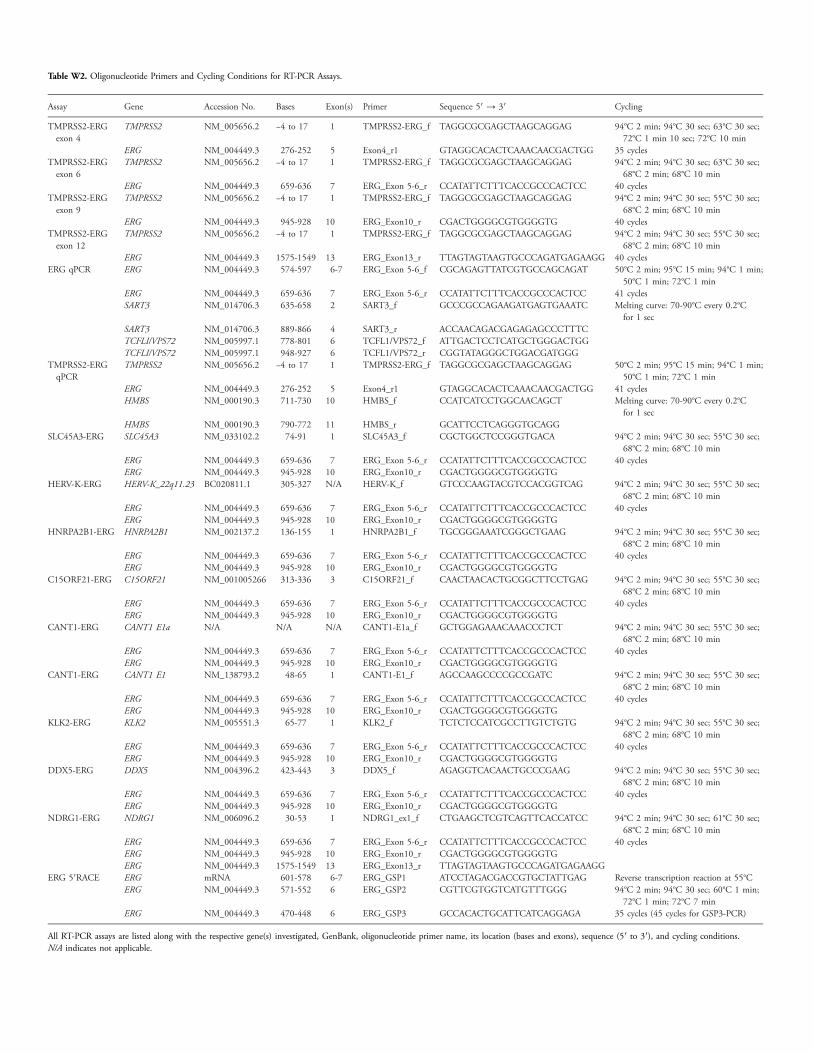
Table W2. Oligonucleotide Primers and Cycling Conditions for RT-PCR Assays.
Assay
Gene Accession No. Bases Exon(s) Primer Sequence 5′ → 3′ CyclingTMPRSS2-ERGexon 4
TMPRSS2
NM_005656.2 −4 to 17 1 TMPRSS2-ERG_f TAGGCGCGAGCTAAGCAGGAG 94°C 2 min; 94°C 30 sec; 63°C 30 sec;72°C 1 min 10 sec; 72°C 10 minERG
NM_004449.3 276-252 5 Exon4_r1 GTAGGCACACTCAAACAACGACTGG 35 cycles TMPRSS2-ERGexon 6TMPRSS2
NM_005656.2 −4 to 17 1 TMPRSS2-ERG_f TAGGCGCGAGCTAAGCAGGAG 94°C 2 min; 94°C 30 sec; 63°C 30 sec;68°C 2 min; 68°C 10 minERG
NM_004449.3 659-636 7 ERG_Exon 5-6_r CCATATTCTTTCACCGCCCACTCC 40 cycles TMPRSS2-ERGexon 9TMPRSS2
NM_005656.2 −4 to 17 1 TMPRSS2-ERG_f TAGGCGCGAGCTAAGCAGGAG 94°C 2 min; 94°C 30 sec; 55°C 30 sec;68°C 2 min; 68°C 10 minERG
NM_004449.3 945-928 10 ERG_Exon10_r CGACTGGGGCGTGGGGTG 40 cycles TMPRSS2-ERGexon 12TMPRSS2
NM_005656.2 −4 to 17 1 TMPRSS2-ERG_f TAGGCGCGAGCTAAGCAGGAG 94°C 2 min; 94°C 30 sec; 55°C 30 sec;68°C 2 min; 68°C 10 minERG
NM_004449.3 1575-1549 13 ERG_Exon13_r TTAGTAGTAAGTGCCCAGATGAGAAGG 40 cycles ERG qPCR ERG NM_004449.3 574-597 6-7 ERG_Exon 5-6_f CGCAGAGTTATCGTGCCAGCAGAT 50°C 2 min; 95°C 15 min; 94°C 1 min;50°C 1 min; 72°C 1 min
ERG NM_004449.3 659-636 7 ERG_Exon 5-6_r CCATATTCTTTCACCGCCCACTCC 41 cycles SART3 NM_014706.3 635-658 2 SART3_f GCCCGCCAGAAGATGAGTGAAATC Melting curve: 70-90°C every 0.2°Cfor 1 sec
SART3 NM_014706.3 889-866 4 SART3_r ACCAACAGACGAGAGAGCCCTTTC TCFLI/VPS72 NM_005997.1 778-801 6 TCFL1/VPS72_f ATTGACTCCTCATGCTGGGACTGG TCFLI/VPS72 NM_005997.1 948-927 6 TCFL1/VPS72_r CGGTATAGGGCTGGACGATGGGTMPRSS2-ERGqPCR
TMPRSS2
NM_005656.2 −4 to 17 1 TMPRSS2-ERG_f TAGGCGCGAGCTAAGCAGGAG 50°C 2 min; 95°C 15 min; 94°C 1 min;50°C 1 min; 72°C 1 minERG
NM_004449.3 276-252 5 Exon4_r1 GTAGGCACACTCAAACAACGACTGG 41 cycles HMBS NM_000190.3 711-730 10 HMBS_f CCATCATCCTGGCAACAGCT Melting curve: 70-90°C every 0.2°Cfor 1 sec
HMBS NM_000190.3 790-772 11 HMBS_r GCATTCCTCAGGGTGCAGGSLC45A3-ERG
SLC45A3 NM_033102.2 74-91 1 SLC45A3_f CGCTGGCTCCGGGTGACA 94°C 2 min; 94°C 30 sec; 55°C 30 sec;68°C 2 min; 68°C 10 minERG
NM_004449.3 659-636 7 ERG_Exon 5-6_r CCATATTCTTTCACCGCCCACTCC 40 cycles ERG NM_004449.3 945-928 10 ERG_Exon10_r CGACTGGGGCGTGGGGTGHERV-K-ERG
HERV-K_22q11.23 BC020811.1 305-327 N/A HERV-K_f GTCCCAAGTACGTCCACGGTCAG 94°C 2 min; 94°C 30 sec; 55°C 30 sec;68°C 2 min; 68°C 10 minERG
NM_004449.3 659-636 7 ERG_Exon 5-6_r CCATATTCTTTCACCGCCCACTCC 40 cycles ERG NM_004449.3 945-928 10 ERG_Exon10_r CGACTGGGGCGTGGGGTGHNRPA2B1-ERG
HNRPA2B1 NM_002137.2 136-155 1 HNRPA2B1_f TGCGGGAAATCGGGCTGAAG 94°C 2 min; 94°C 30 sec; 55°C 30 sec;68°C 2 min; 68°C 10 minERG
NM_004449.3 659-636 7 ERG_Exon 5-6_r CCATATTCTTTCACCGCCCACTCC 40 cycles ERG NM_004449.3 945-928 10 ERG_Exon10_r CGACTGGGGCGTGGGGTGC15ORF21-ERG
C15ORF21 NM_001005266 313-336 3 C15ORF21_f CAACTAACACTGCGGCTTCCTGAG 94°C 2 min; 94°C 30 sec; 55°C 30 sec;68°C 2 min; 68°C 10 minERG
NM_004449.3 659-636 7 ERG_Exon 5-6_r CCATATTCTTTCACCGCCCACTCC 40 cycles ERG NM_004449.3 945-928 10 ERG_Exon10_r CGACTGGGGCGTGGGGTGCANT1-ERG
CANT1 E1a N/A N/A N/A CANT1-E1a_f GCTGGAGAAACAAACCCTCT 94°C 2 min; 94°C 30 sec; 55°C 30 sec;68°C 2 min; 68°C 10 minERG
NM_004449.3 659-636 7 ERG_Exon 5-6_r CCATATTCTTTCACCGCCCACTCC 40 cycles ERG NM_004449.3 945-928 10 ERG_Exon10_r CGACTGGGGCGTGGGGTGCANT1-ERG
CANT1 E1 NM_138793.2 48-65 1 CANT1-E1_f AGCCAAGCCCCGCCGATC 94°C 2 min; 94°C 30 sec; 55°C 30 sec;68°C 2 min; 68°C 10 minERG
NM_004449.3 659-636 7 ERG_Exon 5-6_r CCATATTCTTTCACCGCCCACTCC 40 cycles ERG NM_004449.3 945-928 10 ERG_Exon10_r CGACTGGGGCGTGGGGTGKLK2-ERG
KLK2 NM_005551.3 65-77 1 KLK2_f TCTCTCCATCGCCTTGTCTGTG 94°C 2 min; 94°C 30 sec; 55°C 30 sec;68°C 2 min; 68°C 10 minERG
NM_004449.3 659-636 7 ERG_Exon 5-6_r CCATATTCTTTCACCGCCCACTCC 40 cycles ERG NM_004449.3 945-928 10 ERG_Exon10_r CGACTGGGGCGTGGGGTGDDX5-ERG
DDX5 NM_004396.2 423-443 3 DDX5_f AGAGGTCACAACTGCCCGAAG 94°C 2 min; 94°C 30 sec; 55°C 30 sec;68°C 2 min; 68°C 10 minERG
NM_004449.3 659-636 7 ERG_Exon 5-6_r CCATATTCTTTCACCGCCCACTCC 40 cycles ERG NM_004449.3 945-928 10 ERG_Exon10_r CGACTGGGGCGTGGGGTGNDRG1-ERG
NDRG1 NM_006096.2 30-53 1 NDRG1_ex1_f CTGAAGCTCGTCAGTTCACCATCC 94°C 2 min; 94°C 30 sec; 61°C 30 sec;68°C 2 min; 68°C 10 minERG
NM_004449.3 659-636 7 ERG_Exon 5-6_r CCATATTCTTTCACCGCCCACTCC 40 cycles ERG NM_004449.3 945-928 10 ERG_Exon10_r CGACTGGGGCGTGGGGTG ERG NM_004449.3 1575-1549 13 ERG_Exon13_r TTAGTAGTAAGTGCCCAGATGAGAAGGERG 5′RACE
ERG mRNA 601-578 6-7 ERG_GSP1 ATCCTAGACGACCGTGCTATTGAG Reverse transcription reaction at 55°C ERG NM_004449.3 571-552 6 ERG_GSP2 CGTTCGTGGTCATGTTTGGG 94°C 2 min; 94°C 30 sec; 60°C 1 min;72°C 1 min; 72°C 7 min
ERG NM_004449.3 470-448 6 ERG_GSP3 GCCACACTGCATTCATCAGGAGA 35 cycles (45 cycles for GSP3-PCR)All RT-PCR assays are listed along with the respective gene(s) investigated, GenBank, oligonucleotide primer name, its location (bases and exons), sequence (5′ to 3′), and cycling conditions.N/A indicates not applicable.
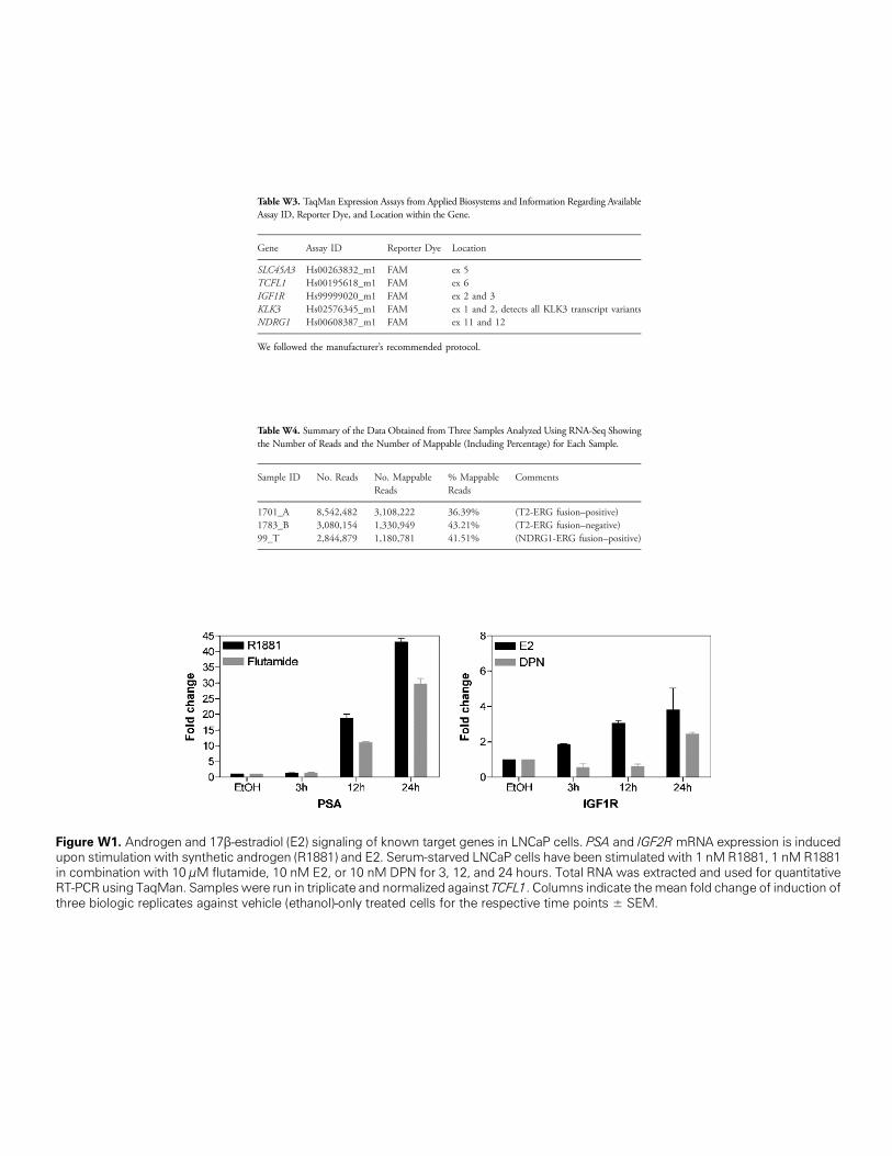
Figure W1. Androgen and 17β-estrupon stimulation with synthetic andin combination with 10 μM flutamidRT-PCR using TaqMan. Samples wethree biologic replicates against ve
Table W3. TaqMan Expression Assays from Applied Biosystems and Information Regarding AvailableAssay ID, Reporter Dye, and Location within the Gene.
Gene
adiol (E2rogen (Re, 10 nMre run inhicle (eth
Assay ID
) signaling of k1881) and E2.E2, or 10 nMtriplicate andanol)-only trea
Reporter Dye
nown targeSerum-starDPN for 3,
normalized ated cells fo
Location
SLC45A3
Hs00263832_m1 FAM ex 5 TCFL1 Hs00195618_m1 FAM ex 6 IGF1R Hs99999020_m1 FAM ex 2 and 3 KLK3 Hs02576345_m1 FAM ex 1 and 2, detects all KLK3 transcript variants NDRG1 Hs00608387_m1 FAM ex 11 and 12We followed the manufacturer’s recommended protocol.
Table W4. Summary of the Data Obtained from Three Samples Analyzed Using RNA-Seq Showingthe Number of Reads and the Number of Mappable (Including Percentage) for Each Sample.
Sample ID
No. Reads No. MappableReads% MappableReads
t genes inved LNCaP12, and 24gainst TCFr the respe
Comments
1701_A
8,542,482 3,108,222 36.39% (T2-ERG fusion–positive) 1783_B 3,080,154 1,330,949 43.21% (T2-ERG fusion–negative) 99_T 2,844,879 1,180,781 41.51% (NDRG1-ERG fusion–positive)LNCaP cells. PSA and IGF2R mRNA expression is inducedcells have been stimulated with 1 nM R1881, 1 nM R1881hours. Total RNA was extracted and used for quantitativeL1. Columns indicate themean fold change of induction ofctive time points ± SEM.
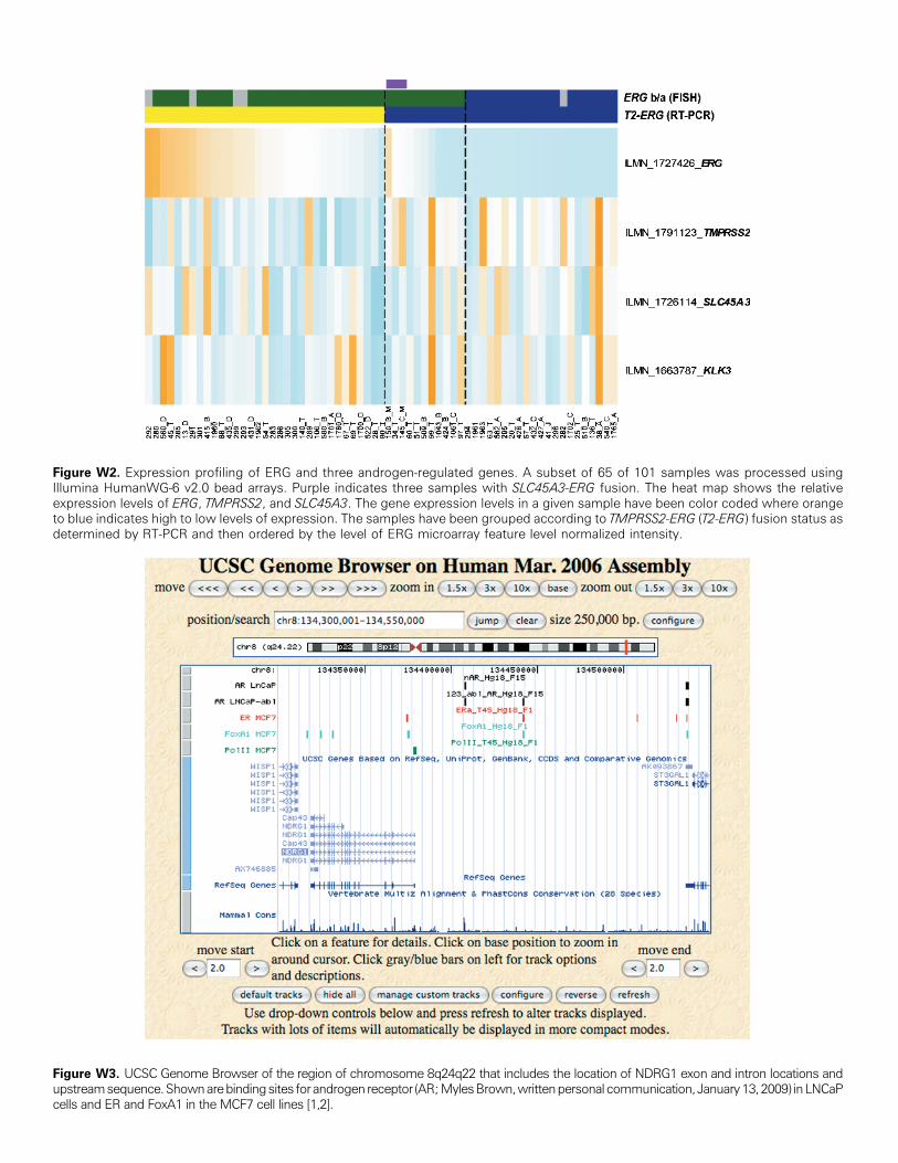
Figure W2. Expression profiling of ERG and three androgen-regulated genes. A subset of 65 of 101 samples was processed usingIllumina HumanWG-6 v2.0 bead arrays. Purple indicates three samples with SLC45A3-ERG fusion. The heat map shows the relativeexpression levels of ERG, TMPRSS2, and SLC45A3. The gene expression levels in a given sample have been color coded where orangeto blue indicates high to low levels of expression. The samples have been grouped according to TMPRSS2-ERG (T2-ERG) fusion status asdetermined by RT-PCR and then ordered by the level of ERG microarray feature level normalized intensity.
Figure W3. UCSC Genome Browser of the region of chromosome 8q24q22 that includes the location of NDRG1 exon and intron locations andupstreamsequence.Shownarebindingsites forandrogen receptor (AR;MylesBrown,writtenpersonal communication, January13,2009) inLNCaPcells and ER and FoxA1 in the MCF7 cell lines [1,2].
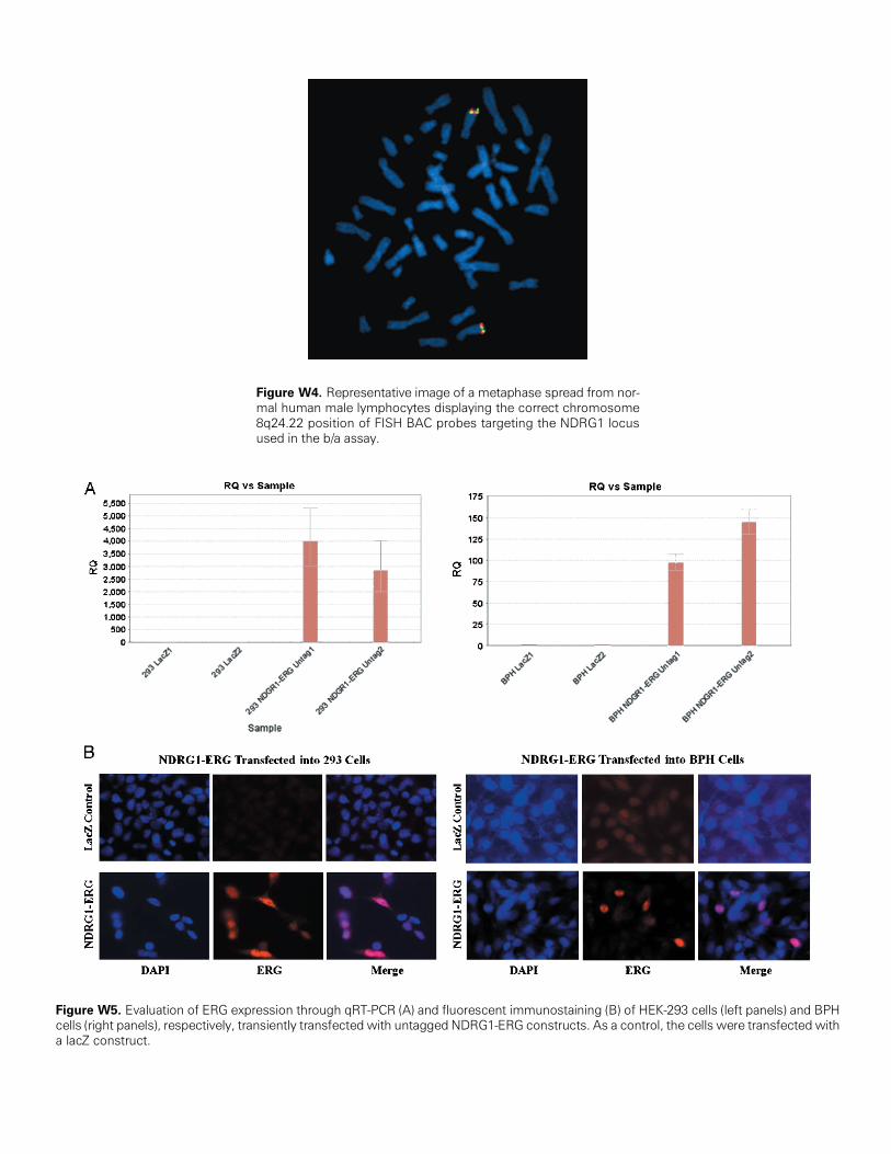
Figure W4. Representative image of a metaphase spread from nor-mal human male lymphocytes displaying the correct chromosome8q24.22 position of FISH BAC probes targeting the NDRG1 locusused in the b/a assay.
Figure W5. Evaluation of ERG expression through qRT-PCR (A) and fluorescent immunostaining (B) of HEK-293 cells (left panels) and BPHcells (right panels), respectively, transiently transfected with untagged NDRG1-ERG constructs. As a control, the cells were transfected witha lacZ construct.