Embed Size (px)
Citation preview

The clinical neuropsychiatry of
multiple sclerosis
A. Feinstein, MPhil, PhD, MRCPsych, FRCP(C)
Sunnybrook Health Science Centre,University of Toronto, Canada

published by the press syndicate of the university of cambridgeThe Pitt Building, Trumpington Street, Cambridge, United Kingdom
cambridge unive rsity pressThe Edinburgh Building, Cambridge CB2 2RU, UK http://www.cup.cam.ac.uk40 West 20th Street, New York, NY 10011-4211, USA http://www.cup.org10 Stamford Road, Oakleigh, Melbourne 3166, Australia
© Anthony Feinstein 1999
This book is in copyright. Subject to statutory exceptionand to the provisions of relevant collective licensing agreements,no reproduction of any part may take place withoutthe written permission of Cambridge University Press
First published 1999
Printed in the United Kingdom at the University Press, Cambridge
Typeset in Minion 10/12 [vn]
A catalogue record for this book is available from the British Library
Library of Congress Cataloguing in Publication data
Feinstein, A. (Anthony), 1956–The clinical neuropsychiatry of multiple sclerosis / A. Feinstein.
p. cm.Includes index.ISBN 0 521 57274 61. Multiple sclerosis – Complications. 2. Multiple sclerosis – Patients – Mental health.3. Psychological manifestation of general diseases. I. Title.[DNLM: 1. Multiple Sclerosis – psychology. 2. Multiple Sclerosis – complications.3. Mental Disorders – etiology. WL 360 f299C 1999]
RC377.F45 1999616.8'34–dc21 98–46888 CIPDNLM/DLCfor Library of Congress
ISBN 0 521 57274 6 hardback
Every eVort has been made in preparing this book to provide accurate and up-to-dateinformation which is in accord with accepted standards and practice at the time of publication.Nevertheless, the authors, editors and publisher can make no warranties that the informationcontained herein is totally free from error, not least because clinical standards are constantlychanging through research and regulation. The authors, editors and publisher thereforedisclaim all liability for direct or consequential damages resulting from the use of materialcontained in this book. Readers are strongly advised to pay careful attention to informationprovided by the manufacturer of any drugs or equipment that they plan to use.
Contents
Acknowledgements ix
1 Multiple sclerosis: diagnosis and deWnitions 1
2 Multiple sclerosis and depression 26
3 Multiple sclerosis and bipolar aVective disorder 51
4 Multiple sclerosis and pathological laughing and crying 65
5 Multiple sclerosis and psychosis 80
6 Cognitive impairment in multiple sclerosis 96
7 The natural history of cognitive change in multiplesclerosis 121
8 Cognitive impairment in multile sclerosis: detection,management and signiWcance 131
9 Neuroimaging correlates of cognitive dysfunction 145
10 Multiple sclerosis: a subcortical, white matter dementia? 176
Index 197
vii
1
1
Multiple sclerosis: diagnosis anddefinitions
Many a chapter, monograph and paper on multiple sclerosis begins with theobservation that MS is the commonest disabling neurological disease aVec-ting young and middle-aged adults. Since the Wrst clinical description of thedisease in the late 1830s, attention has largely focused on neurologicalmanifestations and it is only over the past decade that clinicians, researchers,and indeed the patients themselves, have become more aware of the behav-ioural changes that may accompany MS. A burgeoning literature devoted tothe neuropsychiatry of MS attests to this new-found interest, although thosewith knowledge of the medical history of MS may Wnd themselves a littleperplexed as to why it has taken so long for this interest to ignite. Descrip-tions of altered mentation in MS patients predate the writings of the man Wrstcredited with naming the condition over a century ago, the French behav-ioural neurologist Jean-Martin Charcot (Stenager, 1991).
Before describing the psychiatric and cognitive changes associated withMS, reference will be made to the neurology and pathology of the disorder.This chapter therefore begins with a summary of the pathogenesis, pathology,signs and symptoms, diagnosis and diVerential diagnosis of multiple scler-osis. With the book’s emphasis on mentation, this introduction will by designbe brief and those seeking more detailed explanations are encouraged toconsult the many texts speciWcally devoted to these aspects. This chapter will,however, discuss in depth the research guidelines for diagnosing MS andfurnish clear deWnitions for terms that apply directly to the disease. Thesepoints are important, for they will clarify at the outset many descriptive termsthat appear in the MS research literature and are used throughout this book.The chapter will conclude with a discussion on rating disability and howbehavioural changes may aVect this assessment.
Epidemiology
In the United Kingdom the lifetime risk is 1:800, which translates intoapproximately 60 000 people with the disease (Compston, 1990). In theUnited States, the Wgure is at least four times that. There is a recognition thatsome cases of MS go undetected in life, appearing as a chance Wnding atpostmortem (Gilbert and Sadler, 1983). Estimates that up to 20% of cases fall
2 Clinical neuropsychiatry of multiple sclerosis
into this category (Mackay and Hirano, 1967), introduces a cautionary notein interpreting the epidemiological data. Generally, MS is seen with greaterfrequency as the distance from the equator increases in either hemisphere(Gonzalez-Scarano et al., 1986; Skegg et al., 1987). It is twice as common inwomen as in men and, although may occur at any age, onset in early adult lifeis commonest. The etiology is unknown, and both genetic and environmentalinXuences are considered important. The 25% monozygotic concordancerate (Ebers and Bulman, 1986) attests to the former, while evidence ofenvironmental inXuences comes from three main sources. Migration studieshave demonstrated that those who emigrate during childhood assume therisk of the country of adoption (Dean, 1967), disease epidemics have beenreported in isolated communities such as the Faroe Islands (Kurtzke andHyllested, 1979), and marked variations in prevalence have been found ingenetically homogeneous populations (Miller et al., 1990).
Clinical features
The disorder may present with diverse neurological signs that vary con-siderably between patients. Initial symptoms, which reXect the presence anddistribution of the plaques, commonly involve numbness or tingling in thelimbs or weakness aVecting one or more limbs, loss of vision or impairedvisual acuity, diplopia, facial numbness, vertigo, dysathria, ataxia and urinaryfrequency or urgency and fatigue. As MS is predominantly a white matterdisease, symptoms referrable to cortical (grey) matter involvement are con-sidered rare. Thus, dementia, aphasia, seizures, pain, abnormal and involun-tary movement, muscle atrophy and fasciculations although possible, are sounusual they may cast doubt on the diagnosis (Rolak, 1996). The course ofthe disease is variable and initially impossible to predict. Approximately5–10% of patients show a steady progression of disability from the onset ofthe disease. The remainder run a relapsing–remitting course, of which 20–30% never become seriously disabled and continue to function productively20–25 years after symptom onset (Sibley, 1990). However, the largest group(almost 60%) enter a phase of progressive deterioration a variable number ofyears after symptom onset. Even within this group there is considerablevariability, with a patient’s condition Xuctuating between relapses, periods ofstability and progressive deterioration.
Pathology
Although the exact pathogenesis of MS is uncertain, there is Wrm evidence ofan autoimmune mediated inXammatory disorder aVecting the central ner-
3Multiple sclerosis
vous system (Lisak, 1986; Vrench-Constant, 1994). The target of the inXam-matory response is myelin, a lipoprotein made by oligodendrocytes andinvesting the axons. Along the length of a nerve, the myelin sheaths areseparated by gaps, the nodes of Ranvier. Nerve transmission is facilitated byimpulses jumping from node to node in a process known as saltatoryconduction. With damage to the myelin (i.e. demyelination), the conductionbecomes impaired, transmission of nerve impulses is delayed and symptomsensue.
Postmortem Wndings have further elucidated the neuropathological chan-ges that occur (Allen, 1991). In patients severely aVected by MS and whocome to autopsy, the brain shows a mild degree of generalized atrophy withsulcal widening and dilatation of the ventricles. Plaques, which show his-tological evidence of demyelination, have a striking predilection for a bilat-eral periventricular distribution, particularly the lateral angles of the lateralventricles, the Xoor of the aqueduct and the fourth ventricle. While plaquesmay also be scattered throughout the white matter, immediate subcorticalmyelin is usually spared and the cortex only rarely involved. When viewed onsagittal section, the relationship of demyelination to the terminal veins maybe seen. In some patients, the cerebrum is relatively spared, the main lesionload involving the optic nerves, brain stem and spinal cord (Allen, 1991).Such a constellation of plaques has major implications for the presence andnature of behavioural and cognitive changes and will be more fully discussedin Chapters 2 and 9.
What exactly occurs in the early stages of demyelination is unclear, and it isthe subject of debate whether demyelination can occur de novo without anobserved immune response and increased cellularity, e.g. an inXux of lym-phocytes associated with perivascular inXammation. In the early stages ofmyelin breakdown, oligodendrocytes are still recognizable. As disease prog-resses, the myelin becomes progressively attenuated, partially detached fromthe axon, and ultimately phagocytozed by invading macrophages. The early,established lesion shows a characteristic pattern of increased cells (mac-rophages, astrocytes), a mixture of intact and disintegrated myelin sheaths,perivascular inXammation (lymphocytes, plasma cells, macrophages),oligodendrocyte loss, preserved axons, and within the grey matter, preser-vation of cell bodies.
In non-acute, but active plaques there is hyperplasia of macrophages andastrocytes and lesions contain myelin lipid degradation products. Perivas-cular inXammation, although present, is sparse. While the edges of activelesions are hypercellular with evidence of normal and disintegrating myelinsheaths, the core of such lesions may resemble older, inactive plaques. As thelesion evolves from an active to non-active phase, signs of inXammationdisappear. Chronic lesions, which generally make up the bulk of the largecharacteristic periventricular lesions seen on MRI or at post-mortem, are
4 Clinical neuropsychiatry of multiple sclerosis
thus hypocellular, demyelinated, gliosed and contain few oligodendrocytes.The demyelinated axons are separated by a heavy concentration of astrocyticprocesses (Vrench-Constant, 1994). The small venules are not inXamed, as inacute lesions, but rather show thickened hyalinized walls (Allen, 1991).Although considered a disease primarily aVecting myelin, there is evidencethat axons denuded of myelin are also susceptible to damage (Paty, 1997).
Irrespective of the stage of the lesion, remyelination may aVect the changesobserved. Remyelination has been noted in acute MS lesions (Prineas et al.,1993), giving rise to thin myelin sheaths in areas previously noted to be free ofmyelin. Newly formed as opposed to surviving oligodendrocytes are thoughtto be the source (Prineas et al., 1989). In chronic lesions where not all themyelin is lost, demyelination and remyelination are thought to be occurringsimultaneously. In MS, remyelination is not complete, perhaps becauserepaired areas are subject to repeated bouts of demyelination leading to eithera reduction in oligodendrocyte precursors (termed 02A progenitor cells), orthe creation of an environment that inhibits their migration (Vrench-Con-stant, 1994).
Imaging studies during an acute attack have shown leakage of contrastenhancing materials, indicative of a breakdown in the blood–brain barrier(BBB). The compromised BBB results in edema and the entry of immunemediators (i.e. antibodies), which may contribute to myelin destruction. Theleakage disappears spontaneously over 4–6 weeks (Miller et al., 1988) andmay be reversed temporarily by the administration of corticosteroids (Bar-khof et al., 1991). Postmortem studies have conWrmed that lesions visualizedon magnetic resonance imaging and computerized axial tomography corre-spond to MS plaques (Ormerod et al., 1987). Furthermore, an in vivo studyof MRI and histological parameters from six biopsy proven cases of inXam-matory demyelination of the central nervous system, has shown that changesobserved on MR imaging correlate with the evolving pattern of lesions, i.e.from acute to less active to chronic (Bruck et al., 1997).
An important observation is that white matter, which appears normal tothe naked eye (NAWM) will, more often than not, show histological abnor-malities. These include microscopic foci of demyelination, diVuse gliosis,perivascular inXammation, deposits of iron, lipofuscin and calcium andcollagenization of small blood vessels (Allen, 1991). Furthermore, this evi-dence of a more diVuse lesion may occur in the absence of signiWcant plaqueformation. The clinical signiWcance of these Wndings is that neuroimaging ofthe brain and spinal cord with standard sequences devised for plaque detec-tion, may mislead the observer into thinking the normal appearing whitematter was indeed normal. Alternative imaging procedures for probing thesemore subtle changes have been devised, namely magnetic resonance spectros-copy, and T1 and T2 relaxation times, and are discussed in Chapter 9.
5Multiple sclerosis
Diagnosis
The diagnosis of multiple sclerosis (MS) carries major implications forpatients and their families. Uncertainty over the future, the ability to work,earn a living and live independently are all issues that readily come to mind. Itis therefore imperative for the clinician to be clear about what symptoms andsigns constitute a diagnosis of MS. In addition, making an early, correctdiagnosis is assuming added importance because, for the Wrst time, the MSpatient is facing a choice of treatment options.
The diagnosis of MS is essentially a clinical one and requires that apatient of an appropriate age has had at least two episodes of neurologicaldisturbance, implicating diVerent sites in the central white matter. A num-ber of investigations may help the clinician establish the presence and site ofwhite matter lesions, thereby facilitating a diagnosis. It is, however, impor-tant to realize that these investigations (neuroimaging, evoked potentialsand cerebrospinal Xuid electrophoresis) are not speciWc for multiple scler-osis and should thus be viewed only as helpful adjuncts to the clinicalpresentation.
From a research perspective, correctly diagnosing MS is equally important.Researchers across sites need to talk the same language and, while well-deWned clinical criteria are essential, they cannot stand apart from advancesin technology. A recognition of the need to bring coherence, to what may bewidely divergent neurological presentations, has prompted researchers overthe years to come up with a series of diagnostic guidelines. For many yearsthose of Schumacher (1965) suYced, but in response to improved laboratoryand clinical procedures these have given way to revised criteria (Poser et al.,1983).
The Poser Committee’s Recommendations
The Poser Committee that convened in Washington, DC in 1982 com-prehensively reviewed historical and clinical symptomatology in MS, im-munological observations, CSF tests, a variety of neurophysiological, psycho-physiological and neuropsychological procedures, neuroimaging procedures(CT and MRI), and urological studies of bladder, bowel and sexual function.They concluded that revisions to existing criteria were essential in order toconduct multicentre, therapeutic trials, to compare epidemiological data, toevaluate new diagnostic procedures and to estimate disease activity (Poser etal., 1983; Poser, 1984). It was also clear to Poser and his committee thatphysicians diVered in their use of MS-related terminology (e.g. relapse,remission, etc.), so new deWnitions were included with the diagnostic criteria.They are still used today. Given the pivotal place they have assumed in MS
6 Clinical neuropsychiatry of multiple sclerosis
research, and because they deWne concepts and categories that occurthroughout this book, a detailed description follows.
Definitions
AgeFor research purposes, age was limited to 10–59 years in order to minimizecontamination by patients suVering from other disorders. However, it isrecognized that patients may present outside this range, although suchoccurrences are rare.
Attack (bout, episode, exacerbation, relapse)This was deWned as the occurrence of a symptom or symptoms of neurologi-cal dysfunction, with or without objective conWrmation, lasting more than 24hours. The completely subjective nature of the symptoms were stressed,although it was acknowledged that medical corroboration would strengthenthe case. Individual symptoms that were transient, such as Lhermitte’s sign,i.e. sudden paresthesia following neck Xexion, or vertigo lasting a few sec-onds, were not considered evidence of an attack.
Clinical evidence of a lesionThis refers to the demonstration of abnormal signs on examination by acompetent clinician. These signs are acceptable, even if no longer present,provided they were elicited and recorded earlier by an examiner.
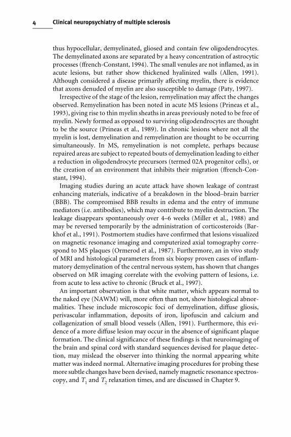
Paraclinical evidence of a lesionProcedures, other than the clinical examination, that can demonstrate theexistence of a lesion in the CNS are termed paraclinical evidence. The lesionmay or may not have produced symptoms and signs of neurological dysfun-ction in the past. The procedures include evoked potential studies (Fig. 1.1),neuroimaging, most notably magnetic resonance imaging (MRI) (Fig. 1.2),and expert urological assessment.
Typical of multiple sclerosisCertain sites within the CNS are more likely to be aVected by demyelinationthan others, with the result that symptoms related to these sites occur morefrequently. Grey matter lesions producing symptoms such as aphasia, seiz-ures and alterations in consciousness should not be considered in making thediagnosis. However, the presence of these symptoms, in the presence of atypical clinical presentation of MS, should not invalidate the diagnosis.
RemissionA deWnite improvement in signs, symptoms or both that have been present
LR
LL
Fig. 1.1. Visual-evoked potentials in a 33-year-old female with clinically definite MS. Notethe mildly delayed conduction in the right optic nerve, compatible with optic neuritis.
7Multiple sclerosis
for at least 24 hours is called a remission. For this to be considered clinicallysigniWcant, the remission should last for a period of at least 1 month.
Separate lesionsSeparate signs or symptoms cannot be accounted for by a single lesion. Anexample given is brainstem infarction, which may give rise to the simul-taneous presentation of internuclear ophthalmoplegia, facial weakness andsigns of corticospinal tract involvement. Similarly, optic neuritis aVectingboth eyes simultaneously is excluded. Should the second eye become invol-ved within 15 days of the other, then convention holds that it is still regardedas a single lesion. Thus, only lesions involving distinctly diVerent parts of theCNS satisfy the criterion.
Laboratory supportThis refers only to immunological abnormalities detected in the CSF, namelyincreased production of immunoglobulin G (IgG) and the presence ofoligoclonal bands in the absence of such bands in the serum (Fig. 1.3).
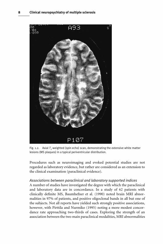
Fig. 1.2. Axial T2-weighted (spin echo) scan, demonstrating the extensive white matterlesions (MS plaques) in a typical periventricular distribution.
8 Clinical neuropsychiatry of multiple sclerosis
Procedures such as neuroimaging and evoked potential studies are notregarded as laboratory evidence, but rather are considered as an extension tothe clinical examination (paraclinical evidence).
Associations between paraclinical and laboratory supported indicesA number of studies have investigated the degree with which the paraclinicaland laboratory data are in concordance. In a study of 62 patients withclinically deWnite MS, Baumhefner et al. (1990) noted brain MRI abnor-malities in 97% of patients, and positive oligoclonal bands in all but one ofthe subjects. Not all reports have yielded such strongly positive associations,however, with Pirttila and Nurmiko (1995) noting a more modest concor-dance rate approaching two-thirds of cases. Exploring the strength of anassociation between the two main paraclinical modalities, MRI abnormalities
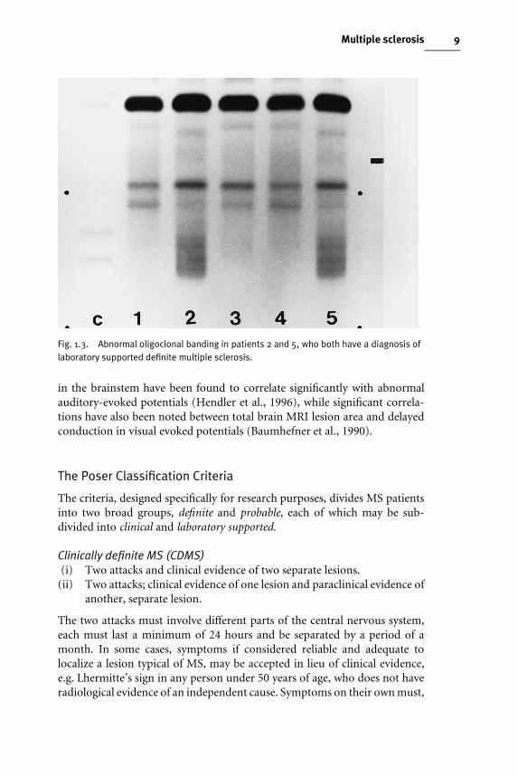
Fig. 1.3. Abnormal oligoclonal banding in patients 2 and 5, who both have a diagnosis oflaboratory supported definite multiple sclerosis.
9Multiple sclerosis
in the brainstem have been found to correlate signiWcantly with abnormalauditory-evoked potentials (Hendler et al., 1996), while signiWcant correla-tions have also been noted between total brain MRI lesion area and delayedconduction in visual evoked potentials (Baumhefner et al., 1990).
The Poser Classification Criteria
The criteria, designed speciWcally for research purposes, divides MS patientsinto two broad groups, deWnite and probable, each of which may be sub-divided into clinical and laboratory supported.
Clinically definite MS (CDMS)(i) Two attacks and clinical evidence of two separate lesions.
(ii) Two attacks; clinical evidence of one lesion and paraclinical evidence ofanother, separate lesion.
The two attacks must involve diVerent parts of the central nervous system,each must last a minimum of 24 hours and be separated by a period of amonth. In some cases, symptoms if considered reliable and adequate tolocalize a lesion typical of MS, may be accepted in lieu of clinical evidence,e.g. Lhermitte’s sign in any person under 50 years of age, who does not haveradiological evidence of an independent cause. Symptoms on their own must,
10 Clinical neuropsychiatry of multiple sclerosis
however, only be considered with extreme caution and, if possible, cor-roboration from friend or relative should be sought if the attack was notrecorded by a physician.
Paraclinical evidence that aids in diagnosis includes CT and MRI, evokedpotentials, hyperthermia challenge and specialized urological studies. Of noteis the recommendation that neuropsychological evidence of impaired cog-nition in someone under 50 years, although suggestive of MS, was not speciWcenough to be considered diagnostic. This recommendation, which was madein 1983, predated the plethora of studies from later in the decade thatunequivocally demonstrated the presence of clinically signiWcant cognitivedysfunction in approximately 40% of community-based MS patients (Rao etal., 1991a: McIntosh-Michaelis et al., 1991). To date, however, impairedcognition is still not one of the acceptable paraclinical signs.
Laboratory-supported definite MS (LSDMS)Laboratory support comes from increased IgG in the CSF, with normal levelsin the serum or oligoclonal bands in the CSF, but not in the serum.
(i) Two attacks; either clinical or paraclinical evidence of one lesion andCSF IgG or oligoclonal bands.
(ii) One attack; clinical evidence of two separate lesions; and CSF IgG oroligoclonal bands.
(iii) One attack; clinical evidence of one lesion and paraclinical evidence ofanother separate lesion; CSF IgG or oligoclonal bands.
The two attacks must involve diVerent parts of the CNS, each must last 24hours and be separated by a month. One of the episodes must involve a partof the CNS distinct from that demonstrated by the clinical or paraclinicalevidence. Unlike CDMS, historical information cannot be substituted forclinical evidence. Whether the evidence is clinical or paraclinical, both lesionsmust not have been present at the time of the Wrst examination and must beseparated by at least a month. This time factor is to reduce the possibility ofincluding a case of acute disseminated encephalomyelitis.
In patients with progressive MS from symptom onset, clinical or parac-linical evidence of the second lesion should not have been present at the timeof symptom onset. If the second lesion was present, the patient can only bedeemed to have had MS once symptom progression had taken place for 6months.
Clinically probable multiple sclerosis (CPMS)(i) Two attacks and clinical evidence of one lesion.
(ii) One attack and clinical evidence of two separate lesions.(iii) One attack; clinical evidence of one lesion and paraclinical evidence of
another separate, lesion.
11Multiple sclerosis
The two attacks must involve separate parts of the CNS. Historical infor-mation cannot replace clinical evidence, and the restrictions discussed underlaboratory supported deWnite multiple sclerosis also apply.
Laboratory supported probable multiple sclerosis (LSPMS)(i) Two attacks and CSF IgG or oligoclonal bands.
The two attacks must involve diVerent parts of the CNS, must be separated bya minimum of a month and each must have lasted 24 hours.
In summary, the Poser committee acknowledge that there will always bepatients who defy easy categorization. The experienced neurologist will haveto rely on intuition and accumulated clinical skill in arriving at diagnoses forthis group. The criteria as outlined above are primarily for research purposes.Furthermore, there is a recommendation that clinical trials and researchprotocols should be limited to patients in one of the two deWnite groups. Thecategory of probable was designed for the purpose of prospectively evaluatingnew diagnostic methods.
Clinically isolated lesions
Patients with clinically isolated lesions (CIL) are of particular interest as theyare frequently the forerunners of MS. In attempting to describe the naturalhistory of psychiatric and cognitive abnormalities in MS, the study of suchpatients aVords a valuable opportunity to document the earliest evidence ofdysfunction before patients progress to the full syndrome. Throughout thebook, reference will be made to patients with CIL and a brief description ofthese conditions is therefore given.
Optic neuritis
Acute unilateral optic neuritis (ON) in adults is the presenting feature of MSin 20% of cases, over three-quarters of patients going on to develop MS(Francis et al., 1987). It is characterized by the rapid development of visualloss, usually accompanied by pain with symptoms progressing for 3–4 weeksand then resolving over 2–3 months, recovery to 6/9 vision occurring ingreater than 90% of patients (McDonald, 1983). MRI with contrast enhan-cement may reveal lesions within the optic nerves (Fig. 1.4). In addition, 60%of adults presenting with clinically isolated optic neuritis display one or moreasymptomatic white matter brain lesions on MRI which appear indistin-guishable from those seen in MS (Ormerod et al., 1987). The presence ofthese lesions is associated with a high risk of progression to clinically deWniteMS within 5 years (Miller et al., 1992), but MS should still not be diagnosed at
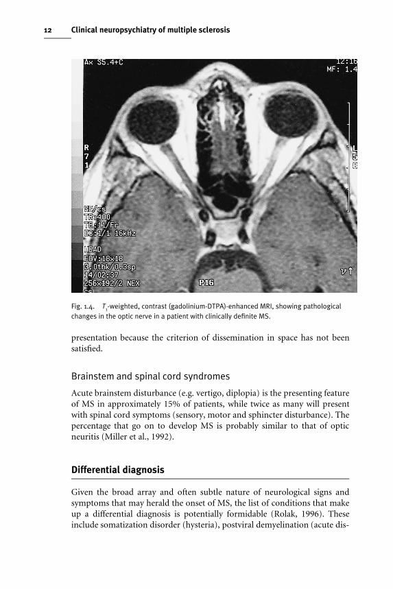
Fig. 1.4. T1-weighted, contrast (gadolinium-DTPA)-enhanced MRI, showing pathologicalchanges in the optic nerve in a patient with clinically definite MS.
12 Clinical neuropsychiatry of multiple sclerosis
presentation because the criterion of dissemination in space has not beensatisWed.
Brainstem and spinal cord syndromes
Acute brainstem disturbance (e.g. vertigo, diplopia) is the presenting featureof MS in approximately 15% of patients, while twice as many will presentwith spinal cord symptoms (sensory, motor and sphincter disturbance). Thepercentage that go on to develop MS is probably similar to that of opticneuritis (Miller et al., 1992).
Differential diagnosis
Given the broad array and often subtle nature of neurological signs andsymptoms that may herald the onset of MS, the list of conditions that makeup a diVerential diagnosis is potentially formidable (Rolak, 1996). Theseinclude somatization disorder (hysteria), postviral demyelination (acute dis-

13Multiple sclerosis
seminated encephalomyelitis), vasculitis aVecting the CNS (either primary orsecondary conditions such as lupus erythematosus), retroviral infectionssuch as acquired immune deWciency syndrome (AIDS), cerebrovascularaccidents (stroke), metachromatic leukodystrophy and tumours (metastases,lymphoma).
To the neuropsychiatrist, dealing primarily with the behavioural sequelaeof MS, the somatizing patient masquerading with MS-like symptoms canpresent a considerable therapeutic challenge (Aring, 1965). A follow-up of400 patients, referred to neurologists and subsequently found not to haveMS, revealed 14 with primarily psychiatric problems (Murray and Murray,1984). These patients were more likely to be female, hospital employees orhave a friend with MS and suVer from anxiety, depression and somatizationdisorder, the latter formerly called hysteria. Conversely, there are patientswith MS, who may be incorrectly dismissed as ‘hysterical’. Skegg et al. (1988)were able to identify 91 patients with MS (a point prevalence of 0.08%), ofwhom 16% had been referred to a psychiatrist between the onset of neur-ological symptoms and the diagnosis of MS. Although neurological symp-toms were present at the time in the majority of patients, these had beenoverlooked by the psychiatrist in all but two cases. Instead, patients weregiven diagnoses, such as hysterical personality disorder or conversion disor-der.
The clinical course of multiple sclerosis
In describing the clinical course of MS, diYculties have also been presentwith respect to terminology (Whitaker et al., 1995), the situation provinganalogous to the imprecision that surrounded the diagnosis of MS and thedeWnition of terms such as relapse, remission, etc. While there is generalrecognition that the course of MS shows individual variability, and thatphysical disability usually follows either a relapsing–remitting or steadilyprogressive course, what is meant by these terms has demanded clariWcation.A tightening up of terminology is not only important from a researchperspective, where clear deWnitions of patient subgroups are essential forvalid data interpretation, but also for correctly assigning patients to par-ticular treatments. The question of which patients would beneWt from whichtreatments is one of crucial importance to physicians looking for clearguidelines in their clinical practice.
DiVerences amongst researchers and clinicians in deWning terms thatdescribe the course and severity of MS have stemmed from a reliance onverbal descriptors as opposed to biological markers. This recognition led toan international survey of MS researchers, with the aim of assessing ag-reement pertaining to the various descriptive terms currently in use (Lublin
14 Clinical neuropsychiatry of multiple sclerosis
and Reingold, 1996). The survey supplied deWnitions of the following diseasecourses and types: relapsing–remitting (RR), relapsing–progressive (RP),primary progressive (PP), secondary progressive (SP), benign and malignant.DeWnitions of each of these terms were included in the survey, but space wasalso made available for researchers to provide their own deWnitions if theydisagreed with those enclosed. Of the 215 surveys mailed out, 125 (58%) werereturned. The results led to the National Multiple Sclerosis Society (USA)providing a set of consensus deWnitions, which are given below.
Clinical course definitions
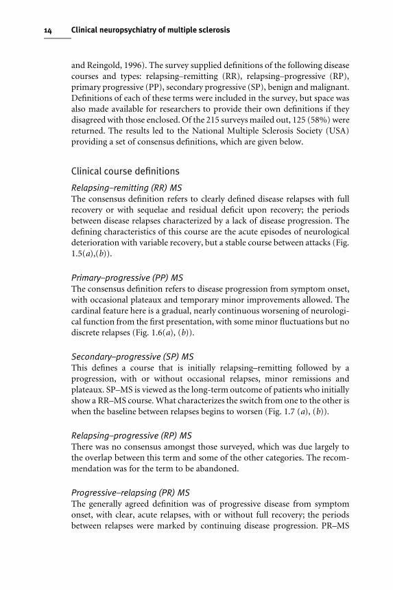
Relapsing–remitting (RR) MSThe consensus deWnition refers to clearly deWned disease relapses with fullrecovery or with sequelae and residual deWcit upon recovery; the periodsbetween disease relapses characterized by a lack of disease progression. ThedeWning characteristics of this course are the acute episodes of neurologicaldeterioration with variable recovery, but a stable course between attacks (Fig.1.5(a),(b)).
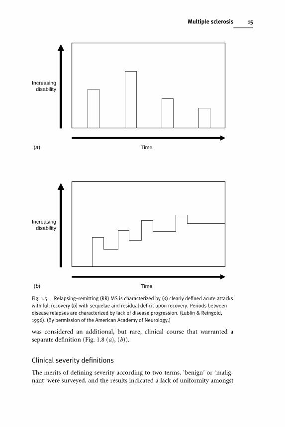
Primary–progressive (PP) MSThe consensus deWnition refers to disease progression from symptom onset,with occasional plateaux and temporary minor improvements allowed. Thecardinal feature here is a gradual, nearly continuous worsening of neurologi-cal function from the Wrst presentation, with some minor Xuctuations but nodiscrete relapses (Fig. 1.6(a), (b)).
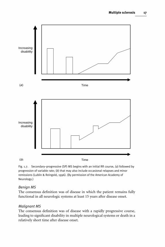
Secondary–progressive (SP) MSThis deWnes a course that is initially relapsing–remitting followed by aprogression, with or without occasional relapses, minor remissions andplateaux. SP–MS is viewed as the long-term outcome of patients who initiallyshow a RR–MS course. What characterizes the switch from one to the other iswhen the baseline between relapses begins to worsen (Fig. 1.7 (a), (b)).
Relapsing–progressive (RP) MSThere was no consensus amongst those surveyed, which was due largely tothe overlap between this term and some of the other categories. The recom-mendation was for the term to be abandoned.
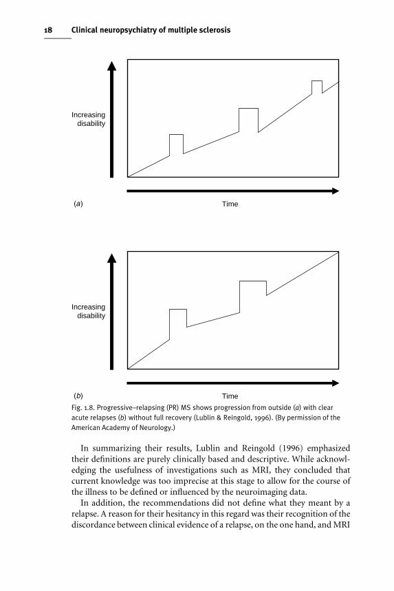
Progressive–relapsing (PR) MSThe generally agreed deWnition was of progressive disease from symptomonset, with clear, acute relapses, with or without full recovery; the periodsbetween relapses were marked by continuing disease progression. PR–MS
Time
Increasingdisability
Time
Increasingdisability
Fig. 1.5. Relapsing–remitting (RR) MS is characterized by (a) clearly defined acute attackswith full recovery (b) with sequelae and residual deficit upon recovery. Periods betweendisease relapses are characterized by lack of disease progression. (Lublin & Reingold,1996). (By permission of the American Academy of Neurology.)
15Multiple sclerosis
was considered an additional, but rare, clinical course that warranted aseparate deWnition (Fig. 1.8 (a), (b)).
Clinical severity definitions
The merits of deWning severity according to two terms, ‘benign’ or ‘malig-nant’ were surveyed, and the results indicated a lack of uniformity amongst
Time
Increasingdisability
Time
Increasingdisability
Fig. 1.6. Primary–progressive (PP) MS is characterized by disease showing progression ofdisability from outset (a) without plateaux or remissions or (b) with occasional plateauxand temporary minor improvements (Lublin & Reingold, 1996). (By permission of theAmerican Academy of Neurology.)
16 Clinical neuropsychiatry of multiple sclerosis
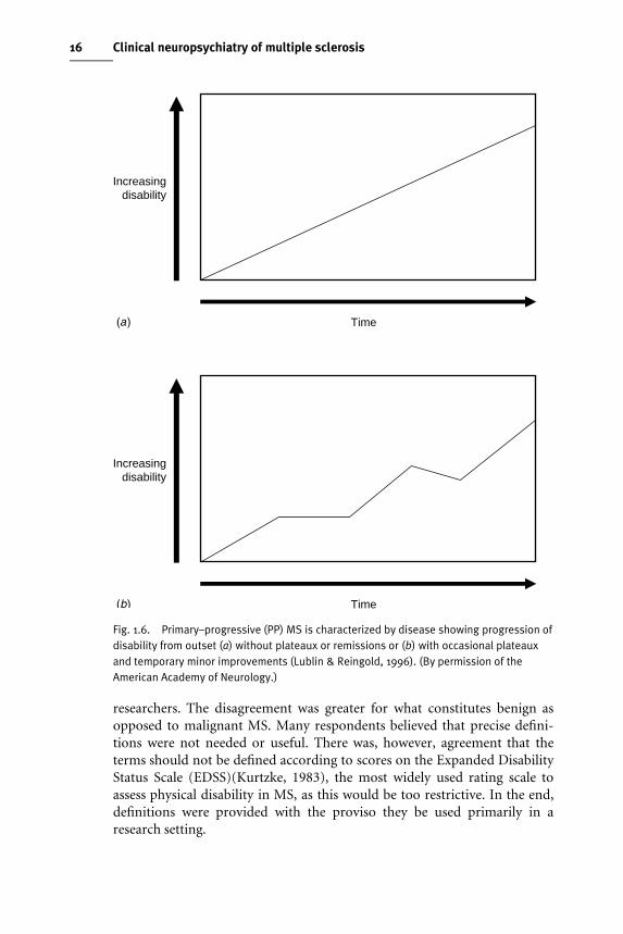
researchers. The disagreement was greater for what constitutes benign asopposed to malignant MS. Many respondents believed that precise deWni-tions were not needed or useful. There was, however, agreement that theterms should not be deWned according to scores on the Expanded DisabilityStatus Scale (EDSS)(Kurtzke, 1983), the most widely used rating scale toassess physical disability in MS, as this would be too restrictive. In the end,deWnitions were provided with the proviso they be used primarily in aresearch setting.
Time
Increasingdisability
Time
Increasingdisability
Fig. 1.7. Secondary–progressive (SP) MS begins with an initial RR course, (a) followed byprogression of variable rate; (b) that may also include occasional relapses and minorremissions (Lublin & Reingold, 1996). (By permission of the American Academy ofNeurology.)
17Multiple sclerosis
Benign MSThe consensus deWnition was of disease in which the patient remains fullyfunctional in all neurologic systems at least 15 years after disease onset.
Malignant MSThe consensus deWnition was of disease with a rapidly progressive course,leading to signiWcant disability in multiple neurological systems or death in arelatively short time after disease onset.
Time
Increasingdisability
Time
Increasingdisability
Fig. 1.8. Progressive–relapsing (PR) MS shows progression from outside (a) with clearacute relapses (b) without full recovery (Lublin & Reingold, 1996). (By permission of theAmerican Academy of Neurology.)
18 Clinical neuropsychiatry of multiple sclerosis
In summarizing their results, Lublin and Reingold (1996) emphasizedtheir deWnitions are purely clinically based and descriptive. While acknowl-edging the usefulness of investigations such as MRI, they concluded thatcurrent knowledge was too imprecise at this stage to allow for the course ofthe illness to be deWned or inXuenced by the neuroimaging data.
In addition, the recommendations did not deWne what they meant by arelapse. A reason for their hesitancy in this regard was their recognition of thediscordance between clinical evidence of a relapse, on the one hand, and MRI
19Multiple sclerosis
and neuropathological signs of relapse on the other. Nevertheless, the term isused repeatedly throughout their deWnitions, and they therefore advise that,for the purpose of a clinical trial, what is meant by relapse will need to bedeWned by consensus amongst investigators as part of the protocol. This viewrepresents a clear departure from the clinical guidelines laid out by Poser etal. (1983) and illustrates a recognition that procedures such as MRI have,over the intervening 15 years, reached a level of sophistication suYcient toinXuence how researchers view the dynamic nature of the MS lesion.
Welcome as these guidelines are, the diYculty is assigning disease course topatients relates, in part, to the changes in neurological state that occur withtime. Goodkin et al. (1989) prospectively followed a group of 254 MSpatients over a 1 to 5-year-period (mean 2.6 years). They reported thatadherence to the initial assigned disease course varied considerably. Thus,30% of patients with chronic–progressive disease had become stable, 32%with stable disease had become chronic–progressive, 20% of relapsing–remitting patients had stabilized, while a similar percentage had deterioratedto a chronic–progressive phase. Furthermore, patients with either stable orrelapsing–remitting (44%) disease switched as frequently to a chronic–progressive phase as patients with the latter reverted to a stable or relapsing–remitting state. The former would now be called secondary progressivedisease, but the study was completed before the subdivisions of primary andsecondary entered the lexicon. The implications of this study are considerablefor, given the dynamic nature of the disease process, they beg the question ofhow valid is the assignment of disease course? Patients who qualify forinterferon beta-1b therapy by virtue of having relapsing–remitting MS, mayin fact have had a secondary–progressive course a few months back. Are thesepatients any diVerent from those relapsing–remitting patients who have notshown a similar transformation? If so, what are the implications for treat-ment? The answer to these conundrums are not yet known. There is, how-ever, an awareness that the disease is seldom static. Clearly deWned deWnitionsthat carry broad agreement will ensure that if, and when, change occurs,those treating and researching multiple sclerosis patients continue to speakthe same language.
Rating neurological impairment in multiple sclerosis
The yardstick by which neurological disability is rated in MS patients is theExpanded Disability Status Scale (EDSS)(Kurtzke, 1983). The scale, routinelyused in clinical and research settings, represents a reWnement of earliermethods devised to assess physical disability in MS (Kurtzke, 1955; Kurtzke,1970). The scale consists of eight ‘functional systems (FS)’, namely pyra-midal, cerebellar, brainstem, sensory, bowel and bladder, visual, cerebral (ormental) and a miscellaneous category termed ‘other’. Each of these

Table 1.1. Expanded Disability Status Scale (EDSS)
0 Normal neurologic exam (all grade 0 in functional systems (FS). Cerebralgrade 1 acceptable.
1.0 No disability, minimal signs in one FS (i.e. grade 1 excluding cerebral grade1)
1.5 No disability. minimal signs in more than one FS (more than one grade 1excluding cerebral grade 1)
2.0 Minimal disability in one FS (one FS grade 2, others 0 or 1)2.5 Minimal disability in two FS (two FS grade 2, others 0 or 1)3.0 Moderate disability in one FS (one FS grade 3, others 0 or 1), or mild
disability in three or four FS (three/four FS grade 2, others 0 or 1) thoughfully ambulatory
3.5 Fully ambulatory, but with moderate disability in one FS (one grade 3) andone or two FS grade 2; or two FS grade 3; or Wve FS grade 2 (others 0 or 1)
4.0 Fully ambulatory without aid, self-suYcient, up and about some 12 hours aday despite relatively severe disability consisting of one FS grade 4 (others 0or 1), or combinations of lesser grades exceeding limits of previous steps.Able to walk without aid or rest some 500 metres.
4.5 Fully ambulatory without aid, up and about much of the day, able to worka full day, may otherwise have some limitation of full activity or requireminimal assistance; characterized by relatively severe disability, usuallyconsisting of one FS grade 4 (others 0 or 1) or combinations of lesser gradesexceeding limits of previous steps. Able to walk without aid or rest for some300 metres.
5.0 Ambulatory without aid or rest for about 200 metres; disability severeenough to impair full daily activities (eg. to work full day without specialprovisions). (Usual FS equivalents are one grade 5 alone, others 0 or 1; orcombinations of lesser grades usually exceeding speciWcations for step 4.0.)
5.5 Ambulatory without aid or rest for about 100 metres; disability severeenough to preclude full daily activities. (Usual FS equivalents are one grade5 alone, others 0 or 1; or combinations of lesser grades usually exceedingthose for step 4.0.)
6.0 Intermittent or unilateral constant assistance (cane, crutch or brace)required to walk about 100 metres with or without resting. (Usual FSequivalents are combinations with more than two FS grade 3+.)
6.5 Constant bilateral assistance (canes, crutches or braces) required to walkabout 20 metres without resting. (Usual FS equivalents are combinationswith more than two FS grade 3+.)
7.0 Unable to walk beyond 5 metres even with aid, essentially restricted towheelchair; wheels self in standard wheelchair and transfers alone; up andabout in wheelchair some 12 hours a day. (Usual FS equivalents arecombinations with more than one FS grade 4+; very rarely, pyramidal grade5 alone.)
7.5 Unable to take more than a few steps; restricted to wheelchair; may need aidin transfer; wheels self, but cannot carry on in standard wheelchair a fullday; may require motorized wheelchair. (Usual FS equivalents arecombinations with more than one FS grade 4+.)
20 Clinical neuropsychiatry of multiple sclerosis