Embed Size (px)
Citation preview
ACR Appropriateness Criteria® Multiple Brain Metastases
EVIDENCE TABLE
* See Last Page for Key 2014 Review Videtic Page 1
Reference Study Type Patients/ Events
Study Objective (Purpose of Study) Study Results Study
Quality 1. Posner JB. Management of brain
metastases. Rev Neurol (Paris). 1992;148(6-7):477-487.
Review/Other-Tx
N/A To outline the modalities available for treating CNS metastases and the results one can expect.
Patients with known cancer and neurological symptoms should all undergo appropriate diagnostic tests which include either CT scan or magnetic resonance imaging and, if a lesion is found and a definitive diagnosis cannot be established, biopsy. Single or solitary brain metastases in patients with good systemic performance status should be strongly considered for surgical extirpation which will both make the diagnosis and deliver definitive treatment to the lesion. Patients with poor systemic performance status and/or multiple brain metastases are candidates for WBRT. WBRT is also indicated in patients after successful surgical extirpation of a single metastasis. The role of focal RT and chemotherapy in the treatment of brain metastases is still being evaluated. Preliminary evidence suggests that focal RT is probably useful for the treatment of relapsed metastases and that chemotherapy may be useful in the primary treatment of small or asymptomatic brain metastases.
4
2. Wen PY, Black PM, Loeffler JS. Metastatic brain cancer. In: DeVita VT, Hellman S, Rosenberg SA, eds. Cancer, principles & practice of oncology. 6th ed. Philadelphia, PA: Lippincott Williams & Wilkins; 2001:2655-2670.
Review/Other-Tx
N/A Book chapter. Book chapter. 4
3. Lassman AB, DeAngelis LM. Brain metastases. Neurol Clin. 2003;21(1):1-23, vii.
Review/Other-Tx
N/A To review the epidemiology, clinical features, treatment, and prognosis of brain metastases from system malignancies.
No results stated in abstract. 4
ACR Appropriateness Criteria® Multiple Brain Metastases
EVIDENCE TABLE
* See Last Page for Key 2014 Review Videtic Page 2
Reference Study Type Patients/ Events
Study Objective (Purpose of Study) Study Results Study
Quality 4. Nussbaum ES, Djalilian HR, Cho KH,
Hall WA. Brain metastases. Histology, multiplicity, surgery, and survival. Cancer. 1996;78(8):1781-1788.
Review/Other-Tx
729 patients To evaluate the disease patterns and survival in patients with metastatic brain tumors with special emphasis on the differences between specific tumor types and the impact of surgical intervention.
Primary tumor histologic type in order of descending frequency included NSCLC, breast carcinoma, SCLC, malignant melanoma, renal cell carcinoma, gastrointestinal carcinoma, uterine/vulvar carcinoma, and unknown primary carcinoma. There were 384 patients (53%) with a single brain metastasis, which was encountered most commonly in patients with prostate carcinoma and least often in patients with SCLC. Multiple metastases were present in 345 patients (47%). The median duration from diagnosis to presentation with a brain metastasis was 12 months, ranging from 3 months for patients with NSCLC to 53 months for patients with breast carcinoma. The median duration from presentation with brain metastases to death was 4 months, ranging from 3 months for patients with SCLC to 13 months for patients with prostate carcinoma. Median survival from presentation with brain metastases to death was 5 months for patients with single lesions and 3 months for patients with multifocal disease (P=0.0001). Median survival for patients with a single lesion was 11 months with surgery and 3 months without surgery (P=0.0001). Surgery did not significantly influence survival in patients with multiple metastases.
4
5. Delattre JY, Krol G, Thaler HT, Posner JB. Distribution of brain metastases. Arch Neurol. 1988;45(7):741-744.
Review/Other-Tx
134 patients To identify the number and site of brain metastases on CT.
The charts of patients with brain metastases from a primary tumor originating outside the lung revealed that the incidence of lung and spine metastases was the same, whether the primary tumor was pelvic or gastrointestinal or from another site. These data suggest that the high incidence of subtentorial lesions in patients with pelvic and gastrointestinal primary tumors cannot be explained by arterial embolization alone, and that this peculiar distribution is probably not explained by seeding of the brain through Batson's plexus.
4
ACR Appropriateness Criteria® Multiple Brain Metastases
EVIDENCE TABLE
* See Last Page for Key 2014 Review Videtic Page 3
Reference Study Type Patients/ Events
Study Objective (Purpose of Study) Study Results Study
Quality 6. Posner JB, Chernik NL. Intracranial
metastases from systemic cancer. Adv Neurol. 1978;19:579-592.
Review/Other-Tx
3,219 autopsies
An epidemiologic study to assess the nature of CNS metastases in patients.
No results stated in abstract. 4
7. Barani IJ, Larson DA, Berger MS. Future directions in treatment of brain metastases. Surg Neurol Int. 2013;4(Suppl 4):S220-230.
Review/Other-Tx
729 patients To review current state of brain metastases management and discusses various treatment controversies with focus on future clinical trials.
The published data demonstrate continued evolution of clinical trials and management strategies designed to minimize and/or prevent cognitive decline following RT management of brain metastases. Hippocampal avoidance WBRT and radiosurgery treatments for multiple brain metastases are discussed along with preliminary results of RTOG 0614, a trial of memantine therapy to prevent cognitive decline following WBRT. Trial results appear to support the use of memantine for prevention of cognitive decline.
4
8. Medical Research Council. Clinical Trials Unit. Does radiotherapy improve patient’s quality of life when lung cancer has spread to the brain?. December 16, 2013. Available from: http://www.ctu.mrc.ac.uk/research_areas/study_details.aspx?s=27. Identifier: ISRCTN3826061.
Review/Other-Tx
Ongoing A randomized controlled trial that aims to find whether giving brain RT adds anything to steroids and supportive care in patients with lung cancer.
This trial is still recruiting study subjects and results are not available yet.
4
9. Gaspar L, Scott C, Rotman M, et al. Recursive partitioning analysis (RPA) of prognostic factors in three Radiation Therapy Oncology Group (RTOG) brain metastases trials. Int J Radiat Oncol Biol Phys. 1997;37(4):745-751.
Review/Other-Tx
1,200 patients
To analyze the relative contributions of pretreatment variables to the survival of patients with brain metastases using an interactive, nonparametric statistical technique known as RPA; to define the influence of treatment variations on survival among patients enrolled on three consecutive RTOG randomized trials and to identify patient subgroups or stages.
According to the RPA tree the best survival (median: 7.1 months) was observed in patients <65 years of age with a KPS of at least 70, and a controlled primary tumor with the brain the only site of metastases. The worst survival (median: 2.3 months) was seen in patients with a KPS >70. All other patients had relatively minor differences in observed survival, with a median of 4.2 months.
4
10. Sperduto PW, Berkey B, Gaspar LE, Mehta M, Curran W. A new prognostic index and comparison to three other indices for patients with brain metastases: an analysis of 1,960 patients in the RTOG database. Int J Radiat Oncol Biol Phys. 2008;70(2):510-514.
Review/Other-Tx
1,960 patients
To introduce a new prognostic index for patients with brain metastases and compare it with 3 published indices.
RPA and the new GPA had the most statistically significant differences between classes (P<0.001 for all classes).
4
ACR Appropriateness Criteria® Multiple Brain Metastases
EVIDENCE TABLE
* See Last Page for Key 2014 Review Videtic Page 4
Reference Study Type Patients/ Events
Study Objective (Purpose of Study) Study Results Study
Quality 11. Sperduto PW, Chao ST, Sneed PK, et al.
Diagnosis-specific prognostic factors, indexes, and treatment outcomes for patients with newly diagnosed brain metastases: a multi-institutional analysis of 4,259 patients. Int J Radiat Oncol Biol Phys. 2010;77(3):655-661.
Observational-Tx
5,067 patients
To identify significant diagnosis-specific prognostic factors and indexes (Diagnosis-Specific GPA).
The significant prognostic factors varied by diagnosis. For NSCLC and SCLC, the significant prognostic factors were KPS, age, presence of extracranial metastases, and number of brain metastases, confirming the original GPA for these diagnoses. For melanoma and renal cell cancer, the significant prognostic factors were KPS and the number of brain metastases. For breast and gastrointestinal cancer, the only significant prognostic factor was the KPS. Two new diagnosis-specific-GPA indexes were thus designed for breast/gastrointestinal cancer and melanoma/renal cell carcinoma. The median survival by GPA score, diagnosis, and treatment were determined.
2
12. Sperduto PW, Kased N, Roberge D, et al. Summary report on the graded prognostic assessment: an accurate and facile diagnosis-specific tool to estimate survival for patients with brain metastases. J Clin Oncol. 2012;30(4):419-425.
Observational-Tx
3,940 patients
To present the updated diagnosis-specific GPA indices in a single, unified, user-friendly report to allow ease of access and use by treating physicians.
Significant prognostic factors varied by diagnosis. For lung cancer, prognostic factors were KPS, age, presence of extracranial metastases, and number of brain metastases, confirming the original Lung-GPA. For melanoma and renal cell cancer, prognostic factors were KPS and the number of brain metastases. For breast cancer, prognostic factors were tumor subtype, KPS, and age. For gastrointestinal cancer, the only prognostic factor was the KPS. The median survival times by GPA score and diagnosis were determined.
2
13. Nieder C, Berberich W, Schnabel K. Tumor-related prognostic factors for remission of brain metastases after radiotherapy. Int J Radiat Oncol Biol Phys. 1997;39(1):25-30.
Observational-Tx
108 patients To study CT determined response to external beam RT as well as influence of tumor-related factors, especially of tumor volume, on remission and to evaluate whether particular subgroups of metastases are controlled by low-dose RT.
In univariate analysis local result was significantly influenced by each of the 4 parameters mentioned above. Complete remission was observed in 37% of metastases from small-cell carcinoma, 35% of those from breast cancer, 25% of those from squamous-cell carcinoma, and 14% of those from nonbreast adenocarcinoma. The rate was 52% for metastases <0.5 cm3 and 0% for those >10 cm3. In multivariate analysis, small volume and no necrosis were the most important prognostic factors for complete remission. PFS was influenced by best local result.
2
ACR Appropriateness Criteria® Multiple Brain Metastases
EVIDENCE TABLE
* See Last Page for Key 2014 Review Videtic Page 5
Reference Study Type Patients/ Events
Study Objective (Purpose of Study) Study Results Study
Quality 14. Swift PS, Phillips T, Martz K, et al. CT
characteristics of patients with brain metastases treated in RTOG study 79-16. Int J Radiat Oncol Biol Phys. 1993;25(2):209-214.
Observational-Dx
779 patients To present the analysis of these CT scans with the intent of identifying radiologic characteristics which would predict for potentially prolonged survival.
Complete pre- and post-treatment CT evaluation of the brain was performed in 779 of the 859 patients entered into RTOG protocol 7916, a phase III study of the role of misonidazole combined with RT in the treatment of brain metastases. Pretreatment scan findings of mass effect, midline shift, massive edema, central necrosis, location of sentinel lesion, and number of lesions were correlated with length of survival for all patients as well as for each treatment group. The only characteristics that showed a statistically significant difference in survival in the overall group were the presence of ≤3 lesions and the presence of a midline shift. The actual benefit in OS, however, was found to be only 3 weeks. The volume of the largest lesion prior to treatment did not correlate well with survival, nor did location of lesions. The time to response, number of responders and absolute decrease in number of lesions were similar for the 4 treatment arms. Patients who responded to cranial treatment had a significantly prolonged survival over those who did not respond. No CT characteristic evaluated in this study showed value as a clinically relevant prognosticator for patients with brain metastases for the overall group.
3
ACR Appropriateness Criteria® Multiple Brain Metastases
EVIDENCE TABLE
* See Last Page for Key 2014 Review Videtic Page 6
Reference Study Type Patients/ Events
Study Objective (Purpose of Study) Study Results Study
Quality 15. Li J, Bentzen SM, Renschler M, Mehta
MP. Relationship between neurocognitive function and quality of life after whole-brain radiotherapy in patients with brain metastasis. Int J Radiat Oncol Biol Phys. 2008;71(1):64-70.
Observational-Tx
208 patients To examine the relationship between neurocognitive function and quality of life in patients with brain metastases after WBRT.
At baseline, all neurocognitive function tests showed statistically significant correlations with activities of daily living, which became stronger at 4 months. A similar observation was made with Functional Assessment of Cancer Therapy-Brain-specific. Neurocognitive function scores from previous visits predicted activities of daily living (P<0.05 for seven of eight tests) or Functional Assessment of Cancer Therapy-Brain-specific. Scores on all 8 neurocognitive function tests deteriorated before activities of daily living decline (net lead time 9-153 days); and scores on 6 of 8 neurocognitive function tests deteriorated before Functional Assessment of Cancer Therapy-Brain-specific (net lead time 9-82 days).
2
16. Borgelt B, Gelber R, Larson M, Hendrickson F, Griffin T, Roth R. Ultra-rapid high dose irradiation schedules for the palliation of brain metastases: final results of the first two studies by the Radiation Therapy Oncology Group. Int J Radiat Oncol Biol Phys. 1981;7(12):1633-1638.
Experimental-Tx
26 patients, first study; 33 patients,
second study
To present the final results of the 2 optional ultra-rapid, high dose arms of the first 2 RTOG studies.
Comparisons were made with 143 control patients randomized by the same participating institutions to receive a more protracted course of irradiation (2000, 3000 or 4000 rad/l-4 weeks). Responses of patients receiving ultra-rapid treatment, as assessed by the percent who had improvement in neurologic function, was comparable to that of patients receiving the more protracted schedules. Promptness of neurologic function improvement, treatment morbidity and median survival were also comparable to those of patients receiving 2000 to 4000 rad. However, the duration of improvement, time to progression of neurologic status and rate of complete disappearance of neurologic symptoms were generally less for those patients who received 1000 or 1200 rad.
1
ACR Appropriateness Criteria® Multiple Brain Metastases
EVIDENCE TABLE
* See Last Page for Key 2014 Review Videtic Page 7
Reference Study Type Patients/ Events
Study Objective (Purpose of Study) Study Results Study
Quality 17. Chatani M, Matayoshi Y, Masaki N,
Inoue T. Radiation therapy for brain metastases from lung carcinoma. Prospective randomized trial according to the level of lactate dehydrogenase. Strahlenther Onkol. 1994;170(3):155-161.
Experimental-Tx
162 patients A prospective randomized trial to determine the best treatment schedule for RT on brain metastasis from lung carcinoma.
The final results showed the facts that: 1) the most important prognostic factor, according to Cox's multivariate analysis, was also the level of LDH in the second trial, 2) the incidence of acute side effects showed the trend toward depending upon a single dose, ie, group A (3 Gy/fraction); 35% vs group B (2.5 Gy/fraction); 21% (P=0.165) and group C (3 Gy/fraction); 23% vs group D (4 Gy/fraction); 45% (P=0.044), 3) median survival time and 1-year survival rates were 5.4 months and 21% in group A; 4.8 months and 17% in group B; 3.4 months and 6% in group C; and 2.4 months and 4% in group D, respectively, and survival curves showed no statistically significant difference between the 2 treatment groups in each LDH group, 4) improvement in neurologic function appeared to increase with total dosage escalation, ie, 41% in group A vs 45% in group B and 35% in group C vs 21% in group D (P=0.13).
1
18. Chatani M, Teshima T, Hata K, Inoue T. Prognostic factors in patients with brain metastases from lung carcinoma. Strahlenther Onkol. 1986;162(3):157-161.
Experimental-Tx
70 patients To investigate the effectiveness of different time-dose RT schemes (ie, 30 Gy/10 fractions/2 weeks vs 50 Gy/20 fractions/4 weeks) and the prognostic factors on the palliation for patients with brain metastases from lung carcinoma.
Patients were entered into 2 randomly allocated trials in order to investigate the effectiveness of different time-dose RT schemes and the prognostic factors on the palliation for patients with brain metastases from lung carcinoma. The most important factors for predicting poor prognosis in this series, which were shown by stepwise proportional hazard (Cox) model, were LDH and general performance status. In normal LDH group, the most important factors for predicting poor prognosis were multiplicity of brain metastases (P<0.001), treatment methods (P<0.0005) and age (P<0.0053). In high LDH group any items were not shown for meeting of the 0.05 significant level.
1
ACR Appropriateness Criteria® Multiple Brain Metastases
EVIDENCE TABLE
* See Last Page for Key 2014 Review Videtic Page 8
Reference Study Type Patients/ Events
Study Objective (Purpose of Study) Study Results Study
Quality 19. Haie-Meder C, Pellae-Cosset B,
Laplanche A, et al. Results of a randomized clinical trial comparing two radiation schedules in the palliative treatment of brain metastases. Radiother Oncol. 1993;26(2):111-116.
Experimental-Tx
216 patients To compare 2 schedules of brain irradiation in a randomized trial: one course of 18 Gy/3 fractions/3 days vs the same fractionation followed by a second course of RT with a 1-month time interval.
The second course was identical to the first one or delivered 25 Gy/10 fractions/14 days. No difference in OS, nor in the neurologic response or in the incidence of complications was demonstrated. Two clinical factors appeared to be prognostic of the OS: the presence of multiple brain metastases and the presence of extracerebral metastases.
1
20. Harwood AR, Simson WJ. Radiation therapy of cerebral metastases: a randomized prospective clinical trial. Int J Radiat Oncol Biol Phys. 1977;2(11-12):1091-1094.
Experimental-Tx
101 patients To compare the effectiveness of a single high-dose treatment to that of a fractionated regimen.
In the 101 patients analyzed, no statistical difference between the 2 fractionation schemes was demonstrated in terms of survival, frequency or degree of response to treatment, complication rate or local control rate. The results are compared to similar retrospective and prospective series, leading to the conclusion that a single dose of 1000 rad provided as good palliation as fractionated schedules.
1
21. Kurtz JM, Gelber R, Brady LW, Carella RJ, Cooper JS. The palliation of brain metastases in a favorable patient population: a randomized clinical trial by the Radiation Therapy Oncology Group. Int J Radiat Oncol Biol Phys. 1981;7(7):891-895.
Experimental-Tx
255 patients To present the results of a third RTOG clinical trial which investigates prospectively the use of a higher dose schedule compared with the “best standard treatment” in a highly selected, favorable group of patients with brain metastases.
The palliative effectiveness of a short, intensive course of brain irradiation (3000 rad in 2 weeks) was compared to that of a high-dose course (5000 rad in 4 weeks) in a randomized RTOG clinical trial. 80% of the 255 evaluable patients had lung primaries, 7% breast, and 13% other or unknown primaries. Patients with evidence of extra-cranial metastases, uncontrolled primaries, or Class IV Neurologic Function were excluded. 41% of Class II Neurologic Function and 71% of Class III Neurologic Function patients improved in neurologic function class. For Class II Neurologic Function patients, a significantly greater improvement rate was obtained with the short course than with the long course.
1
ACR Appropriateness Criteria® Multiple Brain Metastases
EVIDENCE TABLE
* See Last Page for Key 2014 Review Videtic Page 9
Reference Study Type Patients/ Events
Study Objective (Purpose of Study) Study Results Study
Quality 22. Murray KJ, Scott C, Greenberg HM, et al.
A randomized phase III study of accelerated hyperfractionation versus standard in patients with unresected brain metastases: a report of the Radiation Therapy Oncology Group (RTOG) 9104. Int J Radiat Oncol Biol Phys. 1997;39(3):571-574.
Experimental-Tx
445 patients To compare 1-year survival and acute toxicity rates between an accelerated hyperfractionated RT (1.6 Gy b.i.d.) to a total dose of 54.4 Gy vs an accelerated fractionation of 30 Gy in 10 daily fractions in patients with unresected brain metastasis.
The average age in both groups was 60 years; nearly two-thirds of all patients had lung primaries. Of the 429 eligible and analyzable patients, the median survival time was 4.5 months in both arms. The 1-year survival rate was 19% in the accelerated fractionation arm vs 16% in the accelerated hyperfractionated arm. No difference in median or 1-year survival was observed among patients with solitary metastasis between treatment arms. RPA classes have previously been identified and patients with a KPS of 70 or more, a controlled primary tumor, <65 years of age, and brain metastases only (RPA class I), had a 1-year survival of 35% in the accelerated fractionation arm vs 25% in the accelerated hyperfractionated arm (P=0.95). In a multivariate model, only age, KPS, extent of metastatic disease (intracranial metastases only vs intra- and extracranial metastases), and status of primary (controlled vs uncontrolled) were statistically significant (at P<0.05). Treatment assignment was not statistically significant. Overall Grade III or IV toxicity was equivalent in both arms, and one fatal toxicity at 44 days secondary to cerebral edema was seen in the accelerated hyperfractionated arm.
1
23. Davey P, Hoegler D, Ennis M, Smith J. A phase III study of accelerated versus conventional hypofractionated whole brain irradiation in patients of good performance status with brain metastases not suitable for surgical excision. Radiother Oncol. 2008;88(2):173-176.
Experimental-Tx
90 patients A phase III study was proposed to test the accelerated regimen described by Vecht and determine whether the improvement in survival in patients of good performance with brain metastases not suitable for resection could be replicated.
Both arms of the study were balanced by RPA class. The median survival was 19 weeks in both arms. Subset analysis showed time to retreatment for intracranial relapse was 14 weeks in the control arm and 32 weeks in the accelerated arm (P=0.03). Trends for more severe epilation and improved neurological function in the accelerated arm did not reach statistical significance. OS was associated with RPA class and colorectal pathology.
1
ACR Appropriateness Criteria® Multiple Brain Metastases
EVIDENCE TABLE
* See Last Page for Key 2014 Review Videtic Page 10
Reference Study Type Patients/ Events
Study Objective (Purpose of Study) Study Results Study
Quality 24. Borgelt B, Gelber R, Kramer S, et al. The
palliation of brain metastases: final results of the first two studies by the Radiation Therapy Oncology Group. Int J Radiat Oncol Biol Phys. 1980;6(1):1-9.
Review/Other-Tx
1,812 total patients in 2
studies
Two sequential phase III randomized RTOG studies to determine palliative effectiveness in patients with metastatic brain disease.
5 schedules of WBRT ranging from 4000 rad/4 weeks to 2000 rad/l week have been evaluated in 2 sequential phase III studies. Improvement in neurologic function status and maintenance of improved or stable neurologic function were utilized as measures of response. All treatment schedules were comparable with respect to frequency of improvement, duration of improvement, time to progression, survival and palliative index. Important prognosticators of response included initial neurologic function and general performance status. Administration of steroids during irradiation favored more rapid improvement; for neurologic-function-3 patients, it increased the overall frequency of improvement.
4
25. Graham PH, Bucci J, Browne L. Randomized comparison of whole brain radiotherapy, 20 Gy in four daily fractions versus 40 Gy in 20 twice-daily fractions, for brain metastases. Int J Radiat Oncol Biol Phys. 2010;77(3):648-654.
Experimental-Tx
113 patients To compare the intracranial control rate and quality of life for 2 radiation fractionation schemes for cerebral metastases.
The patient age range was 28-83 years (mean 62). Of the 113 patients, 41 had undergone surgical resection, and 74 patients had extracranial disease (31 concurrent and 43 stable). The median survival time was 6.1 months in Arm A and 6.6 months in Arm B, and the overall 5-year survival rate was 3.5%. Intracranial progression occurred in 44% of Arm A and 64% of Arm B patients (P=.03). Salvage surgery or RT was used in 4% of Arm A patients and 21% of Arm B patients (P=.004). Death was attributed to CNS progression in 32% of patients in Arm A and 52% of patients in Arm B (P=.03). The toxicity was minimal, with a minor increase in short-term cutaneous reactions in Arm A. The patients' quality of life was not impaired by the more intense treatment in Arm A.
1
ACR Appropriateness Criteria® Multiple Brain Metastases
EVIDENCE TABLE
* See Last Page for Key 2014 Review Videtic Page 11
Reference Study Type Patients/ Events
Study Objective (Purpose of Study) Study Results Study
Quality 26. Priestman TJ, Dunn J, Brada M, Rampling
R, Baker PG. Final results of the Royal College of Radiologists' trial comparing two different radiotherapy schedules in the treatment of cerebral metastases. Clin Oncol (R Coll Radiol). 1996;8(5):308-315.
Experimental-Tx
533 patients To compare 2 WBRT regimens (30 Gy/10 fractions/2 weeks vs 12 Gy/2 fractions on consecutive days) for the treatment of patients with symptomatic cerebral metastases.
Of these patients 533 were eligible for analysis: 270 assigned to the 2-fraction arm and 263 to the 10-fraction arm. The 2 groups were well balanced with respect to patient characteristics. Median survival was 77 days with 2 fractions (95% CI, 68–89) and 84 days for the longer schedule (95% CI, 67–102). Analysis of the survival curves showed a marginal advantage for 10 fractions (P=0.04). Performance status (P=0.0001), site of primary tumor (P=0.006), dose of dexamethasone (P=0.004), age (P=0.04) and randomization treatment (P=0.03) were independent factors associated with survival. The classification of patients into good or poor risk groups based on these factors, excluding treatment, showed highly significant differences in survival (P<0.0001). Predictive models suggested that any benefit attributable to the longer RT schedule was confined to those in a good prognostic group (these patients formed 22% of the study population). Radiation related side effects, other than alopecia, were seen in 12% of patients receiving 2 fractions and 8% of those given 10 fractions. The short survival of many patients hampered the assessment of response, but overall responses were seen in 39% of those given 2 fractions and 44% of patients receiving 10 fractions.
1
27. Millender L, Wara W. Pallative care. In: Hansen EK, Roach M, eds. Handbook of evidence-based radiation oncology. New York, NY: Springer; 2007:xi, 536 p.
Review/Other-Tx
N/A Book chapter. Book chapter. 4
ACR Appropriateness Criteria® Multiple Brain Metastases
EVIDENCE TABLE
* See Last Page for Key 2014 Review Videtic Page 12
Reference Study Type Patients/ Events
Study Objective (Purpose of Study) Study Results Study
Quality 28. Andrews DW, Scott CB, Sperduto PW, et
al. Whole brain radiation therapy with or without stereotactic radiosurgery boost for patients with one to three brain metastases: phase III results of the RTOG 9508 randomised trial. Lancet. 2004;363(9422):1665-1672.
Experimental-Tx
333 patients To assess whether SRS provided any therapeutic benefit in a randomized multi-institutional trial directed by the RTOG.
From January, 1996, to June, 2001, we enrolled 333 patients from 55 participating RTOG institutions – 167 were assigned WBRT and SRS and 164 were allocated WBRT alone. Univariate analysis showed that there was a survival advantage in the WBRT and SRS group for patients with a single brain metastasis (median survival time 6.5 vs 4.9 months, P=0.0393). Patients in the stereotactic surgery group were more likely to have a stable or improved KPS score at 6 months' follow-up than were patients allocated WBRT alone (43% vs 27%, respectively; P=0.03). By multivariate analysis, survival improved in patients with an RPA class 1 (P<0.0001) or a favorable histological status (P=0.0121).
1
29. DeAngelis LM, Delattre JY, Posner JB. Radiation-induced dementia in patients cured of brain metastases. Neurology. 1989;39(6):789-796.
Review/Other-Tx
12 patients To assess radiation-induced dementia in patients cured of brain metastases.
Within 5 to 36 months (median, 14) all patients developed progressive dementia, ataxia, and urinary incontinence causing severe disability in all and leading to death in 7. No patient had tumor recurrence when neurologic symptoms began. Cortical atrophy and hypodense white matter were identified by CT in all. Contrast-enhancing lesions were seen in 3 patients; 2 of the lesions yielded radionecrosis on biopsy. Autopsies on 2 patients revealed diffuse chronic edema of the hemispheric white matter in the absence of tumor recurrence. Corticosteroids and ventriculoperitoneal shunt offered significant but incomplete improvement in some patients. The total dose of WBRT was only 2,500 to 3,900 cGy, but daily fractions of 300 to 600 cGy were employed.
4
ACR Appropriateness Criteria® Multiple Brain Metastases
EVIDENCE TABLE
* See Last Page for Key 2014 Review Videtic Page 13
Reference Study Type Patients/ Events
Study Objective (Purpose of Study) Study Results Study
Quality 30. Regine WF, Scott C, Murray K, Curran
W. Neurocognitive outcome in brain metastases patients treated with accelerated-fractionation vs. accelerated-hyperfractionated radiotherapy: an analysis from Radiation Therapy Oncology Group Study 91-04. Int J Radiat Oncol Biol Phys. 2001;51(3):711-717.
Experimental-Tx
445 patients To evaluate neurocognitive outcome as measured by the MMSE among patients with unresectable brain metastases randomly assigned to accelerated fractionation vs accelerated hyperfractionated WBRT.
The median survival was 4.5 months for both arms. The average change in MMSE at 2 and 3 months was a drop of 1.4 and 1.1, respectively, in the accelerated fractionation arm as compared to a drop of 0.7 and 1.3, respectively, in the accelerated hyperfractionated arm (P=NS). Overall, 91 patients at 2 months and 23 patients at 3 months had both follow-up MMSE and CT/magnetic resonance imaging documentation of the status of their brain metastases. When an analysis was performed taking into account control of brain metastases, a significant effect on MMSE was observed with time and associated proportional increase in uncontrolled brain metastases. At 2 months, the average change in MMSE score was a drop of 0.6 for those whose brain metastases were radiologically controlled as compared to a drop of 1.9 for those with uncontrolled brain metastases (P=0.47). At 3 months, the average change in MMSE score was a drop of 0.5 for those whose brain metastases were radiologically controlled as compared to a drop of 6.3 for those with uncontrolled brain metastases (P=0.02).
1
ACR Appropriateness Criteria® Multiple Brain Metastases
EVIDENCE TABLE
* See Last Page for Key 2014 Review Videtic Page 14
Reference Study Type Patients/ Events
Study Objective (Purpose of Study) Study Results Study
Quality 31. Aoyama H, Shirato H, Tago M, et al.
Stereotactic radiosurgery plus whole-brain radiation therapy vs stereotactic radiosurgery alone for treatment of brain metastases: a randomized controlled trial. Jama. 2006;295(21):2483-2491.
Experimental-Tx
132 patients To determine if WBRT combined with SRS results in improvements in survival, brain tumor control, functional preservation rate, and frequency of neurologic death.
The median survival time and the 1-year actuarial survival rate were 7.5 months and 38.5% (95% CI, 26.7%–50.3%) in the WBRT + SRS group and 8.0 months and 28.4% (95% CI, 17.6%–39.2%) for SRS alone (P=.42). The 12-month brain tumor recurrence rate was 46.8% in the WBRT + SRS group and 76.4% for SRS alone group (P<.001). Salvage brain treatment was less frequently required in the WBRT + SRS group (n = 10) than with SRS alone (n = 29) (P<.001). Death was attributed to neurologic causes in 22.8% of patients in the WBRT + SRS group and in 19.3% of those treated with SRS alone (P=.64). There were no significant differences in systemic and neurologic functional preservation and toxic effects of radiation.
1
32. Mehta MP, Rodrigus P, Terhaard CH, et al. Survival and neurologic outcomes in a randomized trial of motexafin gadolinium and whole-brain radiation therapy in brain metastases. J Clin Oncol. 2003;21(13):2529-2536.
Experimental-Tx
401 patients A phase III randomized trial evaluated survival as well as neurologic and neurocognitive function in patients with brain metastases from solid tumors receiving WBRT with or without MGd.
401 (251 NSCLC) patients were enrolled. There was no significant difference by treatment arm in survival (median, 5.2 months for MGd vs 4.9 months for WBRT; P=.48) or time to neurologic progression (median, 9.5 months for MGd vs 8.3 months for WBRT; P=.95). Treatment with MGd improved time to neurologic progression in patients with lung cancer (median, not reached for MGd vs 7.4 months for WBRT; P=.048, unadjusted). By investigator, MGd improved time to neurologic progression in all patients (median, 4.3 months for MGd vs 3.8 months for WBRT; P=.018) and in lung cancer patients (median, 5.5 months for MGd vs 3.7 months for WBRT; P=.025). MGd improved neurocognitive function in lung cancer patients.
1
ACR Appropriateness Criteria® Multiple Brain Metastases
EVIDENCE TABLE
* See Last Page for Key 2014 Review Videtic Page 15
Reference Study Type Patients/ Events
Study Objective (Purpose of Study) Study Results Study
Quality 33. Meyers CA, Smith JA, Bezjak A, et al.
Neurocognitive function and progression in patients with brain metastases treated with whole-brain radiation and motexafin gadolinium: results of a randomized phase III trial. J Clin Oncol. 2004;22(1):157-165.
Experimental-Tx
401 patients To report the neurocognitive findings in a phase III randomized trial evaluating survival and neurologic and neurocognitive function in patients with brain metastases from solid tumors receiving WBRT with or without MGd.
401 patients were enrolled (251 with NSCLC, 75 with breast cancer, and 75 with other cancers); 90.5% patients had impairment of one or more neurocognitive tests at baseline. Neurocognitive test scores of memory, fine motor speed, executive function, and global neurocognitive impairment at baseline were correlated with brain tumor volume and predictive of survival. There was no statistically significant difference between treatment arms in time to neurocognitive progression. Patients with lung cancer (but not other types of cancer) who were treated with MGd tended to have improved memory and executive function (P=.062) and improved neurologic function as assessed by a blinded events review committee (P=.048).
1
34. Brown PD, Asher AL, Farace E. Adjuvant whole brain radiotherapy: strong emotions decide but rational studies are needed. Int J Radiat Oncol Biol Phys. 2008;70(5):1305-1309.
Review/Other-Tx
N/A A review to examine the effect of adjuvant WBRT on tumor control, OS, and cognitive function and the results of ongoing studies assessing the role of adjuvant WBRT.
For patients with limited disease and good performance status, treatment typically involves a combination of focal measures (eg, surgical resection or radiosurgery) for the radiographically apparent disease, followed by adjuvant WBRT to treat subclinical disease. Because of concerns regarding the toxicity of WBRT, especially neurocognitive deterioration, many have advocated withholding adjuvant WBRT. Recently published studies have shed more light on the efficacy of adjuvant WBRT and the neurocognitive effects of WBRT. However, the inclusion of neurocognitive and quality-of-life data in clinical trials are still required to better define the role of adjuvant WBRT. Currently, two Phase III trials are underway, one in Europe and one in North America that will determine the effect of adjuvant WBRT on patients' quality of life, neurocognitive function, and survival.
4
ACR Appropriateness Criteria® Multiple Brain Metastases
EVIDENCE TABLE
* See Last Page for Key 2014 Review Videtic Page 16
Reference Study Type Patients/ Events
Study Objective (Purpose of Study) Study Results Study
Quality 35. Chang EL, Wefel JS, Hess KR, et al.
Neurocognition in patients with brain metastases treated with radiosurgery or radiosurgery plus whole-brain irradiation: a randomised controlled trial. Lancet Oncol. 2009;10(11):1037-1044.
Experimental-Tx
58 patients To clarify whether elective WBRT should be given with SRS, or deferred and to propose that patients treated with SRS plus WBRT would have inferior neurocognitive function based on the Hopkins Verbal Learning Test–Revised compared with patients treated with SRS alone.
After 58 patients were recruited (n=30 in the SRS alone group, n=28 in the SRS plus WBRT group), the trial was stopped by the data monitoring committee according to early stopping rules on the basis that there was a high probability (96%) that patients randomly assigned to receive SRS plus WBRT were significantly more likely to show a decline in learning and memory function (mean posterior probability of decline 52%) at 4 months than patients assigned to receive SRS alone (mean posterior probability of decline 24%). At 4 months there were 4 deaths (13%) in the group that received SRS alone, and 8 deaths (29%) in the group that received SRS plus WBRT. 73% of patients in the SRS plus WBRT group were free from CNS recurrence at 1 year, compared with 27% of patients who received SRS alone (P=0.0003). In the SRS plus WBRT group, 1 case of grade 3 toxicity (seizures, motor neuropathy, depressed level of consciousness) was attributed to radiation treatment. In the group that received SRS, 1 case of grade 3 toxicity (aphasia) was attributed to radiation treatment. Two cases of grade 4 toxicity in the group that received SRS alone were diagnosed as radiation necrosis.
1
ACR Appropriateness Criteria® Multiple Brain Metastases
EVIDENCE TABLE
* See Last Page for Key 2014 Review Videtic Page 17
Reference Study Type Patients/ Events
Study Objective (Purpose of Study) Study Results Study
Quality 36. Brown PD, Pugh S, Laack NN, et al.
Memantine for the prevention of cognitive dysfunction in patients receiving whole-brain radiotherapy: a randomized, double-blind, placebo-controlled trial. Neuro Oncol. 2013;15(10):1429-1437.
Experimental-Tx
508 patients To determine the protective effects of memantine on cognitive function in patients receiving WBRT.
Of 554 patients who were accrued, 508 were eligible. Grade 3 or 4 toxicities and study compliance were similar in the 2 arms. There was less decline in delayed recall in the memantine arm at 24 weeks (P=.059), but the difference was not statistically significant, possibly because there were only 149 analyzable patients at 24 weeks, resulting in only 35% statistical power. The memantine arm had significantly longer time to cognitive decline (HR 0.78, 95% CI; 0.62-0.99, P=.01); the probability of cognitive function failure at 24 weeks was 53.8% in the memantine arm and 64.9% in the placebo arm. Superior results were seen in the memantine arm for executive function at 8 (P=.008) and 16 weeks (P=.0041) and for processing speed (P=.0137) and delayed recognition (P=.0149) at 24 weeks.
1
ACR Appropriateness Criteria® Multiple Brain Metastases
EVIDENCE TABLE
* See Last Page for Key 2014 Review Videtic Page 18
Reference Study Type Patients/ Events
Study Objective (Purpose of Study) Study Results Study
Quality 37. Gondi V, Mehta MP, Pugh S, et al.
Memory Preservation With Conformal Avoidance of the Hippocampus During Whole-Brain Radiation Therapy for Patients With Brain Metastases: Primary Endpoint Results of RTOG 0933. Int J Radiat Oncol Biol Phys. 2013;87(5):1186.
Experimental-Tx
100 patients A single-arm phase 2 study of hippocampal WBRT for brain metastases with a primary cognitive endpoint and pre-specified comparison to a historical control of WBRT without hippocampal avoidance.
76% of patients were RPA class II. Two treatment-related grade 3 adverse events were reported (fatigue, headache); no treatment-related grade 4-5 events were observed. Median survival was 6.8 months (95% CI; 4.8–10.9 months). 3 patients (4.5%) had progression in the hippocampal avoidance region, consistent with expected event-rate. 42 patients were analyzable at 4 months. Mean relative decline in Hopkins Verbal Learning Test-Delayed Recall from baseline to 4 months was 7.0% (95% CI: -4.7% to 18.7%), which was significant in comparison to the historical control (PZ.0003). Mean relative decline in Hopkins Verbal Learning Test-Recall and Hopkins Verbal Learning- Immediate Recognition from baseline to 4 months was 3.6% (95% CI: -2.9% to 10.1%) and 1.6% (95% CI: -2.8% to 6.0%), respectively. 29 patients were analyzable at 6 months with a mean relative decline in Hopkins Verbal Learning Test-Delayed Recall, Hopkins Verbal Learning Test-Recall and Hopkins Verbal Learning- Immediate Recognition from baseline to 6 months of 2.0% (95% CI: -9.2% to 13.1%), -3.0% (95% CI: -12.0% to 5.9%) and 0.7% (95% CI: -3.1% to 4.4%), respectively.
1
ACR Appropriateness Criteria® Multiple Brain Metastases
EVIDENCE TABLE
* See Last Page for Key 2014 Review Videtic Page 19
Reference Study Type Patients/ Events
Study Objective (Purpose of Study) Study Results Study
Quality 38. DeAngelis LM, Currie VE, Kim JH, et al.
The combined use of radiation therapy and lonidamine in the treatment of brain metastases. J Neurooncol. 1989;7(3):241-247.
Experimental-Tx
58 patients A randomized prospective study to determine whether the addition of lonidamine to WBRT can improve the response rate of patients with brain metastases.
All patients received 3000 cGy of WBRT. 58 patients were enrolled; 31 received lonidamine plus WBRT and 27 received WBRT alone. There was no significant difference in response rate or survival between the treatment groups. Lonidamine blood levels were measured in 30 of the 31 patients who received the drug, and were therapeutic (≥15 micrograms/ml) in 50%. Survival and response rate were unaffected by the presence or absence of a therapeutic lonidamine level. The most common side-effects of lonidamine were myalgia, testicular pain, anorexia, and ototoxicity; however, only 2 patients had to discontinue the drug because of intolerable myalgias. No serious organ toxicity or myelosuppression was observed.
1
39. Komarnicky LT, Phillips TL, Martz K, Asbell S, Isaacson S, Urtasun R. A randomized phase III protocol for the evaluation of misonidazole combined with radiation in the treatment of patients with brain metastases (RTOG-7916). Int J Radiat Oncol Biol Phys. 1991;20(1):53-58.
Experimental-Tx
859 patients To evaluate the role of misonidazole combined with RT in the treatment of brain metastasis.
Of the 779 cases, 773 are dead (99%). Median survival is 3.9 months, with 60% alive at 3 months, 35% at 6 months, and 15% at 1 year. Survival was evaluated by treatment arm, misonidazole status, and fractionation scheme; none showed any statistical significance. Favorable prognostic factors were assessed (age <60, KPS of 70-100, controlled primary and brain metastasis only) in each treatment arm and no difference was found. Brain metastasis was cause of death in 1/3, and 19%–33% of patients were retreated.
1
ACR Appropriateness Criteria® Multiple Brain Metastases
EVIDENCE TABLE
* See Last Page for Key 2014 Review Videtic Page 20
Reference Study Type Patients/ Events
Study Objective (Purpose of Study) Study Results Study
Quality 40. Phillips TL, Scott CB, Leibel SA, Rotman
M, Weigensberg IJ. Results of a randomized comparison of radiotherapy and bromodeoxyuridine with radiotherapy alone for brain metastases: report of RTOG trial 89-05. Int J Radiat Oncol Biol Phys. 1995;33(2):339-348.
Experimental-Tx
72 patients To determine if the addition of bromodeoxyuridine to RT prolongs survival when compared to RT alone in patients with brain metastases.
There was no significant difference between the 2 treatment arms (P=0.904). The study was open from October 1989 to March 1993 and accrued 72 patients. Only 1 patient in the RT only arm remains alive. The 2 treatment arms were balanced with respect to all stratification variables. Toxicity due to RT was similar in both arms. Bromodeoxyuridine caused significant Grade 4 and 5 hematologic and skin toxicity in 5 patients. Two patients died due to hematologic toxicity and 1 from a Stevens-Johnson type skin reaction. Phenytoin played a role in the skin reactions and ranitidine was associated with the hematologic deaths. Ranitidine was eliminated, bromodeoxyuridine was discontinued after any hematologic toxicity, and no further Grade 4 or 5 toxicities were seen. The median survival was 6.12 months in the RT group and 4.3 in the bromodeoxyuridine group (P=0.904). Patients with solitary brain metastases had significantly better survival (P=0.031).
1
ACR Appropriateness Criteria® Multiple Brain Metastases
EVIDENCE TABLE
* See Last Page for Key 2014 Review Videtic Page 21
Reference Study Type Patients/ Events
Study Objective (Purpose of Study) Study Results Study
Quality 41. Ushio Y, Arita N, Hayakawa T, et al.
Chemotherapy of brain metastases from lung carcinoma: a controlled randomized study. Neurosurgery. 1991;28(2):201-205.
Experimental-Tx
100 patients To evaluate the effects of chemotherapy in patients with brain metastases from lung carcinoma.
Patients were randomly divided into 3 groups at the time of diagnosis or after surgery for metastases. Group A received RT alone; Group B received RT and chloroethylnitrosoureas (methyl-CCNU, 100-120 mg/m2, or ACNU 80-100 mg/m2, every 6-8 weeks), and Group C received RT and a combination of chloroethylnitrosoureas and tegafur (300 mg/m2, daily). Of the 100 patients, 88 could be evaluated. The reduction rates of the tumors of the patients in whom tumor was not surgically removed or not totally removed were compared. Complete resolution of the tumor was noted in 29%, 69%, and 63% of the patients in Groups A, B, and C, respectively. Tumor regression of ≥50% was seen in 36%, 69%, and 74% of the patients in Groups A, B, and C, respectively. The difference in the response rates of Groups A and C was statistically significant (P<0.05). Median survival after the start of treatment for brain metastasis was 27, 30.5, and 29 weeks in Groups A, B and C, respectively. There was 1 long-term survivor (>5 years) in Group A, 3 in Group B, and 1 in Group C. The main cause of death was deterioration attributable to the primary lesion or systemic metastasis, and no statistical difference was noted in survival time among the groups.
1
ACR Appropriateness Criteria® Multiple Brain Metastases
EVIDENCE TABLE
* See Last Page for Key 2014 Review Videtic Page 22
Reference Study Type Patients/ Events
Study Objective (Purpose of Study) Study Results Study
Quality 42. Suh JH, Stea B, Nabid A, et al. Phase III
study of efaproxiral as an adjunct to whole-brain radiation therapy for brain metastases. J Clin Oncol. 2006;24(1):106-114.
Experimental-Tx
515 patients To determine whether efaproxiral, an allosteric modifier of hemoglobin, improves survival in patients with brain metastases when used as an adjunct to WBRT.
The study consisted of 515 eligible patients (efaproxiral arm, n = 265; control arm, n = 250). The median survival time was 5.4 months for the efaproxiral arm vs 4.4 months for the control arm (HR = 0.87; P=.16). For the subgroup of patients with NSCLC or breast cancer, the median survival time was 6.0 and 4.4 months, respectively (HR = 0.82; P=.07). Cox multiple regression analysis demonstrated a significant reduction in the risk of death for the efaproxiral arm in both primary populations. Further analysis indicated that the benefit may be restricted to the subgroup of patients with breast cancer. Response rates (radiographic complete response plus partial response) improved by 7% (P=.10) and 13% (P=.01) for all patients and for NSCLC and breast cancer patients in the efaproxiral arm, respectively. The most common severe adverse event in patients treated with efaproxiral was hypoxemia, which was reversible and effectively managed with supplemental oxygen in most patients.
1
ACR Appropriateness Criteria® Multiple Brain Metastases
EVIDENCE TABLE
* See Last Page for Key 2014 Review Videtic Page 23
Reference Study Type Patients/ Events
Study Objective (Purpose of Study) Study Results Study
Quality 43. Mehta MP, Shapiro WR, Phan SC, et al.
Motexafin gadolinium combined with prompt whole brain radiotherapy prolongs time to neurologic progression in non-small-cell lung cancer patients with brain metastases: results of a phase III trial. Int J Radiat Oncol Biol Phys. 2009;73(4):1069-1076.
Experimental-Tx
554 patients To determine the efficacy of MGd in combination with WBRT for the treatment of brain metastases from NSCLC.
Of 554 patients, 275 were randomized to WBRT and 279 to WBRT + MGd. Treatment with MGd was well tolerated, and 92% of the intended doses were administered. The most common MGd-related Grade 3+ adverse events included liver function abnormalities (5.5%), asthenia (4.0%), and hypertension (4%). MGd improved the interval to neurologic progression compared with WBRT alone (15 vs 10 months; P=0.12, HR = 0.78) and the interval to neurocognitive progression (P=0.057, HR = 0.78). The WBRT patients required more salvage brain surgery or radiosurgery than did the WBRT + MGd patients (54 vs 25 salvage procedures, P<0.001). A statistically significant interaction between the geographic region and MGd treatment effect (which was in the prespecified analysis plan) and between treatment delay and MGd treatment effect was found. In North American patients, where treatment was more prompt, a statistically significant prolongation of the interval to neurologic progression, from 8.8 months for WBRT to 24.2 months for WBRT + MGd (P=0.004, HR = 0.53), and the interval to neurocognitive progression (P=0.06, HR = 0.73) were observed.
1
44. Berk L, Berkey B, Rich T, et al. Randomized phase II trial of high-dose melatonin and radiation therapy for RPA class 2 patients with brain metastases (RTOG 0119). Int J Radiat Oncol Biol Phys. 2007;68(3):852-857.
Experimental-Tx
126 patients To determine if high-dose melatonin for RTOG RPA Class 2 patients with brain metastases improved survival over historical controls, and to determine if the time of day melatonin was given affected its toxicity or efficacy.
Neither of the randomized groups had survival distributions that differed significantly from the historic controls of patients treated with WBRT. The median survivals of the morning and evening melatonin treatments were 3.4 and 2.8 months, while the RTOG historical control survival was 4.1 months.
1
45. Knisely JP, Berkey B, Chakravarti A, et al. A phase III study of conventional radiation therapy plus thalidomide versus conventional radiation therapy for multiple brain metastases (RTOG 0118). Int J Radiat Oncol Biol Phys. 2008;71(1):79-86.
Experimental-Tx
332 patients To compare WBRT with WBRT combined with thalidomide for patients with brain metastases not amenable to resection or radiosurgery.
Enrolled in the study were 332 patients. Of 183 accrued patients, 93 were randomized to receive WBRT alone and 90 to WBRT and thalidomide. Median survival was 3.9 months for both arms. No novel toxicities were seen, but thalidomide was not well tolerated in this population. 48% of patients discontinued thalidomide because of side effects.
1
ACR Appropriateness Criteria® Multiple Brain Metastases
EVIDENCE TABLE
* See Last Page for Key 2014 Review Videtic Page 24
Reference Study Type Patients/ Events
Study Objective (Purpose of Study) Study Results Study
Quality 46. Sperduto PW, Wang M, Robins HI, et al.
A phase 3 trial of whole brain radiation therapy and stereotactic radiosurgery alone versus WBRT and SRS with temozolomide or erlotinib for non-small cell lung cancer and 1 to 3 brain metastases: Radiation Therapy Oncology Group 0320. Int J Radiat Oncol Biol Phys. 2013;85(5):1312-1318.
Experimental-Tx
126 patients A trial was to investigate whether the addition of either temozolomide or erlotinib would improve outcomes, specifically survival, when added to WBRT + SRS in NSCLC patients with 1 to 3 brain metastases.
After 126 patients were enrolled, the study closed because of accrual limitations. The median survival times for WBRT + SRS, WBRT + SRS + temozolomide, and WBRT + SRS + erlotinib were qualitatively different (13.4, 6.3, and 6.1 months, respectively), although the differences were not statistically significant. Time to CNS progression and performance status at 6 months was better in the WBRT + SRS arm. Grade 3 to 5 toxicity was 11%, 41%, and 49% in arms 1, 2, and 3, respectively (P<.001).
1
47. Neuhaus T, Ko Y, Muller RP, et al. A phase III trial of topotecan and whole brain radiation therapy for patients with CNS-metastases due to lung cancer. Br J Cancer. 2009;100(2):291-297.
Experimental-Tx
320 patients To compare the efficacy of a combined radiochemotherapy with topotecan and WBRT vs WBRT alone in patients with brain metastases from lung cancer.
A total of 320 patients with CNS-metastases due to SCLC or NSCLC were projected. The primary end point was OS, whereas second end points were local response and PFS. However, until the cutoff date of study completion (ie, a study duration of 34 months), only a total of 96 (radiochemotherapy: 47, WBRT: 49) patients had been recruited, and so an analysis was performed at that time point. Although the numbers of grade 3/4 non-hematological toxicities (besides alopecia 115 (radiochemotherapy/WBRT: 55 out of 60) were evenly distributed, the 25 hematological events occurred mainly in the combined treatment arm (24 out of 1). Local response, evaluated 2 weeks after treatment, was assessable in 44 (radiochemotherapy/WBRT: 23 out of 21) patients, showing complete response in 8 (3 out of 5), partial response in 17 (11 out of 6), stable disease in 14 (8 out of 6) and progressive disease in 5 (1 out of 4) patients (all differences N/S). Neither OS (radiochemotherapy/WBRT: median (days)): 87 out of 95, range 3–752/4–433; HR 1.32; 95% CI (0.83; 2.10)) nor PFS (median (days)): 71 out of 66, range, 3–399/4–228; HR 1.28, 95% CI (0.73; 2.43) differed significantly.
1
ACR Appropriateness Criteria® Multiple Brain Metastases
EVIDENCE TABLE
* See Last Page for Key 2014 Review Videtic Page 25
Reference Study Type Patients/ Events
Study Objective (Purpose of Study) Study Results Study
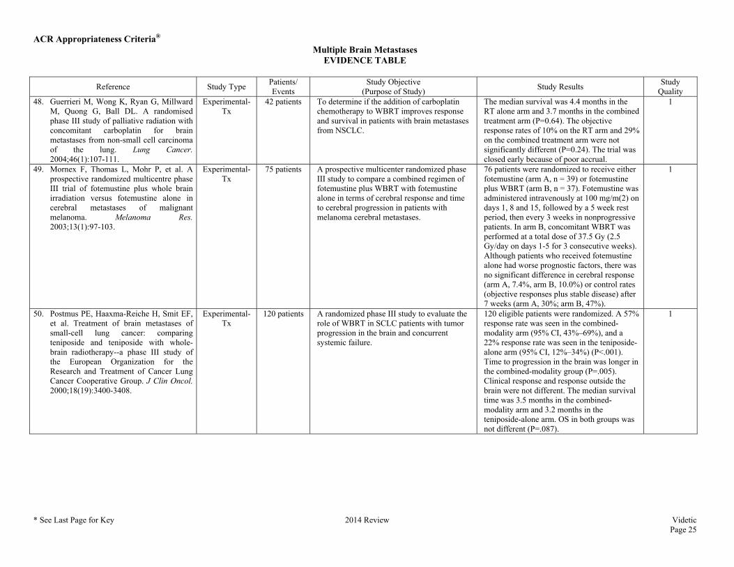
Quality 48. Guerrieri M, Wong K, Ryan G, Millward
M, Quong G, Ball DL. A randomised phase III study of palliative radiation with concomitant carboplatin for brain metastases from non-small cell carcinoma of the lung. Lung Cancer. 2004;46(1):107-111.
Experimental-Tx
42 patients To determine if the addition of carboplatin chemotherapy to WBRT improves response and survival in patients with brain metastases from NSCLC.
The median survival was 4.4 months in the RT alone arm and 3.7 months in the combined treatment arm (P=0.64). The objective response rates of 10% on the RT arm and 29% on the combined treatment arm were not significantly different (P=0.24). The trial was closed early because of poor accrual.
1
49. Mornex F, Thomas L, Mohr P, et al. A prospective randomized multicentre phase III trial of fotemustine plus whole brain irradiation versus fotemustine alone in cerebral metastases of malignant melanoma. Melanoma Res. 2003;13(1):97-103.
Experimental-Tx
75 patients A prospective multicenter randomized phase III study to compare a combined regimen of fotemustine plus WBRT with fotemustine alone in terms of cerebral response and time to cerebral progression in patients with melanoma cerebral metastases.
76 patients were randomized to receive either fotemustine (arm A, n = 39) or fotemustine plus WBRT (arm B, n = 37). Fotemustine was administered intravenously at 100 mg/m(2) on days 1, 8 and 15, followed by a 5 week rest period, then every 3 weeks in nonprogressive patients. In arm B, concomitant WBRT was performed at a total dose of 37.5 Gy (2.5 Gy/day on days 1-5 for 3 consecutive weeks). Although patients who received fotemustine alone had worse prognostic factors, there was no significant difference in cerebral response (arm A, 7.4%, arm B, 10.0%) or control rates (objective responses plus stable disease) after 7 weeks (arm A, 30%; arm B, 47%).
1
50. Postmus PE, Haaxma-Reiche H, Smit EF, et al. Treatment of brain metastases of small-cell lung cancer: comparing teniposide and teniposide with whole-brain radiotherapy--a phase III study of the European Organization for the Research and Treatment of Cancer Lung Cancer Cooperative Group. J Clin Oncol. 2000;18(19):3400-3408.
Experimental-Tx
120 patients A randomized phase III study to evaluate the role of WBRT in SCLC patients with tumor progression in the brain and concurrent systemic failure.
120 eligible patients were randomized. A 57% response rate was seen in the combined-modality arm (95% CI, 43%–69%), and a 22% response rate was seen in the teniposide-alone arm (95% CI, 12%–34%) (P<.001). Time to progression in the brain was longer in the combined-modality group (P=.005). Clinical response and response outside the brain were not different. The median survival time was 3.5 months in the combined-modality arm and 3.2 months in the teniposide-alone arm. OS in both groups was not different (P=.087).
1
ACR Appropriateness Criteria® Multiple Brain Metastases
EVIDENCE TABLE
* See Last Page for Key 2014 Review Videtic Page 26
Reference Study Type Patients/ Events
Study Objective (Purpose of Study) Study Results Study
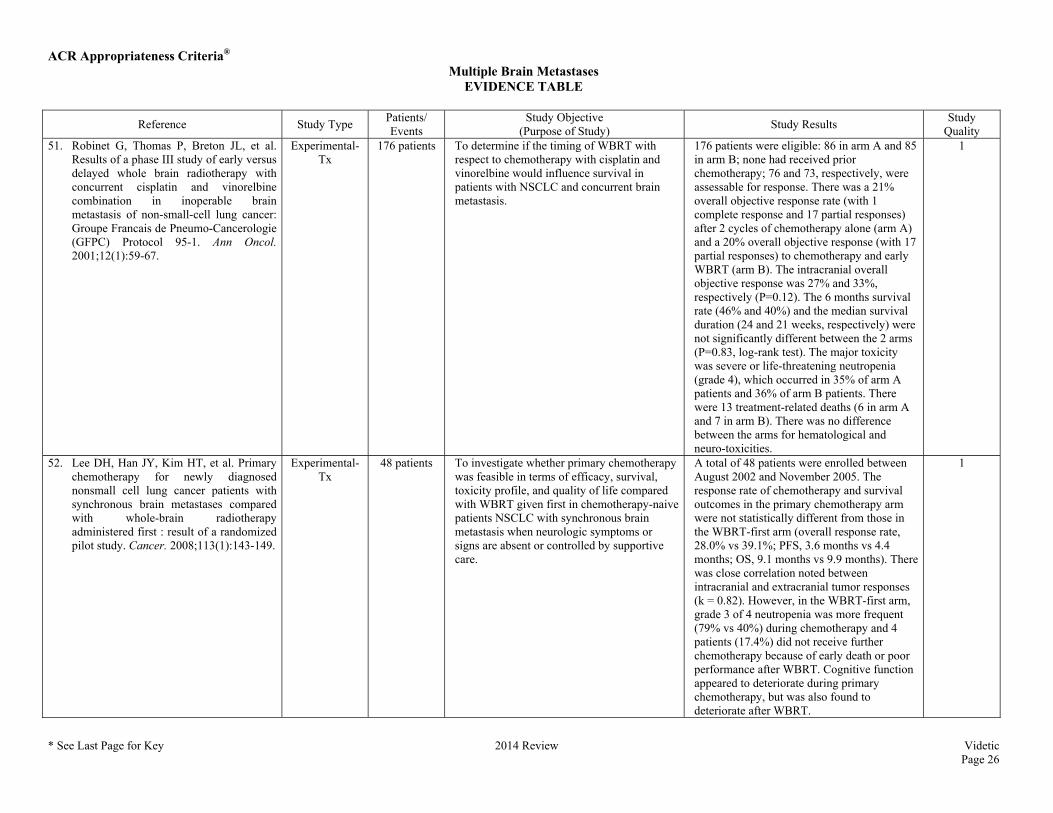
Quality 51. Robinet G, Thomas P, Breton JL, et al.
Results of a phase III study of early versus delayed whole brain radiotherapy with concurrent cisplatin and vinorelbine combination in inoperable brain metastasis of non-small-cell lung cancer: Groupe Francais de Pneumo-Cancerologie (GFPC) Protocol 95-1. Ann Oncol. 2001;12(1):59-67.
Experimental-Tx
176 patients To determine if the timing of WBRT with respect to chemotherapy with cisplatin and vinorelbine would influence survival in patients with NSCLC and concurrent brain metastasis.
176 patients were eligible: 86 in arm A and 85 in arm B; none had received prior chemotherapy; 76 and 73, respectively, were assessable for response. There was a 21% overall objective response rate (with 1 complete response and 17 partial responses) after 2 cycles of chemotherapy alone (arm A) and a 20% overall objective response (with 17 partial responses) to chemotherapy and early WBRT (arm B). The intracranial overall objective response was 27% and 33%, respectively (P=0.12). The 6 months survival rate (46% and 40%) and the median survival duration (24 and 21 weeks, respectively) were not significantly different between the 2 arms (P=0.83, log-rank test). The major toxicity was severe or life-threatening neutropenia (grade 4), which occurred in 35% of arm A patients and 36% of arm B patients. There were 13 treatment-related deaths (6 in arm A and 7 in arm B). There was no difference between the arms for hematological and neuro-toxicities.
1
52. Lee DH, Han JY, Kim HT, et al. Primary chemotherapy for newly diagnosed nonsmall cell lung cancer patients with synchronous brain metastases compared with whole-brain radiotherapy administered first : result of a randomized pilot study. Cancer. 2008;113(1):143-149.
Experimental-Tx
48 patients To investigate whether primary chemotherapy was feasible in terms of efficacy, survival, toxicity profile, and quality of life compared with WBRT given first in chemotherapy-naive patients NSCLC with synchronous brain metastasis when neurologic symptoms or signs are absent or controlled by supportive care.
A total of 48 patients were enrolled between August 2002 and November 2005. The response rate of chemotherapy and survival outcomes in the primary chemotherapy arm were not statistically different from those in the WBRT-first arm (overall response rate, 28.0% vs 39.1%; PFS, 3.6 months vs 4.4 months; OS, 9.1 months vs 9.9 months). There was close correlation noted between intracranial and extracranial tumor responses (k = 0.82). However, in the WBRT-first arm, grade 3 of 4 neutropenia was more frequent (79% vs 40%) during chemotherapy and 4 patients (17.4%) did not receive further chemotherapy because of early death or poor performance after WBRT. Cognitive function appeared to deteriorate during primary chemotherapy, but was also found to deteriorate after WBRT.
1
ACR Appropriateness Criteria® Multiple Brain Metastases
EVIDENCE TABLE
* See Last Page for Key 2014 Review Videtic Page 27
Reference Study Type Patients/ Events
Study Objective (Purpose of Study) Study Results Study
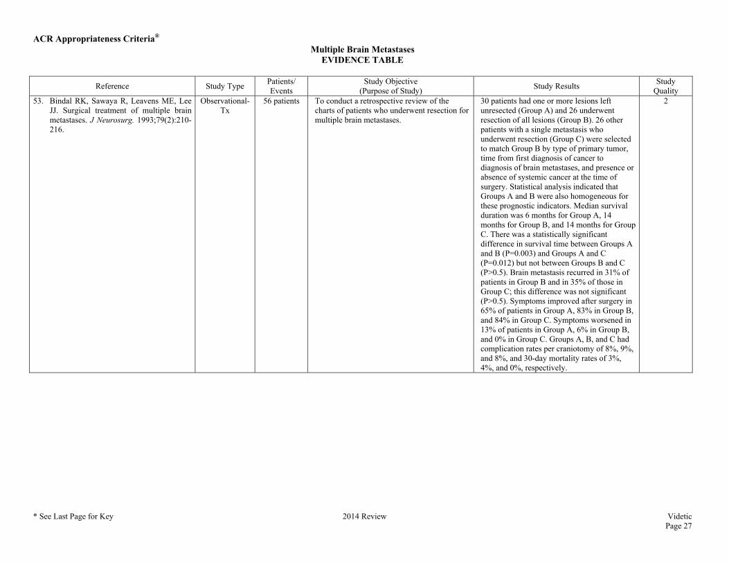
Quality 53. Bindal RK, Sawaya R, Leavens ME, Lee
JJ. Surgical treatment of multiple brain metastases. J Neurosurg. 1993;79(2):210-216.
Observational-Tx
56 patients To conduct a retrospective review of the charts of patients who underwent resection for multiple brain metastases.
30 patients had one or more lesions left unresected (Group A) and 26 underwent resection of all lesions (Group B). 26 other patients with a single metastasis who underwent resection (Group C) were selected to match Group B by type of primary tumor, time from first diagnosis of cancer to diagnosis of brain metastases, and presence or absence of systemic cancer at the time of surgery. Statistical analysis indicated that Groups A and B were also homogeneous for these prognostic indicators. Median survival duration was 6 months for Group A, 14 months for Group B, and 14 months for Group C. There was a statistically significant difference in survival time between Groups A and B (P=0.003) and Groups A and C (P=0.012) but not between Groups B and C (P>0.5). Brain metastasis recurred in 31% of patients in Group B and in 35% of those in Group C; this difference was not significant (P>0.5). Symptoms improved after surgery in 65% of patients in Group A, 83% in Group B, and 84% in Group C. Symptoms worsened in 13% of patients in Group A, 6% in Group B, and 0% in Group C. Groups A, B, and C had complication rates per craniotomy of 8%, 9%, and 8%, and 30-day mortality rates of 3%, 4%, and 0%, respectively.
2
ACR Appropriateness Criteria® Multiple Brain Metastases
EVIDENCE TABLE
* See Last Page for Key 2014 Review Videtic Page 28
Reference Study Type Patients/ Events
Study Objective (Purpose of Study) Study Results Study
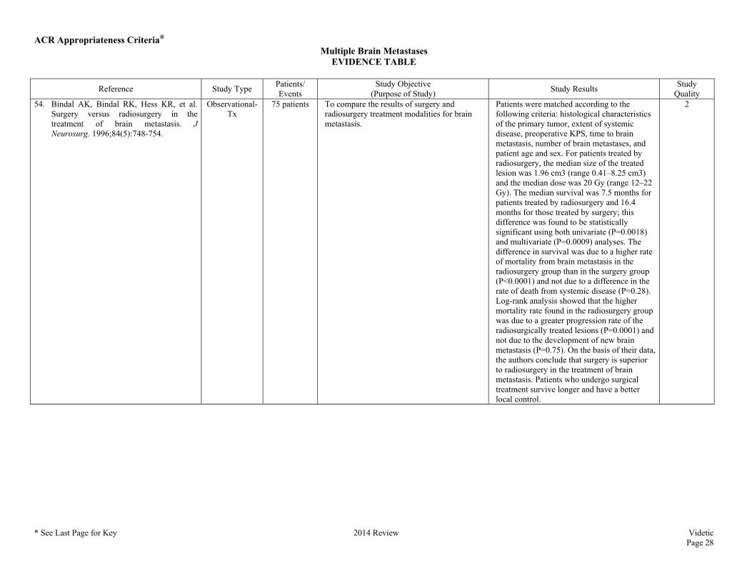
Quality 54. Bindal AK, Bindal RK, Hess KR, et al.
Surgery versus radiosurgery in the treatment of brain metastasis. J Neurosurg. 1996;84(5):748-754.
Observational-Tx
75 patients To compare the results of surgery and radiosurgery treatment modalities for brain metastasis.
Patients were matched according to the following criteria: histological characteristics of the primary tumor, extent of systemic disease, preoperative KPS, time to brain metastasis, number of brain metastases, and patient age and sex. For patients treated by radiosurgery, the median size of the treated lesion was 1.96 cm3 (range 0.41–8.25 cm3) and the median dose was 20 Gy (range 12–22 Gy). The median survival was 7.5 months for patients treated by radiosurgery and 16.4 months for those treated by surgery; this difference was found to be statistically significant using both univariate (P=0.0018) and multivariate (P=0.0009) analyses. The difference in survival was due to a higher rate of mortality from brain metastasis in the radiosurgery group than in the surgery group (P<0.0001) and not due to a difference in the rate of death from systemic disease (P=0.28). Log-rank analysis showed that the higher mortality rate found in the radiosurgery group was due to a greater progression rate of the radiosurgically treated lesions (P=0.0001) and not due to the development of new brain metastasis (P=0.75). On the basis of their data, the authors conclude that surgery is superior to radiosurgery in the treatment of brain metastasis. Patients who undergo surgical treatment survive longer and have a better local control.
2
ACR Appropriateness Criteria® Multiple Brain Metastases
EVIDENCE TABLE
* See Last Page for Key 2014 Review Videtic Page 29
Reference Study Type Patients/ Events
Study Objective (Purpose of Study) Study Results Study
Quality 55. Kocher M, Soffietti R, Abacioglu U, et al.
Adjuvant whole-brain radiotherapy versus observation after radiosurgery or surgical resection of one to three cerebral metastases: results of the EORTC 22952-26001 study. J Clin Oncol. 2011;29(2):134-141.
Experimental-Tx
359 patients A phase III trial to assess whether adjuvant WBRT increases the duration of functional independence after surgery or radiosurgery of brain metastases.
Of 359 patients, 199 underwent radiosurgery, and 160 underwent surgery. In the radiosurgery group, 100 patients were allocated to observation, and 99 were allocated to WBRT. After surgery, 79 patients were allocated to observation, and 81 were allocated to adjuvant WBRT. The median time to WHO performance status more than 2 was 10.0 months (95% CI, 8.1 to 11.7 months) after observation and 9.5 months (95% CI, 7.8 to 11.9 months) after WBRT (P=.71). OS was similar in the WBRT and observation arms (median, 10.9 v 10.7 months, respectively; P=.89). WBRT reduced the 2-year relapse rate both at initial sites (surgery: 59% to 27%, P<.001; radiosurgery: 31% to 19%, P=.040) and at new sites (surgery: 42% to 23%, P=.008; radiosurgery: 48% to 33%, P=.023). Salvage therapies were used more frequently after observation than after WBRT. Intracranial progression caused death in 78 (44%) of 179 patients in the observation arm and in 50 (28%) of 180 patients in the WBRT arm.
1
56. Soffietti R, Kocher M, Abacioglu UM, et al. A European Organisation for Research and Treatment of Cancer phase III trial of adjuvant whole-brain radiotherapy versus observation in patients with one to three brain metastases from solid tumors after surgical resection or radiosurgery: quality-of-life results. J Clin Oncol. 2013;31(1):65-72.
Experimental-Tx
359 patients A phase III trial to compare adjuvant WBRT with observation after either surgery or radiosurgery of a limited number of brain metastases in patients with stable solid tumors. Here, we report the health-related quality-of-life results.
Compliance was 88.3% at baseline and dropped to 45.0% at 1 year; thus, only the first year was analyzed. Overall, patients in the observation only arm reported better health-related quality-of-life scores than did patients who received WBRT. The differences were statistically significant and clinically relevant mostly during the early follow-up period (for global health status at 9 months, physical functioning at 8 weeks, cognitive functioning at 12 months, and fatigue at 8 weeks). Exploratory analysis of all other health-related quality-of-life scales suggested worse scores for the WBRT group, but none was clinically relevant.
1
ACR Appropriateness Criteria® Multiple Brain Metastases
EVIDENCE TABLE
* See Last Page for Key 2014 Review Videtic Page 30
Reference Study Type Patients/ Events
Study Objective (Purpose of Study) Study Results Study
Quality 57. Pollock BE, Brown PD, Foote RL,
Stafford SL, Schomberg PJ. Properly selected patients with multiple brain metastases may benefit from aggressive treatment of their intracranial disease. J Neurooncol. 2003;61(1):73-80.
Observational-Tx
52 patients To determine whether properly selected patients with multiple brain metastases benefit from aggressive treatment of their intracranial disease.
Tumor histology included lung (n = 18, 35%), breast (n = 11, 21%), renal (n = 6, 12%), melanoma (n = 6, 12%), and other (n = 11, 21%). 20 patients (39%) had progressed after prior RT. Treatment included multiple craniotomies and tumor resection (n = 5, 10%), radiosurgery (n = 31, 60%), or resection and radiosurgery (n= 16, 30%). Median survival was 15.5 months. The 1- and 2-year actuarial survivals were 63% and 27%, respectively. Multivariate analysis found RTOG RPA Class (1 vs 2/3) correlated with improved survival (RR = 2.60, 95% CI 1.13–5.97, P=0.03). Class 1 patients (KPS ≥70, age <65 years, and controlled primary with no extracranial metastases) survived a median of 19 months whereas Class 3 patients (KPS <70) survived 8 months. Class 2 patients (all other patients) survived a median of 13 months. 35 patients (67%) had intracranial progression at a median of 8.0 months. Intracranial progression was local (n = 6), distant (n = 23), or local and distant (n = 6); 26 patients with intracranial progression underwent additional brain tumor treatments. Multivariate analysis found patients with radiosensitive tumors (lung, breast, other) had fewer intracranial recurrences compared to patients with radio-resistant tumors (melanoma, renal, sarcoma) (RR = 2.43, 95% CI 1.13–5.10, P=0.02).
1
58. Iwadate Y, Namba H, Yamaura A. Significance of surgical resection for the treatment of multiple brain metastases. Anticancer Res. 2000;20(1B):573-577.
Observational-Tx
138 patients To investigate the role of surgery in the treatment of multiple brain metastases when performed with RT.
The median survival was 8.7 and 9.2 months for the Single Group and the Multiple Group, respectively (not statistically different). The median survival was 9.6, 12.4, 3.7, and 4.5 months for Groups A, B, C, and D, respectively. Survival duration differed significantly between Groups A/B and Groups C/D (P<0.05).
1
ACR Appropriateness Criteria® Multiple Brain Metastases
EVIDENCE TABLE
* See Last Page for Key 2014 Review Videtic Page 31
Reference Study Type Patients/ Events
Study Objective (Purpose of Study) Study Results Study
Quality 59. Kondziolka D, Patel A, Lunsford LD,
Kassam A, Flickinger JC. Stereotactic radiosurgery plus whole brain radiotherapy versus radiotherapy alone for patients with multiple brain metastases. Int J Radiat Oncol Biol Phys. 1999;45(2):427-434.
Observational-Tx
27 patients To examine brain tumor disease control, patient survival, morbidity, and the need for further brain tumor management.
The study was stopped at an interim evaluation at 60% accrual. 27 patients were randomized (14 to WBRT alone and 13 to WBRT plus radiosurgery). The groups were well matched to age, sex, tumor type, number of tumors, and extent of extracranial disease. The rate of local failure at 1 year was 100% after WBRT alone but only 8% in patients who had boost radiosurgery. The median time to local failure was 6 months after WBRT alone (95% CI, 3.5–8.5) in comparison to 36 months (95% CI, 15.6–57) after WBRT plus radiosurgery (P=0.0005). The median time to any brain failure was improved in the radiosurgery group (P=0.002). Tumor control did not depend on histology (P=0.85), number of initial brain metastases (P=0.25), or extent of extracranial disease (P=0.26). Patients who received WBRT alone lived a median of 7.5 months, while those who received WBRT plus radiosurgery lived 11 months (P=0.22). Survival did not depend on histology or number of tumors, but was related to extent of extracranial disease (P=0.02). There was no neurologic or systemic morbidity related to SRS.
1
60. Mahmood U, Kwok Y, Regine WF, Patchell RA. Whole-brain irradiation for patients with brain metastases: still the standard of care. Lancet Oncol. 2010;11(3):221-222; author reply 223.
Review/Other-Tx
N/A No abstract available. No abstract available. 4
ACR Appropriateness Criteria®
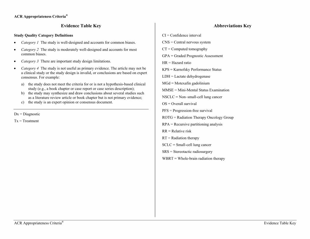
ACR Appropriateness Criteria® Evidence Table Key
Evidence Table Key
Study Quality Category Definitions
Category 1 The study is well-designed and accounts for common biases.
Category 2 The study is moderately well-designed and accounts for most common biases.
Category 3 There are important study design limitations.
Category 4 The study is not useful as primary evidence. The article may not be a clinical study or the study design is invalid, or conclusions are based on expert consensus. For example:
a) the study does not meet the criteria for or is not a hypothesis-based clinical study (e.g., a book chapter or case report or case series description);
b) the study may synthesize and draw conclusions about several studies such as a literature review article or book chapter but is not primary evidence;
c) the study is an expert opinion or consensus document.
Dx = Diagnostic
Tx = Treatment
Abbreviations Key
CI = Confidence interval
CNS = Central nervous system
CT = Computed tomography
GPA = Graded Prognostic Assessment
HR = Hazard ratio
KPS = Karnofsky Performance Status
LDH = Lactate dehydrogenase
MGd = Motexafin gadolinium
MMSE = Mini-Mental Status Examination
NSCLC = Non–small-cell lung cancer
OS = Overall survival
PFS = Progression-free survival
ROTG = Radiation Therapy Oncology Group
RPA = Recursive partitioning analysis
RR = Relative risk
RT = Radiation therapy
SCLC = Small-cell lung cancer
SRS = Stereotactic radiosurgery
WBRT = Whole-brain radiation therapy