Embed Size (px)
Citation preview
Musculoskeletal Imaging of the Digits
Arash David Tehranzadeh, MD
UCSD MSK Radiology
May 11th, 2006
Musculoskeletal Imaging of the Digit
Anatomy & Internal Derangement The Extensor System The Flexor System
Soft Tissue Masses & Tumors
Musculoskeletal Imaging of the Digit
Anatomy & Internal Derangement The Extensor System The Flexor System
Soft Tissue Masses & Tumors
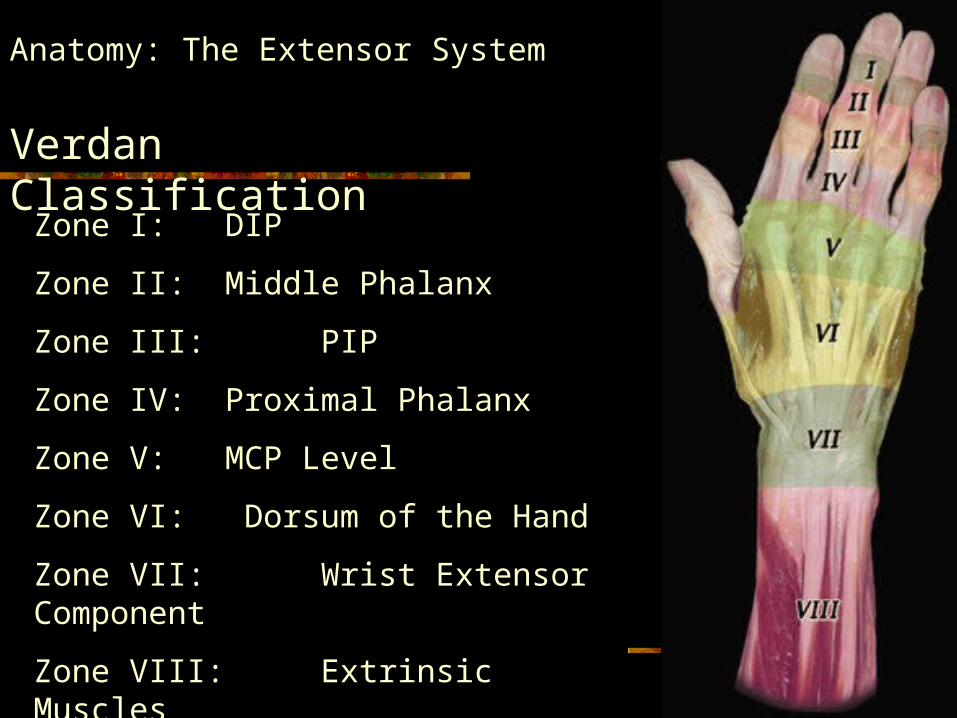
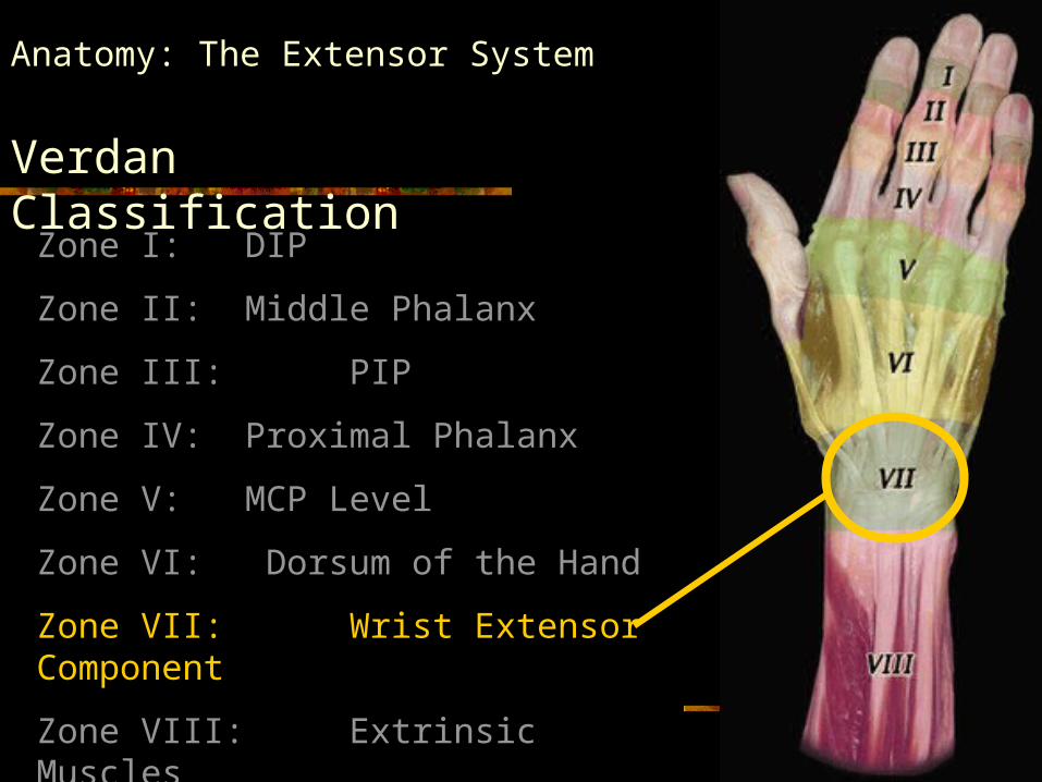
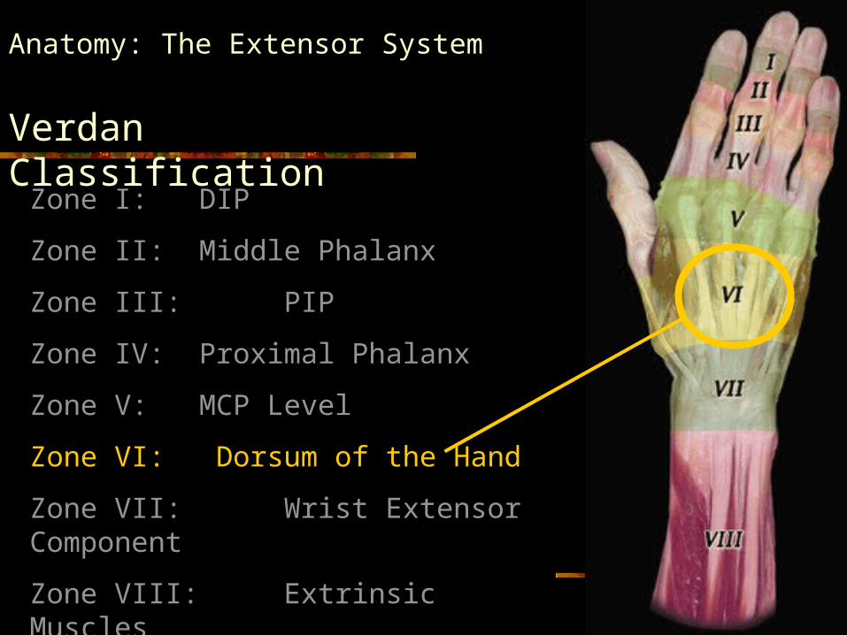
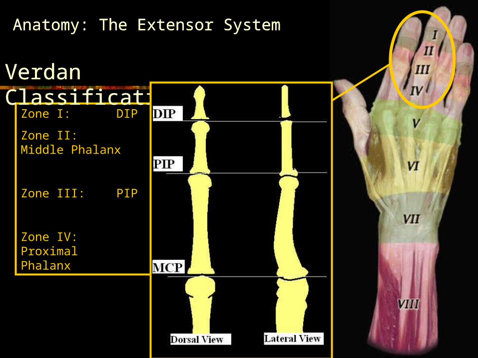
Anatomy: The Extensor System
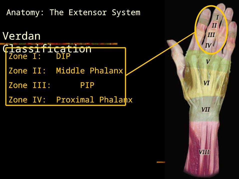
Verdan Classification
Zone I: DIP
Zone II: Middle Phalanx
Zone III: PIP
Zone IV: Proximal Phalanx
Zone V: MCP Level
Zone VI: Dorsum of the Hand
Zone VII: Wrist Extensor Component
Zone VIII: Extrinsic Muscles
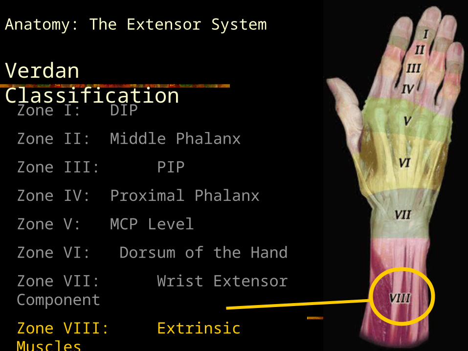
Anatomy: The Extensor System
Verdan Classification
Zone I: DIP
Zone II: Middle Phalanx
Zone III: PIP
Zone IV: Proximal Phalanx
Zone V: MCP Level
Zone VI: Dorsum of the Hand
Zone VII: Wrist Extensor Component
Zone VIII: Extrinsic Muscles
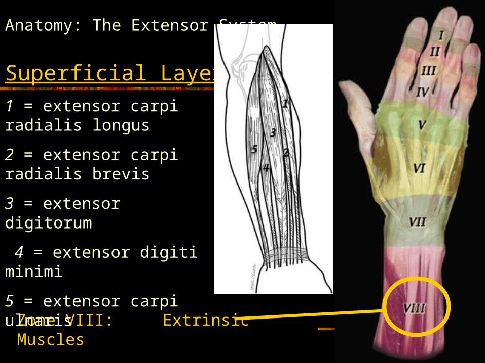
Anatomy: The Extensor System
Superficial Layer
Zone VIII: Extrinsic Muscles
1 = extensor carpi radialis longus
2 = extensor carpi radialis brevis
3 = extensor digitorum
4 = extensor digiti minimi
5 = extensor carpi ulnaris

Anatomy: The Extensor System
Deep Layer
Zone VIII: Extrinsic Muscles
6 = supinator
7 = abductor pollicis longus
8 = extensor pollicis brevis
9 = extensor pollicis longus
10 = extensor indicis
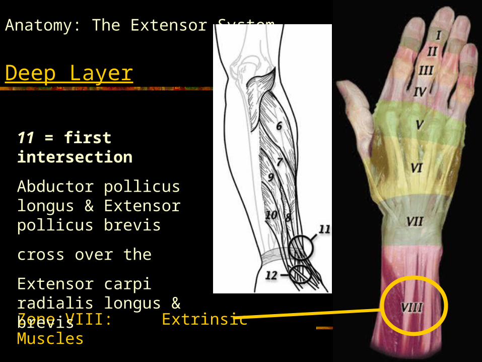
Anatomy: The Extensor System
Deep Layer
Zone VIII: Extrinsic Muscles
11 = first intersection
Abductor pollicus longus & Extensor pollicus brevis
cross over the
Extensor carpi radialis longus & brevis
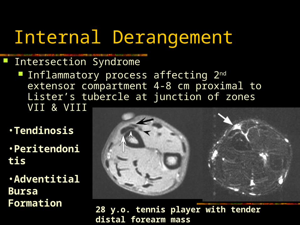
Internal Derangement Intersection Syndrome
Inflammatory process affecting 2nd extensor compartment 4-8 cm proximal to Lister’s tubercle at junction of zones VII & VIII
•Tendinosis
•Peritendonitis
•Adventitial Bursa Formation
28 y.o. tennis player with tender distal forearm mass
Anatomy: The Extensor System
Zone I: DIP
Zone II: Middle Phalanx
Zone III: PIP
Zone IV: Proximal Phalanx
Zone V: MCP Level
Zone VI: Dorsum of the Hand
Zone VII: Wrist Extensor Component
Zone VIII: Extrinsic Muscles
Verdan Classification
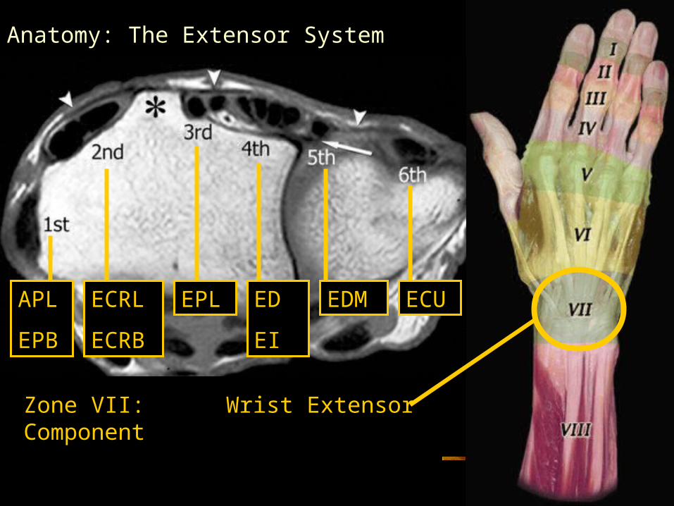
Anatomy: The Extensor System
Zone VII: Wrist Extensor Component
APL
EPB
ECRL
ECRB
EPL ED
EI
EDM ECU
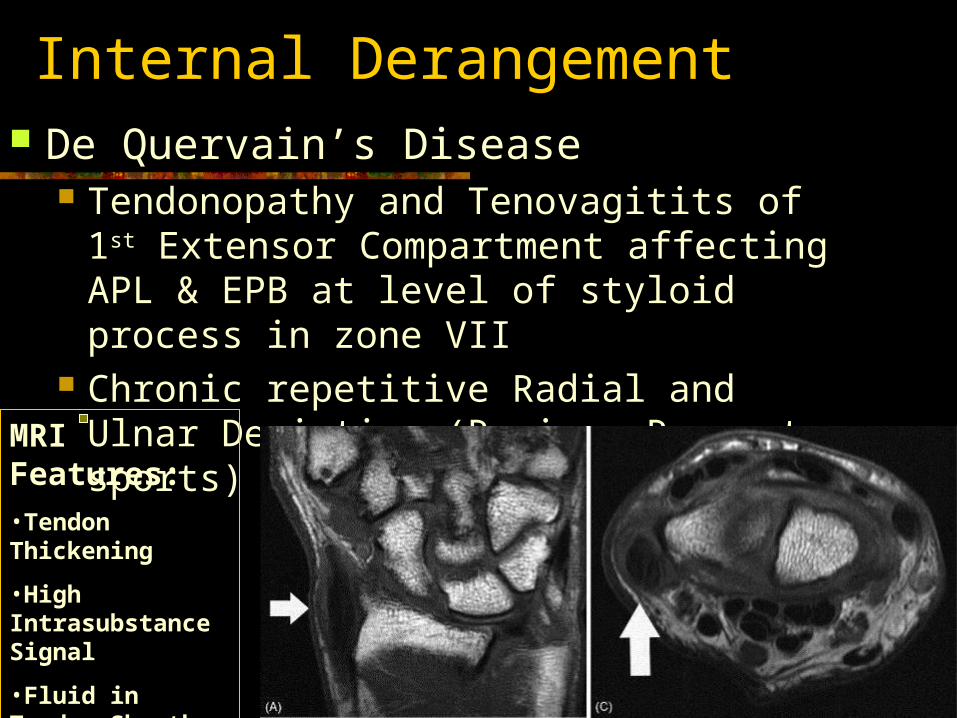
Internal Derangement De Quervain’s Disease
Tendonopathy and Tenovagitits of 1st Extensor Compartment affecting APL & EPB at level of styloid process in zone VII
Chronic repetitive Radial and Ulnar Deviation (Rowing, Racquet sports)
MRI Features:•Tendon Thickening
•High Intrasubstance Signal
•Fluid in Tendon Sheath
•Adj. Soft tissue edema
Anatomy: The Extensor System
Verdan Classification
Zone I: DIP
Zone II: Middle Phalanx
Zone III: PIP
Zone IV: Proximal Phalanx
Zone V: MCP Level
Zone VI: Dorsum of the Hand
Zone VII: Wrist Extensor Component
Zone VIII: Extrinsic Muscles
Anatomy: The Extensor System
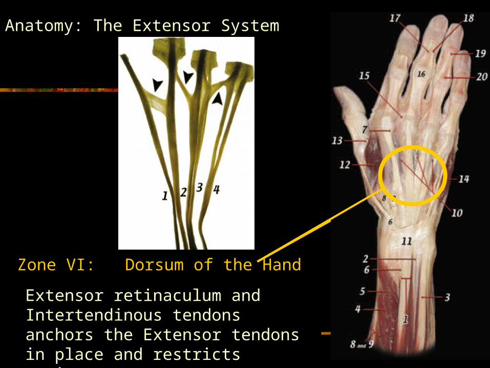
Zone VI: Dorsum of the Hand
Extensor retinaculum and Intertendinous tendons anchors the Extensor tendons in place and restricts motion.
Anatomy: The Extensor System
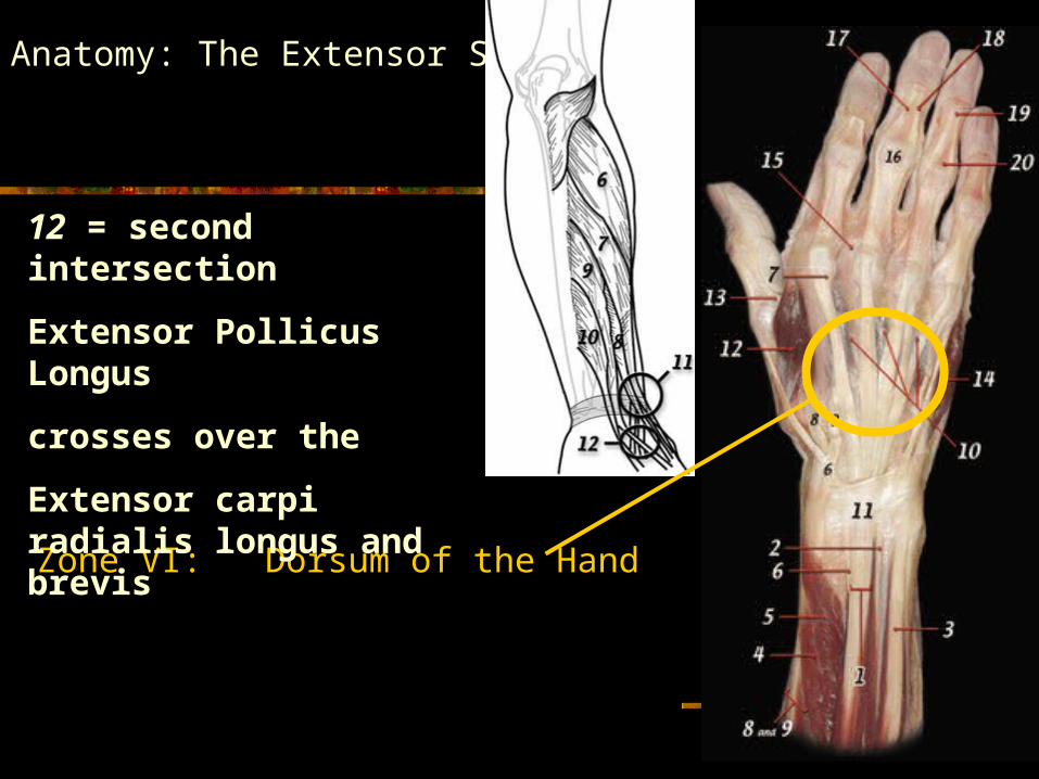
Zone VI: Dorsum of the Hand
12 = second intersection
Extensor Pollicus Longus
crosses over the
Extensor carpi radialis longus and brevis

Anatomy: The Extensor System
Zone I: DIP
Zone II: Middle Phalanx
Zone III: PIP
Zone IV: Proximal Phalanx
Zone V: MCP Level
Zone VI: Dorsum of the Hand
Zone VII: Wrist Extensor Component
Zone VIII: Extrinsic Muscles
Verdan Classification
Anatomy: The Extensor System
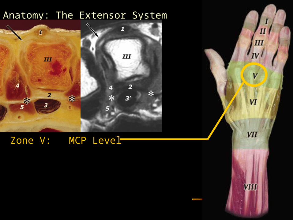
Zone V: MCP Level
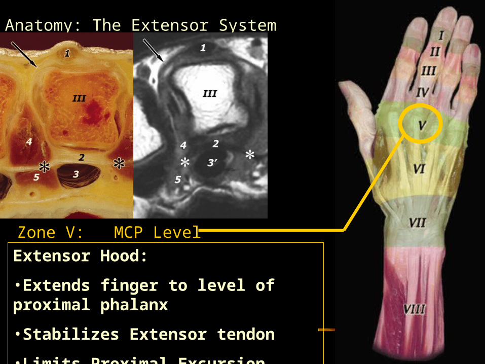
Anatomy: The Extensor System
Zone V: MCP Level
Extensor Hood:
•Extends finger to level of proximal phalanx
•Stabilizes Extensor tendon
•Limits Proximal Excursion
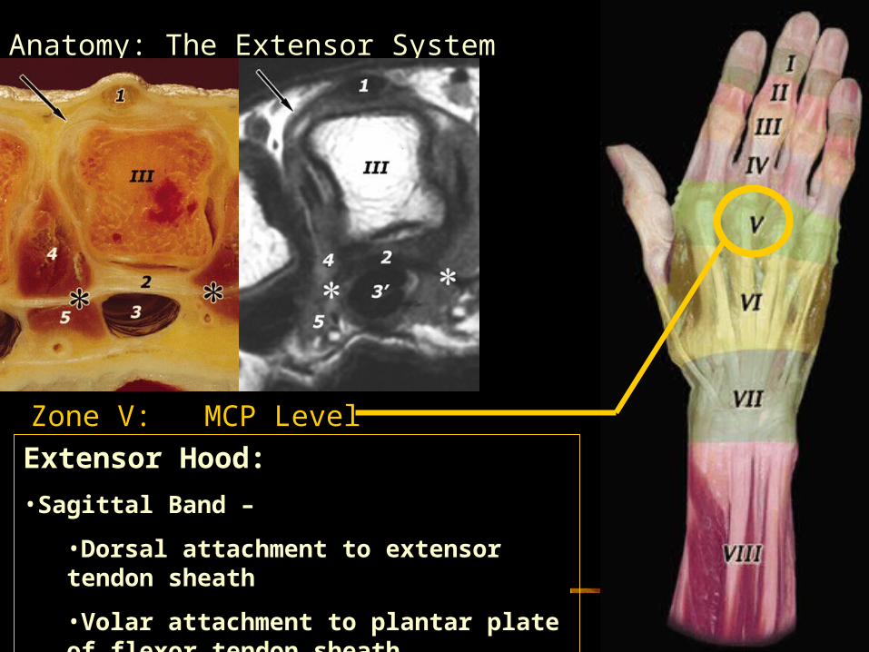
Anatomy: The Extensor System
Zone V: MCP Level
Extensor Hood:
•Sagittal Band –
•Dorsal attachment to extensor tendon sheath
•Volar attachment to plantar plate of flexor tendon sheath
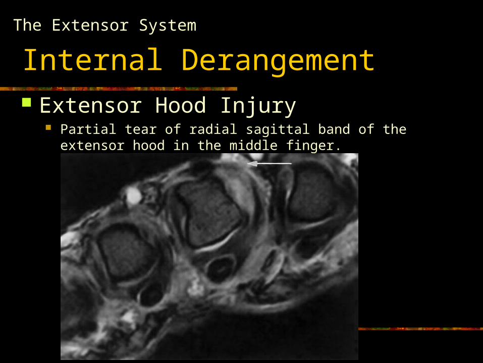
Internal Derangement Extensor Hood Injury
Partial tear of radial sagittal band of the extensor hood in the middle finger.
The Extensor System

Anatomy: The Extensor System
Verdan Classification
Zone I: DIP
Zone II: Middle Phalanx
Zone III: PIP
Zone IV: Proximal Phalanx
Zone V: MCP Level
Zone VI: Dorsum of the Hand
Zone VII: Wrist Extensor Component
Zone VIII: Extrinsic Muscles
Anatomy: The Extensor System
Verdan Classification
Zone I: DIP
Zone II: Middle Phalanx
Zone III: PIP
Zone IV: Proximal Phalanx
Anatomy: The Extensor System
Verdan Classification
Zone I: DIP
Zone II: Middle Phalanx
Zone III: PIP
Zone IV: Proximal Phalanx
Anatomy: The Extensor System
Verdan Classification
Zone I: DIP
Zone II: Middle Phalanx
Zone III: PIP
Zone IV: Proximal Phalanx
Anatomy: The Extensor System
Verdan Classification
Zone I: DIP
Zone II: Middle Phalanx
Zone III: PIP
Zone IV: Proximal Phalanx
Anatomy: The Extensor System
Zone I: DIP
Zone II: Middle Phalanx
Zone III: PIP
Zone IV: Proximal Phalanx
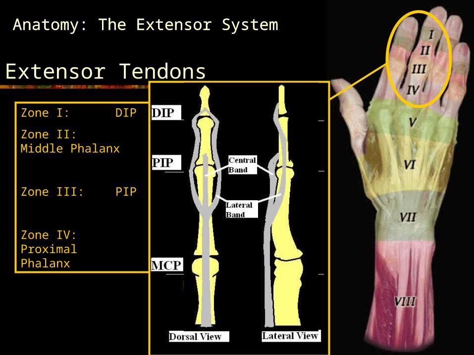
Anatomy: The Extensor System
Extensor Tendons
Zone I: DIP
Zone II: Middle Phalanx
Zone III: PIP
Zone IV: Proximal Phalanx
Anatomy: The Extensor System
Zone I: DIP
Zone II: Middle Phalanx
Zone III: PIP
Zone IV: Proximal Phalanx
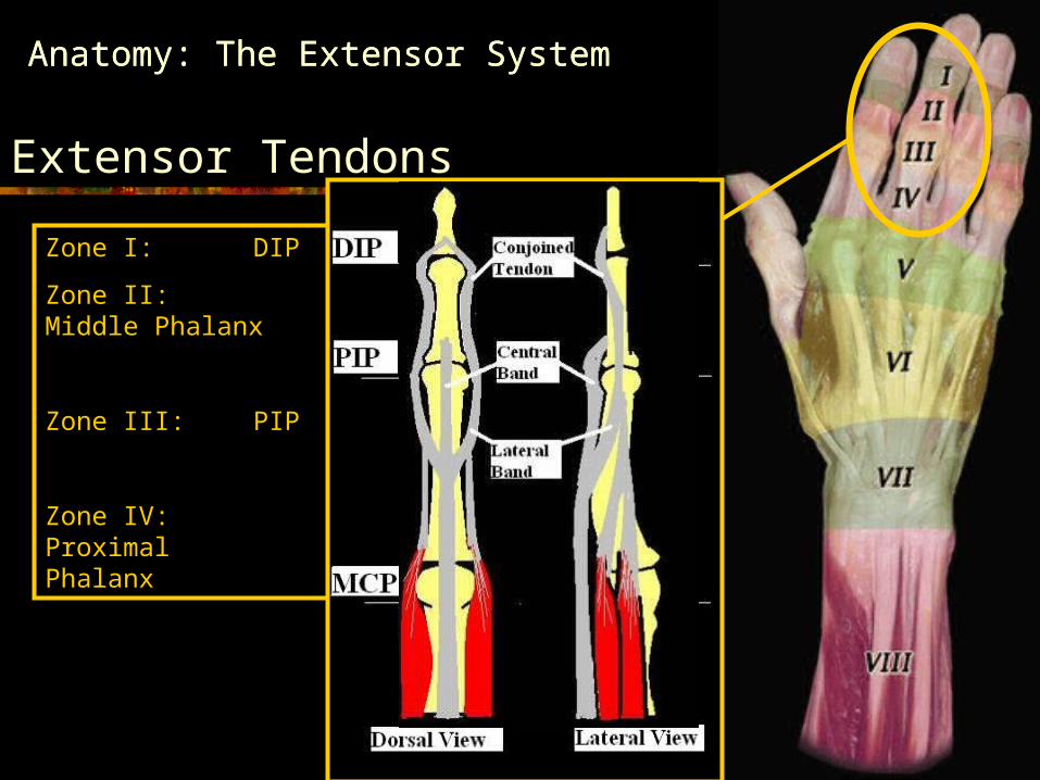
Anatomy: The Extensor System
Extensor Tendons
Zone I: DIP
Zone II: Middle Phalanx
Zone III: PIP
Zone IV: Proximal Phalanx
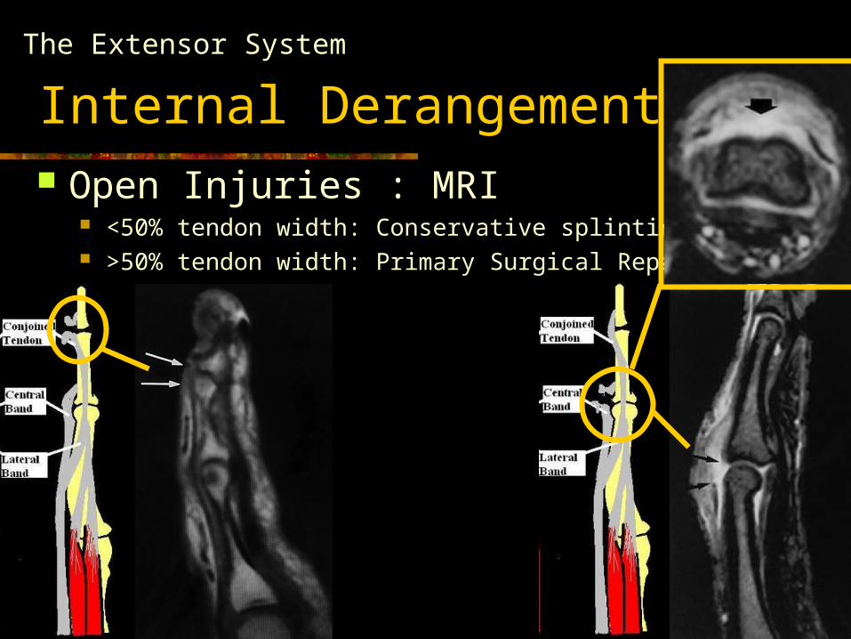
Internal Derangement: Open Injuries : MRI
<50% tendon width: Conservative splinting >50% tendon width: Primary Surgical Repair
The Extensor System
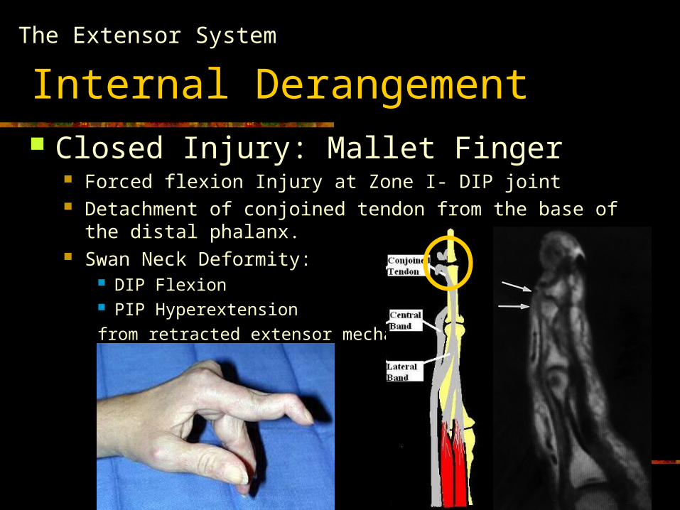
Internal Derangement Closed Injury: Mallet Finger
Forced flexion Injury at Zone I- DIP joint Detachment of conjoined tendon from the base of the distal
phalanx. Swan Neck Deformity:
DIP Flexion PIP Hyperextension
from retracted extensor mechanism
The Extensor System
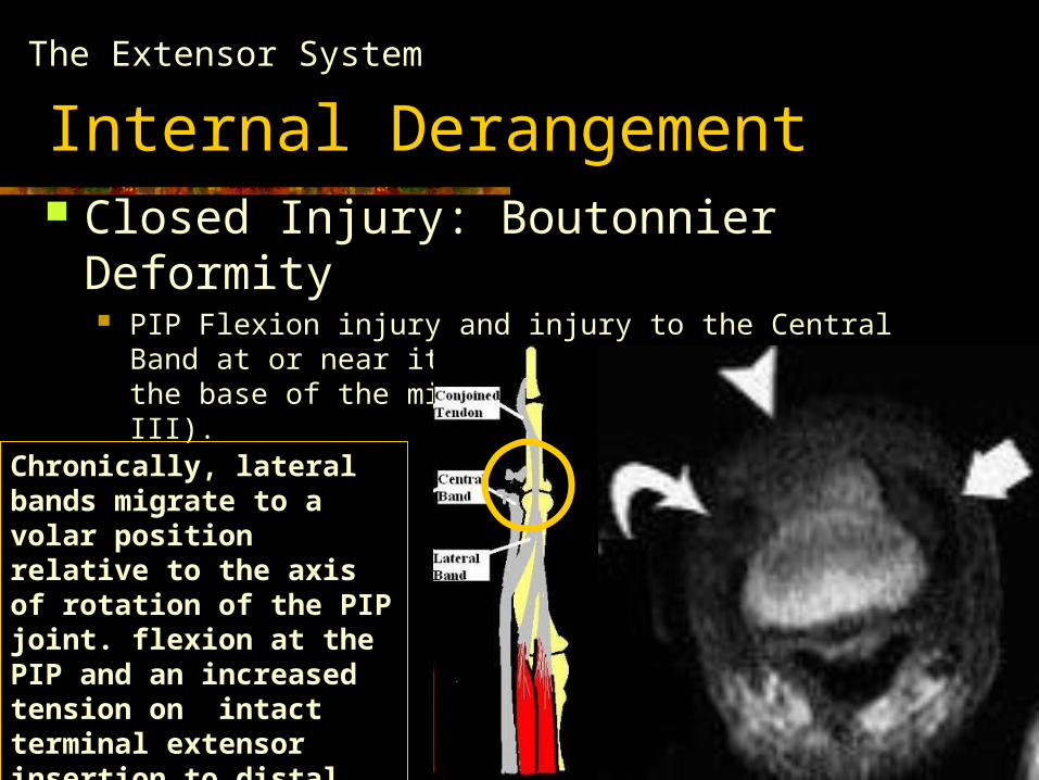
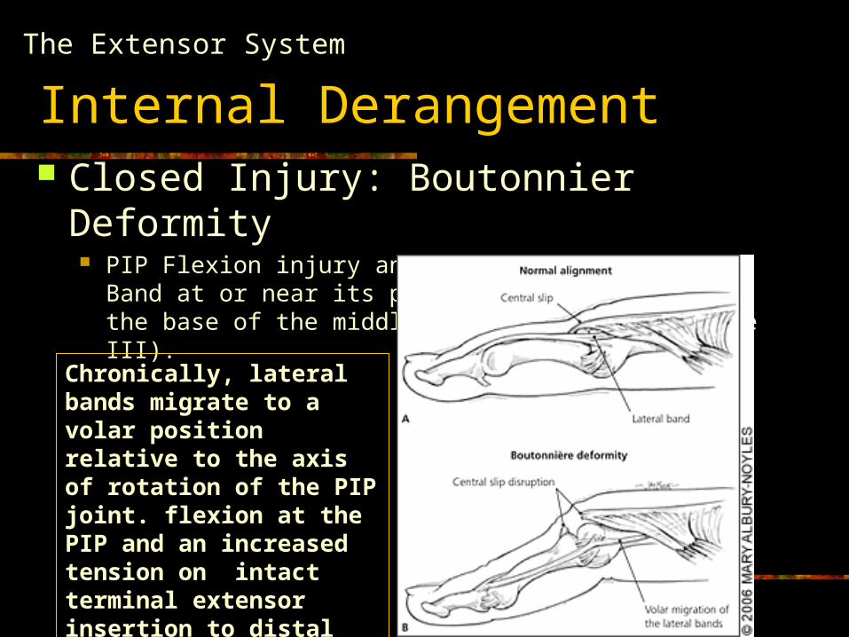
Internal Derangement Closed Injury: Boutonnier Deformity
PIP Flexion injury and injury to the Central Band at or near its point of attatchement at the base of the middle phalanx (Zone II / Zone III).
The Extensor System
Chronically, lateral bands migrate to a volar position relative to the axis of rotation of the PIP joint. flexion at the PIP and an increased tension on intact terminal extensor insertion to distal tuft base
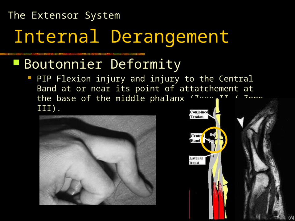
Internal Derangement Boutonnier Deformity
PIP Flexion injury and injury to the Central Band at or near its point of attatchement at the base of the middle phalanx (Zone II / Zone III).
The Extensor System
Musculoskeletal Imaging of the Digit
Anatomy & Internal Derangement The Extensor System The Flexor System
Soft Tissue Masses & Tumors
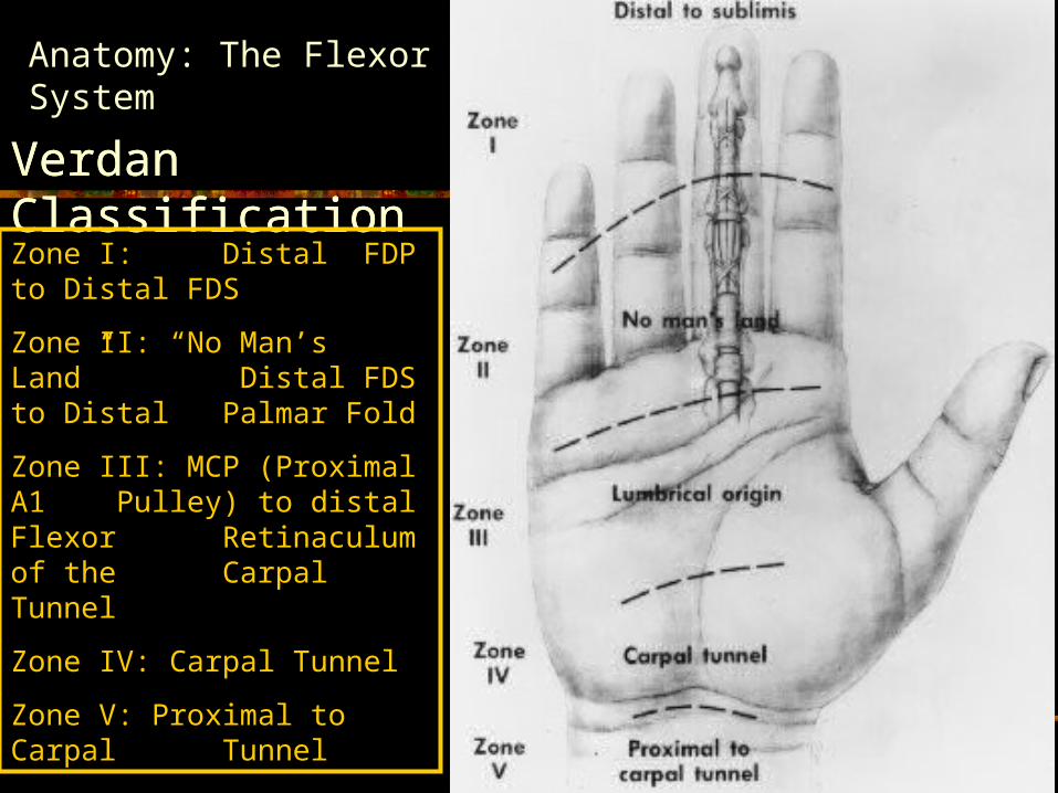
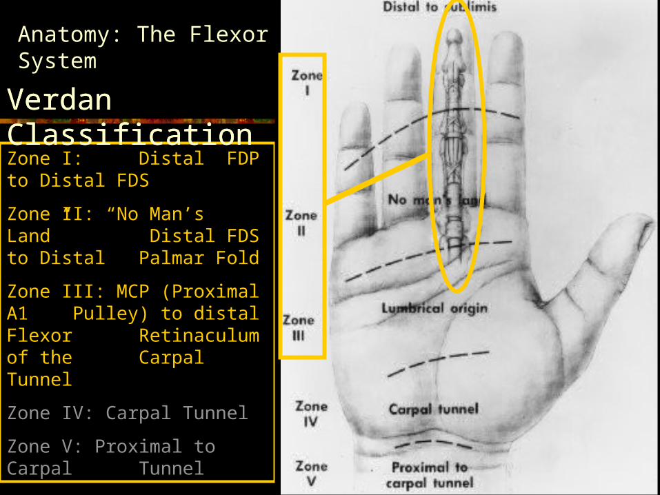
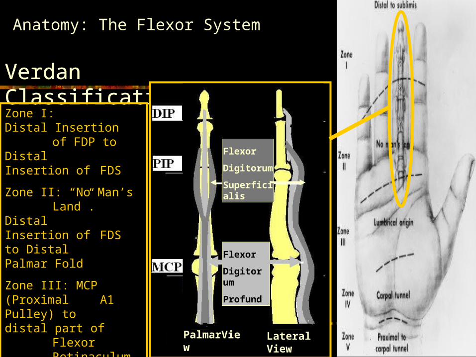
Anatomy: The Flexor System
Verdan ClassificationVerdan Classification
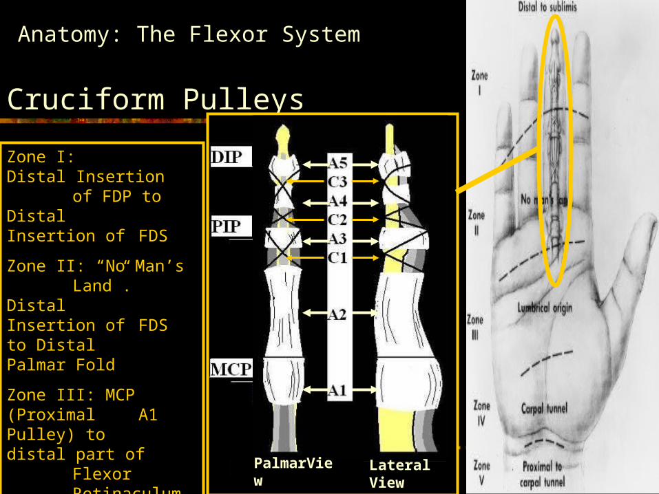
Zone I: Distal FDP to Distal FDS
Zone II: “No Man’s Land” Distal FDS to Distal Palmar Fold
Zone III: MCP (Proximal A1 Pulley) to distal Flexor Retinaculum of the Carpal Tunnel
Zone IV: Carpal Tunnel
Zone V: Proximal to Carpal Tunnel
Anatomy: The Flexor System
Verdan Classification
Zone I: Distal FDP to Distal FDS
Zone II: “No Man’s Land” Distal FDS to Distal Palmar Fold
Zone III: MCP (Proximal A1 Pulley) to distal Flexor Retinaculum of the Carpal Tunnel
Zone IV: Carpal Tunnel
Zone V: Proximal to Carpal Tunnel
Verdan Classification
Verdan Classification
Anatomy: The Flexor System
Verdan Classification
Zone I: DIP
Zone II: Middle Phalanx
Zone III: PIP
Zone IV: Proximal Phalanx
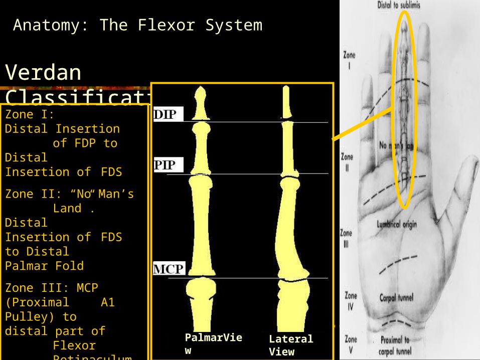
PalmarView Lateral View
Zone I: Distal Insertion of FDP to Distal Insertion of FDS
Zone II: “No Man’s Land”. Distal Insertion of FDS to Distal Palmar Fold
Zone III: MCP (Proximal A1 Pulley) to distal part of Flexor
Retinaculum of the Carpal Tunnel
Verdan Classification
Anatomy: The Flexor System
Verdan Classification
Zone I: DIP
Zone II: Middle Phalanx
Zone III: PIP
Zone IV: Proximal Phalanx
Flexor
Digitorum
Superficialis
Flexor
Digitorum
Profundus
PalmarView Lateral View
Zone I: Distal Insertion of FDP to Distal Insertion of FDS
Zone II: “No Man’s Land”. Distal Insertion of FDS to Distal Palmar Fold
Zone III: MCP (Proximal A1 Pulley) to distal part of Flexor
Retinaculum of the Carpal Tunnel
Anatomy: The Flexor System
Internal Derangement
Flexor Tendon Injuries
•Open > Closed
•Zone II Lesions: Most Frequent, Worst Prognosis
•Tendon-bone attatchement not as strong as Extensor system.
•Higher degree of tendon retraction
•Extent of gap between torn ends may be overestimated due to tendon retraction
•Isolated FDS avulsion Uncommon
Anatomy: The Flexor System
Internal Derangement
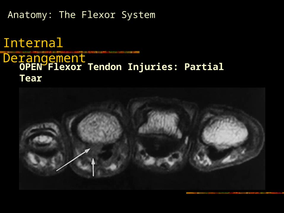
OPEN Flexor Tendon Injuries: Partial Tear
Anatomy: The Flexor System
Internal Derangement
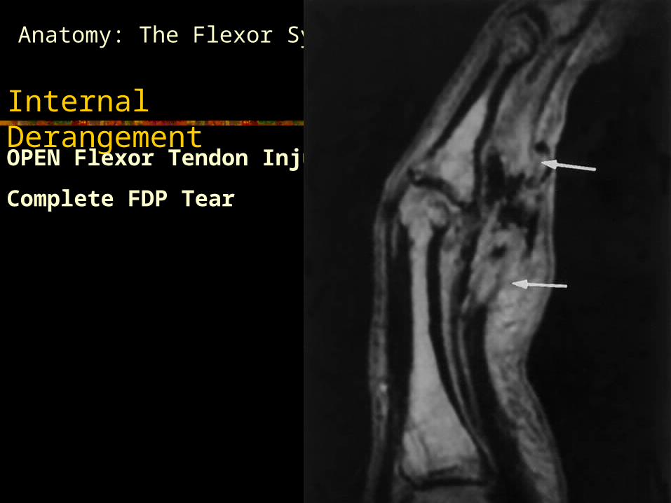
OPEN Flexor Tendon Injuries:
Complete FDP Tear
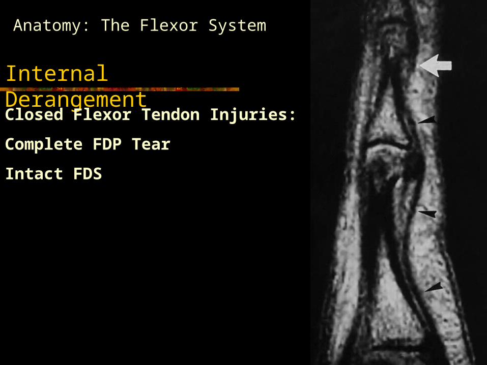
Anatomy: The Flexor System
Internal Derangement
Closed Flexor Tendon Injuries:
Complete FDP Tear
Intact FDS
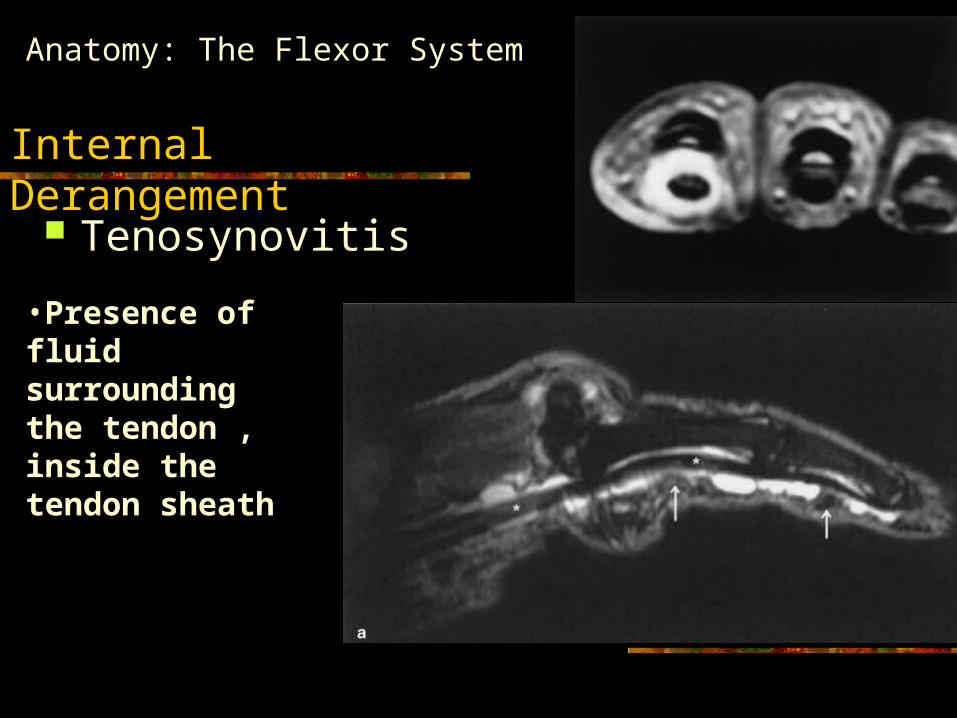
Tenosynovitis
Anatomy: The Flexor System
Internal Derangement
•Presence of fluid surrounding the tendon , inside the tendon sheath
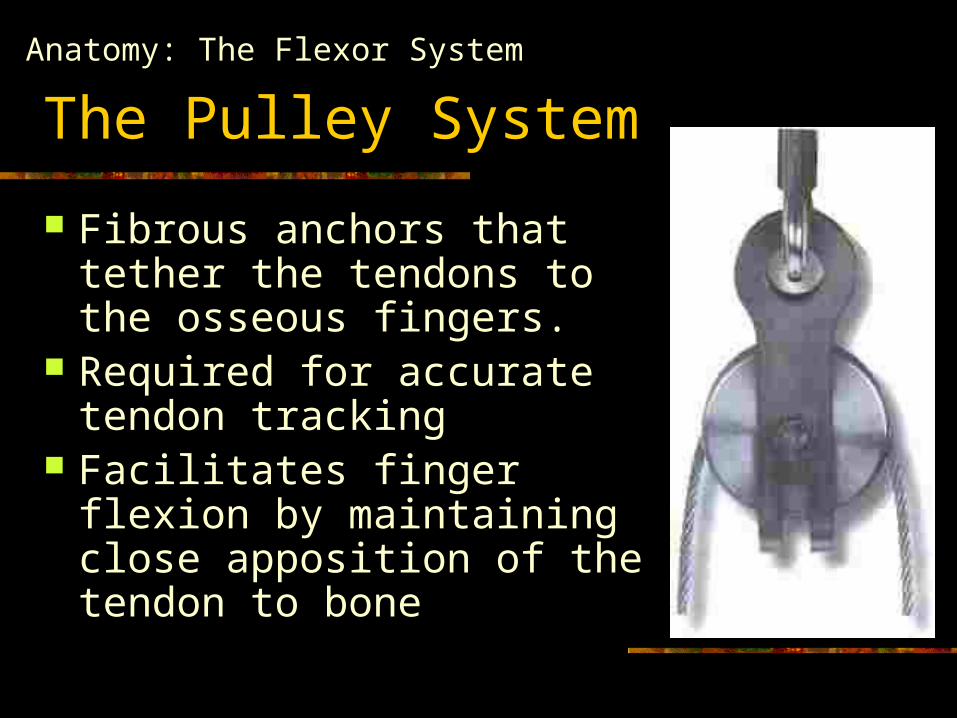
The Pulley System
Fibrous anchors that tether the tendons to the osseous fingers.
Required for accurate tendon tracking
Facilitates finger flexion by maintaining close apposition of the tendon to bone
Anatomy: The Flexor System
Anatomy: The Flexor System
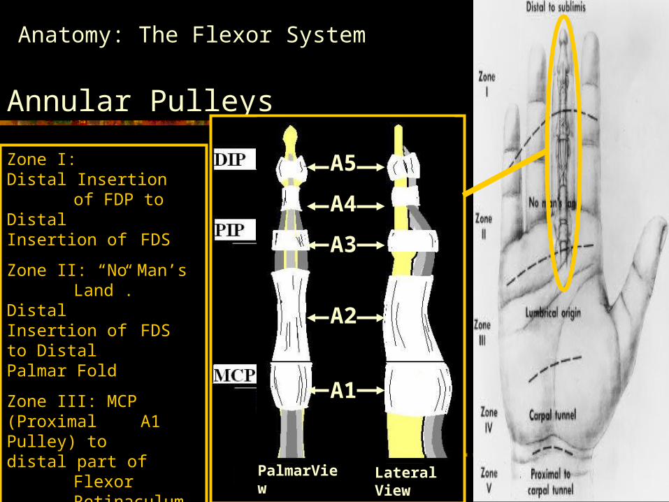
Annular Pulleys
Zone I: DIP
Zone II: Middle Phalanx
Zone III: PIP
Zone IV: Proximal Phalanx
Flexor
Digitorum
Superficialis
Flexor
Digitorum
Profundus
A5
A4
A3
A2
A1
PalmarView Lateral View
Zone I: Distal Insertion of FDP to Distal Insertion of FDS
Zone II: “No Man’s Land”. Distal Insertion of FDS to Distal Palmar Fold
Zone III: MCP (Proximal A1 Pulley) to distal part of Flexor
Retinaculum of the Carpal Tunnel
Anatomy: The Flexor System
Cruciform Pulleys
Zone I: DIP
Zone II: Middle Phalanx
Zone III: PIP
Zone IV: Proximal Phalanx
Flexor
Digitorum
Superficialis
Flexor
Digitorum
Profundus
A5
A4
A3
A2
A1
PalmarView Lateral View
Zone I: Distal Insertion of FDP to Distal Insertion of FDS
Zone II: “No Man’s Land”. Distal Insertion of FDS to Distal Palmar Fold
Zone III: MCP (Proximal A1 Pulley) to distal part of Flexor
Retinaculum of the Carpal Tunnel
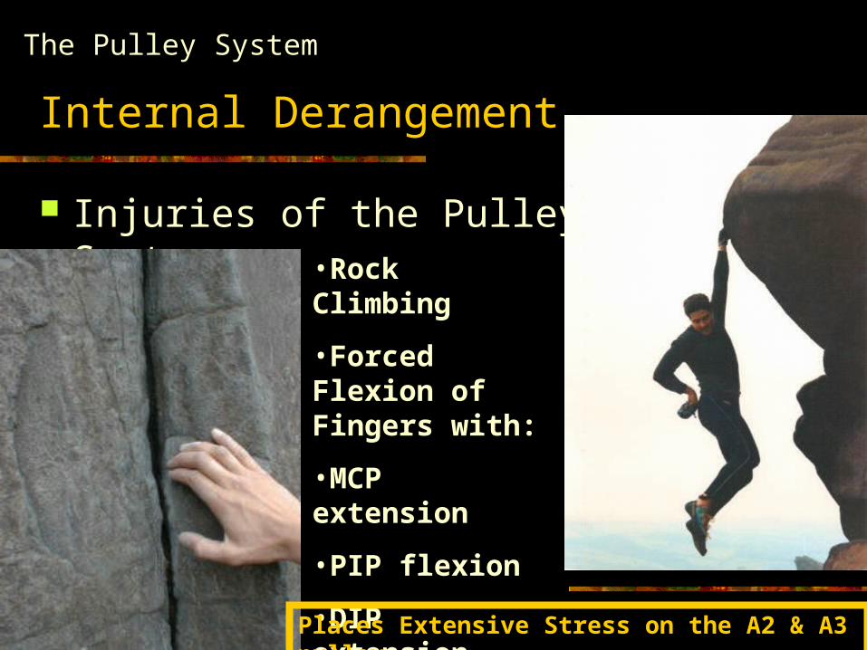
Internal Derangement
Injuries of the Pulley System
The Pulley System
•Rock Climbing
•Forced Flexion of Fingers with:
•MCP extension
•PIP flexion
•DIP extension
Places Extensive Stress on the A2 & A3 pulley
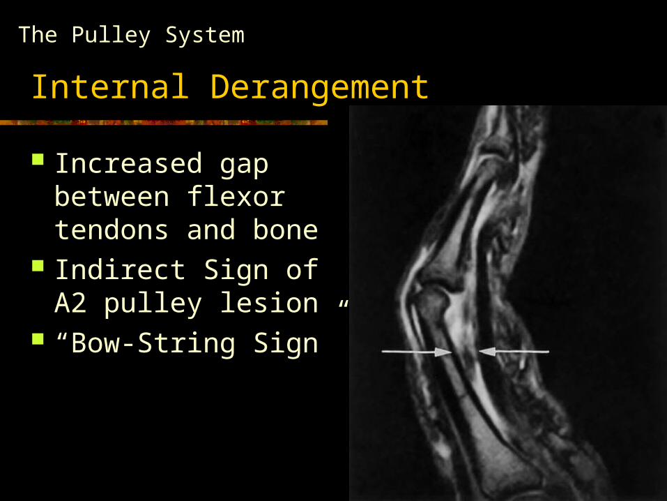
Internal Derangement
Increased gap between flexor tendons and bone
Indirect Sign of A2 pulley lesion
“Bow-String Sign”
The Pulley System
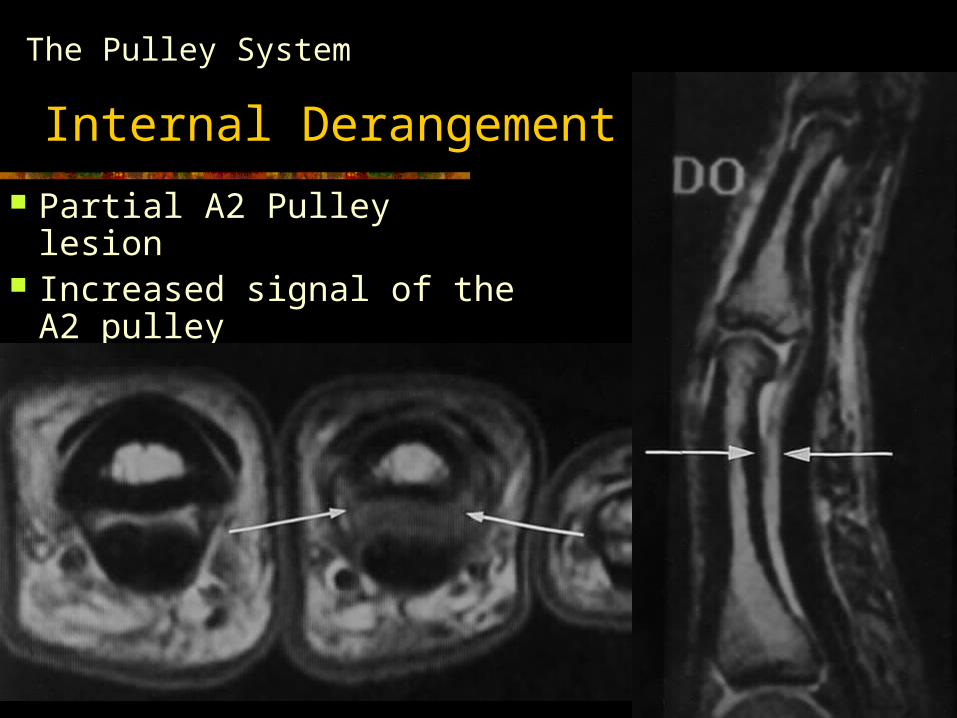
Internal Derangement
Partial A2 Pulley lesion Increased signal of the
A2 pulley
The Pulley System
Anatomy: The Flexor System
Internal Derangement
Flexor Tendon Repair
According to Strickland et al.
•Injuries involving < 60%of tendon cross sectional area should not be repaired
•Injuries involving > 60% repair with traditional core suture method supplemented with epitendinous suturing
Musculoskeletal Imaging of the Digit
Anatomy & Internal Derangement The Extensor System The Flexor System
Soft Tissue Masses and Tumors
Finger: Soft Tissue Masses & Tumors
Synovial Cyst Herniation of the
synovial membrane through joint capsule.
Ganglion Cyst Rarely communicates
with the synovium of a tendon sheath or joint.
Well-circumscribed homogeneous lesions T2 hyperintensity.
Enhancement of a thin wall after IV gadolinium
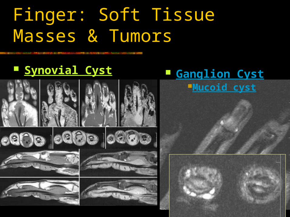
Finger: Soft Tissue Masses & Tumors
Synovial Cyst Ganglion CystMucoid cyst
Finger: Soft Tissue Masses & Tumors
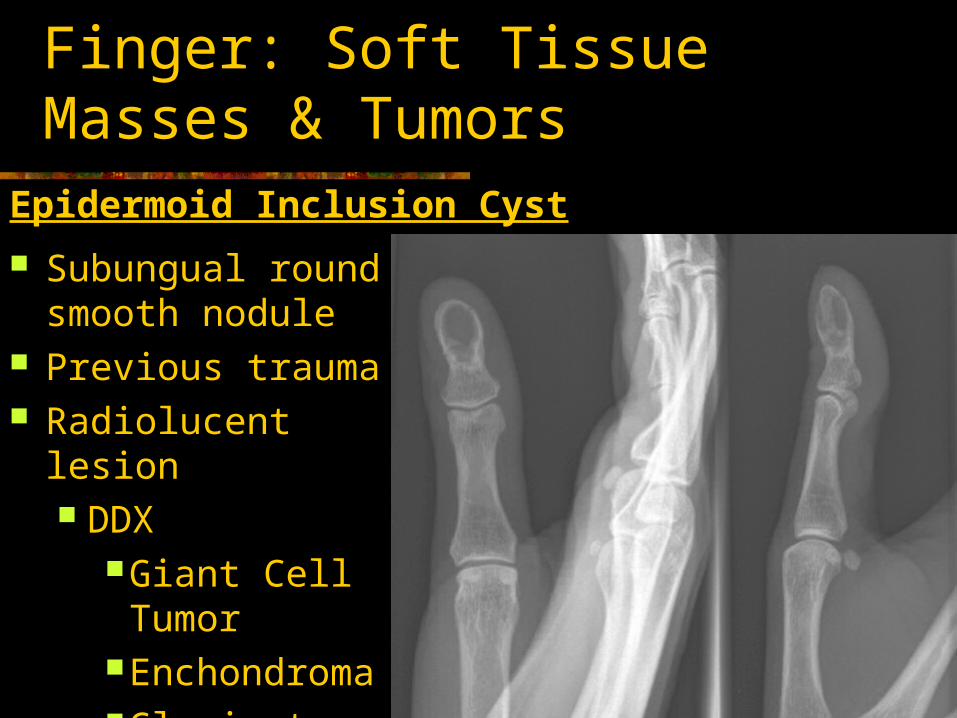
Epidermoid Inclusion Cyst
Subungual round smooth nodule
Previous trauma Radiolucent lesion
DDXGiant Cell TumorEnchondromaGlomic tumor
Finger : Soft Tissue Masses & Tumors
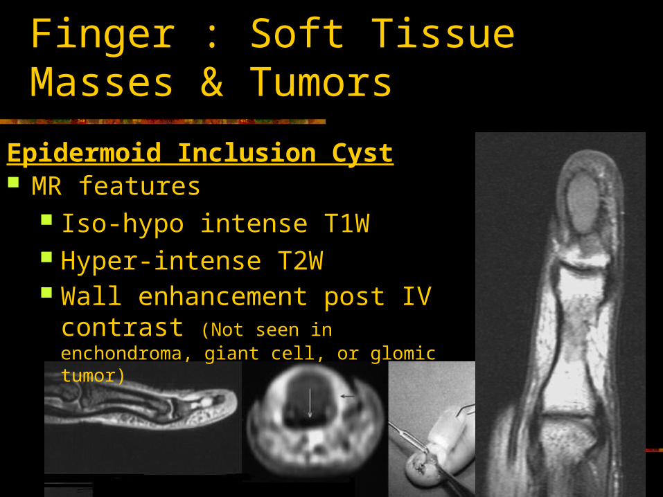
MR features Iso-hypo intense T1W Hyper-intense T2W Wall enhancement post IV
contrast (Not seen in enchondroma, giant cell, or glomic tumor)
Epidermoid Inclusion Cyst
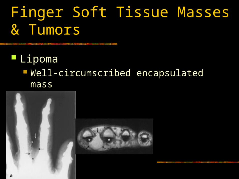
Lipoma Well-circumscribed encapsulated mass Iso-intense to fat
Finger Soft Tissue Masses & Tumors
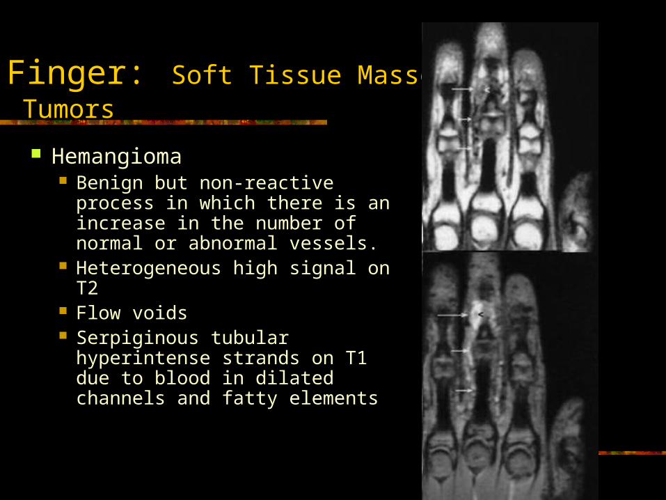
Hemangioma Benign but non-reactive process
in which there is an increase in the number of normal or abnormal vessels.
Heterogeneous high signal on T2
Flow voids Serpiginous tubular
hyperintense strands on T1 due to blood in dilated channels and fatty elements
Finger: Soft Tissue Masses & Tumors
Finger: Soft Tissue Masses & Tumors
Malignant vascular tumors of the finger are rare and when tumoral necrosis is present the DDX may include
Angiosarcoma (most frequently up to 20 years old) Epitheloid sarcoma Kaposi’s Sarcoma
Giant Cell Tumor of Tendon Sheath Second most common tumors of the hand Classified into the common localized type and the rare
diffuse type . Painless masses Most commonly occur in patients aged 30-50 years, with a
peak incidence in those aged 40-50 years. Associated with degenerative joint disease, especially in
the distal interphalangeal (DIP) joint Masses occur along the volar aspect of the hand and
fingers and are most commonly adjacent to the DIP joint. Index and long fingers most commonly involved
Finger: Soft Tissue Masses & Tumors
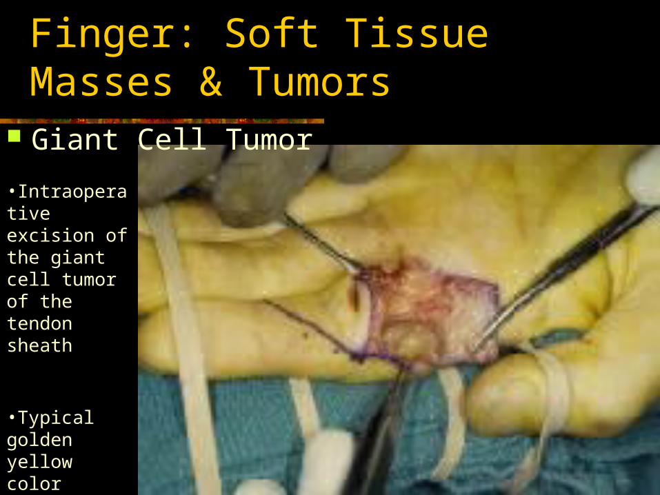
•Intraoperative excision of the giant cell tumor of the tendon sheath
•Typical golden yellow color secondary to hemosiderin deposition
Finger: Soft Tissue Masses & Tumors
Giant Cell Tumor
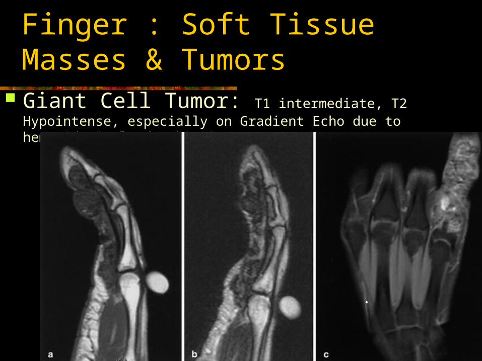
Giant Cell Tumor: T1 intermediate, T2 Hypointense, especially on Gradient Echo due to hemosiderin layden histiocytes
Finger : Soft Tissue Masses & Tumors
Finger: Soft Tissue Masses & Tumors
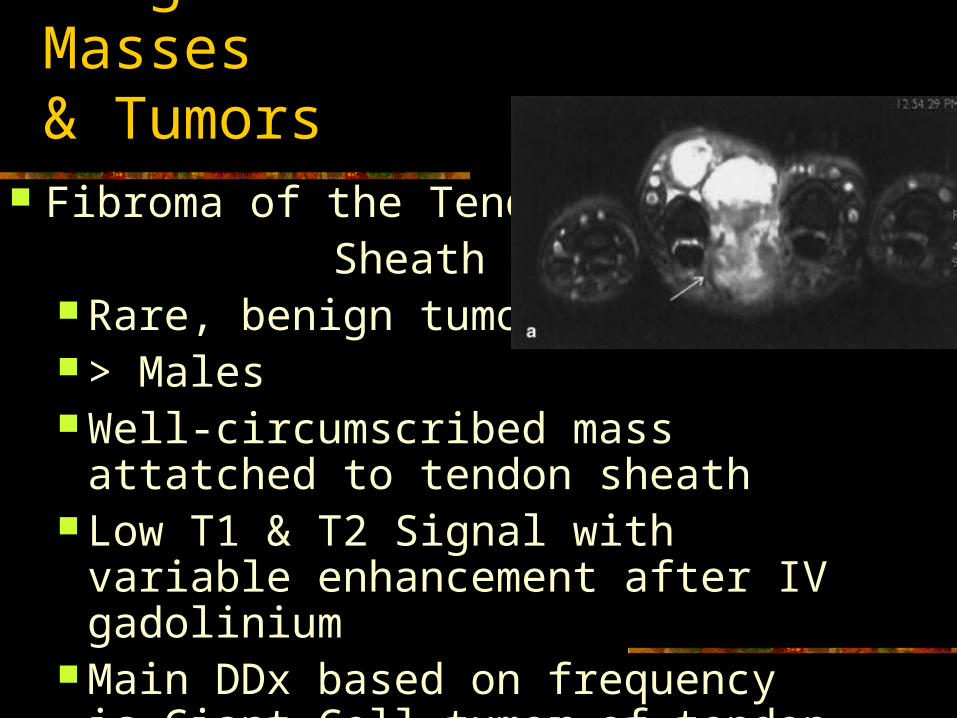
Fibroma of the Tendon Sheath
Rare, benign tumor > Males Well-circumscribed mass attatched to
tendon sheath Low T1 & T2 Signal with variable
enhancement after IV gadolinium Main DDx based on frequency is Giant
Cell tumor of tendon sheath.
Finger: Soft Tissue Masses & Tumors
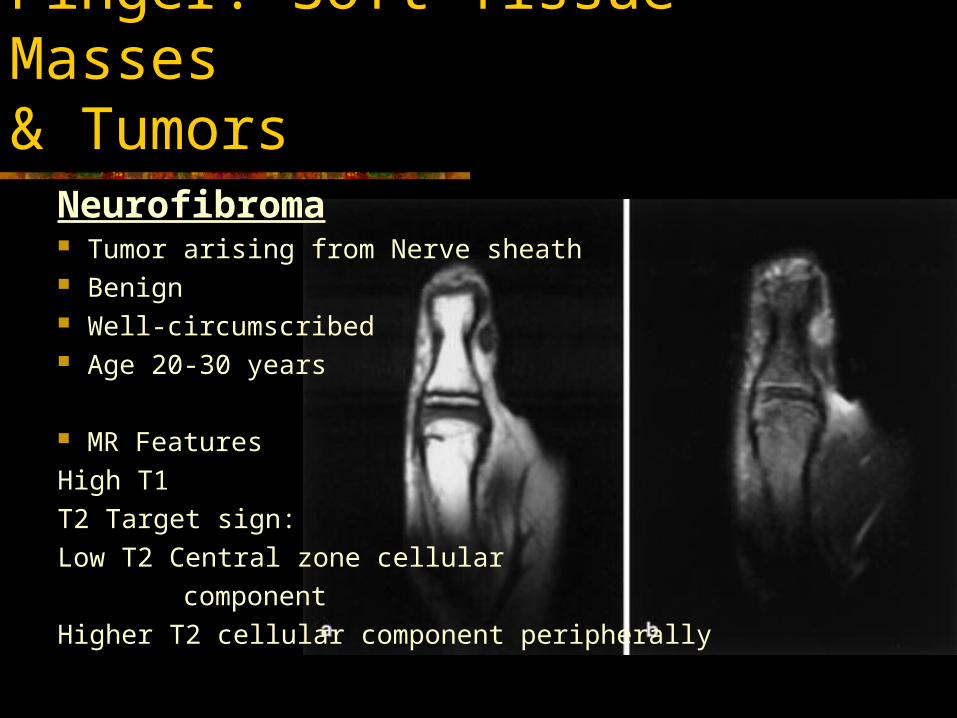
Neurofibroma Tumor arising from Nerve sheath Benign Well-circumscribed Age 20-30 years
MR Features
High T1
T2 Target sign:
Low T2 Central zone cellular
component
Higher T2 cellular component peripherally
Finger: Soft Tissue Masses & Tumors
Chondroma
•Well defined nodule of cartilage unattatched to bone.
•Age 10-70 years old
•Slow growing masses causing pain or tenderness
•Small (<3cm), firm and often mobile
•Calcification 33-70%
•Extrinsic bone erosion may be seen.
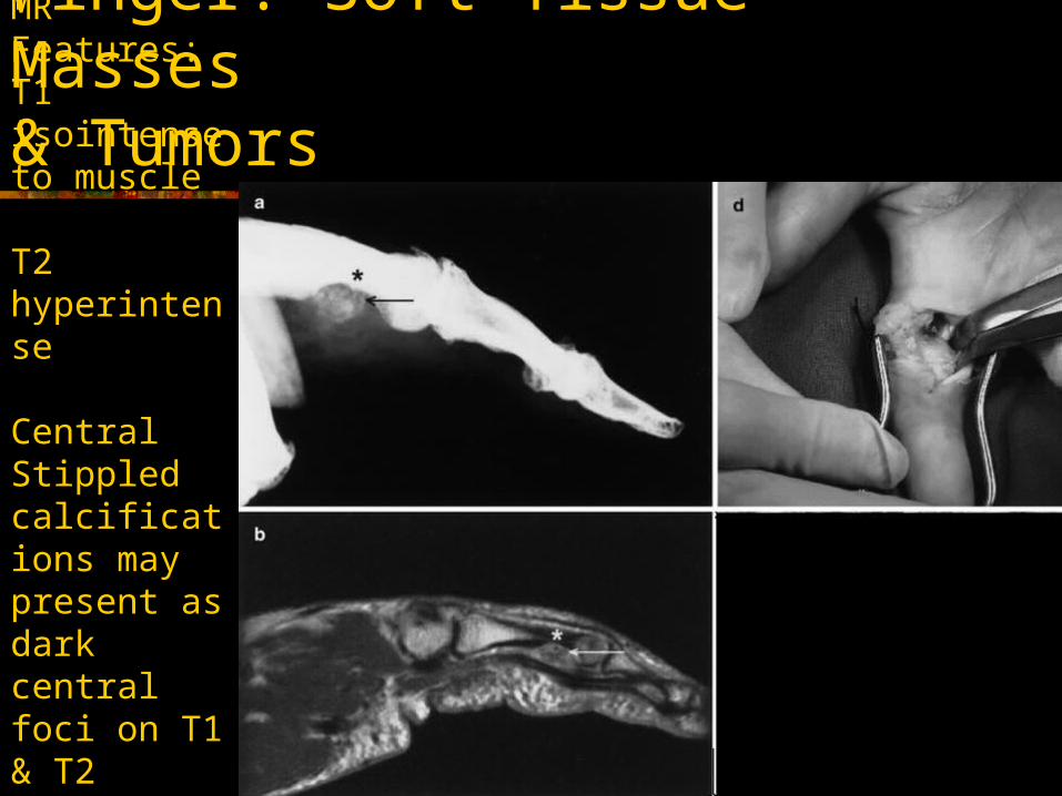
MR Features:T1 isointense to muscle
T2 hyperintense
Central Stippled calcifications may present as dark central foci on T1 & T2
Finger: Soft Tissue Masses & Tumors
J A. Clavero, et al. Extensor Mechanism of the Fingers: MR Imaging-Anatomic Correlation. Radiographics. 2003;23:593-611.)© RSNA 2003.
Milford L: Tendon injuries. In: Crenshaw AH (ed). Campbell's Operative Orthopaedics, edition 7, St. Louis, CV Mosby Co, 1987
Costa et al. MRI Features of Intersection Syndrome of the Forearm. Am J Roentgenol. 2003 Nov;181(5):1245-9
Leggit et al. Acute Finger injuries: Part I. Tendons and Ligaments. American Family Physician. Vol 73, No.5 (March 1, 2006).
Ragheb. Et al. MR Imaging of the finger tendons: Normal anatomy and commonly encountered pathology. European Journal of Radiology 56 (2005) 296-306.
Boyer et al. Flexor Tendon Repair and Rehabilitation: State of the Art in 2002. J. Bone Joint Surg. Am, September 3, 2002; 84 (9): 1684-1706.
Hauger et al. Pulley System in the Fingers: Normal Anatomy and Simulated Lesions in Cadavers at MR Imaging, CT, and US with and without Contrast Material Distention of the Tendon Sheath. Radiology 2000; 217: 201-212
Horcajadas et al. Ultrasound and MR findings in tumor and tumor-like lesions of the fingers. European Radiology. 2003: April Vol 13, No 4.672-685
References