Embed Size (px)
Citation preview

Mutations in NOTCH1 cause aortic valve diseaseVidu Garg1,5, Alecia N. Muth1†, Joshua F. Ransom1†, Marie K. Schluterman1, Robert Barnes3,4, Isabelle N. King1,5†,Paul D. Grossfeld6 & Deepak Srivastava1,2,4,5†
Calcification of the aortic valve is the third leading cause of heartdisease in adults1. The incidence increases with age, and it is oftenassociated with a bicuspid aortic valve present in 1–2% of thepopulation2. Despite the frequency, neither the mechanisms ofvalve calcification nor the developmental origin of a two, ratherthan three, leaflet aortic valve is known. Here, we show thatmutations in the signalling and transcriptional regulatorNOTCH1 cause a spectrum of developmental aortic valveanomalies and severe valve calcification in non-syndromic auto-somal-dominant human pedigrees. Consistent with the valvecalcification phenotype, Notch1 transcripts were most abundantin the developing aortic valve of mice, and Notch1 repressed theactivity of Runx2, a central transcriptional regulator of osteoblastcell fate. The hairy-related family of transcriptional repressors(Hrt), which are activated by Notch1 signalling, physically
interacted with Runx2 and repressed Runx2 transcriptionalactivity independent of histone deacetylase activity. These resultssuggest that NOTCH1 mutations cause an early developmentaldefect in the aortic valve and a later de-repression of calciumdeposition that causes progressive aortic valve disease.
Abundant evidence suggests a major inherited component to theaetiology of aortic valve disease in children and adults3,4. The mostsevere type of aortic valve obstruction in children results in failure ofthe fetal left ventricle to grow, a condition known as hypoplastic leftheart syndrome. About 10% of relatives of hypoplastic left heartsyndrome patients have bicuspid aortic valve, often undiagnosed,suggesting a common genetic aetiology with phenotypic hetero-geneity5. The valve calcification often observed in bicuspid aorticvalve is a result of inappropriate activation of osteoblast-specific geneexpression6, but the mechanism is unknown.
LETTERS
Figure 1 | NOTCH1 mutations segregate with familial aortic valvedisease. a, Kindred with five generations (indicated with Roman numerals)affected by congenital heart disease and valve calcification. Participatingmembers of each generation are indicated numerically. Deceased familymembers (slash) were unavailable for mutation analysis. Squares, males;circles, females. b, Cardiac phenotype in affected family members. AI, aorticinsufficiency; AS, aortic stenosis; AV, aortic valve; BAV, bicuspid aortic valve;
TOF, tetralogy of Fallot; VSD, ventricular septal defect. c, Sequencechromatogram of affected family members. d, Kindred with three membersaffected by congenital heart disease. e, Cardiac phenotype of familyB. DORV, double-outlet right ventricle; HLV, hypoplastic left ventricle; MA,mitral atresia; MS, mitral stenosis. f, Sequence chromatogram of affectedmembers in family B. g, Schematic of normal trileaflet aortic valve, bicuspidaortic valve and calcified aortic valve.
1Departments of Pediatrics, 2Molecular Biology and 3Internal Medicine, and 4the McDermott Center for Human Growth and Development, 6000 Harry Hines Boulevard, RoomNA8.124, University of Texas Southwestern Medical Center, Dallas, Texas 75390-9148, USA. 5Children’s Medical Center, Dallas, Texas 75235, USA. 6Department of Pediatrics,Division of Cardiology, University of California, San Diego 92123, USA. †Present address: Gladstone Institute of Cardiovascular Disease and Department of Pediatrics, Universityof California, San Francisco, 1650 Owens Street, San Francisco, California 94158, USA.
Vol 437|8 September 2005|doi:10.1038/nature03940
270© 2005 Nature Publishing Group
We identified a family of European–American descent spanningfive generations with 11 cases of congenital heart disease (Fig. 1a).Clinical evaluations demonstrated autosomal-dominant inheritanceof congenital heart disease. Nine affected family members had aorticvalve disease (Fig. 1b,g). In eight, an abnormal aortic valve was theonly cardiac malformation; six had bicuspid aortic valve, and sevendeveloped calcific aortic stenosis, including three cases in the settingof a three leaflet valve. One family member (IV-4) had an associatedabnormal mitral valve, resulting in mitral stenosis, and a ventricularseptal defect. An isolated ventricular septal defect or tetralogy ofFallot with a bicuspid pulmonary valve was identified in twoother affected family members (III-4 and IV-1, respectively). Fourfamily members have required aortic valve replacement for severecalcification. No cardiac conduction abnormalities, neurologicaldeficits, or other birth defects were identified. Detailed clinicalphenotype information for this family is shown in SupplementaryFig. 1a.
A genome-wide scan of available family members revealed linkageof the congenital heart disease phenotype to a single locus onchromosome 9q34-35 between D9S1826 and D9qter (logarithm ofodds (LOD) score, 3.5, v ¼ 0), spanning approximately 3 megabases(,9 cM) (for haplotype data, see Supplementary Fig. 2). Review of30 known (and 57 predicted) genes revealed NOTCH1, whichencodes a transmembrane receptor (2,556 amino acids) that func-tions in a highly conserved intracellular signalling pathway involvedin cellular differentiation, cell fate and lateral inhibition7. Directsequencing of NOTCH1 in an affected patient revealed a hetero-zygous C-to-T transition of nucleotide 3322 that predicted a prema-ture stop codon instead of arginine at position 1108 in theextracellular domain (Fig. 1c). All affected subjects who wereclinically evaluated had the R1108X mutation, suggesting autoso-mal-dominant inheritance of the disease phenotype with completepenetrance (Fig. 1a). The mutant allele was not detected in un-affected family members or in 1,136 unrelated subjects of diverseethnicity (Supplementary Fig. 3), making it unlikely that R1108X is arare polymorphism. In the proband (the index case), sequencingof 100 additional regulatory genes essential for, or expressedduring, cardiac development identified no other linked mutations,consistent with a monogenic aetiology (V.G. and D.S., unpublishedobservations).
Direct sequencing of NOTCH1 in a smaller, unrelated Hispanicfamily with aortic valve disease revealed a second mutation thatsegregated with three affected family members, all with bicuspidaortic valve (Fig. 1d, e). The proband (III-1) also had mitral valveatresia, hypoplastic left ventricle and double-outlet right ventricle;his sibling (III-2) and mother (II-1) had aortic valve calcification andstenosis. Family member II-2 had an ascending aortic aneurysm(Fig. 1e; see also Supplementary Fig. 1b) but no aortic valve disease. Asingle base pair deletion at position 4515 that segregated with aorticvalve disease in this family was not found in 1,138 ethnicallydiverse controls (Fig. 1f; see also Supplementary Fig. 3). Thisdeletion resulted in a frameshift mutation (H1505del) thatpredicted a severely altered protein containing 74 incorrect aminoacids at the carboxy terminus of the extracellular domain followedby a premature stop codon (Fig. 1f). These NOTCH1 mutationsgenerate truncated transcripts that probably undergo nonsense-mediated decay8 and provide compelling genetic evidence thatNOTCH1 haploinsufficiency results in human congenital heartdisease, although dominant-negative effects cannot formally beruled out.
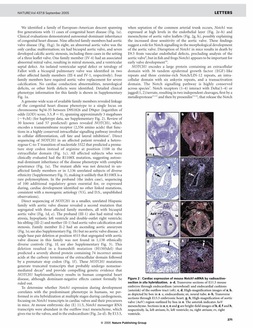
To determine whether Notch1 expression during developmentcorrelates with the predominant phenotype in humans, we per-formed in situ hybridization at multiple stages during cardiogenesis,focusing on Notch1 transcripts in cardiac valves and their precursorsin mice. At mouse embryonic day (E) 11.5, Notch1 messenger RNAtranscripts were abundant in the outflow tract mesenchyme, whichgives rise to the valves, and in the endocardium (Fig. 2a–d). By E13.5,
when septation of the common arterial trunk occurs, Notch1 wasexpressed at high levels in the endothelial layer (Fig. 2e–h) andmesenchyme of aortic valve leaflets (Fig. 2g, h), possibly explainingthe increased dose sensitivity of the aortic valve. These findingssuggest a role for Notch signalling in the morphological developmentof the aortic valve. Disruption of Notch1 in mice results in death byE9.5 from vascular endothelial defects, precluding analysis of theaortic valve9, but in fish and frogs Notch1 appears to be important forearly valve development10.NOTCH1 encodes a large protein containing an extracellular
domain with 36 tandem epidermal growth factor (EGF)-likerepeats and three cysteine-rich Notch/LIN-12 repeats, an intra-cellular domain with six ankyrin repeats, and a transactivationdomain. The Notch signalling pathway is highly conservedacross species7. Notch receptors (1–4) interact with Delta(1–4) orjagged(1, 2)/serrate, resulting in two independent cleavages, first by ametalloprotease11,12 and then by presenilin13,14, that release the Notch
Figure 2 | Cardiac expression of mouse Notch1 mRNA by radioactive-section in situ hybridization. a–d, Transverse sections of E11.5 mouseembryos through endocardium (arrowhead) and endocardial cushions(asterisk) of the outflow tract (oft). c, d, High-magnification images of a, b,as depicted by box in a. e, endocardium; nt, neural tube. e–h, Transversesections through E13.5 embryonic heart. g, h, High-magnification of aorticvalve (AoV) region outlined by box in e. The asterisk indicates AoVmesenchyme. Sections in a, c, e and g are bright-field images of b, d, f and h,respectively. la, left atrium; lv, left ventricle; ra, right atrium; rv, rightventricle.
NATURE|Vol 437|8 September 2005 LETTERS
271© 2005 Nature Publishing Group
intracellular domain from the membrane, in a manner similar to thatfirst described for sterol response element binding protein15. Notchintracellular domain translocates to the nucleus, where it interactswith the DNA-binding protein CSL (CBF-1, suppressor of hairless,and Lag-1) to activate downstream target genes, including membersof the hairy/enhancer of split (Hes) family of transcriptional repres-sors. This pathway participates in cell fate determination anddifferentiation during organogenesis throughout the embryo and isregulated by glycosylation of the extracellular EGF-like repeats inNotch1 (ref. 16).
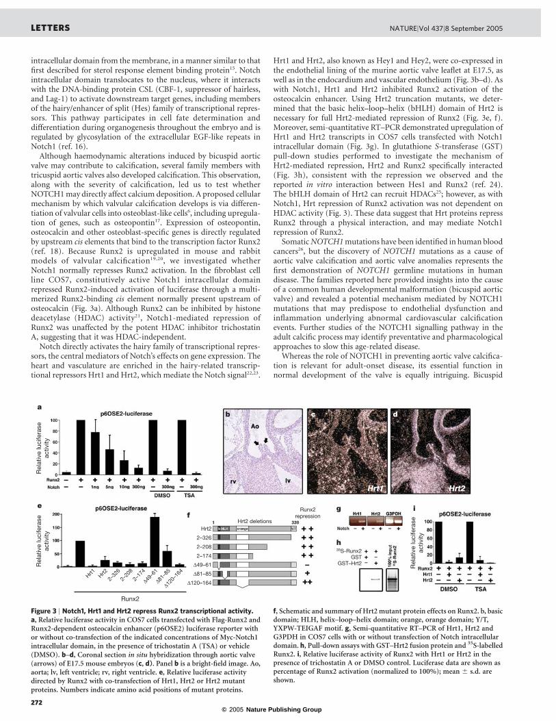
Although haemodynamic alterations induced by bicuspid aorticvalve may contribute to calcification, several family members withtricuspid aortic valves also developed calcification. This observation,along with the severity of calcification, led us to test whetherNOTCH1 may directly affect calcium deposition. A proposed cellularmechanism by which valvular calcification develops is via differen-tiation of valvular cells into osteoblast-like cells6, including upregula-tion of genes, such as osteopontin17. Expression of osteopontin,osteocalcin and other osteoblast-specific genes is directly regulatedby upstream cis elements that bind to the transcription factor Runx2(ref. 18). Because Runx2 is upregulated in mouse and rabbitmodels of valvular calcification19,20, we investigated whetherNotch1 normally represses Runx2 activation. In the fibroblast cellline COS7, constitutively active Notch1 intracellular domainrepressed Runx2-induced activation of luciferase through a multi-merized Runx2-binding cis element normally present upstream ofosteocalcin (Fig. 3a). Although Runx2 can be inhibited by histonedeacetylase (HDAC) activity21, Notch1-mediated repression ofRunx2 was unaffected by the potent HDAC inhibitor trichostatinA, suggesting that it was HDAC-independent.
Notch directly activates the hairy family of transcriptional repres-sors, the central mediators of Notch’s effects on gene expression. Theheart and vasculature are enriched in the hairy-related transcrip-tional repressors Hrt1 and Hrt2, which mediate the Notch signal22,23.
Hrt1 and Hrt2, also known as Hey1 and Hey2, were co-expressed inthe endothelial lining of the murine aortic valve leaflet at E17.5, aswell as in the endocardium and vascular endothelium (Fig. 3b–d). Aswith Notch1, Hrt1 and Hrt2 inhibited Runx2 activation of theosteocalcin enhancer. Using Hrt2 truncation mutants, we deter-mined that the basic helix–loop–helix (bHLH) domain of Hrt2 isnecessary for full Hrt2-mediated repression of Runx2 (Fig. 3e, f).Moreover, semi-quantitative RT–PCR demonstrated upregulation ofHrt1 and Hrt2 transcripts in COS7 cells transfected with Notch1intracellular domain (Fig. 3g). In glutathione S-transferase (GST)pull-down studies performed to investigate the mechanism ofHrt2-mediated repression, Hrt2 and Runx2 specifically interacted(Fig. 3h), consistent with the repression we observed and thereported in vitro interaction between Hes1 and Runx2 (ref. 24).The bHLH domain of Hrt2 can recruit HDACs25; however, as withNotch1, Hrt repression of Runx2 activation was not dependent onHDAC activity (Fig. 3). These data suggest that Hrt proteins repressRunx2 through a physical interaction, and may mediate Notch1repression of Runx2.
Somatic NOTCH1mutations have been identified in human bloodcancers26, but the discovery of NOTCH1 mutations as a cause ofaortic valve calcification and aortic valve anomalies represents thefirst demonstration of NOTCH1 germline mutations in humandisease. The families reported here provided insights into the causeof a common human developmental malformation (bicuspid aorticvalve) and revealed a potential mechanism mediated by NOTCH1mutations that may predispose to endothelial dysfunction andinflammation underlying abnormal cardiovascular calcificationevents. Further studies of the NOTCH1 signalling pathway in theadult calcific process may identify preventative and pharmacologicalapproaches to slow this age-related disease.
Whereas the role of NOTCH1 in preventing aortic valve calcifica-tion is relevant for adult-onset disease, its essential function innormal development of the valve is equally intriguing. Bicuspid
Figure 3 | Notch1, Hrt1 and Hrt2 repress Runx2 transcriptional activity.a, Relative luciferase activity in COS7 cells transfected with Flag-Runx2 andRunx2-dependent osteocalcin enhancer (p6OSE2) luciferase reporter withor without co-transfection of the indicated concentrations of Myc-Notch1intracellular domain, in the presence of trichostatin A (TSA) or vehicle(DMSO). b–d, Coronal section in situ hybridization through aortic valve(arrows) of E17.5 mouse embryos (c, d). Panel b is a bright-field image. Ao,aorta; lv, left ventricle; rv, right ventricle. e, Relative luciferase activitydirected by Runx2 with co-transfection of Hrt1, Hrt2 or Hrt2 mutantproteins. Numbers indicate amino acid positions of mutant proteins.
f, Schematic and summary of Hrt2mutant protein effects on Runx2. b, basicdomain; HLH, helix–loop–helix domain; orange, orange domain; Y/T,YXPW-TEIGAF motif. g, Semi-quantitative RT–PCR of Hrt1, Hrt2 andG3PDH in COS7 cells with or without transfection of Notch intracellulardomain. h, Pull-down assays with GST–Hrt2 fusion protein and 35S-labelledRunx2. i, Relative luciferase activity of Runx2 with Hrt1 or Hrt2 in thepresence of trichostatin A or DMSO control. Luciferase data are shown aspercentage of Runx2 activation (normalized to 100%); mean ^ s.d. areshown.
LETTERS NATURE|Vol 437|8 September 2005
272© 2005 Nature Publishing Group

and even unicuspid aortic valves typically contain a ridge where thevalve leaflets did not separate in utero (Fig. 1g). In extreme cases,blood flow may be so restricted that the left ventricle fails to grow,resulting in hypoplastic left heart syndrome, the most frequent causeof death in children with congenital heart disease. As bicuspid aorticvalve and hypoplastic left heart syndrome may represent extremes ofthe aortic valve disease spectrum, the discovery of NOTCH1 as acause of bicuspid aortic valve and a hypoplastic left ventricle in thesame family suggests that NOTCH1 mutations may be the geneticbasis for hypoplastic left heart syndrome in some patients. Futurestudies of NOTCH1 mutations in this population may reveal those atrisk for a subset of severe congenital heart lesions.
METHODSClinical phenotype evaluation and DNA collection. The congenital heartdisease families and individuals were ascertained for genetic linkage analysesat Children’s Medical Center, Dallas (University of Texas Southwestern MedicalCenter) and the University of California, San Diego. Clinical evaluations andgenetic studies were performed in accordance with human subject guidelinesafter informed consent according to the protocol approved by the individualInstitutional Review Boards. Family members were studied by history, physicalexamination, 12-lead electrocardiogram, and echocardiography. Medicalrecords were reviewed for individuals who had died. All phenotypic informationwas reviewed by cardiologists. Genomic DNA for genetic analyses was extractedfrom peripheral lymphocytes.Genetic linkage analysis. Autosomal genome linkage analysis was performedwith 372 polymorphic DNA markers at ,10-cM intervals (ABI Mapping Setv2.5). Markers were genotyped in all family members, and linkage analysis wasperformed with GENEHUNTER as described27,28. In brief, initial linkage analysisof family A, assuming 90% penetrance and a disease allele frequency of 1.5%,demonstrated the highest LOD score on chromosome 9q34-35. Phenotypicanalysis assuming 100% penetrance yielded a single peak at 9q34-35 and amaximum LOD score of 3.5.Identification of NOTCH1 mutations. All NOTCH1 exons were sequencedbidirectionally to search for sequence variations in the probands of families Aand B. Exons containing R1108X and H1505del mutations were amplified byPCR for each additional family member and sequenced bidirectionally.Sequences of the 42 primer pairs for the 34 NOTCH1 exons are available onrequest. PCR amplification was performed with the BD Biosciences AdvantageGC Genomic PCR kit following the manufacturer’s instructions, with annealingat 60 8C. Screening of identified human NOTCH1 mutations was performedwith allelic discrimination assays and the ABI Prism 7900 HT SequenceDetection System using TaqMan probes on DNA from participants in the DallasHeart Study, as described29.Radioactive-section in situ hybridization. 35S-labelled antisense riboprobeswere synthesized with T7 RNA polymerase (MAXIScript, Ambion) from 400-bppartial mouse Notch1 cDNA or plasmids encoding Hrt1 and Hrt2. With theseriboprobes, radioactive-section in situ hybridization was performed on paraffin-embedded sections of E11.5, E13.5 and E17.5 mouse embryos, as described22.Luciferase assays. COS7 cells were transfected using Fugene 6 (Roche) accordingto the manufacturer’s instructions. The reporter plasmid (250 ng), p6OSE2luciferase18, and CMV b-galactosidase expression plasmid (50 ng) to control fortransfection efficiency were transfected along with Runx2 expression plasmid(100 ng) and Hrt1, Hrt2, Notch1 intracellular domain and Hrt2 deletionexpression plasmids (300–1,000 ng). Hrt2 deletion constructs were generated asdescribed30 and protein levels of mutants kept constant. To inhibit HDAC activity,trichostatin A diluted to 0.1mM in dimethyl sulphoxide (DMSO) was added 24 hbefore collecting cell lysates. Simultaneous duplicate experiments with anidentical amount of DMSO served as a control. Immunoblots verified appro-priate protein expression. Luciferase activity was measured 40 h after transienttransfection as described30 and was normalized to LacZ expression to generaterelative luciferase activity (Fig. 3a) or expression of Hrt and Runx2 proteins(Fig. 3e). At least three independent experiments were performed in duplicate.Quantitative RT–PCR. Total RNA from COS7 cells collected 40 h after transfec-tion with empty vector and Notch1 intracellular domain expression plasmidwas purified with the Trizol method (Invitrogen). Total RNA (1 mg) wasreverse transcribed using the Superscript First Strand Synthesis System forRT–PCR (Invitrogen). PCR analysis was performed using primers specific forHrt1 and Hrt2. Glyceraldehyde 3-phosphate dehydrogenase (G3PDH) RNA wasamplified as a loading control. An annealing temperature of 56 8C was used forPCR analysis. Negative controls for each sample used non-reverse-transcribedRNA.
GST pull-down assay. Mouse GST–Hrt2 fusion proteins were purified withglutathione Sepharose 4 Fast Flow beads (Roche) for 12 h and washed twice inbinding buffer (300 mM NaCl, 50 mM Tris-HCl, pH 8.0, 1 mM EDTA, 1% TritonX-100, 1% NP-40, 0.1% SDS, 0.5 mM dithiothreitol). 35S-labelled Runx2 proteinwas synthesized using the T7 TNT coupled reticulocyte lysate system (Promega)according to the manufacturer’s instructions. Labelled protein was incubatedwith GST fusion protein (2mg) for 8–10 h at 4 8C in binding buffer with 1 mg ofnonfat dried milk to compete for nonspecific interactions. Bound proteins wereanalysed by SDS–PAGE and autoradiography.
Received 2 May; accepted 17 June 2005.Published online 17 July 2005.
1. American Heart Association, Heart Disease and Stroke Statistics—2004 Update(American Heart Association, Dallas, Texas, 2004).
2. Hoffman, J. I. & Kaplan, S. The incidence of congenital heart disease. J. Am.Coll. Cardiol. 39, 1890–-1900 (2002).
3. Cripe, L., Andelfinger, G., Martin, L. J., Shooner, K. & Benson, D. W. Bicuspidaortic valve is heritable. J. Am. Coll. Cardiol. 44, 138–-143 (2004).
4. Horne, B. D., Camp, N. J., Muhlestein, J. B. & Cannon-Albright, L. A. Evidencefor a heritable component in death resulting from aortic and mitral valvediseases. Circulation 110, 3143–-3148 (2004).
5. Loffredo, C. A. et al. Prevalence of congenital cardiovascular malformationsamong relatives of infants with hypoplastic left heart, coarctation of the aorta,and d-transposition of the great arteries. Am. J. Med. Genet. 124A, 225–-230(2004).
6. Rajamannan, N. M., Gersh, B. & Bonow, R. O. Calcific aortic stenosis: frombench to the bedside—emerging clinical and cellular concepts. Heart 89,801–-805 (2003).
7. Artavanis-Tsakonas, S., Rand, M. D. & Lake, R. J. Notch signalling: cellfate control and signal integration in development. Science 284, 770–-776(1999).
8. Frischmeyer, P. A. et al. An mRNA surveillance mechanism that eliminatestranscripts lacking termination codons. Science 295, 2258–-2261 (2002).
9. Krebs, L. T. et al. Notch signalling is essential for vascular morphogenesis inmice. Genes Dev. 14, 1343–-1352 (2000).
10. Timmerman, L. A. et al. Notch promotes epithelial-mesenchymal transitionduring cardiac development and oncogenic transformation. Genes Dev. 18,99–-115 (2004).
11. Wen, C., Metzstein, M. M. & Greenwald, I. SUP-17, a Caenorhabditis elegansADAM protein related to Drosophila KUZBANIAN, and its role in LIN-12/NOTCH signalling. Development 124, 4759–-4767 (1997).
12. Sotillos, S., Roch, F. & Campuzano, S. The metalloprotease-disintegrinKuzbanian participates in Notch activation during growth and patterning ofDrosophila imaginal discs. Development 124, 4769–-4779 (1997).
13. Struhl, G. & Adachi, A. Nuclear access and action of notch in vivo. Cell 93,649–-660 (1998).
14. Struhl, G. & Greenwald, I. Presenilin is required for activity and nuclear accessof Notch in Drosophila. Nature 398, 522–-525 (1999).
15. Brown, M. S. & Goldstein, J. L. The SREBP pathway: regulation of cholesterolmetabolism by proteolysis of a membrane-bound transcription factor. Cell 89,331–-340 (1997).
16. Haines, N. & Irvine, K. D. Glycosylation regulates Notch signalling. Nature Rev.Mol. Cell Biol. 4, 786–-797 (2003).
17. O’Brien, K. D. et al. Osteopontin is expressed in human aortic valvular lesions.Circulation 92, 2163–-2168 (1995).
18. Ducy, P., Zhang, R., Geoffroy, V., Ridall, A. L. & Karsenty, G. Osf2/Cbfa1: atranscriptional activator of osteoblast differentiation. Cell 89, 747–-754(1997).
19. Steitz, S. A. et al. Smooth muscle cell phenotypic transition associated withcalcification: upregulation of Cbfa1 and downregulation of smooth musclelineage markers. Circ. Res. 89, 1147–-1154 (2001).
20. Rajamannan, N. M. et al. Atorvastatin inhibits hypercholesterolemia-inducedcellular proliferation and bone matrix production in the rabbit aortic valve.Circulation 105, 2660–-2665 (2002).
21. Vega, R. B. et al. Histone deacetylase 4 controls chondrocyte hypertrophyduring skeletogenesis. Cell 119, 555–-566 (2004).
22. Nakagawa, O., Nakagawa, M., Richardson, J. A., Olson, E. N. & Srivastava, D.HRT1, HRT2, and HRT3: a new subclass of bHLH transcription factors markingspecific cardiac, somitic, and pharyngeal arch segments. Dev. Biol. 216, 72–-84(1999).
23. Nakagawa, O. et al. Members of the HRT family of basic helix-loop-helixproteins act as transcriptional repressors downstream of Notch signalling. Proc.Natl Acad. Sci. USA 97, 13655–-13660 (2000).
24. McLarren, K. W. et al. The mammalian basic helix loop helix protein HES-1binds to and modulates the transactivating function of the runt-related factorCbfa1. J. Biol. Chem. 275, 530–-538 (2000).
25. Iso, T. et al. HERP, a novel heterodimer partner of HES/E(spl) in Notchsignalling. Mol. Cell. Biol. 21, 6080–-6089 (2001).
26. Weng, A. P. et al. Activating mutations of NOTCH1 in human T cell acutelymphoblastic leukemia. Science 306, 269–-271 (2004).
NATURE|Vol 437|8 September 2005 LETTERS
273© 2005 Nature Publishing Group
27. Garg, V. et al. GATA4 mutations cause human congenital heart defects andreveal an interaction with TBX5. Nature 424, 443–-447 (2003).
28. Kruglyak, L., Daly, M. J., Reeve-Daly, M. P. & Lander, E. S. Parametric andnonparametric linkage analysis: a unified multipoint approach. Am. J. Hum.Genet. 58, 1347–-1363 (1996).
29. Cohen, J. et al. Low LDL cholesterol in individuals of African descent resultingfrom frequent nonsense mutations in PCSK9. Nature Genet. 37, 161–-165 (2005).
30. Kathiriya, I. S. et al. Hairy-related transcription factors inhibit GATA-dependentcardiac gene expression through a signal-responsive mechanism. J. Biol. Chem.279, 54937–-54943 (2004).
Supplementary Information is linked to the online version of the paper atwww.nature.com/nature.
Acknowledgements The authors thank the families for their participation; theDivisions of Pediatric Cardiology and Pediatric Cardiothoracic Surgery atChildren’s Medical Center Dallas for assistance with clinical information;McDermott Center for Human Growth and Development for assistance withlinkage analysis and allelic discrimination assays; Dallas Heart Study participants
and investigators for DNA samples; members of the Molecular Histology Corelaboratory for radioactive-section in situ hybridization; J. C. Cohen andH. H. Hobbs for discussions and critical review of this manuscript; K. Ivey forgraphics assistance; C. Butler, A. Garg and D. Srivastava for assistance withblood collection; G. Karsenty for Runx2 expression and p6OSE2 reporterplasmids; and L. Kedes for the GST–Hrt2 expression plasmid. This work wassupported by grants from NICHD/NIH and March of Dimes Birth DefectsFoundation to V.G., and NHLBI/NIH, March of Dimes Birth Defects Foundationand the Donald W. Reynolds Cardiovascular Clinical Research Center to D.S.J.F.R. was supported by a training grant from NIH; I.N.K. is an NICHD/NIHfellow of the Pediatric Scientist Development Program; and D.S. is anEstablished Investigator of the American Heart Association.
Author Information Reprints and permissions information is available atnpg.nature.com/reprintsandpermissions. The authors declare no competingfinancial interests. Correspondence and requests for materials should beaddressed to V.G. ([email protected]) or D.S.([email protected]).
LETTERS NATURE|Vol 437|8 September 2005
274© 2005 Nature Publishing Group