Embed Size (px)
Citation preview

NAME: _______________________ OPTION GROUP: ____________________
Cell Structure, Tissues &
Viruses
Workbook

T R L J. Version 2.0, 2018 2
Instructions
• Regular revision throughout the year is essential. It’s vital you keep a track of what you understand and what you don’t understand. This booklet is
designed to help you do this. Use the following key to note how well you understand the work after your revision. Put the letter R, A or G in the
table. If you place an R or an A then you should make a note of what you are struggling with and the end of this book under the relevant section and
seek help with this.
Key
• R = Red. I am not confident about my knowledge and understanding.
• A = Amber. I am fairly confident about my knowledge and understanding.
• G = green. I am very confident about my knowledge and understanding.
STUDY CHECKLIST AND ASSESSMENT OBJECTIVES

T R L J. Version 2.0, 2018 3
The following points are what you need to know, revise and answer questions on.
Place an R, A or G when you have revised and make notes of what you do not understand in the relevant section at the back of this booklet.
Cell Structure and Organisation. Unit 1, Section 2, Topics a, b, c, d Ultra Structure of Cells – Topic a,c.
1. Can you identify the following organelles from an electron micrograph and a drawing of a cell:
Mitochondria, chloroplast, rough endoplasmic reticulum, smooth endoplasmic reticulum, Golgi body, ribosome, vacuole, plasma (cell) membrane, centrioles, cilia, nucleus, nucleolus, nuclear membrane, nuclear pores, lysosomes, plasmodesmata and vesicles.
2. Can you state the functions of all the organelles in point 1?
3. Do you know the relative sizes of the organelles in point 1?
4. Can you describe the cell theory?
5. Do you understand the term: “organelles provide membrane bound compartments within the cell”?
6. Can you draw a fully labelled diagram of a mitochondria and a chloroplast?
7. Can you state the similarities and differences between a chloroplast and a mitochondrion?
8. Can you explain why mitochondria can look different when seen under the electron micrograph or in diagrams?
9. Can you explain why all organelles can be seen by an electron microscope but only the nucleus and chloroplast can be seen by the light microscope.
10. Can you compare the structure of a plant cell with that of an animal cell?
11. Can you fully describe the fate of amino acids from when they enter the cell to when they leave the cell as proteins? You must be able to describe the functions of the: nucleus, nucleolus, ribosome, rough endoplasmic reticulum, Golgi body and cell membrane. Also there is a link to membrane structure and transport with regard to endocytosis and exocytosis.
Prokaryotic and Eukaryotic cells – Topic b,c.
12. Can you compare and contrast a prokaryotic and a eukaryotic cell?
13. Can you recognise a prokaryotic cell from a diagram and electron micrograph?

T R L J. Version 2.0, 2018 4
Assessment Objective Description AO1 Demonstrate knowledge and understanding of scientific ideas, processes, techniques and procedures.
AO2 Apply knowledge and understanding of scientific ideas, processes, techniques and procedures:
• In a theoretical context
• In a practical context
• When handling qualitative data
• When handling quantitative data
AO3 Analyse, interpret and evaluate scientific information, ideas and evidence, including in relation to issues, to:
• Make judgments and reach conclusions
• Develop and refine practical design and procedures
The ability to select, organise and communicate information and ideas coherently using appropriate scientific conventions and vocabulary will be tested
across the AO.
14. Can you draw and fully label a prokaryotic cell?
Tissues and Organs – Topic d.
15. Can you define a tissue and an organ?
16. Can you name several different tissues and organs?
17. Can you describe the function of several tissues in relation to their structure?
18. Can you recognise tissues from a diagram?
Viruses – Topic b,c.
19. Can you describe the basic structure of a virus?
20. Can you compare the structure of a virus with a eukaryotic and prokaryotic cell?
Application of Knowledge
21. Can you apply your knowledge of cells and tissues to unfamiliar scenarios?

T R L J. Version 2.0, 2018 5
Below is a list of some key words and phrases you will need to learn and understand.
1. 70s ribosome 2. 80s ribosome 3. Aerobic respiration 4. ATP 5. Cell 6. Cell membrane 7. Cell wall 8. Centimetre (cm) 9. Centrioles 10. Chloroplast 11. Chromatin 12. Ciliated columnar epithelium 13. Cilli 14. Cisternae 15. Columnar epithelium 16. Cristae 17. Cuboidal epithelium 18. Double membrane 19. Electron 20. Endocytosis 21. Endoplasmic reticulum 22. Eukaryotic 23. Exocytosis 24. Flagella 25. Flattened sacs 26. Free ribosome 27. Goblet cell 28. Golgi body 29. Granum 30. Highly folded inner membrane 31. Histones 32. Hydrolytic enzymes 33. Increases the surface area for chemical
reactions 34. Intercellular 35. Interconnected flattened sacs 36. Inter-granum lamellae 37. Intermembrane space 38. Intracellular 39. Light microscope 40. Lysosomes 41. Magnification 42. Matrix 43. Micrometre (µm) 44. Microvilli 45. Millimetre (mm)
46. Mitochondria 47. Murein 48. Nanometre (nm) 49. Nuclear envelope 50. Nuclear pore 51. Nuclear pore 52. Nucleolus 53. Organ 54. Organ system 55. Peptidoglycan 56. Phospholipid bilayer 57. Plane of cut 58. Plasmid 59. Plasmodesmata 60. Prokaryotic 61. proteins 62. Resolution 63. Rough endoplasmic reticulum 64. Scale 65. Scanning electron microscope 66. Self-replication 67. Smooth endoplasmic reticulum 68. Stalked particle 69. Starch grain 70. Stroma 71. Thylakoid 72. Tissue 73. Transmission electron microscope 74. Vesicle 75. Wavelength of light 76. Wavelength of an electron
WORD BANK

T R L J. Version 2.0, 2018 6
In this section, you will:
1. learn how to use a light microscope to view specimens.
2. Learn how to calibrate a light microscope.
3. Learn how to calculate the actual length/width of a specimen viewed with the light microscope.
4. Learn to use the magnification equation.
5. Have further practise of using scientific notation and equivalent units of length– which was
introduced in
Using a light microscope.
• Do not drag the microscope along the bench – this leaves black marks.
• Always remove slides from the stage and hand them back in.
• Replace the microscope back where you got it from with the power cord coiled up and the plastic
cover replaced over the microscope. If using a battery-operated microscope re-plug the
microscope back into the power. These microscopes will be in the fume hood.
Viewing specimens with the light microscope.
1. Parts of the microscope.
MICROSCOPY

T R L J. Version 2.0, 2018 7
2. Setting up the light microscope to view specimens.
1. Plug in and turn on the microscope.
2. Set objective lens to X4
3. Place slide on the stage secured in place by the clips.
4. Bring the specimen into focus by using the course focus knob first then then use the fine focus knob
to make fine adjustments so the image is crisp and clear.
5. You can then change the objective lens magnification to x10 and then x40 re-focusing each time
with the fine focus knob.
3. Calibrating the light microscope.
Make notes below on how this is done.

T R L J. Version 2.0, 2018 8
4. Calculating the actual length/width of a specimen using the calibration value from step 3.
Make notes below on how this is done, below.
In cell biology, all measurements of length are made in meters. However, for cell biology the unit of meter
is too large, so we use sub units of a meter – these are shown in the table below:
Name of Unit Symbol of unit
Size of unit compared to 1m expressed as standard
notation
Size of unit compared to 1m expressed in scientific
notation
Symbol of unit
Meter m 1
Centimetre cm 0.01
Millimetre mm 0.001
Micrometre µm 0.000001
Nanometre nm 0.000000001
Scientific notation is a way to express very large or very small numbers in a more concise way. A number
written in scientific notation has a number referred to as the base. The base is then followed by x10 which
has a number as a superscript to it. This number is known as the power, or exponent. So, a number
written in scientific notation has the following form:
a x 10b where:
a = the base number.
b = the exponent or power. The exponent is a non-zero whole number that can also have a negative value,
i.e.
a x 10-b For cell measurements, the negative exponent will be used.
MATHS – THE SIZE OF CELLS AND ORGANELLES UNITS OF LENGTH

T R L J. Version 2.0, 2018 9
0.1nm
1nm
10nm
100nm
1µm
10µm
100µm
1mm
1cm
Ele
ctro
n M
icro
sco
pe Li
ght
Mic
rosc
op
e
Nak
ed E
ye
Leaf
tissue
Plant cell
Animal cell
Nucleus
Golgi Body
Vesicle
Ribosome
DNA
Below is a summary of the relative sizes of organelles, tissue and a leaf and what type of microscope is
needed to see them.
1. Complete the above table on page 8.
2. The following questions ask you to make various analyses of the numbers in the table on page 8.
(a) What is the relationship between the number of zeros and the exponent?
(b) Explain why the exponents are negative?

T R L J. Version 2.0, 2018 10
(c) For the following questions show your workings out.
How many times:
(i) Smaller is a nanometer compared to a centimetre?
(ii) Larger is a millimetre than a micrometer?
(iii) Smaller is a cenimeter than a meter?
(iv) Smaller is a millimetre than a meter?
(v) Smaller is a micrometer than a meter?
(vi) Smaller is a nanometer than a meter?
3. Consider the diagram below which shown a hypothetical situation where two cells are casting
shadows. The diagrams are not drawn to scale.

T R L J. Version 2.0, 2018 11
The length of the shadow of both cells is measured. The height of cell 2 is known. Calculate the
length of x. Show your calculations.
4. The average diameter of a human hair is 80µm. Using this as a new standard unit of length, called
the hairsbreadth (hb), express the following as hairbreadths or multiples of hairbreadths.
(a) The average human height of 1.8m
(b) One meter (1.0m)
(c) The length of a small bacterium of 200nm
5. The average length a man’s beard grows in 1 second is 1nm.
(a) Calculate the length of growth in 1 day in µm. Show your workings out.
Cell 1
3µm
0.1µm 8µm Cell 2
x

T R L J. Version 2.0, 2018 12
(b) Express your answer to part a in (i) nm and (ii) mm. Show your workings out.
6. Explain and prove the following relationships:
(a) 1nm = 0.001µm
(b) 1µm = 0.001mm
(c) 1mm = 1000µm
(d) 1µm = 1000nm

T R L J. Version 2.0, 2018 13
7 A student was making measurements of cells using an electron microscope. One measurement was
35.4µm. The student wanted to express his measurement in nanometres. He tried two different
methods to make the conversion but got two different answers. The answers were 35400nm and
35.4nm.
(a) Explain which answer is correct.
(b) Suggest the method(s) the student used to get the answers.

T R L J. Version 2.0, 2018 14
Light Microscope Electron Microscope
2.1.1 Key Features of the light and electron microscope
2.1.1 The cell theory
Key Points of the cell theory
The cell theory is a scientific theory that has been developed and expanded of many decades as a
consequence of the improvements in microscopes. The cell theory has the following basic principles:
1. All living things are composed of cells and cell products
2. New cells are formed by the division of pre-existing cells.
3. The cell contains inherited information that is used as instructions for growth, functioning and
development.
4. The cell is the functional unit of life, with the chemical reactions of life taking place within
cells.
2.1 a,c: THE CELL THEORY, COMPARISON OF THE
LIGHT AND ELECTRON MICROSCOPES & THE
IMAGES THEY PRODUCE
T R L J. Version 2.0, 2018 15
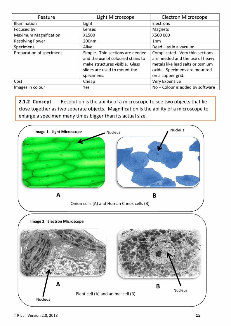
Feature Light Microscope Electron Microscope Illumination Light Electrons
Focused by Lenses Magnets
Maximum Magnification X1500 X500 000
Resolving Power 200nm 1nm
Specimens Alive Dead – as in a vacuum
Preparation of specimens Simple. Thin sections are needed and the use of coloured stains to make structures visible. Glass slides are used to mount the specimens.
Complicated. Very thin sections are needed and the use of heavy metals like lead salts or osmium oxide. Specimens are mounted on a copper grid.
Cost Cheap Very Expensive
Images in colour Yes No – Colour is added by software
Nucleus A B
Plant cell (A) and animal cell (B) Nucleus
Image 2. Electron Microscope
A Onion cells (A) and Human Cheek cells (B)
B
Nucleus Image 1. Light Microscope Nucleus
2.1.2 Concept Resolution is the ability of a microscope to see two objects that lie
close together as two separate objects. Magnification is the ability of a microscope to
enlarge a specimen many times bigger than its actual size.

T R L J. Version 2.0, 2018 16
Light Microscope Electron Microscope
The inside of a plant and animal cells are filled with organelles that lie
very close together to each other. This means that the distance or
gaps between the organelles in very small. In order to see these
organelles as separate objects the light waves must be able to pass
between them. As the wavelength of light is long the resolution of the
light microscope is very limited because the light cannot pass between
the many organelles. This makes the cell appear empty apart from the
nucleus/chloroplast (which can be seen due to their large size) even if
a high magnification is used. A low resolution can make an image
blurry.
To be able to see the smaller organelles, like the mitochondrion, the
electron microscope uses electrons. Electrons are able to pass
between the gaps between the smaller organelles so creating a very
high resolution and producing very detailed images of the cell.
2.1.3 Explanation of resolution
Low resolution creating a
blurry image. Only large
organelles like the
nucleus can be seen.
High resolution creating a
clear non-blurry image.
Small organelles like the
mitochondrion, shown
here, can be seen.
T R L J. Version 2.0, 2018 17
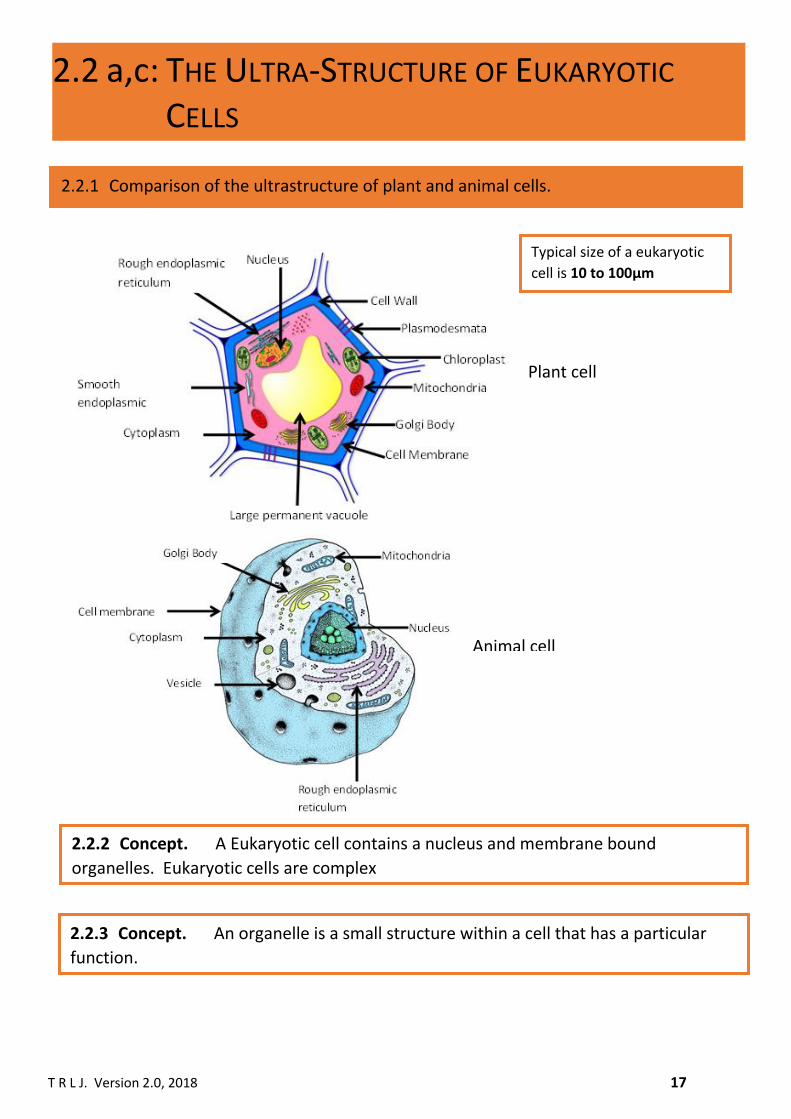
2.2 a,c: THE ULTRA-STRUCTURE OF EUKARYOTIC
CELLS
2.2.1 Comparison of the ultrastructure of plant and animal cells.
Animal cell
2.2.3 Concept. An organelle is a small structure within a cell that has a particular
function.
Plant cell
2.2.2 Concept. A Eukaryotic cell contains a nucleus and membrane bound
organelles. Eukaryotic cells are complex
Typical size of a eukaryotic
cell is 10 to 100µm

T R L J. Version 2.0, 2018 18
Typical size of the:
Nucleus is 10µm
Nucleolus is 1-2 µm
2.2.5 The nucleus, DNA, nuclear membrane and the nucleolus.
2.2.4 Concept. Organelles form compartments within cells. Each organelle is
specialised to perform a specific function. This allows biochemical reactions to be
organised within the cell and also permits different biochemical reactions to occur at
the same time. Within each organelle are the right conditions, e.g. pH and enzymes to
perform the function of that organelle.
Nuclear pores
The nucleus is present in all plant and animal cells except for the red blood cell. The nucleus is a large
organelle having a typical diameter of about 10µm. There is only one nucleus per cell. The nucleus
contains the DNA which is seen as dark staining under the electron microscope. The DNA is coiled
around protein spheres called histones to form chromatin. Also within the nucleus is a darkly stained
round structure called the nucleolus which has a typical diameter of 1-2 µm. The nucleolus is the region
of the nucleus where ribosomal RNA (rRNA) is made which is a component of ribosomes.
The nucleus is one of three organelles that have a double membrane. The membrane of the nucleus is
called the nuclear envelope. The nuclear envelope has many holes in it called nuclear pores. These
allow the movement of substances like mRNA and ribosomes out of the nucleus. Because the nucleus
contains DNA it has the function of regulating all cellular activities like protein synthesis and cell
division. DNA is the code - called the genetic code - containing the instructions to make proteins.
Nucleolus
Nuclear pore
Nuclear Envelope
Nucleoplasm
Chromatin

T R L J. Version 2.0, 2018 19
Small subunit
Large subunit
ribosome
cisternae
Electron Micrograph
of the RER
The ribosomes on the surface of the RER have a
structure represented by the drawing opposite.
They are composed of a large and a small subunit.
Both subunits are composed of rRNA and protein.
There are two basic types of ribosome called 70S
and 80S. The difference between the two is the
size of the ribosome, 70S being smaller than the
80S ribosome. Later you will learn that cells can
be grouped into prokaryotes or eukaryotes. It is
the 80S ribosome that is found in eukaryotic cells.
The 70S ribosome is found in prokaryotic cells as
well as inside the mitochondria and chloroplast
that will be covered later in this section.
Ribosomes are the site of protein synthesis where
amino acids are joined together by peptide bonds.
2.2.6 The Rough Endoplasmic Reticulum (RER) and Ribosomes
cisternae
ribosome
Drawing of the RER
The RER is an extensive network of flattened
interconnected cavities that are called cisternae
that span throughout the cytoplasm and around
the nucleus forming a regular parallel
arrangement. The RER is an extension of the outer
nuclear envelope. On the surface of the cisternae
are ribosomes – this is why we call this organelle
the rough ER as the ribosomes give a rough
appearance to the surface of the cisternae when
seen with the electron microscope.
The function of the ribosome component of the
RER is discussed in the box below. The function of
the endoplasmic reticulum component of the RER is
to transport proteins through the cell. The RER will
also form transport vesicles that contain the
protein, so they can be transported to the Golgi
Body.
Typical size of the:
Ribosome is 20nm
RER is extensive but its mass is very small.

T R L J. Version 2.0, 2018 20
Typical size of the:
SER is extensive but its
mass is very small.
2.2.7 The Smooth Endoplasmic Reticulum (SER)
The SER forms from the RER. The SER has an interconnected
tubular structure and no ribosomes on its surface. The tubular
structure form an irregular arrangement in the cytoplasm.
One of the chief functions of the SER is the synthesis of lipids
(including phospholipids) and steroids. Steroids are a type of
lipid with the sex hormones testosterone and oestrogen being
examples. Other functions of the SER include: storage of calcium
ions (usually in muscle cells where the SER is called the
sarcoplasmic reticulum) and the detoxification of drugs and
poisons (occurring in the liver).
Based on the above functions of the SER, it can be found in high
abundance in liver cells, muscle cells and the ovaries and testes.
Drawing of the SER
Electron Micrograph
of the SER

T R L J. Version 2.0, 2018 21
Vesicles
Cisternae
Drawing of the Golgi Body
Electron Micrograph of the Golgi Body
Typical size of the Golgi Body: the size is
variable, but the number of Golgi Bodies in a
cell is one, the mass is very small.
2.2.8 Golgi Body and lysosomes
Lysosomes are spherical structures with a size of 0.1 to 1.0µm. They contain around 50 different hydrolytic enzymes. Lysosomes are produced by the Golgi Body. Lysosomes have the following functions:
• Digestion of material that the cell takes in from the environment, e.g. bacteria.
• Digestion of worn-out organelles and biological molecules.
The Golgi Body consists of flattered sacs called cisternae. These cisternae are not interconnected.
Surrounding the Golgi body are many vesicles. Vesicles from the RER Join and form cisternae at one
end of the Golgi body, while at the other end secretory vesicles bud off and transport proteins to the
cell membrane where they are released by exocytosis.
The Golgi Body functions to:
• Produce lysosomes.
• Modify proteins by adding carbohydrates to them.
• Packaging proteins into vesicles for export from the cell.
• Transporting and sorting lipids.

T R L J. Version 2.0, 2018 22
2.2.9 Cell Membrane
The cell membrane has a bilayer structure made up of phospholipids. Between the phospholipids are
various proteins. The function of the cell many is to regulate the entry and exit of substances into and
out of the cell.
The membrane structure and function will be covered in detail in the membrane transport topic.
Typical size of the cell membrane is 7-9nm thick.
Drawing of the Cell Membrane
Electron Micrograph of the Cell Membrane

T R L J. Version 2.0, 2018 23
The mitochondria is the site of aerobic respiration. Here Glucose is converted into molecules of ATP
(adenosine triphosphate).
Each mitochondria have the following structural feature3s:
• Double membrane (outer and inner)
• Inner membrane is folded into cristae. On the cristae are stalked particles. The cristae is
the site of aerobic respiration.
• Intermembrane space between the outer and inner membrane.
• A fluid matrix.
• 70s ribosomes
• Circular DNA. This allows the mitochondrion to self-replicate.
Typical size of mitochondria is 6µm.
Drawing of the mitochondria
Electron Micrograph of the mitochondria
2.2.10 Mitochondria
T R L J. Version 2.0, 2018 24
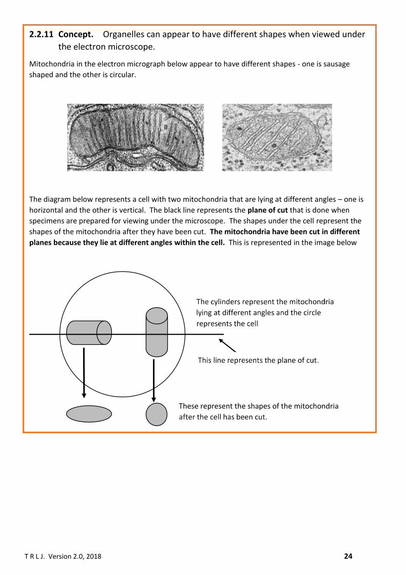
2.2.11 Concept. Organelles can appear to have different shapes when viewed under
the electron microscope.
Mitochondria in the electron micrograph below appear to have different shapes - one is sausage
shaped and the other is circular.
The diagram below represents a cell with two mitochondria that are lying at different angles – one is
horizontal and the other is vertical. The black line represents the plane of cut that is done when
specimens are prepared for viewing under the microscope. The shapes under the cell represent the
shapes of the mitochondria after they have been cut. The mitochondria have been cut in different
planes because they lie at different angles within the cell. This is represented in the image below

T R L J. Version 2.0, 2018 25
Double membrane made up of an
outer and inner membrane
Granum made up of thylakoids
Stroma
Lipid
globule
Chloroplasts are the organelles in which photosynthesis occurs. Chlorophyll is the organic molecules
that is needed to absorb light by the chloroplast. The chloroplast has the following structural features:
• A double membrane.
• Starch grains.
• A liquid stroma.
• 70s ribosomes.
• Circular DNA. This allows the chloroplast to self-replicate.
• Membranous structures called thylakoids.
• Thylakoids stack on top of each other to form grana.
• Thylakoids can link together by lamella.
Typical size of chloroplast is 10µm.
Drawing of the chloroplast
Electron Micrograph of the chloroplast
2.2.12 Chloroplast

T R L J. Version 2.0, 2018 26
2.2.13 Plasmodesmata
Drawing of the plasmodesmata
Electron Micrograph of the plasmodesmata
2.2.14 Vacuole
2.2.15 Cellulose Cell Wall
Plasmodesmata are narrow pores in the cell wall that connects neighbouring plant cells. Water can
move through the Plasmodesmata between the cells.
Plant cells have a large permanent vacuole. The vacuole has
a membrane called the tonoplast. The vacuole contains cell
sap which is a solution of sugars and amino acids dissolved
in water. The vacuole has several functions that include:
1. Water enter the vacuole by osmosis to make the cell
turgid.
2. Act as food storage.
3. The accumulation of waste products.
4. In some pants the vacuole contains coloured pigments,
e.g. beetroot cells. Electron Micrograph of the vacuole
Plants have a cell wall made of cellulose. The cell wall has the following functions:
1. It provides mechanical strength and support to cells.
2. The cell wall is freely permeable to water and substances dissolved in water.
3. Allows cells to become turgid when water enter the cell by osmosis.

T R L J. Version 2.0, 2018 27
2.2.16 Centrioles
2.2.17 Cilia
Centrioles are two short bundles of microtubules (arranged in triplets) that are positioned at right
angles to each other. They are located in the cytoplasm near the nucleus. The function of the
centrioles is to form spindle fibres during cell division.
Drawing of the Centrioles
Electron Micrograph of the centrioles
Drawing of the cilia
Electron Micrograph of the cilia
Cilia are hair like structures that project from the surface of certain cells. They can be found in the
trachea of the lungs where they are covered in mucus which traps bacteria and other particles. The
cilia then waft the mucus out of the lungs. This prevents infections occurring in the lungs.

T R L J. Version 2.0, 2018 28
• Make your own revision notes on this section using the cell structure, tissues and viruses Cornell
notes booklet. Follow the instructions in this booklet on how to use the Cornell template.
• Answer the questions on the following pages.
1. Place the names of organelles in the table below based on the feature/function.
Feature/Function of Organelle Name of Organelle(s)
Adds carbohydrates to proteins
Allows mRNA to leave the nucleus
Can self-replicate
Contains DNA
Contains ribosomes
Contains starch grains
Has a bilayer
Has a double membrane
Has an intermembrane space
Has no Membrane
Has thylakoids
Is an extension of the outer nuclear envelope
Is membrane bound
Made from β-glucose
Makes fats
Makes lysosomes
Site of ribosome synthesis
Has a tubular structure
Made of cisternae
Makes ATP
Contains hydrolytic enzymes
Makes secretory vesicles
Has interconnected cavities
ACTIVITIES/QUESTIONS ON SECTION 2.1 AND 2.2
T R L J. Version 2.0, 2018 29
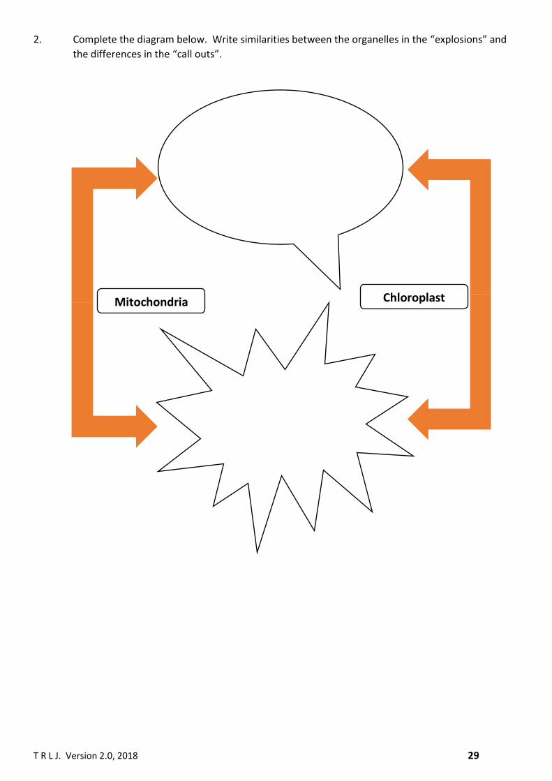
2. Complete the diagram below. Write similarities between the organelles in the “explosions” and
the differences in the “call outs”.
Mitochondria Chloroplast

T R L J. Version 2.0, 2018 30
3. The image below is of an organelle. Name, fully label and state the function of this organelle.
RER SER
Name of organelle:
____________________
Function:
____________________________

T R L J. Version 2.0, 2018 31
4. State the name of the organelle below and describe what is happening.
5. What is the name and function of the organelle below and is there more than one of this organelle
in the image.
6. The diagram on the next page is of a cell from the pancreas. The pancreas has many functions
that include the production of the hormone insulin and digestive enzymes like trypsin. Fill in the
boxes numbered 1 to 6 with the most suitable description as to the function of the various
organelles in the pancreatic cell. You need to name all the organelles as well.

T R L J. Version 2.0, 2018 32
6
5
4
3
2
1

T R L J. Version 2.0, 2018 33
7. The image below has been produced using a microscope.
(a) What type of cell is shown in the image? Explain your answer.
(b) Explain what microscope was used to produce this image.
(c) Calculate the magnification of the image.

T R L J. Version 2.0, 2018 34
8. Identify the word or phrase that describes the following statements.
(i) A description of the inner membrane of the mitochondria.
(ii) A stack of thylakoids.
(iii) The liquid in a chloroplast.
(iv) The contents of lysosomes.
(v) The site of protein synthesis.
(vi) Starch grains are found in this organelle.
(vii) This reaction occurs in mitochondria.
(viii) The ability of a microscope to enlarge a specimen.
(ix) The site of rRNA synthesis.
(x) An organelle that occurs in pairs.
(xi) Secretory vesicles are made by this organelle.

T R L J. Version 2.0, 2018 35
(xii) The Golgi body is composed of these structures.
(xiii) Mitochondria, chloroplasts and the nucleus have this structure in common.
(xiv) A feature of chloroplasts and mitochondria allowed by the presence of DNA.
(xv) A feature of the light microscope that limits its resolution.
(xvi) The name for the inner mitochondrial membrane.
(xvii) An organelle found in bacterial cells.
(xviii) An organelle vital for cell division.

T R L J. Version 2.0, 2018 36
Question
Number 2
Which AO is being Tested
– AO1, AO2, AO3
Which number from the
revision checklist is being
tested
i
ii
iii
iv
v
vi
vii
vii
a
b
c
vii
iv

T R L J. Version 2.0, 2018 37
2.3.2. The structure of a bacterial cell – a typical prokaryotic cell.
Prokaryotic cells have the following features:
• No membrane bound organelles
• No nucleus
• A mesosome. This is an infolding of the cell membrane and is where respiration occurs.
• A size of 0.1-10µm.
• A slime capsule for protection.
• A cell wall made form peptidoglycan (also called murein).
• Have flagella.
• 70s ribosomes
• Have chromosomal DNA which does not have histones. DNA is in the cytoplasm.
• Have plasmid DNA.
Drawing of a bacterial cell
2.3: PROKARYOTIC CELLS
2.3.1 Concept. Prokaryotic cells do not have any membrane bound organelles or a nucleus. They do have a ribosome (ribosomes do not have a membrane) but it is a small ribosome so is called 70s. Prokaryotic cells are bacteria.

T R L J. Version 2.0, 2018 38
1. The table below shows some features of eukaryotic and prokaryotic cells. However, mistakes
have been made. Fill in the blank table with all the mistakes corrected.
PROKARYOTES EUKARYOTES Bacteria Animals
Cell wall present in some bacteria Cell wall present in fungi
10 times smaller than eukaryotes 10 times bigger than prokaryotes
DNA has histones DNA lacks histones and is circular
70s ribosomes 80s ribosomes
PROKARYOTES EUKARYOTES
2. “Mitochondria have a different structure to prokaryotic cells”. Discuss this statement.
ACTIVITIES/QUESTIONS ON SECTION 2.3

T R L J. Version 2.0, 2018 39
Name of epithelial
tissue
Structure Location Function
Squamous
Flat, thin cells with an irregular shape
Blood vessels, bowmen’s capsule, alveoli.
Provides a smooth surface in blood vessels, allows diffusion to occur in the alveoli.
Cuboidal
Cube shaped cells.
Kidney tubules. Forms tubes.
Columnar
Rectangular shaped cell with a large nucleus.
Digestive system. Secretion of mucus (when goblet cells are present), absorption of nutrients.
Ciliated columnar
Rectangular shaped cells with a large nucleus and cilia.
Trachea of the lungs, fallopian tubes.
In the trachea the cilia move bacteria and other particles trapped in mucus out of the lungs. In the fallopian tubes the cilia waft and help move the egg cell towards the uterus.
2.4: TISSUES, ORGANS AND ORGAN SYSTEMS
2.4.1 Definition: A tissue is a collection of cells with a similar structure and a similar function that lie on a basement membrane.
2.4.4 Epithelial tissues
2.4.2 Definition: An organ is a collection of different tissues, having a different structure and a different function.
2.4.3 Definition: An organ system is a group of different organs that work together to perform one or more functions.
T R L J. Version 2.0, 2018 40
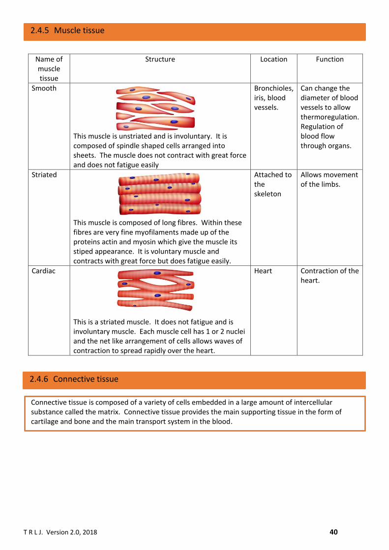
Name of muscle tissue
Structure Location Function
Smooth This muscle is unstriated and is involuntary. It is composed of spindle shaped cells arranged into sheets. The muscle does not contract with great force and does not fatigue easily
Bronchioles, iris, blood vessels.
Can change the diameter of blood vessels to allow thermoregulation. Regulation of blood flow through organs.
Striated This muscle is composed of long fibres. Within these fibres are very fine myofilaments made up of the proteins actin and myosin which give the muscle its stiped appearance. It is voluntary muscle and contracts with great force but does fatigue easily.
Attached to the skeleton
Allows movement of the limbs.
Cardiac This is a striated muscle. It does not fatigue and is involuntary muscle. Each muscle cell has 1 or 2 nuclei and the net like arrangement of cells allows waves of contraction to spread rapidly over the heart.
Heart Contraction of the heart.
2.4.6 Connective tissue
2.4.5 Muscle tissue
Connective tissue is composed of a variety of cells embedded in a large amount of intercellular substance called the matrix. Connective tissue provides the main supporting tissue in the form of cartilage and bone and the main transport system in the blood.

T R L J. Version 2.0, 2018 41
Name of connective
tissue
Structure Location Function
Cartilage
The matrix of cartilage contains chondrin and embedded in it are chondrocytes and fine fibres of collagen. The resulting tissue is hard but flexible.
Found at the end of bones, in the ear, noise, and respiratory system. Comprises the skeleton of cartilaginous fish and sharks.
Support and protection.
Bone
The matrix of bone is made up of collagen together with inorganic substances like calcium and phosphopurs. These components, along with bone cells, called osteocytes, are arranged in circles called lamella that form an Haversion system. The Haversion canel contains blood vessels and nerves.
skeleton Support and movement.

T R L J. Version 2.0, 2018 42
Name of Organ system
Organs Tissues in each organ
Circulatory Heart, blood vessels Cardiac muscle, smooth muscle, nervous tissue, connective tissue, epithelia
Respiratory Lungs Smooth muscle, elastic tissue, connective, nervous tissue, epithelia.
Digestive Intestines, stomach Smooth muscle, elastic tissue, connective, nervous tissue, epithelia,
Renal kidney Epithelia tissue, nervous tissue
Nervous Brain Nervous tissue
2.5: VIRUSES
2.4.7 Organ Systems
Drawing of a bacteriophage
Electron micrograph of a bacteriophage
2.5.1 Structure of viruses

T R L J. Version 2.0, 2018 43
Electron micrograph of influenza virus
Drawing of a typical animal virus
Viruses any extremely small structures that range in size from 20nm to 300nm. Viruses can infect both animal and bacterial cells. Viruses that infect bacteria are called bacteriophages. Viruses do not have a typical cell structure – they have no organelles and can only survive and reproduce by infecting host cells and taking over their biochemical process to produce for viruses. In this regard viruses are describe as obligate intracellular parasites. A typical animal virus consists only of an outer protein coat embedded in which are protein antigens that makes the viruses pathogenic. Inside the virus with either be DNA or RNA. Viruses with RNA are called retroviruses.
Nucleic acid
Protein coat (capsid)
Surface antigens

T R L J. Version 2.0, 2018 44
1. Complete the crossword
PUTTING IT ALL TOGETHER

T R L J. Version 2.0, 2018 45
2. Complete the following multiple-choice questions.
(a) Which statement about mitochondria is incorrect
(i) They produce ATP
(ii) They can make their own proteins.
(iii) They have an intramembrane space.
(iv) They have a double membrane.
(b) What is/are the advantage(s) of using an electron microscope
I. Very high resolution
II. Very high magnification
III. The possibility of examining living material
(i) I only
(ii) I and II only
(iii) II and III only
(iv) I, II and III
(c) In the diagram below macromolecules are being transported to the exterior of the cell.
What is the name of this process?
(i) Exocytosis
(ii) Pinocytosis
(iii) Endocytosis
(iv) Phagocytosis

T R L J. Version 2.0, 2018 46
(d) What are the structures labelled I, II and III in the diagram below.
(e) The diagram below shows a longitudinal section through a chloroplast
(i) I
(ii) II
(iii) III
(iv) IV
I II III
i Outer membrane Crista Matrix
ii Outer membrane Crista Stroma
iii Plasma membrane Inner membrane Matrix
iv Plasma membrane Inner membrane Stroma

T R L J. Version 2.0, 2018 47
(f) Which processes are represented by the labels in the diagram below?
3. There are two passages below, mistake 1 and mistake 2, that contain incorrect information. Read
the passages underlining the mistakes and write the correction above the mistake.
MISTAKE 1
Organelles are intercellular structures that all have a membrane. The largest organelle is the ribosome
that can be free floating in the cytoplasm or found on the surface of the smooth endoplasmic reticulum.
There are two organelles that have a double membrane – these are the mitochondria and the Golgi Body.
The function of the mitochondrion is to make energy by anaerobic respiration. The anaerobic respiration
occurs on the cisternae of the mitochondrion which forms a highly folded inner membrane. The Golgi
body receives proteins from the ribosome via a vesicle. The vesicle fuses with the Golgi Body and the
protein enters the Golgi Body to be modified with sugar molecules.
MISTAKE 2
The smooth endoplasmic reticulum is an extension of the outer nuclear envelope and consists of
interconnected cristae. Chloroplasts contain the inorganic molecule xanthophyll which absorbs light
I II
i A phagocyte ingesting a microbe by exocytosis
Digestion of the microbe with the help of the Golgi body
ii A Phagocyte ingesting a microbe by endocytosis
Digestion of the microbe with the help of a lysosome
iii A phagocyte ingesting a microbe by exocytosis
Digestion of the microbe with the help of a lysosome
iv A phagocyte ingesting a microbe by endocytosis
Digestion of the microbe with the help of the Golgi body

T R L J. Version 2.0, 2018 48
energy to allow photosynthesis to occur. Chloroplasts have a liquid matrix in which are found membrane
bound structures called thylakoids. The nucleus has many nuclear pores that allow the transport of DNA
out of the nucleus and into the cytoplasm. Within the nucleus is a darkly stained region called the
nucleolus where mRNA is synthesised. The cell membrane is composed of phospholipids and proteins.
The phospholipids form a monolayer that prevents intercellular contents from leaking out. The thickness
of the cell membrane is approximately 7-9µm.
4. Assertion & Reason
Each of the following questions consists of two statements, one which is an “Assertion”, the other a
“Reason”. You must determine whether each statement is correct or incorrect, i.e. true or false. If both
are true, you must decide whether the reason is an accurate explanation for the assertion. You then need
to selection one option (from A to E) as shown below:
Option Assertion Reason A True True – and the reason explains the assertion. B True True – But the reason does not explain the assertion. C True False D False True E False False

T R L J. Version 2.0, 2018 49
Question Assertion Reason Answer
1 The electron microscope has a low resolution.
Because of the high wavelength of electrons.
2 Bacterial cells do not have organelles. Because they are prokaryotes.
3 A virus is an obligate intracellular parasite. Because it relies on other cells for the synthesis of new viruses.
4 The cell membrane is a bilayer. Because it is composed of a layer of phospholipids.
5 Mitochondria and chloroplasts are not similar in structure.
Because they both have 80s ribosomes.
6 Cells of the ovary will have abundant smooth endoplasmic reticulum.
Because they make proteins.
7 The inner mitochondrial membrane forms cisternae.
Because a large area is needed for reaction to take place.
8 rRNA is made in the nucleolus. Because DNA contains the genetic code that is needed to make a protein.
9 The cell wall of plants has a high tensile strength.
Because its made from a polymer of alpha glucose.
10 Water can travel from one plant cell to another.
Because the cell wall is fully permeable.
11 Benedict’s reagent turns brick red when heated with maltose.
Because maltose is a non-reducing sugar.
12 Chloroplasts and mitochondria were once though to have originated from prokaryotic cells by the endosymbiotic theory.
Because mitochondria and chloroplasts have a cell.
14 Respiration occurs in the mitochondria. Because the oxygen concentration in the mitochondria is high.
15 The number of mitochondria in cells can increase.
Because mitochondria can divide.
16 Proteins are modified after they are synthesised.
Because they are transported through the cell in vesicles.
17 When a tube containing a suspension of nuclei and ribosomes was spun at 1000 times the force of gravity the ribosomes formed a pellet at the bottom of the tube, but the nuclei did not.
Because ribosomes are heavier than the nuclei.
18 The nucleus can be seen with the aid of a light microscope.
Because the light microscope has a magnification of X5000.
19 Plant cells do not burst when they become turgid.
Because the vacuole expands to accommodate the excess water.
20 The number of bacteria present in the human body, i.e. in the colon, the skin etc exceed the total number of cells that make up the human body.
Because bacterial cells are approximately 10 times smaller than eukaryotic cells.

T R L J. Version 2.0, 2018 50
5. Distinguish between the structures of the following pairs of organelles.
(i) Mitochondria and the nucleus.
(ii) A virus and a prokaryotic cell.
(iii) The rough endoplasmic reticulum and the smooth endoplasmic reticulum.

T R L J. Version 2.0, 2018 51
ANSWERING QUESTIONS WITH QER
The long answer question requires you to write at length about a topic. The quality of your extended
responses (QER) will also be assessed. The mark scheme will have the following areas:
1. Indicative Content – this is the biological concepts and terminology you are expected to use in your
answer.
2. 7-9 marks – to gain these top marks your answer will show:
The candidate constructs an articulate, integrated account correctly linking relevant points, such as
those in the indicative content, which shows sequential reasoning. The answer fully addresses the
question with no irrelevant inclusions or significant omissions. The candidate uses scientific
conventions and vocabulary appropriately and accurately.
3. 4-6 marks – to gain these marks your answer will show:
The candidate constructs an account correctly linking some relevant points, such as those in the
indicative content showing some reasoning. The answer addresses the question with some
omissions. The candidate uses scientific conventions and vocabulary appropriately and accurately.
4. 1-3 marks – to gain these marks your answer will show:
The candidate makes some relevant points, such as those in the indicative content, showing limited
reasoning. The answer addresses the question with significant omissions. The candidate has
limited use of scientific conventions and vocabulary.

T R L J. Version 2.0, 2018 52
Complete the QER questions number 6 below.
6. Cells perform all the processes and reactions that are required to keep organisms alive and
functioning correctly. This includes aerobic respiration, digestion of food and the synthesis of
macromolecules. For a cell to perform these functions it requires the correct organelles in high
abundance. The electron micrographs below are of two different cells from a mammal (cell A and
cell B) – the images have different magnifications. One cell is from the testes, which only become
metabolically active at puberty, and the other cell is from a region of the pancreas called the islets
of Langerhans. Cells in the islets of Langerhans are metabolically active from birth and function in
regulating blood glucose levels.
Describe the structure and function of the organelles visible in the electron micrograph images and so
identify the function of the cells. [9]
Cell A Cell B

T R L J. Version 2.0, 2018 53

T R L J. Version 2.0, 2018 54

T R L J. Version 2.0, 2018 55
1. The unicellular yeast Saccharomyces cerevisiae divides by budding off a small new cell that then
grows to full size. During its growth, the new cell synthesises new cytoplasm, which increases its
volume, and a new cell membrane, which increases its surface area. The micrograph below shows
a yeast cell undergoing budding. The shape of the yeast cells can be approximated to a sphere
ai. Determine the diameter of the parent cell and the new cell.
aii Calculate the volume of each cell using the formula for the volume of a sphere.
𝑉 =4
3𝜋𝑟3
APPLICATION AND EXTENSION

T R L J. Version 2.0, 2018 56
aiii. How much new cytoplasm will the new cell have to synthesis as it matures?
As the cell grows, its cell membrane needs to expand to contain the increased volume of the cell
bi Calculate the surface area of each cell using the formula for the surface area of a sphere.
𝐴 = 4𝜋𝑟2
bii How much area of the new cell membrane will the new cell have to synthesise as it
matures?
c when the cell matures, it will be approximately how many times greater in volume and how
many times greater in surface area that its current size?

T R L J. Version 2.0, 2018 57
2. To determine the structure and function of organelles scientists must first isolate them from cells
by cell fractionation. This procedure is illustrated below.
a. Cell fractionation requires precise conditions and procedures to allow the isolation of the
organelles. Suggest a function for the following conditions and procedures:
i homogenization.
ii ice cold temperatures

T R L J. Version 2.0, 2018 58
iii the use of a buffer
iv the use of isotonic solutions
b. Centrifugation is a method where specimens are spun in a circular motion at very high speeds in
an instrument called a centrifuge. The speed at which the centrifuge is spinning is often expressed
as a force value as compared to the force of gravity.
i. Using your own knowledge and the diagram, explain why differential centrifugation was used
and what organelles/subcellular structures would be found in the pellet of the tubes
centrifuged for 10 mins, 20 mins, 60 mins and 3 hours.
ii. Imagine a protein called X, that is destined to span the cell membrane. Assume that the
mRNA carrying the genetic message for protein X has already been used to make the protein
by ribosomes in a cell culture. If you fractionate the cells in which fraction would you find
protein X? Explain by describing its transit through the cell.

T R L J. Version 2.0, 2018 59
3. Mitochondria and chloroplasts display similarities with bacteria that led to the endosymbiont
theory. This theory states that an early ancestor of eukaryotic cells engulfed an oxygen-using non-
photosynthetic prokaryotic cell. Eventually, the engulfed cell formed a relationship with the host
cell in which it was enclosed, becoming an endosymbiont. Indeed, over the course of evolution, the
host cell and its endosymbiont merged into a single organism, a eukaryotic cell with a
mitochondrion. At least one of these cells may have then taken up a photosynthetic prokaryote,
becoming the ancestor of eukaryotic cells that contain chloroplasts. This theory is consistent with
the many structural features of mitochondria and chloroplasts. Mitochondria and chloroplasts have
developed into autonomous structures within eukaryotic cells.
a. Explain how the endosymbiont theory is “consistent with many structural features of
mitochondria and chloroplasts”.

T R L J. Version 2.0, 2018 60
b. Draw an annotated diagram(s) to summarise the evolution of eukaryotic plant and animal
cells.
c. Explain how mitochondria and chloroplasts can act autonomously within eukaryotic cells.
T R L J. Version 2.0, 2018 61
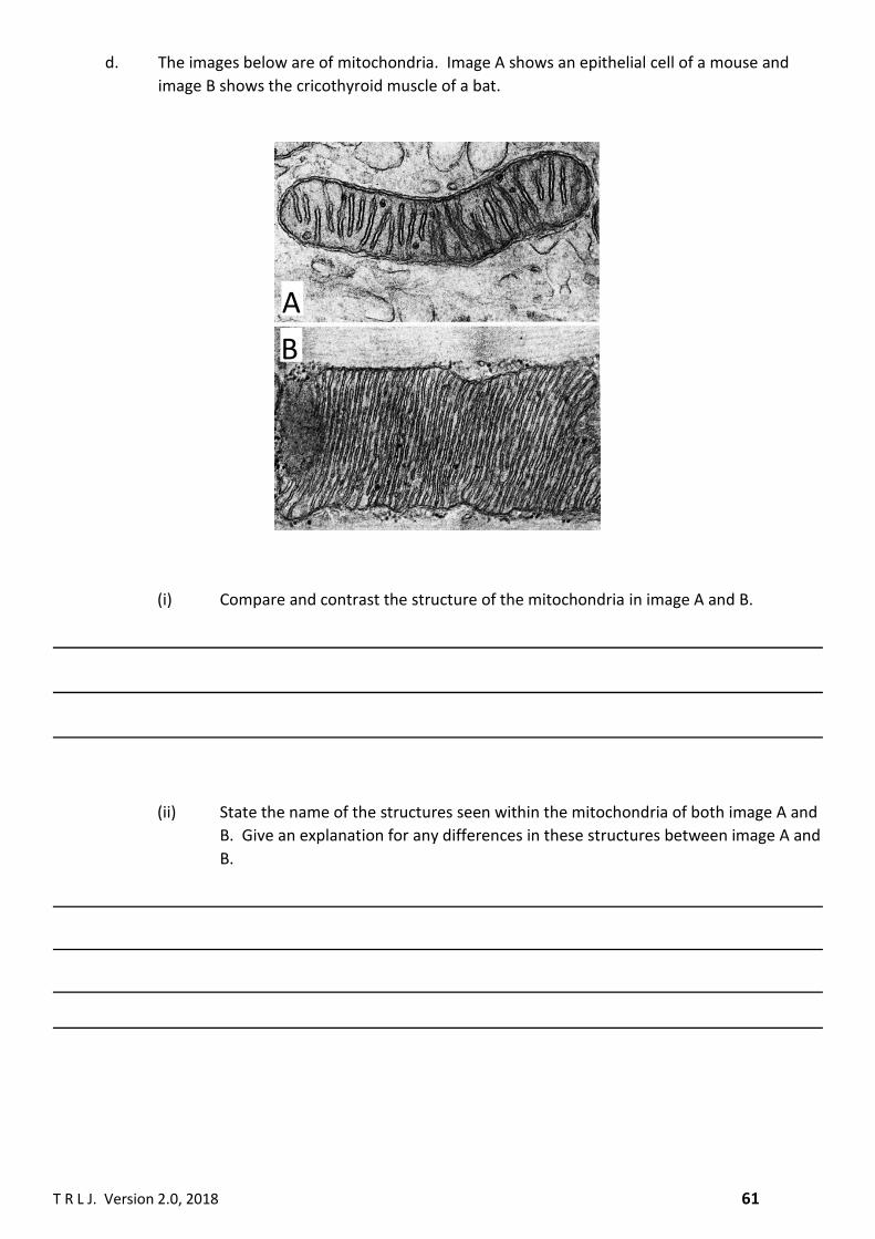
d. The images below are of mitochondria. Image A shows an epithelial cell of a mouse and
image B shows the cricothyroid muscle of a bat.
(i) Compare and contrast the structure of the mitochondria in image A and B.
(ii) State the name of the structures seen within the mitochondria of both image A and
B. Give an explanation for any differences in these structures between image A and
B.
A
B

T R L J. Version 2.0, 2018 62
4. The acronym “GERL” stands for Golgi – Endoplasmic Reticulum – Lysosomal complex. In the space
below draw an annotated diagram in which you show the function(s) of each of the named
organelles and how the organelles are dependent and interact with each other in the complex.

T R L J. Version 2.0, 2018 63
Comprehension 1
Bacterial cells have no nucleus and no membrane bound organelles. All other cells do have a nucleus 1
and a variety of other organelles bound by a membrane including mitochondria, lysosomes, nuclei and 2
endoplasmic reticulum. These organelles are separated from each other and the cytoplasm by their 3
membranes forming separate compartments. 4
The endoplasmic reticulum (ER) is an extensive membranous network of interconnected tubes 5
and sacs that forms part of the endomembrane system. The ER can only be seen by the electron 6
microscope and account for over 50% of the total membrane types of the cell. When the ER was first 7
discovered by cell biologists it was though that the ER was formed from invaginations of the cell 8
membrane, however, no real evidence was found to support this hypothesis. 9
There are two types of ER: smooth ER (SER) and Rough ER (RER) (figure 1). RER accounts for 10
35% of the total membrane types of liver cells and 60% of pancreatic endocrine cells, while the SER 11
accounts for 16% in liver cells and <1% in pancreatic endocrine cells. 12
Despite having different structures, the RER and SER are connected to each other. The functions of the 13
RER and the SER are, also, different. The RER is involved with the production of proteins, but the SER is 14
not. The SER is involved in a wide range of important activities that include: storage of calcium, 15
carbohydrate metabolism, detoxification of drugs and poisons and the synthesis of steroids. Because 16
of these diverse functions the SER is found, for example, in high abundance in the cells of the liver and 17
muscle. 18
Liver cells and the SER play an important role in blood glucose homeostasis. In outline, when 19
blood glucose levels are too high glucose will enter liver cells and, with the aid of the hormone insulin 20
and several enzymes, will be converted to glycogen granules which are located near the SER. The 21
reverse of this process occurs when blood glucose levels are too low. In outline glycogen is broken 22
down to a phosphorylated from of glucose called glucose-6-phosphate. For glucose to enter the blood 23
the phosphate must be removed – the enzyme that catalyses this removal is called glucose-6-24
phosphatase and is located imbedded in the membrane of the SER. 25
The intracellular environment of eukaryotic cells is a dynamic one with the recycling of cellular 26
components common. Lysosomes are primarily responsible for the breakdown of organelles and 27
other, extracellular structures like bacteria that are engulfed by the cell, into their constituent parts by 28
the action of a diverse number of different hydrolytic enzymes. Lysosomes are small membrane-29
bound organelles and were discovered in the 1950’s by the Belgian biochemist Christian de Duve. De 30
Duve named them lysosomes from the lysis action of the enzymes and from the Greek word “somes” 31
READING AND COMPREHENSION
B A
Figure 1. SER and RER
X
T R L J. Version 2.0, 2018 64
which means particles. The importance of the lysosomal processing of cellular material becomes 32
obvious when mutations in the genes that code for the hydrolytic enzymes cause a spectrum of 33
disease collectively called lysosomal storage diseases (LSD). These mutations cause the production of 34
defective enzymes which can no longer catalyse the breakdown of defective cellular components, like 35
mitochondria or molecules like carbohydrates. LSD can be fatal and approximately 50 deaths a year 36
from LSD occur in the UK. 37
(a) The author states that bacteria have no nucleus and no membrane bound organelles (line
1). Can bacteria make proteins? Explain your answer.
(b) Suggest an advantage of eukaryotic cells being “separated into compartments” (line 4).
(c) What is the scientific name for “an extensive membranous network of interconnected
tubules and sacs” (lines 5-6).
(d) What part of the nuclear membrane connects with the rough endoplasmic reticulum?
(e) Explain the different percentages of SER and RER in liver and pancreatic endocrine cells.

T R L J. Version 2.0, 2018 65
(f) Describe how the smooth endoplasmic reticulum helps control blood glucose levels (lines
19 to 25).
(g) Using figure 1 A and B:
(i) Identify which image is the SER and which is the RER. Explain how you arrived at your
decision.
(ii) Using the arrow labelled X in image B, calculate the actual size of the structure.
(h) Lysosomes are essential to the normal functioning of cells (lines 32 to 37).
(i) Using your knowledge of the Lock and Key theory for enzyme action and the information
in the text, explain why mutations cause enzyme to be unable to catalyse the breakdown
of cellular components (lines 34 to 36).
38

T R L J. Version 2.0, 2018 66
(ii) Lysosomes can digest carbohydrates (line 36). Contrast the structure of cellulose and
amylose.
(iii) Lysosomal storage disease (LSD) in a genetic disease. Describe the role of DNA in LSD.
(iv) The UK population is 65.64 million. Unfortunately, every year in the UK 50 people die
from lysosomal storage disease (lines 36 to 37). Lysosomal storage disease is a rare
genetic disease and the total UK deaths from all genetic diseases, in the UK, is 12000.
(iv.i) Calculate the percentage of total deaths of people dying from LSD. Show your calculation
steps.
(iv.ii) Calculate the probability of dying from LSD. Show your calculation steps.
(iv.iii) Calculate the number of deaths from LSD per 1 000 000 people. Show your calculation
steps.

T R L J. Version 2.0, 2018 67
(iv.iv) Explain the advantage of expressing deaths per 1 million people.