Embed Size (px)
Citation preview
The Revue of Aeronautical Medicine and Psychology________________________Volume 18 - Year 2014 – Nr. 1 (66)
NATIONAL INSTITUTE OF AEROSPACE MEDICINE
“General Dr. Av. Victor Anastasiu” Romanian Society of Aerospace Medicine
Aeronautical Medicine and Psychology
Revue
Bilingual Edition
President ADRIAN MACOVEI M.D. Vice-presidents R SVAN HRISTEA M.D.
DRAGO POPESCU M.D. Ph.D.
General secretary DRAGO VLAD M.D. Treasurer EUGENIA GIGEA M.D. Past presidents CONSTANTIN R DUIC M.D. (1992-2001)
Prof. MARIAN MACRI M.D., Ph.D. (2001-2014) Honorary members Prof. MARIAN MACRI M.D., Ph.D.
SILVIO FINKELSTEIN M.D.
Editorial college Silvio Finkelstein – Former chief, AVMED, ICAO; Former President and Emeritus Member of International Academy of Aerospace Medicine Acad. prof. VICTOR VOICU M.D., Ph.D.
Chief editor MIRELA ANGHEL M.D., Ph.D.
Prof. SILVIU DIACONESCU M.D., Ph.D. – Member of Romanian Academy of Medical Sciences Prof. NICOLAE IRJI M.D., Ph.D. Prof. TUDOREL CIUREA M.D., Ph.D. - Member of Romanian Academy of Medical Sciences Prof. MIHAI ANI EI M.D., Ph.D. Prof. MARIAN MACRI M.D., Ph.D. – Member of International Academy of Aerospace Medicine
Editors Prof. SORIN ARAM M.D., Ph.D. MARIUS BOAR M.D., Ph.D. ILIE CAPANU M.D. Assoc.prof. ADRIANA HRISTEA M.D., Ph.D. SORIN PERLEA M.D. DOINA TRANDAFIR Psyh.
Editorial secretary S NDULESCU MIHAI M.D.
Technical editor CRISTINA CIUCHILAN
The materials published in this journal are peer reviewed by the members
of the editorial college Quarterly journal
COPYRIGHT: any reproduction is allowed, tax free, on the condition of a precise quoting
of the journal issue and date in which the article was published.
1

The Revue of Aeronautical Medicine and Psychology, Volume 18 Year 2014 No. 1 (66) Revista de Medicin i Psihologie Aeronautic , Volumul 18, Anul 2014, Nr. 1 (66)
C O N T E N T/C U P R I N S
Issues of physiology and pathophysiology / Probleme de fiziologie i fiziopatologie 1. BMD Analysis By means of Osteodensitometry DXA and Specific Lab Tests in the
Aeronautical Personnel / Analiza densit ii minerale osoase prin osteodensitometrie DXA i analize specifice de laborator la personalul aeronavigant ………….……5/57
Glogojeanu Remus Relu M.D., Dogaru Bombonica Gabriela M.D.
Issues of medical management / Probleme de management medical 2. Mechanisms of allergen specific immunotherapy / Mecanismele imunoterapiei
specifice cu alergene ……………………………………………………………….…..21/71 Violeta Perlea M.D., Sorin Perlea M.D., Prof. Dimitrie Dragomir M.D., Ph.D.
3. The administration of midazolam as a way of anxiolysis and amnesia / Administrarea midazolamului ca modalitate de anxioliz i amnezie …….…….29/79
Cristian Georgescu M.D., Ph.D., Magdalena Diaconu M.D., Cristina Ple a M.D., Adrian Tase M.D., Ph.D., Monica ân u
4. Chronic venous insufficiency / Insuficien a venoas cronic ………………….....33/83 Florin S vulescu M.D., R svan Hristea M.D., Cristian Dumitru M.D., Bogdan Merticariu M.D., Cristian Cîrlan M.D.
Issues of medical clinics / Probleme de clinic medical 5. Changes cardiovascular parameters in patients who received midazolam sedation /
Varia iile parametrilor cardiovasculari la pacien ii care au primit sedare cu midazolam ………………………………………………………………………….…….37/87
Cristian Georgescu M.D., Ph.D., Magdalena Diaconu M.D., Cristina Ple a M.D., Adrian Tase M.D., Ph.D., Monica ân u
6. Mesenteric venous thrombosis / Tromboza mezenteric venoas ………………..43/93 Florin S vulescu M.D., R svan Hristea M.D., Cristian Dumitru M.D., Bogdan Merticariu M.D., Cristian Cîrlan M.D.
7. Mitral Valve Prolapse and the Infective Endocarditis / Prolapsul de valv mitral i endocardita infec ioas ……………………………………………………………….. 47/97
Corina Grosu M.D.
Issues of psychology / Probleme de psihologie 8. Psychological evaluation system in aviation: design, context, principles, issues /
Sistem de evaluare psihologic în avia ie: design, context, principii, probleme …... 51/103 Vasile Gherghina, psychologist
Certificate of accreditation / Certificat de acreditare …………………………….109
3

The Revue of Aeronautical Medicine and Psychology________________________Volume 18 - Year 2014 – Nr. 1 (66)
BMD ANALYSIS BY MEANS OF OSTEODENSITOMETRY DXA AND SPECIFIC LAB TESTS
IN THE AERONAUTICAL PERSONNEL Glogojeanu Remus Relu M.D.1, Dogaru Bombonica Gabriela M.D.2
Summary The current clinical study investigates two groups consisting of 51 aeronautical individuals and 34 non-aeronautical individuals, which were compared and observed after rigorous clinical and paraclinical testing, as it was stipulated in the study protocol: clinical test, lab analysis, osteodensitometry DXA. The following lab analysis were performed: total serum calcium, ionic serum calcium, glycaemia, alkaline phosphatase, total serum proteins, osteocalcin, and parathormone, which revealed statistically significant differences between the two groups according to all the studied parameters with the exception of glycaemia. Bone density was measured using the method of Dual Energy X-ray Absorptiometry and it showed major differences between aeronautical and non-aeronautical individuals; the development of osteoporosis depends largely on the length of service in the aeronautical activity and mainly on the total number of flight hours. Keywords: osteoporosis, osteodensitometry DXA, aeronautical personnel.
1 The National Institute for Aeronautics and Space Medicine “General Doctor Victor Anastasiu”, Bucharest, Romania 2 University of Medicine and Pharmacy, Cluj-Napoca, Romania
Introduction The statistical analysis was performed on
the two studied groups after considering 23 risk factors in the development of osteoporosis: biological factors (age and sex); anthropometric factors (weight and height); lifestyle and nutrition (consumption of tobacco, alcohol, coffee, dairy products, sweets, meat and meat products, and medication that can contribute to osteoporosis); physical activities, endocrine disorders and a family history of fractures or osteoporosis. The results of our research lead to the conclusion that there are no significant differences between aeronautical and non-aeronautical individuals, the two groups are homogenous and totally compatible statistically as far as the occurrence of osteoporosis is concerned. In order to find out whether the aeronautical personnel present a lower bone density due to typical work conditions (altitude, radiations, etc.) as compared to the witness group, some significant osteodensitometry DXA and lab analysis were performed for all the participants in this study
(total serum calcium, ionic serum calcium, glycaemia, alkaline phosphatase, total serum proteins, osteocalcin, and parathormone).
Hypothesis Considering that the study on the risk
factors proved that the groups are homogenous and compatible from a statistical point of view, without presenting differences in risk factors, the possible differences that may occur between BMD and the specific lab tests are due to that feature which distinguishes the two groups: flight activity.
Material and method The aeronautical and non-aeronautical
groups have undergone osteodensitometry DXA and lab testing.
DXA systems use photonic fascicles with two distinct energy levels collimated and captured after they had covered the investigated segment [1]. If the attenuation of the high energy fascicle is approximately equal both at the level of soft tissues and of the bone tissue, the low energy fascicle is more attenuated at bone level.
5
BMD Analysis By means of Osteodensitometry DXA and Specific Lab Tests in the Aeronautical Personnel
Glogojeanu Remus Relu M.D., Dogaru Bombonica Gabriela M.D.
As a result of mathematical induction on two equations with two unknowns, the difference between the two measurements result in a calculus that determines the attenuation caused by the bone and, through a direct connection with this one, its mineral density.
In all the cases, the incidence of the examined lumbar spine was the anteroposterior position. The patient was asked to lay down in dorsal decubitus position, at the center of the examination table, his legs and hips flexed to 90 degrees (may need padded support). This position results in the relaxation of the lumbar spine and the alignment of the intervertebral disc spaces with the X-ray fascicle. The laser beam should be positioned on the median line of the body, five cm below the umbilicus. If we position the subject in this way, we obtain the correct image which guarantees a precise measurement of the BMD.
The Equipment used
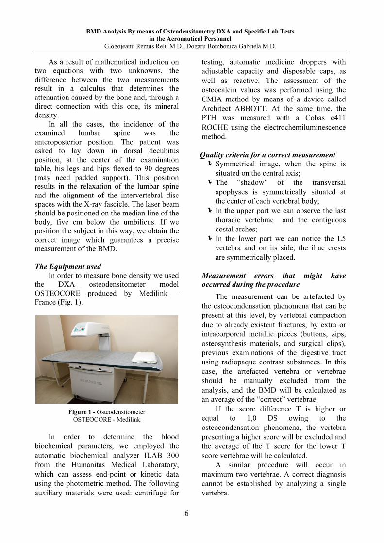
In order to measure bone density we used the DXA osteodensitometer model OSTEOCORE produced by Medilink – France (Fig. 1).
Figure 1 - Osteodensitometer OSTEOCORE - Medilink
In order to determine the blood
biochemical parameters, we employed the automatic biochemical analyzer ILAB 300 from the Humanitas Medical Laboratory, which can assess end-point or kinetic data using the photometric method. The following auxiliary materials were used: centrifuge for
testing, automatic medicine droppers with adjustable capacity and disposable caps, as well as reactive. The assessment of the osteocalcin values was performed using the CMIA method by means of a device called Architect ABBOTT. At the same time, the PTH was measured with a Cobas e411 ROCHE using the electrochemiluminescence method. Quality criteria for a correct measurement
Symmetrical image, when the spine is situated on the central axis; The “shadow” of the transversal apophyses is symmetrically situated at the center of each vertebral body; In the upper part we can observe the last thoracic vertebrae and the contiguous costal arches; In the lower part we can notice the L5 vertebra and on its side, the iliac crests are symmetrically placed.
Measurement errors that might have occurred during the procedure
The measurement can be artefacted by the osteocondensation phenomena that can be present at this level, by vertebral compaction due to already existent fractures, by extra or intracorporeal metallic pieces (buttons, zips, osteosynthesis materials, and surgical clips), previous examinations of the digestive tract using radiopaque contrast substances. In this case, the artefacted vertebra or vertebrae should be manually excluded from the analysis, and the BMD will be calculated as an average of the “correct” vertebrae.
If the score difference T is higher or equal to 1,0 DS owing to the osteocondensation phenomena, the vertebra presenting a higher score will be excluded and the average of the T score for the lower T score vertebrae will be calculated.
A similar procedure will occur in maximum two vertebrae. A correct diagnosis cannot be established by analyzing a single vertebra.
6
BMD Analysis By means of Osteodensitometry DXA and Specific Lab Tests in the Aeronautical Personnel
Glogojeanu Remus Relu M.D., Dogaru Bombonica Gabriela M.D.
Contraindications Any of the following aspects represents
contraindications of the BMD measurement at the level of the lumbar spine using the DXA osteodensitometer. They became exclusion criteria when the study groups were formed:
pregnancy; vertebral osteosynthesis materials; major vertebral distortions (vertebral compaction, scoliosis); recent examinations using contrast substances or radioactive isotopes; recent ingestion of calcium; the inability to stay still during examination; overweight limit (120-130 kg), or a higher abdominal circumference than the distance between the examination table and the mobile arm.
Results and debates The DXA devices usually show the
following results: the surface of the analyzed area (cm2); bone mineral content – BMC (g); bone mineral density BMD = BMC/area (g/cm2); the average value of the BMD for the young person in that particular area; T-score – standard deviation of the measured value as compared to the average adult age; BMD average value for the individuals belonging to a specific area according to sex and age; the Z-score – standard deviation of the measured value as compared to the average value for the individuals belonging to a specific area according to sex and age [2].
This is the number of standard deviations that the measured BMD value deviates from the average value of the population having the age and gender of the patient. The Z-score does not correspond to the OMS criteria to diagnose osteoporosis but it is useful for the premenopause women, men under 50 and children as well as for monitoring the efficiency of the osteoporotic treatment. It reports the BMD value measured at the average DMO in the young individual, that is exactly on reaching peak bone mass. The T-score is considered normal when it is between +2,5 and -1 [3].
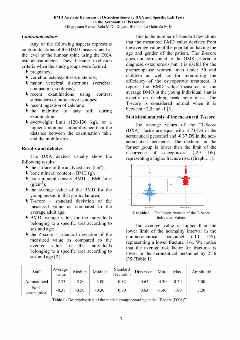
Statistical analysis of the measured T-score The average values of the “T-Score
(DXA)” factor are equal with -2.73 DS in the aeronautical personnel and -0.37 DS in the non-aeronautical personnel. The medium for the former group is lower than the limit of the occurrence of osteoporosis. (-2.5 DS), representing a higher fracture risk. (Graphic 1).
Graphic 1 – The Representation of the T-Score
Individual Values
The average value is higher than the lower limit of the normality interval in the non-aeronautical personnel (-1.0 DS), representing a lower fracture risk. We notice that the average risk factor for fractures is lower in the aeronautical personnel by 2.36 DS (Table 1).
Staff Average value Median Module Standard
Deviation Dispersion Min. Max. Amplitude
Aeronautical -2.73 -2.80 -3.00 0.82 0.67 -4.30 0.70 5.00 Non-
aeronautical -0.37 -0.50 -0.30 0.80 0.63 -1.40 1.80 3.20
Table 1 - Descriptive data of the studied groups according to the “T-score (DXA)”
7
BMD Analysis By means of Osteodensitometry DXA and Specific Lab Tests in the Aeronautical Personnel
Glogojeanu Remus Relu M.D., Dogaru Bombonica Gabriela M.D.
The dispersions of the T-score for both groups are homogenous, according to the
Levene test that assesses the equality of dispersions (Sig.>0.05) (Table 2).
Levene Test indicating the homogeneity of dispersions Welch test for the equality of the media
Levene Statistic df1 df2 Sig. Welch
Statistic df1 df2 Sig.
0.016 1 83 0.901 175.424 1 72 0.000
Table 2 – Tests of homogeneity dispersions in terms of T-Score
Under these circumstances, the results of the test Anova lead us to conclude that there are significant differences between the two groups in terms of the T-SCORE (DXA) factor (Sig.<0.05). Thus, we shall reject the null hypothesis and support instead the alternative hypothesis according to which there are significant differences between the two groups as far as the T-Score is concerned
(DXA), in the sense that the aeronautical individuals have a higher fracture risk than the non-aeronautical personnel who present a lower risk factor. The index number for the amplification of the effect (1.45) comes to support the results and it indicates a huge difference between the two groups regarding the T-SCORE (DXA). (Table 3).
Source of variation SS df MS F F critic Sig. Amplification effect
Between groups 113.60 1 113.601 173.518 3.956 0.000 1.45 Inside groups 54.34 83 0.655 Total 167.94 84
Table 3 – Unifactorial ANOVA Test for the T-Score
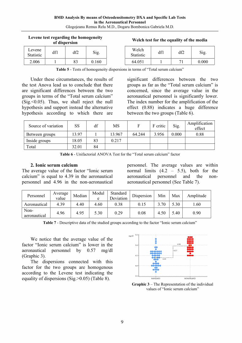
Specific medical analysis 1. Total serum calcium
The average value of the factor “Total serum calcium” equals 8.68 in the
aeronautical personnel and 9.51 in the non aeronautical personnel. The average values are within normal limits (8.6 - 10.3), both for the aeronautical personnel and the non-aeronautical personnel (See Table 4).
Personnel Average value Median Module Standard
Deviation Dispersion Min Max Amplitude
Aeronautical 8.68 8.70 8.80 0.46 0.22 7.70 10.10 2.40 Non-
aeronautical 9.51 9.40 9.20 0.47 0.22 8.80 10.20 1.40
Table 4 – Descriptive data of the studied groups according to the factor “Total serum calcium”
We notice that the average value of the
factor “Total serum calcium” is lower in the aeronautical personnel by 0.83 mg/dl (Graphic 2).
The dispersions connected with this factor for the two groups are homogenous according to the Levene test indicating the equality of dispersions (Sig.>0.05) (Table 5).
Graphic 2 – Representation of the individual values of “Total serum calcium”
8
BMD Analysis By means of Osteodensitometry DXA and Specific Lab Tests in the Aeronautical Personnel
Glogojeanu Remus Relu M.D., Dogaru Bombonica Gabriela M.D.
Levene test regarding the homogeneity of dispersion Welch test for the equality of the media
Levene Statistic df1 df2 Sig. Welch
Statistic df1 df2 Sig.
2.006 1 83 0.160 64.051 1 71 0.000
Table 5 - Tests of homogeneity dispersions in terms of “Total serum calcium”
Under these circumstances, the results of the test Anova lead us to conclude that there are significant differences between the two groups in terms of the “Total serum calcium” (Sig.<0.05). Thus, we shall reject the null hypothesis and support instead the alternative hypothesis according to which there are
significant differences between the two groups as far as the “Total serum calcium” is concerned, since the average value in the aeronautical personnel is significantly lower. The index number for the amplification of the effect (0.88) indicates a huge difference between the two groups (Table 6).
Source of variation SS df MS F F critic Sig. Amplification effect
Between groups 13.97 1 13.967 64.244 3.956 0.000 0.88 Inside groups 18.05 83 0.217 Total 32.01 84
Table 6 - Unifactorial ANOVA Test for the “Total serum calcium” factor
2. Ionic serum calcium The average value of the factor “Ionic serum calcium” is equal to 4.39 in the aeronautical personnel and 4.96 in the non-aeronautical
personnel. The average values are within normal limits (4.2 – 5.5), both for the aeronautical personnel and the non-aeronautical personnel (See Table 7).
Personnel Average
value Median Module
Standard Deviation Dispersion Min Max Amplitude
Aeronautical 4.39 4.40 4.60 0.38 0.15 3.70 5.30 1.60 Non-aeronautical 4.96 4.95 5.30 0.29 0.08 4.50 5.40 0.90
Table 7 - Descriptive data of the studied groups according to the factor “Ionic serum calcium”
We notice that the average value of the
factor “Ionic serum calcium” is lower in the aeronautical personnel by 0.57 mg/dl (Graphic 3).
The dispersions connected with this factor for the two groups are homogenous according to the Levene test indicating the equality of dispersions (Sig.>0.05) (Table 8).
Graphic 3 – The Representation of the individual values of “Ionic serum calcium”
9
BMD Analysis By means of Osteodensitometry DXA and Specific Lab Tests in the Aeronautical Personnel
Glogojeanu Remus Relu M.D., Dogaru Bombonica Gabriela M.D.
Levene test regarding the homogeneity of dispersion Welch test for the equality of the media
Levene Statistic df1 df2 Sig. Welch Statistic df1 df2 Sig. 2.653 1 83 0.107 61.676 1 82 0.000
Table 8 - Tests of homogeneity dispersions in terms of “Ionic serum calcium”
Under these circumstances, the results of the test Anova lead us to conclude that there are significant differences between the two groups in terms of the “Ionic serum calcium” (Sig.<0.05). Thus, we shall reject the null hypothesis and support instead the alternative hypothesis according to which there are significant differences between the
two groups as far as the “Ionic serum calcium” is concerned. The above-mentioned results are also supported by the index number for the amplification of the effect (0.81), which indicates a considerable difference between the two groups regarding the “Ionic serum calcium” (Table 9).
Source of variation SS df MS F F critic Sig. Amplification
effect Between groups 6.69 1 6.687 54.986 3.956 0.000 0.81 Inside groups 10.09 83 0.122 Total 16.78 84
Table 9 - Unifactorial ANOVA Test for the “Ionic serum calcium” factor
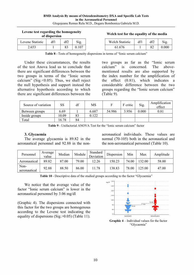
3. Glycaemia The average glycaemia is 89.82 in the
aeronautical personnel and 92.88 in the non-
aeronautical individuals. These values are normal (70-105) both in the aeronautical and the non-aeronautical personnel (Table 10).
Personnel Average value Median Module Standard
Deviation Dispersion Min Max Amplitude
Aeronautical 89.82 87.00 79.00 12.26 150.23 74.00 132.00 58.00 Non-aeronautical 92.88 88.50 86.00 11.78 138.83 78.00 125.00 47.00
Table 10 - Descriptive data of the studied groups according to the factor “Glycaemia”
We notice that the average value of the factor “Ionic serum calcium” is lower in the aeronautical personnel by 3.06 mg/dl (Graphic 4). The dispersions connected with this factor for the two groups are homogenous according to the Levene test indicating the equality of dispersions (Sig.>0.05) (Table 11).
Graphic 4 – Individual values for the factor “Glycaemia”
10
BMD Analysis By means of Osteodensitometry DXA and Specific Lab Tests in the Aeronautical Personnel
Glogojeanu Remus Relu M.D., Dogaru Bombonica Gabriela M.D.
Levene test regarding the homogeneity of dispersion Welch test for the equality of the media
Levene Statistic df1 df2 Sig. Welch
Statistic df1 df2 Sig.
0.073 1 83 0.788 1.331 1 73 0.252 Table 11 - Tests of homogeneity dispersions in terms of “Glycaemia”
Under these circumstances, the results of
the test Anova lead us to conclude that there are no significant differences between the two groups in terms of the factor “Glycaemia” (Sig.<0.05). Thus, we shall accept the null hypothesis according to which there are no significant differences between the two
groups regarding the factor “Glycaemia.” The above-mentioned results are also supported by the index number for the amplification of the effect (0.13), which indicates an insignificant difference between the two groups regarding “Glycaemia” (Table 12).
Source of variation SS df MS F F critic Sig. Amplification effect
Between groups 190.87 1 190.871 1.310 3.956 0.256 0.13 Inside groups 12092.94 83 145.698 Total 12283.81 84
Table 12 - Unifactorial ANOVA Test for the “Glycaemia” factor
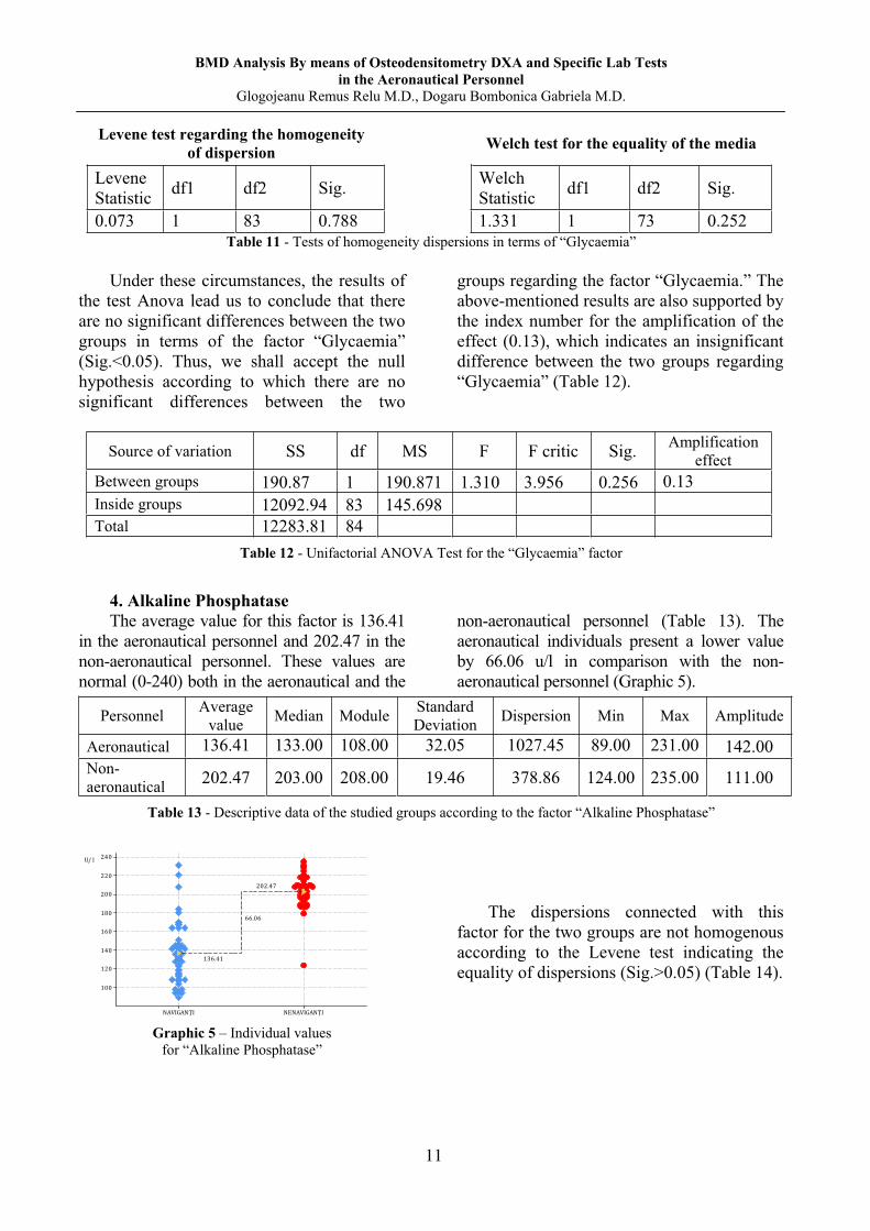
4. Alkaline Phosphatase The average value for this factor is 136.41
in the aeronautical personnel and 202.47 in the non-aeronautical personnel. These values are normal (0-240) both in the aeronautical and the
non-aeronautical personnel (Table 13). The aeronautical individuals present a lower value by 66.06 u/l in comparison with the non-aeronautical personnel (Graphic 5).
Personnel Average value Median Module Standard
Deviation Dispersion Min Max Amplitude
Aeronautical 136.41 133.00 108.00 32.05 1027.45 89.00 231.00 142.00 Non-aeronautical 202.47 203.00 208.00 19.46 378.86 124.00 235.00 111.00
Table 13 - Descriptive data of the studied groups according to the factor “Alkaline Phosphatase”
Graphic 5 – Individual values
for “Alkaline Phosphatase”
The dispersions connected with this
factor for the two groups are not homogenous according to the Levene test indicating the equality of dispersions (Sig.>0.05) (Table 14).
11
BMD Analysis By means of Osteodensitometry DXA and Specific Lab Tests in the Aeronautical Personnel
Glogojeanu Remus Relu M.D., Dogaru Bombonica Gabriela M.D.
Levene test regarding the homogeneity of dispersion Welch test for the equality of the media
Levene Statistic df1 df2 Sig. Welch Statistic df1 df2 Sig. 7.495 1 83 0.008 139.466 1 82 0.000
Table 14 - Tests of homogeneity dispersions in terms of “Alkaline Phosphatase”
In order to check the statistical hypothesis, we shall employ the Welch test. The results provided by this test prove that there are no significant differences between the two groups in terms of the factor “Alkaline Phosphatase” (Sig.<0.05). Therefore, we shall reject the null hypothesis and support instead the alternative hypothesis according to which
there are significant differences between the two groups as far as “Alkaline Phosphatase” is concerned. The above-mentioned results are also supported by the index number for the amplification of the effect (1.18), which indicates a significant difference between the two groups regarding the factor “Alkaline Phosphatase,” as it is shown in the attached graphic (See Table 15).
Source of variation SS df MS F F critic Sig. Amplification effect
Between groups 89020.9 1 89020.87 115.675 3.956 0.000 1.18 Inside groups 63874.8 83 769.58 Total 152895.7 84
Table 15 - Unifactorial ANOVA Test for “Alkaline Phosphatase”
5. Total serum proteins The average values for the factor “Total
serum proteins” represent 6.82 in the aeronautical personnel and 7.23 in the
non-aeronautical personnel, being situated within normal limits in both cases (6.0-8.3) (Table 16).
Personnel Average value Median Module Standard
Deviation Dispersion Min Max Amplitude
Aeronautical 6.82 6.70 6.80 0.64 0.41 6.00 8.20 2.20 Non-aeronautical
7.23 7.20 6.90 0.43 0.19 6.50 8.10 1.60
Table 16 - Descriptive data of the studied groups according to the factor “Total serum proteins”
We notice that the average value of the factor “Total serum proteins” is lower in the aeronautical personnel by 0.41 g/dl (Graphic 6). The dispersions connected with this factor for the two groups are homogenous according to the Levene test indicating the equality of dispersions (Sig.>0.05). Under these circumstances, the results offered by the Welch test prove that there are considerable differences between the two groups concerning the factor “Total serum proteins” (Table 17).
Graphic 6 – Individual values
for “Total serum proteins”
12

BMD Analysis By means of Osteodensitometry DXA and Specific Lab Tests in the Aeronautical Personnel
Glogojeanu Remus Relu M.D., Dogaru Bombonica Gabriela M.D.
Levene test regarding the homogeneity of dispersion Welch test for the equality of the media
Levene Statistic df1 df2 Sig. Welch Statistic df1 df2 Sig. 4.562 1 83 0.036 12.037 1 83 0.001
Table 17 - Tests of homogeneity dispersions in terms of “Total serum proteins”
Thus, we shall reject the null hypothesis and support instead the alternative hypothesis according to which there are significant differences between the two groups as far as the “Total serum proteins” is concerned. The
above-mentioned results are also supported by the index number for the amplification of the effect (0.35), which indicates an average to considerable difference between the two groups (Table 18).
Source of variation SS df MS F F critic Sig. Amplification effect
Between groups 3.31 1 3.312 10.375 3.956 0.002 0.35 Inside groups 26.50 83 0.319 Total 29.81 84
Table 18 - Unifactorial ANOVA Test for “Total serum proteins” 6. Osteocalcin The average values of osteocalcin are
equal to 34.66 in the aeronautical personnel and 135.60 in the non-aeronautical personnel. The average value the aeronautical personnel
is lower than the lower limit of the normal limit (46.63 - 171.38). The average value the non-aeronautical personnel is within normal limits (See table 19).
Personnel Average
value Media
n Modul
e Standard Deviation
Dispersion Min Max Amplitude
Aeronautical 34.66 37.51 44.21 16.61 275.76 5.98 98.90 92.92 Non-aeronautical
135.6 149.06
- 29.90 893.87 69.74 168.17 98.43
Table 19 - Descriptive data of the studied groups according to the factor “Osteocalcin”
We notice that the average value of the factor “Osteocalcin” is lower in the aeronautical personnel by 100.93 ng/ml (Graphic 7). The dispersions connected with this factor for the two groups are not homogenous according to the Levene test indicating the equality of dispersions (Sig.>0.05).
Under these circumstances, the results of the test Anova lead us to conclude that there are significant differences between the two groups in terms of the factor “Osteocalcin” (Sig.<0.05). (Table 20).
Graphic 7 – Individual values
for “Osteocalcin”
13
BMD Analysis By means of Osteodensitometry DXA and Specific Lab Tests in the Aeronautical Personnel
Glogojeanu Remus Relu M.D., Dogaru Bombonica Gabriela M.D.
Levene test regarding the homogeneity of dispersion Welch test for the equality of the media
Levene Statistic df1 df2 Sig. Welch Statistic df1 df2 Sig. 19.120 1 83 0.000 321.384 1 47 0.000
Table 20 - Tests of homogeneity dispersions in terms of “Osteocalcin”
As a consequence, we reject the null hypothesis and support the alternative hypothesis according to which there are significant differences between the two groups as far as the factor “Osteocalcin” is
concerned. The above-mentioned results are also supported by the index number for the amplification of the effect (2.19), which indicates a considerable difference between the two groups (Table 21).
Source of variation SS df MS F F critic Sig. Amplification effect
Between groups 207816.1 1 207816.1 398.483 3.956 0.000 2.19 Inside groups 43286.0 83 521.5 Total 251102.1 84
Table 21 - Unifactorial ANOVA Test for “Osteocalcin”
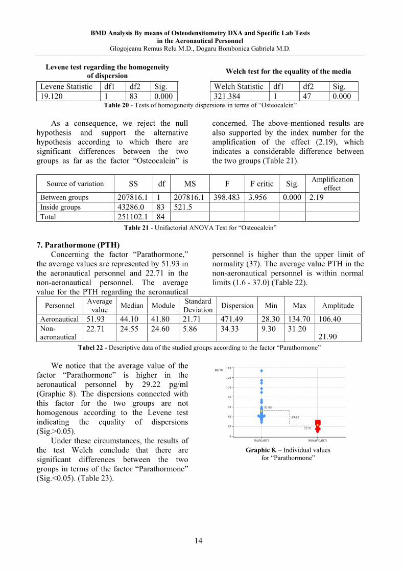
7. Parathormone (PTH) Concerning the factor “Parathormone,”
the average values are represented by 51.93 in the aeronautical personnel and 22.71 in the non-aeronautical personnel. The average value for the PTH regarding the aeronautical
personnel is higher than the upper limit of normality (37). The average value PTH in the non-aeronautical personnel is within normal limits (1.6 - 37.0) (Table 22).
Personnel Average value Median Module Standard
Deviation Dispersion Min Max Amplitude
Aeronautical 51.93 44.10 41.80 21.71 471.49 28.30 134.70 106.40 Non-aeronautical
22.71 24.55 24.60 5.86 34.33 9.30 31.20 21.90
Tabel 22 - Descriptive data of the studied groups according to the factor “Parathormone”
We notice that the average value of the factor “Parathormone” is higher in the aeronautical personnel by 29.22 pg/ml (Graphic 8). The dispersions connected with this factor for the two groups are not homogenous according to the Levene test indicating the equality of dispersions (Sig.>0.05).
Under these circumstances, the results of the test Welch conclude that there are significant differences between the two groups in terms of the factor “Parathormone” (Sig.<0.05). (Table 23).
Graphic 8. – Individual values
for “Parathormone”
14

BMD Analysis By means of Osteodensitometry DXA and Specific Lab Tests in the Aeronautical Personnel
Glogojeanu Remus Relu M.D., Dogaru Bombonica Gabriela M.D.
Levene test regarding the homogeneity of dispersion Welch test for the equality of the media
Levene Statistic df1 df2 Sig. Welch Statistic df1 df2 Sig. 11.463 1 3 0.001 83.242 1 60 0.000
Table 23 - Tests of homogeneity dispersions in terms of “Parathormone”
As a consequence, we reject the null hypothesis and support the alternative hypothesis according to which there are major differences between the two groups as far as the factor “Parathormone” is
concerned. The above-mentioned results are also supported by the index number for the amplification of the effect (0.84), which indicates a considerable difference between the two groups (Table 24).
Source of variation SS df MS F F critic Sig. Amplification effect
Between groups 17413.7 1 17413.7 58.498 3.956 0.000 0.84 Inside groups 24707.4 83 297.7 Total 42121.1 84
Tabel 24 - Unifactorial ANOVA Test for “Parathormone”
Pearson Bilateral Correlations Correlation is a statistical method used
to determine the interdependent relationship between two or more variables.
The correlation coefficient represents a quantitative value that describes the relationship between two or more variables and can range from –1 to +1. A perfect relationship is closer to +1 or –1, whereas a total lack of relationship is closer to 0.
The correlation can be positive or negative. In a positive correlation, as the value of
one of the variables increases, the value of the second independent variable increases. In this case, the value of the correlation, given by the correlation coefficient r ranges from above 0 to below or equal to 1.
In a negative correlation, the value of the dependent variable decreases, while the value of the second independent variable increases. In this case, the value of correlation, given by the correlation coefficient r, ranges from -1 to 0.
When there is no relationship between variables, the value of the correlation r is 0. The higher the correlation coefficient in absolute value is, the more dependent the two variables are. The interpretation of a correlation between two variables involves the analysis of the elements:
intensity of correlation, produced by the value of the correlation coefficient; sense of correlation, only in case of a directional correlation, produced by the sign of the correlation coefficient; significance level, which can be at 005 or 0.01. Our analysis emphasizes the correlation
coefficients together with the value of the corresponding signification (Sig.) If this value is lower than the signification level 0.05, it shows a significant intensity correlation between the two variables. The correlation coefficients are marked with an asterisk (*) after the last decimal;
The correlation coefficients with the respective value Sig. Lower than the significance level 0.01 show a remarkably huge correlation of intensity between the two variables. In this case the correlation coefficients are followed by two asterisks (**). The correlation coefficients that are representative from the point of view of the correlation of intensity are in bold. At the same time, we include a graphic representation through dots the pairs of value that correspond to two variables, focusing on the right which expresses an evolution tendency, due to the fact that the correlation sense can be graphically perceived, as well.
15
BMD Analysis By means of Osteodensitometry DXA and Specific Lab Tests in the Aeronautical Personnel
Glogojeanu Remus Relu M.D., Dogaru Bombonica Gabriela M.D.
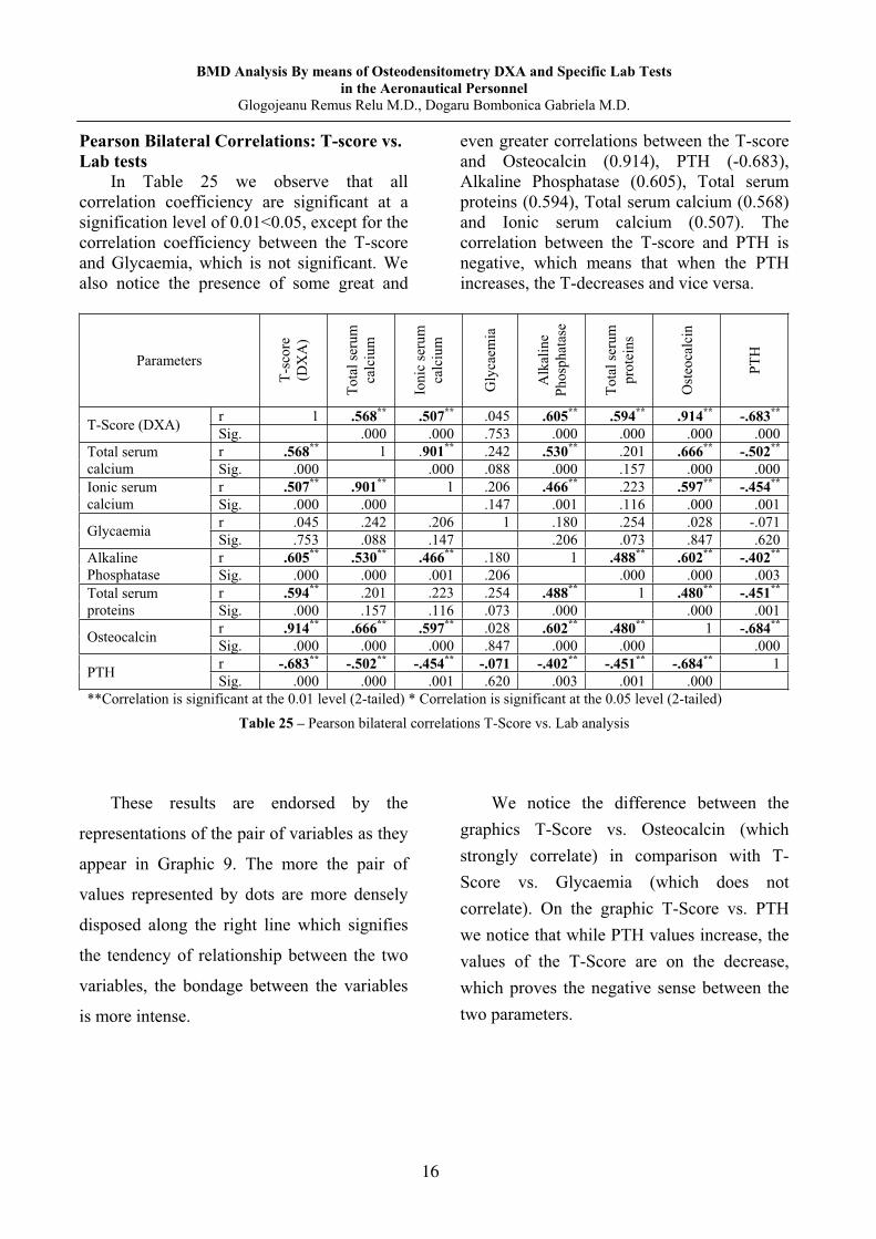
Pearson Bilateral Correlations: T-score vs. Lab tests
In Table 25 we observe that all correlation coefficiency are significant at a signification level of 0.01<0.05, except for the correlation coefficiency between the T-score and Glycaemia, which is not significant. We also notice the presence of some great and
even greater correlations between the T-score and Osteocalcin (0.914), PTH (-0.683), Alkaline Phosphatase (0.605), Total serum proteins (0.594), Total serum calcium (0.568) and Ionic serum calcium (0.507). The correlation between the T-score and PTH is negative, which means that when the PTH increases, the T-decreases and vice versa.
Parameters
T-sc
ore
(D
XA
)
Tota
l ser
um
calc
ium
Ioni
c se
rum
ca
lciu
m
Gly
caem
ia
Alk
alin
e Ph
osph
atas
e
Tota
l ser
um
prot
eins
Ost
eoca
lcin
PTH
T-Score (DXA) r 1 .568** .507** .045 .605** .594** .914** -.683** Sig. .000 .000 .753 .000 .000 .000 .000
Total serum calcium
r .568** 1 .901** .242 .530** .201 .666** -.502** Sig. .000 .000 .088 .000 .157 .000 .000
Ionic serum calcium
r .507** .901** 1 .206 .466** .223 .597** -.454** Sig. .000 .000 .147 .001 .116 .000 .001
Glycaemia r .045 .242 .206 1 .180 .254 .028 -.071 Sig. .753 .088 .147 .206 .073 .847 .620
Alkaline Phosphatase
r .605** .530** .466** .180 1 .488** .602** -.402** Sig. .000 .000 .001 .206 .000 .000 .003
Total serum proteins
r .594** .201 .223 .254 .488** 1 .480** -.451** Sig. .000 .157 .116 .073 .000 .000 .001
Osteocalcin r .914** .666** .597** .028 .602** .480** 1 -.684** Sig. .000 .000 .000 .847 .000 .000 .000
PTH r -.683** -.502** -.454** -.071 -.402** -.451** -.684** 1 Sig. .000 .000 .001 .620 .003 .001 .000
**Correlation is significant at the 0.01 level (2-tailed) * Correlation is significant at the 0.05 level (2-tailed)
Table 25 – Pearson bilateral correlations T-Score vs. Lab analysis
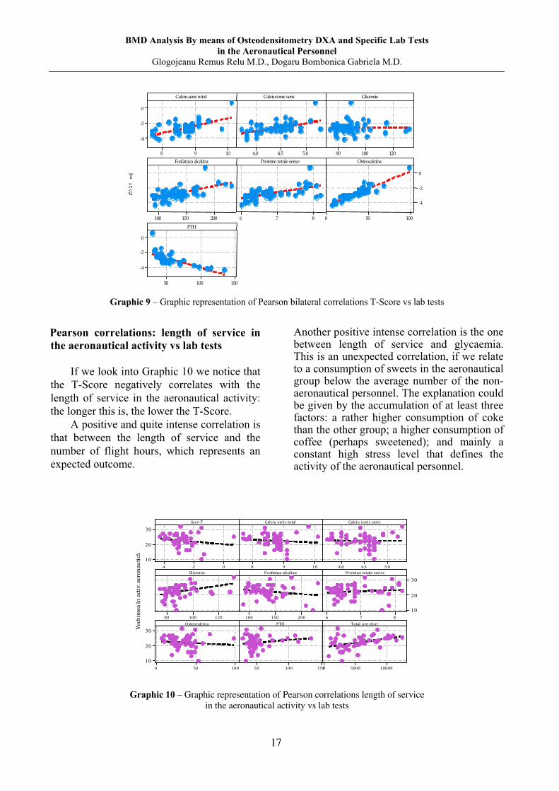
These results are endorsed by the
representations of the pair of variables as they
appear in Graphic 9. The more the pair of
values represented by dots are more densely
disposed along the right line which signifies
the tendency of relationship between the two
variables, the bondage between the variables
is more intense.
We notice the difference between the graphics T-Score vs. Osteocalcin (which strongly correlate) in comparison with T-Score vs. Glycaemia (which does not correlate). On the graphic T-Score vs. PTH we notice that while PTH values increase, the values of the T-Score are on the decrease, which proves the negative sense between the two parameters.
16
BMD Analysis By means of Osteodensitometry DXA and Specific Lab Tests in the Aeronautical Personnel
Glogojeanu Remus Relu M.D., Dogaru Bombonica Gabriela M.D.
109 8
0 -2 -4
5.04.54.0 120 10080
200 150 100 876 100 500
0 -2 -4
150100 50
0 -2 -4
Calciu seric total
ScorT
Calciu ionic seric Glicemie
Fosfataza alcalina Proteine totale serice Osteocalcina
PTH
Graphic 9 – Graphic representation of Pearson bilateral correlations T-Score vs lab tests
Pearson correlations: length of service in the aeronautical activity vs lab tests
If we look into Graphic 10 we notice that
the T-Score negatively correlates with the length of service in the aeronautical activity: the longer this is, the lower the T-Score.
A positive and quite intense correlation is that between the length of service and the number of flight hours, which represents an expected outcome.
Another positive intense correlation is the one between length of service and glycaemia. This is an unexpected correlation, if we relate to a consumption of sweets in the aeronautical group below the average number of the non-aeronautical personnel. The explanation could be given by the accumulation of at least three factors: a rather higher consumption of coke than the other group; a higher consumption of coffee (perhaps sweetened); and mainly a constant high stress level that defines the activity of the aeronautical personnel.
30
20
10
30
20
10
30
20
10
Graphic 10 – Graphic representation of Pearson correlations length of service in the aeronautical activity vs lab tests
17
BMD Analysis By means of Osteodensitometry DXA and Specific Lab Tests in the Aeronautical Personnel
Glogojeanu Remus Relu M.D., Dogaru Bombonica Gabriela M.D.
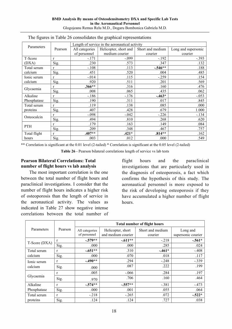
The figures in Table 26 consolidates the graphical representations
Parameters Pearson
Length of service in the aeronautical activity All categories of personnel
Helicopter, short and medium courier
Short and medium courier
Long and supersonic courier
T-Score (DXA)
r -.171 -.099 -.192 -.393 Sig. .230 .573 .347 .132
Total serum calcium
r -.108 .113 -.546** .188 Sig. .451 .520 .004 .485
Ionic serum calcium
r -.014 .115 -.259 .154 Sig. .920 .511 .201 .569
Glycaemia r .366** .316 .160 .476 Sig. .008 .065 .435 .062
Alkaline Phosphatase
r -.186 -.176 -.463* -.053 Sig. .190 .311 .017 .845
Total serum proteins
r .119 .138 .085 .000 Sig. .407 .428 .679 1.000
Osteocalcin r -.098 -.042 -.226 -.134 Sig. .494 .810 .268 .620
PTH r .179 .163 .149 .084 Sig. .209 .348 .467 .757
Total flight hours
r .407** .421* .814** .162 Sig. .003 .012 .000 .549
** Correlation is significant at the 0.01 level (2-tailed) * Correlation is significant at the 0.05 level (2-tailed)
Table 26 - Pearson bilateral correlations length of service vs lab tests
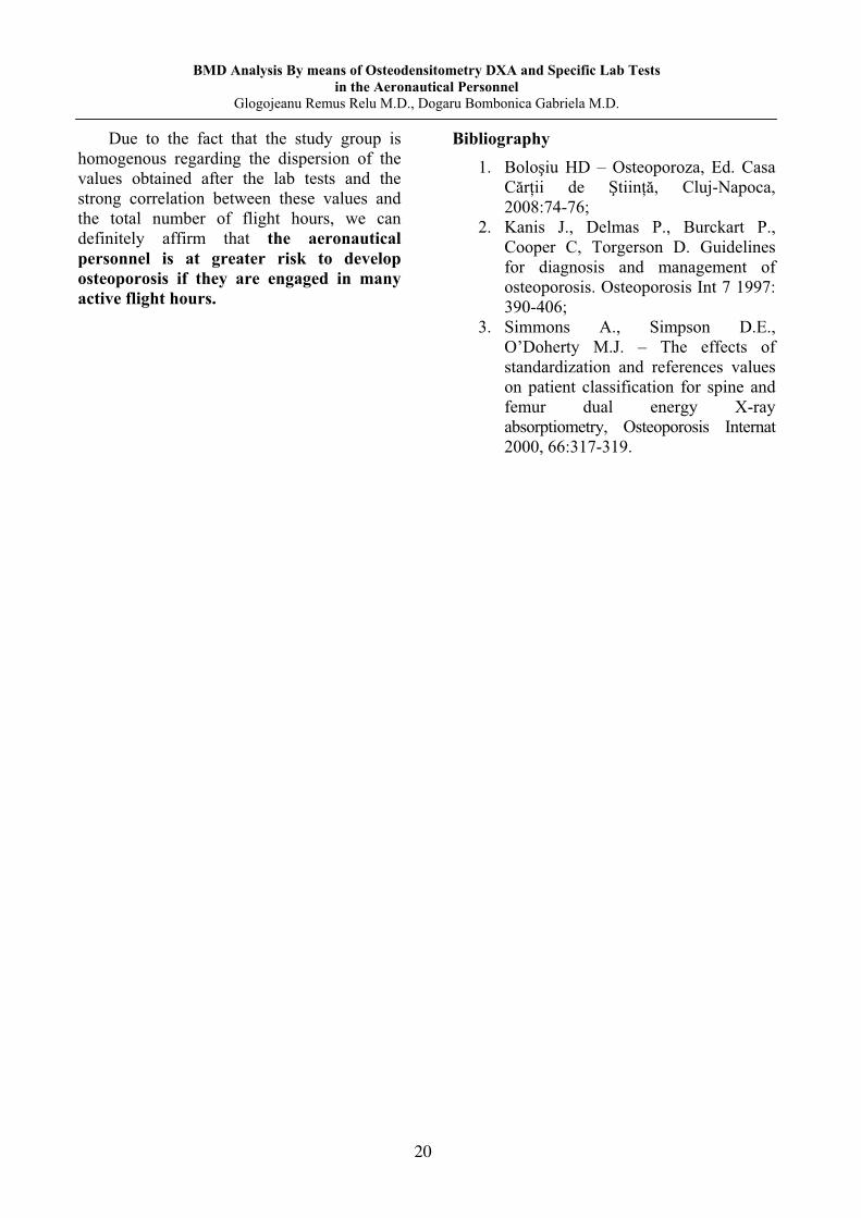
Pearson Bilateral Correlations: Total number of flight hours vs lab analysis
The most important correlation is the one between the total number of flight hours and paraclinical investigations. I consider that the number of flight hours indicates a higher risk of osteoporosis than the length of service in the aeronautical activity. The values as indicated in Table 27 show negative intense correlations between the total number of
flight hours and the paraclinical investigations that are particularly used in the diagnosis of osteoporosis, a fact which confirms the hypothesis of this study. The aeronautical personnel is more exposed to the risk of developing osteoporosis if they have accumulated a higher number of flight hours.
Parameters Pearson Total number of flight hours
All categories of personnel
Helicopter, short and medium courier
Short and medium courier
Long and supersonic courier
T-Score (DXA) r -.579** -.611** -.218 -.561* Sig. .000 .000 .285 .024
Total serum calcium
r -.651** .310 -.461* -.408 Sig. .000 .070 .018 .117
Ionic serum calcium
r -.490** .294 -.248 -.339 Sig. .000 .087 .222 .199
Glycaemia r .005 -.066 .284 .197 Sig. .970 .706 .160 .464
Alkaline Phosphatase
r -.574** -.557** -.381 -.473 Sig. .000 .001 .055 .064
Total serum proteins
r -.218 -.265 .072 -.522* Sig. .124 .124 .727 .038
18

BMD Analysis By means of Osteodensitometry DXA and Specific Lab Tests in the Aeronautical Personnel
Glogojeanu Remus Relu M.D., Dogaru Bombonica Gabriela M.D.
Parameters Pearson Total number of flight hours
All categories of personnel
Helicopter, short and medium courier
Short and medium courier
Long and supersonic courier
Osteocalcin r -.645** -.667** -.337 -.464 Sig. .000 .000 .092 .070
PTH r .524** .528** .159 .191 Sig. .000 .001 .437 .480
Total flight hours
r .407** .421* .814** .162 Sig. .003 .012 .000 .549
**Correlation is significant at the 0.01 level (2-tailed) * Correlation is significant at the 0.05 level (2-tailed) Table 27 - Pearson Bilateral Correlations: Total number of flight hours vs lab analysis
Graphic 11 shows a perfect homogeneity
of the studied group regarding the dispersion of the values analyzed as well as a strong correlation between these values and the number of flight hours. As a consequence of
the intensity of this correlation, we might undoubtedly state that the aeronautical individuals who have accumulated more flight hours have a higher risk of developing osteoporosis.
Graphic 11 – Graphic Representation of Pearson correlations total number of flight hours vs lab analysis
Conclusions
BMD measured using the DXA method is significantly low in the aeronautical personnel and the average T-Score indicates the occurrence of osteoporosis. As far as the non-aeronautical personnel is concerned, the average T-Score for DXA is within normal limits.
The average values for Total serum calcium and Ionic serum calcium are normal in both groups; nevertheless, they are lower in the aeronautical personnel. Alkaline serum phosphatase (which has long been the most commonly used bone formation marker) has a
lower average value, still within normal limits in the aeronautical individuals. Osteocalcin (specific osteoblast marker) presents lower average values in aeronautical personnel, being below the inferior limit of the normal limits. The PTH average value (stimulator of bone resorption) is higher in the aeronautical personnel than in the witness group and goes beyond the superior limit of the normal interval. Considering all the above-mentioned results, we can conclude that the aeronautical personnel is more exposed to the risk of developing osteoporosis than the witness group.
19
BMD Analysis By means of Osteodensitometry DXA and Specific Lab Tests in the Aeronautical Personnel
Glogojeanu Remus Relu M.D., Dogaru Bombonica Gabriela M.D.
Due to the fact that the study group is homogenous regarding the dispersion of the values obtained after the lab tests and the strong correlation between these values and the total number of flight hours, we can definitely affirm that the aeronautical personnel is at greater risk to develop osteoporosis if they are engaged in many active flight hours.
Bibliography 1. Bolo iu HD – Osteoporoza, Ed. Casa
C r ii de tiin , Cluj-Napoca, 2008:74-76;
2. Kanis J., Delmas P., Burckart P., Cooper C, Torgerson D. Guidelines for diagnosis and management of osteoporosis. Osteoporosis Int 7 1997: 390-406;
3. Simmons A., Simpson D.E., O’Doherty M.J. – The effects of standardization and references values on patient classification for spine and femur dual energy X-ray absorptiometry, Osteoporosis Internat 2000, 66:317-319.
20

The Revue of Aeronautical Medicine and Psychology________________________Volume 18 - Year 2014 – Nr. 1 (66)
MECHANISMS OF ALLERGEN SPECIFIC IMMUNOTHERAPY Violeta Perlea M.D.1, Sorin Perlea M.D.2, Prof. Dimitrie Dragomir M.D., Ph.D.3
Summary Allergen specific immunotherapy is the only etiologic treatment of the allergic diseases. The cellular and molecular mechanisms, which form the basis of the efficiency of this therapy with multiple ways of administration, are complex and just partly understood. Evidence accumulated in the recent years proves the acquisition of peripheral T-cell tolerance and regulatory T-cells occurrence – CD4+CD25+Treg 1 that produce inhibiting cytokines such as IL-10 or TGF- as fundamental mechanisms developed in patients under treatment. These cells influence the production of IgG4 type blocking antibodies and inhibit the activation of those cells involved in allergic inflammatory response – mast cells, basophils and eosinophil cells. Specific immunotherapy with sublingual administration is based on the natural tolerogenic characteristics of oral mucosa, selecting mostly a considerable local response and it certainly may become a prime candidate among other administration methods. Key words: allergen specific immunotherapy, peripheral T-cells tolerance, regulatory T- cells, sublingual specific immunotherapy
1 Medical Center for Ambulatory Diagnosis and Treatment „Acad. t. Milcu”, Bucharest 2 National Institute of Aerospace Medicine „General Dr. Av. Victor Anastasiu”, Bucharest 3 MedLife Children´s Hospital, Bucharest
Generalities Allergen specific immunotherapy (SIT) is
the only causal treatment of allergic diseases; it consists in a controlled exposure to the allergen, in order to increase clinic and immunological tolerance. It is not administered in a competing way against the classical pharmacotherapy; on the contrary, it represents a modifier of the disease development. In the context of the correct patient selection this method is highly efficient.
SIT leads to an immunological and clinical tolerance, which lasts beyond the moment of treatment interruption. Both the subcutaneous immunotherapy and the sublingual one, which are usually the main administration routes, result in inducing an immune tolerance by mechanisms considerably overlapping one the other with discrepancies in the range of systemic immunological modifications (1). This range is not necessarily connected to the clinical response.
The SIT objectives are: acquiring peripheral T-cells tolerance, increasing the activation threshold of mast cells and
basophils and decreasing the histamine release (2).
SIT Mechanisms The main step in developing the normal
immune response to allergens is inducing the tolerance state, produced by cellular and humoral modifications (3). Acquiring allergen tolerance represents the basis for SIT usage and allergic diseases progression block.
Throughout years numerous mechanisms have been suggested in order to explain the efficiency and, partly, the persistence in time of immunotherapy effects: antigen blocking or generating blocking IgG type antibodies or antiidiotip antibodies, IgE synthesis blocking or its receptors, decreasing cellular recruitment and activation, tachyphylaxis, Th1/Th2 switch, T cells anergy, B cells tolerance, autacoids action – histamine etc. (4).
Nowadays, two main phenomena are considered to be the basis of acquired immune tolerance as a result of SIT usage: peripheral T cells tolerance and regulatory T cells production able to produce proinflammatory cytokines (5).
21

Mechanisms of allergen specific immunotherapy Violeta Perlea M.D., Sorin Perlea M.D., Prof. Dimitrie Dragomir M.D., Ph.D.
Complex mechanisms connected to T and
B cells regulation are involved in acquiring allergen tolerance (6). Besides “professional” regulatory T cells, there is a big number of cells with regulatory properties, most of them being characterized by the fact that they produce IL-10 and / or TGF- , with the role of down-regulating the proinflammatory immune response during an allergic reaction: B lymphocytes, dendritic cells, macrophages and NK cells.
The immune tolerance may be accompanied by immune deviation, by anergy or T cells apoptosis, with a role in maintaining cellular homeostasis. There are studies which have proved allergen induced lymphocyte apoptosis in case of patients under specific immunotherapy treatment (7).
SIT effects on T cells The immune tolerance state is
conditioned by peripheral T cells tolerance, characterized by regulatory T cells production (8). Regulatory T cells have specific antigenic effects, but also non-specific ones. They suppress T cells helper with different antigenic specifics, modulate specific allergenic antibodies production, diminish IgE synthesis and have suppressive effects on those cells that have an essential role in allergic inflammation – mast cells, eosinophils and basophils (9).
Some regulatory T cells are present in thymus since birth - FOXP3+CD4+CD25+. Others are inducible under exposure to allergen conditions - CD4+CD25+Treg1, when their autocrine activity is stimulated and they secrete IL-10, TGF- and IFN- . The cytokines secretion profile is influenced by the local microenvironment. Treg1 cells secrete either IL-10 and TGF- only, or IL-10 and IFN-y. Treg1 cells have an important role at the beginning of SIT, they increase in the first 3-6 months, and then they decrease after a year approximately, but remain at bigger levels than those previous to treatment (10).
Both types of regulatory cells, the constitutional ones and the inducible ones, are important both in the allergic diseases development and in SIT effects. Specific allergen Th1, Th2, Treg1 cells recognize the same epitopes and they can be found in
variable proportions in healthy persons as well as in allergic patients. Treg1 cells may be found in a significant amount in healthy persons, having a protective role against nonpathogenic environmental factors (11). For both the allergic persons and the healthy ones the immune response is generated by the balance between the Th2 and Treg1 lymphocytes subsets (11).
IL-10 has complex actions: it diminishes specific IgE synthesis, it increases IgG4 type antibodies production, it decreases proinflammatory cytokines synthesis, mucoadhesive particles expression and mast cells, eosinophils and basophils migration, it inhibits dendritic cells maturation, MHC class II molecules expression and that of costimulating molecules (12). It also diminishes IL5 and eosinophilic activation (13).
TGF- has actions which partly overlap those of IL10 in what specific IgE synthesis and inflammatory cell migration are concerned. It inhibits cells proliferation and differentiation, it increases specific allergen IgA production and it induces FOXP3 – the main transcription factor for developing and functioning of regulatory T cells (14), it decreases the expression of Fc RI high affinity receptor on the Langerhans cells.
Regulatory T cells inhibit mast cells activation, preventing OX40-OX40 ligand interaction (15).
Other regulatory cells have been identified: CD8+reg (16), NKreg (17), CD24hiCD27+B regulatory B cells, with suppressive role, capable of generating IL10, with a role in developing, proliferating and maintaining CD4+ and Treg1 lymphocytes (18).
The specific tolerance is acquired by multiple mechanisms, involving other immunodepressive factors: antigen 4 associated with cytotoxic T lymphocytes, programmed-death-1 (11).
SIT Effects on Antibodies Production The central mechanism in antibodies
production is B cells activation. After allergen exposure in allergic
persons a B lymphocytes stimulation naturally occurs, followed by an increased production of total IgE, specific IgE allergen and specific IgG4 allergen. The activation and
22
Mechanisms of allergen specific immunotherapy Violeta Perlea M.D., Sorin Perlea M.D., Prof. Dimitrie Dragomir M.D., Ph.D.
lymphocytes switch are generated by Th2 cytokines: IL4, IL13 and by costimulating molecules, by CD40-CD40 ligand interaction, and also by Toll like receptor (19).
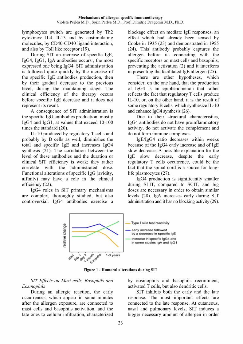
During SIT an increase of specific IgE, IgG4, IgG1, IgA antibodies occurs , the most expressed one being IgG4. SIT administration is followed quite quickly by the increase of the specific IgE antibodies production, then by their gradual decrease to the previous level, during the maintaining stage. The clinical efficiency of the therapy occurs before specific IgE decrease and it does not represent its result.
A consequence of SIT administration is the specific IgG antibodies production, mostly IgG4 and IgG1, at values that exceed 10-100 times the standard (20).
IL-10 produced by regulatory T cells and probably by B cells as well, diminishes the total and specific IgE and increases IgG4 synthesis (21). The correlation between the level of these antibodies and the duration or clinical SIT efficiency is weak; they rather correlate with the administrated dose. Functional alterations of specific IgG (avidity, affinity) may have a role in the clinical efficiency (22).
IgG4 roles in SIT primary mechanisms are complex, thoroughly studied, but also controversial. IgG4 antibodies exercise a
blockage effect on mediate IgE responses, an effect which had already been sensed by Cooke in 1935 (23) and demonstrated in 1955 (24). This antibody probably captures the allergen before its connecting with the specific receptors on mast cells and basophils, preventing the activation (2) and it interferes in presenting the facilitated IgE allergen (25).
There are other hypotheses, which consider, on the one hand, that the production of IgG4 is an epiphenomenon that rather reflects the fact that regulatory T cells produce IL-10, or, on the other hand, it is the result of some regulatory B cells, which synthesize IL-10 and enhance IgG4 synthesis (26).
Due to their structural characteristics, IgG4 antibodies do not have proinflammatory activity, do not activate the complement and do not form immune complexes.
IgE/IgG4 ratio decreases within weeks because of the IgG4 early increase and of IgE slow decrease. A possible explanation for the IgE slow decrease, despite the early regulatory T cells occurrence, could be the fact that the spinal cord is a source for long-life plasmocytes (27).
IgG4 production is significantly smaller during SLIT, compared to SCIT, and big doses are necessary in order to obtain similar levels (28). IgA increases early during SIT administration and it has no blocking activity (29).
Figure 1 - Humoral alterations during SIT
SIT Effects on Mast cells, Basophils and
Eosinophils During an allergic reaction, the early
occurrences, which appear in some minutes after the allergen exposure, are connected to mast cells and basophils activation, and the late ones to cellular infiltration, characterized
by eosinophils and basophils recruitment, activated T cells, but also dendritic cells.
SIT inhibits both the early and the late response. The most important effects are connected to the late response. At cutaneous, nasal and pulmonary levels, SIT induces a bigger necessary amount of allergen in order
23

Mechanisms of allergen specific immunotherapy Violeta Perlea M.D., Sorin Perlea M.D., Prof. Dimitrie Dragomir M.D., Ph.D.
to trigger the early/late response, but also to a low response to non-specific stimuli (30).
SIT effects on the precocious response vary and have a low intensity. However, after SIT, an important decrease of the early response magnitude occurs (31), as a consequence of the increase of mast cells and basophilic activation threshold and of mediate IgE histamine release. The regulatory T cells and IL10 modulate this activation threshold (32).
IL-10 exerts a depressive role on eosinophils. Subcutaneous SIT inhibits eosinophilic seasonal priming (33).
Regulatory T cells directly inhibit dependent FceRI mast cell degranulation. Basophil degranulation occurs more rapidly than specific IgE synthesis decrease and then cutaneous reactivity diminution. Mast cells and basophil desensitization are followed by T cells tolerogenic status (34).
Figure 2 - Cellular Modifications during SIT
The Roles of Histamine in peripheral tolerance induced by SIT
One cannot refer to peripheral immune
tolerance induced by SIT without mentioning the complex effects of histamine. Out of the four receptors by which histamine exerts its pleiotropic effects, HR2 receptor has an essential role in acquiring peripheral tolerance and in suppressing the immune inflammatory response (35). This receptor has a suppressive role, in contrast with HR1 receptor, whose stimulation is responsible for most of the stimulating effects of histamine.
HR2 receptor has a wide distribution, both on nonimmune cells and on those involved in the immune response: T and B lymphocytes, eosinophils, monocytes, dendritic cells. HR1 receptor expression at T lymphocytes level is inhibited by ultra-rush type immunotherapy, allowing HR2 receptor expression and activation (36).
The main mechanisms which interfere with immune tolerance are mediated by HR2 receptor: IL10 production by dendritic cells and Th2 lymphocytes, suppressive action of TGF- being enhanced and synthetized antibodies isotype (37).
Figure 3 - The Role of Histamine in regulating
immune processes (from 35)
Particularities of the Immune Mechanisms in SLIT
The specific immunotherapy with sublingual administration – SLIT represents a non-invasive administration strategy of SIT, more widely spread in recent times. The easy administration and the significantly more reduced side effects are the main reasons why both doctors and patients have changed their approach. Numerous clinical studies and analyses try to answer the problems regarding
24
Mechanisms of allergen specific immunotherapy Violeta Perlea M.D., Sorin Perlea M.D., Prof. Dimitrie Dragomir M.D., Ph.D.
the comparison between the two administration forms and how efficient they really are. Meta-analyses on adults (38), but on children as well (39) have shown that SLIT is an efficient alternative, with a very good safety profile.
SLIT action mechanisms are local and general. The idea of quite a significant overlap with SCIT is accepted when general mechanisms are involved, centered by some regulatory T cells synthesis, cells which produce IL10. Another idea is that of obtaining immune tolerance by immune deviation, anergy or apoptosis. The systemic immune modifications seem to be smaller than in the case of SCIT (1). In the following, we are going to insist on the local mechanisms, which are fundamental for explaining oral immune tolerance, both constitutional and therapeutic.
The efficiency and the very good safety profile of SLIT are connected to the natural immunological protolerogenic particularities of the oral mucosa (40), which, even if it is bombarded by food and infectious antigens, rarely presents allergic or inflammatory responses. A whole set of structural and functional factors co-work in a complex connection which has been just partly explained.
Oral mucosa is thin, well vascularized and permeable, factors which facilitate SLIT administration (41). Due to the fact that antigen cells quickly take it over, the oral enzymatic degradation of allergenic extracts is limited.
The bacterial flora which can be found at this level, approximately 800 bacteria species (among which Streptococcus, Haemophillus, Prevotella, Veilonella are better represented), has protolerogenic and proinflammatory effects (42). There is speculation that differences in the oral and oropharynx flora composition may influence the response to SLIT (43).
At the level of the oral mucosa there are resident cells – antigen presenting cells, lymphoid cells, and proinflammatory ones, but also lymphoid structures organized in the shape of Waldeyer ring of tonsils and adenoidal vegetation. The superficial cervical ganglions (submandibular and those of
internal jugular vein) are stations where antigen presentation is produced. The mucosal associated lymphoid tissue – MALT is weakly expressed at the level of oral mucosa. Antigen presentation can be done by dendritic cells having a small degree of maturation in the basal lamina, in the oral lymphoid loci, where they directly come into contact with T lymphocytes (44).
There are more antigen presenting cells: myeloid dendritic cells – mDCc, plasmoid dendritic cells – pDCc, Langerhans cells and macrophages. The fundamental cells for presenting antigen are the myeloid dendritic ones; they represent the primary target in SLIT. These cells, which have been proved to be Langerhans CD207 (45), have certain characteristics.
On one hand, they constitutionally express the high affinity receptor to Fc RI allergen – the expression and the expression level being correlated to IgE level, but also to other receptors: low affinity Fc RII receptor, Fc R CD16, CD32, CD64 receptors and molecules of the MHC histocompatibility major complex belonging to I and II class, CD80, CD86, CD40 costimulating molecules.
The weak expression of some maturation CD83 molecules and of some (CCR)7 chemoreceptors responsible for dendritic cells migration to lymphoid organs explains, in part, the tolerogenic properties of these cells. TLR2 and TLR4 toll-like receptors stimulation by infectious antigens at the level of oral cavity influences the up-regulation of the expression of some coinhibiting B7H1 and B7H3 molecules with tolerogenic role or it influences IL10 synthesis and the induction of a FOXP3+ transcription factor which induces regulatory T cells secreting IL10 and TGF (46).
The resident lymphoid cells are located in lamina propria, near the cells which present antigen. Most of them are CD4+ cells, both effector and regulatory ones: Th1, Th2, Th17, which have a role in the anti-infectious defense, not in SLIT though, where the inducible cells from naive T cells are more important (47).
Other cells, the proinflammatory ones, are involved in immune events. Mast cells and eosinophils are also present in the oral mucosa, but in a small number (48), possibly
25
Mechanisms of allergen specific immunotherapy Violeta Perlea M.D., Sorin Perlea M.D., Prof. Dimitrie Dragomir M.D., Ph.D.
connected to the pruritus and local edema which may occur during the immunotherapy. The administration in other areas of the oral cavity, with a small number of mast cells and eosinophils – the vestibular area – may be a solution for the patients with local reactions to sublingual administration (49).
Adjuvants utilization in SLIT aims at increasing the efficiency and at decreasing the allergen doses which are administered. TLR expressed in a great amount at the surface of oral dendritic cells are a target for the adjuvants – non-pathogenic bacteria (fermentation bacteria), bacteria toxins which lead to a Th1 high response against the Th2 response (41). If mucoadhesivity is increased using carbohydrates polymers such as chitosan or maltodextrin, the duration of the contact with the oral mucosa is prolonged and it facilitates the allergen phagocytosis (50, 51).
Even if one hundred years have passed since the first SIT academic results were reported (52), the history of this chapter is far from being ended. Getting thorough knowledge about the action mechanisms brings new possibilities of intervention which may increase the administration efficiency and safety regarding this only etiologic therapy, which modifies the natural course of the disease, preventing allergic disease from advancing and occurrence of new sensitization.
Bibliography
1. Scadding G, Durham S. - Mechanisms of sublingual immunotherapy. Immunol Allergy Clin N Am 31 (2011) 191–209;
2. Akdis CA, Akdis M. - Mechanisms of allergen-specific immunotherapy. J Allergy Clin Immunol 2011, 127(1):18-27, quiz 28-9;
3. Jutel M, Akdis CA. - Immunological mechanisms of allergen-specific immunotherapy. Allergy 2011; 66:725–732;
4. Ring J, Gutermuth J. - 100 years of hyposen itization: history of allergen-specific immunotherapy (ASIT). Allergy 2011; 66:713–724;
5. Fujita K, et al. - Clinical and Translational Allergy 2012; 2:2-8;
6. Akdis M, Burgler S, Crameri R, Eiwegger T, Fujita H, Gomez E, et al. - Interleukins, from 1 to 37, and interferon- : receptors, functions, and roles in diseases. J Allergy Clin Immunol 2011; 127:701-21, e1-70;
7. Ohta K, Yamashita N. - Apoptosis of eosinophils and lymphocytes in allergic inflammation. J Allergy Clin Immunol 1999; 104:14-21;
8. Akdis M, Blaser K, Akdis CA. - T regulatory cells in allergy: novel concepts in the pathogenesis, prevention, and treatment of allergic diseases. J Allergy Clin Immunol 2005; 116:961–968; quiz 969;
9. Ozdemir C, Akdis M, Akdis CA. - T regulatory cells and their counterparts: masters of immune regulation. Clin Exp Allergy 2009; 39:626–639;
10. Mobs C, Slotosch C, Loffler H, Jakob T, Hertl M, Pfutzner W. - Birch pollen immunotherapy leads to differential induction of regulatory T cells and delayed helper T cell immune deviation. J Immunol 2010; 184:2194–2203;
11. Akdis M, et al. - Immune responses in healthy and allergic individuals are characterized by a fine balance between allergen-specific T regulatory 1and T helper 2 cells. J Exp Med 2004, 199 (11):1567-75;
12. Akdis CA, Akdis M. - Mechanisms and treatment of allergic disease in the big picture of regulatory T cells. J Allergy Clin Immunol 2009, 123(4):735-46, quiz 747-8;
13. Schandene L, et al. - B7/CD28-dependent IL-5 production by human resting T cells is inhibited by IL-10. J Immunol 1994, 152(9):4368-74;
14. Klunker S, Chong MM, Mantel PY, Palomares O, Bassin C, Ziegler M, Rückert B, Meiler F, Akdis M, Littman DR, Akdis CA. - Transcription factors RUNX1 and RUNX3 in the induction and suppressive function of Foxp3+ inducible regulatory T cells. J Exp Med 2009; 206:2701-15;
15. Gri G, et al. - CD4+CD25+ regulatory T cells suppress mast cell degranulation and allergic responses through OX40-OX40L interaction. Immunity 2008, 29(5):771-81;
26

Mechanisms of allergen specific immunotherapy Violeta Perlea M.D., Sorin Perlea M.D., Prof. Dimitrie Dragomir M.D., Ph.D.
16. Zhou J, Appleton SE, Stadnyk A, Lee
TD, Nashan BA. - CD8 (+) T regulatory cells mediate kidney allograft prolongation after oral exposure to alloantigen. Transpl Int 2008; 21:679–687;
17. Deniz G, Erten G, Kucuksezer UC, Kocacik D, Karagiannidis C, Aktas E, et al. - Regulatory NK cells suppress antigen-specific T cell responses. J Immunol 2008; 180:850–857;
18. Fujimoto M. - Regulatory B cells in skin and connective tissue diseases. J Dermatol Sci 2010, 60(1):1-7;
19. Stavnezer J, Guikema JE, Schrader CE. - Mechanism and regulation of class switch recombination. Annu Rev Immunol 2008; 26:261-92;
20. Van der Giessen M, Homan WL, van Kernbeek G, Aalberse RC, Dieges PH. - Subclass typing of IgG antibodies formed by grass pollen-allergic patients during immunotherapy. Int Arch Allergy Appl Immunol 1976; 50:625–640;
21. Akdis CA, Blesken T, Akdis M, Weuthrich B, Blaser K. - Role of interleukin 10 in specific immunotherapy. J Clin Invest 1998; 102:98-106;
22. Michils A, Mairesse M, Ledent C, Gossart B, Baldassarre S, Duchateau J. - Modified antigenic reactivity of anti-phospholipase A2 IgG antibodies in patients allergic to bee venom: conversion with immunotherapy and relation to subclass expression. J Allergy Clin Immunol 1998; 102:118-26, IIb;
23. Cooke RA, Barnard JH, Hebald S, Stull A. - Serologic evidence of immunity with coexisting sensitization in a type of human allergy (hay fever). J Exp Med 1935; 62:733–750;
24. Cooke RA, Menzel AE, Kessler WR, Myers PA. - The antibody mechanisms of ragweed allergy; electrophoretic and chemical studies. I: The blocking antibody. J Exp Med 1955; 101:177–196;
25. Wachholz PA, Soni NK, Till SJ, Durham SR. - Inhibition of allergen-IgE binding to B cells by IgG antibodies after grass pollen immunotherapy. J Allergy Clin Immunol 2003; 112:915-22, Ib;
26. Aalberse R. - The role of IgG antibodies in allergy and immunotherapy. Allergy 2011; 66 (Suppl. 95):28–30;
27. Radbruch A, Muehlinghaus G, Luger EO, Inamine A, Smith KG, Dorner T, et al. - Competence and competition: the challenge of becoming a long-lived plasma cell. Nat Rev Immunol 2006; 6:741–750;
28. Rossi RE, Monasterolo G, Coco G, et al. - Evaluation of serum IgG4 antibodies specific to grass pollen allergen components in the follow up of allergic patients undergoing subcutaneous and sublingual immunotherapy. Vaccine 2007; 25(5):957–964;
29. Jutel M, Akdis M, Budak F et al. - IL-10 and TGF- cooperate in the regulatory T cell response to mucosal allergens in normal immunity and specific immunotherapy. Eur J Immunol 2003; 33:1205-14;
30. Rak S, Lowhagen O, Venge P. - The effect of immunotherapy on bronchial hyperresponsiveness and eosinophil cationic protein in pollen-allergic patients. J Allergy Clin Immunol 1988; 82(3 pt 1):470–480;
31. Iliopoulos O, Proud D, Adkinson NF Jr, et al. - Effects of immunotherapy on the early, late, and rechallenge nasal reaction to provocation with allergen: changes in inflammatory mediators and cells. J Allergy Clin Immunol 1991; 87:855–66;
32. Pierkes M, Bellinghausen I, Hultsch T, Metz G, Knop J, Saloga J. - Decreased release of histamine and sulfidoleukotrienes by human peripheral blood leukocytes after wasp venom immunotherapy is partially due to induction of IL-10 and IFN- production of T cells. J Allergy Clin Immunol 1999; 103:326-32;
33. Hakansson L, Heinrich C, Rak S, et al. - Priming of eosinophil adhesion in patients with birch pollen allergy during pollen season: effect of immunotherapy. J Allergy Clin Immunol 1997; 99(4):551–562;
34. Akdis CA, Akdis M, Blesken T, Wymann D, Alkan S, Mueller U, et al. - Epitopespecific T cell tolerance to
27
Mechanisms of allergen specific immunotherapy Violeta Perlea M.D., Sorin Perlea M.D., Prof. Dimitrie Dragomir M.D., Ph.D.
phospholipase A2 in bee venom immunotherapy and recovery by IL-2 and IL-15 in vitro. J Clin Invest 1996; 98:1676-83;
35. Jutel M, Blaser K, Akdis CA. - Histamine receptors in immune regulation and allergen specific immunotherapy. Immunol Allergy Clin N Am 2006; 26:245-259;
36. Jutel M, Zak-Nejmark T, Wrzyyszcz M, et al. - Histamine receptor expression on peripheral blood CD4 + lymphocytes is influenced by ultrarush bee venom immunotherapy. Allergy 1997; 52 (Suppl 37):88;
37. Jutel M, Watanabe T, Klunker S, et al. - Histamine regulates T-cell and antibody responses by differential expression of H1 and H2 receptors. Nature 2001; 413:420– 5;
38. Wilson DR, Lima MT, Durham SR. - Sublingual immunotherapy for allergic rhinitis: systematic review and meta-analysis. Allergy 2005; 60:4–12;
39. Penagos M, Compalati E, Tarantini F, Baena-Cagnani R, Huerta J, Passalacqua G, et al. - Efficacy of sublingual immunotherapy in the treatment of allergic rhinitis in pediatric patients 3 to 18 years of age: a metaanalysis of randomized, placebo-controlled, double-blind trials. Ann Allergy Asthma Immunol 2006; 97:141–148;
40. Novak N, Haberstok J, Bieber T, Allam J-P. - The immune privilege of the oral mucosa. Trends Mol Med. 2008; 14:191–198;
41. Novak N, Bieber T, Allam J-P. - Immunological mechanisms of sublingual allergen-specific immunotherapy. Allergy 2011; 66:733–739;
42. Relman DA. - Learning about who we are. Nature 2012; 486:194-5;
43. Moingeon P. - Update on Immune Mechanism Associated with Sublingual Immunotherapy: Practical Implications for the Clinician. J Allergy Clin Immunol: In Practice 2013; 1:228-41;
44. Jotwani R, Palucka AK, Al-Quotub M, Nouri-Shirazi M, Kim J, Bell D, et al. - Mature dendritic cells infiltrate the T cell-rich region of oral mucosa in chronic periodontitis: in situ, in
vivo, and in vitro studies. J Immunol 2001; 167:4693–4700;
45. Allam JP, Novak N, Fuchs C, Asen S, Berge S, Appel T, et al. - Characterization of dendritic cells from human oral mucosa: a new Langerhans cell type with high constitutive Fc RI expression. J Allergy Clin Immunol 2003; 112:141–148;
46. Allam JP, Peng WM, Appel T, Wenghoefer M, Niederhagen B, Bieber T, et al. - Toll-like receptor 4 ligation enforces tolerogenic properties of oral mucosal Langerhans cells. J Allergy Clin Immunol 2008; 121:368–374;
47. Allam JP, Duan Y, Winter J, Stojanovski G, Fronhoffs F, Wenghoefer M, et al. - Tolerogenic T cells, Th1/Th17 cytokines and TLR2/TLR4 expressing dendritic cells predominate the microenvironment within distinct oral mucosal sites. Allergy 2011; 66:532-9;
48. Marcucci F, Sensi L, Incorvaia C, Di Cara G, Moingeon P, Frati F. - Oral reactions to sublingual immunotherapy: a bioptic study. Allergy 2007; 62:1475–1477;
49. Allam JP, Stojanovski G, Friedrichs N, Peng W, Bieber T, et al. - Distribution of Langerhans cells and mast cells within the human oral mucosa: new application sites of allergens in sublingual immunotherapy? Allergy 2008; 63:720 –727;
50. Razafindratsita A, Saint-Lu N, Mascarell L, Berjont N, Bardon T, Betbeder D, et al. - Improvement of sublingual immunotherapy efficacy with a mucoadhesive allergen formulation. J Allergy Clin Immunol 2007; 120:278-85;
51. Saint Lu N, Tourdot S, Razafindrasita A, Mascarell L, Berjont N, Chabre H, et al. - Targeting the allergen to oral dendritic cells with mucoadhesive chitosan particles enhances tolerance induction. Allergy 2009; 64:1003-13;
52. Noon L. - Prophylactic inoculations against hay fever. Lancet 1911; 1:1572–1573
28
The Revue of Aeronautical Medicine and Psychology________________________Volume 18 - Year 2014 – Nr. 1 (66)
THE ADMINISTRATION OF MIDAZOLAM AS A WAY OF ANXIOLYSIS AND AMNESIA
Cristian Georgescu M.D., Ph.D.1, Magdalena Diaconu M.D.1, Cristina Ple a M.D.2, Adrian Tase M.D., Ph.D.3, Monica ân u3
Summary Sedation may be defined as the use of pharmacological agents to produce sufficient depression of consciousness, which results in a state of drowsiness and anxiolysis without the loss of verbal communication [1]. The study refers to two groups of patients who underwent endourologic surgery; the first batch was given only loco-regional anesthesia with bupivacaine (Marcain) in a percentage of 0.5 %, and for the second lot it was used a technique of sedation with Midazolam. The check of the degree of pain was performed using VAS (Visual Analogue Scale), and the degree of sedation with Ramsay score. Keywords: Ramsay score, VAS score, sedation, Midazolam.
1 Craiova County Emergency Hospital 2 Bucharest Universitary Central Military Emergency Hospital 3 Pitesti County Emergency Hospital
Introduction Benzodiazepines interact with specific
receptors in the central nervous system, especially in the cerebral cortex. The binding to the benzodiazepine receptors enhances the inhibitory effects of neurotransmitters [2, 4, 5]. Midazolam, which has a half-life plasma of 2-4 hours, is often used during endoscopic maneuvers, with marked sedative effect and the advantage of producing amnesia [3, 4]. Midazolam requires careful titration in order to avoid overdose and apnea [6]. Airway control may be lost at any time, so patients should be monitored carefully and appropriate equipment must be available [1].
Purpose and objectives
The aim of this therapy is to provide anxiolysis, analgesia and amnesia, without damaging the neurological status of the patient.
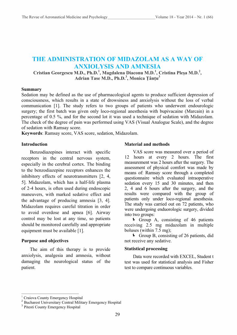
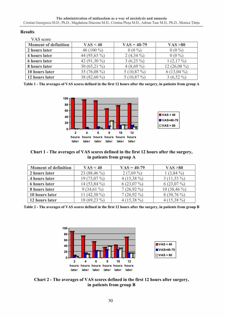
Material and methods VAS score was measured over a period of
12 hours at every 2 hours. The first measurement was 2 hours after the surgery. The assessment of physical comfort was made by means of: Ramsay score through a completed questionnaire which evaluated intraoperative sedation every 15 and 30 minutes, and then 2, 4 and 6 hours after the surgery, and the results were compared with the group of patients only under loco-regional anesthesia. The study was carried out on 72 patients, who were undergoing enduorologic surgery, divided into two groups:
Group A, consisting of 46 patients receiving 2.5 mg midazolam in multiple boluses (within 7.5 mg);
Group B, consisting of 26 patients, did not receive any sedative.
Statistical processing Data were recorded with EXCEL, Student t
test was used for statistical analysis and Fisher test to compare continuous variables.
29
The administration of midazolam as a way of anxiolysis and amnesia Cristian Georgescu M.D., Ph.D., Magdalena Diaconu M.D., Cristina Ple a M.D., Adrian Tase M.D., Ph.D., Monica ân u
88.46
7.693.84
73.07
15.3811.53
53.84
23.0723.0734.61
26.9238.46 42.3
26.9230.76
69.23
15.3815.38
0
20
40
60
80
100
2hourslater
4hourslater
6houralater
8hourslater
10hourslater
12hourslater
VAS < 40
VAS=40-79VAS > 80
Results VAS score
Moment of definition VAS < 40 VAS = 40-79 VAS >80 2 hours later 46 (100 %) 0 (0 %) 0 (0 %) 4 hours later 44 (95,65 %) 2 (4,34 %) 0 (0 %) 6 hours later 42 (91,30 %) 3 (6,25 %) 1 (2,17 %) 8 hours later 30 (65,21 %) 4 (8,69 %) 12 (26,08 %) 10 hours later 35 (76,08 %) 5 (10,87 %) 6 (13,04 %) 12 hours later 38 (82,60 %) 5 (10,87 %) 3 (6,52 %)
Table 1 - The averages of VAS scores defined in the first 12 hours after the surgery, in patients from group A
100
0 0
95.65
4.340
91.3
6.252.17
65.21
8.69
26.08
76.08
10.87 13.04
82.6
10.876.52
0
20
40
60
80
100
2hourslater
4hourslater
6hourslater
8hourslater
10hourslater
12hourslater
VAS < 40VAS=40-79VAS > 80
Chart 1 - The averages of VAS scores defined in the first 12 hours after the surgery,
in patients from group A
Moment of definition VAS < 40 VAS = 40-79 VAS >80 2 hours later 23 (88,46 %) 2 (7,69 %) 1 (3,84 %) 4 hours later 19 (73,07 %) 4 (15,38 %) 3 (11,53 %) 6 hours later 14 (53,84 %) 6 (23,07 %) 6 (23,07 %) 8 hours later 9 (34,61 %) 7 (26,92 %) 10 (38,46 %) 10 hours later 11 (42,30 %) 7 (26,92 %) 8 (30,76 %) 12 hours later 18 (69,23 %) 4 (15,38 %) 4 (15,38 %)
Table 2 - The averages of VAS scores defined in the first 12 hours after the surgery, in patients from group B
Chart 2 - The averages of VAS scores defined in the first 12 hours after surgery, in patients from group B
30

The administration of midazolam as a way of anxiolysis and amnesia Cristian Georgescu M.D., Ph.D., Magdalena Diaconu M.D., Cristina Ple a M.D., Adrian Tase M.D., Ph.D., Monica ân u
Ramsay score
Moment of definition Ramsay = 2 Ramsay = 3 Ramsay = 4 15 minutes later 5 (10,87 %) 11 (23,91 %) 30 (65,21 %) 30 minutes later 8 (17,39 %) 15 (32,60 %) 23 (50 %) At the end of the surgery 3 (6,52 %) 16 (34,78 %) 27 (58,69 %)
Table 3 - Intraoperative definition of Ramsay score in patients from group A
10,8723,91
65,21
17,39
32,6
50
6,52
34,78
58,69
0
20
40
60
80
100
15 minutes later 30 minutes later At the end of thesurgery
Ramsay=2Ramsay=3Ramsay=4
Chart 3 - Intraoperative definition of Ramsay score in patients from group A
Moment of definition Ramsay = 2 Ramsay = 3 Ramsay = 4 2 hours later 44 (95,65 %) 2 (4,34 % ) 0 (0 %) 4 hours later 45 (97,82 %) 1 (2,17 %) 0 (0 %) 6 hours later 46 (100 %) 0 (0 %) 0 (0 %)
Table 4 - Definition of Ramsay score in patients from group A during the postoperative period
95,65
4,34 0
97,82
2,17 0
100
0 00
20
40
60
80
100
2 hours later 4 hours later 6 hours later
Ramsay=2Ramsay=3Ramsay=4
Chart 4 - Determination Ramsay score in group A patients during the postoperative period
Discussion of results
Anxiolysis, amnesia and sedative effects are observed at lower doses. Benzodiazepines do not have direct analgesic properties [2, 5, 6]. Midazolam causes anterograde amnesia and awakening is delayed in comparison to other agents, intellectual capacity is diminished and postoperative sedation is often prolonged [3, 4, 6].
2 hours postoperatively, all 46 patients in group A (100 %) as compared to the 23 patients of a total of 26 patients from group B (88.46 %) described mild pain (VAS < 40).
8 hours after the surgery 30 out of 46 patients from group A (65.21 %), as compared to 9 out of 26 patients from group B (34.61 %), described mild pain (VAS < 40).
12 hours postoperatively in 3 patients out of 46 patients from group A (6.52 %) as
31

The administration of midazolam as a way of anxiolysis and amnesia Cristian Georgescu M.D., Ph.D., Magdalena Diaconu M.D., Cristina Ple a M.D., Adrian Tase M.D., Ph.D., Monica ân u
compared to 4 patients out of 26 patients from group B (15.38 %) described high intensity pain (VAS > 80).
There is an improvement in VAS score for group A compared to group B, so patients in group A showed a higher satisfaction rate compared to group B.
Intraoperatively, 15 minutes later there was observed the Ramsay score = 4 (the patient is asleep, but responds to verbal stimulation) at a number of 5 patients (10.86 %), 30 minutes later the number of patients increases to 8 patients (17.39 %), and decreases to the end of the operation to 3 patients (6.52 %) .
Ramsay score = 2 (patient is awake, quiet), 15 minutes intraoperatively, the score was met in a total of 30 patients (65.21 %), decreasing to the end of surgery to 27 patients (58.69 %).
Postoperatively, 2, 4 and 6 hours later, Ramsay score = 4 was not recorded in any patient, Ramsay = 2, 2 hours later was met in 44 patients (95.65 %), 4 hours later it was measured in 45 patients (97.82 %), and 6 hours later all patients were completely awake.
Lot B was not given any sedative, which is why Ramsay score was not calculated.
Conclusions 1. sedation with midazolam, during loco-
regional anesthesia increases the wellbeing;
2. midazolam sedation increases patients’ satisfaction;
3. midazolam sedation improved VAS values compared to patients not receiving any sedation;
4. patients in group A showed a significantly higher percentage of intra-anesthetic comfort compared to group B.
Bibliography 1. Alan R. Aitkenhead - Textbook of
Anaesthesia, Fourth Edition, 2001, International Edition; Elsevier Science, pag. 192, 201, 208-209;
2. Clinical Anesthesiology, 4 th Edition G. Edward Morgan, Jr., Maged S., Mikhail, Michael J. Murray, Copyright © 2006, 2002 by the McGraw – Hill Companies, inc., Section II, Clinical Pharmacology, Chapter 8, Nonvolatile Anaesthetic Agents – Opioids;
3. Acalovschi I. - Anestezie clinic , Ed. Clusium, edi ia II, 2005, pag. 196-200, 206-209, 220-222, 224-229;
4. Baron R., Janig W. - The role of the sympathetic nervous system in pain processing. In: Villanueva L, Dickenson AH, Ollat H (Eds). The Pain System in Normal and Pathological States: A Primer For Clinicians, Progress in Pain Research and Management, Seattle: IASP Press, 2004; (31): 193-210;
5. Fassoulaki A. et al. - Multimodal analgesia with gabapentin and local anesthetics prevents acute and chronic pain after breast surgery, Anesth Analg 2005; 101(5): 1427- 1432;
6. Georgescu C.C., V.A. Voicu, Florica Popescu – Farmacologie (curs), Ed. Medical Universitar , Craiova, 2005, pag. 109-116.
32
The Revue of Aeronautical Medicine and Psychology________________________Volume 18 - Year 2014 – Nr. 1 (66)
CHRONIC VENOUS INSUFFICIENCY Florin S vulescu M.D.1, R svan Hristea M.D.2, Cristian Dumitru M.D.2,
Bogdan Merticariu M.D.1, Cristian Cîrlan M.D.1
Summary Chronic venous insufficiency represents a lesions complex that affects the lower limb veins and which evolves slowly and gradually, with chronic and complications appearance over time. Chronic venous insufficiency is characterized by symptoms and signs installed as a result of structural and functional alterations of the veins. In the following we will present theoretical data about venous insufficiency: classification, symptomatology, treatment.
1 Second Surgical Department, Carol Davila Central Military Hospital, Bucharest, Romania 2 Surgical Department, National Institute of Aerospace Medicine, Bucharest, Romania
Introduction Chronic venous insufficiency represents a
lesions complex that affects the lower limb veins and which evolves slowly and gradually, with chronic and complications appearance over time. Long-term consequences represent the real danger of neglecting the disease, affecting about 5% of the population. Chronic venous insufficiency consists of two types that clinically are quite similar, but different in terms of etiopathogenesis, therapeutics and prognosis: chronic venous insufficiency and subfascial chronic venous insufficiency. Heavy legs, aching legs, and also varicose veins can cause serious health problems. When veins do not allow the blood to return to the heart, problems such as deep vein thrombosis, pulmonary embolism or death can appear.
Definition Chronic venous insufficiency is
characterized by symptoms and signs installed as a result of structural and functional alterations of the veins.
Characteristic symptoms are sensation of heavy legs, sensation of tired legs, predominantly nocturnal cramps, pain, itching, burning sensation or sensation of swollen leg.
Characteristic symptoms are sensation of heavy legs, sensation of tired legs, predominantly nocturnal cramps, pain, itching, burning sensation or sensation of swollen leg.
Signs include telangiectasias, reticular veins, varicose veins, edema, trophic skin changes such as lipodermatoscleroza
dermatitis, pigmentation and in final evolutionary stages - ulceration.
Epidemiology Worldwide, 10-33% of adult women and
10-20% of men have chronic venous insufficiency. In the European countries the prevalence is even greater, of 20 to 50%.
In Romania, recent epidemiological studies with national relevance have shown a IVC prevalence of 32%.
How does chronic venous insufficiency act The causes of chronic venous
insufficiency are multiple and admit both genetic factors, inherited hereditary as well as environmental or occupational factors, injuries or accidents. To these are added predisposing factors like physical inactivity, obesity, hormonal changes, hard and difficult sports, chronic cough, exposure to high temperature and smoking. Manifestations of the disease can range from the cosmetic to severe suffering, like the type of varicose ulcers which can seriously affect leg functions and can immobilize the patient. There are several degrees of manifestations, from the simple and mild to the severe impairment of vessels. Among these, we recall spider veins, varicose veins, swelling of the lower limbs, especially visible in the evening, after effort, with leg swelling, skin hyperpigmentation or its irreversible coloring.
Aesthetic aspect causes the patient to do a checkup, even if, if asked, he will recognize the existence of other sufferings that are caused by the disease, sufferings that he
33
Chronic venous insufficiency Florin S vulescu M.D., R svan Hristea M.D., Cristian Dumitru M.D., Bogdan Merticariu M.D., Cristian Cîrlan M.D.
ignored, such as the feeling of heavy legs, swollen legs in the evening, pain or tingling. Finally, dramatic events may occur, such as open varicose ulcers or serious complications of venous thrombosis or thrombophlebitis.
The clinical picture is dominated by three signs that appear successively: edema, crown phlebitis and leg ulcer, at which a few other signs are added, such as the large varicose veins ectasies or compensatory collateral circulation, intracutaneous veins ectasies, hypodermic, dermatosclerosis, skin depigmentation hyperpigmentation, skin atrophy or eczema.
Prevention of chronic venous insufficiency Movement, sport, a normal diet and
consequently a normal weight, quitting smoking and avoiding all forms of excessive heat on the legs, including hot waxing, helps prevent chronic venous insufficiency. Sauna, hard massages, prolonged exposure to the sun and traumas should be avoided. It is necessary to drink plenty of fluids, try to move and wear a pair of elastic socks during a plane flight. They will protect you and, in addition, when you land, you will not have the feeling of swollen legs and difficulty while walking.
Treatment The treatment of chronic venous
insufficiency is complex and it is based on the stage of the disease and includes drug therapy (systemic or local), compressive therapy, local treatment of venous ulcer, sclerotherapy, endovascular therapy and surgery.
The treatment of chronic venous insufficiency is determined depending on the stage of the disease, according to the following protocol, systemic treatment can be initiated including by the family doctor.
1. Patients with Chronic Venous Insufficiency in the CEAP stage COs
Description of the stage according to the CEAP classification, as revised:
patients with no palpable or visible signs of chronic venous insufficiency but with characteristic symptoms of: pain, sensation of heavy leg, sensation of swollen legs, muscular cramps,
itching, skin irritation and any other symptoms attributable to chronic venous insufficiency.
Therapeutic possibilities: changing the lifestyle; systemic treatment: diosmin (450 mg) +
hesperidin (50 mg), micronized - 2 tablets daily, chronic treatment;
elastic retainer according to each case - elastic stocking tailored to the individual.
2. Patients with chronic venous insufficiency CEAP stage C1
Description of the stage according to the CEAP classification, as revised:
patients with spider veins (intradermal Conflu and dilated venules with a diameter less than 1 mm) or reticular veins (subdermal dilated veins, with a diameter between 1 and 3 mm, tortuous).
Therapeutic possibilities: changing the lifestyle; systemic treatment: diosmin (450 mg) +
hesperidin (50 mg), micronized - 2 tablets daily, chronic treatment;
elastic retainers - depending on each case; sclerotherapy.
3. Patients with chronic venous insufficiency CEAP stage C2
Description of the patients according to the CEAP classification, as revised:
patients with varicose veins - dilated subcutaneous veins greater than 3 mm in diameter in standing position. They may involve the saphenous vein, tributaries to the saphena veins or non saphenous veins. Most often they have a tortuous appearance.
Therapeutic possibilities: changing the lifestyle; systemic treatment: diosmin (450 mg) +
hesperidin (50 mg), micronized - 2 tablets daily, chronic treatment;
elastic retainers - depending on each case; sclerotherapy; Surgical treatment3.
3 The technique will be selected for each case aside and will depend on the equipment and the experience of the medical center
34
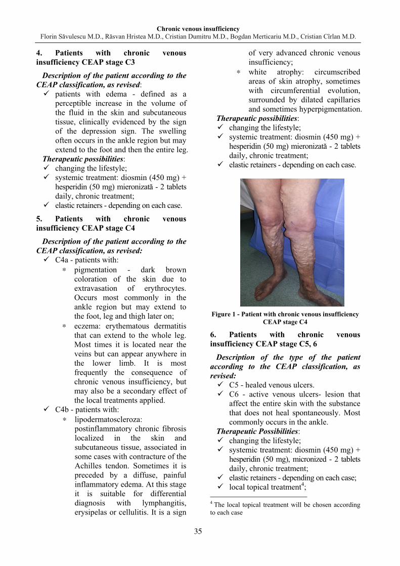
Chronic venous insufficiency Florin S vulescu M.D., R svan Hristea M.D., Cristian Dumitru M.D., Bogdan Merticariu M.D., Cristian Cîrlan M.D.
4. Patients with chronic venous insufficiency CEAP stage C3
Description of the patient according to the CEAP classification, as revised:
patients with edema - defined as a perceptible increase in the volume of the fluid in the skin and subcutaneous tissue, clinically evidenced by the sign of the depression sign. The swelling often occurs in the ankle region but may extend to the foot and then the entire leg.
Therapeutic possibilities: changing the lifestyle; systemic treatment: diosmin (450 mg) +
hesperidin (50 mg) mieronizat - 2 tablets daily, chronic treatment;
elastic retainers - depending on each case.
5. Patients with chronic venous insufficiency CEAP stage C4
Description of the patient according to the CEAP classification, as revised:
C4a - patients with: pigmentation - dark brown
coloration of the skin due to extravasation of erythrocytes. Occurs most commonly in the ankle region but may extend to the foot, leg and thigh later on;
eczema: erythematous dermatitis that can extend to the whole leg. Most times it is located near the veins but can appear anywhere in the lower limb. It is most frequently the consequence of chronic venous insufficiency, but may also be a secondary effect of the local treatments applied.
C4b - patients with: lipodermatoscleroza:
postinflammatory chronic fibrosis localized in the skin and subcutaneous tissue, associated in some cases with contracture of the Achilles tendon. Sometimes it is preceded by a diffuse, painful inflammatory edema. At this stage it is suitable for differential diagnosis with lymphangitis, erysipelas or cellulitis. It is a sign
of very advanced chronic venous insufficiency;
white atrophy: circumscribed areas of skin atrophy, sometimes with circumferential evolution, surrounded by dilated capillaries and sometimes hyperpigmentation.
Therapeutic possibilities: changing the lifestyle; systemic treatment: diosmin (450 mg) +
hesperidin (50 mg) mieronizat - 2 tablets daily, chronic treatment;
elastic retainers - depending on each case.
Figure 1 - Patient with chronic venous insufficiency
CEAP stage C4
6. Patients with chronic venous insufficiency CEAP stage C5, 6
Description of the type of the patient according to the CEAP classification, as revised:
C5 - healed venous ulcers. C6 - active venous ulcers- lesion that
affect the entire skin with the substance that does not heal spontaneously. Most commonly occurs in the ankle.
Therapeutic Possibilities: changing the lifestyle; systemic treatment: diosmin (450 mg) +
hesperidin (50 mg), micronized - 2 tablets daily, chronic treatment;
elastic retainers - depending on each case; local topical treatment4;
4 The local topical treatment will be chosen according to each case
35
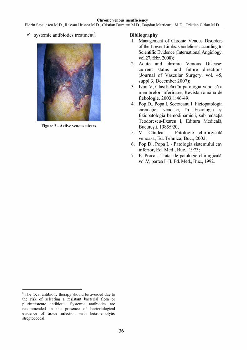
Chronic venous insufficiency Florin S vulescu M.D., R svan Hristea M.D., Cristian Dumitru M.D., Bogdan Merticariu M.D., Cristian Cîrlan M.D.
systemic antibiotics treatment5.
Figure 2 - Active venous ulcers
5 The local antibiotic therapy should be avoided due to the risk of selecting a resistant bacterial flora or plurirezistente antibiotic. Systemic antibiotics are recommended in the presence of bacteriological evidence of tissue infection with beta-hemolytic streptococcal
Bibliography 1. Management of Chronic Venous Disorders
of the Lower Limbs: Guidelines according to Scientific Evidence (International Angiology, vol 27, febr. 2008);
2. Acute and chronic Venous Disease: current status and future directions (Journal of Vascular Surgery, vol. 45, suppl 3, December 2007);
3. Ivan V, Clasific ri în patologia venoas a membrelor inferioare, Revista român de flebologie. 2003;1:46-49;
4. Pop D., Popa I, Socoteanu I. Fiziopatologia circula iei venoase, în Fiziologia i fiziopatologia hemodinamicii, sub redac ia Teodorescu-Exarcu I, Editura Medical , Bucure ti, 1985:920;
5. V. Cândea - Patologie chirurgical venoas , Ed. Tehnic , Buc., 2002;
6. Pop D., Popa I. - Patologia sistemului cav inferior, Ed. Med., Buc., 1973;
7. E. Proca - Tratat de patologie chirurgical , vol.V, partea I+II, Ed. Med., Buc., 1992.
36
The Revue of Aeronautical Medicine and Psychology________________________Volume 18 - Year 2014 – Nr. 1 (66)
CHANGES CARDIOVASCULAR PARAMETERS IN PATIENTS WHO RECEIVED MIDAZOLAM SEDATION
Cristian Georgescu M.D., Ph.D.1, Magdalena Diaconu M.D.1, Cristina Ple a M.D.2, Adrian Tase M.D., Ph.D.3, Monica ân u3
Summary Benzodiazepines have minimal depressant cardiovascular effects even at doses of anesthetic induction. Blood pressure, cardiac output and peripheral vascular resistance, usually have a slight decline, while the heart rate increases sometimes [1]. The study refers to two groups of patients, the first batch having been given midazolam. Cardiovascular parameters were monitored, preanesthetically, intraoperatively and postoperatively for all lots. Keywords: pre-anesthetic examination, intraoperative, and postoperative cardiovascular parameters.
1 Craiova County Emergency Hospital 2 Bucharest Universitary Central Military Emergency Hospital 3 Pitesti County Emergency Hospital
Introduction Sedation is used both during general anesthesia used in laborious interventions as well as in
intravenous anesthesia and regional anesthesia as an adjunct of regional blocks, thus facilitating sleep and anxiolysis, and it is also used as a routine in intensive care services, especially in patients ventilated mechanically [2].
Operative trauma leads to the stimulation of several types of receptors and of several ways of transmission, each one with a particular sensitivity to the analgesic or analgesic adjuvant, only one class of drugs not being able to counter all these mechanisms [3, 4, 5].
The literature points out that midazolam produces an initial decline in BP (blood pressure) of 5-10 % and increases the heart rate by 10-20 % [6].
Purpose and objectives: assessing the development of cardiovascular parameters in patients undergoing loco-regional anesthesia, following adhibition of sedation processes.
Material and methods All the 72 patients undergoing various endourologic interventions with loco-regional
anesthesia, were divided into 2 groups of patients: Group A, consisting of 46 patients received 2.5 mg midazolam in multiple boluses (within 7.5 mg).
Group B, consisting of 26 patients did not receive any sedative. Cardiovascular parameters were monitored during surgery, for the 2 groups and in the case of
the first group which received sedation, at intervals of 15 and 30 minutes and after the surgery they were measured at 15, 30 and 60 minutes.
At preanesthetic examination there were measured systolic BP (SBP) and diastolic (DBP) in the 2 groups of patients and there were obtained the following average values:
Group A: SBP = 135 + 15 mm Hg, DBP = 75 + 10 mm Hg, AV (heart rate) = 75 + 10 beats / minute;
Group B: SBP = 140 + 15 mm Hg, DBP = 80 + 10 mm Hg, AV = 80 + 10 beats / minute.
37
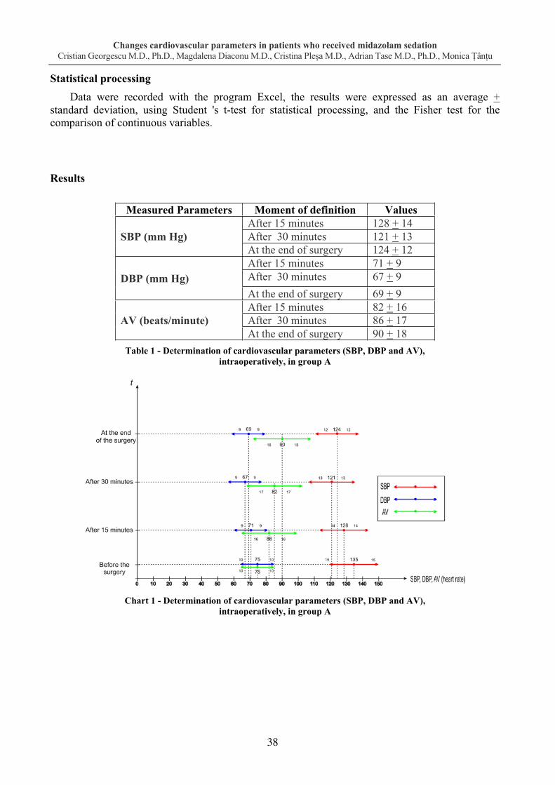
Changes cardiovascular parameters in patients who received midazolam sedation Cristian Georgescu M.D., Ph.D., Magdalena Diaconu M.D., Cristina Ple a M.D., Adrian Tase M.D., Ph.D., Monica ân u
Statistical processing
Data were recorded with the program Excel, the results were expressed as an average + standard deviation, using Student 's t-test for statistical processing, and the Fisher test for the comparison of continuous variables.
Results
Measured Parameters Moment of definition Values
SBP (mm Hg) After 15 minutes 128 + 14 After 30 minutes 121 + 13 At the end of surgery 124 + 12
DBP (mm Hg) After 15 minutes 71 + 9 After 30 minutes 67 + 9 At the end of surgery 69 + 9
AV (beats/minute) After 15 minutes 82 + 16 After 30 minutes 86 + 17 At the end of surgery 90 + 18
Table 1 - Determination of cardiovascular parameters (SBP, DBP and AV), intraoperatively, in group A
Chart 1 - Determination of cardiovascular parameters (SBP, DBP and AV), intraoperatively, in group A
38
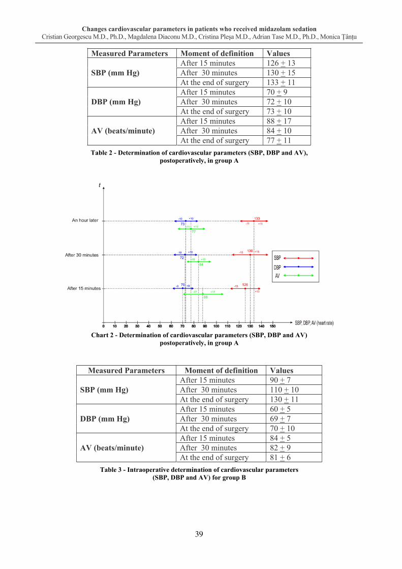
Changes cardiovascular parameters in patients who received midazolam sedation Cristian Georgescu M.D., Ph.D., Magdalena Diaconu M.D., Cristina Ple a M.D., Adrian Tase M.D., Ph.D., Monica ân u
Measured Parameters Moment of definition Values
SBP (mm Hg) After 15 minutes 126 + 13 After 30 minutes 130 + 15 At the end of surgery 133 + 11
DBP (mm Hg) After 15 minutes 70 + 9 After 30 minutes 72 + 10 At the end of surgery 73 + 10
AV (beats/minute) After 15 minutes 88 + 17 After 30 minutes 84 + 10 At the end of surgery 77 + 11
Table 2 - Determination of cardiovascular parameters (SBP, DBP and AV), postoperatively, in group A
Chart 2 - Determination of cardiovascular parameters (SBP, DBP and AV) postoperatively, in group A
Measured Parameters Moment of definition Values
SBP (mm Hg) After 15 minutes 90 + 7 After 30 minutes 110 + 10 At the end of surgery 130 + 11
DBP (mm Hg) After 15 minutes 60 + 5 After 30 minutes 69 + 7 At the end of surgery 70 + 10
AV (beats/minute) After 15 minutes 84 + 5 After 30 minutes 82 + 9 At the end of surgery 81 + 6
Table 3 - Intraoperative determination of cardiovascular parameters (SBP, DBP and AV) for group B
39

Changes cardiovascular parameters in patients who received midazolam sedation Cristian Georgescu M.D., Ph.D., Magdalena Diaconu M.D., Cristina Ple a M.D., Adrian Tase M.D., Ph.D., Monica ân u
Chart 3 - Intraoperative determination of cardiovascular parameters, (SBP, DBP and AV) for group B
Measured Parameters Moment of definition Values
SBP (mm Hg) After 15 minutes 130 + 10 After 30 minutes 135 + 12 At the end of surgery 136 + 12
DBP (mm Hg) After 15 minutes 78 + 10 After 30 minutes 79 + 10 At the end of surgery 80 + 10
AV (beats/minute) After 15 minutes 78 + 10 After 30 minutes 79 + 10 At the end of surgery 80 + 10
Table 4 - Postoperative averages of cardiovascular parameters (SBP, DBP and AV) for group B
Chart 4 - Postoperative averages of cardiovascular parameters (SBP, DBP and AV) for group B
40

Changes cardiovascular parameters in patients who received midazolam sedation Cristian Georgescu M.D., Magdalena Diaconu M.D., Cristina Ple a M.D., Adrian Tase M.D., Ph.D., Monica ân u
Discussion of results
For patients in group B there is a certain cardio-vascular instability, TA decreasing in the first 15 minutes, rather suddenly in some patients, requiring the administration of Ephedrine in a percentage of 5%, in repeated boluses of 10 mg, which leads to an increase in TA and stabilization of the patient, but also to an increase in AV.
Intraoperatively, we found that, in group B, the SBP decreased quite enough 15 minutes later after the spinal, reaching the value of 90 + 7 mm Hg, requiring administration of Ephedrine 5% (10-20 mg), which resulted in the increase of TA, as well as of AV to 84 + 5 beats/minute.
A decrease in BP was also produced after 30 minutes, with a value of 110 + 10 mm Hg, requiring only a small amount of Ephedrine (about 5 mg) so that the AV did not increase significantly, having a value of 82 + 6 beats/minute. At the end of the surgery, AV and TA recorded values close to the preanesthetic ones: 130 + 11 mm Hg and 81 +6 beats/minute. Postoperatively, DBP, SBP and AV recorded after 15 minutes, 30 minutes and one hour values close to the reference.
SBP and DBP in group A decreased in the first 15 minutes from the beginning of the surgery with 5% of the preanesthetic value, and after 30 minutes decreased by 10%. AV increased, in group A, after the first 15 minutes during surgery by 10% and by 15% after 30 minutes, reaching a 20% increase from the value of preanesthetic.
It was observed that both DBP and SBP did not decrease intraoperatively very much as compared to the preanesthetic value, the largest decrease being recorded 30 minutes after the start of the surgery, by 10% of the baseline.
Preanesthetic values were: SBP = 135 + 15 mm Hg and DBP = 75 + 10 mm Hg, and after 30 minutes they decreased as follows: SBP = 121 + 13 mm Hg and DBP = 67 +9 mm Hg.
Following the evolution of AV, it has been an increase of 10 to 20% during surgery. Whereas preanesthetically, AV = 75 + 10 beats/minute after 15 minutes it increased by 10% (82 + 16 beats/minute), after 30 minutes it increased by 15% (86 + 17 beats/minute) and at the end of the surgery it had an increase of 20% of the preanesthetic value (90 + 18 beats/ minute). These increases of AV began to decrease postoperatively after interrupting midazolam, and within one hour they were very close to preanesthetic value (77 + 11 beats/ minute).
If intraoperatively for patients in group A, who were sedated with midazolam, the largest decrease in BP was recorded after 30 minutes, with about 10% of the preanesthetic, after the surgery, since the interruption of midazolam it was observed that the values of both systolic and diastolic blood pressure began to get closer to the preanesthetic value 30 minutes right after the end of surgery. Thus, 30 minutes later SBP = 130 + 15 mm Hg and DBP = 72 + 10 mm Hg , and one hour later SBP = 133 + 11 mm Hg and DBP = 73 + 10 mm Hg.
Conclusions sedation with midazolam appears to provide better hemodynamic stability, less reducing DBP, SBP and AV values as compared to the case in which sedation was not used;
applying techniques for intraoperative sedation with midazolam reduced significantly the intraoperative and early postoperative risk (after the first 6 hours) of hypertensive spurts and episodes of tachyarrhythmia induced by surgery stress;
after the administration of midazolam, intraoperatively, BP, both systolic and diastolic, did not decrease significantly in comparison to the preanesthetic value;
AV began to decline after the surgery, after the interruption of midazolam, an hour later being very close to the preanesthetic value.
41

Changes cardiovascular parameters in patients who received midazolam sedation Cristian Georgescu M.D., Ph.D., Magdalena Diaconu M.D., Cristina Ple a M.D., Adrian Tase M.D., Ph.D., Monica ân u
Bibliography
1. Clinical Anesthesiology, 4 th Edition G. Edward Morgan, Jr., Maged S., Mikhail, Michael J. Murray, Copyright © 2006, 2002 by the McGraw – Hill Companies, inc., Section II, Clinical Pharmacology, Chapter 8, Nonvolatile Anaesthetic Agents – Opioids;
2. Claudiu Zdrehu - Recomand ri pentru analgezie i sedare la pacientul critic, Timi oara, 2009, www.atitimisoara.ro, 145-146;
3. Berggren U, Gordh T, Grama D, Haglund U, Rastad J, Arvidsson D. - Laparoscopic versus open
cholecystectomy: hospitalization, sick leave, analgesia and trauma responses. Br J Surg; 1994; 81:1362-1365;
4. Hensel M, Schwenk W, Bloch A, et al. - The role of anesthesiology in fast track concepts in colonic surgery. Anaesthesist; 2006; 55: 80 -92;
5. Michaloliakou L, Chung F, Sharma S. - Preoperative multimodal analgesia facilitates recovery after ambulatory laparocopic cholecistectomy. Anesth Analg; 1996; 82: 44-51;
6. Acalovschi I. - Anestezie clinic , Ed. Clusium, edi ia II, 2005, pag. 196-200, 206-209, 220-222, 224-229.
42
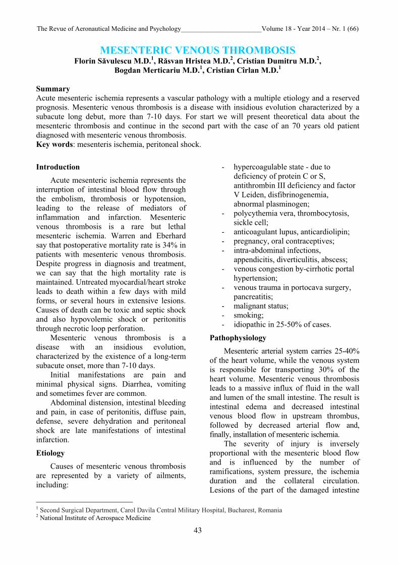
The Revue of Aeronautical Medicine and Psychology________________________Volume 18 - Year 2014 – Nr. 1 (66)
MESENTERIC VENOUS THROMBOSIS Florin S vulescu M.D.1, R svan Hristea M.D.2, Cristian Dumitru M.D.2,
Bogdan Merticariu M.D.1, Cristian Cîrlan M.D.1
Summary Acute mesenteric ischemia represents a vascular pathology with a multiple etiology and a reserved prognosis. Mesenteric venous thrombosis is a disease with insidious evolution characterized by a subacute long debut, more than 7-10 days. For start we will present theoretical data about the mesenteric thrombosis and continue in the second part with the case of an 70 years old patient diagnosed with mesenteric venous thrombosis. Key words: mesenteris ischemia, peritoneal shock.
1 Second Surgical Department, Carol Davila Central Military Hospital, Bucharest, Romania 2 National Institute of Aerospace Medicine
Introduction Acute mesenteric ischemia represents the
interruption of intestinal blood flow through the embolism, thrombosis or hypotension, leading to the release of mediators of inflammation and infarction. Mesenteric venous thrombosis is a rare but lethal mesenteric ischemia. Warren and Eberhard say that postoperative mortality rate is 34% in patients with mesenteric venous thrombosis. Despite progress in diagnosis and treatment, we can say that the high mortality rate is maintained. Untreated myocardial/heart stroke leads to death within a few days with mild forms, or several hours in extensive lesions. Causes of death can be toxic and septic shock and also hypovolemic shock or peritonitis through necrotic loop perforation.
Mesenteric venous thrombosis is a disease with an insidious evolution, characterized by the existence of a long-term subacute onset, more than 7-10 days.
Initial manifestations are pain and minimal physical signs. Diarrhea, vomiting and sometimes fever are common.
Abdominal distension, intestinal bleeding and pain, in case of peritonitis, diffuse pain, defense, severe dehydration and peritoneal shock are late manifestations of intestinal infarction.
Etiology Causes of mesenteric venous thrombosis
are represented by a variety of ailments, including:
- hypercoagulable state - due to deficiency of protein C or S, antithrombin III deficiency and factor V Leiden, disfibrinogenemia, abnormal plasminogen;
- polycythemia vera, thrombocytosis, sickle cell;
- anticoagulant lupus, anticardiolipin; - pregnancy, oral contraceptives; - intra-abdominal infections,
appendicitis, diverticulitis, abscess; - venous congestion by-cirrhotic portal
hypertension; - venous trauma in portocava surgery,
pancreatitis; - malignant status; - smoking; - idiopathic in 25-50% of cases.
Pathophysiology Mesenteric arterial system carries 25-40%
of the heart volume, while the venous system is responsible for transporting 30% of the heart volume. Mesenteric venous thrombosis leads to a massive influx of fluid in the wall and lumen of the small intestine. The result is intestinal edema and decreased intestinal venous blood flow in upstream thrombus, followed by decreased arterial flow and, finally, installation of mesenteric ischemia.
The severity of injury is inversely proportional with the mesenteric blood flow and is influenced by the number of ramifications, system pressure, the ischemia duration and the collateral circulation. Lesions of the part of the damaged intestine
43
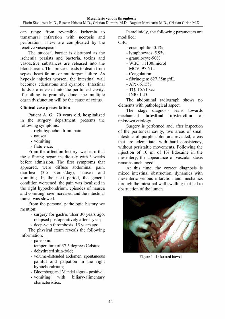
Mesenteric venous thrombosis Florin S vulescu M.D., R svan Hristea M.D., Cristian Dumitru M.D., Bogdan Merticariu M.D., Cristian Cîrlan M.D.
can range from reversible ischemia to transmural infarction with necrosis and perforation. These are complicated by the reactive vasospasm.
The mucosal barrier is disrupted as the ischemia persists and bacteria, toxins and vasoactive substances are released into the bloodstream. This process leads to death from sepsis, heart failure or multiorgan failure. As hypoxic injuries worsen, the intestinal wall becomes edematous and cyanotic. Intestinal fluids are released into the peritoneal cavity. If nothing is promptly done, the multiple organ dysfunction will be the cause of exitus.
Clinical case presentation Patient A. G., 70 years old, hospitalized
in the surgery department, presents the following symptoms:
- right hypochondrium pain - nausea - vomiting - flatulence.
From the affection history, we learn that the suffering began insidiously with 3 weeks before admission. The first symptoms that appeared, were diffuse abdominal pain, diarrhea (3-5 stools/day), nausea and vomiting. In the next period, the general condition worsened, the pain was localized in the right hypochondrium, episodes of nausea and vomiting have increased and the intestinal transit was slowed.
From the personal pathologic history we mention:
- surgery for gastric ulcer 30 years ago, relapsed postoperatively after 1 year;
- deep-vein thrombosis, 15 years ago. The physical exam reveals the following
information: - pale skin; - temperature of 37.5 degrees Celsius; - dehydrated skin-fold; - volume-distended abdomen, spontaneous
painful and palpation in the right hypochondrium;
- Bloomberg and Mandel signs – positive; - vomiting with biliary-alimentary
characteristics.
Paraclinicly, the following parameters are modified: CBC:
- eosinophilic: 0.1% - lymphocytes: 5.9% - granulocyte-90% - WBC: 11100/microl - MCV: 97.6 fL - Coagulation: - fibrinogen: 627.35mg/dL - AP: 66.15% - TQ: 15.71 sec - INR: 1.45 The abdominal radiograph shows no
elements with pathological aspect. The stage diagnosis leans towards
mechanical intestinal obstruction of unknown etiology.
Surgery is performed and, after inspection of the peritoneal cavity, two areas of small intestine of purple color are revealed, areas that are edematiate, with hard consistency, without peristaltic movements. Following the injection of 10 ml of 1% lidocaine in the mesentery, the appearance of vascular stasis remains unchanged.
At this time, the correct diagnosis is mixed intestinal obstruction, dynamics with mesenteric venous infarction and mechanics through the intestinal wall swelling that led to obstruction of the lumen.
Figure 1 - Infarcted bowel
44
Mesenteric venous thrombosis Florin S vulescu M.D., R svan Hristea M.D., Cristian Dumitru M.D., Bogdan Merticariu M.D., Cristian Cîrlan M.D.
Figure 2 - Mesentery appearance after injection
with lidocaine 1%
Diagnosis Laboratory tests are modified, most of times they often present:
- increasing hematocrit; - hyperleukocytosis; - modifications of ionogram indicating
dehydration; - increase in urea and creatinine; - increased hepatic amylase; - FA-growth; - decrease in the alkaline reserve; - increase in lactate; - metabolic acidosis.
Peritoneal puncture can extract, in case of intestinal gangrene, a serosanguinolent and fetid liquid, which studied in Gram colorations reveals numerous leukocytes and bacteria. We can also inject a physiologic serum solution with xenon into the peritoneum, which will be retained by ischemic intestinal, insufficient circulation will not be able to train it.
Abdominal radiography has the appearance of paralytic ileus. Specific images of Myocardial loop are described: the loop is uniformly dilated with a net shape, without air-liquid level, immobile or opaque loop.
CT helps exclude other causes of abdominal pain. It has a sensitivity of 71-96% and a specificity of 92-94%. It may show intestinal pneumatosis, gas in the portal veins, swelling of the intestinal wall or of the mesentery, the mesenteric strangulation and solid organ infarction. Bowel wall edema is the most common sign.
Magnetic resonance IMR reveals the same pathological aspects as CT scan, with a sensitivity of 100% and specificity of 91%, it has the advantage that it is more accurate than the CT but not as practical due to high cost and time required for examination.
Angiography has a sensitivity of 88%. This shows the level and type of the arterial obstruction, the collateral network, the movement of the contrast product in the intestinal lumen, the delay or absence of the opacification of the portal system. The most accurate investigation in the diagnosis of venous mesenteric thrombosis is the laparotomy that allows the surgical treatment.
Differential diagnosis is made with the following conditions:
- abdominal infections; - digestive obstructions; - aortic dissection, - gallstones; - pancreatitis; - myocardial infarction, - bacterial pneumonia; - acute pyelonephritis; - ectopic pregnancy.
Treatment Surgical treatment is indicated in
mesenteric infarction and its complications, it aims the excision of the non-viable segment and anastomosis with margins in healthy tissue. Thrombectomy is indicated in the case of large caliber vein or the portal vein, but most often is doomed to failure. Laparoscopic technique shows no advantages over the classic one, making possible even the damage of the blood flow in the segment found at the edge of the ischemia.
Preoperative Preparation Replacing lost fluids and correction of
acid-base imbalances are necessary and do not represent a contraindication for surgery, but only an indication to delay it for a short time. Given the possible major loss of blood from the digestive tract, it is preferable to be able to do blood transfusion without hesitation. Antibiotic treatment should be instituted
45

Mesenteric venous thrombosis Florin S vulescu M.D., R svan Hristea M.D., Cristian Dumitru M.D., Bogdan Merticariu M.D., Cristian Cîrlan M.D.
urgently using broad-spectrum medication, with imperative character and should also impose anticoagulation.
Intraoperative details You can perform two types of surgery
procedures: - reconstructive surgeries such as
embolectomy, which can be done, provided that the intestinal changes are still reversible: preserved contractility, presence of pulsation on small vessels;
- mutilating surgery, bowel resection. The preferred method is the supra median and subombilical approach, peritoneal fluid is taken for bacteriological examination and antibiogram and the degree and extent of lesions of small bowel and right colon are assessed. Segmental bowel resection will be extended to the healthy tissue to allow restoration of continuity digestive simultaneously.
Of great importance as operator time is the visual inspection of the small intestine, and we will look for color, areas of swelling, bleeding and transmission of peristaltic movements. Among the means of intraoperative diagnosis the most useful proved to be the ultraviolet lamp examination after fluorescein injection.
Postoperatively Postoperative treatment includes
surveillance of vital signs, hemoglobin, identification of signs of sepsis and hemorrhage when appropriate. It was observed that in 60% of cases re-thrombosis occurs in the area of anastomosis, therefore it is essential to continue anticoagulant therapy started preoperatively. To avoid dynamic ileus prokinetic drugs are recommended.
Complications Due to hypercoagulable conditions, the
patient is prone to deep venous thrombosis, a risk that can be removed with a suitable
anticoagulant therapy and early mobilization of the patient. For better monitoring it is indicated to use a catheter Swan-Ganz. Given the hypovolemic status, renal failure is possible, a complication that can be eliminated through proper hydration and proper diuretics doses. Other possible complications are: internal bleeding, sepsis, and short bowel syndrome.
Prognosis Mesenteric venous thrombosis has a
mortality of 34% and a recurrence rate of 25% in the absence of anticoagulant therapy. Surgery combined with proper anticoagulation shows a recurrence rate of about 3-5%. Of all the causes of mesenteric ischemia, mesenteric venous thrombosis has the best prognosis. Bibliography 1. Eugen Tarcoveanu - Tehnici chirurgicale,
2003; 2. Swartz DE, Felix El - Acute mesenteric
venous thrombosis following laparoscopic Roux-en-Y gastric by-pass. JSLS. Apr-JUN 2004;
3. Dada FB, Balan AD - Recurrent primary mesenteric venous thrombosis. South Med J. oct 1987;
4. Ellis DJ, Brandt LJ - Mesenteric venous thrombosis. Dec 1994;
5. Hassan HA, Raufman JP - Mesenteric venous thrombosis. South Med J. Jun 1999;
6. Preventza OA, Habib FA, Young SC - Portal vein thrombosis: an unusual complication of laparoscopic cholecystectomy. JSLS. Jan-Mar 2005;
7. Roman RJ, Loeb PM - Massive colonic dilatation as initial presentation of mesenteric vein thrombosis. Dig Dis Sci. Mar 1987;
8. James AW, Rabl C, Westphalen AC, et al. - Portomesenteric venous thrombosis after laparoscopic surgery: a systematic literature review. Arch Surg. Jun 2009.
46
The Revue of Aeronautical Medicine and Psychology________________________Volume 18 - Year 2014 – Nr. 1 (66)
MITRAL VALVE PROLAPSE AND THE INFECTIVE ENDOCARDITIS
Corina Grosu M.D.1 Summary Introduction: data regarding MVP-IE association, and especially regarding the importance of antibiotics prophylaxis is controversial. Material and methods: selection and analyzing data from 5 patients with MVP from a lot of 50 cases of IE with surgical indication. Results: MVP prevalence in our lot 10%, mortality 20%, diabetes as negative prognosis factor, but without effect, bacteriological profile: S. aureus and negative cultures. Discussions: our data are in concordance with modern literature data and contradicts previous decades data. This data does not recommend routine prophylaxis for MVP, but MVP requires an increase attention from clinicians. Conclusion: our lot data support for low risk of IE in MVP, and if happened, the correct treatment gives good results, regardless of bacteriological profile. Clinical monitoring and early detection of IE signs are more important the routine antibiotics prophylaxis. Key words: MVP, IE, risk, prophylaxis
1 National Institute of Aerospace Medicine
Introduction Mitral valve prolapse (MVP) was
traditionally associated as a risk factor for infective endocarditis (IE). Both IE pathology and morphology seem to support this link, but epidemiological evidence has never been strong. In the last decade, changes in epidemiological profile in IE led to changes in both risk factors and in indications for prophylaxis.
MVP results from total or partial movement of mitral leaflets during systole. Pathological base is either chords elongation, chords rupture or enlargement of mitral ring. General population prevalence is 2-3%. There is genetic, autosomal dominant component (1).
Clinical diagnosis may be made by auscultation (apical click or apical murmur in late systole), but confirmation is made by ultrasound, Doppler being preferred.
MVP is asymptomatic in most patients, especially in youngs, but in elders may trigger sever valvular dysfunction with indication for surgery, embolic phenomena or IE.
So, IE may be a complication of MVP and little is known on how to prevent IE occurrence.
Today, prevention in IE is subject to debates, current guidelines (2) restricting
mandatory antibiotic prophylaxis to a series of severe dysfunctions not including MVP.
Prophylaxis must not oversee basic hygiene measures, especially on skin and oral cavity, having being proved that oral bacteriemia occurred daily (3).
It is well established that IE requires the non-bacterial thrombotic endocarditis (NBTE), for which MVP anomalies are a good precursor. The regurgitation flux is not involved in NBTE formation and cannot be used as a predictor (4).
Baddour describes Streptococcus viridans as common etiology, age above 40 and other germs being reported as negative prognosis factors (5).
Rajamannan recommends risk evaluation of IE on MVP in clinical context, but admits the technical difficulties of such approach, especially for early stages of IE (6).
Chambers recommends more randomized trials to be employed to find indication of valve surgery in MVP, for preventing complications including IE (7).
Chambers also points out the opportunity of collecting more data regarding MVP-IE link, now that mandatory prophylaxis have been restricted to narrower indications (8).
47

Mitral Valve Prolapse and the Infective Endocarditis Corina Grosu M.D.
Material and methods
We have selected all MVP cases from a lot of 50 patients who received surgical treatment in a three years period (2009-2012), data gathered by the author for a larger study regarding surgical indication of IE. All the interventions were made in a single cardiovascular surgery centre.
Our data was not relevant enough for the clinical manifestations of MVP prior to IE.
Data have been processed with a tabelar software, considering risk factors for occurrence and evolution of IE, status at admission (complications, emergency or elective surgery), strain involved, antibiotics treatment and immediate and 1 year survival.
All the cases had native valve IE. There was no control group, without IE.
Results
We have found 5 patients with MVP history out of 50. Our prevalence was 10%. One case died post operatory so we had a 20% mortality.
Sex ratio was 1,5:1 favoring women. Mean age was 60.8 years. Except for one case, all the patients from
the group were overweight (mean BMI 28.04) Only one triggering factor for bacteriemia
was found (fig. 1).
Figure 1 - Factors promoting bacteriemia
From the factors associated with
unfavorable IE prognosis, we have found only diabetes, in two patients (fig. 2)
Figure 2 - Prevalence of diabetes
It worth noting that 4 of the 5 patients had high blood pressure in their history.
Regarding admission status, 4 had IE with no complications and one had CT proved splenical abscesses.
Cultures have been worked in 4 out 5 patients, the fifth having emergency surgery. (fig 3).
Figure 3 - Involved germs
Antibiotical therapy was made according to the guidelines for 8 weeks, except for the emergency case which also died.
Valve replacement was always with St. Jude valve.
Post operatory and one year follow up was good in all 4 surviving cases.
Discussions I present some cases of IE on MVP
associated or no to medical maneuvers: Pecoraro describes a case of IE with
Staphyloccocus lugdunesnsis. Ultrasound found a large vegetation (1.66/1 cm), sign of a
48
Mitral Valve Prolapse and the Infective Endocarditis Corina Grosu M.D.
sub acute endocarditis. Patient complained of recurrent fever and mioarthralguc syndrome. Case characteristic was the low age of the patient, 18 years old (9).
Rigilano describes a case of entrococcal IE following a rectosigmoidoscopy in 67 years old male (10).
Niv describes a IE on 47 years old male with history of MVP, following an esophageal dilatation, one of the few cases reported in literature (11).
On our lot, we have found a single evidence of a triggering factor, a dental maneuver, corresponding with published data scarcity. Blood culture was negative for the case.
Clemens studied a lot of 51 patients with IE, diagnosed according to modified Durack criteria and no additional risk factors. 13 (25%) had MVP in their history, in the control lot (no IE) prevalence being 7%. Relative risk of 8.2 points MVP as a risk factor for IE (12).
Devereux find a 4 times higher prevalence of MVP (16 vs 4, p=0,001) in IE patients versus the control group. Unlinke Clemens, general prevalence is more close to real life data. Devereux concludes that antibiotic prophylaxis may be indicates for man with MVP (13).
Hickey found 20% prevalence of MVP in IE patients, vs 4% in control group (14).
Our lot had a 10% prevalence, smaller than the cited studies, but also in a smaller initial lot. A possible explanation would be changes in epidemiological profile of IE.
A similar result is found by David, on a 840 patients selected for mitral valve interventions, most of the with MVP and 10,3% incidence of IE (15).
Freed finds only 2% EI incidence, without follow up data available (16).
Danchin finds a 5,7 risk ratio, this becoming highly significant (14.5) in patients with clinic murmur (4). Such clinical data were not available in our lot.
Duren finds a 6% EI incidence, on 300 patients with idiopathic MVP. Of those 22,2% died and 33,3% required surgical intervention, data similar with our lot (17).
Nolan points out mild clinical features and good treatment response for patients with
MVP and IE, but results at 5 years were similar with other populations (10% death, 40% recurrences) (18).
For our lot, we had cases with S. aureus and negative hemocultures, both difficult to treat; antibiotical therapy was effective with no one year recurrence. No data are available for 5 years.
Two patients had diabetes, but this did not reflect in the outcome.
It worth noting that the only death in our lot had no associated poor prognosis factors, not even high blood pressure in history. It had too elements of unfavorable prognosis (age greater than 65 and male) but the same profile was shared by another case with survived without complications. The explication could be a sub acute IE with minimal clinical features, which evolved long enough for the patient to develop peripheral abscesses.
Overweight is not cited as a worsen prognosis factor either for MVP or IE but in our lot was present for almost all cases.
Mean age was over 60, at the threshold of aggravating prognosis factor.
In previous years, data have pro and versus antibiotic prophylaxis fro MVP.
Devereux finds a good cost effectiveness ratio for prophylaxis in MVP patients (19).
In a similar timeframe, Bor finds no significant difference between mortality of IE on MVP patients, with or without prophylaxis (20).
Current recommendations include MVP with systolic murmur as low risk and recommends antibiotics prophylaxis only for medical maneuvers in conditions of obvious infection and for patients with aggravating factors (diabetes, age, important amount of blood exposed during procedure, poor hygiene, difficult and/or long procedures) (21).
Conclusion
Our lot data support for low risk of IE in MVP, and if happened, the correct treatment gives good results, regardless of bacteriological profile. Clinical monitoring and early detection of IE signs are more important the routine antibiotics prophylaxis.
49
Mitral Valve Prolapse and the Infective Endocarditis Corina Grosu M.D.
Bibliography
1. Boudoulas, K. D. & Boudoulas, H. Floppy - Mitral Valve (FMV)/Mitral Valve Prolapse (MVP) and the FMV/MVP Syndrome: Pathophysiologic Mechanisms and Pathogenesis of Symptoms. Cardiology 126, 69–80 (2013);
2. Habib, G. et al. - Guidelines on the prevention, diagnosis, and treatment of infective endocarditis (new version 2009) The Task Force on the Prevention, Diagnosis, and Treatment of Infective Endocarditis of the European Society of Cardiology (ESC). Eur. Heart J. 30, 2369–2413 (2009);
3. Roberts, G. J. - Dentists Are Innocent! "Everyday" Bacteremia Is the Real Culprit: A Review and Assessment of the Evidence That Dental Surgical Procedures Are a Principal Cause of Bacterial Endocarditis in Children. Pediatr. Cardiol. 20, 317–325 (1999).
4. Danchin, N. et al. - Mitral valve prolapse as a risk factor for infective endocarditis. The Lancet 333, 743–745 (1989);
5. Baddour, L. M. & Bisno, A. L. - infective Endocarditis Complicating Mitral Valve Prolapse: Epidemiologic, Clinical, and Microbiologic Aspects. Rev. Infect. Dis. 8, 117–137 (1986);
6. Rajamannan, N. M. - The risk of infective endocarditis in the 21st century. Indian Heart J. 65, 452–453 (2013);
7. Chambers, J. B. et al. - Valvular heart disease: a call for global collaborative research initiatives. Heart 99, 1797–1799 (2013);
8. Chambers, J. et al. - The infective endocarditis team: recommendations from an international working group. Heart 100, 524–527 (2014);
9. Pecoraro, R., Tuttolomondo, A., Parrinello, G., Pinto, A. & Licata, G. -Staphylococcus lugdunensis Endocarditis Complicated by Embolism in an 18-Year-Old Woman with Mitral Valve Prolapse. Case Rep. Infect. Dis. 2013, e730924 (2013);
10. Rigilano J, Mahapatra R, Barnhill J & Gutierrez J. - Enterococcal endocarditis following sigmoidoscopy and mitral valve prolapse. Arch. Intern. Med. 144, 850–851 (1984);
11. Niv, Y., Bat, L. & Motro, M. - Bacterial endocarditis after Hurst bougienage in a patient with a benign esophageal stricture and mitral valve prolapse. Gastrointest. Endosc. 31, 265–267 (1985);
12. Clemens, J. D., Horwitz, R. I., Jaffe, C. C., Feinstein, A. R. & Stanton, B. F. - A Controlled Evaluation of the Risk of Bacterial Endocarditis in Persons with Mitral-Valve Prolapse. N. Engl. J. Med. 307, 776–781 (1982);
13. Devereux, R. B. et al. - Complications of mitral valve prolapse: Disproportionate occurrence in men and older patients. Am. J. Med. 81, 751–758 (1986);
14. Hickey, A. J., MacMahon, S. W. & Wilcken, D. E. L. - Mitral valve prolapse and bacterial endocarditis: When is antibiotic prophylaxis necessary? Am. Heart J. 109, 431–435 (1985).
15. David, T. E., Armstrong, S., McCrindle, B. W. & Manlhiot, C. - Late Outcomes of Mitral Valve Repair for Mitral Regurgitation Due to Degenerative Disease. Circulation 127, 1485–1492 (2013);
16. Freed, L. A. et al. - Prevalence and Clinical Outcome of Mitral-Valve Prolapse. N. Engl. J. Med. 341, 1–7 (1999);
17. Düren, D. R., Becker, A. E. & Dunning, A. J. - Long-term follow-up of idiopathic mitral valve prolapse in 300 patients: A prospective study. J. Am. Coll. Cardiol. 11, 42–47 (1988);
18. Nolan CM, Kane JJ & Grunow WA. - Infective endocarditis and mitral prolapse a comparison with other types of endocarditis. Arch. Intern. Med. 141, 447–450 (1981);
19. Devereux, R. B., Frary, C. J., Kramer-Fox, R., Roberts, R. B. & Ruchlin, H. S. - Cost-effectiveness of infective endocarditis prophylaxis for mitral valve prolapse with or without a mitral regurgitant murmur. Am. J. Cardiol. 74, 1024–1029 (1994);
20. Bor, D. H. & Himmelstein, D. U. - Endocarditis prophylaxis for patients with mitral valve prolapse: A quantitative analysis. Am. J. Med. 76, 711–717 (1984);
21. Prendergast, B. D. - The changing face of infective endocarditis. Heart 92, 879–885 (2006).
50

The Revue of Aeronautical Medicine and Psychology________________________Volume 18 - Year 2014 – Nr. 1 (66)
PSYCHOLOGICAL EVALUATION SYSTEM IN AVIATION: DESIGN, CONTEXT, PRINCIPLES, ISSUES
Vasile Gherghina, psychologist1 Summary Psychological selection is especially used for professions requiring high expenses for personnel training or where human errors (or norm violations) have negative professional, social and economic consequences. Psychological assessment when necessary has to take place according to certain reference framework such as the legal one, that specific to the aeronautical environment and the psychological one. The development of a selection system specific to aeronautics has to be based on the study of aptitudes (general and special), personality structure, professional requirements, specialty studies and meta-analyses. Because not all civil pilots are employed in the same manner, since they do not come from the same environments and do not have the same level of flying experience, there have to be different assessment programs, adapted to each particular situation. The ultimate purpose of the assessment is to realize a prognosis regarding the individual’s adaptation capacity to the professional requirements. Keywords: psychological evaluation, pilot selection
1 National Institute of Aerospace Medicine
Introduction The studies of differential and
experimental psychology demonstrated that although individuals have the same qualifications, work with the same machines in the same organizational and environmental conditions achieve different performance mainly due to their multiple intellectual, physical, temperamental, aptitudinal, attitudinal and motivational differences.
The conclusions coming from research in applied psychology in the field of work and ergonomics regarding maximizing efficacy and security for the man-machine-environment systems led to the selection- making and professional orientation of the individuals for their better adaptation to the specificity of the professional activity. The selection was practically born from the fact that people differ among them in many ways.
Psychological selection is especially used for professions requiring high expenses for personnel training or where human errors (or norm violations) have negative professional, social and economic consequences. The specificity of the civil pilot activity represents precisely one which corresponds to such situations. The pilot has to be able to rapidly assess the priorities of his tasks, to combine
information from different sources in order to create a coherent image of the events and to have sufficient psychical resources so as to conceive an action plan while piloting the aircraft. Because, the decision time decreased to a few seconds, a small error can have serious consequences for the team or aircraft. The variety and intensity of the stress factors to which a pilot is subjected, is probably higher than in any other profession.
The effects of the physical and psychical tensions - heat, noise, vibrations, hypoxia, acceleration - on the professional performance are well known. The study of the psychic functions limits, of the way in which the control and display systems are designed, understanding the inter-human relations on board of the aircraft are aspects to which psychological research provided answers. Therefore, we can say that all the results of this research have as final purpose preventing accidents and increasing activity efficiency. Specific reference frameworks in psychological assessment
Psychological assessment when necessary has to take place according to certain reference framework such as the legal one, that specific to the aeronautical environment and the psychological one. All
51
Psychological evaluation system in aviation: design, context, principles, issues Vasile Gherghina, psychologist
these different contexts rise a series of problems for which several clarifications are necessary whose purpose is to find adequate practical solutions.
Legislative Framework Consequently, the European legal context
is represented in this situation by the ,,Acceptable Means Of Compliance And Guidance Material To Part-med, Specific requirements for class 1 medical certificates AMC 1” a normative act which comprises at the psychology chapter four articles. The first if these articles provisions that: „should there be a suspicion or established proof that an applicant has a psychological disorder, then there should be requested the psychologist’s opinion and advice”. In this situation we can ask which are the criteria through which a specialist (psychiatrist and/or neurologist) can base the suspicion that there is a psychological disorder? This article since it is deprived of precision and clarity thus giving way to interpretations should have better mentioned from the beginning the fact that a psychologist needs to set by scientific means the existence of psychic deficiencies which can interfere with doing safely the pilot job. The expression itself of „psychological disorder” is a controversial one since the term „disorder” is specific to the medical (psychiatric) environment and definitely not to the psychological environment (which in the given situation should be: inclination, trend, accentuation, disharmony)
The second article mentions that: „established evidence should be verifiable information from an identifiable source which evokes doubts concerning the mental fitness or personality of particular individual. Sources for this information can be accidents or incidents, problems in training or proficiency checks, delinquency or knowledge relevant to the safe exercise of the privileges of the applicable license”. In this case, should there be provisioned that psychological assessment at least in the ab initio phase should by stand-alone within the medical assessment, then many of the incidents and the situations with problematic potential could certainly be avoided. Psychological examination is useful since it has the means and methods through which it
can anticipate with a degree of certainty which has been proved, some problems related to the deficitary development of the aptitudes required by flying, certain trends in developing negative behavior.
Practically, there should not be diminished the importance of the psychological selection since there is the risk of increasing the number of individuals who can become a constant source of problems related to the safety and efficiency of flying in general (low capacity of acquiring knowledge, deficiencies in the control capacity of the aircraft, errors and omissions which cause incidents and accidents, deficitary abilities of social interactions).
The other two articles make an extremely summary reference, on one hand to the methods an accredited psychologist in the aeronautic environment can use and on the other hand to the fact that the psychological act is finalized by means of a report in which this presents his opinion or advice about the assessed subject.
Professional Framework In what regards the professional framework
specific to the civilian pilot activity, one has to clarify a few matters whose purpose is to nuance the principles which should base and guide the psychological assessment in these particular situations.
Although there are situations when companies employ pilots who do not have flying experience who they train starting with the ab initio phase through theoretical and practical courses, most employers in civil aeronautics prefer to employ experienced pilots in order to mainly reduce training expenses. Traditionally, civil aviation used to employ military pilots, but at present, the number of military pilots tends to decrease, and this is a fact which will more and more determine civil aviation to also train pilots staring with the ab initio phase. Therefore, the psychological selection procedures used in civil aviation, at least for the initial formation phase, will have to be similar as complexity to the military ones.
Pilots who have previous flying experience usually meet minimal conditions
52
Psychological evaluation system in aviation: design, context, principles, issues Vasile Gherghina, psychologist
required when employed, usually consisting in: number of flying hours, licenses, certifications, recommendations and qualifications for different types of aircraft. It is for these situations that there are used different methods (interview, simulator, check flights) so as to establish the existence of a minimally developed capacity through which one will be able to fulfill, in the future activity circumstances, the operational and professional training standards and the capacity to adapt to the new organizational culture. Usually, in these situations, psychological assessment is less required. The emphasis is obviously laid on the professional experience which has already been proved.
Yet, should one employ civil pilots who are not previously flight experienced, one uses intensively the information provided by the psychological assessment. This type of assessment has as purpose to determine, through scientific methods at individual level, the degree of aptitudinal, general and specific development and the potential of professional adaptation.
Therefore, it is not at all unnatural to have differences, in terms of criteria or trials to be used, among assessment types (ab initio, employment, accidents, routine periodical assessments, other circumstances). Each training phase requires different aptitudes, abilities and attitudes and for this one has to use specific methods to identify the individual potential.
Psychological Framework The third reference framework submitted
to this analysis is the one which is truly related to the specific and the context of the psychological assessments. In this context, psychological selection represents an ensemble of instruments, procedures whose purpose is to select candidates for certain jobs so that they could be able to fulfill their professional tasks. Consequently, if all individuals who are taking a training course reach a satisfactory level, and none of them is eliminated during taking the course, then in order to access to the course, psychological assessment becomes unnecessary. Also, if certain subjects become unable to take the entire course, yet the cost and the inconvenience their failure are minimal then is no need for examination.
Yet, if the training costs are high and there are constraints to keep financial losses to minimum, then each training failure becomes significant. Similarly, if one has to deal with a special operational request, such as that related to flying, one has to perform psychological assessment.
Psychological assessment, in this context, seeks to determine the development degree of the adaptational potential and the manner in which an individual possesses a certain natural capacity through which he can match up to several requirements of theoretical and practical training. The natural capacity is synonymous, in this way, with the aptitude (latent potential or the capacity to develop abilities that lead to performance achievement). The psychological test can capture only a certain sample of psychic manifestations: technical thinking, attentional resources, verbal and abstract intelligence, the ability to work with numbers and certain personality traits.
Because the pilot profession - with everything it supposes: specific tasks of piloting, doing complex training systems - is an intensely demanding one from a psychological viewpoint, then realization of psychological assessment represents a necessity which is understood in itself.
The most important thing one has to take into account when doing the psychological assessment is the scientific rigor. Without it, the results one obtains determine the decrease in credibility of the psychological act. The development of a selection system specific to aeronautics has to be based on the study of aptitudes (general and special), personality structure, professional requirements, specialty studies and meta-analyses. The qualitative and quantitative research should guide the practice.
In this way, the fundamental model of te psychological selection includes the following stages: 1. the profession psychological analysis; 2. setting the criterion and the prediction instruments; 3. performance measuring according to the predictor and the professional criterion; 4. predictor assessment according to the criterion.
Also, a system of psychological assessment has to have a very important quality, namely, the predictive validity, that is
53
Psychological evaluation system in aviation: design, context, principles, issues Vasile Gherghina, psychologist
to have that capacity to prognosticate the professional performance. Both the process of determining the predictive validity and that of dealing with all stages for designing a testing system requires a number of 3-5 years, while this is reviewed and refined.
The best predictors for flying performance are represented by the results obtained at the tests for general and special aptitudes, whereas the trials assessing personality structure have a lower predictive validity.
In general, psychological evaluation is completed by realization of a psychological profile that highlights the development of the following factors: fluid and crystallized intelligence, working memory, balance between speed and accuracy of actions, tolerance to stress situations, the ability to divide attention, capacity to risk taking, ability to work in monotonous conditions, the ability to estimate time, assessing distances.
The ultimate purpose of the assessment is to realize a prognosis regarding the individual’s adaptation capacity to the professional requirements.
Conclusions In this complex environment where not all
civil pilots are employed in the same manner, since they do not come from the same environments and do not have the same level of flying experience, there have to be different assessment programs, adapted to each particular situation. Using psychological testing in a manner lacking discernment could lead to distorting the way in which the psychological act is generally considered in civil aeronautics.
The activities related to flying are not deemed dangerous in their own nature, but there is no excuse for being out of focus, incapacity, errors or omissions, and we could certainly say that the whole knowledge activity (description, explanation, application) of the aviation psychologist has as ultimate purpose to prevent accidents and to increase efficiency. It is illustrative in this way what Daniel Maurino says: „(...) the battle for safety is an endless quest, against a restless enemy with infinite resource who will never give up”. And we can also say that in this battle, psychological assessment is an indispensable instrument in order to improve flying safety and efficiency.
Should be noted also that aeronautical psychology is in a sensitive position, since, on one hand it is substantially limited by the confusions and the lack in the normative acts for the selection circumstances, and on the other hand it is intensely demanded to respond to the needs related to the knowledge, explanation and training of the psychological factors involved in flying. Yet, let us hope this situation is a transitory one.
Bibliography 1. Boghaty, Z., (coord.) - Manual de tehnici
i metode în psihologia muncii i organiza ional , Editura Polirom, Ia i, 2007;
2. Hunter, D. R., i Burke, E. - Predicting aircraft pilot-training success: A meta-analysis of published research, Journal of Aviation Psychology, Issue 4, 1994;
3. Hunter, D. R., i Burke, E. - Handbook of Pilot Selection, Cambridge, 1995;
4. Carretta, Thomas, Ree, Malcolm - Pilot candidate selection method (PCSM): what makes it work?, Armstrong Laboratory, Brooks Air Force Base, 1993;
5. Damos, Diane - Pilot Selection Batteries: Shortcomings and Perspectives, The International Journal of Aviation Psychology, Volume 6, Issue 2, 1996;
6. Holban, I. - Probleme de psihologia muncii, Editura tiin ific , Bucure ti, 1970;
7. Green, Roger - Aviation psychology, British Medical Journal Volume 286, London, 1983;
8. Martinussen, M. - Psychological measures as predictors of pilot performance: A meta-analysis, The International Journal of Aviation Psychology, Issue 6, 1996;
9. Minulescu, M. - Teorie i practic în psihodiagnoz , Editura Funda iei România de Mâine, Bucure ti, 2005;
10. Paullin, Cheryl, (coord.) - Review of Aviator Selection, United States Army Research Institute for the Behavioral and Social Sciences, Minnesota, 2006;
11. http://www.easa.europa.eu/agency-measures/acceptable-means-of-compliance-and-guidance-material.php
54

The Revue of Aeronautical Medicine and Psychology________________________Volume 18 - Year 2014 – Nr. 1 (66)
TO THE CONTRIBUTORS
Our journal is opened to any kind of collaboration regarding news in aerospace medicine and psychology, scientific theoretical and practical research, applied in the field of expertise and medical assistance of the aerospace personnel, both civilian and military, in Romania.
We wish to publish both strictly scientific medical papers, and articles which may be used in the process of training of doctors and psychologists working in aviation field. The published materials may also contribute to the general information of a large category of personnel involved in the aerospace domain.
The authors (doctors, psychologists, nurses, aeronautical personnel) will send the manuscripts on the following address: “SOCIETATEA DE MEDICIN AERONAUTIC DIN ROMÂNIA”, Calea Plevnei nr. 134, Sector 1, Bucharest, cod 70786, Tel / Fax 021 3187247, or e-mail it to: [email protected] with the specification “for the journal”.
The authors must write their complete address, phone number (or fax) and their e-mail address, for correspondence. The papers must also comprise a letter of acceptance of being published in our journal, pro bono.
The data and statements in the articles belong fully to the authors, the redaction and publisher declining any responsibility.
The articles, no longer than 4400 printed characters, will be written on a PC, with 2 cm edges, and will comprise:
1. The article; 2. The abstract; 3. Key words; 4. The following structure: introduction, materials and methods, results,
conclusions; 5. Bibliography.
The members of the Romanian Society of Aerospace Medicine have priority in publishing their papers. The editorial team is grateful to authors for sending the articles on magnetic support or by e-mail.
The redaction thanks to all sponsors who helped us and would like to inform about the acceptance of sponsorship and advertising. The redaction
55