Embed Size (px)
Citation preview

© COPYRIG
HT UPM
EFFECTS OF Morinda citrifolia L. LEAF EXTRACT ON HUMAN TLYMPHOBLATIC LEUKEMIA (JURKAT) CELL AND WEHI-3B CELL-
INDUCED MURINE LEUKEMIA
NEGIN AHMADI
FPSK(p) 2016 14

© COPYRIG
HT UPM
EFFECTS OF Morinda citrifolia L. LEAF EXTRACT ON HUMAN T-
LYMPHOBLATIC LEUKEMIA (JURKAT) CELL AND WEHI-3B CELL-
INDUCED MURINE LEUKEMIA
By
NEGIN AHMADI
Thesis Submitted to the School of Graduate Studies, Universiti Putra Malaysia, in
Fulfillment of the Requirements for the Degree of Doctor of Philosophy
June 2016

© COPYRIG
HT UPM
COPYRIGHT
All material contained within the thesis, including without limitation text, logos, icons,
photographs and all other artwork, is copyright material of Universiti Putra Malaysia
unless otherwise stated. Use may be made of any material contained within the thesis for non-commercial purposes from the copyright holder. Commercial use of material
may only be made with the express, prior, written permission of Universiti Putra
Malaysia.
Copyright © Universiti Putra Malaysia
© COPYRIG
HT UPM
i
Abstract of thesis presented to the Senate of Universiti Putra Malaysia in fulfillment of
the requirement for the Degree of Doctor of Philosophy
EFFECTS OF Morinda citrifolia L. LEAF EXTRACT ON HUMAN T-
LYMPHOBLATIC LEUKEMIA (JURKAT) CELL AND WEHI-3B CELL-
INDUCED MURINE LEUKEMIA
By
NEGIN AHMADI
June 2016
Chairman : Professor Suhaila Mohamed, PhD
Faculty : Medicine and Health Sciences
Morinda citrifolia (Rubiaceae) or mengkudu is familiarly known as Ba JiTian, Noni or
Nonu, Indian Mulberry, Nhau and Cheese fruit. There has been no literature reported on
the mechanism of Morinda citrifolia leaf extract and its effects on leukemia. Therefore,
the anti-leukemic effect of Morinda citrifolia leaf extract was examined on a human T-
lymphoblastic leukemia (JURKAT) cells line and on WEHI-3B (myelomonocytic
leukaemia) cell-induced murine leukemia.
MTT assay, fluorescent microscope, flow cytometric analysis after Annexin V-FITC staining, cell cycle, TUNEL assay, and caspase-3, -8 and -9 assays were employed. The
study showed that Morinda citrifolia leaf extract significantly (P<0.05) suppressed
proliferation of JURKAT and WEHI-3B cells in vitro in a time-dependent manner with
an IC50 of 14.5 ± 0.1 and 17.05 ± 0.14 μg/ml at 72 hours. The anti-proliferative effect of
Morinda citrifolia leaf extract on JURKAT and WEHI-3B cells was shown to induce apoptosis
via extrinsic pathway. The study also attempts to determine the effect of Morinda
citrifolia leaf extract and Zerumbone on WEHI-3B cells. The results show that exposure
of WEHI-3B cells to Morinda citrifolia leaf extract along with Zerumbone caused a
greater level of cell progress inhibition than either extract alone.
BALB/c mice were leukemia-induced with a single intraperitoneal injection of WEHI-
3B cells (1×10⁶ cells/animal). The in vivo study revealed that oral Morinda citrifolia leaf extract at doses of 100 mg/kg and 200 mg/kg suppressed the proliferation of leukemic
cells in leukemic mice by the decrease in leukemic cell population in the spleen.
Histopathologic, electron microscopic, immunochemical evaluations and TUNEL assay
studies showed the Morinda citrifolia leaf extract leukemia suppression is via apoptosis.
To determine whether the leukaemia was restricted to the peritoneal cavity, this study
evaluated whether i.p. inoculated WEHI-3 cells were able to migrate to the bone
marrow. It is observed that WEHI-3 cells were present in the marrow of inoculated mice
as early as 24 h post-inoculation, demonstrating the ability of these cells to colonise
secondary sites. This observation demonstrates their aggressive nature, which resulted in

© COPYRIG
HT UPM
ii
the mortality of all animals tested within 30 days. By contrast, under identical
conditions, Morinda citrifolia leaf extract markedly promotes the proliferation of mouse
normal mononuclear bone marrow cells.
Using qRT-PCR Array, on the spleen cells of Morinda citrifolia leaf extract treated
leukemic mice indicated the molecular mechanisms accountable for the effective treatment included anti-inflammatory and immune-modulating responses, JAK-STAT
signaling, and hematopoiesis pathways.
To define potential toxicity of Morinda citrifolia leaf extract, human peripheral blood
mononuclear cells (PBMC) were treated in vitro with serial concentrations of Morinda
citrifolia leaf extract up to 100 µg/mL and normal BALB/c mice treated orally with
Morinda citrifolia leaf extract at doses up to 200 mg/kg. The treatment did not
produce any sign of toxicity in either normal human peripheral mononuclear cells or
mice at any of the doses used, indicating that Morinda citrifolia leaf extract is safe for
parenteral use.
In conclusion, the Morinda citrifolia leaf extract, inhibited the proliferation of leukemia in vitro and in vivo leading to the programmed cell death. The in vivo study on a
zenographic leukemia BALB/c mice model clearly shows that Morinda citrifolia leaf
extract inhibited the proliferation of leukemia via the induction of apoptosis and other
signal transduction pathways.

© COPYRIG
HT UPM
iii
Abstrak tesis yang dikemukakan kepda Senat Universiti Putra Malaysia sebagai
memenuhi keperluan untuk Ijazah Doktor Falsafah
KESAN EKSTRAK DAUN Morinda citrifolia L. SEL LEUKEMIA T-
LIMFOBLAS MANUSIA (JURKAT) DAN LEUKEMIA MENCIT TERARUH
SEL WEHI-3B
Oleh
NEGIN AHMADI
Jun 2016
Pengerusi : Profesor Suhaila Mohamed, PhD
Fakulti : Perubatan dan Sains Kesihatan
Morinda citrifolia (Rubiaceae) atau mengkudu biasanya dikenali sebagai Ba JiTian,
Noni atau Nonu, Indian Mulberry, Nhau dan Cheese fruit. Tiada kajian yang dilaporkan
berkenaan mekanisme ekstrak daun Morinda citrifolia dan kesannya terhadap leukemia.
Oleh itu, kesan antileukemia bagi ekstrak daun Morinda citrifolia telah diuji ke atas
titisan sel leukemia T-limfoblastik manusia (JURKAT) dan leukemia murin yang
dicetuskan oleh sel WEHI-3B (leukemia mielomonosit).
Sel JURKAT dan WEHI-3B diguna untuk menentukan sifat antikanser ekstrak daun M. citrifolia. Assai MTT, mikroskopi pendarfluor, elektron imbasan dan pancaran, analisis
sitometri aliran selepas pewarnaan annexin V-FITC, assai kitaran sel dan TUNEL, assai
kaspase-3, -8, -9 telah diguna. Kajian ini menunjukkan ekstrak daun M. citrifolia
bersandarkan masa pemproliferatan sel JURKAT in vitro dengan IC50 14.5 ± 0.1 dan
17.05 ± 0.14 μg/mL, masing-masing pada jam 72. Kesan antipemproliferatan ekstrak
daun M. citrifolia terhadap sel JURKAT dan WEHI-3B disabitkan dengan pengaruhan
apoptosis melalui arah extrinsik. Kajian ini juga bertujuan untuk menentukan kesan
Morinda citrifolia ekstrak daun dan zerumbon pada sel-sel Wehi-3B. Keputusan
menunjukkan bahawa pendedahan sel Wehi-3B untuk Morinda citrifolia daun ekstrak
bersama-sama dengan zerumbon disebabkan tahap yang lebih besar daripada kemajuan
sel perencatan daripada sama ada mengekstrak sahaja.
Mencit BALB/c diaruh untuk mendapat leukemia dengan suntikan WEHI-3B cell
(1×10⁶ cells/mencit) secara intraperitoneum. Kajian in vivo ini menunjukkan dos oral
ekstrak daun M. citrifolia pada 60 mg/kg telah merencat pemproliferatan sel leukemia
dalam mencit BALB/c yang ternyata dengan penurunan populasi sel leukemia dalam
limpa. Berasaskan penilaian histologi, mikroskopi elektron, imunokimia dan assai
TUNEL, kesan ekstrak daun M. citrifolia ekstrak daun dalam perencatan leukemia ialah
melalui apoptosis.

© COPYRIG
HT UPM
iv
Untuk menentukan sama ada leukemia ini dihadkan kepada rongga peritoneal, kita
dinilai sama ada i.p.-disuntik Wehi-3 sel-sel dapat berhijrah ke sum-sum tulang. Kami
mendapati bahawa Wehi-3 sel terdapat di dalam sumsum tikus disuntik seawal 24 h
selepas inokulasi, menunjukkan keupayaan sel-sel untuk menjajah laman menengah.
Pemerhatian ini menunjukkan sifat agresif mereka, yang mengakibatkan kematian
semua haiwan yang diuji within30 hari. Sebaliknya, di bawah keadaan yang sama, Morinda citrifolia ekstrak daun ketara menggalakkan percambahan tetikus mononuklear
normal sel-sel sum-sum tulang.
Untuk menentukan ketoksikan potensi M. citrifolia ekstrak daun, sel mononukleus darah
periferi manusia (PBMC) telah diperlakukan in vitro dengan kepekatan bersiri M.
citrifolia ekstrak daun sehingga 100 µg/mL dan mencit BALB/c diperlaku secara oral
dengan M. citrifolia ekstrak daun pada dos setinggi hingga 200 mg/kg. Perlakuan ini
tidak menghasilkan sebarang petanda ketoksikan sama ada terhadap PBMC manusia
atau mencit normal pada mana-mana dos yang diguna, menunjukkan ekstrak daun M.
citrifolia adalah selamat untuk diguna secara parenteral.
Setelah menggunakan tatasusunan qRT-PCR pada sel limpa tikus leukemia yang dirawat dengan ekstrak daun Morinda citrifolia, didapati bahawa mekanisme molekul yang
bertanggungjawab ke atas rawatan yang berkesan termasuklah tindak balas antiradang
dan tindak balas modulasi imun, pengisyaratan JAK-STAT dan laluan hemopoiesis.
Kesimpulannya, ekstrak daun M. citrifolia merencatkan proliferasi leukemia in vitro dan
in vivo dan menyebabkan kematian sel terprogram. Kajian in vivo ke atas model tikus
leukemia BALB/c xenograf jelas menunjukkan bahawa ekstrak daun M. citrifolia
merencatkan proliferasi leukemia melalui induksi apoptosis dan laluan transduksi isyarat
yang lain.
© COPYRIG
HT UPM
v
ACKNOWLEDGEMENTS
In the Name of ALLAH, the Beneficent, the Merciful
In the name of ALLAH the most Gracious and the most Merciful, I would like to express
my gratitude to Allah on whom ultimately we depend for sustenance and guidance for
providing me the blessings and giving me strength, ability and knowledge to complete this
work. I would never have succeeded to write and finish this PhD thesis without the
guidance and help of ALLAH. I also would like to ask ALLAH that this project would be
another tool to help in the treatment of cancer patients.
Thanks to everyone who had helped me in completing this work. Initially, I submit my
highest appreciation to my advisor, Professor Dr. Suhaila Mohamed who always believed
in me and never hesitates to provide relentless support and motivation at all times,
encouraged me to further success in my doctoral study and helped in every step of the
way. I certainly would like to thank her for her support in writing my thesis and his
valuable corrections.
I also wish to express grateful to other members of the supervisory committee, my co-
supervisor, Professor Dr. Noordin Bin Mohamed Mustapha, for his understanding,
commitment and support in my study. All thanks go to my co-supervisor, Professor Dr.
Rozita Rosli for her great support.
Indeed I have worked with a great number of people whose contribution in assorted ways
to the research and the making of the thesis deserved special mention. It is entirely
impossible to name all the persons who contributed to the success of this PhD thesis and
it is a pleasure to convey my gratitude to them all in my humble acknowledgment. I just
wish to find the appropriate words to express my thanks for each one of them.
First of all I wish to thank the Universiti Putra Malaysia. In the various laboratories, I have
been aided for many years by a fine technician. I wish to express my gratitude to all
members of UPM-MAKNA Cancer Research Laboratory, Institute of Bioscience (IBS),
for providing invaluable support and assistance during the course of the study, especially
Mrs. Tommini Bte Salleh, Mrs. Nooraini Mohd Ain, Dr. Tan Sheau Wei, Dr. Swee Keong
Yeap, and Mrs. Norhaszalina Md. Jesus.
I wish to express my gratitude to all members of Unit Microscopy, the Laboratory of
Immunotherapeutic and Vaccines (LIVES), and also Professor Dato’ Dr. Tengku Azmi
Ibrahim for their help and assistance to use their labs equipments and without their help
my research will not be completed. I also wish to thank Yap Keng Chee for their
understanding and support. Special thanks go to the crews of the library especially Mr.
Abdul Haleem, in internet lab. I also wish to thank the Institute of Bioscience staff for
their help and support.
In Malaysia, in general and at the Universiti Putra Malaysia in particular, I learned how
to do a PhD research and now I am confident to help others for doing it. In my daily work
I have been blessed with a friendly and cheerful group of fellow students, I would like to
thanks immensely all my kind friends especially Heshu Sulaiman Rahman Mohammad
who sincerely raised me with great care, support and gentle love and Goltaj David

© COPYRIG
HT UPM
vi
Khosravi, Azadeh Bahadoran, Mahnaz Hosseinpour, Nurul Ain Abu Bakar, Lim Swee
Ling, Wan Nurfarahin Wan Osman, Art Aijratul, Rubiatul Bokhari and Nur Adeelah Che
Ahmad Tantowi for their special help and support.
Finally, I would like to thank the many people who have, in one way or another, made this
thesis possible. My apologies for not listing everyone here. Also my regards to all the
academic staff, employees and colleagues at the Universiti Putra Malaysia for their
cooperation and scientific atmosphere during my study.
Last, but not least, I would like to thank all my family members. In the first place special
thanks go to my beloved mother for her prayer and love. She is the one who sincerely
raised me with her caring and gently love. Nothing can really express my feeling and
gratitude towards her for the religious values she had given me. I would like to thank my
sisters, my brothers for their love, prayers and support. Words fail me to express my
appreciation to my husband Farshad and my daughter Selva for their patient, care and
understanding with love.

© COPYRIG
HT UPM

© COPYRIG
HT UPM
viii
This thesis was submitted to the Senate of the Universiti Putra Malaysia and has been
accepted as fulfillment of the requirements for the degree of Doctor of Philosophy. The
members of the Supervisory Committee were as follows:
Suhaila Mohamed, Phd
Professor
Institute of Bioscience
Universiti Putra Malaysia
(Chairman)
Rozita Rosli, Phd Professor
Faculty of Medicine and Health Science
Universiti Putra Malaysia
(Member)
Noordin Bin Mohamed Mustapha, Phd
Professor
Faculty of Veterinary Medicine
Universiti Putra Malaysia
(Member)
BUJANG BIN KIM HUAT, PhD
Professor and Dean
School of Graduate Studies
Universiti Putra Malaysia
Date:

© COPYRIG
HT UPM
ix
Declaration by graduate student
I hereby confirm that:
this thesis is my original work;
quotations, illustrations and citations have been duly referenced;
this thesis has not been submitted previously or concurrently for any other degree at
any institutions;
intellectual property from the thesis and copyright of thesis are fully-owned by
Universiti Putra Malaysia, as according to the Universiti Putra Malaysia (Research)
Rules 2012;
written permission must be obtained from supervisor and the office of Deputy Vice-
Chancellor (Research and innovation) before thesis is published (in the form of
written, printed or in electronic form) including books, journals, modules,
proceedings, popular writings, seminar papers, manuscripts, posters, reports, lecture
notes, learning modules or any other materials as stated in the Universiti Putra
Malaysia (Research) Rules 2012;
there is no plagiarism or data falsification/fabrication in the thesis, and scholarly
integrity is upheld as according to the Universiti Putra Malaysia (Graduate Studies)
Rules 2003 (Revision 2012-2013) and the Universiti Putra Malaysia (Research)
Rules 2012. The thesis has undergone plagiarism detection software
Signature: _____________________________ Date: __________________
Name and Matric No: Negin Ahmadi GS30927

© COPYRIG
HT UPM
x
Declaration by Members of Supervisory Committee
This is to confirm that:
the research conducted and the writing of this thesis was under our supervision;
supervision responsibilities as stated in the Universiti Putra Malaysia (Graduate
Studies) Rules 2003 (Revision 2012-2013) were adhered to.
Signature:
Name of Chairman
of Supervisory
Committee: Professor Dr. Suhaila Mohamed
Signature:
Name of Member
of Supervisory
Committee: Professor Dr. Rozita Rosli
Signature:
Name of Member
of Supervisory
Committee: Professor Dr. Noordin Bin Mohamed Mustapha

© COPYRIG
HT UPM
xi
TABLE OF CONTENTS
Page
ABSTRACT i
ABSTRAK iii
ACKNOWLEDGEMENTS v
APPROVAL vii
DECLARATION ix
LIST OF TABLES xiv
LIST OF FIGURES xvi
LIST OF ABBREVIATIONS xxii
CHAPTER
1 INTRODUCTION 1
2 LITERATURE REVIEW 5
2.1 Cancer 5
2.2 Types of Leukemia 5
2.2.1 Acute Myeloid Leukemia 6
2.2.2 Acute Lymphocytic Leukemia 6
2.2.3 Chronic Myeloid Leukemia 7
2.2.4 Chronic Lymphocytic Leukemia 7
2.2.5 Leukemia therapy 8
2.3 Plants with anticancer effects 9
2.3.1 Morinda citrifolia 12
2.4 Apoptosis Induction by natural products 15
2.4.1 Molecular process involved in apoptosis 16
2.4.2 Intracellular signalling in AML 18
3 ANTILEUKEMIC EFFECT OF MORINDA CITRIFOLIA
LEAF EXTRACT ON HUMAN T-LYMPHBLASTIC
LEUKEMIA AND MURINE CELL LINES
24
3.1 Introduction 24
3.2 Materials and Methods 25
3.2.1 Extract Preparation 26
3.2.2 Cell Culture 26
3.2.3 Cryopreservation 27
3.2.4 Thawing Cryopreserved Cells 27
3.2.5 Cell Viability Assay 27
3.2.6 Cytotoxicity of M. citrifolia leaf extracts, Zerumbone
and M. citrifolia leaf extract-Zerumbone combination
towards Peripheral Blood Mononuclear Cells
28
3.2.7 Fluorescent microscopic observation of apoptosis
using acridine orange and propidium iodide double
staining
28
3.2.8 Annexin V-FITC Assay 29
3.2.9 DNA Cell Cycle Analysis By Flow Cytometry 29
3.2.10 Colourimetric assay of caspases 3, 8 and 9 29
3.2.11 Tunnel Assay 30

© COPYRIG
HT UPM
xii
3.2.12 Statistical Analysis 30
3.3 Results 30
3.3.1 Anti-proliferative Activity of M. citrifolia leaf extract,
Zerumbone and M. citrifolia leaf extract on Leukemic
Cell Line
30
3.3.2 Effect of M. citrifolia leaf extract, Zerumbone and M.
citrifolia leaf extract-Zerumbone combination on
Peripheral Blood Mononuclear Cells
33
3.3.3 Quantification of Apoptosis using AO/PI Double
Staining
33
3.3.4 Quantification of Apoptosis using FITC-conjugated
Annexin-V and PI Staining
35
3.3.5 Effect of M. citrifolia leaf extract, Zerumbone and M.
citrifolia leaf extract-Zerumbone combination on
JURKAT and WEHI-3B Cell Cycle
42
3.3.6 Effects of morinda citrifolia leaf extract on Jurkat and
Wehi-3B cells DNA Fragmentation
49
3.3.7 Effects of M. citrifolia leaf extract, Zerumbone and M.
citrifolia leaf extract-Zerumbone combination on
Caspase Activity of JURKAT and WEHI-3B Cells
53
3.4 Discussion 55
4 EFFECT OF MORINDA CITRIFOLIA LEAF EXTRACT
ON WEHI-3B CELL-INDUCED LEUKEMIA IN BALB/c MICE
60
4.1 Introduction 60
4.2 Materials and Methods 60
4.2.1 Anti-leukemic Effect of Morinda Citrifolia Laef
Extract
61
4.3 PCR Arrays 66
4.3.1 RNA Isolation 66
4.3.2 RT2 Profiler PCR Array System 66
4.4 Statistical Analysis 69
4.5 Results 69
4.5.1 Anti-leukemic Properties of M. citrifolia leaf
extraction on BALB/c Mice
69
4.5.2 Gene Expression Anaklysis 78
4.6 Discussion 90
5 EFFECT OF MORINDA CITRIFOLIA LEAF EXTRACT ON
THE PROLIFERATION OF BONE MARROW
MONONUCLEAR CELLS, APOPTOSIS OF WEHI-3B
LEUKAEMIC CELLS IN A LEUKAEMIA MOUSE MODEL
99
5.1 Introduction 99
5.2 Materials and Methods 99
5.2.1 Experimental animals 100
5.2.2 Cell culture 100
5.2.3 Cell Viability 101
5.2.4 Establishment of the mouse leukaemia model 101
5.2.5 Hematology examination 101
5.2.6 Histopathological study 102

© COPYRIG
HT UPM
xiii
5.2.7 Statistical analysis 102
5.3 Results 102
5.3.1 M.Citrifolia leaf extract inhibits the proliferation of
WEHI-3B cells and induces the proliferation of bone
marrow MNC
102
5.3.2 Establishment of a BALB/c-WEHI-3 leukaemia model 103
5.3.3 Peripheral blood Smear 105
5.3.4 Bon Marrow 105
5.3.5 Histopathological Examination 107
5.4 Discussion 110
6 GENERAL DISCUSSION, CONCLUSION AND
FUTURE WORK
112
6.1 General Discussion 112
6.2 Future Work 114
REFERENCES 115
APPENDICES 129
BIODATA OF STUDENT 133
LIST OF PUBLICATIONS 134

© COPYRIG
HT UPM
xiv
LIST OF TABLES
Tables Page
1.1 Estimated Deaths (All Age Groups) from All Types of Leukemia,
in Malaysia, 2014.
2
2.1 Classification of leukemia and frequencies in children and adults. 5
2.2 French-American-British (FAB) classification of Acute Myeloid
Leukemia.
6
2.3 Established and potential risk factors of adult and childhood
leukemia.
7
2.4 Numerous herbs and its active compound exhibit anti-leukemia
activities via different mechanism of apoptosis.
10
2.5 Component Identified In M. citrifolia Leaf Extract. 15
3.1 Flow cytometric analysis of Annexin V-FITC stained JURKAT
cells treated with M. citrifolia leaf extract IC 50 of 14.5 ± 0.1 μg/ml.
39
3.2 Flow cytometric analysis of Annexin V-FITC stained WEHI-3B
cells treated with M. citrifolia leaf extract IC 50 of 17.05 ± 0.14
μg/ml.
40
3.3 Flow cytometric analysis of Annexin V-FITC stained WEHI-3B
cells treated with M. citrifolia leaf extract and Zerumbone alone and
M. citrifolia leaf extract-Zerumbone combination.
41
3.4 Cell cycle of WEHI-3B cells treated with M. citrifolia leaf extract
IC 50 of 17.05 ± 0.14 μg/ml.
46
3.5 Cell cycle of JURKAT cells treated with M. citrifolia leaf extract
IC 50 of 14.5 ± 0.1 μg/ml.
47
3.6 Cell cycle of WEHI-3B cells treated with M.citrifolia leaf extract
Zerumbone alone and Zerumbone-M.citrifolia leaf extract.
48
3.7 TUNEL flow cytometric analysis of WEHI-3B cells treated with M
.citrifolia leaf extract IC 50 of 17.05 ± 0.14 μg/ml.
51
3.8 TUNEL flow cytometric analysis of JURKAT cells treated with M.
citrifolia leaf extract IC 50 of 14.5 ± 0.1 μg/ml.
52
3.9 Caspase activity in JURKAT cells treated with M. citrifolia leaf
extracts IC 50 of 14.5 ± 0.1 μg/ml.
55
© COPYRIG
HT UPM
xv
3.10 Caspase activity in WEHI-3B cells treated with M. citrifolia leaf
extracts IC 50 of 17.05 ± 0.14 μg/ml
55
4.1 The RBC, HGB, PCV counts (mean±SD). 70
4.2 Total and differential white blood cells count (mean±SD). 71
4.3 Top differentially expressed genes in treated groups compared with
control group from the cDNA PCR array.
79
A1 Histopathology lesion scoring for leukemic mice spleen tissues
after staining with H & E staining.
129
A2 Apoptotic lesion scoring for leukemic mice spleen tissues after
staining with rTdT staining.
129

© COPYRIG
HT UPM
xvi
LIST OF FIGURES
Figure Page
1.1 Estimated new cases of leukemia, lymphoma, and myeloma, 2014 in
malaysia. Cancer Facts and Figures, American Cancer Society, 2014.
1
1.2 Estimated proportion of new cases in 2014 for types of leukemia,
adults and children in malaysia. Cancer Facts and Figures, American
Cancer Society, 2014.
2
2.1 ATRA chemical structure (Yeap et al, 2011) 8
2.2 The mechanisms included in apoptosis (Nadege et al., 2009). 17
2.3 AKT Pathway (The P53 web site). 20
2.4 JAK/STAT Pathway (Ke Shuai et al, 2003). 22
2.5 Types of VEGF (Carmen ruzi et al,.2008) 23
3.1 Zerumbone pure crystals and chemical structure (Heshu et al,. 2013) 25
3.2 Research methodology flow chart. 26
3.3 Cytotoxicity of M. citrifolia leaf extract 72 h treatment on A)
JURKAT cells and B) WEHI-3B cells assessed by MTT test C)
WEHI-B cells treated with M. citrifolia leaf extract and Zerumbone
alone and M. citrifolia leaf extract-Zerumbone combination. The
results are the mean % of absorbance ± SD of three separate
experiments (p<0.05) VS control.
32
3.4 Viability of human peripheral blood mononuclear cells treated with
M. citrifolia leaf extract, Zerumbone alone and M. citrifolia leaf
extract-Zerumbone combination after 72 h treatments. The results
are the mean% of absorbance ± SD of 3 separate experiments. No
significant decreases in cell viability were observed at all
concentrations.
33
3.5 Fluorescent micrograph of double stained AO/PI in JURKAT cells
after treating with M. citrifolia leaf extract (IC50). A) Normal cell
structure in untreated cells (B) Early apoptotic cells showing
membrane blebbing and chromatin condensation after 24h treatment.
(C) Blebbing and nuclear margination after 48h treatment. (D)
Blebbing and nuclear margination after 72h treatment. VC: Viable
cells, CC: Chromatin condensation, BL: Membrane blebbing, MN:
Marginated nucleus, and IN: Necrosis.
34
3.6 Effect of M. citrifolia leaf extract and Zerumbone alone and M.
citrifolia leaf extract-Zerumbone combination on WEHI-3B cells.
35

© COPYRIG
HT UPM
xvii
3.7 Flow cytometric analysis of apoptosis induction by M. citrifolia leaf
extract IC50 of 14.5 ± 0.1 μg/ml in JURKAT cells after staining with
FITC-conjugated annexin-V and PI.
36
3.8 Flow cytometric analysis of apoptosis induction by M. citrifolia leaf
extract IC50 of 17.05 ± 0.14 μg/ml in WEHI-3B cells after staining
with FITC-conjugated annexin-V and PI.
37
3.9 Flow cytometric analysis of apoptosis induction by M. citrifolia leaf
extract-Zerumbone combination in WEHI-3B cells after staining
with FITC-conjugated annexin-V and PI.
38
3.10 Cell cycle analysis of WEHI-3B cells treated with M. citrifolia leaf
extract IC50 of 17.05 ± 0.14 μg/ml after staining with PI. A1-C1:
untreated WEHI-3B cells for 12, 24 and 48h respectively. A2-C2:
The effects of 12, 24 and 48 h, respectively exposure of WEHI-3B
cells treated with M. citrifolia leaf extract. G0/G1, G2/M, and S
indicate the cell phase, and sub G0-G1 refers to the portion of
apoptotic cells.
43
3.11 Cell cycle analysis of JURKAT cells treated with M. citrifolia leaf
extractIC50 of 14.5 ± 0.1 μg/ml after staining with PI. A1-C1:
untreated JURKAT cells for 12, 24 and 48h respectively. A2-C2:
The effects of 12, 24 and 48 h, respectively exposure of JURKAT
cells treated with M. citrifolia leaf extract. G0/G1, G2/M, and S
indicate the cell phase, and sub G0-G1 refers to the portion of
apoptotic cells.
44
3.12 Cell cycle analysis of WEHI-3B cells treated with M. citrifolia leaf
extract and Zerumbone alone and Zerumbone-M. citrifolia leaf
extract in WEHI-3B cells after staining with PI.
45
3.13 Flow cytometric analysis of apoptosis induction in JURKAT cells
IC50 of 14.5 ± 0.1 μg/ml after staining with rTdT.
49
3.14 Flow cytometric analysis of apoptosis induction in WEHI-3B cells
IC50 of 17.05 ± 0.14 μg/ml after staining with rTdT.
50
3.15 Caspases activity in JURKAT cells treated with M. citrifolia leaf
extract IC50 of 14.5 ± 0.1 μg/ml after 12, 24 and 48 hours. One-way
Anova test showed significance (P<0.05) in activity between
untreated and treated cell activities for both caspases-3 and caspase-
8, while caspase-9 did not show significance (P>0.05) change.
53
3.16 Caspases activity in WEHI-3B cells treated with M. citrifolia leaf
extract IC50 17.05 ± 0.14 μg/ml after 12, 24 and 48 hours. One-way
Anova displayed significance (P<0.05) in activity between untreated
and treated cell activities for both caspases-3 and caspase-8, while
caspase-9 did not show significance (P>0.05) change.
54

© COPYRIG
HT UPM
xviii
3.17 Caspases activity in WEHI-3B cells treated with M. citrifolia leaf
extract and Zerumbone alone and Zerumbone-M. citrifolia leaf
extract after 48 h.Independent t-test showed significance (P<0.05) in
activity between control and treated cell activities for caspases-3, -8,
and -9 change.
54
4.1 Research methodology flow chart. 61
4.2 PCR array template 67
4.3 Photomicrograph showing severe leukocytosis in a blood smear of a
mouse (leukemia group). The peripheral blood smear of a mouse
leukemia groups showing leukocytosis. Cells with different sizes
and shapes are seen (Wright’s stain, x 4).
69
4.4 Peripheral blood myeloid (red arrow) and monocytic (black arrow)
cells in leukemic BALB/c mice (400 × Magnification).
70
4.5 Photomicrograph of a section of a spleen of a leukemia group mouse.
The spleen with loss of its normal architecture due to diffused
proliferation of the cells with giant tumor cells formation (yellow
arrowheads) and newly formed blood vessels (black arrowheads)
(H&E, x200).
72
4.6 Photomicrograph of spleen in leukemia BALB/c mice (H & E). A)
Normal spleen B) ATRA–treated represented a significant reduction
in leukaemic cells in comparison to controls. C) M. citrifolia leaf
extract low dose (100mg/kg) group demonstrating significant
reduction in the number of leukaemic cells D) M. citrifolia leaf
extract high dose (200mg/kg) group demonstrating significant
reduction in the number of leukaemic cells. Leukaemic cells (black
arrows), normal cells (white arrow) (400 × Magnification).
73
4.7 Spleen tissue of leukemic BALB/c mice. A) Normal spleen B)
Leukemia group C) ATRA–treated represented significant changes
in leukemic cells in comparison to controls. D) M. citrofolia leaf
extract low dose (100mg/kg) group demonstrating significant
changes in leukemic cells. E) M. citrofolia leaf extracts high dose
(200mg/kg) group demonstrating significant changes in leukemic
cells (1000 × Magnification).
74
4.8 Spleen tissue of BALB/c mice analysed by TUNEL assay. A)
Untreated cells and B) leukemic group. Both groups showing large
number of non-apoptotic cells and a few apoptotic cells (400 ×
Magnification).
75
4.9 Spleen tissue of leukemic BALB/c mice analysed by TUNEL assay.
ATRA-treated. This treatment caused significant increase in number
of apoptotic cells. Non apoptotic cells (blue arrow), apoptotic cells
(pink arrow) (400 × Magnification).
75

© COPYRIG
HT UPM
xix
4.10 Spleen tissue of leukemic BALB/c mice analysed by TUNEL assay.
A) M. citrifolia leaf extract-treated High dose (200 mg/kg) showing
significant apoptosis, B) M. citrifolia leaf extract-treated Low dose
(100mg/kg) showing significant apoptosis. Non apoptotic cells
(yellow arrow), apoptotic cells (White arrow) (400 × Magnification).
76
4.11 Immunohistochemical staining of BALB/c mice spleen tissues with
CD3 T cell marker. A) Untreated control shows positive expression
of CD3 T cell marker. B) Neoplastic cells of leukemia control group
showing significant negative expression of CD3 T-cell marker. C)
M. citrofolia leaf extract low dose (100mg/kg) group D) M. citrofolia
leaf extract high dose (200 mg/kg) group. All treatment groups
showing significant increase in expression of CD3 T-cell marker.
Negative cells (yellow arrow) positive cells (white arrow) (400 ×
Magnification).
77
4.12 Immunohistochemical staining of BALB/c mice spleen tissues with
CD19 B cell marker. A) Control group B) ATRA group C) M.
citrofolia leaf extract low dose (100mg/kg) group D) M. citrofolia
leaf extract high dose (200 mg/kg) group. All treatment groups
showing significant increase in expression of CD19 B-cell marker.
Negative cells (black arrow), positive cells (red arrow) (400 ×
Magnification).
78
4.13 Top differentially expressed genes in treated groups compared with
control group from the cDNA PCR array.
81
4.14 Gene expression analysis with M. citrifolia leaf extracts to compare
with leukemia group.
82
4.15 JAK/STAT pathway with M. citrifolia leaf extract. 82
4.16 Gene expression analysis with M. citrifolia to compare with
leukemia groups.
83
4.17 Cell cycle pathway with M. citrifolia leaf extract. 84
4.18 Gene expression analysis with M. citrifolia to compare with
leukemia groups.
85
4.19 The Ras/Raf/MEK/ERK pathway with M. citrifolia leaf extract. 85
4.20 Gene expression analysis with M. citrifolia to compare with
leukemia groups.
86
4.21 PI3K/PTEN/Akt/mTOR pathway with M. citrifolia leaf extract. 87
4.22 Gene expression analysis with M. citrifolia to compare with
leukemia groups.
88

© COPYRIG
HT UPM
xx
4.23 P53 and MDM2 pathway with M. citrifolia leaf extract. 88
4.24 Gene expression analysis with M. citrifolia to compare with
leukemia groups
89
4.25 The anti-inflammation pathway with M. citrifolia leaf extract. 90
4.26 Mouse RT-PCR Array on spleen tissues. Quantitative RT-PCR
analysis was performed using the comparative threshold cycle
method to calculate fold change in gene expression normalized to
Gapdh and Hsp90ab1 as reference gene.
98
5.1 Research process flow chart. 100
5.2 Proliferation of WEHI-3 leukemia cells and normal bone marrow
MNCs of BALB/c mice in the presence of different concentrations
of M. citrifolia leaf extract. A) MNCs after 72 h treatment with M.
citrifolia leaf extract. B) WEHI-3B and MNCs after 72 h treatment
with M. citrifolia leaf extract. Each value is the mean ± standard
deviation of at least 3 independent assays. * P<0.05.
103
5.3 The effects of M. citrifolia leaf extract on A) the total leukocytes
count and B) the percentage of blast cells of BALB/c mice inoculated
with WEHI-3B leukemia cells in peripheral blood.
104
5.4 The blood cells smear with Wright staining (A) normal mice, (B)
leukemic induced mice without treatment, (C) leukemic induced
mice treated with ATRA, (D) leukemic induced mice treated with M.
citrifolia leaf extract high dose (200mg/ml), (E) leukemic induced
mice treated with M. citrifolia leaf extract low dose (100mg/ml)
(magnification 1000×). Leukemia cells (black arrow).
106
5.5 Giemsa staining of the bone marrow cell smear (A) Normal mice,
(B) leukemic mice group without treatment, (C) leukemic mice
group treated with ATRA, (D) leukemic mice group treated with M.
citrifolia leaf extract high dose (200mg/ml), (E) leukemic mice group
treated with M. citrifolia leaf extract low dose (100mg/ml)
(magnification 1000×). Leukemia cells (black arrow).
107
5.6 Photomicrograph of spleen with H&E staining (A) Normal mice, (B)
leukemic mice group without treatment, (C) leukemic mice group
treated with ATRA, (D) leukemic mice group treated with M.
citrifolia leaf extract high dose (200mg/ml), (E) leukemic mice group
treated with M. citrifolia leaf extract low dose (100mg/ml)
(magnification 1000×). Leukemic cells (black arrow).
108

© COPYRIG
HT UPM
xxi
5.7 Photomicrograph of liver with H&E staining (A) Normal mice, (B)
leukemic mice group without treatment, (C) leukemic mice group
treated with ATRA, (D) leukemic mice group treated with M.
citrifolia leaf extract high dose (200mg/ml), (E) leukemic mice group
treated with M. citrifolia leaf extract low dose (100mg/ml)
(magnification 1000×). Leukemia cells (black arrow).
109
5.8 Kidney of male BALB/c mice treated orally with A) water (control)
B) Leukemia group and C, D) at concentrations 100 or 200 mg/kg
M. citrifolia leaf extract for 14 days. No significant sign of leukemia
induction were observed in the kidney of these mice (400 ×
Magnification).
110
© COPYRIG
HT UPM
xxii
LIST OF ABBREVIATIONS
˚C Degree Cel sius
® Trade Mark
µg Microgram
µl Microl i t r e
µm Micro Meter
Å Angstrom
AA Arachidonic Acid
AB Apoptotic Body
ACUC Animal Care And Use Committee
ADP Adenosine Di Phosphate
Alb Albumin
ALL Acute Lymphocytic Leukemia
ALP Alkaline Phosphatase
ALT Alanine Aminotransferase
AML Acute Myelogenous Leukemia
AO Acridine Orange
AS T Aspartate Aminotransferase
ATCC American Type Culture Collection
Bad Bcl-2-associated death promoter
Bak
1.
2. Bcl-2 homologous antagonist killer
Bax Bcl2 associated x protein
B-cel l B l ymphocyt e
Bcl-2 B cell lymphoma 2
BCL2L12 Bcl-2-like protein 12

© COPYRIG
HT UPM
xxiii
Bcl-xL
BDMA
B cell lymphoma extra large
Benzyl dimethyl amine
BH1-BH3 Bcl-2 homology 1-3
BHT Butylated Hydroxytoulene
Bid BH3 interacting-domain death agonist
Bik Bcl-2-interacting killer
Bim Bcl2-interacting mediator
BL Blebbing of cell membrane
BSA Bovine Serum Albumin
CB Conjugated Bilirubin
CC Chromatin Condensation
CD Cluster Of Differentiation
CLL Chronic Lymphocytic Leukemia
Cm Centimeter
cm2 Square Centimetre
CML Chronic Myelogenous Leukemia
CO2 Carbon Dioxide
COX-2 Cyclooxygenase -2
Creat Creatinine
Cyt-c Cytochrome C
DMSO Dimethyl Sulphoxide
DNA Deoxyribo Nucleic Acid
DTBN 5, 5 Dithiohis-2-Nitrobenzoic Acid
DTT Dithiothreitol
EA Early Apoptosis

© COPYRIG
HT UPM
xxiv
EDTA Ethyl Diamine Tetra Acetic Acid
EFGR
1.
2. Epidermal Growth Factor Receptor
ELISA Enzyme Linked Immunosorbant Assay
dNTP Deoxyribonucleotide
FAB
1.
2. French–American–British Classification
FCS Fetal Calf Serum
FITC Fluorescein Isothiocyanate
G Gage
g Gram
G0/G1 Quiescent / Gap 1
G2/M Gap 2/Mitosis
GGT Γ-Glutamyl Transferase
GSH Glutathione
H&E Haematoxylin And Eosin
h Hour (S)
Hcl Hydrochloric Acid
Hg Mercury
HPLC High Performance Liquid Chromatography
HPO Hydrogenated Palm Oil
HRP Horse Radish Peroxidase
IAP Inhibitor Of Apoptosis Protein
IBS Institute Of Bioscience
IC50 Half-Maximal Inhibitory Concentration
ICAM-1 Intercellular Adhesion Molecule 1
IDT Integrated Dna Technologies

© COPYRIG
HT UPM
xxv
IDTE Integrated Dna Technologies Edta
IgG Immunoglobulin
IKK Inhibitor Of Nuclear Factor Kappa-B Kinase
Inc Incorporation
IκBα Nuclear Factor Of Kappa Light Polypeptide Gene Enhancer In B-
Cells Inhibitor, Alpha
κ Kappa
kDa Kilo Dalton
Kg Kilogram
KH2PO4 Potassium Dihydrogen Phosphate
Kv Kilo Volt
L Litre
LA Late Apoptosis
LD Loading Capacity
LIVES Laboratory Of Immunotherapeutic And Vaccines
mA Milliamp
MAKNA National Cancer Council Malaysia
MDA Malondialdehyde
MeOH Methanol
mg Mil l igram
min Minute
mL Mil l i l i t re
Mm Micromolar
mm Mill imetre
MN Mariginated Nucleus
MTT 3-(4,5-Dimethylthiazol-2-Yl)-2,5-Diphenyltetrazolium Bromide

© COPYRIG
HT UPM
xxvi
n Number
NaCN Sodium Cyanide
NADPH Nicotinamide Adenine Dinucleotide Phosphate
NaOH Sodium Hydroxide
NBT Nitro Blue Tetrazolium
NBT Nitro Blue Tetrazolium
NF-κB Nuclear Factor Kappa-Light-Chain-Enhancer Of Activated B Cells
NH4Cl Ammonium Chloride
NIK NF-Κb Inducing Kinase
NK Natural Killer
nm Nanometer
nmol Nanomole
OD Optical Densi ty
P<0.05 Probability Values Of Less Than Alpha 0.05
PARP Peroxisome Proliferator Activated Receptor
PBS Phosphate Buffer Saline
PBST Phosphate Buffer Solution With Triton X-100
pH Measurement For Hydrogen Ion Concentrat ion
PhD Doctor Of Philosophy
PI Propidium Iodide
PMSF Phenylmethanesulfonylfluoride Or Phenylmethylsulfonyl Fluoride
PS Particle Size
PTA Phosphotungstic Acid
RNAase Ribonuclease Enzyme
RP Reverse Phase

© COPYRIG
HT UPM
xxvii
rpm Round Per Minute
RPMI Roswell Park Memorial Institute Medium
RT Reverse Transcriptase
S Synthesis Phase
SD Standard Deviation
SDS-PAGE Sodium Dodecyl Sulphate -Polyacrylamide Gel
Electrophoresis .
Sec Second (S)
SEM Scanning Electron Microscope
SKHep 1 A Human Cell Line Of Endothelial Origin
SN Secondary Necrosis
SOD Superoxide Dismutase
SPSS Statistical Package For The Social Sciences
survivin Baculoviral Inhibitor Of Apoptosis Repeat-Containing 5
TB Total Bilirubin
TBA Thiobarbituric Acid
TCA Tri-Chloro-Acetic Acid
T-cell T Lymphocyte
TEM Transmission Electron Microscopy
TEP Tetra-Ethoxy Propane
TNF Tissue Necrotizing Factor
TUNEL Tdt-Mediated Dutp Nick-End Labelling
UPM University Putra Malaysia
USA United States Of America
UV Ultra Violet
v /v Volume To Volume
© COPYRIG
HT UPM
xxviii
VC Viable Cell
w/v Weight To Volume
WHO World Health Organization
Z Zingiber
ZER Zerumbone
β-actin Beta Actin
γ Gamma
θ Theta

© COPYRIG
HT UPM
1
CHAPTER 1
INTRODUCTION
Leukemia is a group of heterogeneous neoplastic disorder of white blood cells
characterized by uncontrolled proliferation and blockage in differentiation of
hematopoietic cells (Lee et al., 2007). Basically, leukemia is a cancer of the organ that
involves the blood: the bone marrow and the lymph system (Le Clerct et al., 2002).
Leukaemia is the seventh most common occurring types of cancer in all races in the world
(Alitheen et al., 2012). In 2014, 52,380 people are expected to be diagnosed with leukemia
in malaysia. Decreased in incidence of infectious diseases and increased human life span
caused prevalence of leukaemia (American Cancer Society, 2014). In Malaysia for
myeloid leukemia, the incidence rates were 3.0 and 2.7 per 100,000 populations, in males
and females, respectively. About 52,380 cases of leukemia are detected in 2014, causing
24,090 deaths in malaysia. Males are expected to account for approximately 57 percent of
the new cases of leukemia (American Cancer Society, 2014) (Figures 1.1, 1.2 and Table
1.1).
Figure 1.1 : Estimated new cases of leukemia, lymphoma, and myeloma, 2014 in
malaysia. Cancer Facts and Figures, American Cancer Society, 2014.

© COPYRIG
HT UPM
2
Figure 1.2 : Estimated proportion of new cases in 2014 for types of leukemia, adults
and children in malaysia. Cancer Facts and Figures, American Cancer
Society, 2014.
Table 1.1 : Estimated Deaths (All Age Groups) from All Types of Leukemia, in
Malaysia, 2014.
There are some factors for developing of leukemia: hereditary syndromes such as Down
syndrome, prior chemotherapy, ionizing radiation, smoking and viruses’ infection. The
use of therapeutic herbs in developing countries as cures against leukaemia is prominent.
The plants being investigated for their leukemia therapeutic potential include Hibiscus
cannabinus (Foo et al., 2012), Vernonia amygdalina root (Khalafalla et al., 2009),
Euphorbia formosana (Hsieh et al., 2013), Allium sativum (Abdullah et al., 1988),
Moringa oleifera (Eltayb et al., 2010), Typhonium flagelliforme (Mohan et al., 2010).
Morinda citrifolia (M.c) (Noni) or named mengkudu in Malaysia has been extensively
used for its broad therapeutic effects, including anticancer activity, in both clinical practice

© COPYRIG
HT UPM
3
and laboratory animal models (Monthanapisut et al., 2004). Six substances which are
anthraquinones, epigallocatechin gallate, monoterpenes, terpenoid compounds and
proxeronine had been identified in M. citrifolia that possess cancer preventive effects
(Wang and Su, 2001). M. citrifolia anticancer effects had been reported in human and
animals, both in vivo and in vitro. Damnacanthal, isolated from this herb can inhibit some
human cancers in the colon, pancreas, lung as well as leukemia (Hiramatsu et al., 1993).
Currently, the most widely used anti-leukemia therapies are chemotherapy, radiotherapy,
hormonal therapy, immune therapy and bone marrow transplantation. Generally, most of
these treatments will damage healthy cells and tissues with short to long-term side-effects
(Butler, 2008). To avoid the side-effects extensive research is being conducted to discover
innocuous therapeutic compounds as candidates for next generation anti-leukemic drugs.
Although pharmaceutical companies prefer synthetic compounds to natural materials, the
search for new and effective natural therapeutic agents, which offer better survival rates
and fewer side-effects, still persists among researchers worldwide (Butler, 2008). The
previous logical facts propose that M. citrifolia have possible to be developed as a new
anti-cancer drug against leukemia. Then it can be hypothesized that this plant contains
rich epicatechin and scopoletin and the mode of action of it may be apoptosis in leukemia
cells in vitro (Lim, 2016). There is no information signifying the apoptosis pathways
connected to this plant, that has to be severely evaluated.
This study attempts to determine the effect of M. citrifolia leaf extract on JURKAT T-
lymphoblastic leukemia cells and WEHI-3B cell-induced myelomonocytic leukemia in
mice. Therefore, the objective of this study is to investigate the anti-proliferative and
cytotoxic effects of M. citrifolia leaf extract on human lymphocytic leukemia (JURKAT)
cell line and WEHI-3B cell-induced myelomonocytic leukemia in mice and the
biomolecular mechanisms involved.
Objectives of the Study
Main Objective
To evaluate the in vitro and in vivo anti-leukemic properties of M. citrifolia leaf extract.
Specific Objectives of the Research
1. To assess the cytotoxic and apoptotic effects of M. citrifolia leaf ethanolic extract
and M. citrifolia leaf ethanolic extract-Zerumbone combination on JURKAT and
WEHI-3B cells in vitro.
2. To determine the anti-leukemia effect of M. citrifolia leaf extract in male
BALB/c mice model in vivo.
3. To determine the effect of M. citrifolia leaf extract on the metastasis of WEHI-
3B cell-induced myelomonocytic leukemia to bone marrow and liver in BALB/c
mice model.

© COPYRIG
HT UPM
4
4. To determine the mechanism of action of M. citrifolia leaf extract in leukemic mice
by monitoring changes in gene expression of important cell regulatory pathways
in selected cells.
Significance of the Present Study
Previous studies showed the immune modulatory and antitumor effects of M. citrifolia
fruit juice on cancers induced in laboratory animals (transgenic mice and rats) using
cultured cells (cultured leukemia cell line, K-Ras-NRK cells, Lewis lung carcinoma
(LLC) cells and murine effector cells) and its synergistic effect with anticancer drugs
(Hirazumi et al., 1996; Hirazumi and Furusa1999; Liu et al., 2001; Wang and Su, 2001).
There was a lack of demonstration of the effects of M. citrifolia leaf extract on WEHI-3B
induced leukemia in BALB/c mice. From the outcome of this study, this remedial herb
can be potentially suggested alone or synergistically used in combination with human
leukemia anticancer drugs to prevent and treat leukemia in both man and animals.

© COPYRIG
HT UPM
115
REFERENCES
Abbott, I. and Shimazu, C. (1985). The Geographic Origin of the Plants Most Commonly
Used for Medicine by Hawaiians. Journal of Ethnopharmacology, 14: 213-222.
Abdelwahab, S. I., Abdul, A. B., Devi, N., Ehassan Taha, M. M., Al-Zubairi, A. S.,
Mohan, S., et al. (2010a). Regression of cervical intraepithelial neoplasia by
zerumbone in female Balb/c mice prenatally exposed to diethylstilboestrol:
Involvement of mitochondria-regulated apoptosis. Experimental and Toxicologic
Pathology, 62: 461-469.
Abdelwahab, S. I., Abdul, A. B., Mohan, S., Taha, M. M. E., Syam, S., Ibrahim, M. Y., et
al. (2011b). Zerumbone induces apoptosis in T-acute lymphoblastic leukemia
cells. Leukemia Research, 35: 268-271.
Abdul, A. B., Abdelwahab, S. I., Jalinas, J. B., Al-Zubairi, A. S., and Taha, M. M. (2009b).
Combination of zerumbone and cisplatin to treat cervical intraepithelial
neoplasia in female balb/c mice. International Journal of Gynecologyical
Cancer, 19: 1004-1010.
Abdul, A. B., Abdelwahab, S. I., Jalinas, J., Al-Zubairi, A. S., and Taha, M. M. E. (2009a).
Combination of zerumbone and cisplatin to treat cervical intraepithelial
neoplasia in female balb/c mice. International Journal of Gynecological Cancer,
19: 1004-1010.
Abdul, A., Al-Zubairi, A., Tailan, N., Wahab, S., Zain, Z., Ruslay, S., et al. (2008).
Anticancer activity of natural compound (zerumbone) extracted from Zingiber
zerumbet in human HeLa cervical cancer cells. International Journal of
Pharmacology, 4.
Agarwal, M.L., Talor, W.R., Chernov, M.V., Chernova, O.B. and Stark, G.R. (1998). The
p53 network. Journal of Biochemical Chemistry, 273: 1-4.
Alabsi, A. M., Ali, R., Ideris, A., Rahman, A., Hair, M., Yusoff, K., & Manaf, A. (2012).
Anti-leukemic activity of Newcastle disease virus strains AF2240 and V4-UPM
in murine myelomonocytic leukemia in vivo, 36: 634–645.
Al-Daghri, N. M., Alokail, M. S., Alkharfy, K. M., Mohammed, A. K., Abd-Alrahman,
S. H., Yakout, S. M., … Krishnaswamy, S. (2012). Fenugreek extract as an
inducer of cellular death via autophagy in human T lymphoma Jurkat cells. BMC
complementary and alternative medicine, 12(1), 202.
Alitheen, N. B., Yeap, S. K., Faujan, N. H., Ho, W. Y., Beh, B. K., and Mashitoh, A. R.
(2012). Leukemia and Therapy. American Journal of Immunology, 7: 54-61.
American Cancer Society. (2014). Cancer Facts and Figures. In Acs (Ed.). Atlanta:
American Cancer Society; www.cancer.org.
© COPYRIG
HT UPM
116
Anand, P., Kunnumakara, A. B., Sundaram, C., Harikumar, K. B., Tharakan, S. T., Lai,
O. S., et al. (2008). Cancer is a preventable disease that requires major lifestyle
changes. Pharmaceutical Research, 25: 2097-2116.
Anekpankul, .T, Goto, M., Sasaki, M., Pavasant, P. and Shotipruk, A. (2007). Extraction
of anti-cancer damnacathal from roots of Morinda citrifolia by subcritical water.
Separation and Purification Technology, 55: 343-349.
Anino, L., Vegna, M.L. and Camera, A. (2002). Treatment of adult lymphoblastic
leikemia ALL : long-term follow-up of the GIMEMA ALL 028 randomized
study. Blood, 99: 863-871.
Arpornsuwan, T and Punjanon, T. (2006). Tumor cell-selective antiproliferative effect of
the extract from Morinda citrifolia fruits. Phytotherapy Research, 20: 515-517.
Asano, H., Fukunaga, S., Deguchi, Y., Kawamura, S., and Inaba, M. (2012). Bcl-xL and
Mcl-1 are involved in prevention of in vitro apoptosis in rat late-stage
erythroblasts derived from bone marrow. The Journal of Toxicological Sciences,
37: 23-31.
Aziz, M. Y. A., Omar, A. R., Subramani, T., Yeap, S. K., Ho, W. Y., Ismail, N. H., …
Alitheen, N. B. (2014). Damnacanthal is a potent inducer of apoptosis with
anticancer activity by stimulating p53 and p21 genes in MCF-7 breast cancer
cells. Oncology Letters, 7(5): 1479–1484.
Berger, R., Jennewein, C., Marschall, V., Karl, S., Cristofanon, S., Wagner, L., et al.
(2011). NF-κB Is Required for Smac Mimetic-Mediated Sensitization of
Glioblastoma Cells for γ-Irradiation–Induced Apoptosis. Molecular Cancer
Therapeutics, 10: 1867-1875.
Bettaieb A, Dubrez-Daloz L, Launay S, Plenchette S, Rebe C, Cathelin S and Solary E.
(2003).Curr. Med. Chem. AntiCancer Agents, 3: 307–318.
Bhuiyan, M. N. I., Chowdhury, J. U., and Begum, J. (2008). Chemical investigation of the
leaf and rhizome essential oils of Zingiber zerumbet (L.) Smith from Bangladesh.
Bangladesh Journal of Pharmacology, 4: 9-12.
Boenisch, T., Farmilo, A. J. and Stead, R.H. (2001). Handbook-immunochemical staining
methods. Carpinteria 3rd Edition. DakoCytomation
Bokoch, G.M. (1995). Chemoattractant signaling and leukocyte activation. Blood, 86:
1649-1660.
Bomer, C. (2003). The bcl-2 protein family: sensors and checkpoints for life-or-death
decisions. Mol Immunol, 39: 615-647.
Bontempo, P., Mita, L., Miceli, M., Doto, A., Nebbioso, A., De Bellis, F., … Molinari,
A. M. (2007). Feijoa sellowiana derived natural Flavone exerts anti-cancer
action displaying HDAC inhibitory activities. The international journal of
biochemistry & cell biology, 39(10): 1902–14.

© COPYRIG
HT UPM
117
Burtis, C. A., Ashwood, E. R. and Bruns, D.E. (2006). Tietz textbook of clinical chemistry
and Molecular Diagnostics (4th ed.). Saunders. pp. 2448.
Butler, M. S. (2008). Natural products to drugs: natural product-derived compounds in
clinical trials. Natural Product Reports, 25: 475-516.
Carmen Ruiz de Almodovar, Diether Lambrechts, Massimiliano Mazzone, Peter
Carmeliet. (2008). Role and Therapeutic Potential of VEGF in the Nervous
System. Physiological Reviews, 89 (2): 607-648.
Chane-Ming, J., Vera, R., and Chalchat, J.-C. (2003). Chemical composition of the
essential oil from rhizomes, leaves and flowers of Zingiber zerumbet Smith from
Reunion Island. Journal of Essential Oil Research, 15: 202-205.
Chang, Y.-C., Huang, H.-P., Hsu, J.-D., Yang, S.-F., & Wang, C.-J. (2005). Hibiscus
anthocyanins rich extract-induced apoptotic cell death in human promyelocytic
leukemia cells. Toxicology and Applied Pharmacology, 3: 201–12.
Cheng, X., Xiao, Y., Wang, X., Wang, P., Li, H., Yan, H., & Liu, Q. (2012). Anti-tumor
and pro-apoptotic activity of ethanolic extract and its various fractions from
Polytrichum commune L.ex Hedw in L1210 cells. Journal of
Ethnopharmacology, 143(1): 49–56.
Cheung, H.-H., Liu, X., and Rennert, O. M. (2012). Apoptosis: Reprogramming and the
fate of mature cells. ISRN Cell Biology.
Chung , J.G., Yang, Huang, L.J., warner, F.Y., et al., Teng, C.M., Tsai, S.C., Lin, K.L.,
Wang, S.F. and Kuo, S.C. 2007. Proteomic approach to studying the cytotoxicity
of YC-1 on U937 leukemia cells and antileukemia activity in orthotopic model
of leukemia mice. Proteomics, 7: 3305-3317.
Cimanga, K., Kambu, K., Tona, L., Hermans, N., Apers, S., Tottr, J., Pieters, L. and
Vlietinck, A. (2006). Antiamoebic activity of iridoids from Morinda
morindoides leaves. Planta Medica, 72: 751-753.
Da Fonseca, R. R., Kosiol, C., Vinař, T., Siepel, A., and Nielsen, R. (2010). Positive
selection on apoptosis related genes. FEBS Letters, 584: 469-476.
Dandekar, D. S., Lopez, M., Carey, R. I., and Lokeshwar, B. L. (2005). Cyclooxygenase‐2 inhibitor celecoxib augments chemotherapeutic drug‐induced apoptosis by
enhancing activation of caspase‐3 and‐9 in prostate cancer cells. International
Journal of Cancer, 115: 484-492.
Darzynkiewicz Z, Huang X, Okafuji M, King MA. Cytometric methods to detect
apoptosis. Methods Cell Biol. 2004;75:307–341.
Debbas, M. and White, E. (1993). Wild type p53 mediates apoptosis by E1A, which is
inhibited by E1B. Genes & Development, 7: 546-554.

© COPYRIG
HT UPM
118
Degos, L., & Wang, Z. Y. (2001). All trans retinoic acid in acute promyelocytic leukemia,
oncogene, 17: 7140–7145.
Deng, S., Palu, K., West, B.J., Su, C.X., Zhou, B.N. and Jensen, J.C. (2007).
Lipoxygenase inhibitory constituents of the fruits of noni (Morinda citrifolia)
collected in Tahiti. Journal of Natural Products, 70: 859-862.
Deorukhkar, A., Ahuja, N., Mercado, A., Diagaradjane, P., Mohindra, P., Guha, S., et al.
(2010). Zerumbone, A Sesquiterpene from Southeast Asian Edible Ginger
Sensitizes Colorectal Cancer Cells to Radiation Therapy. International Journal
of Radiation Oncology, 78: S654.
Dewson, G., and Kluck, R. M. (2010). Bcl-2 family-regulated apoptosis in health and
disease. Cell Health and Cytoskeleton, 2: 9-22.
Dittmar, A. (1993). Morinda citrifolia L.—Use in indigenous Samoan medicine. Journal
of Herbs, Spices and Medicine Plants, 1: 77–92.
Dixon, A.R., McMillen, H. and Etkin, N.L., (1999). Ferment this: the transformation of
Noni, a traditional Polynesian medicine (Morinda citrifolia, Rubiaceae).
Ecological Botony, 53: 51–68.
Duve, R. (1980). Highlights of the chemistry and pharmacology of wild ginger (Zingiber
zerumbet Smith. Fiji Agricultural Journal, 42: 41-43.
Eiichi Furusawa, Anne Hirazumi1, Stephen Story and Jarakae Jensen. (2003). Antitumour
Potential of a Polysaccharide-richSubstance from the Fruit Juice of
Morindacitrifolia (Noni) on Sarcoma 180 AscitesTumour in Mice. phytother
Res, 17: 1158–1164.
Elmore, S. (2007). Apoptosis: a review of programmed cell death. Toxicologic
Pathology, 35: 495-516.
Eltayb, M.D. Hussein, and A.N. Amr, (2010) “Antiproliferative action of Moringa
oleiferaLam. Root extracts in Acute Myeloid Leukemia (AML) cell
line,”Journal of Experimental Sciences, 1: 27–28.
Faltynek, C.R, Mauvais, P., Miller, D., Wang, S., Murphy, D., Lehr, R., Kelley, Maycock,
A., Michne, W. and Thunberg, A.L. (1995). Damnacanthal is a highly pot-ent
selective inhibitor of P56 tyrosin kinase activity. Biochemistry, 34: 12404-12441.
Farine, J.P., Legal, L., Moreteau, B. and Le Quere, J.L. (1996). Volatile components of
ripe fruits of Morinda citrifolia and their effects on Drosophila. Phytochemistry,
41: 433-438.
Ferruzzi, L., Turrini, E., Burattini, S., Falcieri, E., Poli, F., Mandrone, M., Fimognari, C.
(2013). Hemidesmus indicus induces apoptosis as well as differentiation in a
human promyelocytic leukemic cell line. Journal of ethnopharmacology,
147(1): 84–91.

© COPYRIG
HT UPM
119
Foo, J. B., Yazan, L. S., Mansor, S. M., Ismail, N., & Ismail, M. (2012). Kenaf seed oil
from supercritical carbon dioxide fluid extraction inhibits the proliferation of
WEHI-3B leukemia cells in viv. journal of medicinal plants research, 6(8):
1429–1436.
Fugh-Berman, A. (2003). The five Minute Herbal and Dietary Supplement Consult.
Philadelphia: Lippincott, 236-237.
Gathy, K., Santiago, L., Chen, E., Huang, M., Graves, L. M., & Mitchell, B. S. (2003).
Induction of apoptosis in IL-3 – dependent hematopoietic cell lines by guanine
nucleotide depletion, Blood: 101(12): 4958–4965.
Gaurav, A. (2011). Comparative analysis of apoptotic function between humans,
chimpanzees and macaques. PhD Thesis, Georgia University, USA.
Gavrieli, Y., Sherman, Y. and Ben-Sasson, S. A. (1992). Identification of
programmmed cell death in situ via specific labeling of nuclear DNA
fragmentation. Journal of Cell Biology, 119: 3493-3501.
Gollob JA, Wilhelm S, Carter C, Kelley SL, editors (2006). Role of Raf kinase in cancer:
therapeutic potential of targeting the Raf/MEK/ERK signal transduction
pathway. Seminars in oncology.
Gu, J. J., Gathy, K., Santiago, L., Chen, E., Huang, M., Graves, L. M., & Mitchell, B. S.
(2003). Induction of apoptosis in IL-3 – dependent hematopoietic cell lines by
guanine nucleotide depletion, Blood, 101(12): 4958–4965.
Guangming, L., Ann, B., Wei-Ya, Ma., Shengmin, S., Chi-Tang, H., and Zigang, D.
(2001). Two Novel Glycosides from the Fruits of Morinda Citrifolia (Noni)
Inhibit AP-1 Transactivation and Cell Transformation in the Mouse Epidermal
JB6 cell line1. Cancer Research, 61: 5749-5756.
Guilloton, F., Jean, C., Thonel, A.C., Laurent, G. and Quillet-Mary, A. (2007). Granzyme
B induction signaling pathway in acute myeloid leukemia cell lines stimulated
by Tumor Necrosis Factor alpha and Fas Ligand. Cellular signaling, 19: 1132-
1140.
Hanahan, D., and Weinberg, R. A. (2011). Hallmarks of cancer: the next generation. Cell,
144: 646-674.
Hassen, S., Ali, N., and Chowdhury, P. (2012). Molecular signaling mechanisms of
apoptosis in hereditary non-polyposis colorectal cancer. World Journal of
Gastrointestinal Pathophysiology, 3: 71.
Hayat, M.A. (1970). Principles and Technique of Electron Microscopy: Biological
applications, 1: pp. 412. Van Nostrand Company, New York.
He Q and Na X (2001). The effects and mechanisms of a novel 2-aminosteroid on
murine WEHI-3B leukemia cells in vitro and in vivo. Leuk Res 25(6): 455-
461.

© COPYRIG
HT UPM
120
Heinicke, R.M. (1985). The pharmacologically active ingredient of Noni. Bulletin of the
National Tropical Botanical Garden, 15: 10–14.
Hengartner, M.O. (2000). The biochemistry of apoptosis. Nature 407: 770-776. Herr, 1.
and Debatin, K.M. 2001. Cellular stress response and apoptosis in - cancer
therapy. Blood, 98: 2603-2614.
Heshu, S.R., Rasedee, A., Ahmad, B.A., Swee, K.Y., Hemn, H.O., Zeenathul, N.A. and
Chee, W.H. (2013). Response of Jurkat Lymphoblastoid Cells to the Growth
Inhibitory Effects of Pure Zerumbone Crystals of Zingiber zerumbet. Research
Updates in Medical Sciences, 1 (Suppl 1): 16
Hiramatsu, T. Imoto, M., Koyano, T. and Umezawa, K. (1993). Induction of nor-mal
phenotypes in ras-transformed cells by damnacanthal from Morinda citrifolia.
Cancer Letters, 73: 161-166.
Hirazumi, A. and Furusawa, E. (1999). An Immunomodulatory polysaccharide rich
substance from the fruit juice of Morinda citrifolia (Noni) with antitumour
activity. Phytotherapy Research, 13: 380-387.
Hirazumi, A., Furusawa, E., Chou, S.C. and Hokama, Y. (1996). Immunomodulation
contributes to the anticancer activity Morinda citrifolia (Noni) fruit juice.
Procedings of the Western Pharmacology Society, 39: 7-9.
Hirazumi, A., Furusuma, E., Chou, S.C. and Hokama, Y. (1994). Anticancer activity of
Morinda citrifolia (Noni) on intraperitonially implanted Lewis lung carcinoma
in syngenic mice. Proceedings of the Western Pharmacology Society, 37: 145-
146.
Hsieh, Y.-J., Chang, C.-J., Wan, C.-F., Chen, C.-P., Chiu, Y.-H., Leu, Y.-L., & Peng, K.-
C. (2013). Euphorbia formosana root extract induces apoptosis by caspase-
dependent cell death via Fas and mitochondrial pathway in THP-1 human
leukemic cells. Molecules (Basel, Switzerland), 18(2): 1949–62.
Hsu, Y. L., Kuo, P. L., and Lin, C. C. (2004). Proliferative inhibition, cell-cycle
dysregulation, and induction of apoptosis by ursolic acid in human non-small
cell lung cancer A549 cells. Life Sciences, 75: 2303-2316.
Hutheyfa, A.H. (2010). Effects of Morinda citrifolia on N-methyl N-nitrosourea induced
peripheral T cell non-Hodgkin’s lymphoma in Sprague Dawley rats. Thesis of
Master of Veterinary Science, Universiti Putra Malaysia.
Johansson, J.T. (1994). The genus Morinda (Morindae, Rubiodeae, Rubiaceae) in New
Caledonia: taxonomy and phylogeny. Opera Botanica, 122: 1-67.
Kang S-H, Jeong S-J, Kim S-H, Kim J-H, Jung JH, et al. (2012) Icariside II Induces
Apoptosis in U937 Acute Myeloid Leukemia Cells: Role of Inactivation of
STAT3-Related Signaling. PLoS ONE 7(4): e28706.

© COPYRIG
HT UPM
121
Kanzawa, T., Kondo, Y., Ito, H., Kondo, S., and Germano, I. (2003). Induction of
autophagic cell death in malignant glioma cells by arsenic trioxide. Cancer
Research, 63: 2103-2108.
Kawaii S, Lansky EP. (2004). Differentiation-promoting activity of pomegranate (Punica
granatum) fruit extracts in HL-60 human promyelocytic leukemia cells. J Med
Food. 7(1):13-8.
Ke Shuai, Bin Liu. (2003). Regulation of JAK–STAT signalling in the immune system.
Nature Reviews Immunology. 3, 900-911.
Kevin, B. (2011). Manipulation of cell death pathways in cancer. PhD thesis, University
of New Jersey, New Brunswick, USA.
Khalafalla, M. M., Abdellatef, E., Daffalla, H. M., Nassrallah, A. A., Lightfoot, D. A.,
Cocchetto, A., & El-shemy, H. A. (2009). Antileukemia activity from root
cultures of Vernonia amygdalina, journal of medicinal plants research, 3(8):
556–562.
Kim E, Kwon K, Shin B, Seo E, and Lee Y. (2005). Scopoletin induces apoptosis in
human promyeloleukemic cells, accompanied by activations of nuclear factor
κB and caspase-3. Life Sci, 77: 824–836.
Koopman, G., Reutelingsperger, C.P., Kuijten, G.A., Keehnen, R.M., Pals, S.T. and Van
Oers, M.H. (1994). Annexin V for flow cytometric detection of
phosphatidylserine expression on B cells undergoing apoptosis. Blood, 84: 1415-
1420.
Krop I, Shaffer AL, Fearon DT and Schlissel MS (1996). The signalling activity of
murine CD19 is regulated during cell development. J Immunol 157(1): 48-56.
Kumar, P., Chu, C., Krishnaiah, D. and Bono, A. (2006). High hydrostatic pressure
extraction of antioxidants from morinda citrifolia fruit process parameters
optimization. Journal of Engineering Science and Technology, 1: 41-49.
Le Clerct, J.M., Billet, A.L. and gelber, R.D. (2002). Tretment of childhood acute
lymphoblastic leukemia: results of Dena-Farber ALL consortium protocol 87-
01. Journal of Clinical Oncology, 20: 237-246.
Ledesma-Martínez, E., Pérez-Cordero, C., Córdova-Galaviz, Y., Sánchez-Tellez, G.,
Huerta-Yepez, S., Aguiñiga-Sánchez, I, Santiago-Osorio, E. (2012). Casein
induces the proliferation of bone marrow mononuclear cells, apoptosis of WEHI-
3 leukaemic cells and increased survival in a leukaemia mouse model. Oncology
letters, 4(3): 461–466.
Lee, S. J., Kim, K. H., Park, J. S., Jung, J. W., Kim, Y. H., Kim, S. K., et al. (2007).
Comparative analysis of cell surface proteins in chronic and acute leukemia cell
lines. Biochemical and Biophysical Research Communications, 357: 620-626.

© COPYRIG
HT UPM
122
Li H, Ji M, Klarmann KD, Keller JR (2010). Repression of Id2 expression by Gfi-1 is
required for B-cell and myeloid development. Blood, 116: 1060–1069.
Lim, G. C. C. (2002). Overview of cancer in Malaysia. Japanese Journal of Clinical
Oncology, 32: S37-S42.
Lin, J. P. (2009). Rutin inhibits the proliferation of murine leukemia WEHI-3B cells in
vivo and promotes immune responses in vivo. Leukemia Research. 33:823–828.
Liu XL, Zhang L, Fu XL, Chen K, and Qian BC. (2001). Effect of scopoletin on PC3 cell
proliferation and apoptosis. Acta Pharmacol Sin, 22: 929.
Luna, L.G. (1968). Manual of histologic staining methods of the armed forces institute of
pathology, 3rd ed., McGraw-Hill Book, New York, pp. 222-258.
Mackenzie, G. G., Carrasquedo, F., Delfino, J. M., Keen, C. L., Fraga, C. G., & Oteiza,
P. I. (2003). Epicatechin, catechin, and dimeric procyanidins inhibit PMA-
induced NF- κ B activation at multiple steps in Jurkat T cells. FASEB J, 18:167.
Maha Abdullah and Zainina Seman (2011). Role of Signaling Pathways in Acute Myeloid
Leukemia, Myeloid Leukemia - Basic Mechanisms of Leukemogenesis, Dr
Steffen Koschmieder (Ed.), ISBN: 978-953-307-789-5.
Malladi, S., Challa-Malladi, M., Bratton, S. B., and Charlene, A. M. (Eds.). (2010).
Apoptosis, Oxford: Elsevier.
Martin, S.J., Reutelingsperger, C.P.M., McGahan, A.J., Rader, J.A., Van Schie, R.,
LaFace, D.M. and Green, D.R. (1995). Early redistribution of plasma membrane
phosphatidylserine is a general feature of apoptosis regardless of the initiating
stimulus: Inhibition by overexpression of Bcl-2 and Abl1. Journal of
experimental medicine, 82: 1545-1556.
Mathivanan, N., Surendiran, G., Srinivasan, K., Sagadevan, E. and Malarvizhi, K. (2005).
Review on the current scenario of Noni research: Taxonomy, distribution, chem-
istry, medicinal and therapeutic valuves of Morinda citrifolia. International.
Journal of Noni Research, 1: 1-9.
Mazzotta, P., A. Kwasnicka and G.J. Kutas, (2001). Cancer chemotherapy: The role
of pharmacological agents in the management of hematological malignancies.
Univ. Toronto Med. J, 79: 38-45.
McClatchey, W. (2002). From Polynesian healers to health food stores: changing
perspectives ofMorinda citrifolia (Rubiaceae). Integrative Cancer Therapies, 1:
110–120.
Melo, J.V. and Chuah, C. (2007). Resistance to imatinib mesylatate in chronic myeloid
leukemia. Cancer Letters, 249: 121-132.

© COPYRIG
HT UPM
123
Metzger, B., Chambeau, L., Begon, D. Y., Faber, C., Kayser, J., Berchem, G., … Wenner,
T. (2011). The human epidermal growth factor receptor (EGFR) gene in
European patients with advanced colorectal cancer harbors infrequent mutations
in its tyrosine kinase domain. BMC Med Genet, 12: 144.
Mitsuzumi H, Kusamiya M, Kurimoto T and Yamamoto I (1998). Requirement of
cytokines for augmentation of the antigen-specific antibody responses by
ascorbate in cultured murine T-cell-depleted splenocytes. Jpn J Pharmacol 78(2):
169-179.
Mohan, S., Abdul, A. B., Abdelwahab, S. I., Al-Zubairi, A. S., Sukari, M. A., Abdullah,
R., … Syam, S. (2010). Typhonium flagelliforme induces apoptosis in CEMss
cells via activation of caspase-9, PARP cleavage and cytochrome c release: its
activation coupled with G0/G1 phase cell cycle arrest. Journal of
ethnopharmacology, 131(3): 592–600.
Mohd Zin, Z., Abdul-Hamid, A. and Osman, A. (2002). Antioxidative activity of extracts
from Mengkudu (Morinda citrifolia L.) root, fruit and leaf. Food Chemistry, 78:
227–231.
Monthanapisut, P., Saensuk, T. and Koontongkaew. S. (2004). Anticancer effect of
Morinda citrifolia (Noni) on oral cancer cells. The preliminary program for
annual scientific meeting, 19th international association for dental research-
Southeast Asia division and 13th Southeast Asia association for dental education
(September 3-6, 2004).
Moon, P.D., Lee, B.H., Jeong, H.J., An, H.J., Park, S.J., Kim, H.R., Ko, S.G., Um, J.Y.,
Hong, S.H., Kim, H.M. (2007). Use of scopoletin to inhibit the production of
inflammatory cytokines through inhibition of the IkappaB/NF-kappaB signal
cascade in the human mast cell line HMC-1. Eur. J. Pharmacol, 555(2-3): 218-
25.
Morton, J.F. (1992). The ocean-going noni, or Indian mulberry (Morinda citrifolia
Rubiaceae) and some of its colorful relatives. Economic Botany, 46: 241 – 256.
Mughal, T.I and Goldman, J.M. (2001). Chronic myeloid leukemia: ST1571 magnifies
the therapeuitic dilemma. European Journal of Cncer 37: 161-168.
Murphy, M. and Levine, A.J. (1998). The role of p53 in apoptosis. In Apoptosis genes.
Ed. Wilson, J.W., Booth, C. and Potten, C.S. Kluwer Academic Publishers. 39-
84.
Nair S, Hebbar V, Shen G, Gopalakrishnan A, Khor TO, Yu S et al (2008). Synergistic
effects of a combination of dietary factors sulforaphane and (-)-epigallocatechin-
3-gallate in HT-29 AP-1human colon carcinoma cells. Pharm Res, 25:387-399.
Nam, N. H. (2006). Naturally occurring NF-kappaB inhibitors. Journal of Medicinal
Chemistry, 6: 945-951.B. Shivananda Nayak , Steve Sandiford, Anderson
Maxwell. (2009). Evaluation of the Wound-healing Activity of Ethanolic Extract
of Morinda citrifolia L. Leaf. eCAM 2009;6(3)351–356.
© COPYRIG
HT UPM
124
Nelson, S.C. (2001). Noni cultivation in Hawaii. Fruit and Nuts, 4: 1–4.
Nicolas, C.S., Peineau, S., Amici, M., Csaba, Z., Fafouri, A., Javalet, C., ... Zhuo M,
Kaang BK, Gressens P, Dournaud P, Fitzjohn SM, Bortolotto ZA, Cho K,
Collingridge GL. (2012). Jak/STAT pathway is involved in synaptic plasticity.
Neuron, 73: 374-390.
O’dea, E., and Hoffmann, A. (2009). NF-kB signalling. WIREs Systems Biology and
Medicine, 1: 107-115.
Papie. MA. (2013). The influence of curcumin and (-)-epicatechin on the genotoxicity
and myelosuppression induced by etoposide in bone marrow cells of male rats.
Drug Chem Toxicol, 36(1): 93-101.
Park, S, Chapuis N, Tamburini J, Bardet V, Cornillet-Lefebvre P, Willems L, Green A,
Mayeux P, Lacombe C, Bouscary D. (2010). Role of the PI3K/AKT and mTOR
signaling pathways in acute myeloid leukemia. Haematologica. Hematology
Journal, 95(5): 819-828
Pawlus, A.D., Su, B.N., Keller, W.J. and Kinghorn, A.D. (2005). An anthraquinone with
potent quinone reductase-inducing activity and other constituents of the fruits of
Morinda citrifolia (noni). Journal of Natural Products, 68: 1720–1722.
Pepper, C., Bentley, P. and Hoy, T. (1996). Reegulation of clinical chemoresistance by
bcl-2 and baxoncoproteins in B-cell chronic lymphocytic leukemia. British
Journal of Heamatology, 95: 513-517.
Perera, F.P., Whyatt, R.M., Jedrychowski, W., Rauh, V., Manchester, D. and Santella
R.M. (1998). Recent developments in molecular epidemiology: a study of
the effects of environmental polycyclic aromatic hydrocarbons on birth
outcomes in Poland. American Journal of Epidemiology, 147: 309-314.
Pretoris, E., Bornman, M.S., Marx, J., Smit, E. and van der Merwe, C.F., (2006).
Ultrastructural effects of low dosage endocrine disrupter chemicals on neural
cells of the chicken embryo model. Hormone and metabolic Research, 38: 639-
649.QIAsymphony® DNA Investigator® Handbook, 2013.
Roehr, B. (2007). Why Sex Matters in Mouse Models. The Scientist.
Rosen DB, Putta S, Covey T, Huang Y-W, Nolan GP, et al. (2010) Distinct Patterns of
DNA Damage Response and Apoptosis Correlate with Jak/Stat and PI3Kinase
Response Profiles in Human Acute Myelogenous Leukemia. PLoS one, 5(8):
e12405.
Salminen, A., Lehtonen, M., Suuronen, T., Kaarniranta, K., and Huuskonen, J. (2008).
Terpenoids: natural inhibitors of NF-κB signaling with anti-inflammatory and
anticancer potential. Cellular and Molecular Life Sciences, 65: 2979-2999.

© COPYRIG
HT UPM
125
Saveleva, O. E., Litvinova, L. S., Anishchenko, E. S., Novitsky, V. V., and Riazantseva,
N. V. (2011). The Role of Transcription Factors in Cytokine-Mediated
Apoptosis of Lymphocytes. International Journal of Biology, 4: 129.
Seong-Hun Ahn, Yeun-Ja Mun, Sung-Won Lee, Sup Kwak, Min-Kyu Choi, Soon-Ki
Baik, Yeong-Mok Kim, and Won-Hong Woo. (2006). Selaginella tamariscina
Induces Apoptosis via a Caspase-3-Mediated Mechanism in Human
Promyelocytic Leukemia Cells. Journal of Medicinal Food, 9(2): 138-144.
Sharif, T., Alhosin, M., Auger, C., Minker, C., Kim, J.-H., Etienne-Selloum, N., …
Schini-Kerth, V. B. (2012). Aronia melanocarpa juice induces a redox-sensitive
p73-related caspase 3-dependent apoptosis in human leukemia cells. PloS one,
7(3): e32526.
Shegokar, R., and Müller, R. H. (2010). Nanocrystals: industrially feasible
multifunctional formulation technology for poorly soluble actives. International
journal of pharmaceutics, 399: 129-139.
Singh, A., Denny, C., and Varghese, V. I. (2010). Apoptosis-A Review. Oral and
Maxillofacial Pathology Journal, 1.
Singh, D.R. and Rai, R.B. (2007). Morinda citrifolia Linn. Ó An important fruit tree of
Andman and Nicobar Islands. Natural Products Radiance, 6: 62-65.
Smith, A.C. (1988). Flora Vitiensis Nova. Morinda Pacific Tropical Botanical Garden,
Lawai, Hawaii, 4: 332-341.
Somchit, M. N., Mak, J. H., Ahmad Bustamam, A., Zuraini, A., Arifah, A. K., Adam, Y.,
et al. (2012). Zerumbone isolated from Zingiber zerumbet inhibits inflammation
and pain in rats. Journal of Medicinal Plant Research, 6: 177-180.
Steelman LS1, Pohnert SC, Shelton JG, Franklin RA, Bertrand FE, McCubrey JA. (2004).
JAK/STAT, Raf/MEK/ERK, PI3K/Akt and BCR-ABL in cell cycle progression
and leukemogenesis. Leukemia,18(2):189-218.
Su, C. C., Yang, J. S., Lin, S. Y., Lu, H. F., Lin, S. S., Chang, Y. H., et al. (2008).
Curcumin inhibits WEHI-3 leukemia cells in BALB/c mice in vivo. In Vivo, 22:
63-68.
Sun, J.-Z., Lu, Y., Xu, Y., Liu, F., Li, F.-Q., Wang, Q.-L., Wu, C.-T., Hu, X.-W. and Duan,
H.-F. (2012), Epidermal growth factor receptor expression in acute myelogenous
leukaemia is associated with clinical prognosis. Hematol. Oncol, 30: 89–97.
Swee-Ling Lim, Yong-Meng Goh, M. Mustapha Noordin, Heshu S. Rahman, Hemn H.
Othman, Nurul Ain Abu Bakar and Suhaila Mohamed. (2016). Morinda
citrifolia edible leaf extract enhanced immune response against lung cancer.
Food and Function, 7: 741-751.

© COPYRIG
HT UPM
126
T. H. Abdullah, O. Kandil, A. Elkadi, and J. Carter, (1998). “Garlic revisited: therapeutic
for the major diseases of our times?” Journal of the National Medical
Association, 80(4): 439–445.
Taha, M. M. E., Abdul, A. B., Abdullah, R., Ibrahim, T. a. T., Abdelwahab, S. I., and
Mohan, S. (2010). Potential chemoprevention of diethylnitrosamine-initiated and
2-acetylaminofluorene-promoted hepatocarcinogenesis by zerumbone from the
rhizomes of the subtropical ginger (Zingiber zerumbet). Chemico-Biological
Interactions, 186: 295-305.
Takano F, Tanaka T, Tsukamoto E, Yahagi N, Fushiya S. (2003) Isolation of (+)-
catechin and (-)-epicatechin from Actinidia arguta as bone marrow cell
proliferation promoting compounds. Planta Med. 69(4):321-6.
Takashima, J.; Ikeda, Y.; Komiyama, K.; Hayashi, M.; Kishida, A. & Ohsaki, A. (2007).
New constituents from the leaves of Morinda citrifolia. Chemical and
Pharmaceutical Bulletin, 55: 343-345.
Tallman, M.S., C.H. Nabhan, J.H. Feusner and J.M. Rowe, (2002). Acute promyelocytic
leukemia: Evolving therapeutic strategies. Blood, 99: 759- 767.
Taskin, E.I., Akgun-Dar, K., Kapucu, A., Osanc, E., Dogruman, H., Eraltan, H. and
Ulukaya, E. (2009). Apoptosis-inducing effects of Morinda citrifolia L. and
doxoru-bicin on the Ehrlich ascites tumor in Balb/c mice. Cell Biochemistry and
Function, 27: 542-546.
Tayarani-Najaran, Z., Mousavi, S. H., Vahdati-Mashhadian, N., Emami, S. A., & Parsaee,
H. (2012). Scutellaria litwinowii induces apoptosis through both extrinsic and
intrinsic apoptotic pathways in human promyelocytic leukemia cells. Nutrition
and cancer, 64(1), 80–8.
Teo IT, Tang JC, Chui CH, Cheng GY, Yau MY, Lau FY, Wong RS, Leung TW, Cheung
F, Ho KP, Cheng CH, Chan AS. (2004). Superoxide anion is involved in the
early apoptosis mediated by Gleditsia sinensis fruit extract. Int J Mol Med. 13(6):
909-13.
Thani W1, Vallisuta O, Siripong P, Ruangwises N. (2010). Anti-proliferative and
antioxidative activities of Thai noni/Yor (Morinda citrifolia Linn.) leaf extract.
Southeast Asian J Trop Med Public Health, 41(2): 482-9.
Theml, H., Diem, H. and Haferlach, T. (2004). Color Atlas of Hematology: Practical
Microscopic and Clinical Diagnosis, 2nd revised edition. Thieme Stuttgart, New
York, pp. 9-20.
Trujillo, A., Mcgee, C., & Cogle, C. R. (2012). Angiogenesis in Acute Myeloid Leukemia
and Opportunities for Novel Therapies. Journal of Oncology, Article ID 128608,
9 pages.

© COPYRIG
HT UPM
127
Tsou, M. F., Tien, N., Lu, C. C., Chiang, J. H., Yang, J. S., Lin, J. P., et al. (2011).
Phenethyl isothiocyanate promotes immune responses in normal BALB/c mice,
inhibits murine leukemia WEHI-3 cells, and stimulates immunomodulations in
vivo. Environmental Toxicology, 27(3): 127-136.
Tweardy, D. J., Morel, P. A., Herberman, R. B., Tweardy, D. J., Morel, P. A., Herberman,
R. B., & Sakurai, M. (1991). Inhibition of Growth of Mouse Leukemic Cell
Lines in Vitro and in Vivo by a Monoclonal Antibody That Recognizes an
Interleukin 3 Receptor-associated Protein Inhibition of Growth of Mouse
Leukemic Cell Lines in Vitro and in Vivo by a Monoclonal Antibody That
Recognizes an Interleukin 3 Receptor-associated, Cancer Res, 51(16): 4355–
4359.
Tzifi, F., Economopoulou, C., Gourgiotis, D., Ardavanis, A., Papageorgiou, S., &
Scorilas, A. (2012). The Role of BCL2 Family of Apoptosis Regulator Proteins
in Acute and Chronic Leukemias. Advances in Hematology, 5: 243-48.
Waki, J. 2005. Morphological variation among noni (Morinda citrifolia) landraces in
Morobe Province, Papua New Guinea. Unpublished Postgraduate Diploma
thesis. Agriculture Department, PNG University of Technology, Lae, Papua
New Guinea.
Wang, M.Y. and Su, C. (2001). Cancer preventive effect of Morinda citrifolia (Noni).
Annals of New York Academy Science, 952: 161-169.
Wang, M.Y., West, B.J., Jensen, C.J., Nowicki, D., Chen, S., Palu, A.K. and Anderson,
G. (2002). Morinda citrifolia (Noni): a literature review and recent advances in
Noni research. Acta Pharmacologica Sinica, 23: 1127-1141.
Wen YF1, Yang JS, Kuo SC, Hwang CS, Chung JG, Wu HC, Huang WW, Jhan JH, Lin
CM, Chen HJ. (2009). Investigation of anti-leukemia molecular mechanism of
ITR-284, a carboxamide analog, in leukemia cells and its effects in WEHI-3
leukemia mice. Biochem Pharmacol, 79(3): 389-98
Weng, H.-Y., Hsu, M.-J., Wang, C.-C., Chen, B.-C., Hong, C.-Y., Chen, M.-C., et al.
(2012). Zerumbone suppresses IKKα, Akt, and FOXO1 activation, resulting in
apoptosis of GBM 8401 cells. Journal of Biomedical Science, 19: 1-11.
West, B.J., White, L.D., C. Jarakae Jensen, J., Fork, A., Utah, U. and Palu, A.K. (2009).
A Double-Blind Clinical Safety Study of Noni Fruit Juice. Pacific Health
Dialog, 15: 21-32.
Willems, L, Chapuis N, Green A, Bardet V, Jacque N, Park S, Ifrah N, Dreyfus F,
Guichard S, Mayeux P et al.. (2012). Targeted inhibition of mtor by azd8055
blocks protein translation and has anti-leukemic activity in acute myeloid
leukemia. Haematologica. Hematology Journal, 95: 0044
Wong SK, Lim YY, Chan EWC. (2012). Evaluation of antioxidant, anti-tyrosinase and
antibacterial activities of selected Hibiscus species. Ethnobotanical
Leaflets,14: 781-796.

© COPYRIG
HT UPM
128
Xian, M., Ito, K., Nakazato, T., Shimizu, T., Chen, C. K., Yamato, K., et al. (2007).
Zerumbone, a bioactive sesquiterpene, induces G2/M cell cycle arrest and
apoptosis in leukemia cells via a Fas and mitochondria mediated pathway.
Cancer Science, 98: 118-126.
Xiao-tian Zhang, Jian Li, Yu Bai, Yu-ping Chu, Jie Li, Yan Li, Ji-fang Gong, Lin Shen.
(2013). A phase II study of triweekly paclitaxel and capecitabine combination
therapy in patients with fluoropyrimidine-platinum-resistant metastatic gastric
adenocarcinoma. Journal of cancer research and therapeutics, 9: 153-157.
Yeap, S. K., Faujan, N. H., Ho, W. Y., Beh, B. K., & Mashitoh, A. R. (2011). Leukemia
and Therapy. American journal of immunology, 7(4): 54–61.
Yu, F. S., Yang, J. S., Lin, H. J., Yu, C. S., Tan, T. W., Lin, Y. T., et al. (2007). Berberine
inhibits WEHI-3 leukemia cells in vivo. In Vivo, 21: 407-412.
Yu, F. S., Yang, J. S., Yu, C. S., Chiang, J. H., Lu, C. C., Chung, H. K., et al. (2011).
Safrole suppresses murine myelomonocytic leukemia WEHI3 cells in vivo, and
stimulates macrophage phagocytosis and natural killer cell cytotoxicity in
leukemic mice. Environmental Toxicology, 28(11): 601-8.
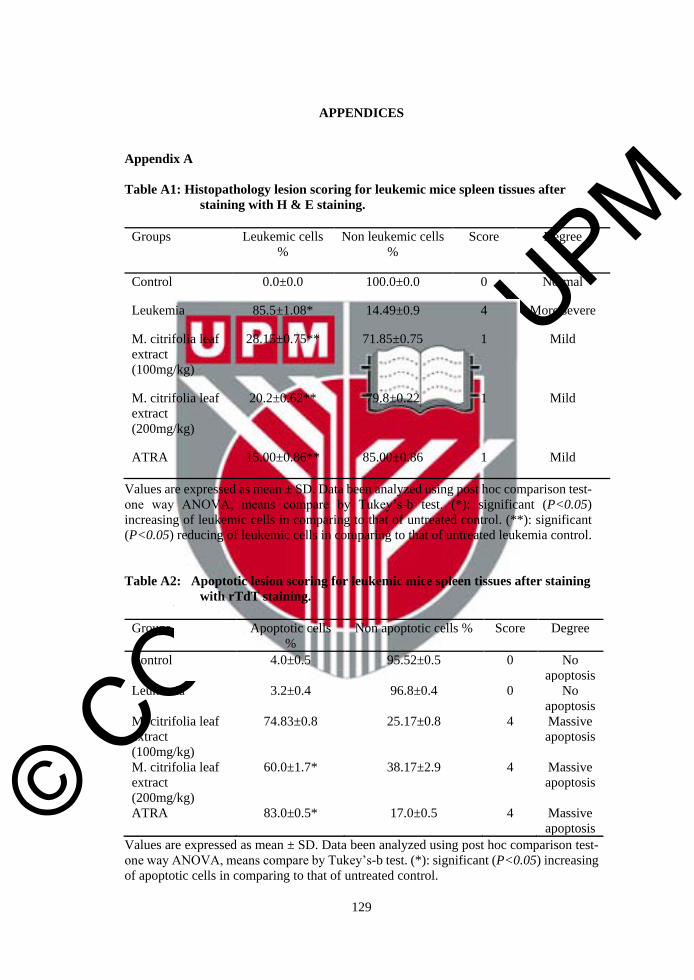
© COPYRIG
HT UPM
129
APPENDICES
Appendix A
Table A1: Histopathology lesion scoring for leukemic mice spleen tissues after
staining with H & E staining.
Groups Leukemic cells
%
Non leukemic cells
%
Score Degree
Control 0.0±0.0 100.0±0.0 0 Normal
Leukemia 85.5±1.08* 14.49±0.9 4 More severe
M. citrifolia leaf
extract
(100mg/kg)
28.15±0.75** 71.85±0.75 1 Mild
M. citrifolia leaf
extract
(200mg/kg)
20.2±0.62** 79.8±0.22 1 Mild
ATRA 15.00±0.86** 85.00±0.86 1 Mild
Values are expressed as mean ± SD. Data been analyzed using post hoc comparison test-
one way ANOVA, means compare by Tukey’s-b test. (*): significant (P<0.05)
increasing of leukemic cells in comparing to that of untreated control. (**): significant
(P<0.05) reducing of leukemic cells in comparing to that of untreated leukemia control.
Table A2: Apoptotic lesion scoring for leukemic mice spleen tissues after staining
with rTdT staining.
Groups Apoptotic cells
%
Non apoptotic cells % Score Degree
Control 4.0±0.5 95.52±0.5 0 No
apoptosis
Leukemia 3.2±0.4 96.8±0.4 0 No
apoptosis
M. citrifolia leaf
extract
(100mg/kg)
74.83±0.8 25.17±0.8 4 Massive
apoptosis
M. citrifolia leaf
extract
(200mg/kg)
60.0±1.7* 38.17±2.9 4 Massive
apoptosis
ATRA 83.0±0.5* 17.0±0.5 4 Massive
apoptosis
Values are expressed as mean ± SD. Data been analyzed using post hoc comparison test-
one way ANOVA, means compare by Tukey’s-b test. (*): significant (P<0.05) increasing
of apoptotic cells in comparing to that of untreated control.

© COPYRIG
HT UPM
130
Histopathology of Tissues
Formalin 10%
Formaldehyde 40% 100 mL
Sodium chloride 9 g
Distilled water 900 mL
Harri’s Haematoxylin Solution
Aluminium potassium sulphate 20 g
Absolute ethanol 10 mL
Mercuric oxide 0.5 g
Hematoxylin crystals 1 g
Distilled water 200 mL
Eosin Solution
Stock Eosin Solution (1%)
Eosin Y 1 g
Distilled water 100 mL
Working Eosin Solution
Stock eosin solution 1 part
Ethanol (80%) 3 part
Harri’s Hematoxylin and Eosin (H & E) Staining
Solution Time period
Xylene 5 min
Ethanol 100% 5 min
Ethanol 70% 5 min
Rinse 2 min
Hematoxylin 5 min
Rinse 2 min
Acid alcohol (1%) 3 sec
Running tap water 2 min
Eosin 1 min
Ethanol 70% 5 min
Ethanol 100% 5 min
Xylene 5 min
Mounting with DPX -

© COPYRIG
HT UPM
131
Appendix B
Electron Microscopy Solutions
Sodium Cocodylate (0.1 M) Solution
Sodium cocodylate 2.14 g
Distilled water 400 mL
Hydrochloric acid 0.2 M
PH=7.4
Resin Mixture
DDSA 6 mL
MNA 5.5 mL
BDMA 0.5 mL
Agar 100 resin 10 mL
Appendix C
Cell Cycle Buffers
Washing Buffer
PBS 100 mL
EDTA 0.2 g
BSA 0.1 g
Sodium azide 0.1 g
Staining Buffer
PBS 100 mL
EDTA 0.00374 g
Triton X-100 0.1 mL
RNAase A (50 mg/mL) 0.1 mL
PI (10 mg/mL) 0.03 mL
Appendix D
TUNEL Assay Solutions
rTdT Incubation Buffer
Equilibration buffer 45 µl
Nucleotide mix 5 µl
rTdT enzyme 1 µl
PBS-triton X-100-BSA

© COPYRIG
HT UPM
132
PBS 1 mL
Triton X-100 0.01 mL
BSA 0.005 g
PBS-PI-RNAase A
PBS 1 mL
PI (10 mg/mL) 0.005 mL
RNAase A (50 mg/mL) 0.01 mL
Appendix E
Wright Stain Reagents
Wright Stain Solution
Wright’s dye 0.3 g
Glycerin 3 mL
Absolute methanol 100 mL
Wright’s Buffer
Potassium phosphate 6.63 g
Dibasic sodium phosphate 2. 56 g
Distilled water 1000 mL
Appendix F
Immunohistochemistry Solutions
Citrate Buffer Solution pH 6.0
Sodium citrate trisodium salt dehydrate 2.94 g
Distilled water 1000 mL
TBST
TBS 1000 mL
Tween-20 1 mL
DAB+ Substrate - Chromogen Solution
DAB-substrate 1 mL
Chromogen 20 µL

© COPYRIG
HT UPM
133
BIODATA OF STUDENT
The author, NEGIN AHMADI was born in 10 May 1975 in Masjedsulaiman city,
Khuzistan region, Southern Iran. She graduated from Isfahan University with a Bachelor
Degree in microbiology in 2000. She ranked 1st out of 5 students for final term with grade
excellent, while ranked 1st in her alumni with grade very good. Thus, she has been
employed by the National Iranian Oil Company (NIOC) until now. In November, 2005,
she started her post-graduate study for the degree of Master of Science in Cell and
Molecular Biology in the Khatam, Tehran, University. In March 2008, she obtained her
master degree, following this; she served at the Azad University as instructor till now.
Since September 2011, she joined the PhD program in Malaysia and supervised by
Professor Dr. Suhaila Mohamed at the Faculty of Medicine and Health Sciences,
Universiti Putra Malaysia. She started her study for the degree of Doctor of Philosophy in
the field of Human Genetics.

© COPYRIG
HT UPM
134
LIST OF PUBLICATIONS
Mahnaz HP, Heshu SR, Ahmad BA, Rasedee A, Negin A. and Hemn H (2014).
Comparison of Apoptotic Inducing Effect of Zerumbone and Zerumbone-Loaded
Nanostructured Lipid Carrier on Human Mammary Adenocarcinoma MDA-MB-231 Cell
Line. Journal of Nanomaterials, Volume 2014, Article ID 742738, 10 pages.
A. CONFERENCE PAPERS
Heshu SR, Rasedee A, Ahmad Bustamam A, Hemn HO, Zeenathul NA, and Negin A
(2013). Zerumbone Loaded Nanostructured Lipid Carrier Regulate the
Expression of Apoptotic Biomarkers in BALB/C Mice Model of Leukemia.
Asia-Pacific Journal of Molecular Medicine, 3 (Suppl 1): P16. (ISI cited)
B. CONFERENCES
Heshu SR, Rasedee A, Ahmad BA, Zeenathul NA, Hemn HO, and Negin A (2013).
Zerumbone Loaded Nanostructured Lipid Carrier Regulate the Expression of
Apoptotic Biomarkers in BALB/C Mice Model of Leukemia. 1st National
Conference for Cancer Research in Conjugation with 5th Regional Conference
on Molecular Medicine (RCMM). 8th – 10th Nov 2013. The Royale Chulan Hotel,
KL, Malaysia. Organized by UKM Molecular Medical Biology Institute
(UMBI). (Poster presentor)
Heshu SR, Rasedee A, Hemn HO, Ahmad BA, Zeenathul NA, How CW and Negin A.
(2013). Immunological Evaluation of Zerumbone Loaded Nanostructured Lipid
Carrier in BALB/c Mice Model of Leukemia. Proceedings of 2nd Malaysia -
Thailand Graduate Forum in Life Sciences and Agriculture (MTGF). 3rd – 7th
Dec 2013. Faculty of Veterinary Medicine, University Putra Malaysia. (Oral
presentor)
Heshu SR, Rasedee A, Hemn HO, Ahmad BA, Zeenathul NA, How CW and Negin A.
(2014). Zerumbone Inhibits Murine WEHI-3B Cells in BALB/c Mice in Vivo.
Proceeding of The World Anti-Cancer Congress & Exhibition 2014 (WACCE).
11th -12th Jan 2014, Dewan Budaya, Universiti Sains Malaysia (USM), Penang,
Malaysia. (Poster presentor)
Heshu S., Rasedee A., Hemn H., Suha A., Ali H., Zahra A., Mahnaz H. and Negin A.
(2014). Proteomic and Genomic Evaluation of Zerumbone-Loaded
Nanostructured Lipid Carrier in BALB/c Mice Model of Leukemia. Proceeding
of Pharmaceutical Science Research Day 2014. 11th -12th Feb 2014. Faculty of
Pharmacy, UKM, Kuala Lumpur, Malaysia. (Oral Presentor)
Mahnaz H., Ahmad BA, Rasedee A., Heshu SR, and Negin A. (2014). Anticancer Effect
of Zerumbone-Loaded Nanostructure Lipid Carrier on Triple Breast Cancer
Cells. Proceeding of Pharmaceutical Science Research Day 2014. 11th -12th Feb
2014. Faculty of Pharmacy, UKM, Kuala Lumpur, Malaysia. (Oral Presentor)
© COPYRIG
HT UPM
135
Heshu SR, Rasedee A, Zeenathul NA, Ahmad BA, Hemn HO, Nozlena BA, Negin A, and
Mahnaz H. (2014). Antileukemic Effect of Zerumbone-Loaded Nanostructured
Lipid Carrier on Murine Leukemic Cell Line. Proceeding of International
Conference on Natural Products-2014. 18th – 19th March 2014. Palm Garden
Hotel, Putrajaya, Malaysia. (Poster presentor)
Hemn HO, Heshu SR, Rasedee A, Zeenathul NA, Ahmad BA, Negin A and Mahnaz H
(2014). Proceeding of International Conference on Natural Products-2014. 18th –
19th March 2014. Palm Garden Hotel, Putrajaya, Malaysia. (Poster presenter)
Mahnaz H, Ahmad B, Abdullah R, Heshu SR, Negin A, Hemn H (2014). In Vitro
Apoptosis Induction by Zerumbone Loaded Nanostructured Lipid Carrier (ZER-
NLC) on MDA-MB-231 Cells. Proceeding of International Conference on
Natural Products-2014. 18th – 19th March 2014. Palm Garden Hotel, Putrajaya,
Malaysia. (Poster presentor)

© COPYRIG
HT UPM
UNIVERSITI PUTRA MALAYSIA
STATUS CONFIRMATION FOR THESIS / PROJECT REPORT AND COPYRIGHT
ACADEMIC SESSION :
TITLE OF THESIS / PROJECT REPORT :
EFFECTS OF Morinda citrifolia L. LEAF EXTRACT ON HUMAN T-LYMPHOBLATIC LEUKEMIA (JURKAT) CELL AND WEHI-3B CELL-INDUCED MURINE LEUKEMIA
NAME OF STUDENT : NEGIN AHMADI
I acknowledge that the copyright and other intellectual property in the thesis/project report
belonged to Universiti Putra Malaysia and I agree to allow this thesis/project report to be placed at
the library under the following terms:
1. This thesis/project report is the property of Universiti Putra Malaysia.
2. The library of Universiti Putra Malaysia has the right to make copies for educationalpurposes only.
3. The library of Universiti Putra Malaysia is allowed to make copies of this thesis for academicexchange.
I declare that this thesis is classified as :
*Please tick (√ )
CONFIDENTIAL (Contain confidential information under Official Secret Act 1972).
RESTRICTED (Contains restricted information as specified by the organization/institution where research was done).
OPEN ACCESS I agree that my thesis/project report to be published as hard copy or online open access.
This thesis is submitted for :
PATENT Embargo from_____________ until ______________ (date) (date)
Approved by:
_____________________ _________________________________________ (Signature of Student) (Signature of Chairman of Supervisory Committee) New IC No/ Passport No.: Name:
Date : Date :
[Note : If the thesis is CONFIDENTIAL or RESTRICTED, please attach with the letter from the organization/institution with period and reasons for confidentially or restricted. ]