Embed Size (px)
Citation preview
CASE REPORT Open Access
Neurocytoma arising from a mature ovaryteratoma: a case reportJuan-Han Yu, Lian-He Yang, Xu-Yong Lin, Shun-Dong Dai, Xue-Shan Qiu and En-Hua Wang*
Abstract: Central neurocytoma/extraventricular neurocytoma is a central nervous system (CNS) tumor composed ofuniform round cells with neuronal differentiation. The typical lesions of central neurocytoma/extraventricularneurocytoma are at the interventricular foramen of the lateral ventricles (central neurocytoma) or brain parenchyma(extraventricular neurocytoma). Mature teratoma is a benign germ cell tumor commonly found in young women.Herein, we report a 24-year-old female with neurocytoma in a mature teratoma of the right ovary. The histologicalexaminations showed mature epidermis, skin appendages, adipose and bone tissues in the tumor; microscopic fociof immature cartilage tissues were also found in some parts. In addition, massive solid sheets and uniform roundtumor cells were found in the neuroectodermal tissues, with the formation of neuropil-like islands. Immunohistochemicalexaminations showed that the tumor cells were synaptophysin- and NeuN-positive but GFAP-negative. Based on thesefindings, the woman was diagnosed with neurocytoma arising from mature ovary teratoma, with microscopic foci ofimmature cartilage tissues. This is the fourth case report of neurocytoma outside the CNS to date.
BackgroundCentral neurocytoma/extraventricular neurocytoma is alow-grade tumor with neuronal differentiation that occursin the central nervous system (CNS), and histologicallycorresponds to WHO grade II. This tumor is predomin-antly found in young adults, and the prognosis is generallygood. Central neurocytoma usually occurs at the lateralventricles, while extraventricular neurocytoma couldoccur at any extraventricular regions in the CNS. Thehistological features and immunohistochemical phenotypesof central and extraventricular neurocytomas are similar:both are composed of round cells with homogeneousmorphology and neuronal differentiation, and the tumorcells are positive for neuronal markers, such as synapto-physin, and sometimes NeuN [1, 2].Mature teratoma is a benign germ cell tumor of the
ovary, commonly found in reproductive women, whichis composed of mature tissues from two or three germlayers. Somatic-type tumors arising from dermoid cystsare very rare, while tumors of CNS arising from matureteratoma are even rarer. Previous studies have reportedthat tumors of CNS arising from mature teratoma aregenerally from glial cells or primitive neuroectodermalcells [3]. Herein, we report a 24-year-old female with
neurocytoma arising from a mature teratoma of the rightovary.
Case presentationA 24-year-old female was admitted to our hospital withamenorrhea for 10 months, and a pelvic mass for 15 days.She had regular menstrual cycles; however, the menstru-ation stopped 10 months earlier for no obvious reasons.Physical examination found no abnormalities. CT examin-ation showed a mass posterior to the uterus with mixeddensity. The anteroposterior and transversal diameters ofthe mass were about 7.3 and 9.0 cm, respectively (Fig. 1).The woman was initially diagnosed with teratoma, andwas surgically treated.
Materials and methodsThe resected specimens were fixed with 10 % neutral-buffered formalin and embedded in paraffin blocks. Tissueblocks were cut into 4-μm slides, deparaffinized in xylene,rehydrated with graded alcohols, and immunostained withthe following antibodies: cytokeratin (CK, AE1/AE3), glialfibrillary acidic protein (GFAP, GA-5), synaptophysin(SP11), NeuN (A60) and Ki67 (MIB-1) (MaiXin, China).Sections were then stained with a streptavidin-peroxidasesystem (KIT-9720, Ultrasensitive TM S-P, MaiXin, China).The chromogen used was diaminobenzidine tetrahydro-chloride substrate (DAB kit, MaiXin, China). All samples
* Correspondence: [email protected] of Pathology, the First Affiliated Hospital and College of BasicMedical Sciences, China Medical University, Shenyang, China
© 2015 Yu et al. Open Access This article is distributed under the terms of the Creative Commons Attribution 4.0 InternationalLicense (http://creativecommons.org/licenses/by/4.0/), which permits unrestricted use, distribution, and reproduction in anymedium, provided you give appropriate credit to the original author(s) and the source, provide a link to the CreativeCommons license, and indicate if changes were made. The Creative Commons Public Domain Dedication waiver (http://creativecommons.org/publicdomain/zero/1.0/) applies to the data made available in this article, unless otherwise stated.
Yu et al. Diagnostic Pathology (2015) 10:171 DOI 10.1186/s13000-015-0406-x
were counterstained with hematoxylin, dehydrated, andmounted. For the negative controls, each sample was in-cubated with PBS, instead of the primary antibodies.
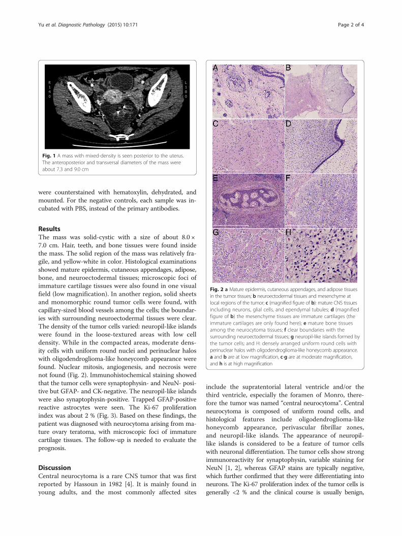
ResultsThe mass was solid-cystic with a size of about 8.0 ×7.0 cm. Hair, teeth, and bone tissues were found insidethe mass. The solid region of the mass was relatively fra-gile, and yellow-white in color. Histological examinationsshowed mature epidermis, cutaneous appendages, adipose,bone, and neuroectodermal tissues; microscopic foci ofimmature cartilage tissues were also found in one visualfield (low magnification). In another region, solid sheetsand monomorphic round tumor cells were found, withcapillary-sized blood vessels among the cells; the boundar-ies with surrounding neuroectodermal tissues were clear.The density of the tumor cells varied: neuropil-like islandswere found in the loose-textured areas with low celldensity. While in the compacted areas, moderate dens-ity cells with uniform round nuclei and perinuclear haloswith oligodendroglioma-like honeycomb appearance werefound. Nuclear mitosis, angiogenesis, and necrosis werenot found (Fig. 2). Immunohistochemical staining showedthat the tumor cells were synaptophysin- and NeuN- posi-tive but GFAP- and CK-negative. The neuropil-like islandswere also synaptophysin-positive. Trapped GFAP-positivereactive astrocytes were seen. The Ki-67 proliferationindex was about 2 % (Fig. 3). Based on these findings, thepatient was diagnosed with neurocytoma arising from ma-ture ovary teratoma, with microscopic foci of immaturecartilage tissues. The follow-up is needed to evaluate theprognosis.
DiscussionCentral neurocytoma is a rare CNS tumor that was firstreported by Hassoun in 1982 [4]. It is mainly found inyoung adults, and the most commonly affected sites
include the supratentorial lateral ventricle and/or thethird ventricle, especially the foramen of Monro, there-fore the tumor was named “central neurocytoma”. Centralneurocytoma is composed of uniform round cells, andhistological features include oligodendroglioma-likehoneycomb appearance, perivascular fibrillar zones,and neuropil-like islands. The appearance of neuropil-like islands is considered to be a feature of tumor cellswith neuronal differentiation. The tumor cells show strongimmunoreactivity for synaptophysin, variable staining forNeuN [1, 2], whereas GFAP stains are typically negative,which further confirmed that they were differentiating intoneurons. The Ki-67 proliferation index of the tumor cells isgenerally <2 % and the clinical course is usually benign,
Fig. 1 A mass with mixed-density is seen posterior to the uterus.The anteroposterior and transversal diameters of the mass wereabout 7.3 and 9.0 cm
Fig. 2 a Mature epidermis, cutaneous appendages, and adipose tissuesin the tumor tissues; b neuroectodermal tissues and mesenchyme atlocal regions of the tumor; c (magnified figure of b): mature CNS tissuesincluding neurons, glial cells, and ependymal tubules; d (magnifiedfigure of b) the mesenchyme tissues are immature cartilages (theimmature cartilages are only found here); e mature bone tissuesamong the neurocytoma tissues; f clear boundaries with thesurrounding neuroectodermal tissues; g neuropil-like islands formed bythe tumor cells; and H: densely arranged uniform round cells withperinuclear halos with oligodendroglioma-like honeycomb appearance.a and b are at low magnification, c-g are at moderate magnification,and h is at high magnification
Yu et al. Diagnostic Pathology (2015) 10:171 Page 2 of 4
thus the current WHO classification assigns the centralneurocytoma to grade II. Subsequently, tumors mimickingcentral neurocytomas but occurring within the cerebrum[5] and spinal cord [6, 7] were reported. Thus, the WHOClassification of Tumors of the Central Nervous System(4th Edition) defined all extraventricular tumors withhistological features and immunophenotypes similar tocentral neurocytoma as extraventricular neurocytoma,and classified them as WHO grade II tumor.The features of the tumor in the present report were as
follows: the tumor formed in the ovary of a 24-year-old fe-male; part of the tumor appeared to be a mature teratomawith microscopic foci of immature cartilage tissues; sheet-like round tumor cells with homogenous morphologywere found in other regions with oligodendroglioma-like honeycomb appearance and abundant neuropil-likeislands. Furthermore, immunohistochemical staining showedthat the tumor cells were synaptophysin- and NeuN-positiveand GFAP-negative. Considering the differentia diagno-sis against clear cell ependymomas and oligodendrogli-omas, neuropil-like islands and diffuse synaptophysinand NeuN positivity are in favor of neurocytoma. A fur-ther differential diagnosis is a teratoma of the ovary withimmature neuroepithelial elements. However, NeuN is notfound in immature neural progenitor cells [8]. Therefore,the pathological diagnosis was a neurocytoma arising froma mature teratoma with microscopic foci of immature car-tilage tissues.Neurocytomas outside the CNS are very rare. Only
three such cases have been reported to date [9–11],among which two cases arose from mature teratomas (in
the ovary and adrenal gland, respectively), and the otheroccurred in the pelvis of a male patient. In all cases, atleast one histological feature of neurocytoma was found;and more importantly, the tumor cells were positive forneuronal markers including synaptophysin and NSE,which confirmed that the tumors were neurocytoma outsidethe CNS. These patients were followed up for 6 months,1 year, and 3 years, respectively, and no sign of recurrencewas found. However, the prognosis remains unclear.Microscopic foci of immature cartilage tissues werefound in one visual field (low magnification) in the presentcase, which was not reported in the previous cases. Thus,we recommended regular follow-up to our patient.
ConclusionIn summary, neurocytoma outside the CNS rarely occurs.When pathological features similar to neurocytoma arefound during the diagnosis of teratomas, the pathologistsshould carefully perform immunohistochemical examina-tions for confirmation and differentiation.
ConsentWritten informed consent was obtained from the patientfor publication of this case report and accompanyingimages. A copy of the written consent is available forreview by the Editor-in Chief of this Journal.
Competing interestsThe authors declare that they have no competing interests.
Authors’ contributionsYJH and LXY participated in the histopathological evaluation, performed theliterature review, acquired photomicrographs and drafted the manuscript.YLH and DSD carried out the immunohistochemical stains evaluation. QXSconceived and designed the study. WEH gave the final histopathologicaldiagnosis and revised the manuscript. YJH and WEH edited the manuscript.All the authors read and approved the final manuscript.
FundingThis work was supported by grants from the National Natural ScienceFoundation of China (No. 81301837 to Juan-Han Yu and No. 81301930 toLian-He Yang).
Received: 29 July 2015 Accepted: 28 August 2015
References1. Figarella B, Pellissier JF, Daumas D, Delisle MB, Pasquier B, Parent M, et al.
Central neu-rocytomas. Critical evaluation of a small-cell neuronal tumor.Am J Surg Pathol. 1992;16:97–109.
2. Soylemezoglu F, Onder S, Tezel GG, Berker M. Neuronal nuclear antigen(NeuN): a new tool in the diagnosis of central neurocytoma. Pathol ResPract. 2003;199:463–8.
3. Luo CC, Huang CS, Chu SM, Chao HC, Yang CP, Hsueh C. Retroperitonealteratomas in infancy and childhood. Pediatr Surg Int. 2005;21:536–40.
4. Hassoun J, Gambarelli D, Grisoli F, Pellet W, Salamon G, Pelissier JF, et al.Central neurocytoma. An electron-microscopic study of two cases. ActaNeuropathol. 1982;56:151–6.
5. Nishio S, Takeshita I, Kaneko Y, Fukui M. Cerebral neurocytoma. A newsubset of benign neuronal tumors of the cerebrum. Cancer. 1992;70:529–37.
6. Coca S, Moreno M, MartosJA RJ, Barcena A, Vaquero J. Neurocytoma ofspinal cord. Acta Neuropathol. 1994;87:537–40.
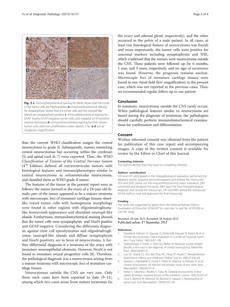
Fig. 3 a: Immunohistochemical staining for NeuN shows that the nucleiof the tumor cells are NeuN-positive; b: immunohistochemical stainingfor synaptophysin shows that the tumor cells and the neuropil-likeislands are synaptophysin-positive; c: immunohistochemical staining forGFAP shows GFAP-negative tumor cells and trapped GFAP-positivereactive astrocytes; d: immunohistochemical staining for Ki-67 showstumor cells with low proliferation index (about 2 %). a-d are atmoderate magnification
Yu et al. Diagnostic Pathology (2015) 10:171 Page 3 of 4
7. Tatter SB, Borges LF, Louis DN. Central neurocytomas of the cervical spinalcord. Report of two cases. J Neurosurg. 1994;81:288–93.
8. Gusel’nikova VV, Korzhevskiy DE. NeuN as a neuronal nuclear antigen andneuron differentiation marker. Acta Nat. 2015;7(2):42–7.
9. Hirschowitz L, Ansari A, Cahill DJ, Bamford DS, Love S. Central neurocytomaarising within a mature cystic teratoma of the ovary. Int J Gynecol Pathol.1997;16:176–9.
10. Friedrichs N, Vorreuther R, Fischer HP, Wiestler OD, Buettner R. Neurocytomaarising in the pelvis. Virchows Arch. 2003;443(2):217–9.
11. Ersoz S, Kucuk H, Mungan S, Turgutalp H, Imamoglu M, Kosucu P.Neurocytoma arising in an adrenal gland mature teratoma. Fetal PediatrPathol. 2011;30(5):275–9.
Submit your next manuscript to BioMed Centraland take full advantage of:
• Convenient online submission
• Thorough peer review
• No space constraints or color figure charges
• Immediate publication on acceptance
• Inclusion in PubMed, CAS, Scopus and Google Scholar
• Research which is freely available for redistribution
Submit your manuscript at www.biomedcentral.com/submit
Yu et al. Diagnostic Pathology (2015) 10:171 Page 4 of 4


![PARIPEX - INDIAN JOURNAL OF RESEARCH | Volume-8 | Issue-10 ... · teratoma is known as a monodemal teratoma.[1] Immature teratoma (IT) is a preferred term for the malignant ovarian](https://img.pdfslide.net/doc/110x75/603e5f8d2bf3bd27e47c8252/paripex-indian-journal-of-research-volume-8-issue-10-teratoma-is-known.jpg)