Embed Size (px)
Citation preview
1
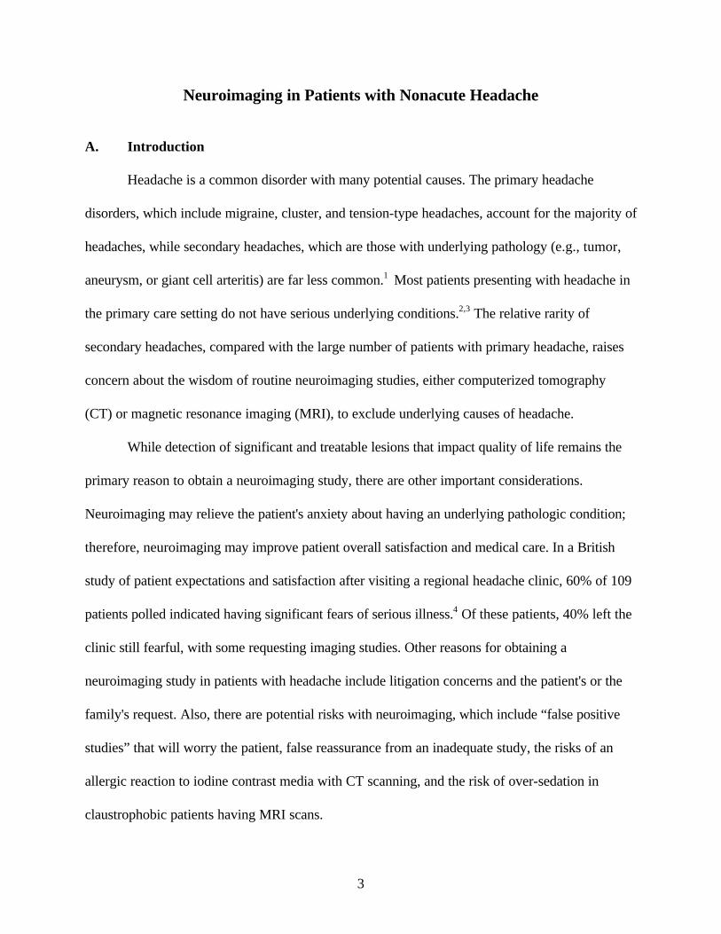
Evidence-Based Guidelines in the Primary Care Setting: Neuroimaging in Patients with Nonacute Headache
Benjamin M. Frishberg, MD, FAAN Associate Clinical Professor of Neurology, University of California,
San Diego, CA
Jay H. Rosenberg, MD, FAAN Department of Neurology, Southern California Permanente Medical Group, and Clinical Professor
of Neurology, Voluntary Faculty, UCSD School of Medicine, San Diego, CA
David B. Matchar, MD
Professor of Medicine and Director, Center for Clinical Health Policy Research, Duke University Medical Center, Durham, NC
Douglas C. McCrory, MD, MHSc
Assistant Professor, Department of Medicine, Assistant Research Professor, Center for Clinical Health Policy Research, Duke University Medical Center, Durham, NC
Research Associate, Center for Health Services Research in Primary Care Durham Veterans Affairs Medical Center, Durham, NC
Michael P. Pietrzak, MD, FACEP
Alexandria, VA
Todd D. Rozen, MD Assistant Professor of Neurology Thomas Jefferson University, Jefferson Headache Center,
Philadelphia, PA
Stephen D. Silberstein, MD, FACP Professor of Neurology, Thomas Jefferson University, and Director of Jefferson Headache
Center, Philadelphia, PA
US Headache Consortium:§ American Academy of Family Physicians
American Academy of Neurology American Headache Society
American College of Emergency Physicians* American College of Physicians-American Society of Internal Medicine
American Osteopathic Association National Headache Foundation
§The US Headache Consortium participants: J. Keith Campbell, MD; Frederick G. Freitag, DO; Benjamin Frishberg, MD; Thomas T. Gilbert, MD, MPH; David B. Matchar, MD; Douglas C. McCrory, MD, MHSc; Donald B.

2
Penzien, PhD; Michael P. Pietrzak, MD, FACEP; Nabih M. Ramadan, MD; Jay H. Rosenberg, MD; Todd D. Rozen, MD; Stephen D. Silberstein, MD, FACP; Eric M. Wall, MD, MPH; and William B. Young, MD. *Endorsement by ACEP means that ACEP agrees with the general concepts in the guidelines and believes that the developers have begun to define a process of care that considers the best interests of patients with migraine headache. Copyright © by the American Academy of Neurology: Licensed to the members of the US Headache Consortium
3
Neuroimaging in Patients with Nonacute Headache
A. Introduction
Headache is a common disorder with many potential causes. The primary headache
disorders, which include migraine, cluster, and tension-type headaches, account for the majority of
headaches, while secondary headaches, which are those with underlying pathology (e.g., tumor,
aneurysm, or giant cell arteritis) are far less common.1 Most patients presenting with headache in
the primary care setting do not have serious underlying conditions.2,3 The relative rarity of
secondary headaches, compared with the large number of patients with primary headache, raises
concern about the wisdom of routine neuroimaging studies, either computerized tomography
(CT) or magnetic resonance imaging (MRI), to exclude underlying causes of headache.
While detection of significant and treatable lesions that impact quality of life remains the
primary reason to obtain a neuroimaging study, there are other important considerations.
Neuroimaging may relieve the patient's anxiety about having an underlying pathologic condition;
therefore, neuroimaging may improve patient overall satisfaction and medical care. In a British
study of patient expectations and satisfaction after visiting a regional headache clinic, 60% of 109
patients polled indicated having significant fears of serious illness.4 Of these patients, 40% left the
clinic still fearful, with some requesting imaging studies. Other reasons for obtaining a
neuroimaging study in patients with headache include litigation concerns and the patient's or the
family's request. Also, there are potential risks with neuroimaging, which include “false positive
studies” that will worry the patient, false reassurance from an inadequate study, the risks of an
allergic reaction to iodine contrast media with CT scanning, and the risk of over-sedation in
claustrophobic patients having MRI scans.
4
Guidelines are developed to assist the physician in making appropriate choices in work-up
and treatment of patients. They are not designed to supersede clinical judgment when dealing with
individual patients, nor are they designed to prevent imaging in any given situation. The realities
of our medical-legal climate are such that many physicians feel compelled to conduct
neuroimaging studies on patients with headache even though the likelihood of finding pathology is
about the same as in the general population. These studies are generally done to avoid the
possibility of a lawsuit based on failure to diagnose a lesion, unrelated to the headache, which is
an incidental finding during a headache evaluation. The Consortium recognized these realities
when developing the Guidelines, but they did not alter the intention or direction of the Guideline’s
recommendations, which are based on the available evidence. Understanding that individual
physicians have their own risk tolerance, some will find these Guidelines useful, while others will
continue to image patients based on risk management strategies.
Aims of the Guideline
The objective of the US Headache Consortium is to develop scientifically sound, clinically
relevant practice guidelines on chronic headache in the primary care setting. The specific aim of
the Diagnostic Guideline is to provide recommendations for diagnostic testing in nonacute
headache patients (encompassing all headache syndromes that have occurred for at least four
weeks during a patient’s lifetime) based on a comprehensive review and meta-analysis of scientific
evidence.‡ Specifically, the evidenced-based Guideline focuses on three questions:
‡This statement is provided as an educational service of the US Headache Consortium member organizations. It is based on an assessment of current scientific and clinical information. It is not intended to include all possible proper methods of care for choosing to use a specific procedure. Neither is it intended to exclude any reasonable alternative methodologies. These organizations recognize that specific patient care decisions are the prerogative of the patient and the physician caring for the patient, based on all of the circumstances involved.
5
(1) Are particular findings in the history and on the physical examination
helpful in identifying which patients have significant intracranial abnormalities?
(2) What is the frequency of significant secondary causes of nonacute
headache, as detected by CT or MRI, in patients presenting with nonacute
headache and a normal neurological examination?
(3) What evidence exists concerning the relative ability of CT and MRI to
detect significant intracranial lesions among patients with nonacute headache?
The results of the literature review and analysis showed that there is insufficient published
clinical research to support evidence-based Guidelines for any diagnostic testing other than
neuroimaging. Previous reports that reviewed the evidence on the role of electroencephalography
(EEG) found that EEG is not indicated in the routine evaluation of headache.5
Methods
Review of the published clinical evidence available in the medical literature (from January
1966 through August 1998) included English-language studies that estimated the sensitivity,
specificity, or predictive value of a neuroimaging test used in patients presenting with nonacute
headache.6 Studies that assessed observer variation or reproducibility of diagnostic tests for
patients with nonacute headache were included. (A complete overview of methodology is
presented elsewhere.7 )
Identified studies were reviewed with the assistance of two board-certified neurologists.
Neuroimaging results were recorded and classified as either “significant abnormalities,”
“abnormalities possibly related to headache,” “insignificant abnormalities,” (defined in Table 3 of
the "Evidence-based guidelines for migraine headache: overview of program description and
6
methodology"7) or “normal”. To evaluate the medical aspects of neuroimaging, we focused
exclusively on significant abnormalities (defined as abnormalities related to headache that may
require further action [e.g., acute cerebral infarct, neoplastic disease, hydrocephalus, and vascular
abnormalities, e.g., aneurysm or arteriovenous malformation]).
B. Summary of the Evidence
The literature search identified 28 studies that met the inclusion criteria.2,8-33 These studies
varied in setting, design, and other methodological features, as well as in the level of detail in
reporting results.6 Twenty-two of the 28 studies were retrospective; the six prospective studies all
lacked blinding. All 28 studies were reviewed and assigned a quality grade of I through IV (Level
I evidence: independent, blind comparison with a “gold standard” of anatomy, physiology,
diagnosis, or prognosis among a large number of consecutive patients suspected of having the
target condition. Level II evidence: independent, blind comparison with a “gold standard” among
a small number of consecutive patients suspected of having the target condition. Level III
evidence: independent, blind comparison with a “gold standard” among non-consecutive patients
suspected of having the target condition. Level IV evidence: included studies that did not meet
criteria for at least Level III evidence. Additional details are presented elsewhere.6,7) All of the
studies identified received a quality Grade IV. Most of the studies identified did not characterize
the study population in terms of duration of headache disorder. Most included a discussion of the
neurological examination, but did not comment on symptoms. Several studies used populations
having a mix of subjects with both acute and nonacute headaches.
7
Historical and Physical Examination Findings
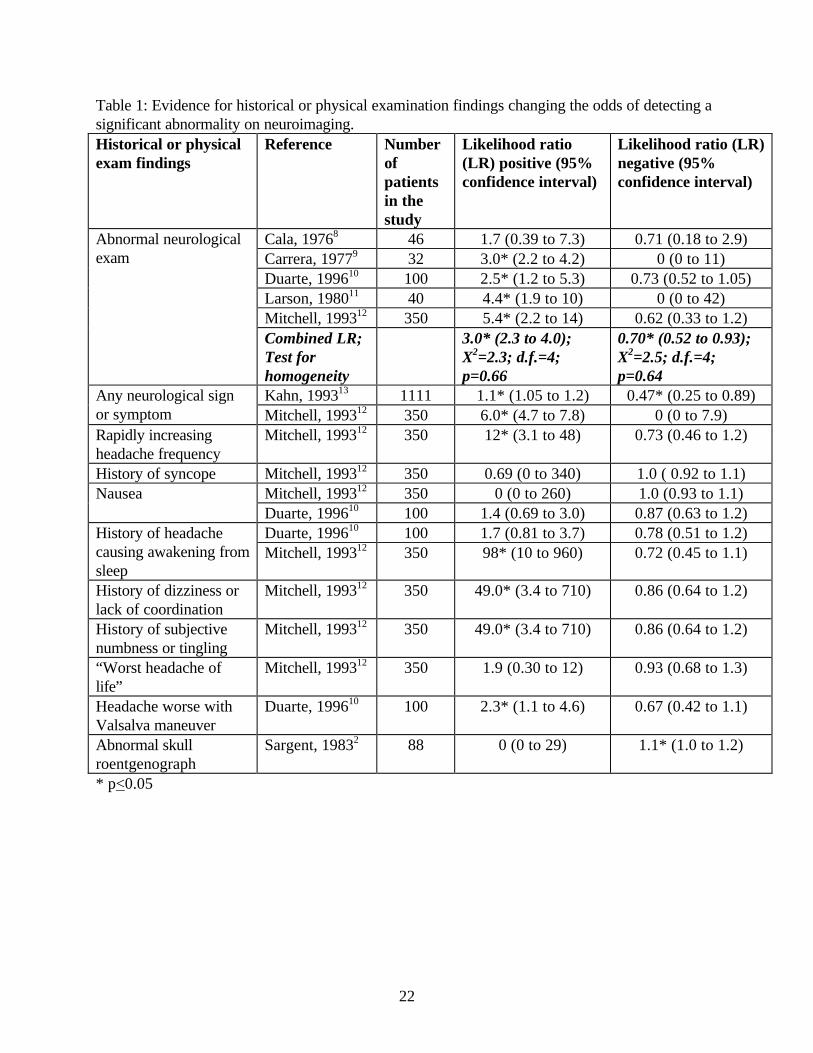
Eight of the 28 studies reported sufficient information to construct tables relating specific
historical or physical examination findings to the occurrence of significant abnormalities on CT or
MRI (Table 1).2,8-13,15
Neurological Findings: Five studies described neuroimaging abnormalities in patients
with neurological findings on examination.8-12 This permitted the calculation of likelihood ratios
for abnormal and normal neurological examination. In four of the five studies, an abnormal
neurological examination (not described consistently in the individual study reports) significantly
increased the likelihood of finding a significant abnormality on neuroimaging. Conversely, the
absence of findings on the neurological examination led to a decreased likelihood of finding a
significant lesion on neuroimaging in all five studies. This reduction approached statistical
significance in two studies.
To describe the efficiency of the screening tests, sensitivity and specificity were combined
to create likelihood ratios. Likelihood ratios are useful because they express the change in odds of
disease for a given finding. As shown in Table 1, values greater than one increase the odds of
disease, while values less than one diminish the odds. Values approaching one have an
increasingly small impact on changing the odds of disease beyond the baseline prevalence. A
calculated combined likelihood ratio of 3.0 (95% confidence interval, 2.3 to 4.0) suggests that
abnormal findings on the neurological examination tripled the odds of finding a significant
intracranial abnormality on neuroimaging.†† Since the prior odds of significant intracranial
abnormality is low (less than 1 in 100), the odds of such an abnormality in the face of an abnormal
neurological examination is still low (i.e., less than 3 in 100). A combined negative likelihood ratio
†† A likelihood ratio greater than one indicates that the diagnostic test results increase the post-test probability of disease.
8
of 0.7 (95% confidence interval, 0.52 to 0.93) suggests that a normal neurological examination
reduced the odds of finding a significant intracranial abnormality on neuroimaging by 30%.
In two studies, the presence of abnormal neurological signs or symptoms (not just signs as
described earlier) significantly increased the likelihood of finding a significant abnormality on
neuroimaging.12,13 The absence of neurological signs and symptoms was associated with a
significantly decreased likelihood of a significant abnormality in one of the two studies. Two
studies related several individual neurological signs and symptoms to the presence of significant
abnormalities on imaging.10,12 The small number of patients with any individual symptoms resulted
in wide confidence intervals (low precision) around the likelihood ratios, which makes
interpretation difficult. In at least one of these studies, the following symptoms were shown to
increase significantly the odds of finding a significant abnormality on neuroimaging:
• rapidly increasing headache frequency,
• history of dizziness or lack of coordination,
• history of subjective numbness or tingling, and
• history of headache causing awakening from sleep.
The absence of these features did not significantly lower the odds of finding a neuroimaging
abnormality.
The same two studies10,12 found that a history of syncope, headache accompanied by
nausea, or the experience of the “worst headache” of one’s life did not significantly increase the
likelihood of finding a significant abnormality on neuroimaging. Similarly, the absence of these
findings did not significantly alter the likelihood of finding an abnormality. For each of the
findings, the studies did not have sufficient power to exclude the possibility of a clinically
important effect.
9
Other Clinical Findings: One study reported that a history of headache worsening with
Valsalva maneuver significantly increased the odds of finding a significant intracranial abnormality
on neuroimaging, most commonly a Chiari malformation.10 However, the lack of this finding
(history of headache worsening with Valsalva maneuver) did not significantly alter the likelihood
of finding an abnormality.
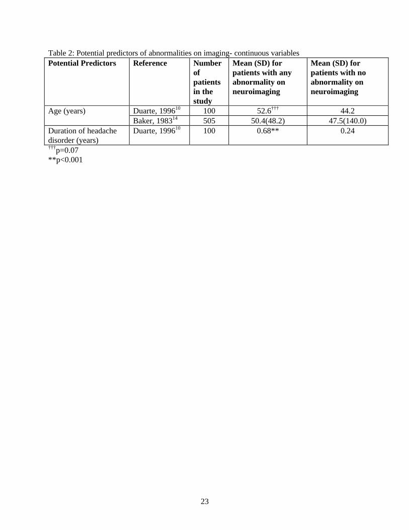
Continuous Variables
The distribution of several continuous variables (age, duration of headache, headache
frequency, and ergot consumption) was reported for subjects with normal versus abnormal
neuroimaging results. One study reported a statistically significant difference in the duration of the
headache disorder between those with and without a significant abnormality on neuroimaging
(Table 2).10 All the patients in this trial had experienced either the recent onset of headaches or a
clear change in the character of their nonacute headaches within the last year. This suggests that
the duration of a headache disorder may be a useful predictor of significant abnormalities among
patients with recent-onset headache symptoms, but not among patients with longstanding
headache disorders.
Two studies described differences in mean age among patients with and without an
abnormality on neuroimaging.10,14 One of these studies found increasing age to be strongly
associated with finding an abnormality; however, the abnormalities considered were mostly
atrophy and old cerebral infarctions.14 The relationship between age and significant abnormalities
in another study found a statistical trend toward older age among those with significant
abnormalities (age 53 vs. 42; p=0.07).10
Other continuous variables (e.g., headache frequency) have not been studied sufficiently to
10
determine whether they might be useful in discriminating between patients likely to have normal
results on imaging and those likely to have abnormal results.
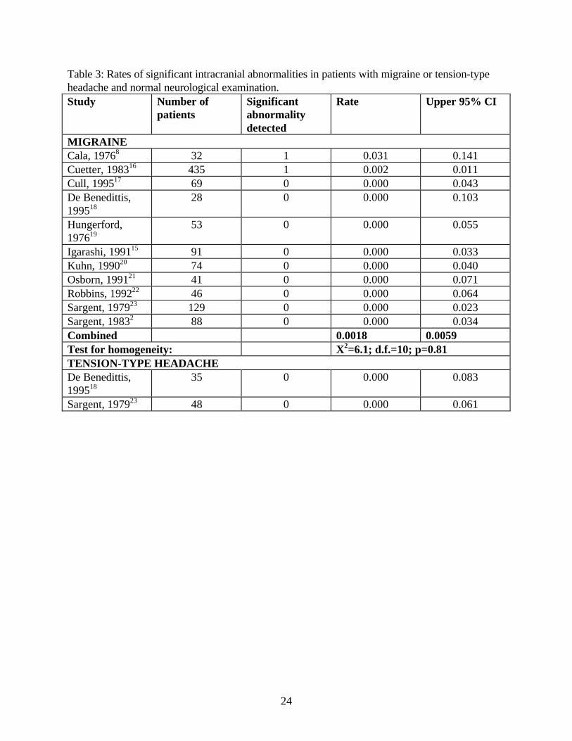
Nonacute Headache and Normal Neurological Examination
The estimates of the rates of significant abnormalities were based on samples of patients
who appeared to have nonacute or recurrent headaches. Candidate articles on this topic were
carefully scrutinized to ensure that patients with acute or new-onset headache (less than four
weeks duration) were excluded or that at least neuroimaging test results were reported separately
for patients with acute versus nonacute headache.
Migraine headache: Among patients with normal neurological examinations and
headaches diagnosed as migraine, the prevalence of significant intracranial abnormalities on
neuroimaging ranged from 0% to 3.1% in 11 studies (Table 3).2,8,15-23 The populations in these
studies were reasonably similar. Results from these studies were combined in a meta-analysis to
yield a summary prevalence of 0.0018 (approximately 0.2%), with an upper 95% confidence limit
of 0.0059 (approximately 0.6%). (This represents a prevalence that is less than the 0.8% incidence
of arteriovenous malformations and the 2.4% incidence of saccular aneurysms found in autopsy
series.34, 35)
Tension-type headache: Two studies reported no patient with significant intracranial
abnormalities among those with normal neurological examinations and headaches diagnosed as
tension-type headaches (one study18 specified as chronic tension-type headache).18,23 However,
both studies were small and included a total of only 83 patients.
Unspecified type of headache: In patients with normal neurological examinations
presenting with headache not described as migraine or tension-type, rates of significant
11
intracranial abnormalities were more variable than among patients with migraine. Among these
patients, the probability of finding significant intracranial abnormalities ranged from 0% to 6.7%
(Table 4).9,11, 12,14,24-29. These rates were not homogeneous; therefore, no combined rates were
calculated.
Other headache types (not specifically described as migraine or tension type): One
"outlier" study10 found a high probability of significant intracranial abnormalities (22.5%) in
patients with either a recent (within 12 months) onset of headache or a recent (within 12 months)
clear change in character of previous headaches. In another study among 27 patients with cluster
headache, no clinically significant findings were observed.30 Another study reported a high
prevalence of significant abnormalities in patients with cough headache, exertional headache, and
sex-induced headache.31 Twelve of 28 patients (43%) with exertional headache had structural
pathology, with 10 having had subarachnoid hemorrhages. They all had new onset of severe
headache. Seventeen of 30 patients with cough-induced headache had a Chiari malformation, and
1 of 14 patients with headache associated with sex had a ruptured aneurysm with explosive
headache.31 This study did not describe neurological examination findings in relation to the
neuroimaging findings.
Relative Effectiveness of CT and MRI
Three studies described the similarities between CT and MRI among patients with
headache.18,20,25 Each study had fewer than 100 patients, and a relatively small fraction of these
patients were imaged using both technologies. No significant abnormalities were detected in these
patients, so the relative effectiveness of the two tests for detecting significant lesions cannot be
determined.
12
One study found that, of 15 patients imaged with both CT and MRI, 11 received the same
diagnosis with the two technologies.18 In the remaining four patients, abnormalities (including
white matter changes) were seen on MRI but not on CT. In a second study, 17 patients were
imaged with both CT and MRI, and 14 of them received identical diagnoses with the two
procedures.25 Differences in the remaining three patients related to the imaging of developmental
venous anomalies. These findings were seen on MRI but not on CT in one case, and were
suspected on CT, but not confirmed on MRI, in the remaining two cases. In the third study, MRI
appeared to be more sensitive than CT in identifying white matter lesions.20 In summary, the
limited number of studies identified and reviewed here suggest that MRI may be more sensitive
than CT for identifying clinically insignificant abnormalities. However, MRI may not be more
sensitive for identifying clinically significant pathology that is relevant to the cause of headache.
Only one study compared the sensitivity of CT with and without enhancement; however, this
study excluded patients with migraine.25
C. Transition from Evidence to Guidelines
While the studies selected for this analysis were rigorously screened for quality, many
failed to define headache duration, specify headache type, or adequately detail the history and
examination of the patients. Studies differed in their size, methods, and endpoints, making it
difficult to perform a meta-analysis. In addition, significant biasing effects were encountered in
this review. In patients presenting with nonacute headache and a normal neurological
examination, most of the studies included in this analysis shared two significant biases that may
cause overestimation of the prevalence of significant abnormalities in the primary care population.
First, because most were conducted at referral centers, the study populations could possibly be
13
skewed toward a higher frequency of pathology (referral filter bias). Second, most studies were
assembled from a series of patients selected for neuroimaging, but provided no description of the
criteria used to select patients. That is, no explanation was given for why some patients and not
others were chosen for scanning, from the larger population of headache patients with normal
neurological examinations. Therefore, the findings summarized above may not be generalizable to
all populations of patients. The evidence gathered is all Level IV and lacks the strength preferred
when making evidence-based guidelines.
D. General Principles of Management
In making decisions about neuroimaging in headache, the US Headache Consortium
identified three consensus-based (not evidence-based) general principles of management:
(1) Testing should be avoided if it will not lead to a change in management.
(2) Testing is not recommended if the individual is not significantly more likely
than anyone else in the general population to have a significant abnormality.
(3) Testing that normally may not be recommended as a population-policy may
make sense at an individual level, resources notwithstanding. For example,
exceptions can be considered for patients who are disabled by their fear of serious
pathology, or for whom the provider is suspicious even in the absence of known
predictors of abnormalities on neuroimaging studies (red flags).
E. Specific Testing Recommendations
14
The consortium considered the available evidence, and through a consensus process
arrived at the following treatment recommendations listed below. Importantly, this list of
recommendations is not all-inclusive, and there are other reasons for performing neuroimaging
studies that are not detailed below due to the lack of published studies in the literature.
Neurological Examination
Finding: An abnormal neurological examination increases the likelihood of finding
significant intracranial pathology (e.g., brain tumor, arteriovenous
malformation, hydrocephalus) on neuroimaging. The absence of any
abnormalities on neurological examination reduces the odds of finding a
significant abnormality on imaging.
Recommendation: Neuroimaging should be considered in patients with nonacute headache
and an unexplained abnormal finding on the neurological examination
(Grade B‡‡).
Neurological Symptoms
Finding: Headache worsened by Valsalva maneuver, headache causing awakening
from sleep, new headache in the older population, or progressively
worsening headache may indicate a higher likelihood of significant
‡‡Grade A. Multiple well-designed randomized clinical trials, directly relevant to the recommendation, yielded a consistent pattern of findings.36
Grade B. Some evidence from randomized clinical trials supported the recommendation, but the scientific support was not optimal. For instance, either few randomized trials existed, the trials that did exist were somewhat inconsistent, or the trials were not directly relevant to the recommendation. An example of the last point would be the case where trials were conducted using a study group that differed from the target group for the recommendation. Grade C. The US Headache Consortium achieved consensus on the recommendation in the absence of relevant randomized controlled trials.
15
intracranial pathology, as reported in several small studies. (One study
reported that a history of headache worsening with Valsalva maneuver
significantly increased the odds of finding a significant intracranial
abnormality on neuroimaging, most commonly a Chiari malformation. In
general, however, the absence of signs and symptoms is less reliable and
informative than their presence.
Recommendation: Evidence is insufficient to make specific recommendations regarding
neuroimaging in the presence or absence of neurological symptoms (Grade
C‡‡).
Migraine and a Normal Neurological Examination
Finding: Meta-analysis of patients with migraine and a normal neurological
examination found a rate of significant intracranial lesions of 0.18%
(2/1000; previously reported rates of finding intracranial lesions with CT
and MRI ranged from 0.3% to 0.4%37). Neuroimaging is thus unlikely to
reveal an abnormality on MRI or CT scanning in patients with migraine and
a normal neurological examination.
Recommendation: Neuroimaging is not usually warranted for patients with migraine and
normal neurological examination. (Grade B‡‡). For patients with atypical
headache features or patients who do not fulfill the strict definition of
migraine (or have some additional risk factor), a lower threshold for
16
neuroimaging may be applied (Grade C‡‡).
Tension-type Headache and Normal Neurological Examination
Finding: In two studies of imaging in patients with tension-type headache (one study
specified as chronic tension-type headache18) and normal neurological
examinations, no significant lesions were demonstrated.
Recommendation: Data were insufficient to make an evidence-based recommendation
regarding the use of neuroimaging for tension-type headache (Grade C‡‡).
Effectiveness of CT vs. MRI
Finding: Based on the limited data in the studies reviewed here, MRI appears to be
more sensitive in finding white matter lesions and developmental venous
anomalies than CT, a result that could be expected based upon the
characteristics of the two technologies. The greater resolution and
discrimination of MRI, however, appears to be of little clinical importance
in the evaluation of patients with nonacute headache. Data were lacking
comparing enhanced with unenhanced CT scans.
Recommendation: Data were insufficient to make any evidence-based recommendations
regarding the relative sensitivity of MRI compared with CT in the
evaluation of migraine or other nonacute headache (Grade C‡‡).
17
F. Future Research
In the studies reviewed in this report, sufficient evidence was available in the literature to
evaluate the rate of significant intracranial lesions among patients with migraine and a normal
neurological examination. However, for patients with tension-type headache and a normal
neurological examination, information was not sufficient to estimate the probability of important
intracranial pathology. Among patients with nonacute headache of an unspecified type and no
neurological signs or symptoms, rates of significant abnormalities were statistically
heterogeneous. Available studies are not sufficient to permit definitive recommendations about
neuroimaging in these groups of patients. Review of the literature on diagnostic imaging in
headache patients does provide a reasonable estimate of the prevalence of significant intracranial
lesions in patients with migraine and normal neurological exam. However, additional research is
needed to determine the prevalence of significant intracranial lesions for patients with tension-type
headache and normal neurologic exam, and for the prognostic importance of various findings on
the headache and neurologic exam for headaches of all types. These include history of syncope,
nausea, history of “worst headache of life,” headache frequency, the presence of risk factors (e.g.,
diabetes mellitus, multiple sclerosis, hypertension, collagen disease, valvular heart disease) and
significant laboratory abnormality (e.g., anticardiolipin antibody, polycythemia, hyperlipidemia).
Additional comparative, well-controlled studies are needed to better understand the
significant differences in sensitivities between MRI and CT neuroimaging for patients with
nonacute headache.
Acknowledgments

18
The authors and US Headache Consortium wish to thank Starr Pearlman, PhD, and
Joanne Okagaki for their help in preparing this manuscript and for their administrative support.
We also wish to acknowledge the scientific advice of Drs. Jes Olesen, Jean Schoenen, Helene
Massiou, Peer Tfelt Hansen, F. Cankat Tulunay, and Kai Jensen.
Funding and Support
The Evidenced-Based Guidelines for Migraine Headache were supported by: Abbott
Laboratories, AstraZeneca, Bristol Myers Squibb, Glaxo Wellcome, Merck, Pfizer, Ortho-McNeil
and the AAN Education & Research Foundation, along with the seven participant member
organizations.
19
G. References
1. Rasmussen BK, Jensen R, Schroll M, Oleson J. Epidemiology of headache in a general population- a prevalence study. J Clin Epidemiol. 1991; 44: 1147-1157. 2. Sargent JD, Solbach P. Medical evaluation of migraineurs: review of the value of laboratory and radiologic tests. Headache. 1983;23(2):62-65. 3. Becker LA, Green LA, Beaufait D, Kirk J, Froom J, Freeman WL. Use of CT scans for the investigation of headache: a report from ASPN, Part 1. J Fam Pract. 1993;37(2):129-134. 4. Fitzpatrick, R, Hopkins A. Referrals to neurologists for headache not due to structural disease. J Neurol Neurosurg Psych. 1981; 44:1061-1067. 5. Practice parameter: the electroencephalogram in the evaluation of headache. Neurology. 1995; 45:1411-1413. 6. McCrory DC, Simel DL, Frishberg BM, Gray RN. Evidence Report: Neuroimaging of Patients Presenting with Headache in the Primary Care Setting. Prepared by Duke University Center for Health Policy Research, Raleigh, NC; January 5, 1999. 7 McCrory DC, Matchar DB, Rosenberg JH, Silberstein, SD. Evidenced-based guidelines for migraine headache: overview of program description and methodology. (http//www.aan.com) 8. Cala LA, Mastaglia FL. Computerized axial tomography findings in a group of patients with migrainous headaches. Proc Aust Assoc Neurol. 1976; 13:35-41. 9. Carrera GF, Gerson DE, Schnur J, McNeil BJ. Computed tomography of the brain in patients with headache or temporal lobe epilepsy: findings and cost-effectiveness. J Comput Assist Tomogr. 1977;1(2):200-203. 10. Duarte J, Sempere AP, Delgado JA, Naranjo G, Sevillano MD, Claveria LE. Headache of recent onset in adults: a prospective population-based study. Acta Neurol Scand. 1996; 94(1): 67-70. 11. Larson EB, Omenn GS, Lewis H. Diagnostic evaluation of headache. Impact of computerized tomography and cost-effectiveness. JAMA. 1980;243(4):359-362. 12. Mitchell CS, Osborn RE, Grosskreutz SR. Computed tomography in the headache patient: is routine evaluation really necessary? Headache. 1993;33(2):82-86. 13. Kahn CE Jr, Sanders GD, Lyons EA, Kostelic JK, MacEwan DW, Gordon WL. Computed tomography for nontraumatic headache: current utilization and cost-effectiveness. Can
20
Assoc Radiol J. 1993;44(3):189-193. 14. Baker HL Jr. Cranial CT in the investigation of headache: cost-effectiveness for brain tumors. J Neuroradiol. 1983;10(2):112-116. 15. Igarashi H, Sakai F, Kan S, Okada J, Tazaki Y. Magnetic resonance imaging of the brain in patients with migraine. Cephalalgia. 1991;11(2):69-74. 16. Cuetter AC, Aita JF. CT scanning in classic migraine. Headache. 1983;23(4):195. 17. Cull RE. Investigation of late-onset migraine. Scott Med J. 1995;40:50-2. 18. De Benedittis G, Lorenzetti A, Sina C, Bernasconi V. Magnetic resonance imaging in migraine and tension-type headache. Headache. 1995; 35(5):264-268. 19. Hungerford GD, du Boulay GH, Zilkha KJ. Computerised axial tomography in patients with severe migraine: a preliminary report. J Neurol Neurosurg Psychiatry. 1976;39(10):990-994. 20. Kuhn MJ, Shekar PC. A comparative study of magnetic resonance imaging and computed tomography in the evaluation of migraine. Comput Med Imaging Graph. 1990;14(2):149-152. 21. Osborn RE, Alder DC, Mitchell CS. MR imaging of the brain in patients with migraine headaches. Am J Neuroradiol. 1991;12(3):521-524. 22. Robbins L, Friedman H. MRI in migraineurs. Headache. 1992;32(10):507-508. 23. Sargent JD, Lawson RC, Solbach P, Coyne L. Use of CT scans in an out-patient headache population: an evaluation. Headache. 1979;19(7):388-390. 24. Akpek S, Arac M, Atilla S, Onal B, Yucel C, Isik S. Cost-effectiveness of computed tomography in the evaluation of patients with headache. Headache. 1995;35:228-230. 25. Demaerel P, Boelaert I, Wilms G, Baert AL. The role of cranial computed tomography in the diagnostic work-up of headache. Headache 1996; 36(6):347-348. 26. Dumas MD, Pexman JH, Kreeft JH. Computed tomography evaluation of patients with chronic headache. Can Med Assoc J. 1994;151(10):1447-1452. 27. Knaus WA, Davis DO. Utilization and cost-effectiveness of cranial computed tomography at a university hospital. J Comput Assist Tomogr 1978; 2(2):209-214. 28. Sotaniemi KA, Rantala M, Pyhtinen J, Myllylä VV. Clinical and CT correlates in the diagnosis of intracranial tumours. J Neurol Neurosurg Psychiatry. 1991; 54:645-647. 29. Weingarten S, Kleinman M, Elperin L, Larson EB. The effectiveness of cerebral imaging
21
in the diagnosis of chronic headache. Arch Intern Med. 1992;152(12):2457-2462. 30. Russell D, Nakstad P, Sjaastad O. Cluster headache--pneumoencephalographic and cerebral computerized axial tomography findings. Headache. 1978;18(5):272-273. 31. Pascual J, Iglesias F, Oterino A, Vazquez-Barquero A, Berciano J. Cough, exertional, and sexual headaches: an analysis of 72 benign and symptomatic cases. Neurology. 1996; 46(6): 1520-1524. 32. Cala LA, Mastaglia FL. Computerized axial tomography in the detection of brain damage, 2: epilepsy, migraine, and general medical disorders. Med J Aust 1980;2(11):616-620. 33. Joseph R, Cook GE, Steiner TJ, Clifford Rose F. Intracranial space-occupying lesions in patients attending a migraine clinic. Practitioner 1985;229(1403):477-481. 34. Housepian EM, Pool JL. A systemic analysis of intracranial aneurysms from the autopsy file of the Presbyterian Hospital 1914-1956. J Neuropathol Exp Neurol 1958; 17:409-423 35. Stebhens WE. Etiology and pathogenesis of intracranial berry aneurysm. In Fox JL, ed. Intracranial Aneurysms, Vol 1. New York: Springer-Verlag; 1983:358-395. 37. Fiore MC, Bailey WC, Cohen SJ, et al. Smoking Cessation. Clinical Practice Guideline No 18. Rockville, MD: US Department of Health and Human Services, Public Health Service, Agency for Health Care Policy and Research. AHCPR Publication No. 96-0692. April 1996. 38. Frishberg, BM. The utility of neuroimaging in the evaluation of headache patients with normal neurological examinations. Neurology 1994;44:1191-1197.
22
Table 1: Evidence for historical or physical examination findings changing the odds of detecting a significant abnormality on neuroimaging. Historical or physical exam findings
Reference Number of patients in the study
Likelihood ratio (LR) positive (95% confidence interval)
Likelihood ratio (LR) negative (95% confidence interval)
Cala, 19768 46 1.7 (0.39 to 7.3) 0.71 (0.18 to 2.9) Carrera, 19779 32 3.0* (2.2 to 4.2) 0 (0 to 11) Duarte, 199610 100 2.5* (1.2 to 5.3) 0.73 (0.52 to 1.05) Larson, 198011 40 4.4* (1.9 to 10) 0 (0 to 42) Mitchell, 199312 350 5.4* (2.2 to 14) 0.62 (0.33 to 1.2)
Abnormal neurological exam
Combined LR; Test for homogeneity
3.0* (2.3 to 4.0); X2=2.3; d.f.=4; p=0.66
0.70* (0.52 to 0.93); X2=2.5; d.f.=4; p=0.64
Kahn, 199313 1111 1.1* (1.05 to 1.2) 0.47* (0.25 to 0.89) Any neurological sign or symptom Mitchell, 199312 350 6.0* (4.7 to 7.8) 0 (0 to 7.9) Rapidly increasing headache frequency
Mitchell, 199312 350 12* (3.1 to 48) 0.73 (0.46 to 1.2)
History of syncope Mitchell, 199312 350 0.69 (0 to 340) 1.0 ( 0.92 to 1.1) Mitchell, 199312 350 0 (0 to 260) 1.0 (0.93 to 1.1) Nausea Duarte, 199610 100 1.4 (0.69 to 3.0) 0.87 (0.63 to 1.2) Duarte, 199610 100 1.7 (0.81 to 3.7) 0.78 (0.51 to 1.2) History of headache
causing awakening from sleep
Mitchell, 199312 350 98* (10 to 960) 0.72 (0.45 to 1.1)
History of dizziness or lack of coordination
Mitchell, 199312 350 49.0* (3.4 to 710) 0.86 (0.64 to 1.2)
History of subjective numbness or tingling
Mitchell, 199312 350 49.0* (3.4 to 710) 0.86 (0.64 to 1.2)
“Worst headache of life”
Mitchell, 199312 350 1.9 (0.30 to 12) 0.93 (0.68 to 1.3)
Headache worse with Valsalva maneuver
Duarte, 199610 100 2.3* (1.1 to 4.6) 0.67 (0.42 to 1.1)
Abnormal skull roentgenograph
Sargent, 19832 88 0 (0 to 29) 1.1* (1.0 to 1.2)
* p<0.05
23
Table 2: Potential predictors of abnormalities on imaging- continuous variables Potential Predictors Reference Number
of patients in the study
Mean (SD) for patients with any abnormality on neuroimaging
Mean (SD) for patients with no abnormality on neuroimaging
Duarte, 199610 100 52.6††† 44.2 Age (years) Baker, 198314 505 50.4(48.2) 47.5(140.0)
Duration of headache disorder (years)
Duarte, 199610 100 0.68** 0.24
†††p=0.07 **p<0.001
24
Table 3: Rates of significant intracranial abnormalities in patients with migraine or tension-type headache and normal neurological examination. Study Number of
patients Significant abnormality detected
Rate Upper 95% CI
MIGRAINE Cala, 19768 32 1 0.031 0.141 Cuetter, 198316 435 1 0.002 0.011 Cull, 199517 69 0 0.000 0.043 De Benedittis, 199518
28 0 0.000 0.103
Hungerford, 197619
53 0 0.000 0.055
Igarashi, 199115 91 0 0.000 0.033 Kuhn, 199020 74 0 0.000 0.040 Osborn, 199121 41 0 0.000 0.071 Robbins, 199222 46 0 0.000 0.064 Sargent, 197923 129 0 0.000 0.023 Sargent, 19832 88 0 0.000 0.034 Combined 0.0018 0.0059 Test for homogeneity: X2=6.1; d.f.=10; p=0.81 TENSION-TYPE HEADACHE De Benedittis, 199518
35 0 0.000 0.083
Sargent, 197923 48 0 0.000 0.061
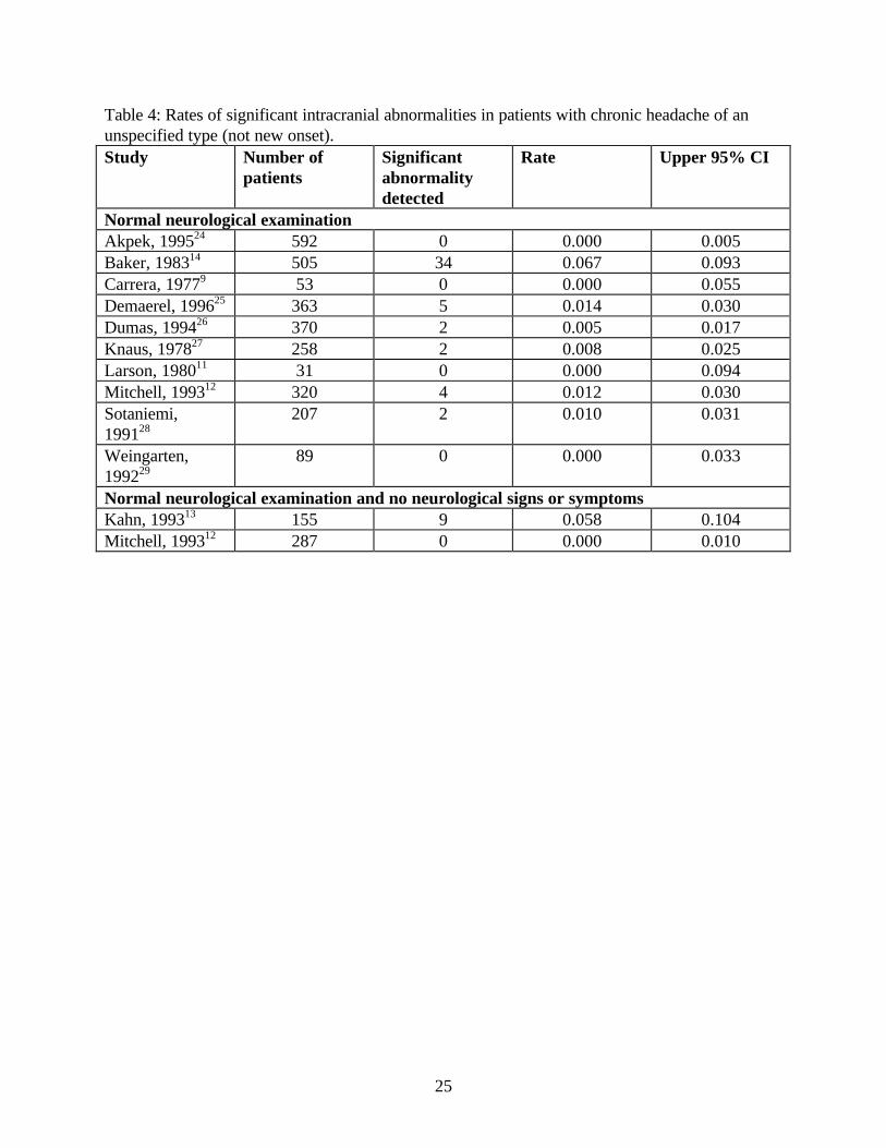
25
Table 4: Rates of significant intracranial abnormalities in patients with chronic headache of an unspecified type (not new onset). Study Number of
patients Significant abnormality detected
Rate Upper 95% CI
Normal neurological examination Akpek, 199524 592 0 0.000 0.005 Baker, 198314 505 34 0.067 0.093 Carrera, 19779 53 0 0.000 0.055 Demaerel, 199625 363 5 0.014 0.030 Dumas, 199426 370 2 0.005 0.017 Knaus, 197827 258 2 0.008 0.025 Larson, 198011 31 0 0.000 0.094 Mitchell, 199312 320 4 0.012 0.030 Sotaniemi, 199128
207 2 0.010 0.031
Weingarten, 199229
89 0 0.000 0.033
Normal neurological examination and no neurological signs or symptoms Kahn, 199313 155 9 0.058 0.104 Mitchell, 199312 287 0 0.000 0.010