Embed Size (px)
Citation preview

Albert L. Rhoton Jr., MD: His Philosophy and Education of Neurosurgeons
Toshio MATSUSHIMA,1,2 Shigeaki KOBAYASHI,3 Tooru INOUE,4 Alice S. RHOTON,5 Alexander L. VLASAK,6 and Evandro de OLIVEIRA7,8
1Graduate School, International University of Health and Welfare, Fukuoka, Fukuoka, Japan;2Neuroscience Center, Fukuoka Sanno Hospital, Fukuoka, Fukuoka, Japan;
3Stroke and Brain Center, Aizawa Hospital, Matsumoto, Nagano, Japan;4Department of Neurosurgery, Fukuoka University, Fukuoka, Fukuoka, Japan;
5Department of OB/GYN, University of Florida, Gainesville, USA;6College of Medicine, University of Florida, Gainesville, USA;
7Department of Neurosurgery, Instituto de Ciencias Neurologicas, Sao Paulo, Brazil;8Department of Neurological Surgery, Mayo Clinic, Jacksonville, USA
Abstract
Dr. Rhoton’s key philosophies included “Keep working hard.”, “Make surgery more accurate, gentle and safe”, “We want perfect anatomical dissections, because we want perfect surgical operations”, “Competence without compassion is worthless. Compassion without competence is meaningless”, “Neurosurgeons share a great professional gift; our lives have yielded an opportunity to help mankind in a unique and exciting way” and “There is no finish line for this effort”. His words reveal his passion for microneurosurgery and infinite love for humankind. Although his reknown rested on his reputation as a researcher, Dr. Rhoton was also a devoted educator. The principal aim behind the enormous amount of work he performed was that of educating neurosurgeons worldwide, so that they could be better surgeons. His work included: (1) numerous dissection courses, (2) numerous lectures and publications including about 160 original papers (3) the textbook “RHOTON” and Rhoton Collection (4) the education of 119 research fellows. The projects directed in his lab, produced the international dissemination of neuroana-tomical knowledge. The ultimate goal of his microsurgical research was to improve the care of patients with neurosurgical diseases around the world. The technical contributions and humble character of Dr. Rhoton should be remembered as we care for patients.
Key words: education, philosophy, Rhoton
Received March 16, 2018; Accepted April 11, 2018
Copyright© 2018 by The Japan Neurosurgical Society This work is licensed under a Creative Commons Attribution-NonCommercial-NoDerivatives International License.
Online June 20, 2018
doi: 10.2176/nmc.ra.2018-0082
Neurol Med Chir (Tokyo) 58, 279–289, 2018
Review ARticle
279
Introduction
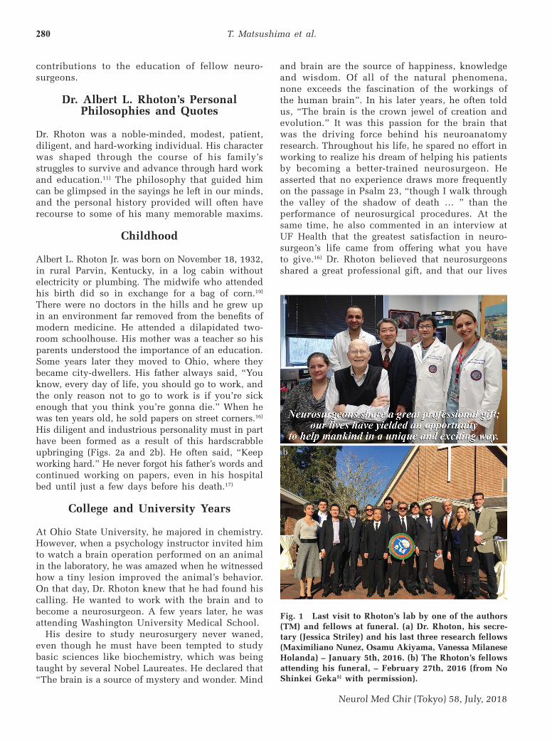
Dr. Albert L. Rhoton Jr., a great pioneer in the study of microneurosurgical anatomy, passed away at the age of 83 on February 21st, 2016. His work is a testimony to the passion he brought to the study of the brain’s anatomy, and the impact is one of the cornerstones in the history of neurosurgery, leading to safer and gentler surgical treatment performed by neurosurgeons all over the world1–14) (Figs. 1a and 1b).
He was, however, not only a keen researcher, but also an enthusiastic and passionate educator. Dr. Rhoton had been interested in teaching since
his youth.15–17) In fact, one of the major reasons he moved to the University of Florida (UF) was to develop a center for training neurosurgeons in microsurgical techniques. Among his many educational achievements are his numerous publications, including the textbook “RHOTON: Cranial Anatomy and Surgical Approaches” and the Rhoton Collection, along with many dissection courses for training neurosurgeons as well as 3-dimensional (3D) lecture presentations.9,11,12,18–23) He accepted research fellows from all over the world to assist in his research and to educate them so that the knowledge and techniques of microneurosurgical anatomy and microsurgery would follow them back to their countries.8)
His outstanding academic achievements and noble character have already been the subjects of many memorial articles.1–6,8,10–14,17,23,24) In this paper, we turn our attention to Dr. Rhoton’s unique personal philosophies that underpinned his research and
T. Matsushima et al.280
Neurol Med Chir (Tokyo) 58, July, 2018
and brain are the source of happiness, knowledge and wisdom. Of all of the natural phenomena, none exceeds the fascination of the workings of the human brain”. In his later years, he often told us, “The brain is the crown jewel of creation and evolution.” It was this passion for the brain that was the driving force behind his neuroanatomy research. Throughout his life, he spared no effort in working to realize his dream of helping his patients by becoming a better-trained neurosurgeon. He asserted that no experience draws more frequently on the passage in Psalm 23, “though I walk through the valley of the shadow of death … ” than the performance of neurosurgical procedures. At the same time, he also commented in an interview at UF Health that the greatest satisfaction in neuro-surgeon’s life came from offering what you have to give.16) Dr. Rhoton believed that neurosurgeons shared a great professional gift, and that our lives
contributions to the education of fellow neuro-surgeons.
Dr. Albert L. Rhoton’s Personal Philosophies and Quotes
Dr. Rhoton was a noble-minded, modest, patient, diligent, and hard-working individual. His character was shaped through the course of his family’s struggles to survive and advance through hard work and education.11) The philosophy that guided him can be glimpsed in the sayings he left in our minds, and the personal history provided will often have recourse to some of his many memorable maxims.
Childhood
Albert L. Rhoton Jr. was born on November 18, 1932, in rural Parvin, Kentucky, in a log cabin without electricity or plumbing. The midwife who attended his birth did so in exchange for a bag of corn.19) There were no doctors in the hills and he grew up in an environment far removed from the benefits of modern medicine. He attended a dilapidated two-room schoolhouse. His mother was a teacher so his parents understood the importance of an education. Some years later they moved to Ohio, where they became city-dwellers. His father always said, “You know, every day of life, you should go to work, and the only reason not to go to work is if you’re sick enough that you think you’re gonna die.” When he was ten years old, he sold papers on street corners.16) His diligent and industrious personality must in part have been formed as a result of this hardscrabble upbringing (Figs. 2a and 2b). He often said, “Keep working hard.” He never forgot his father’s words and continued working on papers, even in his hospital bed until just a few days before his death.17)
College and University Years
At Ohio State University, he majored in chemistry. However, when a psychology instructor invited him to watch a brain operation performed on an animal in the laboratory, he was amazed when he witnessed how a tiny lesion improved the animal’s behavior. On that day, Dr. Rhoton knew that he had found his calling. He wanted to work with the brain and to become a neurosurgeon. A few years later, he was attending Washington University Medical School.
His desire to study neurosurgery never waned, even though he must have been tempted to study basic sciences like biochemistry, which was being taught by several Nobel Laureates. He declared that “The brain is a source of mystery and wonder. Mind
Fig. 1 Last visit to Rhoton’s lab by one of the authors (TM) and fellows at funeral. (a) Dr. Rhoton, his secre-tary (Jessica Striley) and his last three research fellows (Maximiliano Nunez, Osamu Akiyama, Vanessa Milanese Holanda) – January 5th, 2016. (b) The Rhoton’s fellows attending his funeral, – February 27th, 2016 (from No Shinkei Geka5) with permission).
a
b

Rhoton: His Philosophy and Education 281
Neurol Med Chir (Tokyo) 58, July, 2018
Fig. 2 Dr. Rhoton’s photo archives (with permission from the Joyce Rhoton photo archives). (a) High School Graduation, - 1951. (b) South High champion swim team on the newspaper, -1950, Albert circled and younger brother Dick next to coach. (c) Washington University Neurosurgical residency, - 1960, Albert circled. (d) Mayo Clinic Faculty, 1970. (e) Dr. and Mrs. Rhoton (Joyce) with grown children (Eric, Albert, Alice and Laurel), - 2005. (f) Dr. and Mrs. Rhoton at daughter Alice’s medical school graduation ceremony, 1989. (g) Dr. Rhoton and grandson, Alexander, at “White Coat Ceremony”, November 2015.
b
d
f
g
e
c
a

T. Matsushima et al.282
Neurol Med Chir (Tokyo) 58, July, 2018
had yielded an opportunity to help mankind in a unique and exciting way.19)
As a college student, he had another valuable learning experience, which shaped his life. He failed his tests in every subject, including chem-istry, physics and biochemistry, because he needed to work to support himself. After this experience, he borrowed money from friends and obtained scholarships enabling him to continue his studies without financial pressures. The experience taught him not to let financial constraints interfere with his education. During medical school, he was a very intelligent student who enjoyed teaching, and ultimately graduated from medical school with the highest academic standing in the class of 1959.12)
Residency Years
In 1959, Dr. Rhoton went to Columbia Presbyterian Medical Center where he had a 1-year internship followed by 2 years of residency, one in general surgery and the other in neurosurgery. He subse-quently returned to Washington University and Barnes Hospital in St. Louis where he completed his neurosurgery residency under Professor Henry G. Schwarz in 1964 (Fig. 2c). It is noteworthy that he took a year of National Institutes of Health research fellowship in neuroanatomy at Washington Univer-sity Medical School before joining Mayo Clinic. Reminiscing about the neurosurgery he observed during his training, he spoke about the anxieties he endured. “I lay awake many nights, worrying about a patient who was facing a necessary, critical, high-risk operation the next day. The brain surgeries at that time always involved some dangers and those results tended to depend on luck.” During his residency he never saw a facial nerve preserved in a case of acoustic tumor and the pituitary gland preserved in a case of pituitary tumor.19) He was struck by the realization that, with a better knowl-edge of anatomy, he would be able to do a better job for his patients. It was during his residency at Barnes Hospital that he became more interested in teaching than before. Indeed, the atmosphere at the hospital was such that many trainees chose to enter academia by joining the teaching staff of a medical school after completing their residency.
Mayo Clinic Years
Dr. Rhoton began his professional career as a staff neurosurgeon at the Mayo Clinic in 1966 (Fig. 2d). It was around this time that microsurgery, after the introduction of the operative microscope, started to develop. He became convinced that a deeper
knowledge of microneurosurgery and microneurosur-gical anatomy had the potential to improve the care of his patients, and became eager to establish road-maps to access lesions by exploiting a more precise knowledge of anatomy.19) He started to study cranial nerves (C.N.) such as the trigeminal and facial nerves under the operative microscope at Mayo Clinic. He reported the results of this study in a paper entitled “Nervus Intermedius,” in 1968.25) This was the dawn of the study of the microneurosurgical anatomy. He wanted to build on the knowledge already available to create new programs that would promote both further study and teaching. This he later observed, was a consequence of his “missionary instinct”, the urge to set on a journey into unexplored territory to build from scratch a program of his own design, one that could be disseminated to the benefit of patients. At the time, he was offered the option of moving to UF, to develop a program from the ground up. He chose this unknown path as a way to begin and grow the training and educational environment he envisioned. It was a perfect opportunity for a young eager neurosurgeon.
University of Florida Years (1972–2016)
Dr. Rhoton moved to UF in Gainesville in 1972 and became the Chief of the Division of Neurosurgery in the Department of Surgery, with only one neuro-surgeon and two residents.16,24) His motivating desire was to build a program which created roadmaps to make surgery more “accurate, gentle and safe” for patients. His efforts resulted in the establishment of a laboratory, known as the Rhoton Lab, which opened in 1975 as a center for teaching microsur-gery and for the study of the microsurgical anatomy of the brain.8,24) Over the next 40 years, as the Lab grew and developed, its record of achievements was outstanding. Dr. Rhoton accepted a total of 119 young neurosurgeons as neuroanatomical research fellows from both the United States and abroad; the majority of them were from foreign countries. For some of these fellows, English was not a native language, but Dr. Rhoton’s calm demeanor always prevailed and overcame any difficulties in commu-nication. As is well known, during the 40-year period between 1975 and 2016, Dr. Rhoton and his research team published almost 500 publications related to microsurgical anatomy, two supplements for NEUROSURGERY and the textbook “RHOTON”.9) Through the pursuit of this research he educated his fellows and constantly urged them to be more “accurate, gentle and safe”, reminding them that “We want perfect anatomical dissections, because we want perfect surgical operations”. He also stated

Rhoton: His Philosophy and Education 283
Neurol Med Chir (Tokyo) 58, July, 2018
Fig. 3 Dr. Rhoton in the dissection courses and in the seminars. (a) Dr. Rhoton showing the anatomical dissection in the satellite dissection course in the 7th meeting of Japanese Skull Base Surgery in Fukuoka, 1995. (b) Dr. Vinko Dolenc showing his approach in the Gildred Lab, 1988. (c) Dr. Rhoton and one of the senior research fellows (Evandro de Oliveira) in the Hands-on Course in Braga, Portugal, 2012. (d) Dr. Rhoton preparing his lecture with one of the senior research fellows (Toshio Matsushima) in Braga, Portugal, 2012.
a b
c d
that it was the inner discipline of striving toward that perfection that made them improve.26)
Competence and Compassion
Dr. Rhoton began his anatomical research with the aim of taking “better care of my patients” and he never lost sight of this goal (Figs. 2e–g). In his later years, he often talked about competence and compassion, remarking that “Competence without compassion is worthless. Compassion without competence is meaningless.”19) Similarly, he explained that, “I sort of have had a two view, two-handed view, of how I want people in our profession to grow. One of them is competence, which is skill and knowledge, and the other is compassion, which is love and kindness.”16) He added, “Competence and compas-sion need to be developed simultaneously, as the
giant oak develops its root system along with its leaves and branches.”19) His belief was that, “Success should mean giving every patient the feeling that he or she is cared about, no matter how desperate their situation, that their pain is felt, that their anger is understood, and that we care and will do our best.”19)
Future Anatomic Study
Dr. Rhoton’s sense of responsibility toward the future of microsurgical anatomical study was evident in his exhortations to colleagues: “Our work is not complete in any area. Further study will yield new information that will improve our operative approach and operative results in dealing with pathology in each of the areas previously examined. “There is no finish line for this effort.” The study

T. Matsushima et al.284
Neurol Med Chir (Tokyo) 58, July, 2018
of microsurgical anatomy continues to be important in the improvement of old techniques and in the adaptation of these techniques to new situations. In Dr. Rhoton’s view, every year, there were advances in neurological technology that yielded new therapeutic possibilities that must be evaluated and directed in the light of advances in our understanding of anatomy.19) New anatomical knowledge was also to play a major role in the training of neurosurgeons to improve their surgical skills.
Education of Neurosurgeons
Dr. Rhoton contributed significantly to the education of neurosurgeons globally through the following contributions: (1) dissection courses, (2) publica-tions and lectures, and (3) the education of his 119 research fellows. Each of these endeavors will be considered separately below.
Dissection courses For 40 years, Dr. Rhoton hosted and organized
numerous dissection courses (Figs. 3a–d). One type was a 5-day micro-vascular and dissection course held in his lab at UF every month from the late 1970s till the late 1980s, and 2nd type was the cadaver dissection course held from the early 1990s at the AANS, CNS, and/or North American Skull Base Society meetings. In the late 1990s, it was also offered in the Rhoton Lab when it moved to the McKnight Brain Institute. The dissection courses were also held abroad in several places.
In 1975, soon after the Rhoton Lab opened, Dr. Rhoton established the 5-day micro-vascular and dissection courses to train neurosurgeons primarily in the use of the operative microscope.8,24) These courses, consisting of a 3-day rat anastomosis practice and a 2-day dissection of the human cadaveric sphenoid and temporal bones, were epoch-making because, at that time, there were no regularly held practical training courses in the use of microscope. For more than 10 years, the 5-day courses made a crucial contribution to the training of hundreds of neuro-surgeons and residents from all over the world and played an important role in the dissemination of the knowledge and techniques of microsurgery in the field of the neurosurgery. It was because of these initiatives that both the department of neurosur-gery at UF and Dr. Rhoton’s Laboratory became so renowned.24) Many Japanese neurosurgeons attended the courses.
Later in his life Dr. Rhoton recalled the first course: “I still remember and am grateful to each member of the initial group of seven neurosurgeons. During the first afternoon of that course, I walked into the
laboratory and, to my amazement, found seven surgeons working quietly and diligently. Nothing was said for long periods of time. In the midst of this intense endeavor and amazing quietness, I realized that we had tapped into a great force: the desire of neurosurgeons to improve themselves.”19) During every course, Dr. and Mrs. Rhoton invited the participants and lab staff to their own home for dinner. As the participants were from all over the world, these occasions were a marvelous opportu-nity to communicate and learn about neurosurgery in other countries.
At the beginning of 1990s, Dr. Rhoton began to offer a 2-day cadaveric dissection course at the AANS, CNS and/or North American Skull Base Society meetings, titled “Microsurgical Approaches to the Brain, Ventricles and Skull Base”. Dr. Rhoton started to organize a 5-day cadaveric dissection course once or twice a year in the Rhoton Lab, officially named the George Schrader Colter Micro-neurosurgical Anatomy Lab, when it moved to the newly built McKnight Brain Institute in 1998. In each of these dissection courses, about 20 participants dissected whole cadaveric heads and studied not only surgical anatomy but also surgical approaches. Beautiful booklets were prepared and lectures were given in a 3D format that required the participants to wear 3D glasses. Martins provides a detailed description of some of the more recent courses.23) Dr. Rhoton continued the CNS courses for about 20 years and completed the last one in 2013.24)
Publications and lecturesOver a period of 50 years, from the 1960s to the
2010s, Prof. Rhoton was responsible for over 500 publications including about 160 original anatomical papers, the textbook “RHOTON” and the Rhoton Collection.8,9) These publications have played a vital role in the education of neurosurgeons throughout the world. Other cranial and ENT surgeons also refer to these works for surgical guidance.Over 500 publications: Prof. Rhoton preferred publishing his anatomical research results in journals rather than textbooks because this allowed the infor-mation to be accessible to a wider reading audience. He submitted at least two or three original articles every year either to the Journal of Neurosurgery or Neurosurgery. In the beginning, however, Dr. Rhoton was often required to revise these articles because they were regarded as educational and so considered to be less than original. Often, he experienced diffi-culties getting his papers accepted for publication. The themes of the 160 original papers reflect the trend of the time, and track the transitions in the evolution of Dr. Rhoton’s work.9) After pursuing his

Rhoton: His Philosophy and Education 285
Neurol Med Chir (Tokyo) 58, July, 2018
research for nearly 35 years, from 1960s to 2003, and covering every area of the brain, he finally published the great neuroanatomical textbook “RHOTON” in 2003.
From an educational point of view, Dr. Rhoton’s papers on the surgical anatomy of the brain are excellent because they are accurate and comprehensive with beautiful illustrations. This was accomplished in part by incorporating the following features: (1) Many clear and precise figures made using retouched prints, (2) the introduction of certain rules such as “the rule of 3”, (3) new names, that were more comprehensible, applied to, for example, the veins of the posterior fossa, and (4) clarification of the structures of the ventricles by collating anatomical information relating to the neural structures, arteries and veins into one anatomical figure.2,8,9,23,27,28)
Textbook “RHOTON: cranial anatomy and surgical approaches”: The textbook “RHOTON” could be thought of as a bible for doctors majoring in the nervous system. It was the combination of the Millennium Supplement with the issue for Celebrating 25 years of “Neurosurgery”. Dr. Rhoton compiled the efforts of 35 years of research on surgical anatomy into a single book. It was a major undertaking, which he embarked on after he stepping down from his chairmanship and becoming a professor emeritus in 1999. Dr. Michael Apuzzo, the editor of “Neurosurgery” who collaborated with Dr. Rhoton on these publications over many years, praised him thus in an editor’s letter entitled “MIRABILE VISU” “This volume stands as a tribute to the remarkable vision, diligence, and intelligence of Albert L. Rhoton, Jr. It is the concrete legacy of his character and persona. It serves as an example for all of us who would call ourselves neurosurgeons and represents the epitome of the term ‘contribution to the field’ - a notion and goal that is the elusive ‘Holy Grail’ for many of us.”19) This textbook has been translated into Portuguese, Chinese and Japanese.Rhoton collection: In Rhoton’s Lab, a tremendous volume of descriptive photographs of dissections were produced by more than 100 research fellows over four decades. Dr. Rhoton collected this data and considered how it could best be handed down to posterity. Dr. Jon H. Robertson, a friend of Dr. Rhoton, has stated in a paper that, “Recognizing the importance of preserving Dr. Rhoton’s 3D neuro-microanatomical lectures for future neurosurgical education, I began to discuss with him how this might occur in 2008.”11) In accordance with his hope that the entire body of his work might be shared with as many neurosurgeons as possible, free of charge, Dr. Rhoton began in 2010 to discuss with Dr. Robertson and Dr. Jeffrey Sorenson how, with
the financial support of the Neurosurgery Research and Education Foundation (NREF) and the AANS, provisions might be made to archive this treasury of information.11,20,21) Modern web-browser technologies have now made it possible to present his material in new formats. The basic rationale underlying the creation of the Rhoton Collection website was to organize what was a very large set of photographic data into a searchable online database. Dr. Sorenson, the producer and director of the Rhoton Collection, reported that it took more than 2 years for a full-time employee to clean and scan the full collection of slides, which consisted of thousands of images.22) The Rhoton Collection was designed with two goals in mind: to focus the reader’s attention on the images as far as possible and to increase the use of the material by making it readily accessible in any learning environment, including cellular phones and laptops. The Collection typically documents regional anatomy at various stages of dissection, while also providing step-by-step guides to the neurosurgical approaches and the opportunity to view the stereoscopic movies which were featured in Dr. Rhoton’s lectures.Rhoton’s lectures: Dr. Rhoton gave an enormous number of lectures in US and abroad (Figs. 4a and 4b). His lectures were easily understood both because of the clarity of the images and his slow, deliberate speech. Among the count-less images created at Rhoton’s Lab, he selected only those of the highest quality in which the audience would be fascinated by the beauty of his images. At that time, the task of preparing slides that met Dr. Rhoton’s exacting standards was a daunting challenge for his research fellows. Digital cameras have now made it possible to take as many pictures as desired, with the option to delete those that are substandard. Prior to the early 1990s, however, when the fellows only had 35-mm cameras at their disposal, it was neces-sary to take a large number of photographs, and have the film developed and made into color slides before selecting the best among them for Dr. Rhoton’s consideration. Due to his intense desire for the best representations of anatomy, he was often dissatisfied with slides presented to him and many had to be discarded. It was, needless to say, a very time consuming and costly process, but resulted in the highest quality images being preserved for future educational purposes.8,23,27,28)
Two further features made his lectures distinctive. (1) At the beginning of his lectures, as if in imitation of the Socratic mode of teaching, he would pose questions such as “Which appears the most lateral, the internal carotid artery, optic nerve

T. Matsushima et al.286
Neurol Med Chir (Tokyo) 58, July, 2018
or anterior clinoid process?” or “How many bones compose the foramen magnum?” (2) After the 1990s, Dr. Rhoton often used 3D imaging, which was an aid to attaining a more accurate understanding of depth during surgical procedures. With advances in digital imaging techniques in the 2000s, 3D projec-tion regularly became an important tool.9,23)
Education of Research Fellow
Dr. Rhoton often showed the famous painting “Creation of Adam” by Michelangelo Buonar-roti in his lectures and commented that the red cloak behind God was in the shape of a human brain. Referring to this suggestion, Dr. Jeffrey Sorenson wrote, “Dr. Rhoton was very proud of the artistic results his fellows were achieving there
(Fig. 4c). He repeatedly referred to them as modern day Michelangelos, which is not so far-fetched; as their dissections are essentially sculptures created by precise subtraction of material until a master-piece remains. Just as Lorenzo de’ Medici took in the young Michelangelo for training in the finest techniques of sculpture, Dr. Rhoton brought young, bright, motivated people from all over the world to learn the art and craft of anatomical dissection before making world-class contributions of their own. We also know that Michelangelo’s deep interest in human anatomy and the brain led him to perform dissections and contemplate illustrating an anatomy textbook. I am certain that if Dr. Rhoton’s laboratory had existed in Renaissance Florence, Michelangelo would have done a fellowship and we would still be admiring the results centuries later.”21)
Fig. 4 Dr. Rhoton’s visit to Japan. (a) The first visit, the 36th annual meeting of the Japanese Neurosurgical Society, Osaka, 1977. (b) The last visit, the Rhoton Seminar in Saga, Saga, 2010. (c) Dr. Rhoton, Dr. Erdener Timurkaynak, Dr. Shigeaki Kobayashi, and Dr. Rhoton’s Japanese fellows in the Rhoton Seminar in Saga, 2010.
a b
c

Rhoton: His Philosophy and Education 287
Neurol Med Chir (Tokyo) 58, July, 2018
At first, Dr. Rhoton accepted young doctors as research fellows to conduct the research needed to develop his surgical techniques. However, when he moved to UF in 1972, he discovered that there were young neurosurgeons all over the world who aspired to learn and complete fellowships under his guidance to gain a better understanding of microneurosurgical anatomy. It occurred to him that he might educate them by providing the opportu-nity to pursue anatomical study and anastomosis training in his laboratory. Dr. Rhoton directly educated 119 young neurosurgeons, domestic and international, during their research fellowships at UF over a 40-year period from 1975 to 2016. About 100 of these doctors were from outside the USA.8,28) Forty-one Japanese neurosurgeons studied in his Lab. In the early stage of his research, Dr. Rhoton developed close relationships with a few neurosurgery programs in Japan, especially with Kyushu University.24) This latter development came about as a consequence of a courtesy visit by the late Prof. Katsutoshi Kitamura of Kyusyu University to Prof. Rhoton at UF in 1973, in the year following that in which Dr. Rhoton had become chief of the Division of Neurological Surgery. Dr. Rhoton felt very honored and grateful to Prof. K. Kitamura. It became a lifelong friendship and collaboration.
The research methods and pace of work in Rhoton’s Lab were improved in the early 1980s when Evandro de Oliveira (Brazil), Erdener Timur-kaynak (Turkey) and Toshio Matsushima (Japan) worked together.2,8) Subsequently, these three former fellows were entrusted with the task of recom-mending promising young neurosurgeons from their own countries.8,29) As a result, most of the 100 foreign neurosurgeons were from Japan (41), Latin American countries (24: 15 from Brazil, 7 from Argentina, and 1 each from Chile and Mexico) and Turkey (11). Since the fellows studied for a limited period (usually 1 or 2 years) without salary, they were highly motivated to achieve their goals. They worked hard until late in the evening and/or during weekends.2,8) They published a total of about 160 original papers on microneurosurgical anatomy with Dr. Rhoton. These international research fellows have shared the knowledge they gained at UF to many neurosurgeons in their own countries after their return. About 40 of the 119 fellows have now become professors.8)
Dr. Rhoton’s international influence and his foreign research fellows’ achievements
One of Dr. Rhoton’s goals was to teach and mentor as many neurosurgeons as possible. He harbored the
fervent hope that the knowledge obtained at Rhoton’s Lab in UF might be disseminated throughout the world, and his former foreign fellows have played an important role in working toward that goal. After returning to their homes, many of them established annual meetings or seminars in microneurosurgical anatomy, established microneurosurgical laboratories offering dissection courses, and/or translated the English textbook “RHOTON” into languages such as Portuguese, Chinese and Japanese.8,18,29–32) All have contributed in their own ways, to enhancing the understanding of neuroanatomy after they returned to their own countries.
Dr. Rhoton’s Japanese fellows started an annual microsurgical anatomy seminar in 1986. This meeting was established and organized by the Japanese Society for Microsurgical Anatomy, and the 31st meeting was held last year.18,30) The proceedings of these seminars, published in Japanese every year, have been widely read, especially during the period between the late 1980s and the early 1990s. About 3,000 copies of the proceedings were sold every year at a time when the number of Japanese board-certified neurosurgeons was between 3,000 and 4,000. The first International Symposium on Microsurgical Anatomy (ISMA) (Co-Presidents: Toshio Matsushima & Albert L. Rhoton Jr.) was held in 2002 in Matsumoto, Japan, and followed by subsequent international meetings organized by Dr. Erdener Timurkaynak from Ankara, Turkey, Dr. Evandro de Oliveira from Sao Paulo, Brazil and Dr. Turker Kiliq from Istanbul, Turkey, four times in Turkey and once in Brazil, each time with Dr. Rhoton as the invited co-president.5,8,30,31) The international symposium has contributed to the advancement of the professional skills of neurosurgeons through the study of microsurgical anatomy both in the host countries and in the countries of all those attending the meetings.
Some of the fellows established microneurosurgical laboratories in their countries, in some cases naming them “Rhoton’s Lab”.8,29,31,32) Dissection courses similar to those run by Dr. Rhoton at UF have been held in Brazil, Turkey and China. In Brazil, Dr. de Oliveira established the Microsurgery Laboratory of Hospital Beneficencia Portugueesa in Sao Paulo in 1989. Since then, he and his colleagues have held 348 courses attended by a total of 6,896 participants.29) In Turkey, Dr. Timurkaynak established a laboratory in 1985. He organized 255 courses during a 25-year period, with the participation of a total of 1,082 physicians, some of whom came from outside Turkey.31) In Turkey, another Rhoton laboratory has been established. In China, Dr. Xiaoguang Tong first established a laboratory at his own hospital to create

T. Matsushima et al.288
Neurol Med Chir (Tokyo) 58, July, 2018
a 3-month-training course around 2007, and then extended the length of the program while setting up courses in other hospitals in China.32) Many similar initiatives, both large and small, have been seen in Korea, Argentina, Chile, Spain, Portugal and other countries. Rhoton’s former fellows have organized these courses either individually or cooperatively in their own countries.
Conclusion
Dr. Rhoton embarked on his anatomical studies in an effort to provide better care for his patients. His work attracted numerous international research fellows who wanted to research and become better educated in his Lab, thereby contributing to a remarkable scientific body of work. Dr. Rhoton enlarged the scope of his work to include the dissemination of the knowledge obtained at his Lab to elevate the quality and safety of neurosurgery worldwide, contributing greatly to the education of neurosurgeons. It was an undertaking rooted in his philosophy, in his compassion for his fellow human beings, and in the sense of wonder and amazement he felt when he contemplated the human brain. As he fondly observed, “The brain is the crown jewel of creation and evolution.”
Before closing, the authors would like to dedicate this paper to our teacher, friend and father, the late Dr. Albert L. Rhoton, Jr. and his wife, Mrs. Joyce L. Rhoton, who supported his work both implicitly and explicitly.
Acknowledgments
Our deepest appreciation goes to Mrs. Joyce L. Rhoton for her cooperation. We would like to express our gratitude to several former Japanese Rhoton’s fellows for their suggestions and comments. We are also grateful to Mrs. Sumiko Matsushima for her assistance in preparing and completing this manuscript.
Conflicts of Interest Disclosure
The authors report no conflict of interest concerning the materials or methods used in this study or the findings specified in this paper.
References
1) Fernandez-Miranda JC: Prof. Rhoton: master and mentor. J Neurol Surg B Skull Base 77: 288–290, 2016
2) Fernandez-Miranda JC: Prof. Albert L. Rhoton, Jr.: his life and legacy. World Neurosurg 92: 590–596, 2016
3) Friedman WA: Albert Lore Rhoton Jr., MD. World Neurosurg 75: 188–191, 2011
4) Friedman WA: Obituary: Albert Loren Rhoton Jr., MD, 1932–2016. J Neurosurg 125: 1–3, 2016
5) Kobayashi S: Obituary: In memory of the late Prof. Albert L. Rhoton, Jr. No Shinkei Geka 44: 431–435, 2016 (Japanese)
6) Link MJ, Graffeo CS: Albert L. Rhoton, Jr., MD, 1932– 2016. J Neurol Surg B Skull Base 77: 283–285, 2016
7) Matsushima T: Getting to know a neuroscientist: Professor Albert L. Rhoton Jr. Clin Neurosci 26: 1406, 2008 (Japanese)
8) Matsushima T, Lister JR, Matsushima K, et al.: The history of Rhoton’s Lab. Neurosurg Rev 2017. doi:10.1007/s10143-017-0902-4
9) Matsushima T, Matsushima K, Kobayashi S, Lister JR, Morcos JJ: The microneurosurgical anatomy legacy of Albert L. Rhoton Jr., MD: an analysis of transition and evolution over 50 years. J Neurosurg 1–11, 2018
10) Morcos JJ: In memoriam of Dr. Al Rhoton, 2016 North American Skull Base Society – Managed by BSC M
11) Robertson JH: Dr. Al Rhoton, Jr.: friend, mentor, and colleague. J Neurol Surg B Skull Base 77: 291–293, 2016
12) Robertson JH: Rhoton and the United States. World Neurosurg 92: 597–600, 2016
13) Stafford N: Albert L Rhoton Jr. BMJ 353: i2762, 201614) Tubbs RS, Oskouian RJ, Delashaw J: Memorial:
Albert L. Rhoton Jr., MD (1932–2016). Clin Anat 29: 422–423, 2016
15) Rhoton AL Jr., Matsushima T: Neurosurgeon’s life with microsurgical anatomy. Curr Pract Neurosurg 13: 73–77, 2003 (Japanese) (Interview of Dr. AL. Rhoton by T Matsushima)
16) Rhoton AL Jr., Stoyan-Rosenzweig N: “An Oal History Albert L. Rhoton MD”. DVD, University of Florida Health Science Center 071, 2010, Interview of Dr. AL. Rhoton by N Stoyan-Rosenzweig.
17) Rhoton-Vlasak AS, Vlasak AL: Family reflections on our amazing journey with dad – Professor Rhoton. World Neurosurg 92: 653–657, 2016
18) Matsushima T, Kawashima M, Matsushima K, Wanibuchi M: Japanese neurosurgeons and microsurgical anatomy: a historical review. Neurol Med Chir (Tokyo) 55: 276–285, 2015
19) Rhoton AL: Cranial anatomy and surgical approaches. Lippincott Williams & Wilkins: Philadelphia, 2003, pp. iii–vii (Foreword)
20) Rutka JT: Editorial: The Rhoton Collection and Journal of Neurosurgery: expanding the reach of neuroanatomy in the digital print world. J Neuro-surg 125: 4–6, 2016
21) Sorenson J: The Rhoton Collection. J Neurol Surg B Skull Base 77: 294–296, 2016
22) Sorenson J, Khan N, Couldwell W, Robertson J: The Rhoton Collection. World Neurosurg 92: 649–652, 2016
23) Martins C: Rhoton’s lab. World Neurosurg 92: 623–636, 2016
Rhoton: His Philosophy and Education 289
Neurol Med Chir (Tokyo) 58, July, 2018
24) Day AL, Blackburn S: Rhoton and the University of Florida. World Neurosurg 92: 601–605, 2016
25) Rhoton AL, Kobayashi S, Hollinshead WH: Nervus intermedius. J Neurosurg 29: 609–618, 1968
26) Rhoton AL, Jr.: Presidential address: improving ourselves and our specialty. Clin Neurosurg 26: xiii–xix, 1979
27) Peace D: A medical illustrator reflects upon his career working with Albert L. Rhoton Jr. Congress Quarterly Fall 17–21, 2016
28) Barry ME: Art and the role of the Rhoton medical illustrators in his legacy. World Neurosurg 92: 637–648, 2016
29) Wen HT, de Oliveira E: Rhoton and his influence in Latin America neurosurgery. World Neurosurg 92: 606–607, 2016
30) Matsushima T: Rhoton and his influence on Japanese neurosurgery. World Neurosurg 92: 608–613, 2016
31) Timurkaynak E: Rhoton and his influence on Turkish neurosurgery. World Neurosurg 92: 614–616, 2016
32) Tong X: Rhoton and his influence in Chinese neuro-surgery. World Neurosurg 92: 617–622, 2016
Address reprint requests to: Toshio Matsushima, MD, PhD, Neuroscience Center, Fukuoka Sanno Hospital, 3-6-45 Momochihama, Sawara-ku, Fukuoka, Fukuoka 814-0001, Japan.
e-mail: [email protected]