Embed Size (px)
Citation preview
ORIGINAL CONTRIBUTION
Neuroprotective effects of digested polyphenols from wildblackberry species
Lucelia Tavares • Ines Figueira • Gordon J. McDougall •
Helena L. A. Vieira • Derek Stewart • Paula M. Alves •
Ricardo B. Ferreira • Claudia N. Santos
Received: 14 November 2011 / Accepted: 18 January 2012 / Published online: 8 February 2012
� Springer-Verlag 2012
Abstract
Purpose Blackberry ingestion has been demonstrated to
attenuate brain degenerative processes with the benefits
ascribed to the (poly)phenolic components. The aim of this
work was to evaluate the neuroprotective potential of two
wild blackberry species in a neurodegeneration cell model
and compare them with a commercial variety.
Methods This work encompasses chemical characteriza-
tion before and after an in vitro digestion and the assess-
ment of neuroprotection by digested metabolites. Some
studies targeting redox/cell death systems were also per-
formed to assess possible neuroprotective molecular
mechanisms.
Results The three blackberry extracts presented some
quantitative differences in polyphenol composition that
could be responsible for the different responses in the
neurodegeneration cell model. Commercial blackberry
extracts were ineffective but both wild blackberries, Rubus
brigantinus and Rubus vagabundus, presented neuropro-
tective effects. It was verified that a diminishment of
intracellular ROS levels, modulation of glutathione levels
and activation of caspases occurred during treatment. The
last effect suggests a preconditioning effect since caspase
activation was not accompanied by diminution in cell death
and loss of functionality.
Conclusions This is the first time that metabolites
obtained from an in vitro digested food matrix, and tested
at levels approaching the concentrations found in human
plasma, have been described as inducing an adaptative
response.
Keywords Caspase activity � Glutathione balance �In vitro digestion � Neurodegenerative diseases �Wild blackberries
Introduction
In the developed world, the population lifespan is
increasing with a concomitant increase in the incidence
of age-related diseases such as neurodegeneration [1].
Due to the high impact at both financial and social levels
Electronic supplementary material The online version of thisarticle (doi:10.1007/s00394-012-0307-7) contains supplementarymaterial, which is available to authorized users.
L. Tavares � I. Figueira � H. L. A. Vieira �P. M. Alves � R. B. Ferreira � C. N. Santos (&)
Instituto de Tecnologia Quımica e Biologica, Universidade Nova
de Lisboa, Av. da Republica, 2780-157 Oeiras, Portugal
e-mail: [email protected]
G. J. McDougall � D. Stewart
Environmental and Biochemical Science Group, Enhancing
Crop Productivity and Utilisation Theme, The James Hutton
Institute, Dundee DD2 5DA, Scotland, UK
H. L. A. Vieira � P. M. Alves � C. N. Santos
Instituto de Biologia Experimental e Tecnologica,
Apartado 12, 2781-901 Oeiras, Portugal
H. L. A. Vieira
CEDOC@IGC, Faculdade de Ciencias Medicas,
UNL, 1169-056 Lisbon, Portugal
D. Stewart
School of Life Sciences, Heriot Watt University,
Edinburgh EH14 4AS, Scotland, UK
R. B. Ferreira
Departamento de Botanica e Engenharia Biologica, Instituto
Superior de Agronomia, Universidade Tecnica de Lisboa,
Tapada da Ajuda, 1349-017 Lisbon, Portugal
123
Eur J Nutr (2013) 52:225–236
DOI 10.1007/s00394-012-0307-7
[2, 3], strategies to retard or reverse neuronal and
behavioral deficits that occur in aging are urgently
required. Indeed, these foci are areas of intense research
effort, but the therapeutic strategies [4] and delivery of
(pharma) products [5, 6] have been limited. Epidemio-
logical evidence indicates that antioxidant supplementa-
tion may provide neuroprotection against age-related
neurodegenerative disorders, including Parkinson’s dis-
ease, amyotrophic lateral sclerosis and Alzheimer’s dis-
ease [7–9]. Increased dietary intake of antioxidant fruits,
in particular berry fruits, may cause positive and pro-
found impacts on human health, performance and disease
[10]. Their biological properties are attributed to the
wide diversity and high levels of phenolic compounds,
frequently associated with a high antioxidant capacity.
Due to the multitude of phytochemicals found in these
fruits, instead of a single compound, they can promote
complementary, additive and/or synergistic effects [10,
11].
In vitro studies have revealed blackberries as pos-
sessing potent antioxidant, antiproliferative and anti-
inflammatory activities [12, 13]. Moreover, in aged rats,
these fruits were capable of improving performance on
motor tests, which relied on balance and fine motor
coordination, and on measures of spatial working mem-
ory [14]. However, these effects were not accompanied
by an improvement in dopamine release [14] and con-
sequently by an improvement of receptor sensitivity,
events usually related with the observed effects [15, 16].
A large number of pathways and protein kinase cascades,
such as protein kinase C, Nrf2/ARE antioxidant pathway,
pro-survival MEK/ERK and PI3K/AKT pathways among
others, have been reported as targets for phenolic com-
pounds; nevertheless, the target pathways affected remain
unknown [17].
The chemical diversity of plants constitutes an immense
and relatively untapped reservoir of molecules with
potential pharmacological/nutraceutical value. The diver-
sity of Portuguese plants represents a reservoir of phyto-
chemicals as yet poorly characterized and explored. In
particular, in the north of Iberian Peninsula, there are
endemic Rubus species, such as Rubus brigantinus Samp.
and Rubus vagabundus Samp. [18], of which chemical
diversity could be further explored.
The aim of this work is to evaluate the neuroprotec-
tive potential of two endemic blackberry species in a
neurodegeneration cell model. This work encompasses an
in vitro digestion (IVD) to mimic some alterations in
metabolites that fruits are submitted to when ingested.
Some studies targeting redox and cell death systems
were performed to illustrate molecular mechanisms by
which blackberry metabolites could exert beneficial
effects.
Methods and materials
Plant material
Fruits of wild blackberry species (R. brigantinus Samp. and
R. vagabundus Samp.) were collected in September 2009
in Braganca region (northeast region of Portugal) and
frozen. Fruits were collected from several populations,
growing in different locations in order to be representative
of species. For both species, voucher samples were
authenticated and deposited at the herbarium ‘‘Joao de
Carvalho e Vasconcelos’’, Instituto Superior de Agrono-
mia, Lisbon, Portugal (voucher number 716/2010 and
722/2010). For comparison purposes, the commercial
blackberry cv. Apache (Rubus L. subgenus Rubus Watson)
produced in Fataca experimental field (Odemira, Portugal)
was also used. The samples were freeze-dried, ground
without separation of seeds in an IKA M20 mill to pass a
0.5 mm sieve and stored at -80 �C prior to extraction.
Extract preparation
Fruit extracts were prepared using an hydroethanolic
solution (ethanol 50% (v/v)) as previously described [19].
Briefly, 12 mL of ethanol 50% was added for each gram of
blackberry freeze-dried powder. Homogenate was shaken
for 30 min and filtered. Extracts obtained were dried under
vacuum.
In vitro digestion (IVD)
Phytochemical alterations during digestion were mimicked
using the IVD model already described [20]. Briefly, the
undigested extract was submitted to conditions that mimic
the gastric digestion such as adjusted to pH 1.7, the addi-
tion of pepsin and incubation at 37 �C with shaking at
100 rpm for 2 h. After, small intestine conditions were
mimicked by the addition of pancreatin and bile salts,
followed by dialysis with a cellulose tube containing
NaHCO3 to neutralize titratable acidity. After 2 h incuba-
tion at 37 �C, the solution inside the dialysis tubing (IN)
and the solution outside the dialysis tubing (OUT) were
taken.
Chemical characterization
Total phenolic quantification
Determination of total phenolic compounds was performed
by the Folin–Ciocalteau method adapted to microplate
reader [21]. Gallic acid was used as the standard and the
results were expressed as mg of gallic acid equivalents (mg
GAE).
226 Eur J Nutr (2013) 52:225–236
123

Peroxyl radical scavenging capacity determination
Peroxyl radical scavenging capacity was determined by the
ORAC (Oxygen Radical Absorbance Capacity) method as
described by Tavares et al. [19]. The final results were
calculated using the area differences under the fluorescence
decay curves between the blank and the sample and were
expressed as lM trolox equivalents (lM TE).
Determination of phenolic profile by LC–MS
Extracts and digested fractions, containing 500 mg GAE
mL-1, were applied to a C18 column (Synergi Hydro C18
column with polar end capping, 4.6 9 150 mm, Phe-
nomonex Ltd.) and analyzed on a LCQ-DECA system
controlled by the XCALIBUR software (2.0, Thermo-
Finnigan), as reported by Tavares et al. [19]. The LCQ-
Deca system comprised a Surveyor autosampler, pump and
photodiode array (PDA) detector and a Thermo Finnigan
iontrap mass spectrometer.
Animal cell culture
Human neuroblastoma SK-N-MC cells were obtained from
the European Collection of Cell Cultures (ECACC) and
cultured in EMEM (Eagle Minimum Essential medium,
Sigma) supplemented with 2 mM L-glutamine (Sigma),
10% (v/v) heat-inactivated fetal bovine serum (FBS,
Gibco), 1% (v/v) non-essential amino acids (Sigma), 1 mM
sodium pyruvate (Sigma) and containing 50 U penicillin
and 50 lg streptomycin per mL of medium. The cells were
maintained at 37 �C in 5% CO2 and split at sub-confluence
of 70–80% using 0.05% trypsin/EDTA (Gibco).
Cytotoxicity evaluation
Digested fractions were dried under vacuum and dissolved
in cell medium for the cytotoxicity tests by measuring cell
viability as previously described [20]. Briefly, SK-N-MC
neuroblastoma cells were seeded in a 96-well plate using
1.25 9 105 cells mL-1 and grown for 48 h prior to incu-
bation with the IN fractions. Toxicity tests involved 24 h
fractions incubation in the range 0–100 lg GAE mL-1
medium. Cell viability was assessed using the CellTiter-
Blue� Cell Viability Assay (Promega), according to the
manufacture instructions.
Neuroprotective evaluation
To evaluate the neuroprotective effect of fractions, a neu-
rodegeneration cell model already described was used [20].
The model described the treatment of SK-N-MC neuro-
blastoma cells with H2O2 to induce cell death. Briefly, cells
were seeded at 7.4 9 104 cells mL-1, grown for 24 h and
then after 24 h of pre-incubation with medium supple-
mented with non-toxic concentrations of blackberry frac-
tions, the cells were treated with medium containing H2O2
(300 lM). After 24 h, the medium was removed and cells
were washed with PBS and collected by trypsinization.
Cells were then incubated with two fluorescent probes for
30 min at 37 �C. 3,30-Dihexyloxacarbocyanine iodide
(DiOC6(3), 20 nM, Invitrogen) was used to evaluate the
mitochondrial transmembrane potential (DWm), and pro-
pidium iodide (PI, 1 lg mL-1, Invitrogen) was used to
determine cell viability, based on plasma membrane
integrity [22]. A flow cytometer (Partec) was used to ana-
lyze parameters. This cytometer contains a blue solid state
laser (488 nm) with FL1 green fluorescence channel for
DiOC6(3) at 530 nm and a FL3 red fluorescence channel for
PI detection at 650 nm. The acquisition and analysis of the
results were performed with FlowMax� (Partec) software.
Intracellular reactive oxygen species (ROS) production
determination
To evaluate the ability of fractions to reduce ROS levels
produced by cells, the conversion of 20,70-dichlorofluores-
cein diacetate (H2DCFDA, Invitrogen) to fluorescent 20,70-dichlorofluorescein (DCF) was monitored [23, 24]. SK-N-
MC neuroblastoma cells were seeded in a 96-well plate
(1.25 9 105 cells mL-1), grown for 24 h, then washed
with PBS and pre-incubated with fractions prepared in
medium (0.5% (v/v) FBS) for 2 or 24 h. After pre-incu-
bation, cells were washed with PBS and incubated for
30 min at 37 �C with 25 lM H2DCFDA prepared in PBS.
Cells were washed with PBS and then H2O2 (200 or
300 lM) was added. Fluorescence was measured (kex:
485 nm, kem: 530 nm) using a FLx800 Fluorescence
Microplate Reader (Biotek) over 1 h at 37 �C. ROS gen-
eration was calculated as an increase in fluorescent signal
compared with cells not treated with H2O2.
Glutatione (GSH) and glutatione disulfide (GSSG)
quantification
GSH and GSSG were quantified by HPLC after derivati-
zation with orthophthalaldehyde, performed accordingly to
Kand’ar [25] as already described in Tavares et al. [20].
Chromatographic analysis was accomplished using iso-
cratic elution on a C18 analytical column (SupelcosilTM
ABZ ? Plus HPLC Column 15 cm 9 4.6 mm, 3 lm
(Supelco)) at 40 �C on an AcquityTM Ultra Performance
LC system (Waters). The mobile phase was 15% (v/v)
methanol in 25 mM sodium hydrogen phosphate, pH 6.0.
The flow rate was kept constant at 0.7 mL min-1. The
excitation and emission wavelengths were set at 350 and
Eur J Nutr (2013) 52:225–236 227
123
420 nm, respectively. The amount of GSH and GSSG was
quantified from the corresponding peak areas using
Empower� Pro 2.0 software. The concentration of GSH
and GSSG in the samples was determined from standard
curves with ranges 0–100 lM for GSH and 0–5 lM for
GSSG. Values were normalized for total protein content,
determined by Lowry method [26].
Caspase 3/7 activity determination
Caspase activity was determined using the Caspase-GloTM
3/7 assay (Promega). SK-N-MC neuroblastoma cells were
seeded in a 96-well plate (1 9 104 cells mL-1). Cells were
grown for 24 h, then washed with PBS and pre-incubated
with fractions prepared in medium (0.5% (v/v) FBS) for
24 h. After this period, cells were washed again with PBS
and medium containing 300 lM H2O2 was added. Cells
were incubated for 24 h and then 100 lL of proluminescent
caspase 3/7 substrate was added to each well. Cells were
incubated at room temperature for 3 h and luminescent
signal was recorded. Values were normalized for cell via-
bility, determined by flow cytometry, as described above.
Statistical analysis
The results reported in this work are the averages of at least
three independent experiments and are represented as the
mean ± SD. Differences among treatments were detected
by analysis of variance with Tukey HSD (Honest Signifi-
cant Difference) multiple comparison test (a = 0.05) using
SigmaStat 3.10 (Systat).
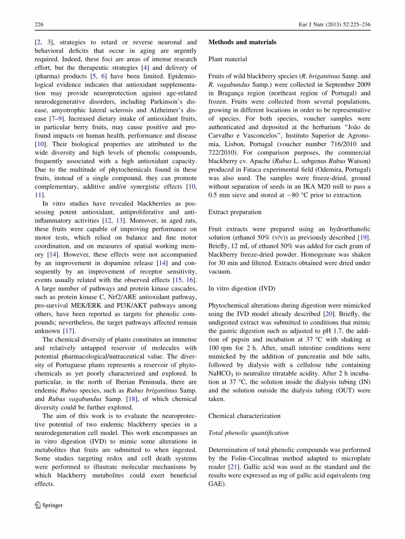
Results
Characterization of the blackberry extracts
Characterization of the three blackberry extracts was per-
formed, and although the total phenolic content (TPC) of
the three blackberries was very similar, their antioxidant
capacity (AC) was different (Table 1). The wild black-
berries had a higher AC compared to the commercial
variety, especially R. brigantinus that had 60% higher
antioxidant capacity than the commercial blackberry.
The in vitro digestion model provided two fractions (IN
and OUT) after pancreatic digestion. The fraction that
passes through the dialysis membrane constitutes the IN
fraction and contains metabolites that equate to those that
should be able to reach serum by paracellular transport.
The material that remains outside the dialysis tubing con-
stitutes the OUT fraction and contains metabolites that
equate to those that reach colon after digestion. After IVD,
the TPC and AC were greatly changed (Table 1), and the
TPC of the IN fractions was reduced to less than 10% of
the original content. The AC values were also reduced in
all samples, but since the reduction was lower than in the
values of TPC, the ratio AC/TPC became higher in the
IN fractions comparatively to the undigested extracts
(Table 1). Concerning these ratios for the IN fraction,
R. brigantinus was the most potent (222 lmol TE mg-1 GAE)
followed by the commercial blackberry (100 lmol TE
mg-1 GAE) and then by R. vagabundus (44 lmol TE mg-1
GAE; Table 1). Moreover, IN fractions had a higher ratio
of AC/TPC than the respective OUT fractions (results not
shown). Since IN fraction equates to the compounds that
could potentially reach the serum through paracellular
transport and at the same time, this fraction seems to be the
most chemically reactive (higher AC/TPC), these factors
led us to choose the IN fraction to be tested in neuropro-
tective studies.
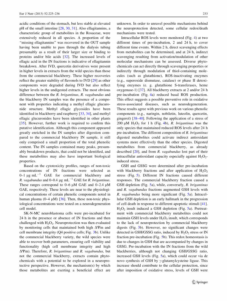
LC–MS analysis (Fig. S1 and Table S1 on Supplemen-
tary material) showed that the polyphenol profiles of the
wild blackberries before digestion were similar to that of
the commercial blackberry (Fig. S1A on Supplementary
material and Fig. 1a). The main differences were in the
amount of anthocyanins (cyanidin-3-O-glucoside, -xylo-
side and -hydroxymethyl-glutaroyl glucoside), which were
lower for the endemic species, and in the quantity of
Table 1 Chemical characterization of commercial blackberry and two wild blackberry species (R. brigantinus and R. vagabundus) extracts
before and after in vitro digestion
Undigested extract IN fraction
TPC (mg GAE
g-1 dw)
AC (lmol TE
g-1 dw)
AC/TPC (lmol TE
mg-1 GAE)
TPC (% of
undigested extract)
AC (% of
undigested extract)
AC/TPC (% of
undigested extract)
Commercial blackberry 27.51 ± 0.98a 221 ± 22c 33 ± 2a 5.5 17 307
R. brigantinus 28.17 ± 3.65a 357 ± 10a 21 ± 3b 0.5 5 1,040
R. vagabundus 31.07 ± 1.47a 274 ± 57b 15 ± 3b 1.7 5 292
For undigested extracts and IN fractions, the total phenolic content (TPC) and antioxidant capacity for peroxyl radical (AC) were determinate and
the ratio AC/TPC was calculated. Values of TPC, AC and AC/TPC in the undigested extract were expressed as mg GAE g-1 dw, lmol TE g-1
dw and lmol TE mg-1 GAE, respectively. Values in the IN fraction were expressed as % of the values determined for the undigested extract.
Statistical significant differences for p \ 0.05 are denoted with different letters (a–c). All values are mean ± SD, n = 3
228 Eur J Nutr (2013) 52:225–236
123
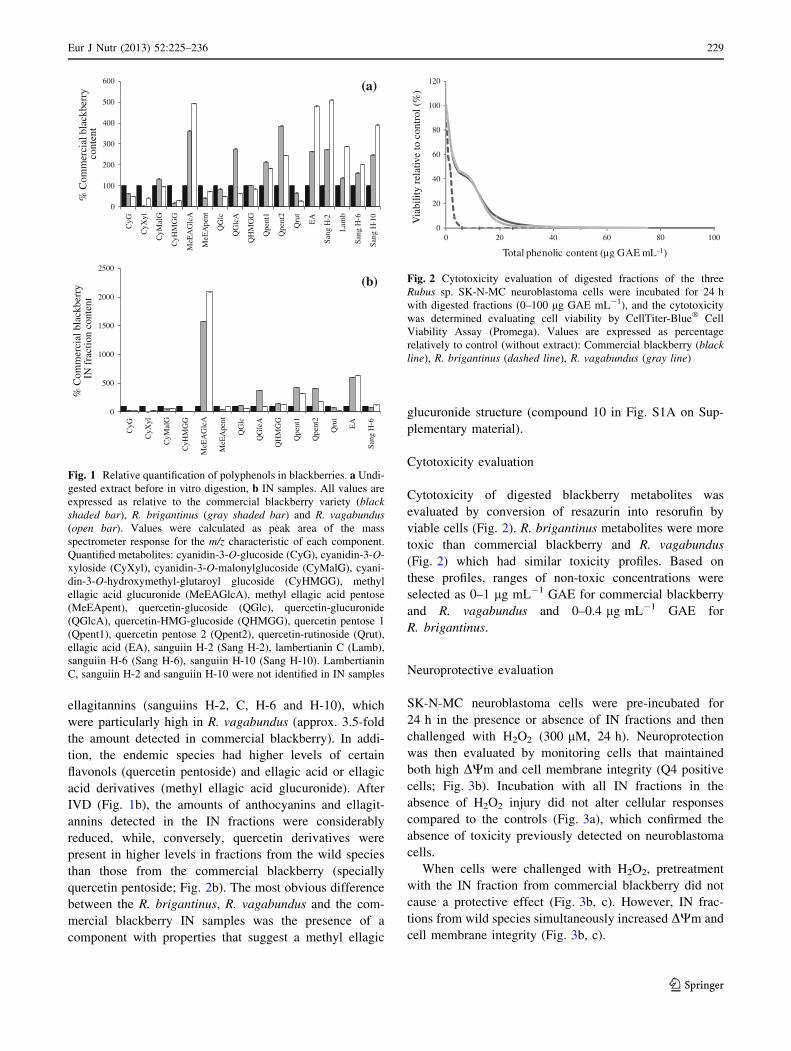
ellagitannins (sanguiins H-2, C, H-6 and H-10), which
were particularly high in R. vagabundus (approx. 3.5-fold
the amount detected in commercial blackberry). In addi-
tion, the endemic species had higher levels of certain
flavonols (quercetin pentoside) and ellagic acid or ellagic
acid derivatives (methyl ellagic acid glucuronide). After
IVD (Fig. 1b), the amounts of anthocyanins and ellagit-
annins detected in the IN fractions were considerably
reduced, while, conversely, quercetin derivatives were
present in higher levels in fractions from the wild species
than those from the commercial blackberry (specially
quercetin pentoside; Fig. 2b). The most obvious difference
between the R. brigantinus, R. vagabundus and the com-
mercial blackberry IN samples was the presence of a
component with properties that suggest a methyl ellagic
glucuronide structure (compound 10 in Fig. S1A on Sup-
plementary material).
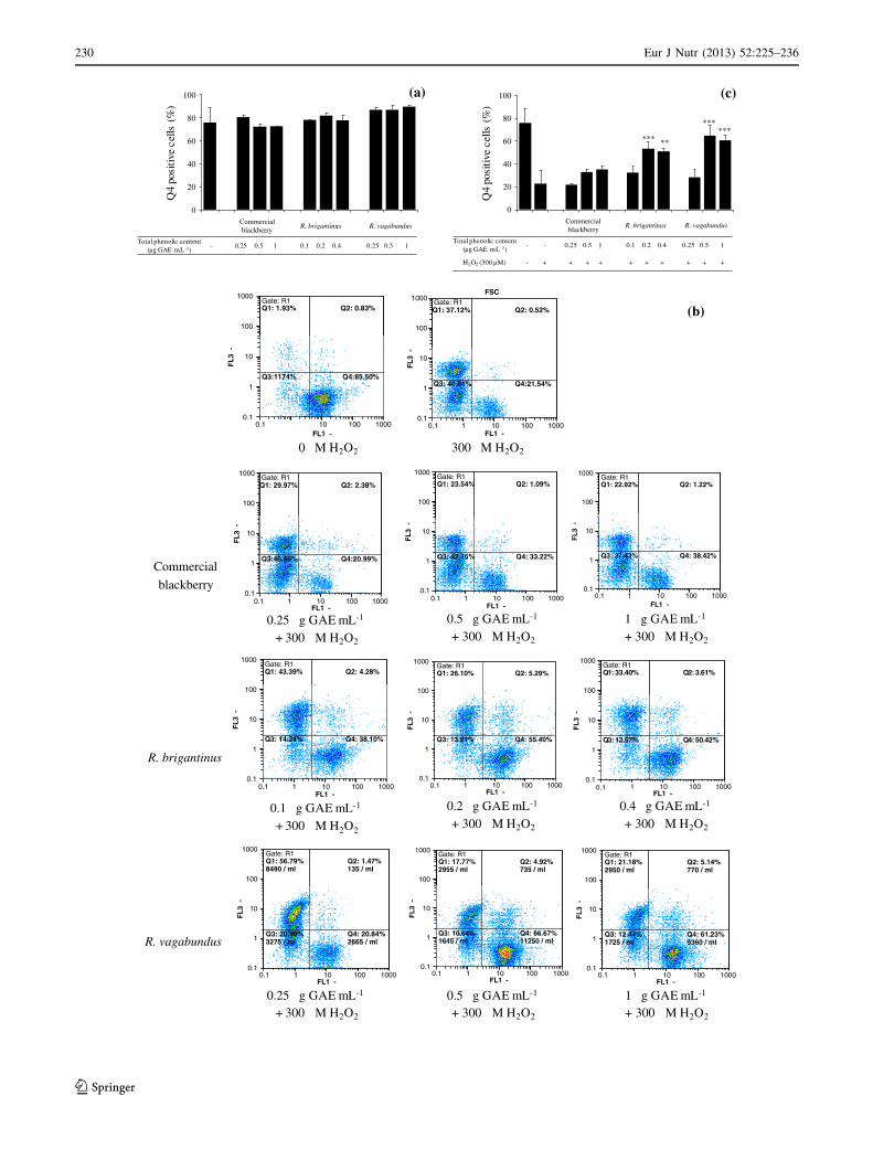
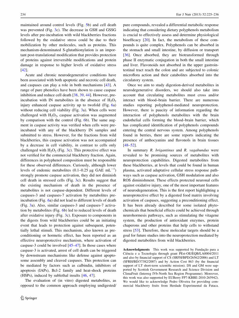
Cytotoxicity evaluation
Cytotoxicity of digested blackberry metabolites was
evaluated by conversion of resazurin into resorufin by
viable cells (Fig. 2). R. brigantinus metabolites were more
toxic than commercial blackberry and R. vagabundus
(Fig. 2) which had similar toxicity profiles. Based on
these profiles, ranges of non-toxic concentrations were
selected as 0–1 lg mL-1 GAE for commercial blackberry
and R. vagabundus and 0–0.4 lg mL-1 GAE for
R. brigantinus.
Neuroprotective evaluation
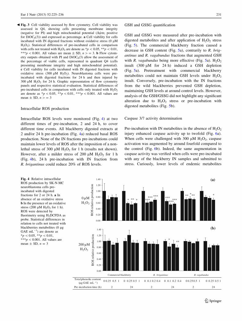
SK-N-MC neuroblastoma cells were pre-incubated for
24 h in the presence or absence of IN fractions and then
challenged with H2O2 (300 lM, 24 h). Neuroprotection
was then evaluated by monitoring cells that maintained
both high DWm and cell membrane integrity (Q4 positive
cells; Fig. 3b). Incubation with all IN fractions in the
absence of H2O2 injury did not alter cellular responses
compared to the controls (Fig. 3a), which confirmed the
absence of toxicity previously detected on neuroblastoma
cells.
When cells were challenged with H2O2, pretreatment
with the IN fraction from commercial blackberry did not
cause a protective effect (Fig. 3b, c). However, IN frac-
tions from wild species simultaneously increased DWm and
cell membrane integrity (Fig. 3b, c).
0
100
200
300
400
500
600
CyG
CyX
yl
CyM
alG
CyH
MG
G
MeE
AG
lcA
MeE
Ape
nt
QG
lc
QG
lcA
QH
MG
G
Qpe
nt1
Qpe
nt2
Qru
t
EA
Sang
H-2
Lam
b
Sang
H-6
Sang
H-1
0
% C
omm
erci
al b
lack
berr
y %
Com
mer
cial
bla
ckbe
rry
(a)
0
500
1000
1500
2000
2500
CyG
CyX
yl
CyM
alG
CyH
MG
G
MeE
AG
lcA
MeE
Ape
nt
QG
lc
QG
lcA
QH
MG
G
Qpe
nt1
Qpe
nt2
Qru
t
EA
Sang
H-6
IN
fra
ctio
n co
nten
t
(b)
cont
ent
Fig. 1 Relative quantification of polyphenols in blackberries. a Undi-
gested extract before in vitro digestion, b IN samples. All values are
expressed as relative to the commercial blackberry variety (blackshaded bar), R. brigantinus (gray shaded bar) and R. vagabundus(open bar). Values were calculated as peak area of the mass
spectrometer response for the m/z characteristic of each component.
Quantified metabolites: cyanidin-3-O-glucoside (CyG), cyanidin-3-O-
xyloside (CyXyl), cyanidin-3-O-malonylglucoside (CyMalG), cyani-
din-3-O-hydroxymethyl-glutaroyl glucoside (CyHMGG), methyl
ellagic acid glucuronide (MeEAGlcA), methyl ellagic acid pentose
(MeEApent), quercetin-glucoside (QGlc), quercetin-glucuronide
(QGlcA), quercetin-HMG-glucoside (QHMGG), quercetin pentose 1
(Qpent1), quercetin pentose 2 (Qpent2), quercetin-rutinoside (Qrut),
ellagic acid (EA), sanguiin H-2 (Sang H-2), lambertianin C (Lamb),
sanguiin H-6 (Sang H-6), sanguiin H-10 (Sang H-10). Lambertianin
C, sanguiin H-2 and sanguiin H-10 were not identified in IN samples
0
20
40
60
80
100
120
0 20 40 60 80 100
Via
bilit
y re
lativ
e to
con
trol
(%
)
Total phenolic content (µg GAE mL-1)
Fig. 2 Cytotoxicity evaluation of digested fractions of the three
Rubus sp. SK-N-MC neuroblastoma cells were incubated for 24 h
with digested fractions (0–100 lg GAE mL-1), and the cytotoxicity
was determined evaluating cell viability by CellTiter-Blue� Cell
Viability Assay (Promega). Values are expressed as percentage
relatively to control (without extract): Commercial blackberry (blackline), R. brigantinus (dashed line), R. vagabundus (gray line)
Eur J Nutr (2013) 52:225–236 229
123
Commercial blackberry
R. brigantinus R. vagabundus
Total phenolic content (µg GAE mL-1)
- 0.25 0.5 1 0.1 0.2 0.4 0.25 0.5 1
0
20
40
60
80
100
Q4
posi
tive
cel
ls (
%)
(a)
Commercial blackberry
R. brigantinus R. vagabundus
Total phenolic content (µg GAE mL-1)
- - 0.25 0.5 1 0.1 0.2 0.4 0.25 0.5 1
H2O2 (300 µM) - + + + + + + + + + +
0
20
40
60
80
100
Q4
posi
tive
cel
ls (
%)
***
******
**
(c)
FSC1000
0.10.1
1
10
100
1000
FL
3 -
0.10.1
1
10
100
FL
3 -
(b)
1 10 100 1000FL1 -
Gate: R1
Gate: R1Gate: R1
10
100
1000
FL
3 -
Gate: R1
10
100
1000
FL
3 -
Gate: R1
10
100
1000
FL
3 -
10 100 1000FL1 -
0 µM H2O2 300 µM H2O2
0.1 1 10 100 10000.1
1
FL1 -0.1 1 10 100 1000
0.1
1
FL1 -0.1 1 10 100 1000
0.1
1
FL1 -
Commercial
blackberry
0.25 µg GAE mL-1
+ 300 µM H2O2
0.5 µg GAE mL-1
+ 300 µM H2O2
1 µg GAE mL-1
+ 300 µM H2O2
Gate: R1
0.10.1
1
10
100
1000
FL
3 -
Gate: R1
0.10.1
1
10
100
1000
FL
3 -
Gate: R1
0.10.1
1
10
100
1000
FL
3 -
Q1: 37.12% Q2: 0.52%
Q3: 40.81% Q4:21.54%
Q1: 1.93% Q2: 0.83%
.Q3:1174% Q4:85.50%
Q1: 29.97% Q2: 2.38% Q1: 23.54% Q2: 1.09% Q1: 22.92% Q2: 1.22%
Q3:46.66% Q4:20.99% Q3: 42.15% Q4: 33.22% Q3: 37.43% Q4: 38.42%
Q1: 43.39% Q2: 4.28%
Q3: 14.24% Q4: 38.10%
Q1: 26.10% Q2: 5.29%
Q3: 13.21% Q4: 55.40%
Q1: 33.40% Q2: 3.61%
Q3: 12.57% Q4: 50.42%
R. brigantinus
1 10 100 1000FL1 -
1 10 100 1000FL1 -
1 10 100 1000FL1 -
Gate: R1
100
1000
-
Q1: 56.79%8490 / ml
Q2: 1.47%135 / ml
Gate: R1
100
1000
-
Q1: 17.77%2955 / ml
Q2: 4.92%735 / ml
Gate: R1
100
1000
-
Q1: 21.18%2950 / ml
Q2: 5.14%770 / ml
0.1 µg GAE mL-1
+ 300 µM H2O2
0.2 µg GAE mL-1
+ 300 µM H2O2
0.4 µg GAE mL-1
+ 300 µM H2O2
0.1 1 10 100 10000.1
1
10
FL1 -
FL
3
Q3: 20.90%3275 / ml
Q4: 20.84%2665 / ml
0.1 1 10 100 10000.1
1
10
FL1 -
FL
3
Q3: 10.64%1645 / ml
Q4: 66.67%11250 / ml
0.1 1 10 100 10000.1
1
10
FL1 -
FL
3
Q3: 12.44%1725 / ml
Q4: 61.23%9360 / mlR. vagabundus
0.25 µg GAE mL-1
+ 300 µM H2O2
0.5 µg GAE mL-1
+ 300 µM H2O2
1 µg GAE mL-1
+ 300 µM H2O2
230 Eur J Nutr (2013) 52:225–236
123
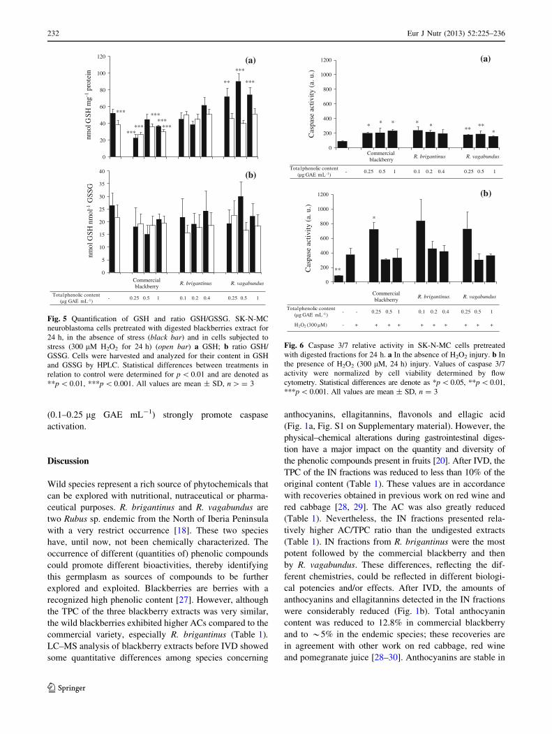
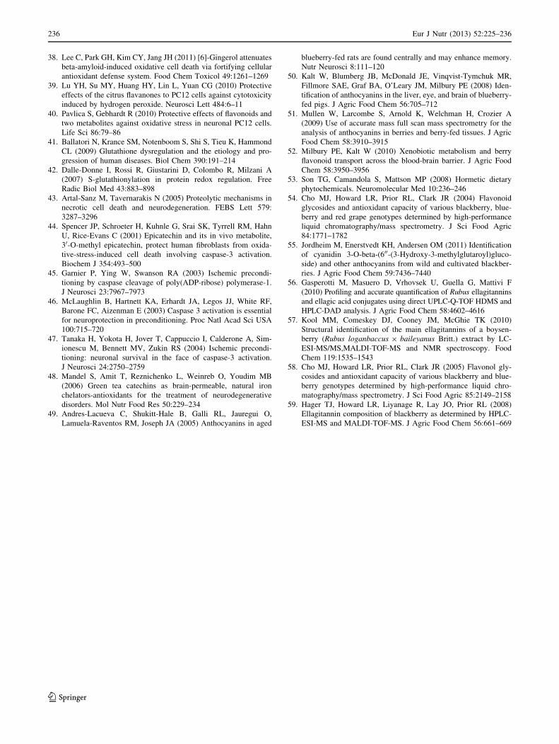
Intracellular ROS production
Intracellular ROS levels were monitored (Fig. 4) at two
different times of pre-incubation, 2 and 24 h, to cover
different time events. All blackberry digested extracts at
2 and/or 24 h pre-incubation (Fig. 4a) reduced basal ROS
production. None of the IN fractions pre-incubations could
maintain lower levels of ROS after the imposition of a non-
lethal stress of 300 lM H2O2 for 1 h (results not shown).
However, after a milder stress of 200 lM H2O2 for 1 h
(Fig. 4b), 24 h pre-incubation with IN fraction from
R. brigantinus could reduce 20% of ROS levels.
GSH and GSSG quantification
GSH and GSSG were measured after pre-incubation with
digested metabolites and after application of H2O2 stress
(Fig. 5). The commercial blackberry fraction caused a
decrease in GSH content (Fig. 5a), contrarily to R. brig-
antinus and R. vagabundus fractions that augmented GSH
with R. vagabundus being more effective (Fig. 5a). H2O2
insult (300 lM for 24 h) induced a GSH depletion
(Fig. 5a). Pretreatment with commercial blackberry
metabolites could not maintain GSH levels under H2O2
insult. Conversely, pre-incubation with the IN fractions
from the wild blackberries prevented GSH depletion,
maintaining GSH levels at around control levels. However,
analysis of the GSH/GSSG did not highlight any significant
alteration due to H2O2 stress or pre-incubation with
digested metabolites (Fig. 5b).
Caspase 3/7 activity determination
Pre-incubation with IN metabolites in the absence of H2O2
injury enhanced caspase activity up to twofold (Fig. 6a).
When cells were challenged with 300 lM H2O2, caspase
activation was augmented by around fourfold compared to
the control (Fig. 6b). Indeed, the same augmentation in
caspase activity was verified when cells were pre-incubated
with any of the blackberry IN samples and submitted to
stress. Curiously, lower levels of endemic metabolites
Commercial blackberry R. brigantinus R. vagabundus
Total phenolic content(µg GAE mL -1)
0 0.25 0.5 1 0 0.25 0.5 1 0 0.1 0.2 0.4 0 0.1 0.2 0.4 0 0.25 0.5 1 0 0.25 0.5 1
Pre-incubation time (h) 2 24 2 24 2 24
0 µM H2O2
200 µM H2O2
0.00
0.20
0.40
0.60
0.80
1.00
1.20
1.40
RO
S re
lativ
e pr
oduc
tion
* ** * ****** *** ***
******** *** *
0.00
0.20
0.40
0.60
0.80
1.00
1.20
1.40
RO
S re
lativ
e pr
oduc
tion
***
(a)
(b)
Fig. 4 Relative intracellular
ROS production by SK-N-MC
neuroblastoma cells pre-
incubated with digested
fractions for 2 or 24 h. a In
absence of an oxidative stress
b In the presence of an oxidative
stress (200 lM H2O2 for 1 h).
ROS were detected by
fluorimetry using H2DCFDA as
probe. Statistical differences in
relation to cells not treated with
blackberries metabolites (0 lg
GAE mL-1) are denote as
*p \ 0.05, **p \ 0.01,
***p \ 0.001. All values are
mean ± SD, n = 3
Fig. 3 Cell viability assessed by flow cytometry. Cell viability was
assessed in Q4, showing cells presenting membrane integrity
(negative for PI) and high mitochondrial potential (Dwm; positive
for DiOC6(3)) and expressed as percentage. a Cell viability for cells
incubated with IN digested fractions without oxidative stress (0 lM
H2O2). Statistical differences of pre-incubated cells in comparison
with cells not treated with H2O2 are denote as *p \ 0.05, **p \ 0.01,
***p \ 0.001. All values are mean ± SD, n [ = 3. b Flow cytom-
etry outputs obtained with PI and DiOC6(3) allow the assessment of
the percentage of viable cells, represented in quadrant Q4 (cells
presenting membrane integrity and high mitochondrial potential).
c Cell viability for cells incubated with IN digested fractions with
oxidative stress (300 lM H2O2). Neuroblastoma cells were pre-
incubated with digested fractions for 24 h and then injured by
300 lM H2O2 for 24 h. Graphic representation of flow cytometry
panels and respective statistical evaluation. Statistical differences of
pre-incubated cells in comparison with cells only treated with H2O2
are denote as *p \ 0.05, **p \ 0.01, ***p \ 0.001. All values are
mean ± SD, n [ = 3
b
Eur J Nutr (2013) 52:225–236 231
123
(0.1–0.25 lg GAE mL-1) strongly promote caspase
activation.
Discussion
Wild species represent a rich source of phytochemicals that
can be explored with nutritional, nutraceutical or pharma-
ceutical purposes. R. brigantinus and R. vagabundus are
two Rubus sp. endemic from the North of Iberia Peninsula
with a very restrict occurrence [18]. These two species
have, until now, not been chemically characterized. The
occurrence of different (quantities of) phenolic compounds
could promote different bioactivities, thereby identifying
this germplasm as sources of compounds to be further
explored and exploited. Blackberries are berries with a
recognized high phenolic content [27]. However, although
the TPC of the three blackberry extracts was very similar,
the wild blackberries exhibited higher ACs compared to the
commercial variety, especially R. brigantinus (Table 1).
LC–MS analysis of blackberry extracts before IVD showed
some quantitative differences among species concerning
anthocyanins, ellagitannins, flavonols and ellagic acid
(Fig. 1a, Fig. S1 on Supplementary material). However, the
physical–chemical alterations during gastrointestinal diges-
tion have a major impact on the quantity and diversity of
the phenolic compounds present in fruits [20]. After IVD, the
TPC of the IN fractions was reduced to less than 10% of the
original content (Table 1). These values are in accordance
with recoveries obtained in previous work on red wine and
red cabbage [28, 29]. The AC was also greatly reduced
(Table 1). Nevertheless, the IN fractions presented rela-
tively higher AC/TPC ratio than the undigested extracts
(Table 1). IN fractions from R. brigantinus were the most
potent followed by the commercial blackberry and then
by R. vagabundus. These differences, reflecting the dif-
ferent chemistries, could be reflected in different biologi-
cal potencies and/or effects. After IVD, the amounts of
anthocyanins and ellagitannins detected in the IN fractions
were considerably reduced (Fig. 1b). Total anthocyanin
content was reduced to 12.8% in commercial blackberry
and to *5% in the endemic species; these recoveries are
in agreement with other work on red cabbage, red wine
and pomegranate juice [28–30]. Anthocyanins are stable in
0
20
40
60
80
100
120
nmol
GS
H m
g-1pr
otei
n
***
***
***
**
***
***
***
***
***
(a)
0
5
10
15
20
25
30
35
40
nmol
GS
H n
mol
- 1G
SS
G
(b)
Commercial blackberry
R. brigantinus R. vagabundus
Total phenolic content(µg GAE mL-1)
- 0.25 0.5 1 0.1 0.2 0.4 0.25 0.5 1
Fig. 5 Quantification of GSH and ratio GSH/GSSG. SK-N-MC
neuroblastoma cells pretreated with digested blackberries extract for
24 h, in the absence of stress (black bar) and in cells subjected to
stress (300 lM H2O2 for 24 h) (open bar) a GSH; b ratio GSH/
GSSG. Cells were harvested and analyzed for their content in GSH
and GSSG by HPLC. Statistical differences between treatments in
relation to control were determined for p \ 0.01 and are denoted as
**p \ 0.01, ***p \ 0.001. All values are mean ± SD, n [ = 3
0
200
400
600
800
1000
1200
Cas
pase
act
ivity
(a.
u.)
* * * * **
****
(a)
Commercial blackberry
R. brigantinus R. vagabundus
Total phenolic content (µg GAE mL-1)
- 0.25 0.5 1 0.1 0.2 0.4 0.25 0.5 1
Commercial blackberry
R. brigantinus R. vagabundus
Total phenolic content (µg GAE mL-1)
- - 0.25 0.5 1 0.1 0.2 0.4 0.25 0.5 1
H2O2 (300 µM) - + + + + + + + + + +
0
200
400
600
800
1000
1200
Cas
pase
act
ivity
(a.
u.)
**
*
(b)
Fig. 6 Caspase 3/7 relative activity in SK-N-MC cells pretreated
with digested fractions for 24 h. a In the absence of H2O2 injury. b In
the presence of H2O2 (300 lM, 24 h) injury. Values of caspase 3/7
activity were normalized by cell viability determined by flow
cytometry. Statistical differences are denote as *p \0.05, **p \0.01,
***p\ 0.001. All values are mean ± SD, n = 3
232 Eur J Nutr (2013) 52:225–236
123
acidic conditions of the stomach, but less stable at elevated
pH of the small intestine [28, 30, 31]. Also ellagitannins, a
characteristic group of metabolites in the Rosaceae, were
extensively reduced in all species. A proportion of the
‘‘missing ellagitannins’’ was recovered in the OUT sample
having been unable to pass through the dialysis tubing
presumably as a result of their larger size or binding to
proteins and/or bile acids [32]. The increased levels of
ellagic acid in the IN fractions is indicative of ellagitannin
breakdown. After IVD, quercetin derivatives were present
in higher levels in extracts from the wild species than those
from the commercial blackberry. These higher recoveries
reflect the greater stability of flavonols to IVD [29] as other
components were degraded during IVD but also reflect
higher levels in the undigested extracts. The most obvious
difference between the R. brigantinus, R. vagabundus and
the blackberry IN samples was the presence of a compo-
nent with properties indicating a methyl ellagic glucuro-
nide structure. Methyl ellagic glycosides have been
identified in blackberry and raspberry [33, 34], and methyl
ellagic glucuronides have been identified in other plants
[35]. However, further work is required to confirm this
putative identification. Although this component appeared
greatly enriched in the IN samples after digestion com-
pared to the commercial blackberry IN sample, it still
only comprised a small proportion of the total phenolic
content. The IN samples contained many peaks, presum-
ably breakdown products, that could not be identified, and
these metabolites may also have important biological
properties.
Based on the cytotoxicity profiles, ranges of non-toxic
concentrations of IN fractions were selected as
0–1 lg mL-1 GAE for commercial blackberry and
R. vagabundus and 0–0.4 lg mL-1 GAE for R. brigantinus.
These ranges correspond to 0–6 lM GAE and 0–2.4 lM
GAE, respectively. These levels are near to the physiologi-
cal concentrations of certain phenolic components noted in
human plasma (0–4 lM) [36]. Then, these non-toxic phys-
iological concentrations were tested on a neurodegeneration
cell model.
SK-N-MC neuroblastoma cells were pre-incubated for
24 h in the presence or absence of IN fractions and then
challenged with H2O2. Neuroprotection was then evaluated
by monitoring cells that maintained both high DWm and
cell membrane integrity (Q4 positive cells; Fig. 3b). Unlike
the commercial blackberry variety, the wild species were
able to recover both parameters, ensuring cell viability and
functionality (high cell membrane integrity and high
DWm). Therefore, R. brigantinus and R. vagabundus, but
not the commercial blackberry, extracts contain phyto-
chemicals with a potential to be explored in a neuropro-
tective perspective. However, the mechanism(s) by which
those metabolites are exerting a beneficial effect are
unknown. In order to unravel possible mechanisms behind
the neuroprotection detected, some cellular redox/death
mechanisms were tested.
Intracellular ROS levels were monitored (Fig. 4) at two
different times of pre-incubation, 2 and 24 h, to cover
different time events. Within 2 h, direct scavenging effects
from metabolites can be determined, and at 24 h, indirect
scavenging resulting from activation/modulation of other
molecular mechanisms can be assessed. Diverse phyto-
chemicals can act directly through scavenging properties or
indirectly through modulation of thiol-containing mole-
cules (such as glutathione), ROS-inactivating enzymes
(e.g., superoxide dismutase, catalase) or phase II detoxi-
fying enzymes (e. g. glutathione S-transferases, heme
oxygenase-1) [37]. All blackberry extracts at 2 and/or 24 h
pre-incubation (Fig. 4a) reduced basal ROS production.
This effect suggests a possible preventive role in oxidative
stress-associated diseases, such as neurodegeneration.
These results agree with previous work on various phenolic
components (e.g., naringin, nobiletin, luteolin, quercetin,
gingerol) [38–40]. Following the application of a stress of
200 lM H2O2 for 1 h (Fig. 4b), R. brigantinus was the
only species that maintained reduced ROS levels after 24 h
pre-incubation. The different composition of R. brigantinus
digested metabolites could activate indirect antioxidant
systems more effectively than the other species. Digested
metabolites from commercial blackberry, as already
described [20], and from R. vagabundus lost part of their
intracellular antioxidant capacity especially against H2O2-
induced stress.
GSH and GSSG were determined after pre-incubation
with blackberry fractions and after application of H2O2
stress (Fig. 5). Different IN fractions caused different
responses. The commercial blackberry fraction caused a
GSH depletion (Fig. 5a), while, conversely, R. brigantinus
and R. vagabundus fractions augmented GSH levels with
R. vagabundus being more significant (Fig. 5a). Intracel-
lular GSH depletion is an early hallmark in the progression
of cell death in response to different apoptotic stimuli [41].
H2O2 insult induced a GSH depletion (Fig. 5a). Pretreat-
ment with commercial blackberry metabolites could not
maintain GSH levels under H2O2 insult, which corresponds
to the lack of neuroprotection by commercial blackberry
digests (Fig. 3b). However, no significant changes were
detected in GSH/GSSG ratio, induced by H2O2 stress or IN
fraction pre-incubation (Fig. 5b). This redox homeostasis is
due to changes in GSH that are accompanied by changes in
GSSG. Pre-incubation with the IN fractions from the wild
blackberries, although not changing GSH/GSSG ratio,
increased GSH levels (Fig. 5a), which could occur via de
novo synthesis of GSH by c-glutamylcysteine ligase. This
increase should contribute to the cellular protection, since
after imposition of oxidative stress, levels of GSH were
Eur J Nutr (2013) 52:225–236 233
123
maintained around control levels (Fig. 5b) and cell death
was prevented (Fig. 3c). The decrease in GSH and GSSG
levels after pre-incubation with wild blackberries fractions
followed by the oxidative stress could be due to their
mobilization by other molecules, such as proteins. This
mechanism-denominated S-glutathionylation is an impor-
tant post-translational modification that provides protection
of proteins against irreversible modifications and protein
damage in response to higher levels of oxidative stress
[42].
Acute and chronic neurodegenerative conditions have
been associated with both apoptotic and necrotic cell death,
and caspases can play a role in both mechanisms [43]. A
range of pure phenolics have been shown to cause caspase
inhibition and reduce cell death [38, 39, 44]. However, pre-
incubation with IN metabolites in the absence of H2O2
injury enhanced caspase activity up to twofold (Fig. 6a)
without reducing cell viability (Fig. 3a). When cells were
challenged with H2O2, caspase activation was augmented
by comparison with the control (Fig. 6b). The same aug-
ment in caspase activity was verified when cells were pre-
incubated with any of the blackberry IN samples and
submitted to stress. However, for the fractions from wild
blackberries, this caspase activation was not accompanied
by a decrease in cell viability, in contrast to cells only
challenged with H2O2 (Fig. 3c). This protective effect was
not verified for the commercial blackberry fraction. Again,
differences in polyphenol composition must be responsible
for these observed differences. Curiously, although lower
levels of endemic metabolites (0.1–0.25 lg GAE mL-1)
strongly promote caspase activation, they did not diminish
cell death in stressed cells (Fig. 3c). Results suggest that
the existing mechanism of death in the presence of
metabolites is not caspase-dependent. Different levels of
caspases-3 and caspases-7 activation by metabolites pre-
incubation (Fig. 6a) did not lead to different levels of death
(Fig. 3a). Also, similar caspases-3 and caspases-7 activa-
tion by metabolites (Fig. 6b) led to reduced levels of death
after oxidative injury (Fig. 3c). Exposure to components in
the digests from wild blackberries could be an initiating
event that leads to protection against subsequent, poten-
tially lethal stimuli. This mechanism, also known as pre-
conditioning or hormetic effect, has been reported as an
effective neuroprotective mechanism, where activation of
caspase-3 could be involved [45–47]. In those cases where
caspase-3 is activated, arrest of cell death can be triggered
by downstream mechanisms like defense against apopto-
some assembly and cleaved caspases. This protection can
be mediated by factors such as calbindin, inhibitors of
apoptosis (IAPs), Bcl-2 family and heat-shock proteins
(HSPs), induced by sublethal insults [46, 47].
The evaluation of (in vitro) digested metabolites, as
opposed to the common approach employing undigested/
pure compounds, revealed a differential metabolic response
indicating that considering dietary polyphenols metabolism
is crucial to effectively assess and determine physiological
bioefficacy [20]. In fact, the metabolism of these com-
pounds is quite complex. Polyphenols can be absorbed in
the stomach and small intestine, by diffusion or transport
[36]. Once absorbed, they are biotransformed through
phase II enzymatic conjugation in both the small intestine
and liver. Flavonoids not absorbed in the upper gastroin-
testinal tract reach the colon and are subjected to colonic
microflora action and their catabolites absorbed into the
circulatory system.
Once we aim to study digestion-derived metabolites in
neurodegenerative disorders, we should also take into
account that circulating metabolites must cross and/or
interact with blood–brain barrier. There are numerous
studies reporting polyphenol-mediated neuroprotection.
However, there is paucity of information regarding the
interaction of polyphenols metabolites with the brain
endothelial cells forming the blood–brain barrier, which
has complicated identification of polyphenols compounds
entering the central nervous system. Among polyphenols
found in berries, there are some reports indicating the
presence of anthocyanins and flavonols in brain tissues
[48–52].
In summary R. brigantinus and R. vagabundus were
revealed to be promising sources of metabolites with
neuroprotection capabilities. Digested metabolites from
these blackberries, at levels that could be found in human
plasma, activated adaptative cellular stress response path-
ways such as caspase activation, GSH modulation and also
ROS diminishment. These effects protected neuronal cells
against oxidative injury, one of the most important features
of neurodegeneration. This is the first report highlighting a
neuroprotective effect by a digested food matrix involving
activation of caspases, suggesting a preconditioning effect.
It has been already described for some isolated phyto-
chemicals that beneficial effects could be achieved through
neurohormesis pathways, such as stimulating the vitagene
system, the production of antioxidant enzymes, protein
chaperons and other proteins that help cells to withstand
stress [53]. Therefore, these molecular targets should be a
goal for future studies into the neuroprotection mediated by
digested metabolites from wild blackberries.
Acknowledgments This work was supported by Fundacao para a
Ciencia e a Tecnologia through grant PEst-OE/EQB/LA0004/2011
and also by financial support of CS (SRFH/BPD/26562/2006) and LT
(SFRH/BD/37382/2007) and by Action Cost 863 (by the financial
support of LT short-term scientific mission). DS and GM were sup-
ported by Scottish Government Research and Science Division and
ClimaFruit (Interreg IVb-North Sea Region Programme). Moreover,
this work was also supported by EUBerry FP7-KBBE-2010-265942).
We would like to acknowledge Pedro Oliveira for providing com-
mercial blackberry fruits from Herdade Experimental da Fataca.
234 Eur J Nutr (2013) 52:225–236
123
We also would like thank to Carlos Aguiar from CIMO, Instituto
Politecnico de Braganca for helping us to identify and collect the wild
species, to Cristina Silva Pereira for providing access to HPLC and
M. Cristina Leitao for the HPLC technical support.
References
1. Lau FC, Shukitt-Hale B, Joseph JA (2006) Beneficial effects of
berry fruit polyphenols on neuronal and behavioral aging. J Agric
Food Chem 86:2251–2255
2. Beking K, Vieira A (2010) Flavonoid intake and disability-
adjusted life years due to Alzheimer’s and related dementias: a
population-based study involving twenty-three developed coun-
tries. Public Health Nutr 13:1403–1409
3. Wimo A, Jonsson L, Gustavsson A, McDaid D, Ersek K, Georges
J, Gulacsi L, Karpati K, Kenigsberg P, Valtonen H (2011) The
economic impact of dementia in Europe in 2008-cost estimates
from the Eurocode project. Int J Geriatr Psychiatry 26:825–832
4. Maiese K, Chong ZZ, Hou J, Shang YC (2009) New strategies for
Alzheimer’s disease and cognitive impairment. Oxid Med Cell
Longev 2:279–289
5. Dumont M, Beal MF (2011) Neuroprotective strategies involving
ROS in Alzheimer disease. Free Radic Biol Med 51:1014–1026
6. Williams P, Sorribas A, Howes MJR (2011) Natural products as a
source of Alzheimer’s drug leads. Nat Prod Rep 28:48–77
7. de Rijk MC, Breteler MM, den Breeijen JH, Launer LJ, Grobbee
DE, van der Meche FG, Hofman A (1997) Dietary antioxidants
and Parkinson disease. The Rotterdam study. Arch Neurol
54:762–765
8. Di Matteo V, Esposito E (2003) Biochemical and therapeutic
effects of antioxidants in the treatment of Alzheimer’s disease,
Parkinson’s disease, and amyotrophic lateral sclerosis. Curr Drug
Targets CNS Neurol Disord 2:95–107
9. Dai Q, Borenstein AR, Wu Y, Jackson JC, Larson EB (2006)
Fruit and vegetable juices and Alzheimer’s disease: the Kame
project. Am J Med 119:751–759
10. Seeram NP (2008) Berry fruits: compositional elements, bio-
chemical activities, and the impact of their intake on human
health, performance, and disease. J Agric Food Chem 56:627–629
11. Shukitt-Hale B, Lau FC, Joseph JA (2008) Berry fruit supple-
mentation and the aging brain. J Agric Food Chem 56:636–641
12. Dai J, Patel JD, Mumper RJ (2007) Characterization of black-
berry extract and its antiproliferative and anti-inflammatory
properties. J Med Food 10:258–265
13. Wang SY, Jiao H (2000) Scavenging capacity of berry crops on
superoxide radicals, hydrogen peroxide, hydroxyl radicals, and
singlet oxygen. J Agric Food Chem 48:5677–5684
14. Shukitt-Hale B, Cheng V, Joseph JA (2009) Effects of black-
berries on motor and cognitive function in aged rats. Nutr Neu-
rosci 12:135–140
15. Joseph JA, Kowatch MA, Maki T, Roth GS (1990) Selective
cross-activation/inhibition of second messenger systems and the
reduction of age-related deficits in the muscarinic control of
dopamine release from perifused rat striata. Brain Res 537:40–48
16. Rutz S, Majchrzak M, Siedschlag V, Barbelivien A, Harati H,
Rothmaier AK, Feuerstein TJ, Jackisch R, Cassel JC (2009) The
modulation of striatal dopamine release correlates with water-
maze performance in aged rats. Neurobiol Aging 30:957–972
17. Ramassamy C (2006) Emerging role of polyphenolic compounds
in the treatment of neurodegenerative diseases: a review of their
intracellular targets. Eur J Pharmacol 545:51–64
18. Castroviejo S (ed) (1986–2007) Flora Iberica. Plantas vasculares
de la Penınsula Iberica e Islas Baleares. Real Jardın Botanico,
CSIC, Madrid
19. Tavares L, Fortalezas S, Carrilho C, McDougall GJ, Stewart D,
Ferreira RB, Santos CN (2010) Antioxidant and antiproliferative
properties of strawberry tree tissues. J Berry Res 1:3–12
20. Tavares L, Figueira I, Macedo D, McDougall GJ, Leitao MC,
Vieira HLA, Stewart D, Alves PM, Ferreira RB, Santos CN
(2012) Neuroprotective effect of blackberry (Rubus sp.) poly-
phenols is potentiated after simulated gastrointestinal digestion.
Food Chem 131:1443–1452
21. Tavares L, Carrilho D, Tyagi M, Barata D, Serra AT, Duarte
CMM, Duarte RO, Feliciano RP, Bronze MR, Chicau P, Espırito-
Santo MD, Ferreira RB, dos Santos CN (2010) Antioxidant
capacity of Macaronesian traditional medicinal plants. Molecules
15:2576–2592
22. Queiroga CS, Almeida AS, Martel C, Brenner C, Alves PM, Vieira
HL (2010) Glutathionylation of adenine nucleotide translocase
induced by carbon monoxide prevents mitochondrial membrane
permeabilization and apoptosis. J Biol Chem 285:17077–17088
23. Wang H, Joseph JA (1999) Quantifying cellular oxidative stress
by dichlorofluorescein assay using microplate reader. Free Radic
Biol Med 27:612–616
24. Wolfe KL, Liu RH (2007) Cellular antioxidant activity (CAA)
assay for assessing antioxidants, foods, and dietary supplements.
J Agric Food Chem 55:8896–8907
25. Kand’ar R, Zakova P, Lotkova H, Kucera O, Cervinkova Z
(2007) Determination of reduced and oxidized glutathione in
biological samples using liquid chromatography with fluorimetric
detection. J Pharm Biomed Anal 43:1382–1387
26. Bensadoun A, Weinstein D (1976) Assay of proteins in the
presence of interfering materials. Anal Biochem 70:241–250
27. Wolfe KL, Kang X, He X, Dong M, Zhang Q, Liu RH (2008)
Cellular antioxidant activity of common fruits. J Agric Food
Chem 56:8418–8426
28. McDougall GJ, Fyffe S, Dobson P, Stewart D (2005) Anthocy-
anins from red wine-their stability under simulated gastrointes-
tinal digestion. Phytochemistry 66:2540–2548
29. McDougall GJ, Fyffe S, Dobson P, Stewart D (2007) Anthocy-
anins from red cabbage-stability to simulated gastrointestinal
digestion. Phytochemistry 68:1285–1294
30. Perez-Vicente A, Gil-Izquierdo A, Garcia-Viguera C (2002)
In vitro gastrointestinal digestion study of pomegranate juice
phenolic compounds, anthocyanins, and vitamin C. J Agric Food
Chem 50:2308–2312
31. Gil-Izquierdo A, Gil MI, Ferreres F (2002) Effect of processing
techniques at industrial scale on orange juice antioxidant and
beneficial health compounds. J Agric Food Chem 50:5107–5114
32. Deaville ER, Green RJ, Mueller-Harvey I, Willoughby I, Frazier
RA (2007) Hydrolyzable tannin structures influence relative
globular and random coil protein binding strengths. J Agric Food
Chem 55:4554–4561
33. Mertz C, Cheynier V, Gunata Z, Brat P (2007) Analysis of
phenolic compounds in two blackberry species (Rubus glaucusand Rubus adenotrichus) by high-performance liquid chroma-
tography with diode array detection and electrospray ion trap
mass spectrometry. J Agric Food Chem 55:8616–8624
34. Mullen W, Yokota T, Lean MEJ, Crozier A (2003) Analysis of
ellagitannins and conjugates of ellagic acid and quercetin in
raspberry fruits by LC-MSn. Phytochemistry 64:617–624
35. Ayoub NA (2010) A trimethoxyellagic acid glucuronide from
Conocarpus erectus leaves: isolation, characterization and assay
of antioxidant capacity. Pharm Biol 48:328–332
36. Manach C, Williamson G, Morand C, Scalbert A, Remesy C (2005)
Bioavailability and bioefficacy of polyphenols in humans. I. Review
of 97 bioavailability studies. Am J Clin Nutr 81:230S–242S
37. Jung KA, Kwak MK (2010) The Nrf2 system as a potential target
for the development of indirect antioxidants. Molecules
15:7266–7291
Eur J Nutr (2013) 52:225–236 235
123
38. Lee C, Park GH, Kim CY, Jang JH (2011) [6]-Gingerol attenuates
beta-amyloid-induced oxidative cell death via fortifying cellular
antioxidant defense system. Food Chem Toxicol 49:1261–1269
39. Lu YH, Su MY, Huang HY, Lin L, Yuan CG (2010) Protective
effects of the citrus flavanones to PC12 cells against cytotoxicity
induced by hydrogen peroxide. Neurosci Lett 484:6–11
40. Pavlica S, Gebhardt R (2010) Protective effects of flavonoids and
two metabolites against oxidative stress in neuronal PC12 cells.
Life Sci 86:79–86
41. Ballatori N, Krance SM, Notenboom S, Shi S, Tieu K, Hammond
CL (2009) Glutathione dysregulation and the etiology and pro-
gression of human diseases. Biol Chem 390:191–214
42. Dalle-Donne I, Rossi R, Giustarini D, Colombo R, Milzani A
(2007) S-glutathionylation in protein redox regulation. Free
Radic Biol Med 43:883–898
43. Artal-Sanz M, Tavernarakis N (2005) Proteolytic mechanisms in
necrotic cell death and neurodegeneration. FEBS Lett 579:
3287–3296
44. Spencer JP, Schroeter H, Kuhnle G, Srai SK, Tyrrell RM, Hahn
U, Rice-Evans C (2001) Epicatechin and its in vivo metabolite,
30-O-methyl epicatechin, protect human fibroblasts from oxida-
tive-stress-induced cell death involving caspase-3 activation.
Biochem J 354:493–500
45. Garnier P, Ying W, Swanson RA (2003) Ischemic precondi-
tioning by caspase cleavage of poly(ADP-ribose) polymerase-1.
J Neurosci 23:7967–7973
46. McLaughlin B, Hartnett KA, Erhardt JA, Legos JJ, White RF,
Barone FC, Aizenman E (2003) Caspase 3 activation is essential
for neuroprotection in preconditioning. Proc Natl Acad Sci USA
100:715–720
47. Tanaka H, Yokota H, Jover T, Cappuccio I, Calderone A, Sim-
ionescu M, Bennett MV, Zukin RS (2004) Ischemic precondi-
tioning: neuronal survival in the face of caspase-3 activation.
J Neurosci 24:2750–2759
48. Mandel S, Amit T, Reznichenko L, Weinreb O, Youdim MB
(2006) Green tea catechins as brain-permeable, natural iron
chelators-antioxidants for the treatment of neurodegenerative
disorders. Mol Nutr Food Res 50:229–234
49. Andres-Lacueva C, Shukitt-Hale B, Galli RL, Jauregui O,
Lamuela-Raventos RM, Joseph JA (2005) Anthocyanins in aged
blueberry-fed rats are found centrally and may enhance memory.
Nutr Neurosci 8:111–120
50. Kalt W, Blumberg JB, McDonald JE, Vinqvist-Tymchuk MR,
Fillmore SAE, Graf BA, O’Leary JM, Milbury PE (2008) Iden-
tification of anthocyanins in the liver, eye, and brain of blueberry-
fed pigs. J Agric Food Chem 56:705–712
51. Mullen W, Larcombe S, Arnold K, Welchman H, Crozier A
(2009) Use of accurate mass full scan mass spectrometry for the
analysis of anthocyanins in berries and berry-fed tissues. J Agric
Food Chem 58:3910–3915
52. Milbury PE, Kalt W (2010) Xenobiotic metabolism and berry
flavonoid transport across the blood-brain barrier. J Agric Food
Chem 58:3950–3956
53. Son TG, Camandola S, Mattson MP (2008) Hormetic dietary
phytochemicals. Neuromolecular Med 10:236–246
54. Cho MJ, Howard LR, Prior RL, Clark JR (2004) Flavonoid
glycosides and antioxidant capacity of various blackberry, blue-
berry and red grape genotypes determined by high-performance
liquid chromatography/mass spectrometry. J Sci Food Agric
84:1771–1782
55. Jordheim M, Enerstvedt KH, Andersen OM (2011) Identification
of cyanidin 3-O-beta-(600-(3-Hydroxy-3-methylglutaroyl)gluco-
side) and other anthocyanins from wild and cultivated blackber-
ries. J Agric Food Chem 59:7436–7440
56. Gasperotti M, Masuero D, Vrhovsek U, Guella G, Mattivi F
(2010) Profiling and accurate quantification of Rubus ellagitannins
and ellagic acid conjugates using direct UPLC-Q-TOF HDMS and
HPLC-DAD analysis. J Agric Food Chem 58:4602–4616
57. Kool MM, Comeskey DJ, Cooney JM, McGhie TK (2010)
Structural identification of the main ellagitannins of a boysen-
berry (Rubus loganbaccus 9 baileyanus Britt.) extract by LC-
ESI-MS/MS,MALDI-TOF-MS and NMR spectroscopy. Food
Chem 119:1535–1543
58. Cho MJ, Howard LR, Prior RL, Clark JR (2005) Flavonol gly-
cosides and antioxidant capacity of various blackberry and blue-
berry genotypes determined by high-performance liquid chro-
matography/mass spectrometry. J Sci Food Agric 85:2149–2158
59. Hager TJ, Howard LR, Liyanage R, Lay JO, Prior RL (2008)
Ellagitannin composition of blackberry as determined by HPLC-
ESI-MS and MALDI-TOF-MS. J Agric Food Chem 56:661–669
236 Eur J Nutr (2013) 52:225–236
123