Embed Size (px)
Citation preview

T he neutron technique aims to answer various scientific questions about the fundamental properties of mate-
rials on an atomic scale, as well as medical applications on a nanometre scale. Researchers use neutron instruments to count scattered neutrons, measure their energies and the angles at which they scatter, and map their final position. In contrast to X-rays and electrons, which interact with the electrons around the atomic nuclei, the neutron interacts with those nuclei. As the neutrons are neutral, they are de-flected only when they strike a core. This capability allows scientists to glean details about the nature of materials ranging from single crystals to thermally isolating materials, from proteins to drug systems, from metals to magnets.
Our user communities performed the following remarkable studies; they completed their outstanding research at ANSTO using neutron instruments in 2020. Five reports include the highlights from high-performance non-linear optical mate-rials reported by Chin-Wei Wang. Two highlights from the SIKA instrument show the first inelastic neutron-scattering study for a SrRuO3 thin film capable of SIKA and the pho-non behaviour of high-zT Sb-doped GeTe thermoelectric materials reported by Chun-Ming Wu and Shin-ichiro Yano, respectively. A SANS study as a new strategy to combat LPS-deficient Gram-negative pathogens with nano-technology is reported by Xiangfeng Lai. Research using a neutron powder diffractometer for a magnetic frustration mate-rial, Mn1.5Cr1.5O4, is described by Chin-Wei Wang. The last texture study for advanced metallurgy reported by E-Wen Huang is based on experiments at the Spallation Neutron Source facility in Oak Ridge National Laboratory. (by Shih-Chun Chung)
Neutron Science

Neutron Science 075
Local Structural Distortion as a New Design Strategy for High-Performance Nonlinear Optical MaterialsA new tungsten bronze compound synthesized with a new molecular design strategy exhibits a strong SHG response, 39 times that of KDP.
N onlinear optical (NLO) materials play a crucial role in modern laser technology by effectively expanding the spectral range of a laser; they
have many important applications in photolithography, spectral analysis, tissue imaging, environmental monitoring, etc. Up to now, the strategies for molecular design of NLO materials have focused on the introduction of NLO-active molecular units with a large polarization, e.g., second-order Jahn-Teller distorted octahedra (Ti4+, Nb5+, Ta5+, Te6+, W6+, Mo6+, etc.), stereochemically active lone-pair cations (Pb2+, Bi3+, I5+, Te4+, etc.), π-orbital anionic groups (BO3, B3O6, CO3, NO3, etc.), and d10 metal cations with a polar displacement, into non-centrosymmetric crystal structures.1 Kun Lin (Uni-versity of Science and Technology Beijing, China) and his coworkers recently proposed that a local structural distortion induced with vacancies, apart from the NLO-active units, can also be employed to improve the NLO effect in solids. Accordingly, a new tungsten bronze (TB) oxide, Pb2(Pb0.15Li0.7□0.15)Nb5O15, PLN (□ representing vacancies), has been successfully designed and prepared, which exhibits a strong second-harmonic generation (SHG) response, 39 times that of KH2PO4 (KDP)(Figs. 1(a) and 1(b)).
Multiple experimental probes were employed to study both average and lo-cal structures of PLN. To study the local structural distortion in PLN, Lin and his coworkers applied an atomic-level scanning transmission electron micro-scope (STEM) and neutron total scattering (Spallation Neutron Source, Oak
Fig. 1: (a) Experimental oscilloscope traces of SHG signals using the method of Kurtz and Perry with a 1064-nm Q-switched laser. (b) Compar-ison of powder SHG response. [Reproduced from Ref. 1]
Fig. 2: SHG response versus geometric local distortion index m for a series of lead-niobate-based TB oxides. PL06N: Pb2.15Li0.6Nb5O15, PLN-Re: Pb2Li0.94Re0.02Nb5O15 (Re = Eu, Gd), PKLN: Pb2K0.5Li0.5Nb5O15, PKN: Pb2KNb5O15, PNN: Pb2NaNb5O15, PRN: Pb2RbNb5O15. The dashed line serves for visual guidance; the dashed circle indicates the A-site with vacancies. [Reproduced from Ref. 1]
Ridge National Laboratory). The obtained local structural information was compared with the averaged structure deduced from single-crystal X-ray diffraction, synchrotron radiation X-ray powder diffraction (BL44B2, SPring-8) and high-resolution neutron powder diffraction (BT-1, National Institute of Standards, and ECHIDNA at Australian Nuclear Science and Technology Organisation (ANSTO)). The struc-tural model of PLN, obtained from NPD and XRD, exhibits a strong site preference of the vacancy. The vacancies are distributed in quadrangular A1 tunnels surrounded by four columns of corner-sharing NbO6 octahedra, of which the Pb sites in A2 tunnels are fully occupied. A semi-quantitative analysis of the TEM data led to the same conclusion. Lin and coworkers also observed that, in the nPDF, the signal asso-ciated with the Nb−O bond length inside the NbO6 octahe-dra in PLN is significantly broadened. This effect indicates that the second-order Jahn-Teller-distorted NbO6 octahedra have a larger distortion on a local scale, confirming the observations from the STEM.
To demonstrate further the feasibility of the proposed design strategy, Lin and his coworkers introduced the local structural distortions on cross-substituting the A-site atoms in the TB structure and discovered a series of new compounds

076 ACTIVITY REPORT 2020
with nominal composition Pb2(Pb,Li,Na,K,Rb,Re)1-xNb5O15 (0 ≤ x ≤ 0.15, Re = rare earth). Interestingly, many of these compounds also exhibit a large SHG response. These workers proposed a new parameter, called local distortion index m, to evaluate the degree of local distortion of these compounds. Please refer to the Supporting Information of Ref. 1 for the definition of local distortion index m. Figure 2 shows the distribution of the SHG response and local distor-tion index m of several NLO materials. The SHG response evidently tends to be strengthened with the enhancement of local distortions.
In summary, the analysis of the local structure unveiled that the structural vacancies at the A1-site strengthen the local distortion and local dipole moments of nearby NbO6 octa-hedra and significantly improve the NLO effect. Lin’s work is a perspective for the search and design of new materials
with an effective NLO performance. (Reported by Chin-Wei Wang)
This report features the work of Kun Lin and his collaborators published in J. Am. Chem. Soc. 142, 7480 (2020).
ANSTO ECHIDNA – High-resolution Powder Diffractometer • NPD, XRD, Total Scattering• Materials Science, Chemistry
Reference1. K. Lin, P. Gong, S. Chu, Q. Li, Z. Lin, H. Wu, Q. Wang, J.
Wang, M. J. Kim, K. Kato, C.-W. Wang, X. Liu, Q. Huang, J. Chen, H. Zhu, J. Deng, X. Xing, J. Am. Chem. Soc. 142, 7480 (2020).
N eutron diffraction is a powerful tool to study magnetic phenomena. It is the best method to determine the magnetic structure and can detect the short-range magnetic correlation between magnetic spins. It is known that competing an-
tiferromagnetic interactions among the magnetic spins on a regular lattice could lead to geometrically magnetic frustration, resulting in the absence of long-range magnetic order or extremely low ordering temperatures. In such systems, the diffuse scattering and unconventional spin dynamics could be observed in a neutron-scattering experiment. Spinel compounds of formula AB2O4 have been studied for a long time. The B-site (16 b of space group F d -3 m) cations form a regular corner- sharing tetrahedral network, which is potentially geometrically frustrated. Hsiung Chou (National Sun Yat-sen University) and his coworkers maintain a great interest in spinel compounds and recently conducted the powder neutron experiments on Mn1.5Cr1.5O4, employing both high-resolution diffractometer ECHIDNA and high-intensity diffractometer WOMBAT of ANSTO.
This work determined the cation distribution and magnetic structure of Mn1.5Cr1.5O4, using the high-resolution NPD data (Fig. 1(a) and 1(b)). The magnetic structure is collinear ferrimagnetic; the uncompensated A- and B-site sublattice produces a net magnetization. The magnetic order parameter obtained from neutron diffraction (Fig. 1(a)) matches satisfactorily with the spontaneous magnetization for the transition temperature and the critical exponent (Fig. 1(a)), but the deviation from the Curie-Weiss behavior occurs at a much higher temperature, T’ ~ 170 K, indicating that diffuse scattering might be detectable in neutron-diffraction experiments. Detailed temperature-dependent measurements were hence conducted employing HIPD WOMBAT (Fig. 2(a)). Figure 2(a) shows that modulation of the background intensities develops below T’ before it eventually collapses onto the magnetic Bragg reflections at TN. The diffuse magnetic scattering weakens but continues to co-exist with the ferrimagnetic phase below TN. The difference between the diffraction patterns collected at 163 K and 63 K clearly reveals the modulation (Fig. 2(b)). Because of the Q dependence of the instrument resolution, the oscillation at higher q is smeared out and becomes shallower, but the oscillation persists. Chou suggested that the liquid-like short-range order develops be-cause of the presence of magnetic frustration in the system.
This observation of diffuse scattering above the magnetic-ordering temperature is neither the first time reported nor a rare case. For example, magnetic diffuse scattering of the textbook example, MnO, was reported in the 1940s.2 The broad diffuse
Magnetic Frustration Induces Liquid-Like Short-Range Ordering in Cubic Mn1.5Cr1.5O4 Short-range magnetic correlation manifested by the liquid-like structure factor is observed at temperatures well above TN in the title compound.
Neutron Science 077
Fig. 1: (a) Left axis shows ZFC and FC M(T) curves of Mn1.5Cr1.5O4 under applied magnetic field 250 Oe. The right axis represents an inverse magnetic- susceptibility curve. (b) Magnetic order parameter of Mn1.5Cr1.5O4 manifested by the integrated intensity of signal (1 1 1). Rietveld plot of Mn1.5Cr1.5O4 neutron powder-diffraction (NPD) data at (c) 65 K and (d) 3.5 K. [Reproduced from Ref. 1]
Fig. 2: (a) Small-angle view of the 2D plot of the temperature-dependent diffraction pattern around the (111) signal shows that the back-ground intensity diffuses below 170 K and then concentrates around the (111) signal below TN. (b) Residual magnetic signal obtained from the difference of diffraction patterns at 163 K and 63 K. [Reproduced from Ref. 1]
signals above the ordering temperature are generally regarded as the precursor of long-range magnetic order; the inverse signal width reflects the magnetic correlation length or the size of the magnetic clusters. Chou and his coworkers demonstrated the oscillatory nature of the mag-netic diffuse-scattering intensities in Mn1.5Cr1.5O4, which could lead to various physical pictures. The ferromagnetic magnetic correlation above TN could refer to the Griffiths phase; the local minimum in the oscillation might refer to a pinch point in the context of spin ice. This work has indeed triggered a motivation to grow single-crystal samples and to expand the neutron-scattering investigations of spinel compounds. (Reported by Chin-Wei Wang)
This report features the work of Hsiung Chou and his collab-orators published in Appl. Phys. Lett. 116, 182406 (2020).
ANSTO WOMBAT – High-intensity Powder DiffractometerANSTO ECHIDNA – High-resolution Powder Diffractometer• NPD• Condensed-matter Physics
References1. G. D. Dwivedi, C. W. Wang, S. M. Kumawat, H. Chou, Appl.
Phys. Lett. 116, 182406 (2020).2. C. G. Shull, J. S. Smart, Phys. Rev. 76, 1256 (1949).

078 ACTIVITY REPORT 2020
A bulk crystal or multiple crystals of mass of order gram are necessary for a successful detection of extremely weak signals. No successful
inelastic neutron scattering (INS) measurement on films has been reported, but most materials are difficult to grow into large high-quality crystals, and some of them can be grown as high-quality single-crystal films. Pushing the INS measurement to the limit of a film is now extremely crucial. Hsiung Chou of National Sun Yat-sen University and his collaborators have used SrRuO3 single-crystal epitaxial thin films (Fig. 1) to investigate the magnon dispersion curve in this ferromagnetic system to extend the limitation of INS measurements and to understand better the underlying mechanisms. This limitation imposes a strong constraint on the applicability of neutron inelastic-scattering instruments. Even though many materials can be grown as single-crystal films, their small mass makes this limitation a significant barrier. If the material has, however, a specific property that has a strong correlation with the low excitation phase along a specific direction and is measured with a highly sensitive and low-noise neutron inelastic-scattering instrument, this limitation might be overcome. Taiwan has built a state-of-the-art neutron inelastic-scattering instrument, SIKA, in Australian Nuclear Science and Technology Organisation (ANSTO), which has the potential to undertake neutron inelastic-scattering measurement on thin films.
Chou’s group has publised an example1 on choosing SrRuO3 (SRO), which is one of few itinerant ferromagnetic materials from the 4d-transition-metal group widely used as a conducting electrode in multilayer device appli-cations, magnetic tunnel junctions, electronic transport tuning, dynamic random-access memory applications, switchable acoustic-wave resonators and spintronic devices, among others. Unexplained phenomena such as the non-saturated magnetization at a large magnetic field, changing of the sign
Fig. 1: Four 1 × 1 cm2 SrRuO3 films, stacked with the film’s surface in contact with each other and placed side by side on an aluminium sample holder. [Courtesy of Chun-Ming Wu]
of Hall coefficients, an unbelievable high upper-limit-temperature (T ≤ 30 K) for Fermi liquid conduction and non-Fermi-liquid behavior within 90 ≤ T/K ≤ 150, are, however, waiting to be explored. Chou’s team has hence a great motivation to challenge the unbreakable limitation for inelastic neutron scattering on a thin film as well as to understand the exotic mag-non dispersion profile of SrRuO3 films.
To confirm that their observed signal is really contributed by a magnon and not by phonons or instrumental er-rors, it was necessary to compare the result at high temperature at which the material is in a paramagnetic state.
First Inelastic Neutron Scattering from a SrRuO3 Thin FilmSIKA makes it possible to investigate the magnetic property of single-crystal epitaxial thin film with inelastic neutron scattering
Fig. 2: Direction [002] shows the largest count difference, which indicates that the magnon disper-sion curve that extends along this direction should the most readily detectable. INS spectra of L = 1.90 at 5 K and 200 K. [Reproduced from Ref. 1]

Neutron Science 079
The same measurement results at 5 K and 200 K are plot-ted on a linear scale (Fig. 2). The center signals at 0 meV overlap perfectly whereas the signal about 0.7 meV at 5 K disappears at 200 K. This observation confirmed a magnon peak and also showed a successful breaking of the concept of an inability to measure INS of films.
Using the present identified magnon signal, they further obtained a dispersion curve, in Fig. 3. Fitting the data points with the dispersion relation yielded J = 1.88 meV. This magnon band gap 0.32 meV is much smaller than Itoh’s 2 meV2 and slightly smaller than 1 meV estimated from a FMR measurement calculated from the anisotropy energy estimated with bulk magnetization.
In summary, Chou and his collaborators successfully demonstrated that measuring a low-energy excitation with inelastic neutron scattering on a single crystal film is pos-sible. Because of the strong strain effect at the STO/SRO interface, the crystal structure of SRO was tuned to a tetrag-onal structure with out-of-plane lattice parameter slightly larger than the in-plane lattice parameter, about 0.1%. This change suppresses the structural distortion and symmetry and produces a smaller magnon gap. (Reported by Chun-Ming Wu)
This report features the work of Hsiung Chou and his collab-orators published in Physical Review B 101, 054403 (2020).
ANSTO SIKA − Cold Neutron Triple-axis Spectrometer• INS• Ferromagnetism, Magnon, Inelastic Neutron Scattering,
Epitaxial Single-Crystal Thin Film
References1. G.-D. Dwivedi, C.-M. Wu, B.-Y. Chen, S. T. Lin, W.-Z. S. J.
Qiu, G.-X. Sun, J.-W. Lynn, J.-W. Chiou, C.-H. Lee, W.-H. Li, S.-I. Yano, H. Chou, Physical Review B 101, 054403 (2020).
2. S. Itoh, Y. Endoh, T. Yokoo, S. Ibuka, J.-G. Park, Y. Kaneko, K.-S. Takahashi, Y. Tokura, N. Nagaosa, Nat. commun 7, 11788 (2016).
Fig. 3: (a) Relation between E and L (or Q). The magnon spectrum follows the dispersion relation (E ∝ Q2). The data points presented as red circles are obtained from Ref. 2. The data points obtained from Chou’s measurements match well with Ref. 2 and are situated well within their FWHM. (b) Magnon dispersions for varied Coulomb U at temperature T = 0. Magnetic coupling J = 0.5 meV Å-3, conduction-band electron density ne = 1 × 10⁻⁴ Å-3 and Ru occupation cRu = 10-3 Å-3. [Reproduced from Ref. 1]

080 ACTIVITY REPORT 2020
A neutron-scattering experiment has been performed on SIKA for Ge0.92Sb0.08Te single crystals that produced a record high zT, 2.2 at 740 K, with an optimal hole carrier concentration ≈ 4 × 1020 cm−3 that simultaneously maximizes the power
factor (PF) ≈ 56 µW cm−1 K−2 and minimizes the thermal conductivity ≈ 1.9 Wm−1 K−1. The Ge0.92Sb0.08Te material exhibits a sig-nificant modification for phonon dispersion with an extra phonon excitation about ≈ 5–6 meV at the Γ point of the Brillouin zone as confirmed. The theoretical support with density-functional theory (DFT) confirmed this phonon excitation, and pre-dicted another higher-energy phonon excitation ≈ 12–13 meV at the W point. These phonon excitations collectively increase the number of phonon decay channels leading to a softening of the phonon frequencies such that a three‐phonon process is dominant in Ge0.92Sb0.08Te, in contrast to a dominant four‐phonon process in pristine GeTe, highlighting the importance of phonon engineering approaches to improve thermoelectric (TE) performance.
Fig. 1: Inelastic neutron scattering tests of GeTe and Sb-doped GeTe. Phonon dispersion relation from S(Q,E) with function of energy transfer E and q along [0K0] for (a) pristine GeTe, and (c) Ge0.92Sb0.08Te crystals with TA and LA branches. The solid circles in (a) were determined with a multi-peak Gaussian function from panel (b), the red dashed lines are for visual guidance. (b) and (d) show phonon energy spectra for energy scans along [0K0] with a constant Q of k = 1–1.5 for GeTe and Ge0.92Sb0.08Te crystals respectively. The open symbols represent the data collected from the triple-axis spectrometer of SIKA; the solid lines in (b) are numerical fits with a multi-peak Gaussian function, and in (d) are for visual guidance. [Reproduced from Ref. 1]
High zT and Its Origin in Sb‐Doped GeTe Single CrystalsA record high zT, 2.2 at 740 K, is reported for Ge0.92Sb0.08Te single crystals, with an optimal hole carrier concentration ≈ 4 × 1020 cm−3 that simultaneously maximizes the power factor (PF) ≈ 56 µW cm−1 K−2 and minimizes the thermal conductivity ≈ 1.9 Wm−1 K−1. Neutron inelastic scattering and DFT calculations revealed the origin.

Neutron Science 081
DFT calculations revealed the origin of the thermoelectric properties of a record high zT of Ge0.92Sb0.08Te single crys-tals. The work on Ge0.92Sb0.08Te highlights the importance of phonon engineering approaches to improve the thermo-electric (TE) performance for future applications. (Reported by Shinichiro Yano)
This report features the work of Yang-Yuan Chen and his collaborators published in Adv. Sci. 7, 2002494 (2020). This paper was chosen as the inside back cover of the Nov.-Dec. 2020 issue.
ANSTO SIKA – Cold Neutron Triple-axis Spectrometer• INS, Phonon• Materials Science, Chemistry, Condensed-matter Physics,
Environmental Science
Fig. 2: Phonon density of states and dispersion relations. Theoretical calculation of partial density of states for (a) GeTe and (b) Ge0.92Sb0.08Te crystals. The red trace in (b) indicates the contribution from Sb. (c) The VCA method based on calculations to examine the effects of partially doped Sb into GeTe on the phonon dispersion and -PDOS. Two Sb-doping levels, 0.02 (blue traces) and 0.08 (orange traces), are shown for a comparison with that of pristine GeTe (green traces). The right panel shows the supercell used in the DFT calculation of GeTe. Similar DFT calculations were performed for GST with varied con-centrations of Sb; the overall phonon frequency softens because of the Sb doping. In pristine GeTe, no phonon mode is present between 0 and 10 meV at the Γ point and 12–13 at the W point. [Reproduced from Ref. 1]
Reference1. R. K. Vankayala, T.-W. Lan, P. Parajuli, F. Liu, R. Rao, S. H. Yu,
T.-L. Hung, C.-H. Lee, S.-I. Yano, C.-R. Hsing, D.-L. Nguyen, C.-L. Chen, S. Bhattacharya, K.-H. Chen, M.-N. Ou, O. Rancu, A. M. Rao, Y.-Y. Chen, Adv. Sci. 7, 2002494 (2020).
Yang-Yuan Chen’s collabora-tors Lotus Hung and Min-Nan Ou (Academia Sinica) have performed several neutron inelastic-scattering experiments on the SIKA spectrometer in ACNS ANSTO (user programs P6776 and P7553). Using 14 days of beam time on SIKA, the team successfully recorded phonon-dispersion data for both GeTe and Ge0.92Sb0.08Te samples. The samples were mounted on the sample stage of SIKA at room temperature. The measurement of phonon dispersion for a thermoelectric material is becoming popular around the world. This work also proved that SIKA is fully capable of measurements of outstand-ing results for publication in a high-impact journal. The group also conducted the theoretical calculations (DFT) to support their findings; this theoretical support is important to com-plete the study.
In summary, the work here has reported a recent study of GeTe and Ge0.92Sb0.08Te single crystals on the cold triple-axis spectrom-eter SIKA. The phonon disper-sion has been measured at SIKA;
082 ACTIVITY REPORT 2020
A ntimicrobial resistance is becoming a severe threat to human health. Nanoparticles have increasingly
emerged as new tools to combat fatal bacterial infections caused by resistant bacteria. Neutron scattering has been utilized to probe the changes in bacterial membrane struc-ture upon nanoparticle actions, whereby the bactericidal process was seen, and the mechanism was deduced.
Hsin-Hui Shen’s group in Monash University, Australia, found that lytropic liquid-crystalline lipid nanoparticles, i.e. cubosomes, were highly bactericidal against lipopoly-saccharide (LPS)-deficient, Acinetobacter baumannii (Ab)
strains.1 The antibiotic-resistant strains of Ab strains are being reported increasingly in clinical settings. To unveil the killing mechanism, biomimetic membrane bilayers were reconstituted to replicate the composition of LPS-deficient strains, whereby neutrons can be employed to investigate the structure changes on bilayers upon cubosome treat-ment.
In their experiment, synthetic 1-palmitoyl-2-oleoyl-snglycero- 3-phosphoethanolamine (16:0−18:1 POPE), 1-palmitoyl-2-oleoyl-sn-glycero-3-phospho-(1'-rac-glycerol) (sodium salt) (16:0−18:1 POPG) and 1',3'-bis-[1,2-dioleoyl-sn-glycero
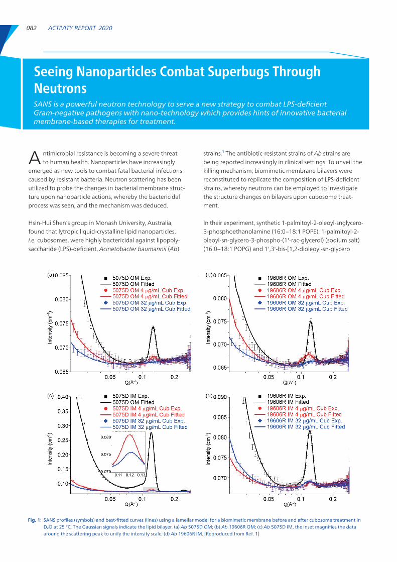
Fig. 1: SANS profiles (symbols) and best-fitted curves (lines) using a lamellar model for a biomimetic membrane before and after cubosome treatment in D2O at 25 °C. The Gaussian signals indicate the lipid bilayer. (a) Ab 5075D OM; (b) Ab 19606R OM; (c) Ab 5075D IM, the inset magnifies the data around the scattering peak to unify the intensity scale; (d) Ab 19606R IM. [Reproduced from Ref. 1]
Seeing Nanoparticles Combat Superbugs Through NeutronsSANS is a powerful neutron technology to serve a new strategy to combat LPS-deficient Gram-negative pathogens with nano-technology which provides hints of innovative bacterial membrane-based therapies for treatment.

Neutron Science 083
bosome treatment. Taken together, these results indicate that cubosomes are bactericidal to A. baumannii on altering the membrane bilayer structure, whilst Ab 19606R is more susceptible to cubosome treatment, which is in accordance with results in vitro in which the minimum inhibitory con-centration of cubosomes against Ab 19606R is 2 μg/mL compared with 4 μg/mL for Ab 5075D. (Reported by Xiang-feng Lai, Monash University, Australia)
This report features the work of Mei-Ling Han, Jian Li, Hsin-Hui Shen and their collaborators, published in ACS Appl. Mater. Interfaces 12, 44485 (2020).
ANSTO Quokka – Small-angle Neutron Scattering• SANS• Antibiotic Resistance, Drag Delivery System, Polymers,
Magnetism, Earth Science
Reference1. X. Lai, Y. Ding, C. M. Wu, X. Chen, J. H. Jiang, H. Y. Hsu, Y.
Wang, A. P. Le Brun, J. Song, M. L. Han, J. Li, H. H. Shen, ACS Appl. Mater. Interfaces 12, 44485 (2020).
-3-phospho]-glycerol (sodium salt) (18:1 Cardiolipin, TOCL) were mixed thoroughly in trichloromethane at molar ratios 37:47:16, 64:26:10, 72:25:3 and 81:17:3 to represent an Ab 5075D outer membrane (OM), Ab 19606R OM, Ab 5075D inner membrane (IM) and Ab 19606R IM, respectively. The mixtures were then dried under N2 and dispersed in D2O with a HEPES buffer using bath sonication. The membrane dispersions were then transferred to quartz cuvettes and characterized at 25 oC to confirm the fabrication of the bilayer. Cubosomes at concentrations 4 and 32 μg/mL were consecutively added to the cuvettes and incubated for 30 min before being subjected to shows small-angle neutron scattering (SANS) measurements. The measurements were conducted on 40-m pinhole instrument QUOKKA at OPAL research reactor in Australian Nuclear Science and Technol-ogy Organisation (ANSTO), Sydney, Australia. Wave vector Q is calculated from θ (scattering angle) and λ (neutron wavelength): Q = 4π sin θ/λ. Two instrument configurations were applied to cover large Q and small Q. Distance L1 = 4 m from source to sample and distance L2 = 3 m at 5 Å and L1 = L2 = 12 m at 6 Å from sample to detector were used to give a Q range 0.0049−0.28 Å−1.
Figure 1 shows SANS profiles of membrane bilayers before and after cubosome treatment. After the cubosome treat-ment, both OM and IM of Ab 5075D and Ab 19606R were evidently significantly disrupted, as indicated by the loss of the bilayer intensity. An analysis of SANS profiles was performed using SasView (version 4.2.2). The data were fitted using lamellar_hg and a Gaussian peak model. The calculated bilayer thicknesses are reported in Table 1. No-tably, the thicknesses of OM and IM of Ab 5075D remained unchanged, while being altered for Ab 19606R after cu-
Table 1: Bilayer thicknesses (δ) before and after cubosome treatment with a lamellar model for the OM and IM of Ab 19606R and Ab 5075D. [Reproduced from Ref. 1]
Membrane δbilayer (Å) δCub 4 (Å) δCub 32 (Å)
Ab 5075D OM 47.2 ± 1.1 47.5 ± 1.2 -
Ab 19606R OM 47.6 ± 0.5 49.2 ± 0.4 -
Ab 5075D IM 51.7 ± 1.5 51.5 ± 0.9 50.9 ± 2.2
Ab 19606R IM 48.3 ± 0.4 45.0 ± 0.7 51.9 ± 2.5
A collaborative team, distinguished by an Innovation in Taiwan Award of the Institute for Biotechnology and Medicine In-dustry and Research Center for Biotechnology and Medicine Policy, has reported an investigation of the bone growth in
additive-manufactured implants using Ti6Al4V and bioactive glass- powder composite.1 E-Wen Huang (National Chiao Tung University & Industrial Technology Research Institute) led the multi-scale characterization team to investigate the underly-ing mechano-biological processes at varied scales due to the hierarchical nature of the bones.2 With customized geometry made by one-step mixing of the bioactive ceramic in the metallic powders, additive-manufacturing implants demonstrate outstanding performance in bone growth. Shao-Ju Shih (National Taiwan University of Science and Technology University) designed the one-step ceramic-metal powder mixing; Nien-Ti Tsou (National Chiao Tung University) invented the healing pattern analysis for implant geometry, and Meng-Huang Wu (Taipei Medical University) performed the surgery.
Neutron Experiments Exploring Advanced MetallurgyAM and HEA are two emerging topics to advance metallurgy. Newly developed additive-man-ufacturing implants and a summary of how neutron experiments reveal the HEA deformation behaviours are reported.

084 ACTIVITY REPORT 2020
Specifically, additive manufacturing (AM) brings high degrees of geometric freedom to the production, but it raises a great challenge to control the microstructure het-erogeneity and anisotropic mechanical properties induced by localized rapid cooling and directional solidification. It is hence important to apply neutron diffraction measure-ments in situ to reveal the mechanical behavior of the AM materials. Under the Neutron Program of NSRRC, Huang showed that, through the generation of a transient phase using AM, the metals are strengthened.3 The plastic anisot-ropy and deformation-induced phase transformation of additive-manufactured metals were revealed. Neutron dif-fraction in situ also unraveled the thermal history about the reversibility of the phase transformation of additive-manu-facturing metals.
Using the advanced light sources at the NSRRC in Taiwan for the multi-scale characterizations, the team applied synchrotron-based high-resolution X-ray microcomputed tomography to microstructured images of the bulk im-plants surgically embedded in the iliac bones at TLS 01A1. Transmission X-ray microscopy is also a non-destructive image microscopy technique, which enables the character-ization of high-resolution X-ray radiography in both two dimensions and the internal microstructure tomography in
three dimensions at TLS 01B1. The direct contacts of new bone, mature bone, and void areas were distinguished. The small- and wide-angle X-ray scattering techniques have been extensively carried out to identify the predominant orientation distribution, degree of crystalline arrangement, as well the shape and size of mineralized hydroxyapatite within the regenerating bone tissues at TLS 23A1. The characterization in crystallographic textures of mineral crys-tallites acquired by X-ray diffraction is capable of evaluating the progress of bone remodeling and bone mineralization during bone healing process. To map the crystallization of the local bone growth, Huang’s group applied the nano X-ray Laue diffraction at TPS 21A for scanning across the boundary from the implants to the mature bone through the new-born bone and at TPS 23A for element mapping.
Although AM enables the freedom for the fabrication, the concept of high-entropy alloys (HEA) opens new space for many further possibilities. To investigate the complicated HEA, in situ neutron diffraction experiments, which simul-taneously measure the bulk performance and the deforma-tion at the lattice level,4 reveal great insight. For example, “a study of lattice elasticity from low entropy metals to medium and high entropy alloys”,5 supported by the Neu-tron Program of NSRRC, is the first research revealing the
The NSRRC Neutron Program supports Huang’s team for the neutron experiments at (a) Australian Nuclear Science and Technology Organisation; (b) Japan Proton Accelerator Research Complex; and (c) Spallation Neutron Source, Oak Ridge National Laboratory. [Photos courtesy of E-Wen Huang]
(c)
(b)(a)

Neutron Science 085
fundamental tensors and proposing the role of stacking faults for HEA using neutron diffraction measurements in situ. The highly cited “in-situ neutron diffraction studies on high-temperature deformation behavior in a CoCrFeMnNi high-entropy alloy”6 is the first paper using neutron diffrac-tion to demonstrate the creep behavior of the HEA. More-over, Huang’s team quantifies the vacancy concentration using neutron diffraction; Huang’s group is the earliest few to point out the elemental effects on HEA vacancy and heterogeneous lattice distortion subjected to quasi- equilibrium heating.7 For improved fatigue life, neutron diffraction measurements explain why HEA are promising for longer lifetime.8
In summary, this report shows systematic research using multi-scale neutron measurements for research on AM and High Entropy Alloys. The underlying mechanisms are revealed for better design and fabrication of the advanced metallurgy. (Reported by E-Wen Huang, National Chiao Tung University)
This report features the work of E-Wen Huang and his collab-orators published in Acta Mater. 201, 412 (2020) and Mater. Chem. Phys. 230, 83 (2019); and the work of K. N. Tu and his collaborators published in Sci. Rep. 9, 14788 (2019).
Neutron Program• Travel support for neutron experiment execution• Magnetic Materials, Superconducting Materials, Energy
materials, Polymer MaterialsTPS 21A X-ray Nanodiffraction• Laue Diffraction• Materials Science, Environment and Energy Science, High
Pressure PhysicsTPS 23A X-ray Nanoprobe• Element Mapping• Materials Science, Semiconducting Polymer, Organic
Transistor, Metal Coordination
TLS 01A1 SWLS – White X-ray• Tomography• Biochemistry, Materials ScienceTLS 01B1 SWLS – X-ray Microscopy• TXM• Biochemistry, Materials ScienceTLS 23A1 IASW – Small/Wide Angle X-ray Scattering• SWAXS• Materials Science, Chemistry, Condensed-matter Physics,
Environmental and Earth Science
References 1. T.-N. Lam, M.-G. Trinh, C.-C. Huang, P.-C. Kung, W.-C.
Huang, W. Chang, L. Amalia, H.-H. Chin, N.-T. Tsou, S.-J. Shih, S.-Y. Chen, C.-C. Wang, P.-I. Tsai, M.-H. Wu, E.-W. Huang, Int. J. Mol. Sci. 21, 7438 (2020).
2. P.-I. Tsai, T.-N. Lam, M.-H. Wu, K.-Y. Tseng, Y.-W. Chang, J.-S. Sun, Y.-Y. Li, M.-H. Lee, S.-Y. Chen, C.-K. Chang, C.-J. Su, C.-H. Lin, C.-Y. Chiang, C.-S. Ku, N.-T. Tsou, S.-J. Shih, C.-C. Wang, E.-W. Huang, Mater. Chem. Phys. 230, 83 (2019).
3. E.-W. Huang, S. Y. Lee, J. Jain, Y. Tong, K. An, N.-T. Tsou, T.-N. Lam, D. Yu, H. Chae, S.-W. Chen, S.-M. Chen, H.-S. Chou, Intermetallics 109, 60 (2019).
4. T.-N. Lam, C.-W. Tsai, B.-K. Chen, B.-H. Lai, H.-C. Liu, T. Kawasaki, S. Harjo, B.-H. Lin, E.-W. Huang, Metall. Mater. Trans. A 51, 5023 (2020).
5. E.-W. Huang, D. Yu, J.-W. Yeh, C. Lee, K. An, S.-Y. Tu, Scripta Mater. 101, 32 (2015).
6. W. Woo, E. W. Huang, J.-W. Yeh, H. Choo, C. Lee, S.-Y. Tu, Intermetallics 62, 1 (2015).
7. E. W. Huang, H.-S. Chou, K. N. Tu, W.-S. Hung, T.-N. Lam, C.-W. Tsai, C.-Y. Chiang, B.-H. Lin, A.-C. Yeh, S.-H. Chang, Y.-J. Chang, J.-J. Yang, X.-Y. Li, C.-S. Ku, K. An, Y.-W. Chang, Y.-L. Jao, Sci. Rep. 9, 14788 (2019).
8. T.-N. Lam, S.Y. Lee, N.-T. Tsou, H.-S. Chou, B.-H. Lai, Y.-J. Chang, R. Feng, T. Kawasaki, S. Harjo, P. K. Liaw, A.-C. Yeh, M.-J. Li, R.-F. Cai, S.-C. Lo, E.-W. Huang, Acta Mater. 201, 412 (2020).