Embed Size (px)
Citation preview
Graduate School for Cellular and Biomedical Sciences
University of Bern
Development of a novel marker vaccine platform for protection against Bluetongue
Virus (BTV)
PhD Thesis submitted by
Stefanie Kochinger
from Austria
for the degree of
Doctor of Veterinary Medicine and Philosophy (DVM,PhD)
Supervisor PD. Dr. Gert Zimmer
Institute of Virology and Immunology Vetsuisse Faculty of the University of Bern
Co‐advisor Prof. Dr. Eliane Marti
Division of Experimental Clinical Research Vetsuisse Faculty of the University of Bern
PhD thesis Stefanie Kochinger 2
Copyright Notice
This document is licensed under the Creative Commons Attribution-Non-Commercial-No derivative works 2.5 Switzerland. http://creativecommons.org/licenses/by‐nc‐nd/2.5/ch/
You are free:
to copy, distribute, display, and perform the work
Under the following conditions:
Attribution. You must give the original author credit.
Non-Commercial. You may not use this work for commercial purposes.
No derivative works. You may not alter, transform, or build upon this work..
For any reuse or distribution, you must take clear to others the license terms of this work.
Any of these conditions can be waived if you get permission from the copyright holder.
Nothing in this license impairs or restricts the author’s moral rights according to Swiss law.
The detailed license agreement can be found at:
http://creativecommons.org/licenses/by‐nc‐nd/2.5/ch/legalcode.de
PhD thesis Stefanie Kochinger 3
Accepted by the Faculty of Medicine, the Faculty of Science and the
Vetsuisse Faculty of the University of Bern at the request of the
Graduate School for Cellular and Biomedical Sciences
Bern, Dean of the Faculty of Medicine
Bern, Dean of the Faculty of Science
Bern, Dean of the Vetsuisse Faculty Bern
PhD thesis Stefanie Kochinger 4
TABLE OF CONTENT
TABLE OF CONTENT 4
ABSTRACT 5
ABBREVIATIONS 7
INTRODUCTION BLUETONGUE 8
History and Epidemiology 8
Bluetongue disease 10
Structure and replication cycle 12
Vaccine strategies 14
INTRODUCTION VSV REPLICONS 16
Vesicular stomatitis virus structure and replication 16
Vesicular stomatitis virus replicon in experimental vaccines 17
Generation of VSV replicon particles expressing foreign antigens 18
AIM OF THE PROJECT 19
RESULTS 20
Unpublished results 20
Published results 29
CONCLUSION AND OUTLOOK. 38
ACKNOWLEDGEMENT 42
CURRICULUM VITAE 43
LIST OF PUBLICATIONS 44
DECLARATION OF ORIGINALITY 45
REFERENCES 46
PhD thesis Stefanie Kochinger 5
ABSTRACT
Bluetongue virus (BTV) is an economically important member of the genus
Orbivirus and closely related to African horse sickness virus (AHSV) and Epizootic
hemorrhagic disease virus (EHDV). Currently, 26 different serotypes of BTV are
known. The virus is transmitted by blood-feeding Culicoides midges and causes
disease (bluetongue [BT]) in ruminants. In 2006/2007, BTV serotype 8 (BTV-8)
caused widespread outbreaks of BT amongst livestock in Europe, which were
eventually controlled employing a conventionally inactivated BTV vaccine.
However, this vaccine did not allow the discrimination of infected from vaccinated
animals (DIVA) by the commonly used VP7 cELISA.
RNA replicon vectors based on propagation-incompetent recombinant vesicular
stomatitis virus (VSV) represent a novel vaccine platform that combines the
efficacy of live attenuated vaccines with the safety of inactivated vaccines. Our
goal was to generate an RNA replicon vaccine for BTV-8, which is safe,
efficacious, adaptable to emerging orbivirus infections , and compliant with the
DIVA principle.
The VP2, VP5, VP3 and VP7 genes encoding the BTV-8 capsid proteins, as well
as the non-structural proteins NS1 and NS3 were inserted into a VSV vector
genome lacking the essential VSV glycoprotein (G) gene. Infectious virus replicon
particles (VRP) were produced on a transgenic helper cell line providing the VSV G
protein in trans. Expression of antigens in vitro was analysed by
immunofluorescence using monoclonal and polyclonal antibodies.
In a pilot study, sheep were immunized with two different VRP-based vaccine
candidates, one comprising the BTV-8 antigens VP2, VP5, VP3, VP7, NS1, and
NS3, the other one containing antigens VP3, VP7, NS1, and NS3. Control animals
PhD thesis Stefanie Kochinger 6
received VRPs containing an irrelevant antigen. Virus neutralizing antibodies and
protection after BTV-8 challenge were evaluated and compared to animals
immunized with the conventionally inactivated vaccine. Full protection was induced
only when the two antigens VP2 and VP5 were included in the vaccine.
To further evaluate if VP2 alone, a combination of VP2 and VP5 or VP5 alone were
necessary for complete protection, we performed a second animal trial.
Interestingly, VP2 as well as the combination of VP2 and VP5 but not VP5 alone
conferred full protection in terms of neutralizing antibodies, and protection from
clinical signs and viremia after BTV-8 challenge.
These results show that the VSV replicon system represents a safe, efficacious
and DIVA-compliant vaccine against BTV as well as a possible platform for
protection against other Orbiviruses, such as AHSV and EHDV.
PhD thesis Stefanie Kochinger 7
ABBREVIATIONS
BTV Bluetongue virus
VSV Vesicular stomatitis virus
VRP Virus replicon particles
GFP or * Green fluorescent protein
IFT Immunofluorscent test/assay
MOI Multiplicity of infection
NS Non-structural protein
VP Viral protein
G Glycoprotein
∆G lacking Glycoprotein
AHSV African horse sickness virus
EHDV Epizootic hemorrhagic disease virus
RNA Ribonucleic acid
kDA kiloDalton
ELISA Enzyme linked immunosorbent assay
dsRNA double-stranded ribonucleic acid
kb kilobases
PhD thesis Stefanie Kochinger 8
INTRODUCTION BLUETONGUE
Bluetongue virus (BTV) is an Orbivirus of the Reoviridae family, and the causative
agent of a haemorrhagic disease (bluetongue [BT]) in ruminants, mainly sheep [1].
There are currently 26 recognised serotypes of BTV, which are transmitted by blood-
feeding Culicoides midges and sometimes vertically in animals [2, 3]. BTV is closely
related to African horse sickness virus (AHSV).
History and Epidemiology
Bluetongue was first described in Merino sheep in the late 19th century in South
Africa and got its name due to severe cyanosis of the tongue of some severely
affected sheep [4]. Later in the 1990s, it was determined that BT was a disease of
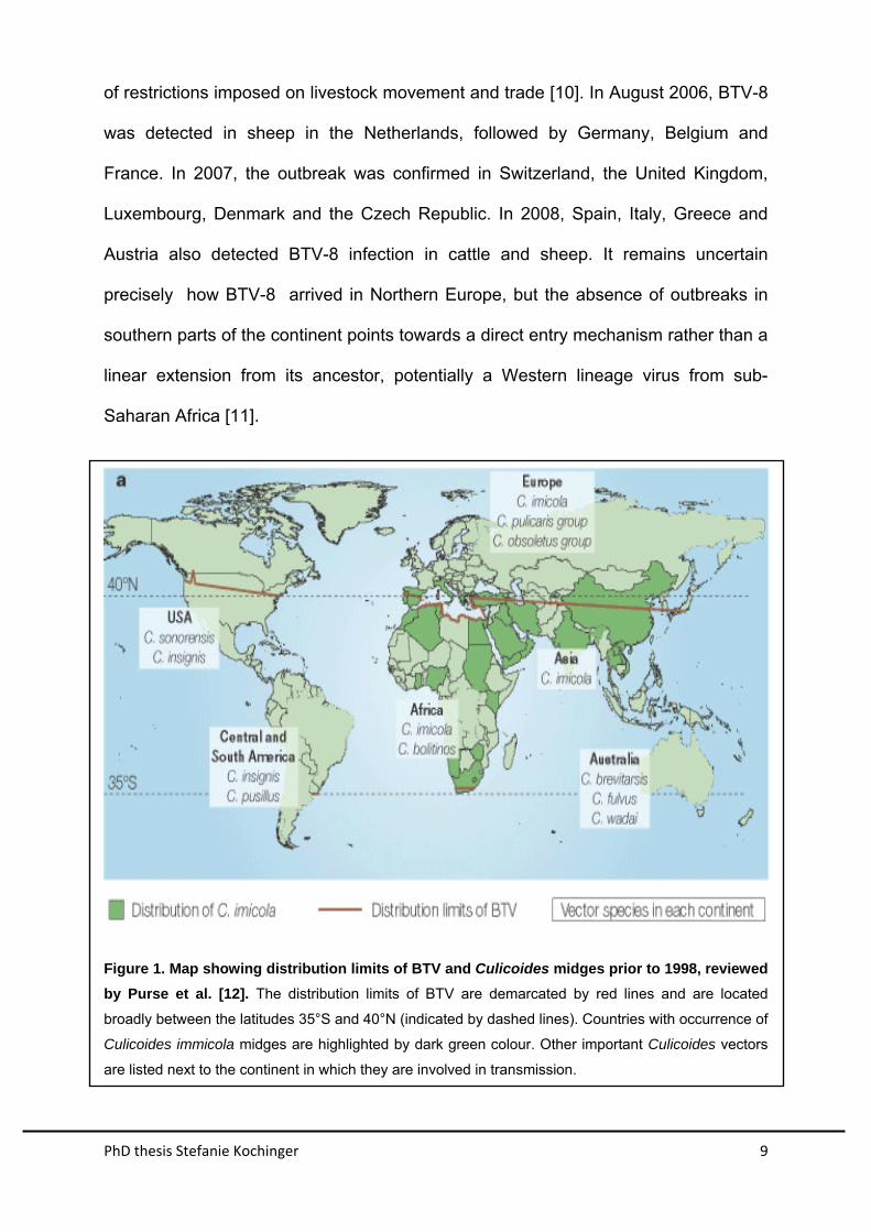
ruminants occurring worldwide from latitudes of approximately 40 degrees North to
35 degrees South [5]. Furthermore, the disease was responsible for considerable
economic losses especially at the incursional margins of the global range of BTV
infection [6] (Figure 1). Although BTV had transiently made brief incursions into
Southern Europe, these did not persist in the continent [5]. However, between 1998
and 2001, eight BTV strains of five different serotypes (BTV-1, 2, 4, 8, 9 and 16)
emerged in several countries around the northern rim of the Mediterranean Basin
where they had previously not been recorded, likely because of climatic changes
which allowed overwintering of the virus in Culicoides vector midges [7].
A novel strain of BTV serotype 8 (BTV-8), which had never previously been detected
in Europe, emerged in 2006 as an epidemic wave in Western and Northern Europe
[4, 8, 9]. This outbreak had a significant economic impact, not only because of
morbidity and mortality caused by the disease in sheep and cattle, but also because
PhD thesis Stefanie Kochinger 9
of restrictions imposed on livestock movement and trade [10]. In August 2006, BTV-8
was detected in sheep in the Netherlands, followed by Germany, Belgium and
France. In 2007, the outbreak was confirmed in Switzerland, the United Kingdom,
Luxembourg, Denmark and the Czech Republic. In 2008, Spain, Italy, Greece and
Austria also detected BTV-8 infection in cattle and sheep. It remains uncertain
precisely how BTV-8 arrived in Northern Europe, but the absence of outbreaks in
southern parts of the continent points towards a direct entry mechanism rather than a
linear extension from its ancestor, potentially a Western lineage virus from sub-
Saharan Africa [11].
Figure 1. Map showing distribution limits of BTV and Culicoides midges prior to 1998, reviewed
by Purse et al. [12]. The distribution limits of BTV are demarcated by red lines and are located
broadly between the latitudes 35°S and 40°N (indicated by dashed lines). Countries with occurrence of
Culicoides immicola midges are highlighted by dark green colour. Other important Culicoides vectors
are listed next to the continent in which they are involved in transmission.

PhD thesis Stefanie Kochinger 10
Figure 2. Distribution of BTV-8 during the outbreak in northern Europe between 2006 and 2007,
reviewed by Saegerman et al. [13]. Monthly distribution of confirmed bluetongue virus 8 (BTV-8)
outbreaks in northern and central Europe from August 17, 2006, through February 1, 2007. After
January 1, 2007, few BTV cases were reported; those that were probably involved animals that had
been infected, but not detected, in 2006.
Bluetongue disease
BTV is transmitted to livestock by blood-feeding Culicoides midges, in Europe mainly
by C. imicola, C. pulicaris and C. obsoletus. In cattle, goats, and wild ruminants, BTV
infection is typically asymptomatic despite prolonged viremia. These host species
represent a potential reservoir for unnoticed dissemination of BTV in ruminant
populations. In sheep, however, BTV infection often results in an acute disease with
associated high morbidity and mortality, depending on the virulence of the virus and
PhD thesis Stefanie Kochinger 11
the sheep breed affected [14]. Following an initial replication in the lymph nodes, BTV
targets endothelium and mononuclear phagocytes [15]. After an incubation period of
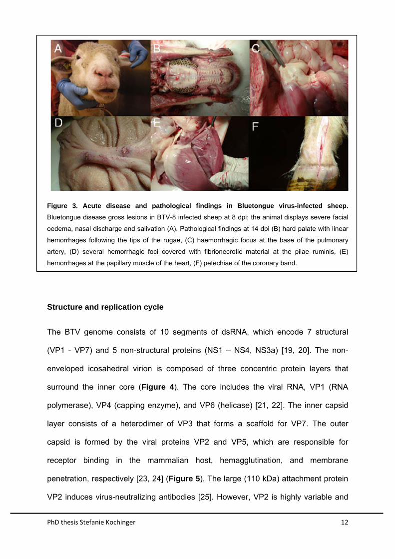
2 to 15 days, clinical signs in the acute phase of disease comprise high fever,
salivation, depression, nasal discharge, hyperaemia and congestion of the muzzle,
lips, face, eyelids and ears leading to tissue oedema (Figure 3 A). Ulcerations and
necrosis of the mucosae of the mouth are often observed, the tongue may become
hyperaemic and oedematous. Lameness due to coronitis, pneumonia or lung
oedema can be observed in severe cases. In cases with fatal outcome, sheep
usually die after 8 to 10 days.
Certain strains of BTV, notably the northern European strain of BTV-8, can cross the
placental barrier, leading to infection of the developing fetus [3]. Hence, abortions
and malformations of offspring are frequently associated with infection of pregnant
animals with certain strains of the virus [16-18].
Frequently observed pathological findings comprise hemorrhages at the base of the
pulmonary artery (A. pulmonalis), the papillary muscles of the left ventricle of the
heart , enlarged haemorrhagic lymph nodes, erosions on ruminal pillars, and ulcers
of the mucosal lining throughout the upper gastrointestinal tract (Figure 3 B-F).
PhD thesis Stefanie Kochinger 12
Figure 3. Acute disease and pathological findings in Bluetongue virus-infected sheep.
Bluetongue disease gross lesions in BTV-8 infected sheep at 8 dpi; the animal displays severe facial
oedema, nasal discharge and salivation (A). Pathological findings at 14 dpi (B) hard palate with linear
hemorrhages following the tips of the rugae, (C) haemorrhagic focus at the base of the pulmonary
artery, (D) several hemorrhagic foci covered with fibrionecrotic material at the pilae ruminis, (E)
hemorrhages at the papillary muscle of the heart, (F) petechiae of the coronary band.
Structure and replication cycle
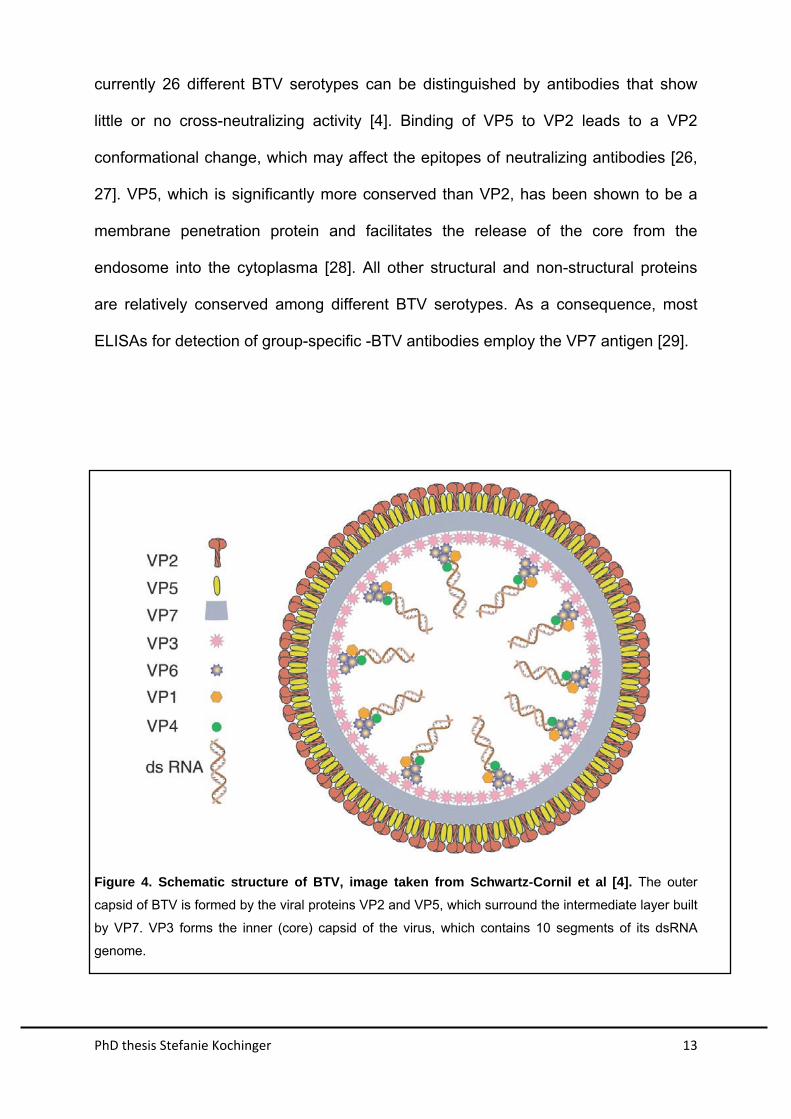
The BTV genome consists of 10 segments of dsRNA, which encode 7 structural
(VP1 - VP7) and 5 non-structural proteins (NS1 – NS4, NS3a) [19, 20]. The non-
enveloped icosahedral virion is composed of three concentric protein layers that
surround the inner core (Figure 4). The core includes the viral RNA, VP1 (RNA
polymerase), VP4 (capping enzyme), and VP6 (helicase) [21, 22]. The inner capsid
layer consists of a heterodimer of VP3 that forms a scaffold for VP7. The outer
capsid is formed by the viral proteins VP2 and VP5, which are responsible for
receptor binding in the mammalian host, hemagglutination, and membrane
penetration, respectively [23, 24] (Figure 5). The large (110 kDa) attachment protein
VP2 induces virus-neutralizing antibodies [25]. However, VP2 is highly variable and
PhD thesis Stefanie Kochinger 13
currently 26 different BTV serotypes can be distinguished by antibodies that show
little or no cross-neutralizing activity [4]. Binding of VP5 to VP2 leads to a VP2
conformational change, which may affect the epitopes of neutralizing antibodies [26,
27]. VP5, which is significantly more conserved than VP2, has been shown to be a
membrane penetration protein and facilitates the release of the core from the
endosome into the cytoplasma [28]. All other structural and non-structural proteins
are relatively conserved among different BTV serotypes. As a consequence, most
ELISAs for detection of group-specific -BTV antibodies employ the VP7 antigen [29].
Figure 4. Schematic structure of BTV, image taken from Schwartz-Cornil et al [4]. The outer
capsid of BTV is formed by the viral proteins VP2 and VP5, which surround the intermediate layer built
by VP7. VP3 forms the inner (core) capsid of the virus, which contains 10 segments of its dsRNA
genome.
PhD thesis Stefanie Kochinger 14
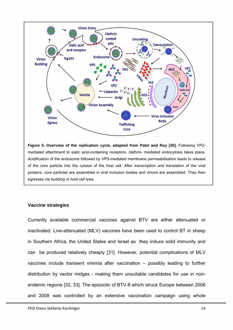
Figure 5. Overview of the replication cycle, adapted from Patel and Roy [30]. Following VP2-
mediated attachment to sialic acid-containing receptors, clathrin- mediated endocytosis takes place.
Acidification of the endosome followed by VP5-mediated membrane permeabilisation leads to release
of the core particle into the cytosol of the host cell. After transcription and translation of the viral
proteins, core particles are assembled in viral inclusion bodies and virions are assembled. They then
egresses via budding or host cell lysis.
Vaccine strategies
Currently available commercial vaccines against BTV are either attenuated or
inactivated. Live-attenuated (MLV) vaccines have been used to control BT in sheep
in Southern Africa, the United States and Israel as they induce solid immunity and
can be produced relatively cheaply [31]. However, potential complications of MLV
vaccines include transient viremia after vaccination – possibly leading to further
distribution by vector midges - making them unsuitable candidates for use in non-
endemic regions [32, 33]. The epizootic of BTV-8 which struck Europe between 2006
and 2008 was controlled by an extensive vaccination campaign using whole
PhD thesis Stefanie Kochinger 15
inactivated BTV-8. Although these vaccines induced strong protection against BTV-8
infection and disease, they did not allow simple serological discrimination of infected
from vaccinated animals (DIVA). Furthermore, vaccine production required the
production of large amounts of infectious virus in cell culture, proper virus
inactivation, and formulation of the antigen with adjuvant, all of which delayed
production and added to the cost of producing the vaccine. Importantly, inactivated
virus vaccines may not be suitable for the control of all serotypes of BTV as, for
example, serotype 25 cannot yet be propagated in cell culture [34].
Much effort has been devoted to the development of alternative vaccine candidates,
to overcome the drawbacks of conventional vaccines. Virus-like particles were
produced using recombinant baculovirus-encoded BTV proteins that were expressed
using baculovirus expression vectors [35, 36]. They proved to be promising
candidates for the production of multi-serotype vaccines, but also caused concerns
regarding potential replication in insect vectors in the field [37]. Other recombinant
viruses encoding different BTV-antigens such as vaccinia viruses [38], canarypox
viruses [39], capripox viruses [40] and herpes viruses [41] have also been proposed.
These studies already revealed the importance of the two components of the outer
BTV shell, VP2 and VP5, for protection [42]
PhD thesis Stefanie Kochinger 16
INTRODUCTION TO VSV REPLICONS
Vesicular stomatitis virus (VSV) structure and replication
VSV is a non-segmented negative-strand RNA virus of the family Rhabdoviridae, and
is known to trigger a robust humoral immune response in many different host species
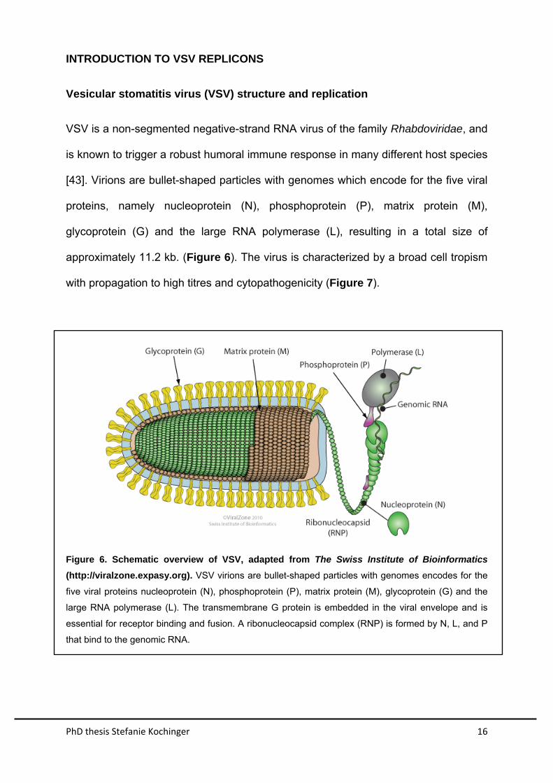
[43]. Virions are bullet-shaped particles with genomes which encode for the five viral
proteins, namely nucleoprotein (N), phosphoprotein (P), matrix protein (M),
glycoprotein (G) and the large RNA polymerase (L), resulting in a total size of
approximately 11.2 kb. (Figure 6). The virus is characterized by a broad cell tropism
with propagation to high titres and cytopathogenicity (Figure 7).
Figure 6. Schematic overview of VSV, adapted from The Swiss Institute of Bioinformatics
(http://viralzone.expasy.org). VSV virions are bullet-shaped particles with genomes encodes for the
five viral proteins nucleoprotein (N), phosphoprotein (P), matrix protein (M), glycoprotein (G) and the
large RNA polymerase (L). The transmembrane G protein is embedded in the viral envelope and is
essential for receptor binding and fusion. A ribonucleocapsid complex (RNP) is formed by N, L, and P
that bind to the genomic RNA.
PhD thesis Stefanie Kochinger 17
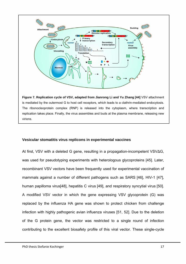
Figure 7. Replication cycle of VSV, adapted from Jianrong Li and Yu Zhang [44] VSV attachment
is mediated by the outermost G to host cell receptors, which leads to a clathrin-mediated endocytosis.
The ribonocleoprotein complex (RNP) is released into the cytoplasm, where transcription and
replication takes place. Finally, the virus assembles and buds at the plasma membrane, releasing new
virions.
Vesicular stomatitis virus replicons in experimental vaccines
At first, VSV with a deleted G gene, resulting in a propagation-incompetent VSV∆G,
was used for pseudotyping experiments with heterologous glycoproteins [45]. Later,
recombinant VSV vectors have been frequently used for experimental vaccination of
mammals against a number of different pathogens such as SARS [46], HIV-1 [47],
human papilloma virus[48], hepatitis C virus [49], and respiratory syncytial virus [50].
A modified VSV vector in which the gene expressing VSV glycoprotein (G) was
replaced by the influenza HA gene was shown to protect chicken from challenge
infection with highly pathogenic avian influenza viruses [51, 52]. Due to the deletion
of the G protein gene, the vector was restricted to a single round of infection
contributing to the excellent biosafety profile of this viral vector. These single-cycle
PhD thesis Stefanie Kochinger 18
virus vector vaccines were effective even though adjuvants were not employed [51,
52].
Generation of VSV replicon particles expressing foreign antigens
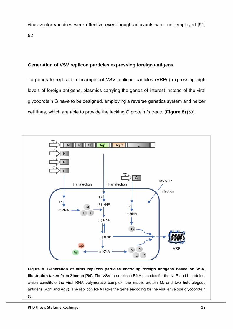
To generate replication-incompetent VSV replicon particles (VRPs) expressing high
levels of foreign antigens, plasmids carrying the genes of interest instead of the viral
glycoprotein G have to be designed, employing a reverse genetics system and helper
cell lines, which are able to provide the lacking G protein in trans. (Figure 8) [53].
Figure 8. Generation of virus replicon particles encoding foreign antigens based on VSV,
illustration taken from Zimmer [54]. The VSV the replicon RNA encodes for the N, P and L proteins,
which constitute the viral RNA polymerase complex, the matrix protein M, and two heterologous
antigens (Ag1 and Ag2). The replicon RNA lacks the gene encoding for the viral envelope glycoprotein
G.
PhD thesis Stefanie Kochinger 19
AIM OF THE PROJECT
Outbreaks of BT disease amongst domestic and wild ruminats can cause
considerable economic loss that reflect high morbidity and mortality as well as
indirect losses because of trade restrictions [10]. In the European outbreak of BTV-8
between 2006 and 2008, an inactivated vaccine was successfully employed.
However, a serological discrimination between infected and vaccinated animals was
not possible.
In this project, we aimed on overcoming these hurdles using propagation-
incompetent VSVG vectors as a new vaccine platform against BTV, which should
be safe, efficacious and conferred the DIVA principle.
First, we generated VSV-virus replicon particles expressing different combinations of
vaccine candidates and studied the expression of recombinant BTV antigens. Next,
we assessed the immune response in a small number of sheep towards VSV VRPs
compared to the inactivated vaccine. Immunized sheep were then infected with
pathogenic BTV-8. Next, we immunized a small number of sheep to test the VRP’s
general suitability, using different combinations of vaccine candidates and comparing
results to animals immunized with the inactivated vaccine before and after infection
with infectious BTV-8 and monitored for viremia, clinical symptoms, and antibody
production. Taking the results from these first experiments into account, we finally
selected propagation-incompetent VSVG vectors expressing the BTV-8 outer
capsid proteins VP2 or VP5 , or a combination of both, and performed a blind
vaccine/challenge study with 24 sheep. The DIVA principle was validated using a
commercially available VP7-based competitive ELISA.
PhD thesis Stefanie Kochinger 20
RESULTS
Unpublished Results
Generation of recombinant VSV replicons expressing BTV antigens
The vaccine vector system is based on a vesicular stomatitis virus (VSV) deletion
mutant that lacks the essential envelope glycoprotein G in the fourth transcription unit
and expresses the enhanced green fluorescent protein at position 5 (VSV*∆G). By
inserting different BTV-8 antigens of interest VP2, VP5, VP3, VP7 and the non-
structural protein NS1 and NS3 into the fourth transcription unit, we were able to
design six different single antigen expression vectors (Figure 9). Three vectors,
expressing two BTV-8 genes from the fourth and fifth transcription unit were also
designed. Recombinant VSV*∆G served as a control vector as it did not express any
BTV antigen. All recombinant vectors were propagated on helper cell lines providing
the VSV glycoprotein G in trans, which yielded infectious virus replicon particles
(VPRs) of approximately 108 infectious units per ml of cell culture supernatant.
PhD thesis Stefanie Kochinger 21
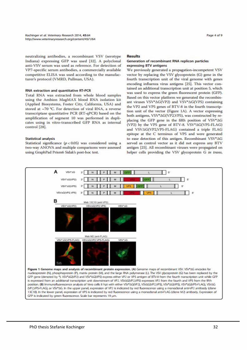
Figure 9. Genome maps of recombinant VSV. (A) VSV encodes for nucleoprotein (N),
phosphoprotein (P), matrix protein (M), glycoprotein (G) and the large RNA polymerase (L). In the
VSV*∆G, the glycoprotein (G) has been replaced by the GFP gene (denoted by *). (B) The vectors
VSV*∆G(VP2), VSV*∆G(VP5), VSV*∆G(VP3), VSV*∆G(VP7), VSV*∆G(NS1), and VSV*∆G(NS3)
express individual antigens of BTV-8 from the fourth transcription unit while GFP (*) is expressed from
an additional transcription unit downstream of the BTV antigen. (C) Double expression vectors
VSV∆G(VP2,VP5), VSV∆G(VP3,VP7), and VSV∆G(NS1, NS3) express two BTV-8 antigens from the
fourth and fifth position.
Characterization of antigen expression in vitro
In order to study the expression of BTV antigens VP2 and VP7, we performed
indirect immunofluorecence analysis (IFA) with Vero cells infected with VRPs using
an MOI of 1.
The BTV-8 VP2-specific monoclonal antibody reacted with cells infected with
VSV*∆G(VP2) and VSV∆G(VP2, VP5), whereas cells infected with VSV*∆G were not
recognized (Figure 10A). Expression of VP7 was detected by a VP7-specific
PhD thesis Stefanie Kochinger 22
monoclonal antibody in cells infected with VSV*∆G(VP7) and VSV∆G(VP3,VP7),
whereas cells infected with VSV*∆G were not recognized (Figure 10B).
Figure 10. Immunofluorecence analysis of Vero cells expressing VP2 and VP7. (A) Detection of
VP2. Cells were infected with either VSV*∆G(VP2), VSV∆G(VP2,VP5), or VSV*∆G. Expression of VP2
is indicated by red fluorescence using a monoclonal anti-VP2 antibody (clone 13C10). Expression of
GFP is indicated by green fluorescence. Scale bar represents 19 µm. (B) Detection of VP7. Cells were
infected with either VSV*∆G(VP7), VSV∆G(VP3,VP7), or VSV*∆G. Expression of VP7 is indicated by
red fluorescence using a monoclonal anti-VP7 antibody (ATCC). Expression of GFP is indicated by
green fluorescence. Scale bar represents 19 µm.
Because specific monoclonal antibody were not available, expression of VP5 and
NS1 was confirmed using modified antigens, containing a triple FLAG epitope at the
C-terminus. The monoclonal anti-FLAG antibody reacted specifically with Vero cells
infected with VSV*∆G(VP5-FLAG), VSV∆G(VP2,VP5-FLAG) and VSV*∆G(NS1-
FLAG) but not with VSV*∆G(VP5)-, VSV*∆G(NS1)-, or VSV*∆G-infected cells
(Figure 11).

PhD thesis Stefanie Kochinger 23
Figure 11. Immunofluorescence analysis of Vero cells expressing VP5 and NS1. (A) Cells were
infected with either VSV*∆G(VP5-FLAG), VSV∆G(VP2,VP5-FLAG), or VSV*∆G. Expression of VP5 is
indicated by red fluorescence using a monoclonal anti-FLAG antibody. (B) Cells infected with either
VSV*∆G(NS1-FLAG), or VSV*∆G. Expression of NS1 in indicated by red fluorescence using a
monoclonal anti-FLAG antibody.
For the remaining BTV antigens VP3 and NS3, monoclonal antibodies were not
available. In order to obtain evidence for expression of these antigens, chickens were
immunized with recombinant VSV replicons. Immune sera were then tested on Vero
cells 24 h post infection with BTV-8 (Figure 12). All sera were found to react
specifically with the infected cells.
Figure 12. Immunofluorecence analysis of BTV-8 infected Vero cells stained with anti-VP3 and
anti-NS3 immune sera. Sera obtained from chicken immunized with VSV-VRPs react with BTV-8
infected cells (red fluorescence). Scale bar represents 24 µm.
Taken together, these data show that infection of Vero cells with virus replicon
particles (VRP) readily led to expression of BTV-8 antigens VP2, VP5, VP7 and NS1.
PhD thesis Stefanie Kochinger 24
Evidence of expression of BTV-8 antigens VP3 and NS3 was obtained indirectly,
since sera of immunized chickens reacted with BTV-8 infected cells. However,
infectious VSV was not produced by the cells due to the lack of VSV G protein
expression. Thus, we refer to the recombinant VSV vectors as virus replicon particles
(VRP).
Analysis of antibody responses in VRP-immunized sheep
To evaluate the immunogenicity of recombinant VRPs in a natural BTV host, nine
seronegative sheep were immunized three times at 3-week intervals. Three animals
were immunized with cell culture supernatant containing at least 1x10⁸ infectious
units of each of the VRPs VSV∆G(VP2,VP5), VSV∆G(VP3,VP7) and
VSV∆G(NS1,NS3). Two animals were immunized with the VRPs VSV∆G(VP3,VP7)
and VSV∆G(NS1,NS3) expressing four antigens in total. Four animals received
VSV*∆G as control. Furthermore, five animals that had been immunized with
inactivated BTV-8 vaccine (BTVpur) two years ahead of the experiment were
included in our analysis. No adverse side effects due to vaccination with VRPs were
observed.
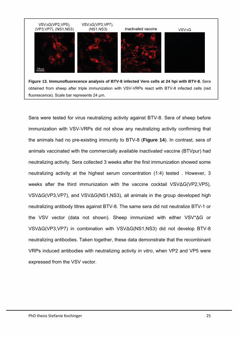
Three weeks after the third immunization, sera were prepared from from sheep and
analyzed for the presence of BTV-specific antibodies by immunofluorecence. All sera
of sheep immunized with VRPs containing BTV antigens and BTVpur reacted
specifically with BTV-8 infected cells, indicating that antibodies had been produced
(Figure 13). In contrast, sera from VSV*∆G-immunized sheep did not react.
PhD thesis Stefanie Kochinger 25
Figure 13. Immunofluorecence analysis of BTV-8 infected Vero cells at 24 hpi with BTV-8. Sera
obtained from sheep after triple immunization with VSV-VRPs react with BTV-8 infected cells (red
fluorescence). Scale bar represents 24 µm.
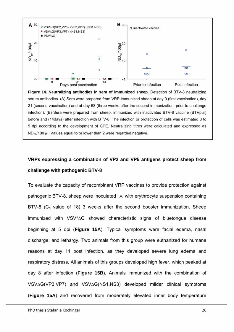
Sera were tested for virus neutralizing activity against BTV-8. Sera of sheep before
immunization with VSV-VRPs did not show any neutralizing activity confirming that
the animals had no pre-existing immunity to BTV-8 (Figure 14). In contrast, sera of
animals vaccinated with the commercially available inactivated vaccine (BTVpur) had
neutralizing activity. Sera collected 3 weeks after the first immunization showed some
neutralizing activity at the highest serum concentration (1:4) tested . However, 3
weeks after the third immunization with the vaccine cocktail VSV∆G(VP2,VP5),
VSV∆G(VP3,VP7), and VSV∆G(NS1,NS3), all animals in the group developed high
neutralizing antibody titres against BTV-8. The same sera did not neutralize BTV-1 or
the VSV vector (data not shown). Sheep immunized with either VSV*∆G or
VSV∆G(VP3,VP7) in combination with VSV∆G(NS1,NS3) did not develop BTV-8
neutralizing antibodies. Taken together, these data demonstrate that the recombinant
VRPs induced antibodies with neutralizing activity in vitro, when VP2 and VP5 were
expressed from the VSV vector.
PhD thesis Stefanie Kochinger 26
Figure 14. Neutralizing antibodies in sera of immunized sheep. Detection of BTV-8 neutralizing
serum antibodies. (A) Sera were prepared from VRP-immunized sheep at day 0 (first vaccination), day
21 (second vaccination) and at day 63 (three weeks after the second immunization, prior to challenge
infection). (B) Sera were prepared from sheep, immunized with inactivated BTV-8 vaccine (BTVpur)
before and (14days) after infection with BTV-8. The infection or protection of cells was estimated 3 to
5 dpi according to the development of CPE. Neutralizing titres were calculated and expressed as
ND50/100 l. Values equal to or lower than 2 were regarded negative.
VRPs expressing a combination of VP2 and VP5 antigens protect sheep from
challenge with pathogenic BTV-8
To evaluate the capacity of recombinant VRP vaccines to provide protection against
pathogenic BTV-8, sheep were inoculated i.v. with erythrocyte suspension containing
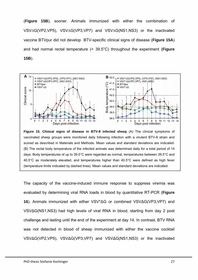
BTV-8 (Cq value of 18) 3 weeks after the second booster immunization. Sheep
immunized with VSV*G showed characteristic signs of bluetongue disease
beginning at 5 dpi (Figure 15A). Typical symptoms were facial edema, nasal
discharge, and lethargy. Two animals from this group were euthanized for humane
reasons at day 11 post infection, as they developed severe lung edema and
respiratory distress. All animals of this groups developed high fever, which peaked at
day 8 after infection (Figure 15B). Animals immunized with the combination of
VSVG(VP3,VP7) and VSVG(NS1,NS3) developed milder clinical symptoms
(Figure 15A) and recovered from moderately elevated inner body temperature
PhD thesis Stefanie Kochinger 27
(Figure 15B), sooner. Animals immunized with either the combination of
VSVG(VP2,VP5), VSVG(VP3,VP7) and VSVG(NS1,NS3) or the inactivated
vaccine BTVpur did not develop BTV-specific clinical signs of disease (Figure 15A)
and had normal rectal temperature (< 39.5°C) throughout the experiment (Figure
15B).
Figure 15. Clinical signs of disease in BTV-8 infected sheep (A) The clinical symptoms of
vaccinated sheep groups were monitored daily following infection with a virulent BTV-8 strain and
scored as described in Materials and Methods. Mean values and standard deviations are indicated.
(B) The rectal body temperature of the infected animals was determined daily for a total period of 14
days. Body temperatures of up to 39.5°C were regarded as normal, temperatures between 39.5°C and
40.5°C as moderately elevated, and temperatures higher than 40.5°C were defined as high fever
(temperature limits indicated by dashed lines). Mean values and standard deviations are indicated.
The capacity of the vaccine-induced immune response to suppress viremia was
evaluated by determining viral RNA loads in blood by quantitative RT-PCR (Figure
16). Animals immunized with either VSV*∆G or combined VSV∆G(VP3,VP7) and
VSV∆G(NS1,NS3) had high levels of viral RNA in blood, starting from day 2 post
challenge and lasting until the end of the experiment at day 14. In contrast, BTV RNA
was not detected in blood of sheep immunized with either the vaccine cocktail
VSV∆G(VP2,VP5), VSV∆G(VP3,VP7) and VSV∆G(NS1,NS3) or the inactivated
PhD thesis Stefanie Kochinger 28
vaccine (BTVpur). Thus, recombinant VRPs expressing VP2 and VP5 induced
protective immunity in sheep that prevented both viremia and bluetongue disease.
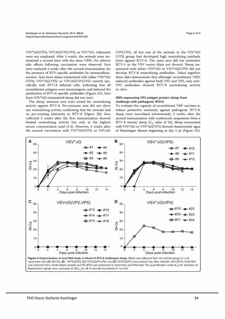
Figure 16. Determination of viral RNA loads in blood of BTV-8 challenged sheep. Blood was
collected from animals of the indicated vaccine groups every second day after infection with BTV-8.
Total RNA was extracted from whole blood samples and RT-qPCR was performed to determine viral
RNA loads. The quantification cycles (Cq) for detection of fluorescence signals were expressed as 50-
Cq for all 14 animals (numbered #1 to #14).

PhD thesis Stefanie Kochinger 29
Published Results
PhD thesis Stefanie Kochinger 30
PhD thesis Stefanie Kochinger 31
PhD thesis Stefanie Kochinger 32
PhD thesis Stefanie Kochinger 33
PhD thesis Stefanie Kochinger 34
PhD thesis Stefanie Kochinger 35
PhD thesis Stefanie Kochinger 36
PhD thesis Stefanie Kochinger 37
PhD thesis Stefanie Kochinger 38
CONCLUSION AND OUTLOOK
The BTV outer capsid proteins VP2 and VP5 represent potential targets for
neutralizing antibodies. However, all neutralizing epitopes identified to date reside on
VP2, and VP5 is thought to exert a conformational effect on VP2 (26). The present
study demonstrates that recombinant VRPs that express the VP2 protein of BTV-8
induce the production of serotype-specific neutralizing antibodies that protect sheep
from disease and viremia following challenge infection with BTV-8. Our data confirm
previous reports that have demonstrated that immunization with VP2 alone is
sufficient to induce a protective immune response [25, 62, 63]. VP5 has been
reported to enhance the protective immune response if present in the vaccine along
with VP2 [27, 38]. Employing a canarypox virus vector expressing VP2 and VP5 of
BTV-17, protection in sheep was achieved, but the relative contributions of VP2 and
VP5 to protection have not been addressed [39]. In a recent study, expression of
both VP2 and VP5 by a recombinant equine herpesvirus vector was necessary to
protect mice against BTV-8 infection, whereas VP2 alone was not fully protective
[41]. In our investigations, VRPs co-expressing VP2 and VP5 did not induce higher
neutralizing antibody titers than VRP expressing only VP2, although one animal
immunized with VP2 was not protected nor did it develop neutralizing antibodies.
This discrepancy between VSV-based VRPs and other vaccines might be due to the
different vector systems, i.e. different expression levels and differences in the
conformation of recombinant VP2, especially if yields of recombinant VP2 are low.
Thus, the proportion of properly folded VP2 might increase in the presence of VP5
given their extensive conformational interaction [24]. If high expression levels of VP2
can be achieved with a given vector system, the amounts of correctly folded VP2
might be sufficient to induce neutralizing antibodies. Antibodies directed to VP5 may
PhD thesis Stefanie Kochinger 39
not have neutralizing properties on their own, probably because this protein is not
accessible for antibodies on the surface of the virion. Furthermore, VP5 is more
conserved than VP2, suggesting that VP5 is subject to less immune pressure.
Conventional BTV vaccines have to be properly inactivated and formulated with
adjuvants before they can be applied to animals once (sheep) or twice (cattle).
Neutralizing antibodies were shown to persist up to four years after immunization [64,
65]. The present VRP vaccine did not depend on adjuvants to induce a protective
immune response in sheep, probably because the RNA virus vector is cytotoxic and
so stimulates the innate immune system via pathogen recognition receptor. Any
adverse side effects that could be caused by adjuvants [66] are thereby eliminated.
However, significant levels of neutralizing serum antibodies were detected only after
the animals had been vaccinated with the VRP vaccine twice. Additional studies are
required to determine the protective effect of a single immunization. In addition, it will
be interesting to see for how long VRP-induced neutralizing antibodies will persist in
sheep.
Despite their efficacy, MLV (live-attenuated) BTV vaccines have inherent
disadvantages, including potential transmission to vector midges, lack of attenuation,
reassortment of gene segments with field strains of the virus, and potential to cross
the placenta to cause reproductive losses and teratogenesis [67]. In contrast, the
propagation-defective VRP vaccine does not cause disease nor will it be able to
revert to virulence. As the VSV G protein which is the major immunogenic VSV
protein is not expressed neither by the VRPs nor in non-helper cells, antibodies
directed against the viral vector are not induced. Accordingly, VRP vaccines are
effective when used repeatedly [51, 52, 68]. Since antibodies directed to VSV G
PhD thesis Stefanie Kochinger 40
protein are lacking, vaccinated animals will appear seronegative in VSV
neutralization tests [69].
Vector vaccines are superior to conventional vaccines, since they can serve as
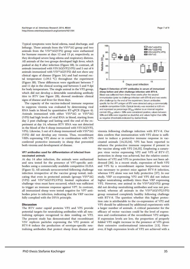
marker vaccines [42]. The VRP vaccine did not contain the VP7 antigen and
therefore allowed the discrimination of infected from vaccinated animals using a
commercially available BTV VP7 ELISA. In contrast, conventional (inactivated) BTV
vaccines, live-attenuated and propagation-defective BTV vaccines [70, 71] do not
comply with the DIVA principle. This has complicated the serological surveillance of
BTV in those European countries that implemented a vaccination campaign to control
the BTV-8 outbreak in 2006. Moreover, the inability to discriminate between infected
and vaccinated animals had a significant impact on economy due to restrictions on
animal trade [31]. The recombinant replicon particles eliminate this problem as they
can be used as marker vaccines.
There is no universal vaccine available that would protect against all 26 currently
known serotypes of BTV. The VRP technology is not restricted to BTV-8 antigens but
can be also used to express VP2 antigen of other BTV serotypes. Thus, in the
scenario where a new BTV serotype emerges in a non-endemic region, a
recombinant VRP vaccine matching this serotype could be rapidly produced and
used for emergency vaccination. This should be particularly valuable for BTV
serotypes and strains for which inactivated vaccines are not readily available
because the corresponding viruses do not propagate well in cell culture.
Furthermore, this generic vaccine platform may be also be relevant to protect horses
against infection with African horse sickness virus. In summary, this study
demonstrates that propagation-incompetent VSV replicon particles can efficiently
protect a natural host against BT disease and viremia. This safe and adjuvant-free

PhD thesis Stefanie Kochinger 41
vaccine technology complies with the DIVA principle, can be easily adapted to other
serotypes or other viruses, and is rapidly available in case of an emerging BTV
outbreak.
PhD thesis Stefanie Kochinger 42
ACKNOWLEDGEMENTS
First, I would like to express my sincere gratitude to my advisor Gert Zimmer for the
help throughout my PhD study and research. My sincere thanks also goes to my co-
advisor Eliane Marti for encouragement, insightful comments and help.
I want to thank external Co-advisor, Jim MacLachlan for sharing his knowledge,
wisdom and experience with me.
Besides my advisors, I would like to thank my excellent mentor Giuseppe Bertoni for
continuous support, help and for being a person who I really learned to admire, both
for his knowledge and his personal resilience.
I thank the Institute of Virology and Immunology, Christian Griot, Kathrin
Summermatter, Martin Hofmann, Nahalie Renevey, Barbara Thür, Andrea
Vögtlin, Lukas Bruckner, Volker Thiel, Arthur Summerfield, Meret Ricklin, and
all my fellow lab mates for their support and motivation throughout the past three
years.
I also thank my dear Austrian (Barbara Matzinger, Georg Schätz, Sonja Prucha,
Viviane Benetka and Verena Küronya) and Swiss (Patricia Neupert, Lena Ferner,
Claudia Wehinger, Roger Büchi, Tobias Meyer, Tobias Struchen and Daniela
Juen) friends for all the fun, time and love you gave me. Friends like you make the
world go round!
Furthermore, I would like to thank my beloved family-first and foremost my gorgeous
mother Christa, my sporty father Günther, my adventurous brother Christian, my
caring sister Tanja and my great aunt Luise for raising me, teaching me and loving
me the way they did and do throughout my life. I could not think of a better family!
I also thank my boyfriend’s parents Regula and Florian Meyer for the nice family
dinners at their place and Samuel for being both, my best friend and love. I truly
admire, appreciate and love you just the way you are!
Last but not least, I thank my Irish Setter dog Diana for being an example of patience
and sweetness.
PhD thesis Stefanie Kochinger 43
CURRICULUM VITAE
CONTACT DETAILS
STEFANIE KOCHINGER Zollikon, Switzerland
Fohrbachstrasse 16 • 8702 Zollikon • Switzerland
+41 79 1247980 • [email protected]
June 27, 1986
PROFESSIONAL EXPERIENCE
INSTITUTE OF VIROLOGY AND IMMUNOLOGY Mittelhäusern, Switzerland
Development of a novel marker vaccine platform against Bluetongue August 2011 – Juli 2014
Execution of BLV-granted project Worked in a BSL-4 facility Conducted reverse genetics, serology, animal trials, qRT-PCR, cell culture, pathology Participation and presentations in national and international conferences
INSTITUTE OF ANIMAL WELFARE Vienna, Austria
Research Intern February 2011 – May 2011
Evaluated breeding facilities for pigs and chickens
CLINICAL VIROLOGY Vienna, Austria
Diagnostics and research assistent October 2010 – Juli 2011
Virological and serological testing of samples Helped implementing QM-system for certification (GLP, ISO) Project: Canine Parvovirus type 2; evaluation of a novel qPCR for diagnostic use Teaching and supervision of students
AGES INSTITUTE OF FOOD SAFETY Vienna, Austria
Intern
Laboratory diagnostics milk and meat September 2011
AGES PHARMMED Vienna, Austria
Intern Registration of human therapeutics July 2009 – August 2009
Wrote and corrected expert opinions on human therapeutics
PhD thesis Stefanie Kochinger 44
EDUCATION
FEDERAL FOOD SAFETY AND VETERINARY OFFICE Berne, Switzerland
Federal veterinary officer studies 2014
GRADUATE SCHOOL FOR CELLULAR AND MICROBIOLOGICAL SCIENCE Berne, Switzerland
DVM-Ph.D. 2011-2014
SVVLD - SCHWEIZER VEREINIGUNG DER VETERINÄR-LABORDIAGNOSTIKER Berne, Switzerland
Fachtierarzt FVH für Labor- und Grundlagenmedizin, Virologie 2011-2014
UNIVERSITY OF VETERINARY MEDICINE Vienna, Austria
Veterinary Medicine 2004-2011
Completed study in minimum required time with excellent success Specialized in Public Health and was intern in different divisions
GYMNASIUM DER DOMINIKANERINNEN Vienna, Austria
Matura 1996 – 2004
PERSONAL
LANGUAGES
English: fluent • German: fluent (native) • French: basic
TOOLS
Microsoft Office • PrismGraph • Adobe Illustrator CS6 • Adobe Photoshop Elements 10
INTERESTS
Jogging and walking with my dog • Meeting with colleagues • Horseback riding Exploring new places • Skiing in winter • Reading books and papers
LIST OF PUBLICATIONS
Kochinger S., Renevey N., Hofmann M. A., Zimmer, G. 2014. Vesicular stomatitis virus replicon expressing the VP2 outer capsid protein of bluetongue virus serotype 8 induces complete protection of sheep against challenge infection. Vet Res 2014, 45:64.

PhD thesis Stefanie Kochinger 45
DECLARATION OF ORIGINALITY
PhD thesis Stefanie Kochinger 46
REFREFERENCES
1. Vet Immunol ImmunopatholDNA RaHtDaT, Prof. Anica Dricu (Ed.)Erasmus, B. J.: Bluetongue in sheep and goats. Aust Vet J 1975, 51:165-170.
2. van der Sluijs MT, Schroer-Joosten DP, Fid-Fourkour A, Vrijenhoek MP, Debyser I, Moulin V, Moormann RJ, de Smit AJ: Transplacental transmission of bluetongue virus serotype 1 and serotype 8 in sheep: virological and pathological findings. PloS one 2013, 8:e81429.
3. Santman-Berends IM, van Wuijckhuise L, Vellema P, van Rijn PA: Vertical transmission of bluetongue virus serotype 8 virus in Dutch dairy herds in 2007. Vet Microbiol 2010, 141:31-35.
4. Schwartz-Cornil I, Mertens PP, Contreras V, Hemati B, Pascale F, Breard E, Mellor PS, MacLachlan NJ, Zientara S: Bluetongue virus: virology, pathogenesis and immunity. Vet Res 2008, 39:46.
5. Mellor PS, Boorman J: The transmission and geographical spread of African horse sickness and bluetongue viruses. Ann Trop Med Parasitol 1995, 89:1-15.
6. Tabachnick WJ: Culicoides variipennis and bluetongue-virus epidemiology in the United States. Annu Rev Entomol 1996, 41:23-43.
7. Mellor PS, Wittmann EJ: Bluetongue virus in the Mediterranean Basin 1998-2001. Vet J 2002, 164:20-37.
8. Maclachlan NJ, Guthrie AJ: Re-emergence of bluetongue, African horse sickness, and other orbivirus diseases. Vet Res 2010, 41:35.
9. Wilson AJ, Mellor PS: Bluetongue in Europe: past, present and future. Philos Trans R Soc Lond B Biol Sci 2009, 364:2669-2681.
10. MacLachlan NJ, Osburn BI: Impact of bluetongue virus infection on the international movement and trade of ruminants. J Am Vet Med Assoc 2006, 228:1346-1349.
11. Rasmussen LD, Rasmussen TB, Belsham GJ, Strandbygaard B, Botner A: Bluetongue in Denmark during 2008. Vet Rec 2010, 166:714-718.
12. Purse BV, Brown HE, Harrup L, Mertens PP, Rogers DJ: Invasion of bluetongue and other orbivirus infections into Europe: the role of biological and climatic processes. Rev Sci Tech 2008, 27:427-442.
PhD thesis Stefanie Kochinger 47
13. Saegerman C, Berkvens D, Mellor PS: Bluetongue epidemiology in the European Union. Emerg Infect Dis 2008, 14:539-544.
14. Worwa G, Thur B, Griot C, Hofmann M, MacLachlan JN, Chaignat V: [Bluetongue disease in Swiss sheep breeds: clinical signs after experimental infection with bluetongue virus serotype 8]. Schweiz Arch Tierheilkd 2008, 150:491-498.
15. Barratt-Boyes SM, MacLachlan NJ: Dynamics of viral spread in bluetongue virus infected calves. Vet Microbiol 1994, 40:361-371.
16. Saegerman C, Bolkaerts B, Baricalla C, Raes M, Wiggers L, de Leeuw I, Vandenbussche F, Zimmer JY, Haubruge E, Cassart D, et al: The impact of naturally-occurring, trans-placental bluetongue virus serotype-8 infection on reproductive performance in sheep. Vet J 2011, 187:72-80.
17. Housawi FM, Abu Elzein EM, Ramadan RO, Gameel AA, Al-Afaleq AI, Al-Mousa J: Abortions, stillbirths and deformities in sheep at the Al-Ahsa oasis in eastern Saudi Arabia: isolation of a bluetongue serogroup virus from the affected lambs. Rev Sci Tech 2004, 23:913-920.
18. Wouda W, Peperkamp NH, Roumen MP, Muskens J, van Rijn A, Vellema P: Epizootic congenital hydranencephaly and abortion in cattle due to bluetongue virus serotype 8 in the Netherlands. Tijdschr Diergeneeskd 2009, 134:422-427.
19. Verwoerd DW, Louw H, Oellermann RA: Characterization of bluetongue virus ribonucleic acid. J Virol 1970, 5:1-7.
20. Belhouchet M, Mohd Jaafar F, Firth AE, Grimes JM, Mertens PP, Attoui H: Detection of a fourth orbivirus non-structural protein. PloS one 2011, 6:e25697.
21. Mertens PP, Brown F, Sangar DV: Assignment of the genome segments of bluetongue virus type 1 to the proteins which they encode. Virology 1984, 135:207-217.
22. Mertens PP, Diprose J, Maan S, Singh KP, Attoui H, Samuel AR: Bluetongue virus replication, molecular and structural biology. Vet Ital 2004, 40:426-437.
23. Hassan SS, Roy P: Expression and functional characterization of bluetongue virus VP2 protein: role in cell entry. J Virol 1999, 73:9832-9842.
24. Zhang X, Boyce M, Bhattacharya B, Zhang X, Schein S, Roy P, Zhou ZH: Bluetongue virus coat protein VP2 contains sialic acid-binding domains,
PhD thesis Stefanie Kochinger 48
and VP5 resembles enveloped virus fusion proteins. Proc Natl Acad Sci U S A 2010, 107:6292-6297.
25. Huismans H, van der Walt NT, Cloete M, Erasmus BJ: Isolation of a capsid protein of bluetongue virus that induces a protective immune response in sheep. Virology 1987, 157:172-179.
26. DeMaula CD, Bonneau KR, MacLachlan NJ: Changes in the outer capsid proteins of bluetongue virus serotype ten that abrogate neutralization by monoclonal antibodies. Virus Res 2000, 67:59-66.
27. Roy P, Marshall JJ, French TJ: Structure of the bluetongue virus genome and its encoded proteins. Curr Top Microbiol Immunol 1990, 162:43-87.
28. Hassan SH, Wirblich C, Forzan M, Roy P: Expression and functional characterization of bluetongue virus VP5 protein: role in cellular permeabilization. J Virol 2001, 75:8356-8367.
29. Boyer TC, Ward MP, Wallace RL, Singer RS: Regional seroprevalence of bluetongue virus in cattle in Illinois and western Indiana. Am J Vet Res 2007, 68:1212-1219.
30. Patel A, Roy P: The molecular biology of Bluetongue virus replication. Virus Res 2013.
31. Savini G, MacLachlan NJ, Sanchez-Vizcaino JM, Zientara S: Vaccines against bluetongue in Europe. Comp Immunol Microbiol Infect Dis 2008, 31:101-120.
32. Veronesi E, Darpel KE, Hamblin C, Carpenter S, Takamatsu HH, Anthony SJ, Elliott H, Mertens PP, Mellor PS: Viraemia and clinical disease in Dorset Poll sheep following vaccination with live attenuated bluetongue virus vaccines serotypes 16 and 4. Vaccine 2010, 28:1397-1403.
33. Batten CA, Maan S, Shaw AE, Maan NS, Mertens PP: A European field strain of bluetongue virus derived from two parental vaccine strains by genome segment reassortment. Virus Res 2008, 137:56-63.
34. Planzer J, Kaufmann C, Worwa G, Gavier-Widen D, Hofmann MA, Chaignat V, Thur B: In vivo and in vitro propagation and transmission of Toggenburg orbivirus. Res Vet Sci 2011, 91:e163-168.
35. Roy P, French T, Erasmus BJ: Protective efficacy of virus-like particles for bluetongue disease. Vaccine 1992, 10:28-32.
36. Roy P, Bishop DH, LeBlois H, Erasmus BJ: Long-lasting protection of sheep against bluetongue challenge after vaccination with virus-like
PhD thesis Stefanie Kochinger 49
particles: evidence for homologous and partial heterologous protection. Vaccine 1994, 12:805-811.
37. Hervas-Stubbs S, Rueda P, Lopez L, Leclerc C: Insect baculoviruses strongly potentiate adaptive immune responses by inducing type I IFN. J Immunol 2007, 178:2361-2369.
38. Lobato ZI, Coupar BE, Gray CP, Lunt R, Andrew ME: Antibody responses and protective immunity to recombinant vaccinia virus-expressed bluetongue virus antigens. Vet Immunol Immunopathol 1997, 59:293-309.
39. Boone JD, Balasuriya UB, Karaca K, Audonnet JC, Yao J, He L, Nordgren R, Monaco F, Savini G, Gardner IA, Maclachlan NJ: Recombinant canarypox virus vaccine co-expressing genes encoding the VP2 and VP5 outer capsid proteins of bluetongue virus induces high level protection in sheep. Vaccine 2007, 25:672-678.
40. Wade-Evans AM, Romero CH, Mellor P, Takamatsu H, Anderson J, Thevasagayam J, Fleming MJ, Mertens PP, Black DN: Expression of the major core structural protein (VP7) of bluetongue virus, by a recombinant capripox virus, provides partial protection of sheep against a virulent heterotypic bluetongue virus challenge. Virology 1996, 220:227-231.
41. Ma G, Eschbaumer M, Said A, Hoffmann B, Beer M, Osterrieder N: An equine herpesvirus type 1 (EHV-1) expressing VP2 and VP5 of serotype 8 bluetongue virus (BTV-8) induces protection in a murine infection model. PloS one 2012, 7:e34425.
42. Calvo-Pinilla E, Castillo-Olivares J, Jabbar T, Ortego J, de la Poza F, Marin-Lopez A: Recombinant vaccines against bluetongue virus. Virus Res 2013.
43. Hastie E, Cataldi M, Marriott I, Grdzelishvili VZ: Understanding and altering cell tropism of vesicular stomatitis virus. Virus Res 2013, 176:16-32.
44. Zhang JLaY: Messenger RNA Cap Methylation in Vesicular Stomatitis Virus, a Prototype of Non‐Segmented Negative‐Sense RNA Virus, Methylation. DNA, RNA and Histones to Diseases and Treatment, Prof Anica Dricu (Ed) 2012.
45. Takada A, Robison C, Goto H, Sanchez A, Murti KG, Whitt MA, Kawaoka Y: A system for functional analysis of Ebola virus glycoprotein. Proc Natl Acad Sci U S A 1997, 94:14764-14769.
46. Kapadia SU, Simon ID, Rose JK: SARS vaccine based on a replication-defective recombinant vesicular stomatitis virus is more potent than one based on a replication-competent vector. Virology 2008, 376:165-172.
PhD thesis Stefanie Kochinger 50
47. Publicover J, Ramsburg E, Rose JK: A single-cycle vaccine vector based on vesicular stomatitis virus can induce immune responses comparable to those generated by a replication-competent vector. J Virol 2005, 79:13231-13238.
48. Lichty BD, Power AT, Stojdl DF, Bell JC: Vesicular stomatitis virus: re-inventing the bullet. Trends Mol Med 2004, 10:210-216.
49. Majid AM, Barber GN: Recombinant Vesicular Stomatitis Virus (VSV) and Other Strategies in HCV Vaccine Designs and Immunotherapy. In Hepatitis C Viruses: Genomes and Molecular Biology. Edited by Tan SL. Norfolk (UK); 2006
50. Kahn JS, Roberts A, Weibel C, Buonocore L, Rose JK: Replication-competent or attenuated, nonpropagating vesicular stomatitis viruses expressing respiratory syncytial virus (RSV) antigens protect mice against RSV challenge. J Virol 2001, 75:11079-11087.
51. Kalhoro NH, Veits J, Rautenschlein S, Zimmer G: A recombinant vesicular stomatitis virus replicon vaccine protects chickens from highly pathogenic avian influenza virus (H7N1). Vaccine 2009, 27:1174-1183.
52. Halbherr SJ, Brostoff T, Tippenhauer M, Locher S, Berger Rentsch M, Zimmer G: Vaccination with recombinant RNA replicon particles protects chickens from H5N1 highly pathogenic avian influenza virus. PloS one 2013, 8:e66059.
53. Berger Rentsch M, Zimmer G: A vesicular stomatitis virus replicon-based bioassay for the rapid and sensitive determination of multi-species type I interferon. PloS one 2011, 6:e25858.
54. Zimmer G: RNA Replicons - A New Approach for Influenza Virus Immunoprophylaxis. Viruses 2010, 2:413-434.
55. Borden EC, Shope RE, Murphy FA: Physicochemical and morphological relationships of some arthropod-borne viruses to bluetongue virus--a new taxonomic group. Physiocochemical and serological studies. J Gen Virol 1971, 13:261-271.
56. Murphy FA, Borden EC, Shope RE, Harrison A: Physicochemical and morphological relationships of some arthropod-borne viruses to bluetongue virus--a new taxonomic group. Electron microscopic studies. J Gen Virol 1971, 13:273-288.
57. Hanika A, Larisch B, Steinmann E, Schwegmann-Wessels C, Herrler G, Zimmer G: Use of influenza C virus glycoprotein HEF for generation of vesicular stomatitis virus pseudotypes. J Gen Virol 2005, 86:1455-1465.
PhD thesis Stefanie Kochinger 51
58. Orru G, Ferrando ML, Meloni M, Liciardi M, Savini G, De Santis P: Rapid detection and quantitation of Bluetongue virus (BTV) using a Molecular Beacon fluorescent probe assay. J Virol Methods 2006, 137:34-42.
59. Worwa G, Hilbe M, Chaignat V, Hofmann MA, Griot C, Ehrensperger F, Doherr MG, Thur B: Virological and pathological findings in Bluetongue virus serotype 8 infected sheep. Vet Microbiol 2010, 144:264-273.
60. Worwa G, Chaignat V, Feldmann J, Thur B: Detection of neutralizing antibodies against Bluetongue virus serotype 8 by an optimized plasma neutralization test. J Virol Methods 2013, 188:168-174.
61. Hoffmann M, Wu YJ, Gerber M, Berger-Rentsch M, Heimrich B, Schwemmle M, Zimmer G: Fusion-active glycoprotein G mediates the cytotoxicity of vesicular stomatitis virus M mutants lacking host shut-off activity. J Gen Virol 2010, 91:2782-2793.
62. Top S, Foucras G, Deplanche M, Rives G, Calvalido J, Comtet L, Bertagnoli S, Meyer G: Myxomavirus as a vector for the immunisation of sheep: protection study against challenge with bluetongue virus. Vaccine 2012, 30:1609-1616.
63. Jabbar TK, Calvo-Pinilla E, Mateos F, Gubbins S, Bin-Tarif A, Bachanek-Bankowska K, Alpar O, Ortego J, Takamatsu HH, Mertens PP, Castillo-Olivares J: Protection of IFNAR (-/-) mice against bluetongue virus serotype 8, by heterologous (DNA/rMVA) and homologous (rMVA/rMVA) vaccination, expressing outer-capsid protein VP2. PloS one 2013, 8:e60574.
64. Oura CA, Edwards L, Batten CA: Evaluation of the humoral immune response in adult dairy cattle three years after vaccination with a bluetongue serotype 8 inactivated vaccine. Vaccine 2012, 30:112-115.
65. Batten CA, Edwards L, Oura CA: Evaluation of the humoral immune responses in adult cattle and sheep, 4 and 2.5 years post-vaccination with a bluetongue serotype 8 inactivated vaccine. Vaccine 2013, 31:3783-3785.
66. Gonzalez JM, Figueras L, Ortega ME, Lozano M, de Arcaute MR, Royo R, Cebrian LM, Ferrer LM, Farinas F, de Jalon JA, De las Heras M: Possible adverse reactions in sheep after vaccination with inactivated BTV vaccines. Vet Rec 2010, 166:757-758.
67. Zientara S, MacLachlan NJ, Calistri P, Sanchez-Vizcaino JM, Savini G: Bluetongue vaccination in Europe. Expert Rev Vaccines 2010, 9:989-991.
68. Schwartz JA, Buonocore L, Suguitan A, Jr., Hunter M, Marx PA, Subbarao K, Rose JK: Vesicular stomatitis virus-based H5N1 avian influenza vaccines
PhD thesis Stefanie Kochinger 52
induce potent cross-clade neutralizing antibodies in rhesus macaques. J Virol 2011, 85:4602-4605.
69. Roberts A, Buonocore L, Price R, Forman J, Rose JK: Attenuated vesicular stomatitis viruses as vaccine vectors. J Virol 1999, 73:3723-3732.
70. Matsuo E, Celma CC, Boyce M, Viarouge C, Sailleau C, Dubois E, Breard E, Thiery R, Zientara S, Roy P: Generation of replication-defective virus-based vaccines that confer full protection in sheep against virulent bluetongue virus challenge. J Virol 2011, 85:10213-10221.
71. Celma CC, Boyce M, van Rijn PA, Eschbaumer M, Wernike K, Hoffmann B, Beer M, Haegeman A, De Clercq K, Roy P: Rapid generation of replication-deficient monovalent and multivalent vaccines for bluetongue virus: protection against virulent virus challenge in cattle and sheep. J Virol 2013, 87:9856-9864.


![5.1 Thorax.ppt [Kompatibilitätsmodus] - cox-radiology.orgcox-radiology.org/content/e1653/e1541/e1863/e1864/e1866/5.1_Thorax.pdf · 6. linke A. pulmonalis 7. rechte A. pulmonalis](https://img.pdfslide.net/doc/110x75/5cf1031088c993d72c8d1629/51-kompatibilitaetsmodus-cox-radiologyorgcox-radiologyorgcontente1653e1541e1863e1864e186651thoraxpdf.jpg)


![Sobotta...V. pulmonalis sinistra Pulmo sinister Oesophagus N. laryngeus recurrens N. vagus [X] Plexus pulmonalis A. pulmonalis dextra Pulmo dexter Bronchus principalis dexter Ductus](https://img.pdfslide.net/doc/110x75/5e5dc0c9b4bb8d4ca0665540/sobotta-v-pulmonalis-sinistra-pulmo-sinister-oesophagus-n-laryngeus-recurrens.jpg)


![[Free Scores.com] Rossini Gioacchino Largo Al Factotum Trascrizione Da Concerto Per Solo Di Pedale Pedal Only e Orchestra Conductore Score 58082](https://img.pdfslide.net/doc/110x75/55cf974b550346d03390cf9b/free-scorescom-rossini-gioacchino-largo-al-factotum-trascrizione-da-concerto.jpg)