Embed Size (px)
Citation preview
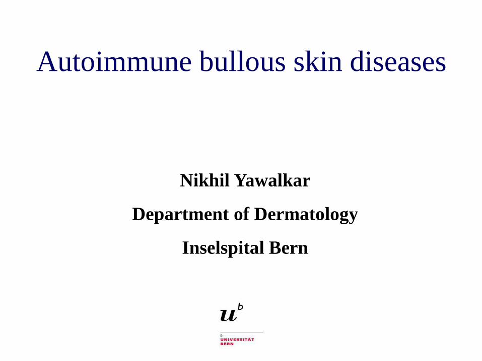
Autoimmune bullous skin diseases
Nikhil Yawalkar
Department of Dermatology
Inselspital Bern
• appearance of blisters or erosions
- skin
- mucous membranes
• characterized by the presence of auto-
antibodies that target distinct adhesion
molecules in the skin
Autoimmune bullous skin diseases
Autoimmune bullous skin diseases
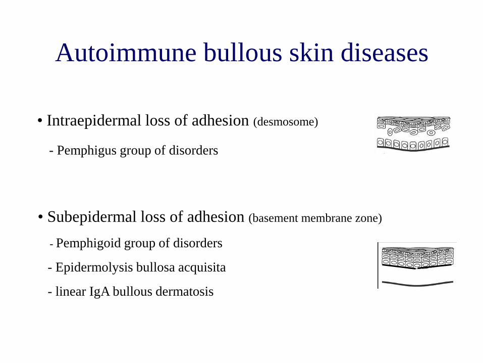
• Intraepidermal loss of adhesion (desmosome)
- Pemphigus group of disorders
• Subepidermal loss of adhesion (basement membrane zone)
- Pemphigoid group of disorders
- Epidermolysis bullosa acquisita
- linear IgA bullous dermatosis
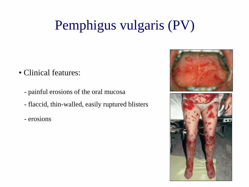
Pemphigus vulgaris (PV)
- painful erosions of the oral mucosa
- flaccid, thin-walled, easily ruptured blisters
- erosions
• Clinical features:
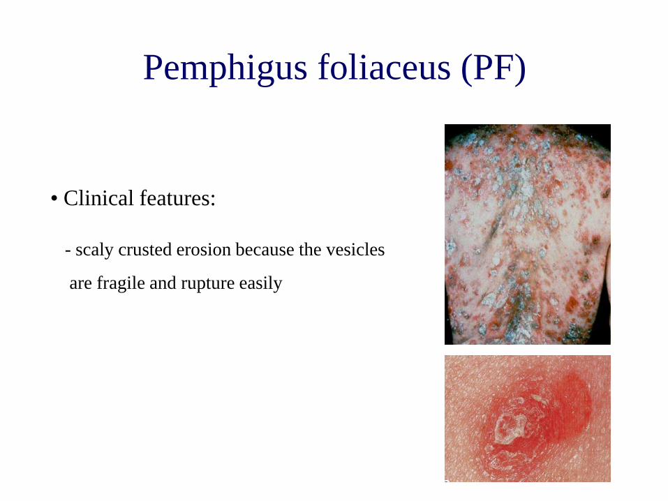
Pemphigus foliaceus (PF)
- scaly crusted erosion because the vesicles
are fragile and rupture easily
• Clinical features:
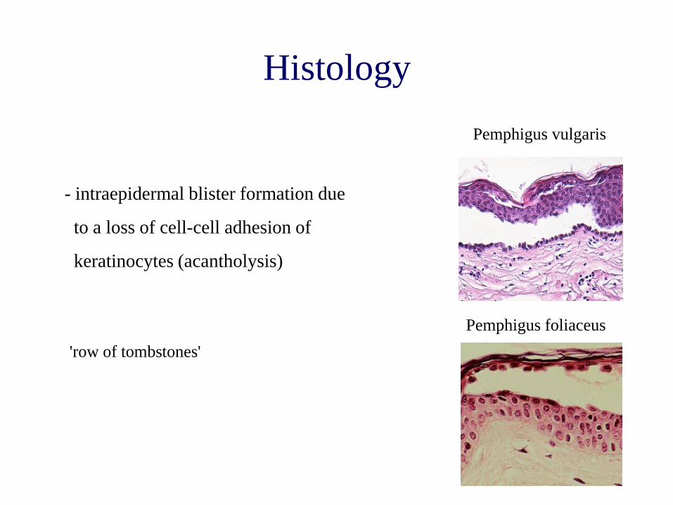
Histology
- intraepidermal blister formation due
to a loss of cell-cell adhesion of
keratinocytes (acantholysis)
'row of tombstones'
Pemphigus foliaceus
Pemphigus vulgaris
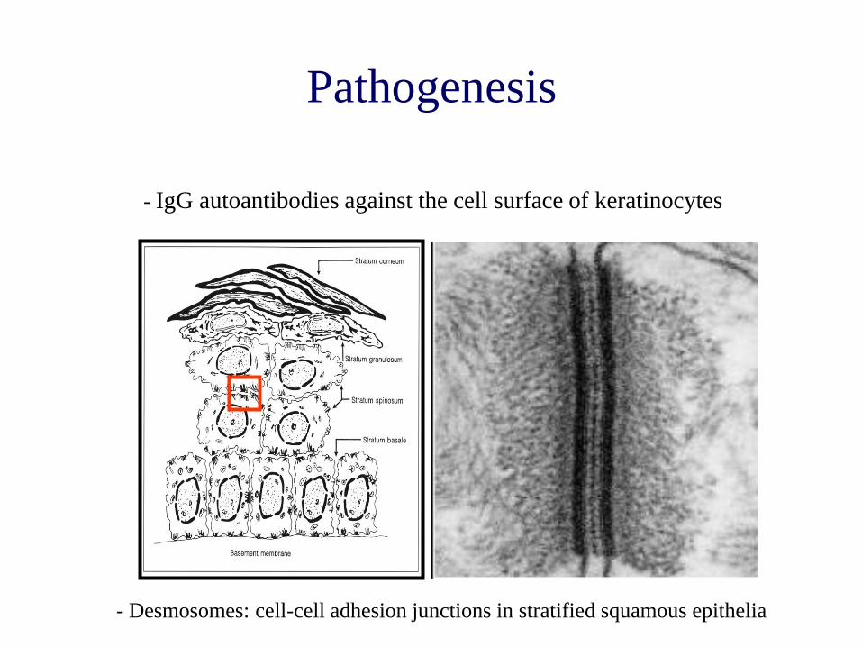
Pathogenesis
- IgG autoantibodies against the cell surface of keratinocytes
- Desmosomes: cell-cell adhesion junctions in stratified squamous epithelia
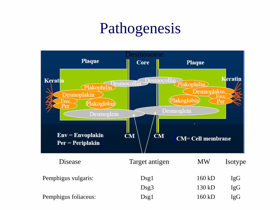
Pathogenesis
Pemphigus vulgaris: Dsg1 160 kD IgG
Dsg3 130 kD IgG
Pemphigus foliaceus: Dsg1 160 kD IgG
Desmosome
Disease Target antigen MW Isotype
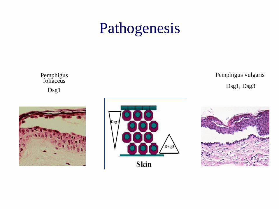
Pathogenesis
Pemphigus foliaceus
Dsg1
Pemphigus vulgaris
Dsg1, Dsg3
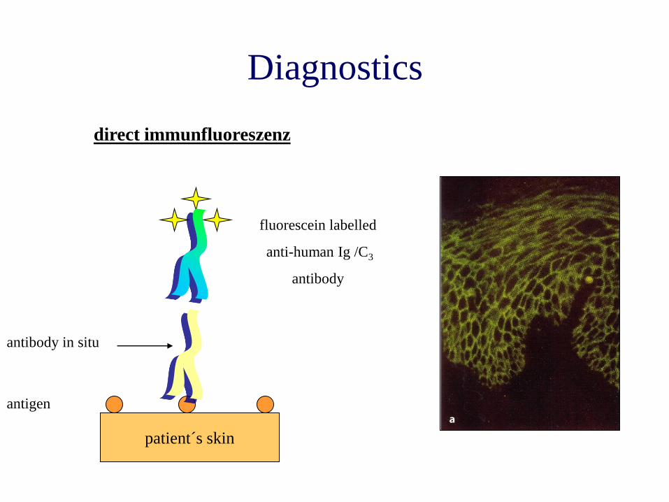
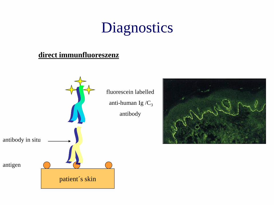
patient´s skin
direct immunfluoreszenz
antigen
antibody in situ
Diagnostics
fluorescein labelled
anti-human Ig /C3
antibody

monkey esophagus
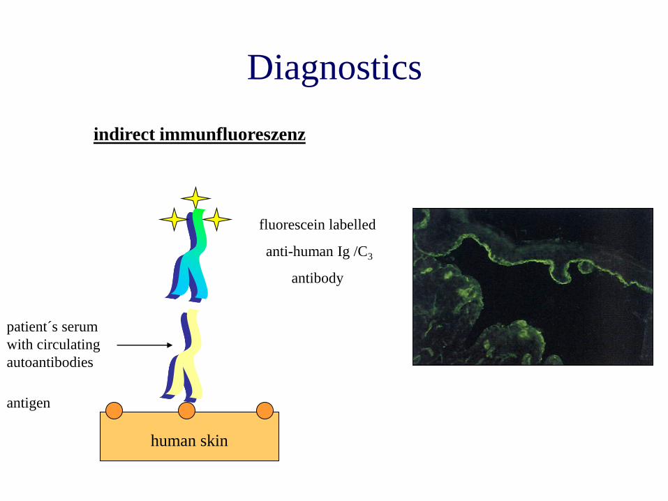
indirect immunfluoreszenz
antigen
Diagnostics
patient´s serum
with circulating
autoantibodies
fluorescein labelled
anti-human Ig /C3
antibody
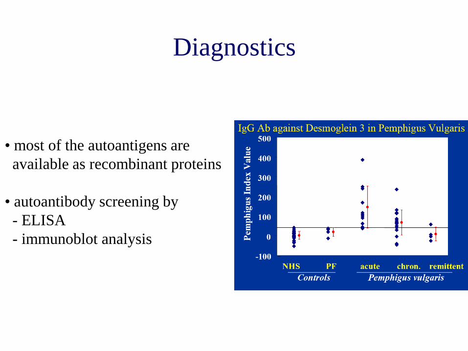
Diagnostics
• most of the autoantigens are
available as recombinant proteins
• autoantibody screening by
- ELISA
- immunoblot analysis

Autoimmune bullous skin diseases
• Intraepidermal loss of adhesion (desmosome)
- Pemphigus group of disorders
• Subepidermal loss of adhesion (basement membrane zone)
- Pemphigoid group of disorders
- Epidermolysis bullosa acquisita
- linear IgA bullous dermatosis
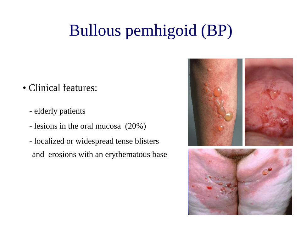
Bullous pemhigoid (BP)
- elderly patients
- lesions in the oral mucosa (20%)
- localized or widespread tense blisters
and erosions with an erythematous base
• Clinical features:
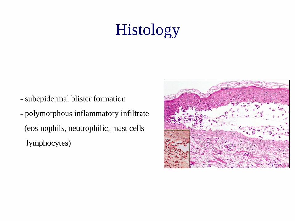
Histology
- subepidermal blister formation
- polymorphous inflammatory infiltrate
(eosinophils, neutrophilic, mast cells
lymphocytes)
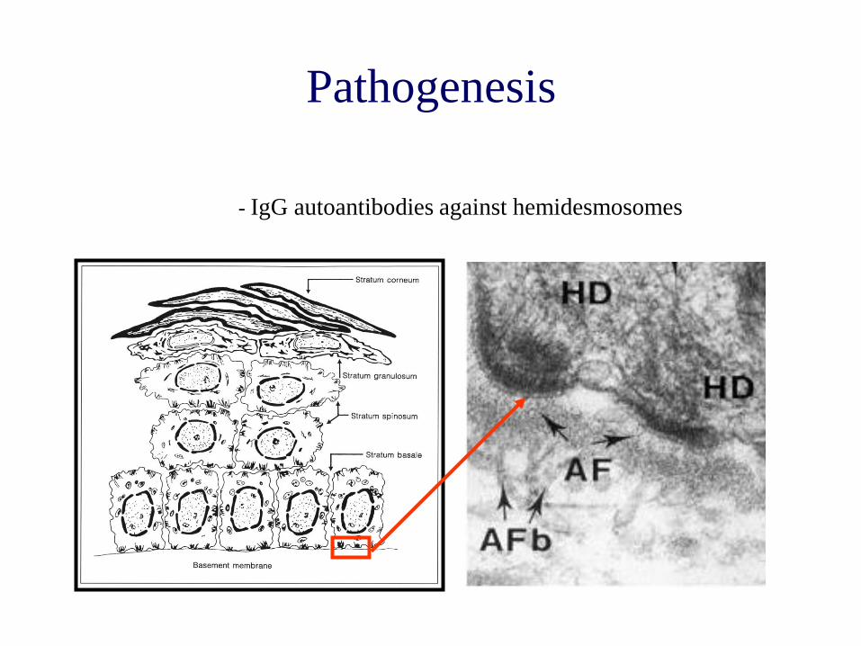
Pathogenesis
- IgG autoantibodies against hemidesmosomes
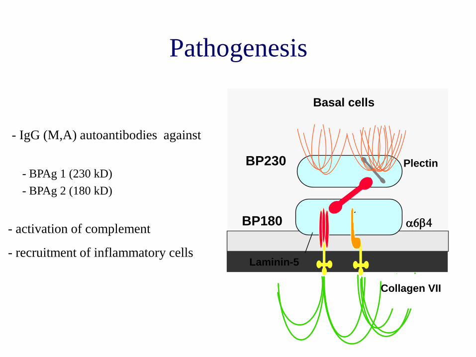
Pathogenesis
- IgG (M,A) autoantibodies against
BP180
Laminin-5
Plectin BP230
Collagen VII
a6b4
Basal cells
- BPAg 1 (230 kD)
- BPAg 2 (180 kD)
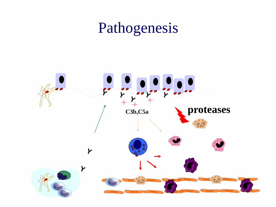
- activation of complement
- recruitment of inflammatory cells
T cell
T cell
proteases
B cell
C3b,C5a
Pathogenesis
T cell
MC
patient´s skin
direct immunfluoreszenz
antigen
antibody in situ
Diagnostics
fluorescein labelled
anti-human Ig /C3
antibody
human skin
indirect immunfluoreszenz
antigen
Diagnostics
patient´s serum
with circulating
autoantibodies
fluorescein labelled
anti-human Ig /C3
antibody
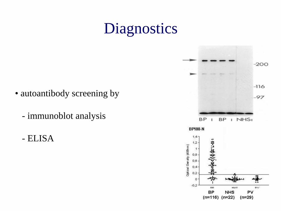
Diagnostics
• autoantibody screening by
- immunoblot analysis
- ELISA
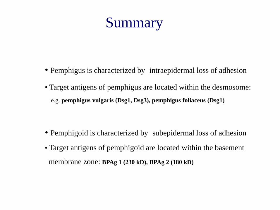
Summary
• Pemphigus is characterized by intraepidermal loss of adhesion
• Target antigens of pemphigus are located within the desmosome:
e.g. pemphigus vulgaris (Dsg1, Dsg3), pemphigus foliaceus (Dsg1)
• Pemphigoid is characterized by subepidermal loss of adhesion
• Target antigens of pemphigoid are located within the basement
membrane zone: BPAg 1 (230 kD), BPAg 2 (180 kD)

![Manifestations buccales du pemphigus paranéoplasique · 2013-02-08 · différencier le pemphigus paranéoplasique du pemphigus vulgaire [41, 63, 75, 83, 93, 100]. Par contre, la](https://img.pdfslide.net/doc/110x75/5f49b405f3d6f653f74e2428/manifestations-buccales-du-pemphigus-paranoplasique-2013-02-08-diffrencier.jpg)