Embed Size (px)
Citation preview
ORIGINAL ARTICLESee related Commentary on page v
Nitric Oxide Appears to Be a Mediator of Solar-SimulatedUltraviolet Radiation-Induced Immunosuppression in Humans
Johanna M. Kuchel, Ross St. C. Barnetson, and Gary M. HallidayDepartment of Medicine (Dermatology),The Melanoma and Skin Cancer Research Institute, Royal Prince Alfred Hospital at The University of Sydney,Sydney, Australia
Topical application of NG-methyl-L-arginine and 2,20 -dipyridyl were used to examine the respective roles ofnitric oxide and reactive oxygen species in solar-simu-lated ultraviolet radiation-induced immunosuppressionin humans in vivo. Immunosuppression was studiedusing a nickel contact hypersensitivity recall model.Ultraviolet radiation dose^responses were generated todetermine the extent to which NG-methyl-L-arginineand 2,20 -dipyridyl a¡ected the immune response. NG-methyl-L-arginine but not 2,20 -dipyridyl protectedthe immune system from ultraviolet radiation-inducedsuppression. Both NG-methyl-L-arginine and 2,20 -dipyridyl inhibited nitrite production. Nitrite is a de-gradation product of peroxynitrite, a cytotoxic media-tor resulting from reactions between nitric oxide andreactive oxygen species. This suggests that nitric oxide,
not its downstream product peroxynitrite, was likely tobe responsible for solar-simulated ultraviolet radiation-induced immunosuppression. In contrast, both nitricoxide and reactive oxygen species were mediators ofsolar-simulated ultraviolet radiation-induced apoptosisand loss of dendritic S-100þ cells (probably Langerhanscells) from the epidermis. It is likely that di¡erent me-chanisms are involved in these ultraviolet-induced end-points and that events in addition to Langerhans celldepletion are important for local immune suppres-sion to recall antigens in humans. Understanding themechanisms of cutaneous ultraviolet-induced oxidativestress will assist in the future design of novel productsthat protect skin from photoaging and skin cancer.Key words: apoptosis/dendritic cells/human/nitric oxide/skin. J Invest Dermatol 121:587 ^593, 2003
The skin is the largest organ of the human body and aprimary target for an array of environmental insults.Ultraviolet (UV) radiation has been identi¢ed as acause of several hazardous cutaneous e¡ects, includ-ing immune suppression, skin cancer (Kripke, 1974),
sunburn (delayed erythema), and premature aging (Gilchrest,1979). More than two decades ago it was recognized that UVmodulates the skin immune system. Highly antigenic UV-in-duced skin cancers in mice are immunologically rejected upontransplantation into normal unirradiated syngeneic mice (Kripke,1974). Pathak and Stratton (1968) were the ¢rst to note the pre-sence of free radicals in the skin after UVexposure. Even a singledose of UVB or UVA can compromise antioxidant enzyme de-fense systems. The resulting free radical load causes damage toimmune cells, attacking cellular components and damagingproteins, lipids, and DNA (Goldstone and Hunt, 1997).Nitric oxide (NO), a gaseous free radical, plays an integral role
in cutaneous function. It is synthesized from L-arginine, molecu-
lar oxygen, and reduced nicotinamide adenine dinucleotide phos-phate (NADPH) by NO synthase (NOS) (Marletta, 1994). In theskin, keratinocytes (Arany et al, 1996), Langerhans cells (Qureshiet al, 1996), dermal ¢broblasts (Wang et al, 1996), and melanocytes(Rocha and Guillo, 2001) all express inducible NOS upon stimu-lation with in£ammatory cytokines and/or lipopolysaccharide.NO itself mainly plays physiologic regulatory roles, but if pro-duced in excess, may combine with superoxide to form peroxy-nitrite (ONOO�), which has cytopathic e¡ects. Peroxynitriteexists in equilibrium with peroxynitrous acid, which may de-grade to nitrate and nitrite, which are both stable end-productsof NO metabolism (Pryor and Squadrito, 1995) (Fig 1). Peroxy-nitrite is a highly toxic reactive nitrogen intermediate that canreact directly with nucleic acids promoting DNA strand breakage(Zingarelli et al, 1996). NO plays a part in UV-induced immunesuppression in rodents partially via e¡ects on dendritic cells(Halliday et al, 1999; Yuen et al, 2002). Synthesized by endothelialcells, NO also acts as an endogenous modulator of leukocyticin¢ltration (Kroncke et al, 1997).The involvement of ferrous iron (Fe2þ ) in catalyzing redox
reactions in vitro is well established. Hydrogen peroxide (H2O2)reacts with iron to generate hydroxyl radicals (OH) via theFenton reaction: H2O2þ Fe2þ-OH�þOH�þ Fe3þ. 2,20 -Di-pyridyl, an aromatic amine, inhibits this reaction by chelating fer-rous iron. An increase in nonheme iron in the mouse and humanepidermis has been demonstrated after solar-simulated UVradiation (ssUV) (Jurkiewicz and Buettner, 1996). The topicalapplication to human skin of an iron chelator, desferrioxamine,has been previously shown to decrease signi¢cantly UV-inducedfree radicals (Jurkiewicz and Buettner, 1996). Topically applied
Address correspondence and reprint requests to: Prof. Gary Halliday,Dermatology Laboratories, Blackburn Building, D06, University ofSydney, Sydney, NSW 2006, Australia. Email: [email protected]: CHS, contact hypersensitivity; DEI, change in erythema
index; EI, erythema index; Fe2þ , ferrous iron; H2O2, hydrogen peroxide;MED, minimum erythemal dose; NOS, nitric oxide synthase iNOS, indu-cible nitric oxide synthase; O2
��, superoxide anion; OH, hydroxyl radical;ONOO�, peroxynitrite; ONOO�Hþ , peroxynitrous acid; SBC, sunburncell; ssUV, solar-simulated ultraviolet radiation.
Manuscript received November 6, 2002; revised February 24, 2003;accepted for publication April 7, 2003
0022-202X/03/$15.00 . Copyright r 2003 by The Society for Investigative Dermatology, Inc.
587

2,20 -dipyridyl, penetrates human skin where it remains for at least24 h (Bissett et al, 1991). It has been shown to prevent photocarci-nogenesis, wrinkling (Bissett et al, 1991; Halliday et al, 1999), andUV immunosuppression (Yuen et al, 2002) in murine models.Very little is known about the e¡ect of UV-induced free
radicals in humans in vivo. By using an analog of L-arginine,NG-methyl-L-arginine (L-NMMA) and an aromatic amine2,20 -dipyridyl, we examined the roles of NO and reactive oxygenspecies (ROS) in ssUV-induced immunosuppression, apoptosis,and Langerhans cell depletion in normal human skin.
MATERIALS AND METHODS
Subjects Volunteers were recruited from the local university, generalpopulation, and hospital sta¡. Subjects were excluded from the study ifthey had abnormal skin at the test site, a history of sun exposure to thelower back within 6 wk prior to entry into the study, were less than 18 yof age, pregnant, lactating, or were photo-sensitive or immune suppressedthrough illness or medication use. All subjects were recruited with approvalfrom both the Central Sydney Area Health Service and the University ofSydney Ethics Committees.Written, informed consent was provided priorto entry into the study.
L-NMMA and 2,20 -dipyridyl topical solutions The base solution wasprepared by combining 1,2-propanediol (Sigma, St Louis, Missouri),absolute ethanol (Selby-Biolab Scienti¢c Pty. Ltd, Clayton, Victoria,Australia) and double distilled water at a ratio of 1:2:1, at roomtemperature; 72.0 mg 2,20 -dipyridyl per ml (Sigma) or 129.6 mgL-NMMA per ml (Sigma) were then dissolved in this base solution.L-NMMA was applied topically to the human volunteers at 0.18 mg percm2 and 2,20 -dipyridyl at 0.1 mg per cm2. The solutions were stored at41C and discarded after 2 mo. The absorbance spectrum was performedusing the same technique used to determine absorbance of commercialsunscreens in Australia. L-NMMA, 2,20 -dipyridyl and base solutions wereeach applied to Mimskin, a 1.5� 40 mm quartz glass plate pro¢led withthe topography of human skin at the same concentration as was applied tohuman subjects (1.4 mL per cm2). After a 15 min dry time, the plate wasirradiated using a Labsphere UV-1000 sun protection factor (SPF)Analyzer (Labsphere, North Sutton, New Hampshire). The results wereblanked against Mimskin without product.
UV source A 1000 W xenon arc lamp with a collimated 7.5 cm squarebeam (Oriel, Stratford, Connecticut) was used for irradiation of humansubjects. The lamp emission was ¢ltered by two, 280 to 380 nm dichroicmirrors, which attenuated the visible and infrared components of thelight source. To provide ssUV, an ‘‘atmospheric attenuation ¢lter’’ (Orielcatalog number 81017) with a spectral cut-o¡ of 290 to 400 nm, was usedto modify the output spectrum. The spectrum was monitored at 2 nmintervals using a calibrated OL-754 spectroradiometer (OptronicLaboratories Inc., Orlando, Florida) (Fig 2). Spectral irradiance wasdetermined at least daily with an IL 1350 broadband radiometer usingSED 038 (UVA) and SED 240 (UVB) detectors (International Light,Newburyport, MA) calibrated against the xenon-arc solar simulator withthe spectroradiometer. The integrated irradiance of the ssUV at the skinsurface measured at 7.5 cm was 5.1 mW per cm2 UVB (290^320 nm),
11.8 mW per cm2 UVAII (320^340 nm), and 39.2 mW per cm2 UVAI(340^400 nm).
Nitrite inhibition by L-NMMA and 2,20 -dipyridyl, measured bythe Greiss assay All equipment and reagents were obtained from GibcoBRL (Life Technologies, Inc., Grand Island, New York) unless otherwisespeci¢ed. COLO-16 cells, a human epidermal squamous carcinoma cellline (Moore et al, 1975), were added to 24-well plates at a concentration of7�105 cells per mL. Cells were cultured in RPMI 1640 medium with 1%N-2-hydroxyethylpiperazine-N0-2-ethane sulfonic acid, 2% L-glutamine,10% fetal calf serum, 0.2% penicillin/streptomycin, 0.5% fungizone (250 mgamphotericin in 250 mg sodium desoxycholate per mL), and 0.2%gentamycin (Sigma). The assay was performed after 24 h when the cellswere con£uent. Media was replaced with either 1 mL phosphate-bu¡eredsaline (PBS), PBS with L-NMMA (0.3 mg per ml) or PBS with 2,20 -dipyridyl (0.18 mg per ml). Doses of L-NMMA and 2,20 -dipyridyl percm2 were the same as applied topically in the immunosuppression studies.The cells were incubated at 371C in 5% CO2 for a further 15 min thenrinsed twice with 1.5 mL of PBS and left in 200 mL of PBS. They wereirradiated with 2.5 J per cm2 ssUV, the dose of ssUV required to causesigni¢cant immunosuppression in the human contact hypersensitivity(CHS) study. One set of wells was sham-irradiated. Immediately afterirradiation, 800 mL of either L-NMMA in PBS, 2,20 -dipyridyl in PBS orPBS alone was added to make up to the original concentration of each testsubstance. The plates were incubated for 15 min at room temperature priorto adding the supernatant to an equal volume of Greiss reagent (1%sulfanilamide, 3.5% phosphoric acid and 0.1% b-naphthylethylenediamine dihydrochloride, added to double distilled water at roomtemperature). Fifteen minutes later, supernatant absorption at 550 nm wasdetermined using a Unicam 8625 UV/Visible re£ectance spectro-photometer (Unicam Ltd, Division of Analytical Technology Inc.,Cambridge, UK). The concentration of the sodium nitrite was interpo-lated from the linear portion of a standard curve using sodium nitrite indouble distilled water as a reference. The study was performed in triplicate.
The e¡ect of L-NMMA and 2,20 -dipyridyl on sunburn The e¡ecton sunburn of L-NMMA and 2,20 -dipyridyl solutions were determinedusing two separate groups of 10 volunteers, age and skin type matched tothe immunosuppression study (¢ve males, 15 females). Minimumerythemal doses (MED) were determined for both a site treated with 1.4mL per cm2 base solution and a site treated with either 1.4 mL per cm2 L-
Figure1. UV-induced production of reactive nitrogen species.L-NMMA inhibits the production of NO through competitive inhibitionof NO synthase. 2,20 -Dipyridyl inhibits the production of ROS via theFenton reaction. NO2
�, nitrite; NO3�, nitrate; ONOO�, peroxynitrite
anion; ONOO�Hþ , peroxynitrous acid; O2��, superoxide anion.
Figure 2. Comparison of the experimental ssUV spectra withsunlight. The solar UV spectral irradiance is the noon solar spectrum ona cloudless October day, in Sydney, Australia, 2001. The spectra weremeasured at 2 nm intervals using a calibrated spectroradiometer (OptronicLaboratories Inc., Orlando, Florida). All spectra were normalized to 1.0 at320 nm.
588 KUCHEL ETAL THE JOURNAL OF INVESTIGATIVE DERMATOLOGY
NMMA or 2,20 -dipyridyl solution. Test solutions were applied on eachvolunteer’s lower back 15 min prior to irradiation. A series of ten 6� 6mm squares were irradiated with increasing, equal increments of ssUV.The MED was de¢ned as the lowest dose of ssUV at which clearlydemarcated erythema was visualized at 24 h postirradiation.
Nickel patch testing To determine the degree of nickel sensitivityof each volunteer, the left side of the lower back was initially patchtested with nickel sulfate (NiSO4.6H2O) in a petrolatum base (TrolabHermal, Reinbeck, Germany). Three concentrations of nickel sulfate, from0.0125% to 5%, with an additional petrolatum-only control, were appliedto the left lateral back of each volunteer, in 9 mm Finn chambers (Epitest,Tuusula, Finland) (Fischer and Rystedt, 1985). The patches remained in situfor 48 h and the reactions were assessed 21 to 28 h after patch removal.Clinically, each reaction was graded from 1 to 10 depending on the degreeof vesiculation and induration.The lowest nickel concentration resulting ina uniform, erythematous reaction with papules was determined for eachsubject. This concentration was then used for the immunosuppressionstudy.
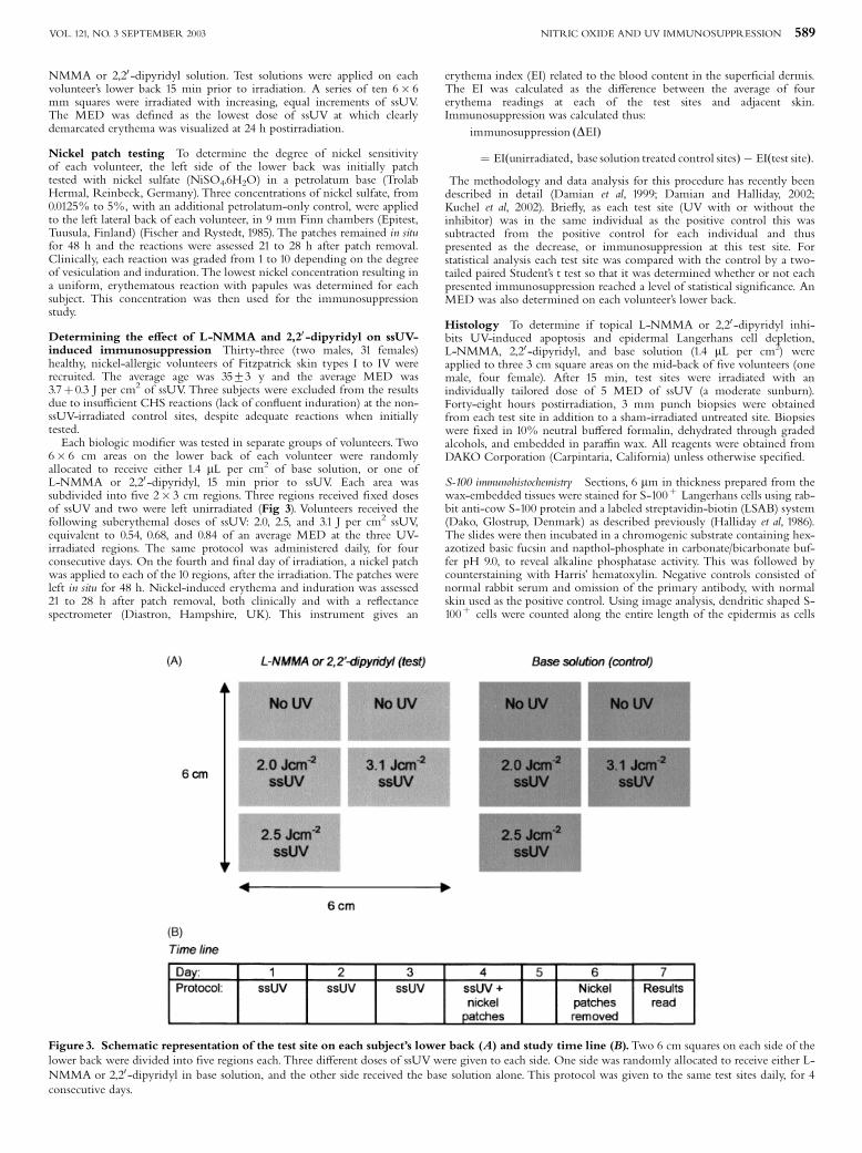
Determining the e¡ect of L-NMMA and 2,20 -dipyridyl on ssUV-induced immunosuppression Thirty-three (two males, 31 females)healthy, nickel-allergic volunteers of Fitzpatrick skin types I to IV wererecruited. The average age was 3573 y and the average MED was3.7þ0.3 J per cm2 of ssUV. Three subjects were excluded from the resultsdue to insu⁄cient CHS reactions (lack of con£uent induration) at the non-ssUV-irradiated control sites, despite adequate reactions when initiallytested.Each biologic modi¢er was tested in separate groups of volunteers. Two
6� 6 cm areas on the lower back of each volunteer were randomlyallocated to receive either 1.4 mL per cm2 of base solution, or one ofL-NMMA or 2,20 -dipyridyl, 15 min prior to ssUV. Each area wassubdivided into ¢ve 2� 3 cm regions. Three regions received ¢xed dosesof ssUV and two were left unirradiated (Fig 3). Volunteers received thefollowing suberythemal doses of ssUV: 2.0, 2.5, and 3.1 J per cm2 ssUV,equivalent to 0.54, 0.68, and 0.84 of an average MED at the three UV-irradiated regions. The same protocol was administered daily, for fourconsecutive days. On the fourth and ¢nal day of irradiation, a nickel patchwas applied to each of the 10 regions, after the irradiation.The patches wereleft in situ for 48 h. Nickel-induced erythema and induration was assessed21 to 28 h after patch removal, both clinically and with a re£ectancespectrometer (Diastron, Hampshire, UK). This instrument gives an
erythema index (EI) related to the blood content in the super¢cial dermis.The EI was calculated as the di¡erence between the average of fourerythema readings at each of the test sites and adjacent skin.Immunosuppression was calculated thus:
immunosuppression ðDEIÞ
¼ EIðunirradiated; base solution treated control sitesÞ � EIðtest siteÞ:
The methodology and data analysis for this procedure has recently beendescribed in detail (Damian et al, 1999; Damian and Halliday, 2002;Kuchel et al, 2002). Brie£y, as each test site (UV with or without theinhibitor) was in the same individual as the positive control this wassubtracted from the positive control for each individual and thuspresented as the decrease, or immunosuppression at this test site. Forstatistical analysis each test site was compared with the control by a two-tailed paired Student’s t test so that it was determined whether or not eachpresented immunosuppression reached a level of statistical signi¢cance. AnMED was also determined on each volunteer’s lower back.
Histology To determine if topical L-NMMA or 2,20 -dipyridyl inhi-bits UV-induced apoptosis and epidermal Langerhans cell depletion,L-NMMA, 2,20 -dipyridyl, and base solution (1.4 mL per cm2) wereapplied to three 3 cm square areas on the mid-back of ¢ve volunteers (onemale, four female). After 15 min, test sites were irradiated with anindividually tailored dose of 5 MED of ssUV (a moderate sunburn).Forty-eight hours postirradiation, 3 mm punch biopsies were obtainedfrom each test site in addition to a sham-irradiated untreated site. Biopsieswere ¢xed in 10% neutral bu¡ered formalin, dehydrated through gradedalcohols, and embedded in para⁄n wax. All reagents were obtained fromDAKO Corporation (Carpintaria, California) unless otherwise speci¢ed.
S-100 immunohistochemistry Sections, 6 mm in thickness prepared from thewax-embedded tissues were stained for S-100þ Langerhans cells using rab-bit anti-cow S-100 protein and a labeled streptavidin-biotin (LSAB) system(Dako, Glostrup, Denmark) as described previously (Halliday et al, 1986).The slides were then incubated in a chromogenic substrate containing hex-azotized basic fucsin and napthol-phosphate in carbonate/bicarbonate buf-fer pH 9.0, to reveal alkaline phosphatase activity. This was followed bycounterstaining with Harris’ hematoxylin. Negative controls consisted ofnormal rabbit serum and omission of the primary antibody, with normalskin used as the positive control. Using image analysis, dendritic shaped S-100þ cells were counted along the entire length of the epidermis as cells
Figure 3. Schematic representation of the test site on each subject’s lower back (A) and study time line (B).Two 6 cm squares on each side of thelower back were divided into ¢ve regions each. Three di¡erent doses of ssUVwere given to each side. One side was randomly allocated to receive either L-NMMA or 2,20 -dipyridyl in base solution, and the other side received the base solution alone. This protocol was given to the same test sites daily, for 4consecutive days.
NITRIC OXIDE AND UV IMMUNOSUPPRESSION 589VOL. 121, NO. 3 SEPTEMBER 2003

per linear millimeter of epidermis. As the S-100 protein is present in bothmelanocytes and Langerhans cells in the epidermis and melanocytes arenot found more than two epidermal cells above the basement membrane(Halliday et al, 1986), only S-100þ cells situated at least two cells abovethe basement membrane were scored. Dermal S-100þ cells were alsocounted in 10 random ¢elds using the � 20 lens of a light microscope. Cellnumbers were expressed as the percentage change in dendritic S-100þ cellnumbers compared with the unirradiated control site, which was normal-ized to 100%.
SBC assay Hematoxylin and eosin staining was performed on6 mm thick sections. Sunburn cells (SBC) are UV-damaged keratinocyteswith pyknotic nuclei and eosinophilic cytoplasm. The histologic criteriafor identi¢cation of SBC were those described byWoodcock and Magnus(1976). Slides were counted blind and in duplicate. Using image analysis(Chromatic Color Image Analysis System, Leitz, Sydney, NSW, Australia),SBC were counted along the entire length of the epidermis and expressedas cells per linear millimeter of epidermis. Counts were expressed as a per-centage of the site receiving ssUV and base solution alone, which was nor-malized to 100%.
Statistical analysis Statview (version 4.51, Abacus Concepts Inc. 1992^95, Berkeley, California) was utilized for statistical analysis. Results arepresented as the meanþ SEM and were considered signi¢cant if po0.05using a paired two-tailed Student’s t test.
RESULTS
L-NMMA and 2,20 -dipyridyl absorption spectra NeitherL-NMMA in solution or the base solution itself absorbed inthe UV portion of the solar spectrum. 2,20 -Dipyridyl absorbedin the UVB range, up to 310 nm, with an absorption maximumof 0.48, which is just less than half that of an opaque object, withan absorption of 1 and much less than a low SPF (9) sunscreen(Fig 4).
L-NMMA and 2,20 -dipyridyl both inhibit nitrite productionin response to ssUV UV-irradiated COLO-16 cells producedsigni¢cantly more nitrites than the unirradiated controls(po0.05) (Fig 5). A signi¢cantly lower nitrite concentration wasproduced by cells irradiated with L-NMMA (12�10�6 g perliter) or 2,20 -dipyridyl (9�10�6 g per liter) treatment comparedwith irradiated control cells (22.5�10�6 g per liter) (po0.05).There was no signi¢cant di¡erence in nitrite productionbetween irradiated cells treated with L-NMMA and 2,20 -dipyridyl (p40.05).
L-NMMA and 2,20 -dipyridyl e¡ects on sunburn Todetermine whether L-NMMA or 2,20 -dipyridyl altered ssUV-induced erythema, two groups of 10 non-nickel allergicvolunteers were recruited. Subjects were irradiated with incre-mental doses of ssUV, with or without topical L-NMMA or2,20 -dipyridyl. L-NMMA increased the MED from 4.3þ 0.2 to4.9þ 0.3. 2,20 -Dipyridyl also increased the MED, from 4.4þ 0.5to 5.770.7.
ssUV-induced suppression of the recall phase of CHSto nickel in humans
L-NMMA inhibits ssUV-induced immunosuppression L-NMMA didnot have any irritant e¡ect either alone, or in combination withnickel. It did not alter the re£ectance readings. No signi¢cant dif-ference was detected between the control unirradiated nickel siteand the site that received L-NMMA plus nickel (0 UV inFig 6A). In this study, only the highest dose of ssUV (3.1 J percm2) resulted in a level of immunosuppression that reached statis-tical signi¢cance (Fig 6A, i.e., a signi¢cant di¡erence betweenthe control unirradiated and irradiated sites). L-NMMA inhibitedthis immunosuppression as the di¡erence between the controlunirradiated base lotion treated and the L-NMMA-treated UV-irradiated site was not signi¢cant (i.e., there was no signi¢cantimmunosuppression). Signi¢cant immunosuppression was notobserved at any UV dose at the sites treated with L-NMMA. At
the highest ssUV dose (3.1 J per cm2), immunosuppression (DEI)was reduced by 58%, from 20.8 with base solution alone, to 8.7with L-NMMA.
2,20 -Dipyridyl does not inhibit ssUV-induced immunosuppression In aseparate experiment, 2,20 -dipyridyl was applied to each test site(Fig 6B). This compound also did not have any irritant e¡ecteither alone, or when applied to a site that received nickel. Therewas no signi¢cant di¡erence detected between the EI readings atthe unirradiated control nickel-treated site and the site receiving2,20 -dipyridyl and nickel (0 UV in Fig 6B). ssUV caused immu-nosuppression in a dose-related manner, with both 2.5 and3.1 J per cm2 resulting in a level of immunosuppression thatreached statistical signi¢cance, after the application of both baseand 2,20 -dipyridyl solution. Signi¢cant immunosuppression
Figure 4. The absorbance spectrum of 2,20 -dipyridyl and L-NMMA,compared with a low SPF sunscreen with titanium dioxide. Solu-tions of L-NMMA and 2,20 -dipyridyl were applied at 1.4 mL per cm2, toMimskin, a 1.5 mm� 40 mm quartz glass plate pro¢led with the topogra-phy of human skin. Sunscreen was applied at a dose of 2.0 mL per cm2. Theplate was irradiated after a 15 min dry time, using a Labsphere UV-1000SPF analyzer (Labsphere, North Sutton, New Hampshire).The results wereblanked against Mimskin without product.
Figure 5. L-NMMA and 2,20 -dipyridyl inhibit nitrite produced inresponse to ssUV. COLO-16 cells in PBS were irradiated with ssUV aftera 15 min incubation in L-NMMA or 2,20 -dipyridyl in PBS. Controls con-sisted of both either irradiated or sham-irradiated cells in PBS. MeanþSEM are shown. �Signi¢cance: po0.05, compared with the ssUV-irra-diated control using a paired two-tailed Student’s t test (n¼ 6).
590 KUCHEL ETAL THE JOURNAL OF INVESTIGATIVE DERMATOLOGY

(DEI) of 21.1 and 15.0 occurred at a dose of 2.5 J per cm2 ssUV,both with and without the iron chelator, respectively. No signi¢-cant immunosuppression occurred at the lower UV dose in thisstudy.Whereas L-NMMA reduced immunosuppression, 2,20 -di-pyridyl increased the level of immunosuppression by 30.471.6%(average increase in immunosuppression over the three doses ofssUV tested), this was not a statistically signi¢cant e¡ect.
L-NMMA and 2,20 -dipyridyl inhibit the loss of S-100þcellsfrom the epidermis in human skin in vivo Skin biopsies were
collected 48 h after application of L-NMMA, 2,20 -dipyridyl orbase solution and irradiation with 5 MED of ssUV. ssUV causeda signi¢cant reduction in epidermal S-100þ dendritic cells anda concomitant increase in dendritic S-100þ cells in the dermis(Fig 7). Both topical L-NMMA and 2,20 -dipyridyl inhibitedS-100þ cell loss from the epidermis. There was no statisticallysigni¢cant di¡erence between S-100þ cells at the unirradiatedcontrol sites and each test site that received L-NMMA or 2,20 -dipyridyl prior to irradiation. There was also no di¡erencein S-100 counts between test sites receiving L-NMMA or 2,20 -dipyridyl, at the concentrations studied. The 3.5-fold increase inS-100þ cells in the dermis post-ssUV, was greater than the2-fold increase that was observed at the test sites receivingL-NMMA or 2,20 -dipyridyl (Fig 7B). This suggests that NOand ROS both mediate migration of dendritic S-100þ cellsfrom the epidermis in response to ssUV.
L-NMMA and 2,20 -dipyridyl both inhibit SBC formation inhuman skin in vivo Skin biopsies were collected 48 h afterapplication of L-NMMA, 2,20 -dipyridyl, or base solution, andirradiation with 5 MED of ssUV. Both L-NMMA and 2,20 -dipyridyl signi¢cantly inhibited SBC formation by 40% and60%, respectively, compared with the test site with base solutiononly (po0.05; n¼ 5; paired Student’s t test) (Fig 8). Nosigni¢cant di¡erence was observed between the UV-irradiated
Figure 6. ssUV-induced immunosuppression is inhibited by (A)L-NMMA but not (B) 2,20 -dipyridyl.Two groups of 15 volunteers wereexposed daily to a range of doses of ssUV (0, 2.0, 2.5, and 3.1 J per cm2).Some skin areas received L-NMMA or 2,20 -dipyridyl, whereas others re-ceived the base solution only. Each skin region received an identical proto-col for each of 4 consecutive days. This was followed by nickel patchtesting. The area that received no ssUV and topical base solution only, wasthe positive, unirradiated control to which all other sites were compared.Nickel-induced erythema was measured by re£ectance spectroscopy. Theresult shown at each test site is the reaction subtracted from the unirra-diated, base solution treated control reaction in the same volunteer (DEI),and is therefore a measure of immunosuppression at that test site. As theexperimental design is to study control and test reactions in the same indi-vidual, these control and test reactions were compared using a paired two-tailed Student’s t test to assess whether the level of immunosuppression ateach test site was signi¢cant. Meanþ SEM are shown. �Signi¢cance;po0.05, compared with unirradiated control site using a paired two-tailedStudent’s t test (n¼15).
Figure 7. ssUV-induced NO and ROS are involved in the loss of S-100þ cells from the epidermis in human skin in vivo. L-NMMA (0.18mg per cm2), 2,20 -dipyridyl (0.1 mg per cm2) and base solution were ap-plied at 1.4 mL per cm2, 15 min prior to 5 MED of ssUV. Forty-eight hourspostirradiation, 3 mm punch biopsies were taken from each test site, and anunirradiated, untreated site. These were para⁄n-embedded and immuno-stained for S-100. Results are presented as the percentage change in S-100þ cells compared with the unirradiated site in the (A) epidermis and(B) dermis. Meanþ SEM are shown. �Signi¢cance; po0.05, comparedwith the unirradiated site using a paired two-tailed Student’s t test (n¼ 5).
NITRIC OXIDE AND UV IMMUNOSUPPRESSION 591VOL. 121, NO. 3 SEPTEMBER 2003
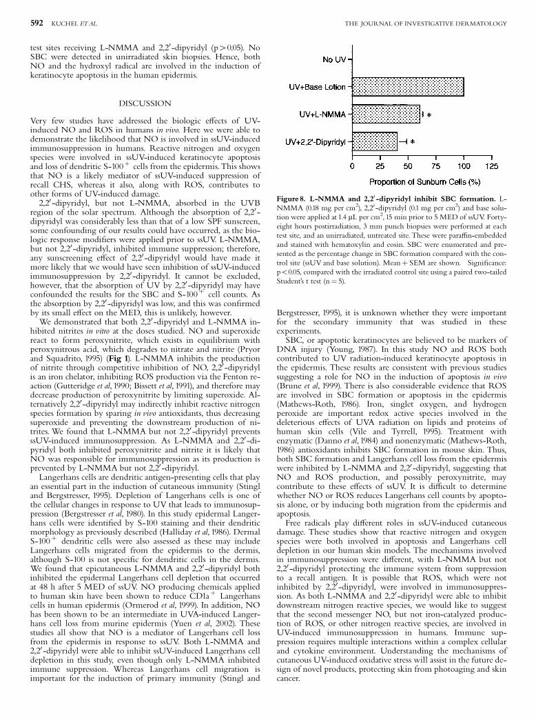
test sites receiving L-NMMA and 2,20 -dipyridyl (p40.05). NoSBC were detected in unirradiated skin biopsies. Hence, bothNO and the hydroxyl radical are involved in the induction ofkeratinocyte apoptosis in the human epidermis.
DISCUSSION
Very few studies have addressed the biologic e¡ects of UV-induced NO and ROS in humans in vivo. Here we were able todemonstrate the likelihood that NO is involved in ssUV-inducedimmunosuppression in humans. Reactive nitrogen and oxygenspecies were involved in ssUV-induced keratinocyte apoptosisand loss of dendritic S-100þ cells from the epidermis.This showsthat NO is a likely mediator of ssUV-induced suppression ofrecall CHS, whereas it also, along with ROS, contributes toother forms of UV-induced damage.2,20 -dipyridyl, but not L-NMMA, absorbed in the UVB
region of the solar spectrum. Although the absorption of 2,20 -dipyridyl was considerably less than that of a low SPF sunscreen,some confounding of our results could have occurred, as the bio-logic response modi¢ers were applied prior to ssUV. L-NMMA,but not 2,20 -dipyridyl, inhibited immune suppression; therefore,any sunscreening e¡ect of 2,20 -dipyridyl would have made itmore likely that we would have seen inhibition of ssUV-inducedimmunosuppression by 2,20 -dipyridyl. It cannot be excluded,however, that the absorption of UV by 2,20 -dipyridyl may haveconfounded the results for the SBC and S-100þ cell counts. Asthe absorption by 2,20 -dipyridyl was low, and this was con¢rmedby its small e¡ect on the MED, this is unlikely, however.We demonstrated that both 2,20 -dipyridyl and L-NMMA in-
hibited nitrites in vitro at the doses studied. NO and superoxidereact to form peroxynitrite, which exists in equilibrium withperoxynitrous acid, which degrades to nitrate and nitrite (Pryorand Squadrito, 1995) (Fig 1). L-NMMA inhibits the productionof nitrite through competitive inhibition of NO, 2,20 -dipyridylis an iron chelator, inhibiting ROS production via the Fenton re-action (Gutteridge et al, 1990; Bissett et al, 1991), and therefore maydecrease production of peroxynitrite by limiting superoxide. Al-ternatively 2,20 -dipyridyl may indirectly inhibit reactive nitrogenspecies formation by sparing in vivo antioxidants, thus decreasingsuperoxide and preventing the downstream production of ni-trites.We found that L-NMMA but not 2,20 -dipyridyl preventsssUV-induced immunosuppression. As L-NMMA and 2,20 -di-pyridyl both inhibited peroxynitrite and nitrite it is likely thatNO was responsible for immunosuppression as its production isprevented by L-NMMA but not 2,20 -dipyridyl.Langerhans cells are dendritic antigen-presenting cells that play
an essential part in the induction of cutaneous immunity (Stingland Bergstresser, 1995). Depletion of Langerhans cells is one ofthe cellular changes in response to UV that leads to immunosup-pression (Bergstresser et al, 1980). In this study epidermal Langer-hans cells were identi¢ed by S-100 staining and their dendriticmorphology as previously described (Halliday et al, 1986). DermalS-100þ dendritic cells were also assessed as these may includeLangerhans cells migrated from the epidermis to the dermis,although S-100 is not speci¢c for dendritic cells in the dermis.We found that epicutaneous L-NMMA and 2,20 -dipyridyl bothinhibited the epidermal Langerhans cell depletion that occurredat 48 h after 5 MED of ssUV. NO producing chemicals appliedto human skin have been shown to reduce CD1aþ Langerhanscells in human epidermis (Ormerod et al, 1999). In addition, NOhas been shown to be an intermediate in UVA-induced Langer-hans cell loss from murine epidermis (Yuen et al, 2002). Thesestudies all show that NO is a mediator of Langerhans cell lossfrom the epidermis in response to ssUV. Both L-NMMA and2,20 -dipyridyl were able to inhibit ssUV-induced Langerhans celldepletion in this study, even though only L-NMMA inhibitedimmune suppression. Whereas Langerhans cell migration isimportant for the induction of primary immunity (Stingl and
Bergstresser, 1995), it is unknown whether they were importantfor the secondary immunity that was studied in theseexperiments.SBC, or apoptotic keratinocytes are believed to be markers of
DNA injury (Young, 1987). In this study NO and ROS bothcontributed to UV radiation-induced keratinocyte apoptosis inthe epidermis. These results are consistent with previous studiessuggesting a role for NO in the induction of apoptosis in vivo(Brune et al, 1999). There is also considerable evidence that ROSare involved in SBC formation or apoptosis in the epidermis(Mathews-Roth, 1986). Iron, singlet oxygen, and hydrogenperoxide are important redox active species involved in thedeleterious e¡ects of UVA radiation on lipids and proteins ofhuman skin cells (Vile and Tyrrell, 1995). Treatment withenzymatic (Danno et al, 1984) and nonenzymatic (Mathews-Roth,1986) antioxidants inhibits SBC formation in mouse skin. Thus,both SBC formation and Langerhans cell loss from the epidermiswere inhibited by L-NMMA and 2,20 -dipyridyl, suggesting thatNO and ROS production, and possibly peroxynitrite, maycontribute to these e¡ects of ssUV. It is di⁄cult to determinewhether NO or ROS reduces Langerhans cell counts by apopto-sis alone, or by inducing both migration from the epidermis andapoptosis.Free radicals play di¡erent roles in ssUV-induced cutaneous
damage. These studies show that reactive nitrogen and oxygenspecies were both involved in apoptosis and Langerhans celldepletion in our human skin models. The mechanisms involvedin immunosuppression were di¡erent, with L-NMMA but not2,20 -dipyridyl protecting the immune system from suppressionto a recall antigen. It is possible that ROS, which were notinhibited by 2,20 -dipyridyl, were involved in immunosuppres-sion. As both L-NMMA and 2,20 -dipyridyl were able to inhibitdownstream nitrogen reactive species, we would like to suggestthat the second messenger NO, but not iron-catalyzed produc-tion of ROS, or other nitrogen reactive species, are involved inUV-induced immunosuppression in humans. Immune sup-pression requires multiple interactions within a complex cellularand cytokine environment. Understanding the mechanisms ofcutaneous UV-induced oxidative stress will assist in the future de-sign of novel products, protecting skin from photoaging and skincancer.
Figure 8. L-NMMA and 2,20 -dipyridyl inhibit SBC formation. L-NMMA (0.18 mg per cm2), 2,20 -dipyridyl (0.1 mg per cm2) and base solu-tion were applied at 1.4 mL per cm2, 15 min prior to 5 MED of ssUV. Forty-eight hours postirradiation, 3 mm punch biopsies were performed at eachtest site, and an unirradiated, untreated site. These were para⁄n-embeddedand stained with hematoxylin and eosin. SBC were enumerated and pre-sented as the percentage change in SBC formation compared with the con-trol site (ssUV and base solution). Meanþ SEM are shown. �Signi¢cance:po0.05, compared with the irradiated control site using a paired two-tailedStudent’s t test (n¼ 5).
592 KUCHEL ETAL THE JOURNAL OF INVESTIGATIVE DERMATOLOGY
We acknowledge with gratitude the time and e¡ort given by the volunteers. Dr. J.Kuchel was supported by an Australian Postgraduate Award. Thank you to StuartDavies for staining the biopsies with anti-S-100 antibody.
REFERENCES
Arany I, Brysk MM, Brysk H, Tyring SK: Regulation of inducible nitric oxidesynthase mRNA levels by di¡erentiation and cytokines in human keratino-cytes. Biochem Biophys Res Commun 220:618^622, 1996
Bergstresser PR, Toews GB, Streilein JW: Natural and perturbed distributions ofLangerhans cells: Responses to ultraviolet light, heterotopic skin grafting, anddinitro£uorobenzene sensitisation. J Invest Dermatol 75:73^77, 1980
Bissett DL, Chatterjee R, Hannon DP: Chronic ultraviolet radiation-induced in-crease in skin iron and the photoprotective e¡ect of topically applied iron che-lators. Photochem Photobiol 54:215^223, 1991
Brune B, von Knethen A, Sandau KB: Nitric oxide (NO): An e¡ector of apoptosis.Cell Death Di¡erentiation 6:969^975, 1999
Damian DL, Halliday GM: Measurement of ultraviolet radiation-induced suppres-sion of recall contact and delayed-type hypersensitivity in humans. Methods28:34^45, 2002
Damian DL, Halliday GM, St Barnetson RC: Sun protection factor measurement ofsunscreens is dependent on minimal erythema dose. Br J Dermatol 141:502^507,1999
Danno K, Horio T,Takigawa M, Imamura S: Role of oxygen intermediates in UV-induced epidermal cell injury. J Invest Dermatol 83:166^168, 1984
Fischer T, Rystedt I: False positive, follicular and irritant patch test reactions of metalsalts. Contact Dermatitis 12:93^98, 1985
Gilchrest BA: Relationship between actinic damage and chronologic aging in kera-tinocyte cultures of human skin. J Invest Dermatol 72:219^223, 1979
Goldstone SD, Hunt NH: Redox regulation of the mitogen-activated protein kinasepathway during lymphocyte activation. Biochim Biophys Acta 1355:353^360, 1997
Gutteridge JM, Maidt L, Poyer L: Superoxide dismutase and Fenton chemistry. Re-action of ferric-EDTA complex and ferric-bipyridyl complex with hydrogenperoxide without the apparent formation of iron (II). Biochem J 269:169^174,1990
Halliday GM, McArdle JP, Knight BA, Muller HK: New methodology for assess-ment of the Langerhans’ cell network. J Pathol 148:127^134, 1986
Halliday GM, Russo PA, Yuen KS, Robertson BO: E¡ect of inhibitors of oxygenradical and nitric oxide formation on UV radiation-induced erythema, immu-nosuppression and carcinogenesis. Redox Rep 4:316^318, 1999
Jurkiewicz BA, Buettner GR: EPR detection of free radicals in UV-irradiated skin:Mouse versus human. Photochem Photobiol 64:918^922, 1996
Kripke ML: Antigenicity of murine skin tumors induced by ultraviolet light. J NatlCancer Inst 53:1333^1336, 1974
Kroncke KD, Fehsel K, Kolb-BachofenV: Nitric oxide: Cytotoxicity versus cytopro-tection�how, why, when, and where? Nitric Oxide 1:107^120, 1997
Kuchel JM, Barnetson RS, Halliday GM: Ultraviolet A augments solar-simulatedultraviolet radiation-induced local suppression of recall responses in humans.J Invest Dermatol 118:1032^1037, 2002
Marletta MA: Nitric oxide synthase: Aspects concerning structure and catalysis. Cell78:927^930, 1994
Mathews-Roth MM: Carotenoids quench evolution of excited species in epidermisexposed to UV-B (290^320 nm) light. Photochem Photobiol 43:91^93, 1986
Moore GE, Merrick SB,Woods LK, Arabasz NM: A human squamous cell carcino-ma cell line. Cancer Res 35:2684^2688, 1975
Ormerod AD, Copeland P, Hay I, Husain A, Ewen SW: The in£ammatory and cy-totoxic e¡ects of a nitric oxide releasing cream on normal skin. J Invest Derma-tol 113:392^397, 1999
Pathak MA, Stratton K: Free radicals in human skin before and after exposure tolight. Arch Biochem and Biophys 123:468^476, 1968
PryorWA, Squadrito GL: The chemistry of peroxynitrite: A product from the reac-tion of nitric oxide with superoxide. AmJ Physiol 268:L699^L722, 1995
Qureshi AA, Hosos J, Xu S, Takashima A, Granstein RD, Lerner EA: Langerhanscells express inducible nitric oxide synthase and produce nitric oxide. J InvestDermatol 107:815^821, 1996
Rocha IM, Guillo LA: Lipopolysaccharide and cytokines induce nitric oxidesynthase and produce nitric oxide in cultured normal human melanocytes.Arch Dermatol Res 293:245^248, 2001
Stingl G, Bergstresser PR: Dendritic cells. A major story unfolds. Immunol Today16:330^333, 1995
Vile GF,Tyrrell RM: UVA radiation-induced oxidative damage to lipids and proteinsin vitro and in human skin ¢broblasts is dependent on iron and singlet oxy-gen. Free Radic Biol Med 18:721^730, 1995
Wang R, GhaharyA, ShenYJ, Scott PG,Tredget EE: Human dermal ¢broblasts pro-duce nitric oxide and express both constitutive and inducible nitric oxidesynthase isoforms. J Invest Dermatol 106:419^427, 1996
Woodcock A, Mangus JA:The sunburn cell in mouse skin: Preliminary quantitativestudies on its production. Br Jrnl Dematol 95:459^468, 1976
Young AR:The sunburn cell. Photodermatology 4:127^134, 1987Yuen KS, Nearn MR, Halliday GM: Nitric-oxide mediated depletion of Langerhans
cells from the epidermis may be involved in UVA radiation-induced immuno-suppression. Nitric Oxide 6:313^318, 2002
Zingarelli B, O’Connor M,Wong H, Salzman AL, Szabo C: Peroxynitrite-mediatedDNA strand breakage activates poly-adenosine diphosphate ribosyl synthetaseand causes cellular energy depletion in macrophages stimulated with bacteriallipopolysaccharide. J Immunol 156:350^358, 1996
NITRIC OXIDE AND UV IMMUNOSUPPRESSION 593VOL. 121, NO. 3 SEPTEMBER 2003