Embed Size (px)
Citation preview

Non-cardiogenic pulmonary edema:
when to think and how to treat?
Muhammad Abdur Rahim
MBBS, FCPS (Medicine)
Assistant Professor
BIRDEM Academy and Ibrahim Medical College

Case 1
• Male, 38 years
• Fever 4 days, abdominal pain and vomiting 2 days, breathlessness 1 day
• Temp 101.2°F, RR 40/min, Pulse 100/min, BP 100/70 mm Hg
• Rash, low oxygen saturation, 77% at room air
• Chest examination – breath sound normal, bilateral diffuse crackles
• Abdomen – distended, tender, no organomegaly, no ascites

CXR ABG
• PO2 (80–100 mm Hg) 82 *
• PCO2 (35–45 mm Hg) 20.8
• HCO3 (20–24 mmol/l) 10.9
• SO2 (%) 94.6
*with 2 liter/min oxygen

• Hb 9.8 gm/dL, HCT 29%
• TC of WBC 7600/cmm of blood
• Platelet 15,000/cmm of blood
• S. amylase 750 U/L (20-85 U/L)
• S. lipase 500 U/L (10-60 U/L)
• Dengue NS 1 positive

USG CT scan
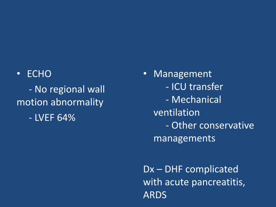
• Management - ICU transfer- Mechanical
ventilation- Other conservative
managements
Dx – DHF complicatedwith acute pancreatitis, ARDS
• ECHO
- No regional wall motion abnormality
- LVEF 64%

Case 2
• A 35-year-old woman, h/o miscarriage
• Past history – treatment for symptomatic anaemia
• Admitted at 24 weeks’ gestation with threatened pre-term labour

• She was given antenatal corticosteroids for fetal lung maturity and magnesium sulfate
• The contractions settled with magnesium sulfate and 28 h after magnesium therapy, her membranes ruptured spontaneously draining clear liquor and 18 h later she delivered a live infant vaginally
• She required transfusion of 1 unit of whole blood
• Within 30 min of delivery, she ‘felt wheezy’ and was found to be profoundly hypoxic
• Pulse 120/min, BP 120/70 mm Hg, RR 24/min, normal JVP, normal heart sounds but-
• Chest examination revealed bilateral crepitations
• ABG was suggestive of hypoxia (saturations of 88% on room air with a partial pressure of oxygen of 6.8 kPa)

• A chest X-ray showed increased pulmonary vasculature

• CT pulmonary angiogram was normal but dependent air space opacities were consistent with acute non-cardiogenic pulmonary oedema
• Echocardiogram – normal valve morphology and good biventricular function
• The placental histology subsequently showed chorioamnionitis

Treatment
• High dependency area
• 60% humidified high-flow oxygen
• Intravenous antibiotics and other supportive managements

Case 3
• A 68-year-old woman , DM, RA
• Progressive SOB and altered mental status
• 2 weeks prior - upper respiratory tract infection
• 1+ pitting edema
• Serum creatinine (Cr) was increased at 4.0 mg/dl
• Urine 3+ proteinuria, 10–15 RBC/HPF, eosinophils(>5%)
• ANA, ANCAs – negative, anti-GBM IgG positive at 272 units/ml (reference range 0–19 units/ml)
• CXR – bilateral infiltrates

Kidney biopsy findings showing FGN with anti-GBM disease. (A) Glomerulus with cellular crescent and mild mesangial matrix expansion. Associated interstitial inflammation is noted (periodic acid–Schiff, 40×). (B) Immunofluorescence staining with IgG demonstrates linear GBM staining as well as segmental smudgy mesangial staining. (C) Electron microscopy shows randomly oriented nonbranching fibrils distributed within mesangium as well as within the peripheral capillary loops.

Treatment
• IV methyl prednisolone followed by oral immunosuppressives
• Haemodialysis

Case 4
• 63-year-old male, smoker, DM, HTN
• 1-h h/o chest pain, sweating, SOB
• Pulse 92/min, BP 165/92 mm Hg, RR 23/min
• Precordium – unremarkable, Chest – bi-basal creps
• ECG – ST-elevation V1-V4
• CXR – pulmonary oedema
• ECHO – antero-septal hypokynesia, EF 47%

Treatment
• Transfer to Cath Lab
• Primary PCI

Difference between cases 1-3 vs case 4


Case 5
Int J Obs Anaes 2009 Jul;18(3):268-71. doi: 10.1016/j.ijoa.2009.02.003. Epub2009 May 22.
Prone positioning for ARDS following blunt chest trauma in late pregnancy.
Kenn S, Weber-Carstens S, Weizsaecker K, Bercker SAbstractAfter a road traffic accident a pregnant patient at 34 weeks of gestation developed ARDS following blunt chest trauma, for which she required mechanical ventilation. Twenty-four hours after the accident, ongoing severe hypoxaemia with atelectasis mainly in the dorsal parts of the lung led to the decision to manage the patient in the prone position. Prone positioning over 8h resulted in a persistent improvement of oxygenation, which allowed extubation the following day. At term, however, our patient was admitted with dyspnoea, chest pain, haemodynamic instability and fetal bradycardia, for which she required emergency caesarean section followed by thoracotomy for haemothorax, from which she eventually made a full recovery. We have demonstrated that prone positioning can be used safely and effectively in a pregnant patient. It might be superior to other therapeutic options for improvement of oxygenation in pregnant patients. Careful positioning avoiding any external abdominal pressure and continuous fetal monitoring are mandatory.

Case 6
Novel diagnostic procedure
Utility of lung ultrasound in near-drowning victimsChristian B Laursen, Jesper Rømhild Davidsen, Poul Henning Madsen
Department of Respiratory Medicine, Odense University Hospital,
Correspondence toDr Christian B Laursen, [email protected]
Summary
Drowning and near-drowning are common causes of accidental death worldwide and respiratory complications such as non-cardiogenic
pulmonary oedema, acute respiratory distress syndrome and pneumonia are often seen. In other settings lung ultrasound can accurately
diagnose these conditions; hence lung ultrasound may have a potential role in the evaluation of drowning or near-drowning victims. In
this case report the authors describe a 71-year-old man who was brought to hospital with acute respiratory failure after a near-drowning
accident. Lung ultrasound showed multiple B-lines on the anterior and lateral surfaces of both lungs, consistent with pulmonary oedema.
Focus assessed transthoracic echocardiography showed no pericardial effusion and a normal global left ventricular function. Based on
these findings the patient was diagnosed as having non-cardiogenic pulmonary oedema. Subsequent chest x-ray showed bilateral
infiltrates consistent with pulmonary oedema. The case report emphasises the clinical value of lung ultrasound in the evaluation of a
near-drowning victim.

Case 7

Case 8

Case 9

Principle of treatment of non-cardiogenic pulmonary oedema
• Admission in HDU/ICU
• Oxygenation including use of mechanical ventilators
• Reduce lung inflammation
• Treatment of underlying cause

Summary
• Non-cardiogenic pulmonary oedema – not an uncommon entity
• May occur in wide-spread clinical contexts –common and uncommon
• High index of suspicion in needed
• Sometimes invasive diagnostics are needed
• Treatment is aimed to increase oxygenation by using ± ventilators and addressing underlying cause

Thank you