Embed Size (px)
Citation preview
Novel Diagnostic Ocular Toxoplasmosis Biomarkers
Jordan Isenberg B.Sc., Department of Pathology, McGill University, Montreal.
Submitted August 2010.
“A thesis submitted to McGill University in partial fulfillment of the requirements of the degree of M.Sc.”
©Jordan Isenberg, 2010.
2
Table of Contents:
Preface .................................................................................................................................................... 3
Abstract ................................................................................................................................................... 5
Résumé ................................................................................................................................................... 6
Chapter 1: Introduction ........................................................................................................................... 7
1.1. Research Rational ................................................................................................................... 7
1.2. Objectives of the research ..................................................................................................... 14
Chapter 2: Review of the Literature for Ocular Toxoplasmosis ............................................................. 25
2.1. Population structure .............................................................................................................. 25
2.2. Ocular manifestations ........................................................................................................... 27
2.3. Treatment of ocular toxoplasmosis ........................................................................................ 32
Chapter 3: Patients and Methods ........................................................................................................... 35
3.1. Serum Samples ..................................................................................................................... 35
3.2. Serum Fractionation .................................................................................................................... 35
3.3. Binding of Fractions to ProteinChip Arrays ................................................................................. 36
3.4. Preparation and Application of Matrix ........................................................................................ 37
3.5. SELDI-ToF-MS Analysis........................................................................................................... 37
3.6. Ciphergen Express Software Analysis ......................................................................................... 38
3.7. Decision Tree Classification........................................................................................................ 39
3.8. Two Dimensional Gel Electrophoresis ........................................................................................ 39
3.8.1. Microscale Isolelectric Focusing in Solution ........................................................................ 39
3.8.2. Protein Gel Elctrophoresis ................................................................................................... 40
3.9. Protein Separation (SDS-PAGE) ................................................................................................. 41
3.10. Protein Identification................................................................................................................. 41
3.11. Protein Function Search ............................................................................................................ 42
3.12. T. gondii Outbreak ................................................................................................................... 42
Chapter 4: Results ................................................................................................................................ 44
4.1. Biomarker Discovery .................................................................................................................. 44
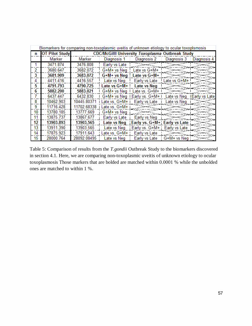
4.2. Biomarker Cross-Validation ........................................................................................................ 56
4.3. Biomarker Identification ............................................................................................................. 60
4.4. Potential Diagnostic Panel........................................................................................................... 64
Chapter 5: Discussion ........................................................................................................................... 66
Chapter 6: Final conclusion and summary .............................................................................................. 75
Chapter 7: Reference List ...................................................................................................................... 78
Appendix .............................................................................................................................................. 85
3
Preface
To say I am a “dwarf standing on the shoulders of giants” is a gross understatement. My
achievements both past, present and future are due entirely to the incredible support I receive
from my family, professors, colleagues, and friends. Over the course of my life I have been
fortunate to be immersed in a nurturing environment and tutored by incredibly caring and
thoughtful individuals. The following manuscript has been put together not only by me but the
countless others who have led me, taught me and fought with me to get to this point. Though it
would be impossible to acknowledge everyone who has been with me over the course of my
studies, I would like to take the time to mention those who had a direct impact.
Without Dr. Miguel N. Burnier Jr. this study would have never occurred. He built a laboratory
for this exact type of interdisciplinary work. The environment he created brings together
physicians and researchers from across McGill and from around the world. This allowed me to
collect samples in Brazil and analyze them seamlessly in three different laboratories scattered
around McGill. I could never have asked for a better supervisor. He allowed me to see my
potential, while providing me with every opportunity to grow. Equally, I want to thank Dr. Phil
Gold for simply being the mentor every student dreams of. He is the most caring, understanding
and brilliant man I know. After every talk we have I have a renewed hope for the future.
4
Thank you to the amazing team at the Henry C. Witelson Ocular Pathology Laboratory. In
particular I would like to single out Dr. Rubens N. Belfort whom without I would have lasted
neither my first week nor last days in the lab without you. His guidance as a friend will not be
forgotten. Christine Straccini the award winning friend and proteomics wizard who taught me
what research is really about as well as how to perform the experiments, analyse the data and run
the statistics. You are the true hero this study. Dr. Bruno F. Fernandes an amazing coach who
always has time to listen to me and guide me especially in how to prepare and edit texts. Dr
Momar Ndao, who helped immensely in the design of this study and the preparation of this
thesis; his willingness to advise me in your capacity of external thesis advisor, and for providing
me with unrestricted access to the CDC/McGill T. gondii outbreak data was a tremendous gift.
Dr Manon Auger for agreeing to act as my internal thesis advisor, her professional guidance was
more help than she can ever imagine. Dr. Bernard Gibbs for performing the protein
identification experiments and aiding me in analysing the data generated. Dr Rubens Belfort Jr.,
who gave me the opportunity to share my ideas with hundreds of people and, of course, along
with Dr. Rubens N. Belfort for providing the patient material and data as well as Dr. Edith
Zorychta for introducing me to the field pathology, developing the program and providing me
with editorial support. Finally, I want to thank Mom, Dad, Grandma, Grandpa, Gregory and
Rachel for being so caring, understanding and patient with me, while providing me with the ideal
environment to succeed. Also, a big thank you to Dr. Catherine Potvin, whose expertise in
phytophysiology might seem out of place in pathology thesis but she showed me that I was
capable of independent research and more importantly thought. To her and everyone else, I am
indebted and ever grateful.
5
Abstract
Purpose: Ocular toxoplasmosis is the most common etiology of posterior uveitis. The high
incidence of macular scarring associated with ocular toxoplasmosis is a leading cause of visual
morbidity. Serum biomarkers of the disease would aid in its diagnosis. This work was designed
as a pilot study to detect potential biomarkers.
Methods: Blood serum samples were collected from four groups of nine patients each; healthy,
uveitic, one single-toxoplasmic event and recurrent events. Protein profiles were generated by
SELDI-ToF-MS and 2-DE. Proteins were sequenced using tandem-MS. A tree-based decision
classification system was developed.
Results: 50 markers of ocular toxoplasmosis and 46 markers of recurrent disease were
discovered by MS; 47% were cross-validated; 14 biomarkers were selected for validation and all
were visualized by SDS-PAGE. 2-DE yielded 57 differentially expressed bands, 20 of which
were excised and identified.
Conclusions: This pilot study sough to elucidate blood serum biomarkers for ocular
toxoplasmosis, as no biomarker for ocular toxoplasmosis exists currently. This study
demonstrates the potential for SELDI-ToF-MS and well as other MS technologies to identify
novel biomarkers for this disease.
6
Résumé
Objectif : La toxoplasmose oculaire est la cause principale de l'uvéite postérieure. Les
cicatrices maculaires associées à la toxoplasmose oculaire sont à la base de la morbidité visuelle.
Des marqueurs sérum sanguin de la maladie en faciliter le diagnostic.
Méthodes : Les échantillons de sérum sanguin ont été recueillis auprès de quatre groupes de
neuf participants composées de patients : sains, avec uvéite, ayant subit un seul événement
toxoplasmique et ayant subit des événements récurrents. Des profils protéiques ont été générés
par SELDI-ToF-MS et le 2-DE. Les protéines ont été séquencées à l'aide de tandem MS. Un
système de classification basé sur des arbres décisionnels a été élaboré.
Résultats : 50 marqueurs de la toxoplasmose oculaire et 46 marqueurs de récidive de la maladie
ont été découverts par MS. 47% de ces marqueurs ont été validés par recoupement et 14
biomarqueurs ont été retenus pour validation. Tous ont été visualisés par SDS-PAGE. 2-DE, ce
qui a abouti à 57 différentiellements exprimés en bandes dont 20 ont été excisées et identifiées.
Conclusions : Cette étude pilote visait à identifier des biomarqueurs sériques potentiels de la
toxoplasmose oculaire. Ici, on a dénoté l’utilité de la technologie SELDI-ToF-MS et des autres
technologies pour identifier de nouveaux biomarqueurs pour cette maladie.
7
Chapter 1: Introduction
1.1. Research Rational
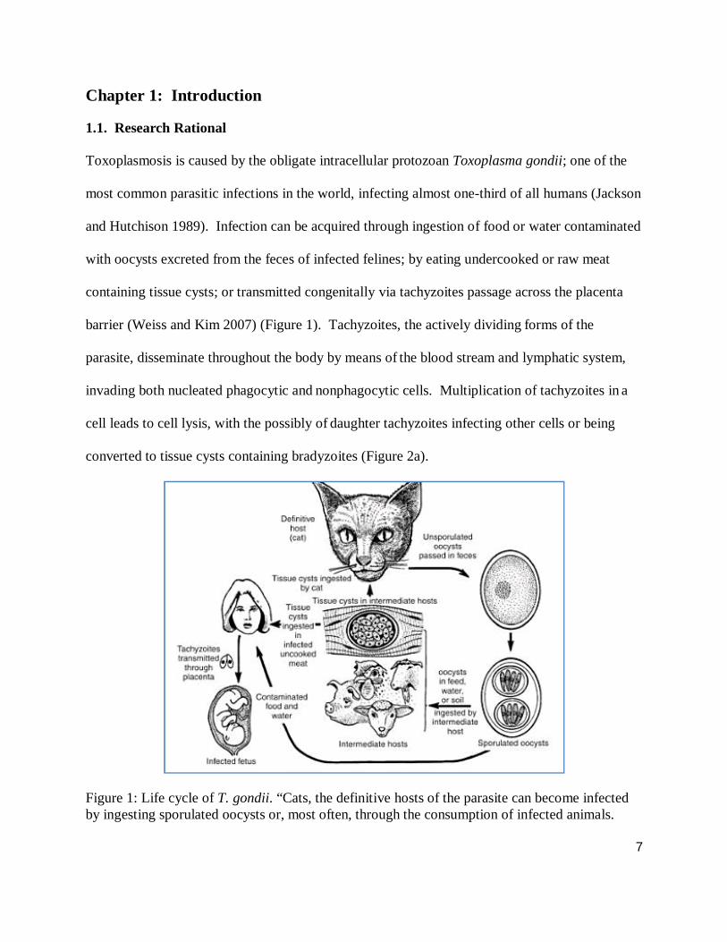
Toxoplasmosis is caused by the obligate intracellular protozoan Toxoplasma gondii; one of the
most common parasitic infections in the world, infecting almost one-third of all humans (Jackson
and Hutchison 1989). Infection can be acquired through ingestion of food or water contaminated
with oocysts excreted from the feces of infected felines; by eating undercooked or raw meat
containing tissue cysts; or transmitted congenitally via tachyzoites passage across the placenta
barrier (Weiss and Kim 2007) (Figure 1). Tachyzoites, the actively dividing forms of the
parasite, disseminate throughout the body by means of the blood stream and lymphatic system,
invading both nucleated phagocytic and nonphagocytic cells. Multiplication of tachyzoites in a
cell leads to cell lysis, with the possibly of daughter tachyzoites infecting other cells or being
converted to tissue cysts containing bradyzoites (Figure 2a).
Figure 1: Life cycle of T. gondii. “Cats, the definitive hosts of the parasite can become infected by ingesting sporulated oocysts or, most often, through the consumption of infected animals.
8
The oocysts are infectious to all mammals and most birds. Toxoplasma can be transmitted to intermediate hosts through oocysts, by carnivorism, or transplacentally. Transplacental transmission is most important in humans and sheep” (Dubey 1986).
Although tissues throughout the body may be infected, in humans, cysts are found to
predominate in the eye and other parts of the central nervous system. Theories explaining
peripheral involvement of the CNS in toxoplasmosis include the passage of the parasite across
the blood-brain-barrier and its poor clearance from the eye, though it is not known whether T.
gondii penetrates these areas relatively easily or whether immunologically privileged sites fail to
eradicate the parasite (Peterson and Remington 1997). Characteristic changes in human
behaviour have been linked to T. gondii’s neurotrophism; impaired reaction times, leading to an
increase in traffic accidents (Flegr, Klose et al. 2009) and an increase in psychiatric symptoms
including schizophrenia as well as mental retardation (Zhu 2009) are but some of the examples.
Even as clinical sequelae of acute and congenital toxoplasmosis are well established, that of
chronic toxoplasma infection remains uncertain (Holliman 1997). The majority of T. gondii
infected patients are asymptomatic with clinical manifestation depending on the patient’s
immune status and the clinical setting. Once an individual is infected by T. gondii, “the retina is
randomly undetectably ‘seeded;’ allowing for the possibility of local recurrences, which causes
irreversible damage to the retina” (Weiss and Kim 2007) (Figure 2a). The most common
pathological conditions resulting from infection are lymphadenopathy and retinochoroiditis
(Montoya and Liesenfeld 2004). Non-ocular infections are not usually serious in otherwise
healthy adults. In contrast, toxoplasmic retinochoroiditis is a progressive, recurring disease that
can cause severe visual morbidity. The severity of the ocular lesion and its propensity to reoccur
9
varies among patients and may be due to the strain of T. gondii, chronic reinfection or the genetic
characteristics of certain patient populations (Belfort-Neto, Nussenblatt et al. 2007).
It has been shown that the reactivation of ocular toxoplasmosis cannot be considered a local
event (Contini, Seraceni et al. 2005) but rather a systemic one (Fortier, Aïssi et al. 1991). Once a
lesion becomes active, parasite levels are detectable as T. gondii enters blood via choroidal
circulation and/or aqueous humour drainage. Although the mechanisms of ocular toxoplasmosis
reactivation are unknown, accumulating evidence suggests that senescent changes in tissue cysts
with release of antigens, trauma, hormonal changes and/or fluctuation of cellular and humoural
immune response could be responsible for disease recurrence (Holland 2000; Silveira, Belfort et
al. 2001). Other theories include that recurrences could be induced by reinfection with a
different strain, as was demonstrated in a murine model (Dao and Fortier 2001)
Ocular toxoplasmosis is the most common etiology of posterior and infectious uveitis, both in
community-based practices of comprehensive ophthalmology and in tertiary referral services
globally (McCannel, Holland et al. 1996). Up to 20 % of infected individuals present with
retinal lesions (Vallochi, Nakamura et al. 2002) of which these infections account for a third of
all infectious uveitis cases (McCannel, Holland et al. 1996). The high incidence of macular
scarring associated with ocular toxoplasmosis, in absolute terms, is a leading cause of visual
morbidity in the developing world causing a highly negative social and economic impact on
those affected. To control ocular toxoplasmosis effectively, efforts should be directed towards
developing a sensitive method for effective diagnosis. Presently there exist no diagnostic assays
available that can determine which patient will develop the recurrent disease.
10
Currently the “gold standard” for toxoplasmosis detection is direct identification of the parasite,
but this method is expensive, time-consuming and relatively insensitive (Chen, Lu et al. 2008).
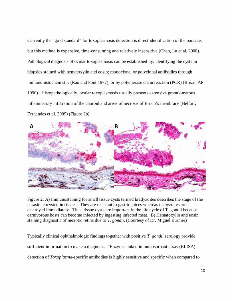
Pathological diagnosis of ocular toxoplasmosis can be established by: identifying the cysts in
biopsies stained with hematoxylin and eosin; monoclonal or polyclonal antibodies through
immunohistochemistry (Rao and Font 1977); or by polymerase chain reaction (PCR) (Brézin AP
1990). Histopathologically, ocular toxoplasmosis usually presents extensive granulomatous
inflammatory infiltration of the choroid and areas of necrosis of Bruch’s membrane (Belfort,
Fernandes et al. 2009) (Figure 2b).
Figure 2: A) Immunostaining for small tissue cysts termed bradyzoites describes the stage of the parasite encysted in tissues. They are resistant to gastric juices whereas tachyzoites are destroyed immediately. Thus, tissue cysts are important in the life cycle of T. gondii because carnivorous hosts can become infected by ingesting infected meat. B) Hematoxylin and eosin staining diagnostic of necrotic retina due to T. gondii. (Courtesy of Dr. Miguel Burnier)
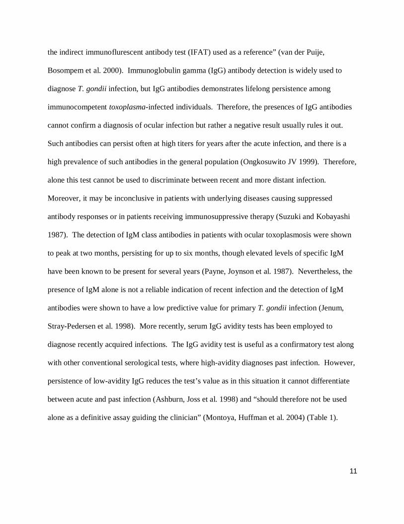
Typically clinical ophthalmologic findings together with positive T. gondii serology provide
sufficient information to make a diagnosis. “Enzyme-linked immunosorbant assay (ELISA)
detection of Toxoplasma-specific antibodies is highly sensitive and specific when compared to
11
the indirect immunoflurescent antibody test (IFAT) used as a reference” (van der Puije,
Bosompem et al. 2000). Immunoglobulin gamma (IgG) antibody detection is widely used to
diagnose T. gondii infection, but IgG antibodies demonstrates lifelong persistence among
immunocompetent toxoplasma-infected individuals. Therefore, the presences of IgG antibodies
cannot confirm a diagnosis of ocular infection but rather a negative result usually rules it out.
Such antibodies can persist often at high titers for years after the acute infection, and there is a
high prevalence of such antibodies in the general population (Ongkosuwito JV 1999). Therefore,
alone this test cannot be used to discriminate between recent and more distant infection.
Moreover, it may be inconclusive in patients with underlying diseases causing suppressed
antibody responses or in patients receiving immunosuppressive therapy (Suzuki and Kobayashi
1987). The detection of IgM class antibodies in patients with ocular toxoplasmosis were shown
to peak at two months, persisting for up to six months, though elevated levels of specific IgM
have been known to be present for several years (Payne, Joynson et al. 1987). Nevertheless, the
presence of IgM alone is not a reliable indication of recent infection and the detection of IgM
antibodies were shown to have a low predictive value for primary T. gondii infection (Jenum,
Stray-Pedersen et al. 1998). More recently, serum IgG avidity tests has been employed to
diagnose recently acquired infections. The IgG avidity test is useful as a confirmatory test along
with other conventional serological tests, where high-avidity diagnoses past infection. However,
persistence of low-avidity IgG reduces the test’s value as in this situation it cannot differentiate
between acute and past infection (Ashburn, Joss et al. 1998) and “should therefore not be used
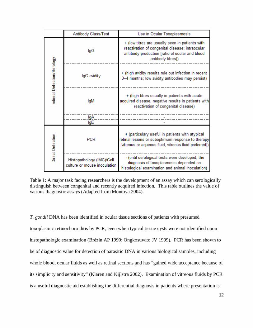
alone as a definitive assay guiding the clinician” (Montoya, Huffman et al. 2004) (Table 1).
12
Table 1: A major task facing researchers is the development of an assay which can serologically distinguish between congenital and recently acquired infection. This table outlines the value of various diagnostic assays (Adapted from Montoya 2004).
T. gondii DNA has been identified in ocular tissue sections of patients with presumed
toxoplasmic retinochoroiditis by PCR, even when typical tissue cysts were not identified upon
histopathologic examination (Brézin AP 1990; Ongkosuwito JV 1999). PCR has been shown to
be of diagnostic value for detection of parasitic DNA in various biological samples, including
whole blood, ocular fluids as well as retinal sections and has “gained wide acceptance because of
its simplicity and sensitivity” (Klaren and Kijlstra 2002). Examination of vitreous fluids by PCR
is a useful diagnostic aid establishing the differential diagnosis in patients where presentation is
13
atypical (Montoya, Parmley et al. 1999; Rothova A, de Boer JH et al. 2008). Nested (n)-PCR is
a reliable diagnostic technique for ocular toxoplasmosis, because of the amount of specimen
required, speed, cost effectiveness, and high specificity in detecting T. gondii DNA in intraocular
fluids (Calderaro A 2006; Mahalakshmi B 2006). Although the sensitivity of n-PCR and real-
time (RT)-PCR techniques are similar, RT-PCR has since replaced n-PCR as a quicker and
simpler technique for quantitatively evaluating ocular samples for the presence of infectious
pathogens (Lin MH 2000; Dworkin LL 2002; Rothova A, de Boer JH et al. 2008). One study
found RT-PCR to have a sensitivity of 38 % for the detection of ocular toxoplamosis from
aqueous humour (Fekkar A 2008). Even though sensitivity was increased to 97 % through the
use of RT-PCR in conjunction with two other biological methods: Western blotting and the
calculation of the Goldmann-Witmer coefficient; current laboratory diagnostics of ocular
toxoplasmosis resulting from the reactivation of a latent infection on samples of aqueous
humour, are inadequate because of the PCR’s low sensitivity (Fekkar, Bodaghi et al. 2008).
Despite the assay’s low sensitivity when sampling ocular fluids, the use of RT-PCR to detect
circulating T. gondii from peripheral blood during a recurrent episode offers not only a reduction
in the potential of product carryover between assays, but also limits patient risks associated with
biopsy retrieval. A randomized controlled trial of 32 patients was able to detect less than 0.5
organisms per sample demonstrating the use of RT-PCR with the fluorescent SYBR Green I in
conjunction with the 529bp gene from the whole blood in patients with Toxoplasma posterior
uveitis, but only in two individuals (Belfort, Isenberg et al. 2010). To date, the respective
contributions of both aqueous humour and peripheral blood in PCR tests for the diagnostic
14
confirmation of ocular toxoplasmosis is still not clear (Fardeau, Romand et al. 2002). No
reliable, clinically available method demonstrating T. gondii in the peripheral blood of ocular
toxoplasmosis patients is able to establish diagnosis in atypical cases, monitor treatment efficacy
and/or to study disease mechanisms including recurrence has been described. Furthermore, no
clinical trial to validate such methods in different settings of patients with toxoplasmosis to been
established or evaluated.
1.2. Objectives of the research
As proteins represent the vast majority of biologically active molecules responsible for cellular
function and with PCR diagnostics proving to be inconclusive, protein biomarkers could be
useful in the future diagnosis of ocular toxoplasmosis. To date, no single, or group of, protein
biomarkers have been described for the diagnosis of either ocular or systemic toxoplasmosis
because the technology needed for the detection of non-invasive biomarkers has only recently
become available. Clinical proteomics, the quantitative study of protein expression between
samples to identify disease-specific proteins, has now been made possible due to the
accumulation of DNA and protein sequence databases, improvements in mass spectrometry
(MS), and the development of computer algorithms for database searching (Graves and Haystead
2002) as well as advances in two-dimensional protein separation.
One of the first methods developed for the analysis of complex protein extracts from cells,
tissues or other biological samples is two-dimensional gel electrophoresis (2-DE). 2-DE is a
multistep process that takes days to complete. Nevertheless, the concept behind the technology
15
is simple: the first dimension is resolved by the isoelectric focusing (IEF), proteins are separated
according to their isoelectric points; following that the second-dimension is resolved through the
use of sodium dodecyl sulphate-polyacrylamide gel electrophoresis (SDS-PAGE), which
separates the proteins according to their molecular weight (O’Farrell, 1975). Using O’Farrell’s
technique the pathophysiology and phenotypes of various diseases including acute lymphoblastic
leukemia and acute myeloid leukemia were able to be elucidated (Hanash et al. 1988).
Despite these successes, 2-DE has been kept from the forefront of biomarker discovery research
even when combined with MS due to concerns over the quantification of protein expression
differences as well as the need for protein characterization and identification (Hanash 2003).
The results obtained by 2-DE are difficult to reproduce, even within a single laboratory, and
more difficult between laboratories. Reasons for this limited reproducibility like the batch-to-
batch variability of carrier ampholytes of the IEF, pH gradient instability over time and cathodic
drift have been largely overcome by the development of immobilized pH gradients (IPGs), the
method employed in this study (Frobel 2009).
These IPGs are based on the use of bifunctional immobiline reagents, ten chemically well-
defined acrylamide derivatives, which form a series of buffers with different pK values between
one and less than 12. They are co-polymerized with the acrylamide matrix and generate
extremely stable pH gradients (Bjellqvist et al., 1982). Nevertheless, conventional 2D-PAGE
analysis is not suitable for high-throughput studies because the samples have to be separated on
individual gels and quantification is time-consuming and inaccurate. As such we chose to
perform 2-DE in parallel with MS.
16
MS is an analytical technique for the determination of the elemental composition of a sample.
Increasing in the past decade MS-based proteomics has become an indispensable tool for
molecular and cellular biology and for the emerging field of systems biology. These include the
study of protein-protein interactions via affinity-based isolations on a small and proteome-wide
scale, the mapping of numerous organelles, the concurrent description of the various organismal
genomes and proteomes, and the generation of quantitative protein profiles from diverse species.
It is precisely the potential for MS to yield a compressive summary of a given biological fluid or
tissue’s proteome, without the need to first carry out protein separations, by gels for example,
which has garnered much interest (Wright Jr. and Cazares et al. 1999). Theoretically, such a
method is well suited for biomarker discovery because of high-throughputs, rapid analysis, and
minimal sample requirement. Indeed, surface-enhanced laser desorption/ionization time-of-flight
mass spectroscopy (SELDI-ToF-MS), has been described and developed to meet these precise
needs (Hutchens and Yip 1993).
The three MS techniques principally used today in biomarker discovery studies are laser
desorption/ionization (LDI), matrix-assisted laser desorption/ionization (MALDI) and SELDI.
All three techniques employ the same general principle of ionizating the solid-state sample by
photoinduction and then detecting the ionized proteins. This is done first by presenting the
sample as either crystals or as a thin film on a sample support called a probe and then using the
energy of a focused laser beam to promote the creation of gaseous ions from solid-state matter
(Scot R. Weinberger, Lee Lomas et al. 2007). The ionized gaseous molecules then enter the
17
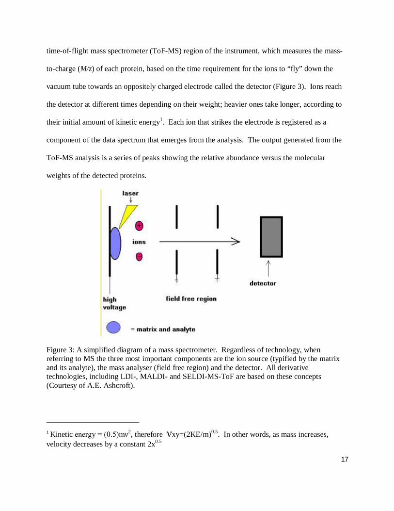
time-of-flight mass spectrometer (ToF-MS) region of the instrument, which measures the mass-
to-charge (M/z) of each protein, based on the time requirement for the ions to “fly” down the
vacuum tube towards an oppositely charged electrode called the detector (Figure 3). Ions reach
the detector at different times depending on their weight; heavier ones take longer, according to
their initial amount of kinetic energy1. Each ion that strikes the electrode is registered as a
component of the data spectrum that emerges from the analysis. The output generated from the
ToF-MS analysis is a series of peaks showing the relative abundance versus the molecular
weights of the detected proteins.
Figure 3: A simplified diagram of a mass spectrometer. Regardless of technology, when referring to MS the three most important components are the ion source (typified by the matrix and its analyte), the mass analyser (field free region) and the detector. All derivative technologies, including LDI-, MALDI- and SELDI-MS-ToF are based on these concepts (Courtesy of A.E. Ashcroft).
1 Kinetic energy = (0.5)mν2, therefore νxy=(2KE/m)0.5. In other words, as mass increases, velocity decreases by a constant 2x0.5
18
The main difference between the three techniques is that in both LDI and MALDI the probe
simply presents the sample to the mass spectrometer for analysis, playing a passive role in the
overall analytical scheme, whereas in SELDI the probe plays an active role in sample
presentation. In practical terms this means that although all three techniques are suitable for the
study of purified protein products; only the SELDI platform allows for the analysis of
heterogeneous materials such as serum.
The study of heterogeneous material by SELDI is enabled by the implementation of two subset
technologies: surface-enhanced affinity capture (SEAC) and surface-enabled neat desorption
(SEND). It is SEAC that allows the probe to accept heterogeneous samples; playing an active
role in the extraction, presentation, structural modification and/or amplification of the sample.
This is accomplished through the use of chemical surface arrays derived from classical
chromographic separation moieties. Through mechanisms including hydrophobic, electrostatic,
coordinate covalent bonding and Lewis-acid/base interactions potential biomarkers bind to
various chemical surface arrays such as reverse phase, ion exchange, immobilized metal affinity
capture and normal phase media. These surfaces have broad binding properties allowing for
their use in de novo biomarker discovery, where large populations of proteins are compared with
the goal of elucidating and then detecting differentially expressed elements. SEAC is the SELDI
technology that has shown the greatest utility thus far. In fact, SEAC is so synonymous with
SELDI that it often referred to as such in the literature. SELDI further differs from LDI and
MALDI due to its SEND technology, a process where the analyte are desorpted and ionized
without the application of a matrix. Rather, SEND is accomplished through the addition of an
19
UV-absorbing compound to the probe’s surface by covalent modification and physical
adsorption.
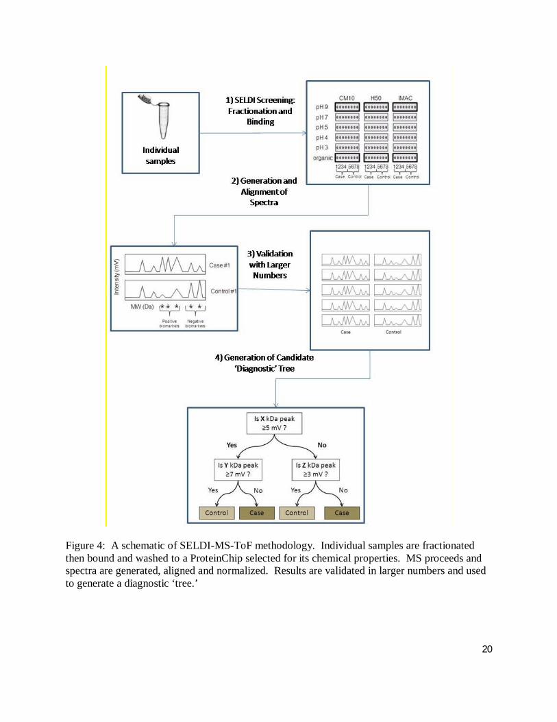
SEAC, SEND and ToF-MS technologies, together with a suite of bioinformatics software, make
up a SELDI-ToF-MS platform capable of identifying differences in protein expression profiles of
two or more distinct samples. This is particularly useful when analyzing complex clinical
samples. Once the spectra of the samples are obtained, software can convert the peaks into one-
dimensional gel view or a simplified map view to more clearly display expression differences
between samples. This results in a list of the MW of proteins whose relative expression differed
across the groups. Proteomic pattern analysis relies on the pattern of proteins observed rather
than the identification of a traceable, binary biomarker (Figure 4). In order to find out the
identities of those proteins, mass and sequence information need to be elucidated by purifying
the proteins followed by a tandem ToF-MS.
20
Figure 4: A schematic of SELDI-MS-ToF methodology. Individual samples are fractionated then bound and washed to a ProteinChip selected for its chemical properties. MS proceeds and spectra are generated, aligned and normalized. Results are validated in larger numbers and used to generate a diagnostic ‘tree.’
21
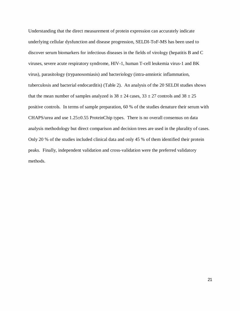
Understanding that the direct measurement of protein expression can accurately indicate
underlying cellular dysfunction and disease progression, SELDI-ToF-MS has been used to
discover serum biomarkers for infectious diseases in the fields of virology (hepatitis B and C
viruses, severe acute respiratory syndrome, HIV-1, human T-cell leukemia virus-1 and BK
virus), parasitology (trypanosomiasis) and bacteriology (intra-amniotic inflammation,
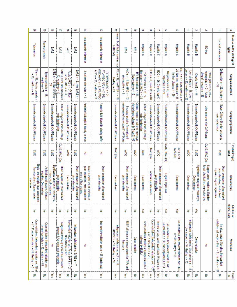
tuberculosis and bacterial endocarditis) (Table 2). An analysis of the 20 SELDI studies shows
that the mean number of samples analyzed is 38 ± 24 cases, 33 ± 27 controls and 38 ± 25
positive controls. In terms of sample preparation, 60 % of the studies denature their serum with
CHAPS/urea and use 1.25±0.55 ProteinChip types. There is no overall consensus on data
analysis methodology but direct comparison and decision trees are used in the plurality of cases.
Only 20 % of the studies included clinical data and only 45 % of them identified their protein
peaks. Finally, independent validation and cross-validation were the preferred validatory
methods.
22
23
Table 2: List of SELDI ID studies summarizing the patient/control populations investigated, sample preparation and data analysis methods used in the studies. The following are the acronyms used in the table above: AF: Amniotic fluid; AFC: AF culture positive; ALT: Adult T-cell leukemia; CHAPS: 3-[(3-Cholamidopropyl)dimethylammonio]-1-propanesulfonate; CM10: Weak cationic exchange array; ELISA: Enzyme-linked immunosorbent assay; H4: Hydrophbic assay (see H50); HAD: HIV-associated dimentia; HAM/TSP:Human T-cell leukemia virus type 1-associated myelopathytropical paraparesis; HCC: Hepatocellular carcinoma; HTLV-1: Human T-cell leukemia virus type 1;IMAC: Immobilized metal affinity array; PTL: Pre-term labour; Q10: Strong anionic exchange array; SAA: Serum amyloid: WCX2: Weak cationic exchange (see CM10). (Adapted from: Hodgetts 2007).
To date, there have been no mass spectroscopic studies performed for either systemic or ocular
toxoplasmosis. As such, two hypotheses have been developed for this study: The first, there
exists differential protein expression between individuals with toxoplasmic retinochoriditis and
non-toxoplasmic uveitis; and the second there exists differential protein expression between
individuals with their first episode of ocular toxoplasmosis and those who have had multiple
episodes.
The purpose of this study is to determine the feasibility of hypothesis-driven proteomics based
research in the elucidation of serum protein biomarkers for an accurate diagnosis of ocular
toxoplasmosis, specifically to aid in determining the etiology of uveitis. Through the uses of
SELDI-ToF-MS, along with 2-DE and LC-MS/MS, this pilot study intends to demonstrate an
ability to differentiate individuals with ocular toxoplasmosis from those with non-toxoplasma
uveitis. Furthermore, it will attempt to effectively group individuals with ocular toxoplasmosis
into either single or multi-episodic ocular toxoplasmosis. The ability to differentiate between
toxoplamosis and non-toxoplasmosis uveitis as well as the type of ocular toxoplasmosis would
confer an advantage to our patients and their physicians.
24
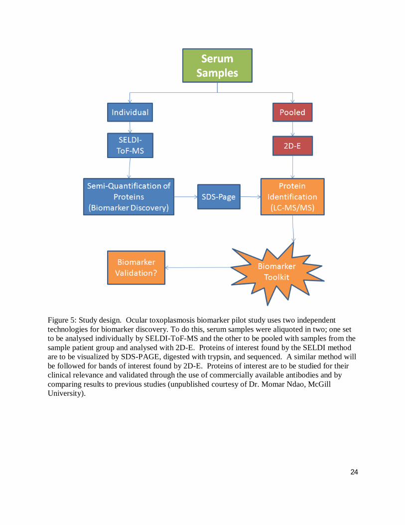
Figure 5: Study design. Ocular toxoplasmosis biomarker pilot study uses two independent technologies for biomarker discovery. To do this, serum samples were aliquoted in two; one set to be analysed individually by SELDI-ToF-MS and the other to be pooled with samples from the sample patient group and analysed with 2D-E. Proteins of interest found by the SELDI method are to be visualized by SDS-PAGE, digested with trypsin, and sequenced. A similar method will be followed for bands of interest found by 2D-E. Proteins of interest are to be studied for their clinical relevance and validated through the use of commercially available antibodies and by comparing results to previous studies (unpublished courtesy of Dr. Momar Ndao, McGill University).
25
Chapter 2: Review of the Literature for Ocular Toxoplasmosis
2.1. Population structure
Despite a sexual phase in the life cycle occurring in feline enterocytes, it was accepted until
recently that the population structure of T. gondii was highly clonal; demonstrating low genetic
variability. Parasitic isolates have been classified into three genetic types: I, II and III, first
based on their virulence in mice then reassessed on the basis of restriction fragment length
polymorphisms (RFLPs) analysis (Howe DK 1995). Less than one percent of the previously
studied strains contain unique genotypes; demonstrating a high divergence in their DNA
sequence, and are consequently considered ‘exotic’ or ‘atypical’ strains (Ajzenberg, Banuls et al.
2004). The low levels of genetic diversity previously reported may be due to a systematic
underestimation of the total number of T. gondii strains; strains in the vast majority of previous
studies were collected from patients and domesticated animals in North America and Europe.
Ajzenberg et al. studied the genetic diversity, clonality and sexuality in T. gondii through the
construction of genetic diversity indices through microsatellite genotypes analysis and the
phylogram development. Results suggested that the global T. gondii population is more diverse
than previously thought. This is not a characteristic of a clonal organism and in this way T.
gondii presents a complex population structure with a mix of clonal and sexual propagation as a
function of the environmental conditions (Ajzenberg 2004).
Brazil as a whole, and the southern regions in particular, has a disproportionately high incidence
and severity of ocular toxoplasmosis when compared to Europe and North America (Holland
26
2003). T. gondii strains from remote and underserved areas, such as the Amazon, are still largely
underrepresented in the phylogeny of the parasite. The use of Brazilian T. gondii isolates along
with recently described markers of genetic characterization demonstrated a higher genetic
variability than has been previously reported (Ajzenberg, Banuls et al. 2004; Lehmann, Graham
et al. 2004). Today PCR-RFLP assays allow for the parasite to be classified as either one of the
three ‘classical’ clonalities or as atypical genotypes (Ajzenberg, Cogne et al. 2002).
The outcome of toxoplasmosis depends on the interaction of many factors, including the
functions of immune system and parasitic factors, such as the inoculum, infective parasite stage,
and genotype of T. gondii isolate. Regardless of the debate surrounding parasite classification,
the type II strain is responsible for more than 70 % of symptomatic human cases in France and
the United States (Howe DK 1997; Nowakowska D 2006). Although there is no patient data
characterizing the difference in strain expression in Brazil for systemic toxoplasmosis, various
studies of wild and farm animals have been carried out demonstrating the high prevalence of
type I, III atypical strains over type II (Dubey JP 2006; Pena, Gennari et al. 2008; Yai, Ragozo et
al. 2009). As such, the type I strain seems to be responsible for the majority of ocular infections
in Brazil (Vallochi AL 2005). In the southern Brazilian city of Erechim the population exhibits a
17 % prevalence of ocular toxoplasmosis. There, the type I and atypical strains predominate
(Jones JL 2006), with the type I strain found in sources of potable water (De Moura L 2006) and
atypical T. gondii genotypes isolated from porcine tongue and diaphragm obtained from local
abattoirs (Belfort and Rasmussen, et al. 2008). Furthermore, a recent analysis of the genotypes
of T. gondii isolated from ocular toxoplasmosis patients from Erechim and Sao Paulo were
27
highly atypical compared to previously described clonal lineages (Khan A 2006). Importantly, it
is “these atypical strains that may be playing an increasingly important role in the acquired
infection ocular manifestations” (Holland 2000), a suggestion that is in-line with the Ajzenberg
hypothesis.
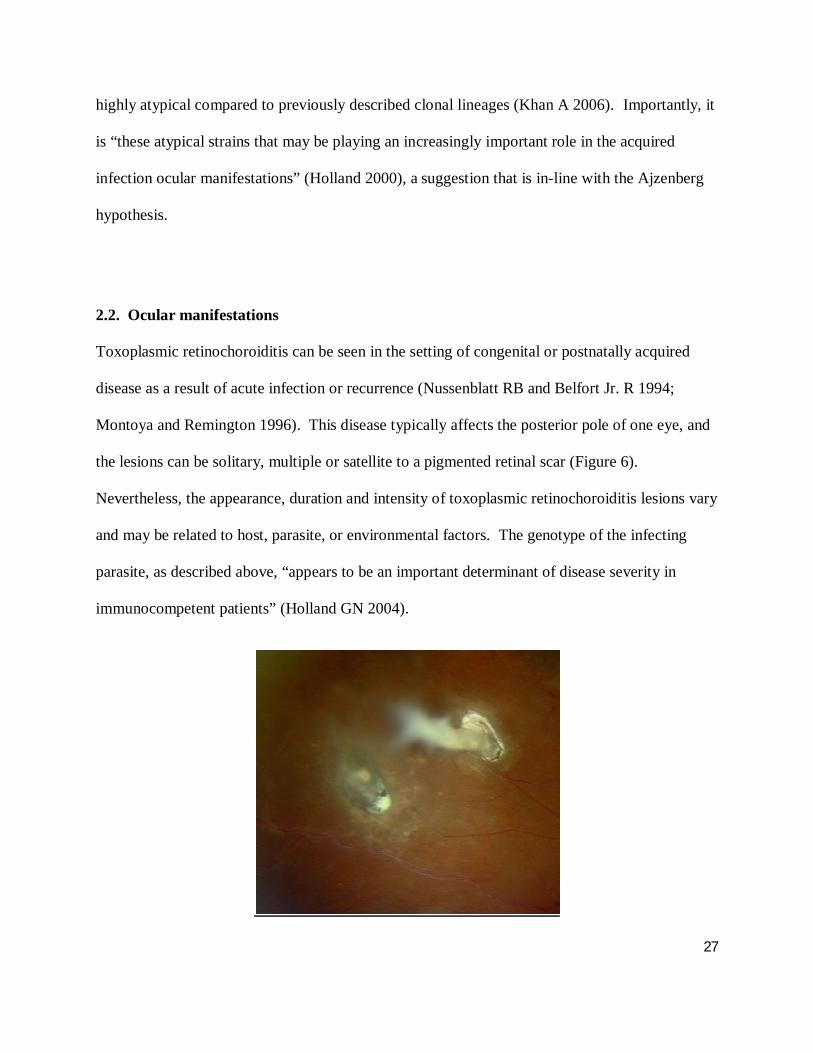
2.2. Ocular manifestations
Toxoplasmic retinochoroiditis can be seen in the setting of congenital or postnatally acquired
disease as a result of acute infection or recurrence (Nussenblatt RB and Belfort Jr. R 1994;
Montoya and Remington 1996). This disease typically affects the posterior pole of one eye, and
the lesions can be solitary, multiple or satellite to a pigmented retinal scar (Figure 6).
Nevertheless, the appearance, duration and intensity of toxoplasmic retinochoroiditis lesions vary
and may be related to host, parasite, or environmental factors. The genotype of the infecting
parasite, as described above, “appears to be an important determinant of disease severity in
immunocompetent patients” (Holland GN 2004).
28
Figure 6: Ocular toxoplasmosis with vitreous strand and vasculitis. (Courtesy of the Henry C. Witelson Ocular Pathology Laboratory, McGill University)
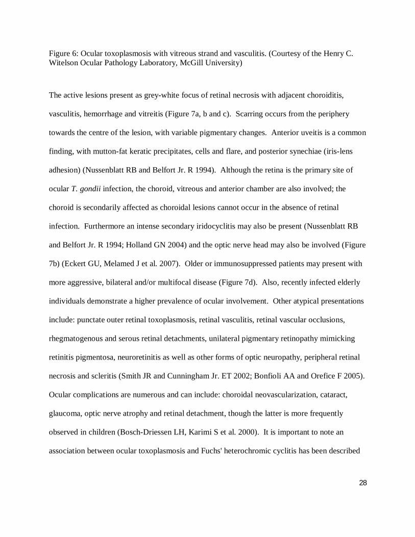
The active lesions present as grey-white focus of retinal necrosis with adjacent choroiditis,
vasculitis, hemorrhage and vitreitis (Figure 7a, b and c). Scarring occurs from the periphery
towards the centre of the lesion, with variable pigmentary changes. Anterior uveitis is a common
finding, with mutton-fat keratic precipitates, cells and flare, and posterior synechiae (iris-lens
adhesion) (Nussenblatt RB and Belfort Jr. R 1994). Although the retina is the primary site of
ocular T. gondii infection, the choroid, vitreous and anterior chamber are also involved; the
choroid is secondarily affected as choroidal lesions cannot occur in the absence of retinal
infection. Furthermore an intense secondary iridocyclitis may also be present (Nussenblatt RB
and Belfort Jr. R 1994; Holland GN 2004) and the optic nerve head may also be involved (Figure
7b) (Eckert GU, Melamed J et al. 2007). Older or immunosuppressed patients may present with
more aggressive, bilateral and/or multifocal disease (Figure 7d). Also, recently infected elderly
individuals demonstrate a higher prevalence of ocular involvement. Other atypical presentations
include: punctate outer retinal toxoplasmosis, retinal vasculitis, retinal vascular occlusions,
rhegmatogenous and serous retinal detachments, unilateral pigmentary retinopathy mimicking
retinitis pigmentosa, neuroretinitis as well as other forms of optic neuropathy, peripheral retinal
necrosis and scleritis (Smith JR and Cunningham Jr. ET 2002; Bonfioli AA and Orefice F 2005).
Ocular complications are numerous and can include: choroidal neovascularization, cataract,
glaucoma, optic nerve atrophy and retinal detachment, though the latter is more frequently
observed in children (Bosch-Driessen LH, Karimi S et al. 2000). It is important to note an
association between ocular toxoplasmosis and Fuchs' heterochromic cyclitis has been described
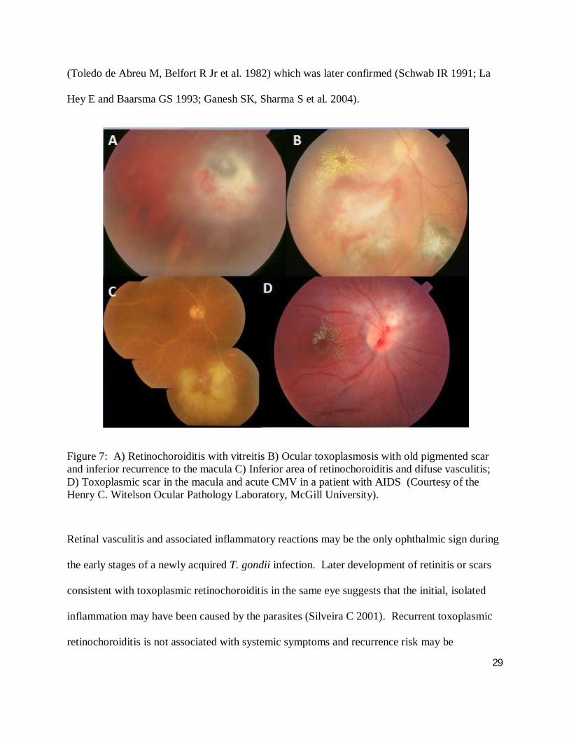
29
(Toledo de Abreu M, Belfort R Jr et al. 1982) which was later confirmed (Schwab IR 1991; La
Hey E and Baarsma GS 1993; Ganesh SK, Sharma S et al. 2004).
Figure 7: A) Retinochoroiditis with vitreitis B) Ocular toxoplasmosis with old pigmented scar and inferior recurrence to the macula C) Inferior area of retinochoroiditis and difuse vasculitis; D) Toxoplasmic scar in the macula and acute CMV in a patient with AIDS (Courtesy of the Henry C. Witelson Ocular Pathology Laboratory, McGill University).
Retinal vasculitis and associated inflammatory reactions may be the only ophthalmic sign during
the early stages of a newly acquired T. gondii infection. Later development of retinitis or scars
consistent with toxoplasmic retinochoroiditis in the same eye suggests that the initial, isolated
inflammation may have been caused by the parasites (Silveira C 2001). Recurrent toxoplasmic
retinochoroiditis is not associated with systemic symptoms and recurrence risk may be
30
influenced by patient age. Ocular lesions may first develop many years after T. gondii infection
and are often asymptomatic (Nussenblatt RB and Belfort Jr. R 1994).
Transplacental transmission of T. gondii to the fetus during pregnancy is another important
source of infection. The mother can transmit the parasite to the fetus if infected during
pregnancy or a few months before conception (Montoya JG and Liesenfeld O 2004). The
infection can result in visual and hearing loss, mental and psychomotor retardation, seizures,
hematological abnormalities, hepatosplenomegaly or death (Montoya JG and Remington JS
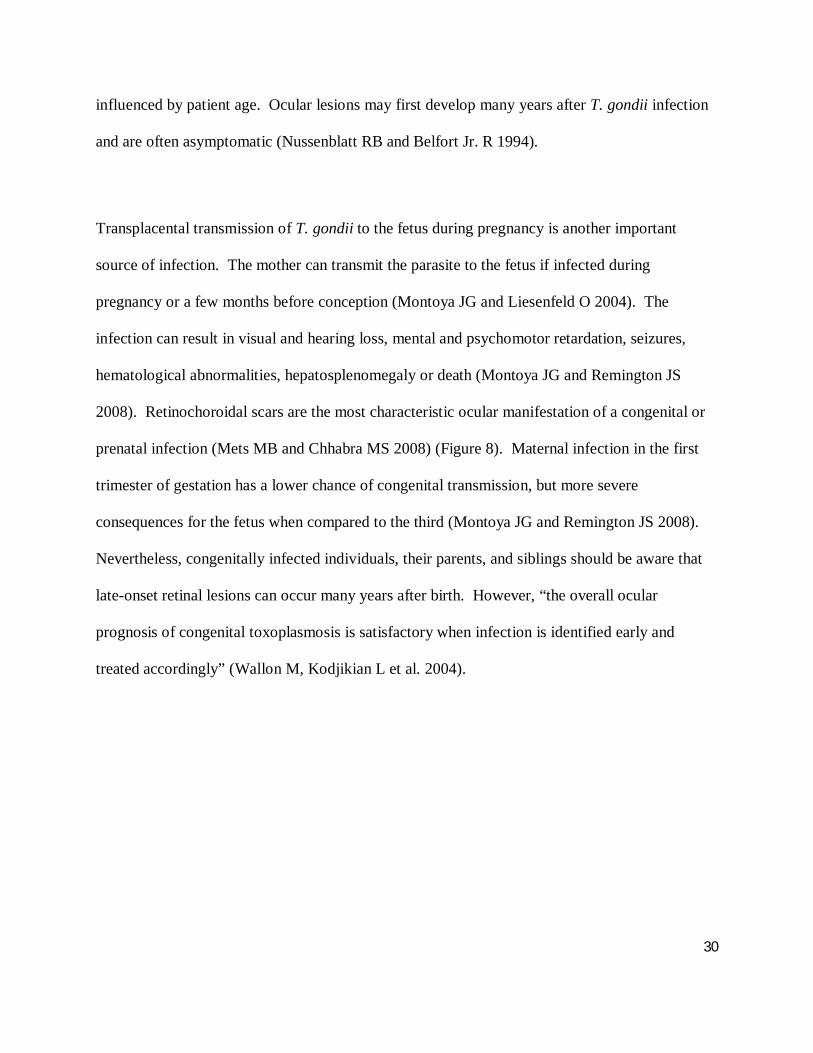
2008). Retinochoroidal scars are the most characteristic ocular manifestation of a congenital or
prenatal infection (Mets MB and Chhabra MS 2008) (Figure 8). Maternal infection in the first
trimester of gestation has a lower chance of congenital transmission, but more severe
consequences for the fetus when compared to the third (Montoya JG and Remington JS 2008).
Nevertheless, congenitally infected individuals, their parents, and siblings should be aware that
late-onset retinal lesions can occur many years after birth. However, “the overall ocular
prognosis of congenital toxoplasmosis is satisfactory when infection is identified early and
treated accordingly” (Wallon M, Kodjikian L et al. 2004).
31
Figure 8: Retinal scars linked by vitreous strand in congenital toxoplasmosis (Courtesy of the Henry C. Witelson Ocular Pathology Laboratory, McGill University).
A British survey assessing the risk of visual impairment in 281 congenitally infected children
with mean follow-up of 4.8 years demonstrated that 17 % of the children presented at least one
retinal lesion. Out of 44 children, where information on visual acuity was available, 9 %
suffered from severe bilateral impairment. Also, 52 % of the children with a posterior pole
lesion and 17 % of those with only peripheral lesions were found to be visually impaired in the
affected eye (Tan HK 2007). Many children with congenital toxoplasmosis present with
substantial retinal damage at birth; yet vision may be remarkably good in the presence of large
macular scars. Active lesions become quiescent with treatment and may recur at any age (Mets
MB 1997).
32
A French study evaluating 430 children treated for congenital toxoplasmosis, found that ocular
involvement was present in 30 % after a median follow-up of 12 years. The overall functional
prognosis of these congenitally infected children was better than would be expected on the basis
of literature findings, with only two of the 130 children suffering bilateral visual impairment
(Kodjikian L 2006). Although it is classically known that only during primary infection the
mother could transmit the infection to the fetus, there are a few reports supporting the possibility
of chronically infected women transmitting the disease congenitally (Silveira C 2003). Reports
have also suggested that T. gondii immune mothers are susceptible to reinfection and therefore
parasite transmission to the fetus (Gavinet, Robert et al. 1997).
2.3. Treatment of ocular toxoplasmosis
Ocular toxoplasmosis therapy may include systemic antimicrobial drugs with or without
corticosteroids. Some ophthalmologists treat all ocular toxoplasmosis cases while others only
those with posterior pole lesions, intense vitreitis, and lesions close to the optic disk or
immunosuppressed patients (Rothova A 1989). Several drugs have been proposed including
pyrimethamine, sulfadiazine, spiramycin, clindamycin, and trimethoprim-sulfamethoxazole
(Pleyer U 2007; Antoniazzi E, Guagliano R et al. 2008).
Results of a study comparing three drug combinations: association of pyrimethamine,
sulphadiazine and corticosteroids; association of clindamycin, sulphadiazine and corticosteroids;
and association of cotrimoxazole (trimethoprim and sulphamethoxazole) with corticosteroids
33
showed no difference in the resolution of inflammatory processes (Rothova A 1989). The same
group showed a reduction in size of the retinal inflammatory lesion for 49 % of the
pyrimethamine-treated patients compared to 20% of the untreated patients (Rothova A 1993).
The most frequent side effects were associated with pyrimethamine and included hematologic
complications such as thrombocytopenia and leucopenia, though folinic acid supplementation is
believed to prevent side effects related to pyrimethamine treatment (Rothova A 1989). It should
be noted however that folic acid does not prevent such complications and should not be used as a
substitute for folinic acid (Belfort, Fernandes et al. 2009). The use of pyrimethamine,
sulfadiazine, and corticosteroids is considered ‘the classical’ therapy for ocular toxoplasmosis
and is the most common drug combination used (Montoya JG and Liesenfeld O 2004). Patients
with active toxoplasmosis may also be treated with trimethoprim-sulfamethoxazole with or
without adjunctive clindamycin and prednisone for four to six weeks. Trimethoprim-
sulfamethoxazole appears to be a safe and effective substitute for sulfadiazine, pyrimethamine,
and folinic acid in treating ocular toxoplasmosis (Opremcak EM 1992; Soheilian M 2005). The
therapeutic benefit from the use of pyrimethamine in combination with azithromycin was similar
to that of pyrimethamine and sulfadiazine. Multidrug therapy with the combination of
pyrimethamine and azithromycin appears to be “an acceptable alternative treatment for sight-
threatening ocular toxoplasmosis” (Bosch-Driessen LH 2002).
As previously mentioned, the causes of recurrences in ocular toxoplasmosis remain unknown.
They may be related to the rupture of dormant retinal cysts (Abreu MT, Belfort Jr R et al. 1987),
or circulating parasites in the peripheral blood (Silveira C, Vallochi AL et al. Manuscript
34
submitted). In some patients, recurrent toxoplasmic retinochoroiditis remains a major problem
and can be associated with severe visual morbidity if disease extends to the macula and optic
disk. Moreover, recurrent disease may cause visual morbidity resulting from inflammation or
complications including retinal detachment and/or choroidal neovascularization. In patients with
frequent recurrences, long-term intermittent treatment with trimethoprim combined with
sulfamethoxazole was shown to reduce the rate of recurrent toxoplasmic retinochoroiditis from
23.8 % to 6.6 % (Silveira C 2002). Nevertheless, traditional short-term treatments of the active
toxoplasmic retinochoroiditis lesions neither prevent subsequent recurrences nor did they have
an effect on visual outcomes or future recurrence rates, with the exception of a poor visual
outcome for patients who received corticosteroids without antiparasitic drugs (Bosch-Driessen
LH 2002). Nevertheless, the relationship between the use of systemic corticosteroids and
reactivation of ocular toxoplasmosis has yet to be elucidated (Morhun PJ, Weisz JM et al. 1996).
Intravitreal injection of clindamicyn with or without steroids may be used in patients that have
contraindication of systemic therapy specific for toxoplasmosis (Aggio FB, Muccioli C et al.
2006; Sobrin L, Kump LI et al. 2007); intravitreal clindamycin injection was associated with
resolution of toxoplasmic retinochoroiditis (Sobrin L, Kump LI et al. 2007). On the other hand,
intravitreal injections of clindamycin and dexamethasone (Kishore K 2001) as well as
subconjunctival injections of clindamycin (Colin J and Harie JC 1989) have demonstrated their
potential to be employed as alternatives to the use of the ‘classical’ anti-toxoplasmic ocular
therapy.
35
Chapter 3: Patients and Methods
3.1. Serum Samples
Blood serum samples were collected from four groups of nine patients each (n=36): healthy,
uveitic (T. gondii IgG negative), those who have had one ocular toxoplasmic event and those
who have had recurrent events (multi-episodic). Serum samples were collected in collaboration
with Clinica Silveira in Erechim, Brazil and the Vision Institute at the Federal University at São
Paulo (UNIFSP), São Paulo, Brazil. Patients provided informed consent while also participating
in prospective clinical study of RT-PCR diagnostics in ocular toxoplasmosis (Belfort, Isenberg et
al. 2010).
3.2. Serum Fractionation
Sera were fractionated using a Ciphergen Q HyperD F strong anion-exchange resin filtration
plate. The filtration plate was re-equilibrated through the addition of: 200 µl rehydration buffer;
50 mM Tris–HCl at pH 9.0; and then placing it on a MicroMix 5 Orbital Vortex (Beckman
Coulter), with a form of 20 and amplitude of 7 for 60 min at room temperature (RT). The
rehydration buffer was then removed by vacuum and the resin was washed four times with 200
µl rehydration buffer and four times with 200 µl U1 solution containing: 1 M urea; 0.2 % 3-[(3-
Cholamidopropyl)dimethylammonio]-1-propanesulfonate (CHAPS); and 50 mM Tris–HCl at pH
9.0. Serum samples were thawed on ice and centrifuged at 17,300 g for 5 min at RT to remove
particulates. Twenty microlitres of sample were added to a v-bottom 96-well microplate (Costar
Corning) with 30 µl of U9 buffer containing; 9 M urea; 2% CHAPS; 50 mM Tris–HCl at pH 9.
36
The microplate was sealed and placed on a MicroMix 5 orbital vortex with a form of 20 and
amplitude of 5 for 20 min at RT. Fifty microlitres of sample were added to the equilibrated resin
with 50 µl of U1 buffer. The filtration plate was sealed and placed on the MicroMix 5 orbital
vortex with a form of 20 and amplitude of 7 for 30 min at RT. The fraction was collected by
vacuum. One hundred microlitres of pH 9 buffer containing 50 mM Tris–HCl and 0.1 % octyl β-
d-glucopyranoside (OGP), buffered to a pH of 9, were added to the wells of the filtration plate
using the Biomek Robot Automation System (Beckman Coulter). The microplate was then
placed on the MicroMix 5 orbital vortex at RT for 10 min and the fraction collected by vacuum.
One hundred microlitres of the following buffers were added in two consecutive applications and
collected by vacuum: pH 9 buffer, pH 7 buffer (50 mM 4-(2-Hydroxyethyl)piperazine-1-
ethanesulfonic acid (HEPES), 0.1% OGP, pH 7); pH 5 buffer (100 mM sodium acetate, 0.1%
OGP, pH 5); pH 4 buffer (100 mM sodium acetate, 0.1% OGP, pH 4); pH 3 buffer (50 mM
sodium citrate, 0.1% OGP, pH 3); and organic wash buffer (33.3% isopropanol, 16.7%
acetonitrile (ACN), 0.1% trifluoroacetic acid (TFA)). The two 100 µl eluants from each pH
fraction were pooled, aliquoted and stored at −20 °C.
3.3. Binding of Fractions to ProteinChip Arrays
Two of the six pH fractions, pH 9 and the organic layer, of serum were profiled on a weak
cation-exchange (CM10), immobilized metal affinity capture (IMAC) and reversed-phase
hydrophobic surface (H50) ProteinChip (Invitrogen Life Science) Arrays according to the
manufacturer’s instructions. All steps were performed at RT. Briefly, the ProteinChip arrays
were placed in a Ciphergen bioprocessor (C503-0006) and washed twice with 200 µl low-
stringency binding buffer containing 0.1 M sodium acetate and 0.1% Triton X-100, buffered to a
37
pH of 4, and placed on a multi-tube vortexer (VWR VX-2500) at speed 1 for 5 min. Each of the
fractions were bound to the chip by adding 10 µl of sample in 90 µl of binding buffer; the
bioprocessor was placed on the multi-tube vortexer for 60 min. The samples were discarded, the
ProteinChip arrays washed three times with 200 µl of binding buffer, placed on the multi-tube
vortexer for 5 min and washed twice with 200 µl 1 mM HEPES buffered to a pH of 7.4 for 1
min. The ProteinChip arrays were air-dried prior to matrix application. Serum samples were all
spotted randomly on the arrays.
3.4. Preparation and Application of Matrix
For protein analysis, a saturated sinapinic acid (SPA) solution was freshly prepared by adding
50% ACN/0.5% TFA solution. Half a microlitre of the matrix was added to each spot on the
ProteinChip array and air-dried prior to adding an additional 0.5 µl of matrix.
3.5. SELDI-ToF-MS Analysis
ProteinChip arrays were read using a Ciphergen PCS4000 SELDI-TOF MS reader (Bio-Rad
Laboratories). Profiles were collected in the low and high ranges: 0–10; and 10–200 kDa. The
intensity and sensitivity of the instrument was adjusted for each of these ranges on each day of
analysis. The instrument was calibrated for dataset collection using an all-in-one peptide
standard (Bio-Rad Laboratories) when collecting data both in the 0–10 and 10–200 kDa ranges,
varying the intensity and sensitivity of collection. Spectra from profiling experiments are an
average of data from 110 laser shots.
38
3.6. Ciphergen Express Software Analysis
Spectra were normalized by total ion current intensity starting and ending at the M/z of the
collection ranges of 0-10 and 10-200 kDa. The spectra were then aligned to a spectrum with the
normalization factor nearest 1. This was only done if the percentage coefficient of variation for
the given spectra was reduced after the alignment. Afterwards, any spectra identified to be
greater than two standards of deviation away from the normal were rejected. First pass data
analysis began when peaks from the different spectra were aligned using the Cluster Wizard
function of Ciphergen Express Software (Bio-Rad Laboratories). Peak detection was completely
automated within the M/z range of analysis, where a peak was automatically detected on the first
pass when its signal-to-noise (S/N) ratio was five and it was three times the valley depth. User-
detected peaks below threshold were deleted and all first-pass peaks were preserved. Clusters
were created within 0.3 % of mass for each peak detected in the first pass for low mass range and
2 % for high mass range. When no peaks were detected, the peak intensity was estimated at the
centre of the cluster. The peaks were manually inspected to determine if they were multi-
charged entities. P-values and the receiver operation characteristic (ROC) values were
calculated through the use of the P-Value Wizard which compared: healthy to uveitis (non-
toxoplasma); one toxoplasmic event to recurrent events; uveitic to one and multiple events; and
healthy to uveitic to one toxoplasmic event to recurrent events. In this way uveitic and T. gondii
specific proteins were removed from the analysis, retaining only makers for one ocular
toxoplasmic event, multiple events and healthy controls from which p-values below 0.05 were
considered statistically significant. Second pass analysis followed a methodology similar to first
pass analysis. Here, all peaks with a p-value of less than 0.001 were retained and relabelled.
The peak detection software was then placed on manual mode with the following settings: detect
39
user labelled peaks only, increase of the mass cluster to 2 % of mass for each peak for the low
mass range. Subsequently, p-value and ROC was calculated using the P-Value Wizard by
comparing one toxoplasmic event to recurrent events.
3.7. Decision Tree Classification
Construction of the decision tree classification algorithm was performed by Ciphergen
Biomarker Pattern software version 5.0 (Bio-Rad Laboratories). Classification trees split the
data into two nodes using one rule at a time in the form of peak intensity. The splitting decisions
in this case were based on the normalized intensity levels of peaks from SELDI protein
expression profile. The process of splitting was continued until terminal nodes were created.
After V-fold cross validation of 50, the accuracy of each classification tree was then challenged
with the blinded test set.
3.8. Two Dimensional Gel Electrophoresis
3.8.1. Microscale Isolelectric Focusing in Solution
Nine serum samples from the uveitic, single event and recurrent groups used in the SELDI-TOF-
MS analysis were thawed and made into pools of 20 µL each. The lysate is formed when 900 µL
of 1.1 x IEF denaturant buffer (Invitrogen Life Sciences), containing: 7.7 M urea; 2.2 M
thiourea; and 4.4% (w/v) CHAPS, 10 µL of a 100 x protease inhibitor cocktail, 20 µL of 1 M
DDT reducing agent, 10 µL of both 0.5 M EDTA and 1 M tris-base were added to the pools
following a half hour RT incubation on a platform rocker. 5.2 µL of 99 % N,N-
dimethyacylatime was then added followed by another incubation period. To 1 mL of the now
40
reduced and alkylated lysate, 2.28 mL of IEF denaturant buffer, 35 µL of carrier ampholytes (pH
3−10) and 20 µL of 1 M DDT, were added for a final volume of 3.5 mL.
The ZOOM Isoelectric Focusing (IEF) Fractionator (Invitrogen Life Sciences) was assembled
according to the manufacturer's instructions. 650 µL of the pooled diluted samples described
above was pipetted into each of the 5 chambers of the instrument: chamber 1 pI 3−4.6; chamber
2 pI 4.6−5.4; chamber 3 pI 5.4−6.2; chamber 4 pI 6.2−7.0; chamber 5 pI 7.0−10.0. The anode
chamber was filled with 17.5 mL of anode buffer containing 8.4 g of urea, 3.0 g of thiourea, and
3.3 mL of Novex IEF Anode buffer (Invitrogen Life Sciences), adjusted to pH 3, and made up to
a total volume of 20 mL with deionized water. The cathode chamber was filled with 17.5 mL of
cathode buffer containing: 8.4 g of urea; 3.0 g of thiourea; and 2 mL of Novex IEF Cathode
buffer (Invitrogen Life Sciences) and made up to a volume of 20 mL with deionized water. IEF
was then performed for three hours: 100 V for 20 min; 200 V for 80 min; and at 600 V for
80min. The current and power were limited to 2 mA and 2 W. This was repeated for each
group. Samples were then removed from wells and placed into three 200 µL aliquots per
chamber and kept at –20 oC until the protein gel was run.
3.8.2. Protein Gel Elctrophoresis
Following isoelectric focusing, each sample was thawed, centrifuged and desalted with 1400 µL
of methanol, 200 µL of chloroform, 800 µL of deionized water, then left to air dry. Samples
were then equilibrated and reduced, according to the manufacture’s recommendations, by the
addition of NuPAGE LDS Sample Buffer and NuPAGE Reducing Agent (Invitrogen Life
41
Science), containing 500 mmol·L-1 DTT. Samples were then heated at 70 oC for ten minutes.
The second-dimension electrophoresis was performed by aligning the ZOOM strip in the well of
a NuPAGE Novex 4% to 12% Bis-Tris ZOOM Gel (Invitrogen Life Science) in a denaturing
running buffer prepared with 25 mL of NuPAGE 20 x SDS Running Buffer made up to a total
volume of 500 mL with deionized water. Samples were loaded into ten wells and Mark12 MW
Marker (Invitrogen) was loaded into two wells, then electrophoresed at 200 V for 35 min.
Following electrophoresis, samples were stained in a freshly prepared Commassie G250 dye
containing 85 mL of methanol, 10 mL phosphoric acid, 42.5 g ammonium sulfate, and 0.25 g
Brilliant Blue completed to a final volume of 250 mL. Gels were left to stain for 48 hours and
then were photographed. Bands demonstrating differential expression, as determined by visual
inspection, were excised and kept in 5 % acetone at 4 oC until protein identification proceeded.
3.9. Protein Separation (SDS-PAGE)
General protein separations are based on MW. Samples were fractionated following the method
described in section 3.2, while one-dimensional gel electrophoresis followed the method
presented in section 3.8.2.
3.10. Protein Identification
Trypsin digestion and liquid chromatography (LC)-MS/MS analysis were carried out on an
Agilent micro LC connected to an ABI Q-STAR mass spectrometer at the Sheldon
Biotechnology Centre, McGill University. The resulting tryptic peptides were matched against
42
both mammalian and other eukaryotic NCBI databases using MASCOT search engine
(http://www.matrixscience.com/) for product ion confirmation.
3.11. Protein Function Search
Differentially expressed proteins identified by MS analysis had protein function assigned for
each. Protein function assignation tools including the Bioinformatic Harvester program
(http://www.harvester.embl.de/) and those at the Human Protein Resource Database
(http://www.hprd.org/) were used in order to determine human protein function in ocular
toxoplasmosis. Only proteins expressed by humans were selected, as no T. gondii proteins were
detected.
3.12. T. gondii Outbreak
An outbreak of toxoplasmosis was reported between November 2001 and January 2002 in Santa
Isabel do Ivai, Brazil, following which a matched case-control study was conducted from
January 15 to February 2, 2002. Serum samples from case-patients and controls were tested for
anti-T. gondii IgM and IgG antibodies (de Moura, Bahia-Oliveira et al. 2006) and an extensive
biomarker study was at undertaken by Dr. Momar Ndao at McGill University’s National
Reference Laboratory for Parasitology in conjunction with the US Center for Disease Control
(CDC) on 200 of those patient samples. This study followed similar protocols as those described
above, though two different SELDI-TOF-MS machines (PBSIIC and PCS4000) were employed
as well as all six fractions generating 10 800 spectra. The same study used th three ProteinChip
arrays and three chip types (results unpublished). Data analysis saw the samples divided into six
43
groups: early vs. late; early vs G+M+; late vs G+M+;· early vs negative; G+M+ vs negative; late
vs negative. All statistically significant biomarkers discovered in this pilot (129) study were
compared to the biomarker outbreak database of 422 peaks, and then diagnosis, p-value as well
as the receiver operating characteristics were noted. Biomarkers are matched to within 1 % of
m/z but the bolded ones are matched to within 0.0001 %, while the m/z sensitivity of the SELDI-
ToF-MS technology is within 0.1 %.
44
Chapter 4: Results
4.1. Biomarker Discovery
Individual serum samples (n=36) from all the training sets were fractionated, analysed and
compared by SELDI-ToF-MS with CM10, H50 and IMAC ProteinChips generating a total of
432 spectra. All MS data were baseline subtracted and normalized using total ion current, and
the peak clusters were generated by Biomarker Wizard software as outlined in section 3.5.
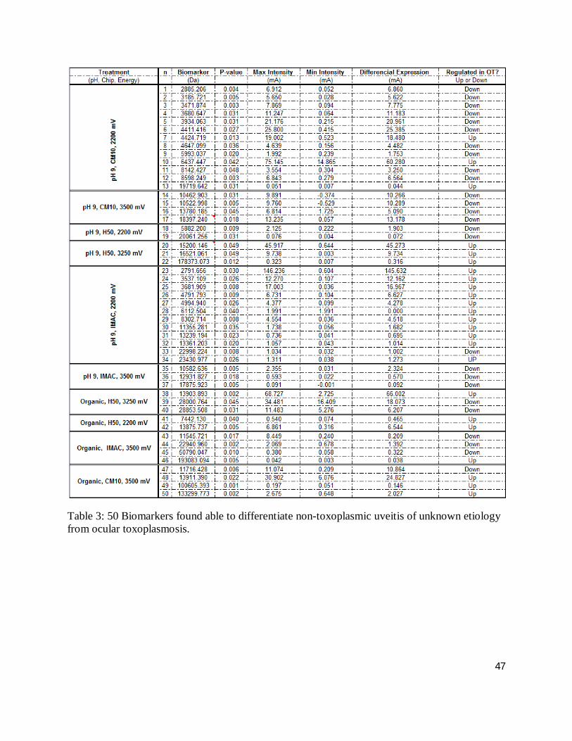
Table 3 shows the results of second pass analysis of the ocular toxoplasmosis compared to non-
toxoplasmic uveitis groups; 50 protein peaks, 52 % of which were down-regulated in the ocular
toxoplasmosis group, were discovered. The m/z of the 50 biomarkers ranged from 2,992 to
193,083 Da demonstrating a m/z mean of 23,174 ± 40,539 Da with a mean p value of 0.020 ±
0.015. The intensity of the samples analysed range from -0.53 to 146.23 mA. The mean
difference in intensity (DI), defined as the two individual samples with the highest and lowest
intensities for a given biomarker, is 12.25 ± 23.7 mA, with seven biomarkers demonstrating a
difference greater than one standard of deviation. Of the biomarkers in the m/z range of 2.7 to
15.2 kDa, 75 % of them up-regulate; suggestive of humoral immune response. Two of the
biomarkers are within 7 Da of each other and are up-regulated suggesting that they may be the
same protein.
45
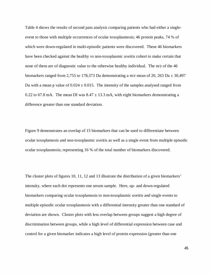
Table 4 shows the results of second pass analysis comparing patients who had either a single-
event to those with multiple occurrences of ocular toxoplasmosis; 46 protein peaks, 74 % of
which were down-regulated in multi-episodic patients were discovered. These 46 biomarkers
have been checked against the healthy vs non-toxoplasmic uveitis cohort to make certain that
none of them are of diagnostic value to the otherwise healthy individual. The m/z of the 46
biomarkers ranged from 2,755 to 178,373 Da demonstrating a m/z mean of 20, 263 Da ± 30,497
Da with a mean p value of 0.024 ± 0.015. The intensity of the samples analysed ranged from
0.22 to 67.8 mA. The mean DI was 8.47 ± 13.3 mA, with eight biomarkers demonstrating a
difference greater than one standard deviation.
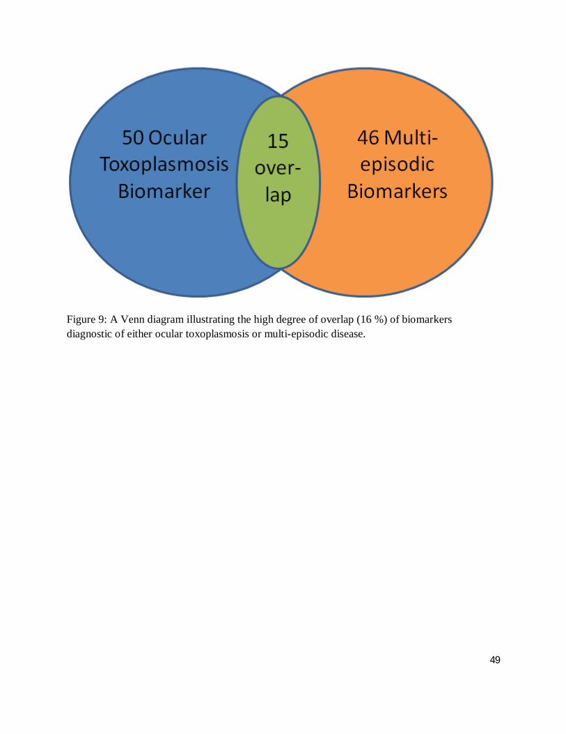
Figure 9 demonstrates an overlap of 15 biomarkers that can be used to differentiate between
ocular toxoplamosis and non-toxoplasmic uveitis as well as a single event from multiple episodic
ocular toxoplasmosis; representing 16 % of the total number of biomarkers discovered.
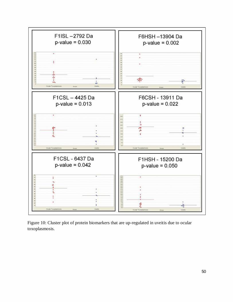
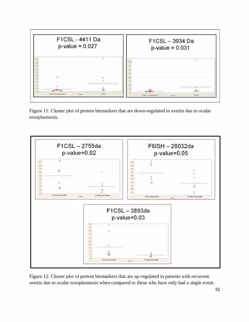
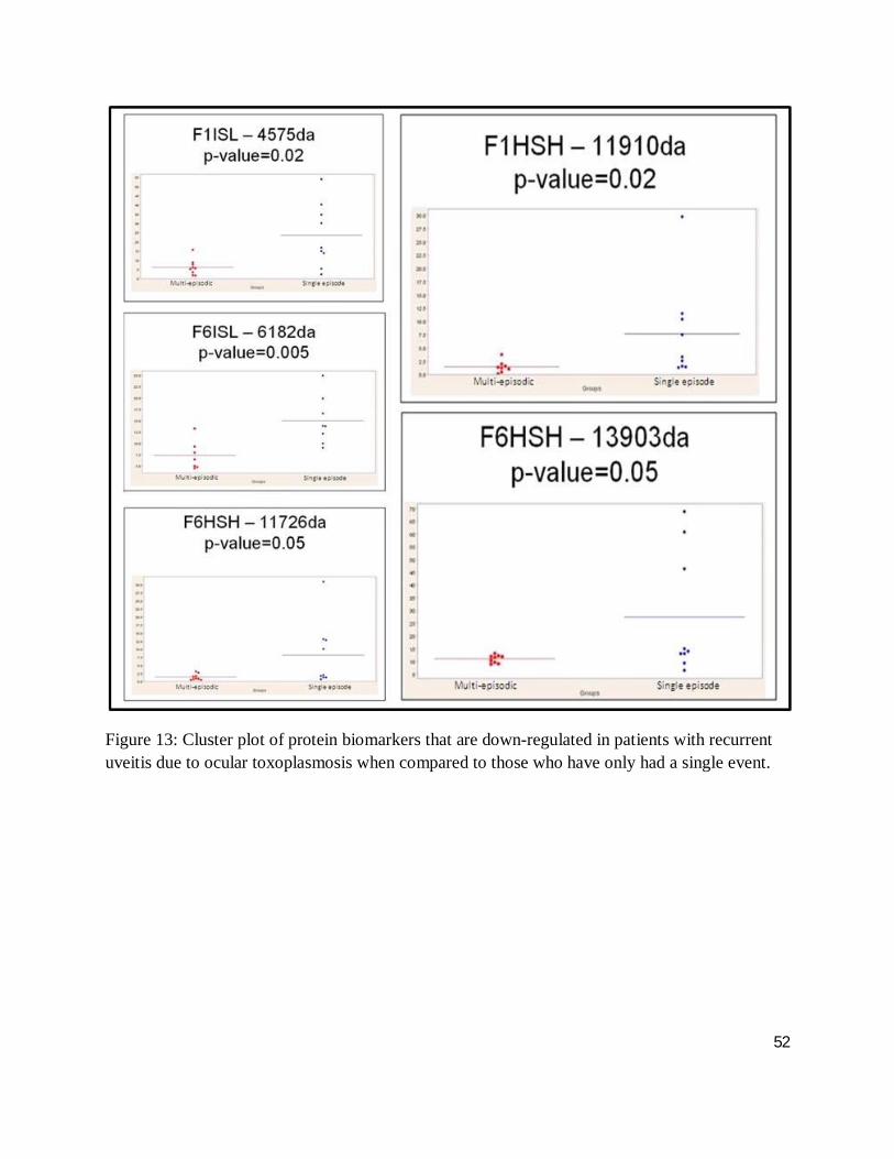
The cluster plots of figures 10, 11, 12 and 13 illustrate the distribution of a given biomarkers’
intensity, where each dot represents one serum sample. Here, up- and down-regulated
biomarkers comparing ocular toxoplamosis to non-toxoplasmic uveitis and single events to
multiple episodic ocular toxoplasmosis with a differential intensity greater than one standard of
deviation are shown. Cluster plots with less overlap between groups suggest a high degree of
discrimination between groups, while a high level of differential expression between case and
control for a given biomarker indicates a high level of protein expression (greater than one
46
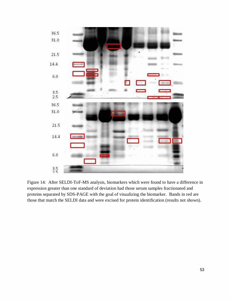
standard deviation), facilitating downstream analysis. As such, figure 14 demonstrates the
visualization of those highly and minimally expressed proteins as outlined in section 3.9
Sera from the same patients used in SELDI-ToF-MS analysis were pooled according to their
group: non-toxoplasmic uveitis, single event, multi-episodic. Figures 15 and 16 demonstrate the
results of two-dimensional gel electrophoresis whereby the pooled sera’s proteins were separated
according to pH and mass. In this way, 57 differentially expressed bands (potential biomarkers)
were detected and excised for downstream protein identification according to section 3.10 with
results featured in section 4.3. Analysis of the banding patterns suggest that 68 % of the bands
are indicative of muli-episodic disease (red) whereas 21 % (blue) and 9 % (green) are markers of
non-toxoplasmic uveitis and single episodes of disease.
47
Table 3: 50 Biomarkers found able to differentiate non-toxoplasmic uveitis of unknown etiology from ocular toxoplasmosis.

48
Table 4: 46 biomarkers able to differentiate between single and multi-episodic ocular toxoplasmic events.
49
Figure 9: A Venn diagram illustrating the high degree of overlap (16 %) of biomarkers diagnostic of either ocular toxoplasmosis or multi-episodic disease.
50
Figure 10: Cluster plot of protein biomarkers that are up-regulated in uveitis due to ocular toxoplasmosis.
51
Figure 11: Cluster plot of protein biomarkers that are down-regulated in uveitis due to ocular toxoplasmosis.
Figure 12: Cluster plot of protein biomarkers that are up-regulated in patients with recurrent uveitis due to ocular toxoplasmosis when compared to those who have only had a single event.
52
Figure 13: Cluster plot of protein biomarkers that are down-regulated in patients with recurrent uveitis due to ocular toxoplasmosis when compared to those who have only had a single event.
53
Figure 14: After SELDI-ToF-MS analysis, biomarkers which were found to have a difference in expression greater than one standard of deviation had those serum samples fractionated and proteins separated by SDS-PAGE with the goal of visualizing the biomarker. Bands in red are those that match the SELDI data and were excised for protein identification (results not shown).
54
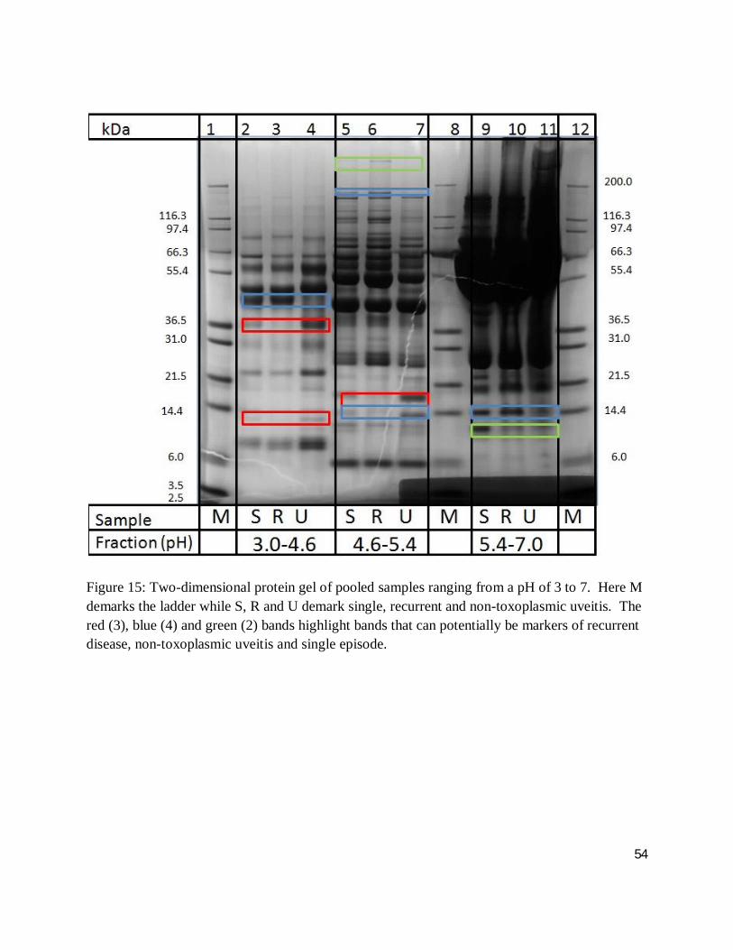
Figure 15: Two-dimensional protein gel of pooled samples ranging from a pH of 3 to 7. Here M demarks the ladder while S, R and U demark single, recurrent and non-toxoplasmic uveitis. The red (3), blue (4) and green (2) bands highlight bands that can potentially be markers of recurrent disease, non-toxoplasmic uveitis and single episode.
55
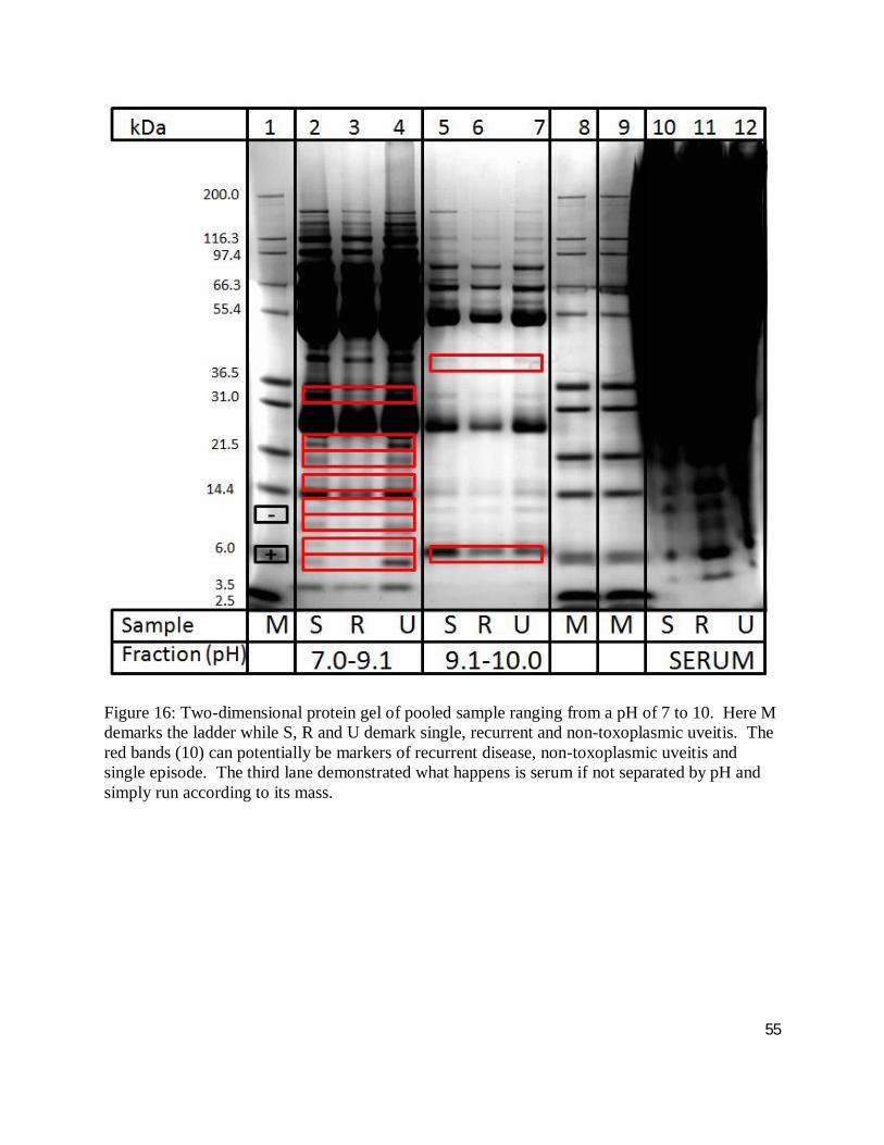
Figure 16: Two-dimensional protein gel of pooled sample ranging from a pH of 7 to 10. Here M demarks the ladder while S, R and U demark single, recurrent and non-toxoplasmic uveitis. The red bands (10) can potentially be markers of recurrent disease, non-toxoplasmic uveitis and single episode. The third lane demonstrated what happens is serum if not separated by pH and simply run according to its mass.
56
4.2. Biomarker Cross-Validation
Table 5 and 6 compare SELDI-ToF-MS results of ocular toxoplasmosis to the protein peaks
found in the CDC/McGill toxoplasmosis outbreak study as outlined in section 3.12. Those
markers that are bolded are matched within 0.0001 % while the unbolded ones are matched to
within 1 %.
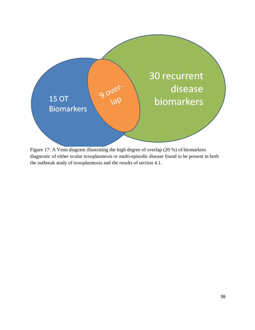
Figure 16 demonstrates an overlap of 9 biomarkers that can be used to differentiate between
ocular toxoplasmosis and non-toxoplasmic uveitis as well as single events from multiple episodic
ocular toxoplasmosis, found in both the outbreak study and the present work, repeated in section
4.1. This represents 20 % of the total number of biomarkers discovered. Interestingly, 60 % of
the markers for ocular toxoplasmosis overlap, compared to only 20 % of those marking recurrent
disease.
57
Table 5: Comparison of results from the T.gondii Outbreak Study to the biomarkers discovered in section 4.1. Here, we are comparing non-toxoplasmic uveitis of unknown etiology to ocular toxoplasmosis Those markers that are bolded are matched within 0.0001 % while the unbolded ones are matched to within 1 %.
58
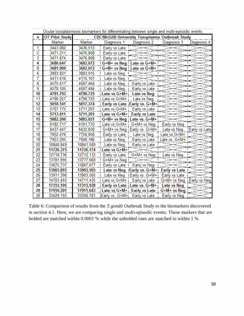
Table 6: Comparison of results from the T.gondii Outbreak Study to the biomarkers discovered in section 4.1. Here, we are comparing single and multi-episodic events. Those markers that are bolded are matched within 0.0001 % while the unbolded ones are matched to within 1 %
59
Figure 17: A Venn diagram illustrating the high degree of overlap (20 %) of biomarkers diagnostic of either ocular toxoplasmosis or multi-episodic disease found to be present in both the outbreak study of toxoplasmosis and the results of section 4.1.
60
4.3. Biomarker Identification
Table 7 lists all 20 biomarkers identified from 2, 3 and 4 of figure 16. Furthermore, the table
describes the function of the proteins and their role in toxoplasmosis. Using the sequencing
results from the 2-DE, 11 % of the markers cross referenced with the toxoplasmosis outbreak
were able to be identified.
In table 8 protein peaks generated by SELDI-ToF-MS analysis were compared to the mass and
sequence information from the protein identification generated from 2-DE in order to elucidate
the identities of those peaks; 16 % of the SELDI generated peaks were identified.
Table 9 employed the sequence information alongside the proteins ‘molecular weight search
score’ to develop an initial diagnostic picture using 2-DE parallel to SELDI-ToF-MS technology.
Hemaglobin subunit beta, serum amyloid and complement C8 are markers of ocular
toxoplasmosis. Nuclease-sensitive element-binding protein (YBOX1) is a marker of single
disease. Ankyrin repeat domain-containing protein 11 (ANKRD11) and peptidyl-prolyl cis-trans
isomerase A (PPIA) are markers of multi-episodic disease. Plasma retinol-binding protein 4
(RBP4), ATP-binding cassette sub-family A member 2 and bullous pemphigoid antigen 1,
isoforms 6/9/10 (Trabeculin-ß) are markers of non-toxoplasmic uveitis.

61
Table 7: A lists of all 20 biomarkers identified from 2, 3 and 4 of figure 16 displaying protein function and involvement in toxoplasmosis. All proteins are human derived.

62
Table 8: Protein peaks generated by SELDI-ToF-MS analysis were compared to the mass and sequence information from the protein identification generated from 2-DE in order to elucidate the identities of those peaks
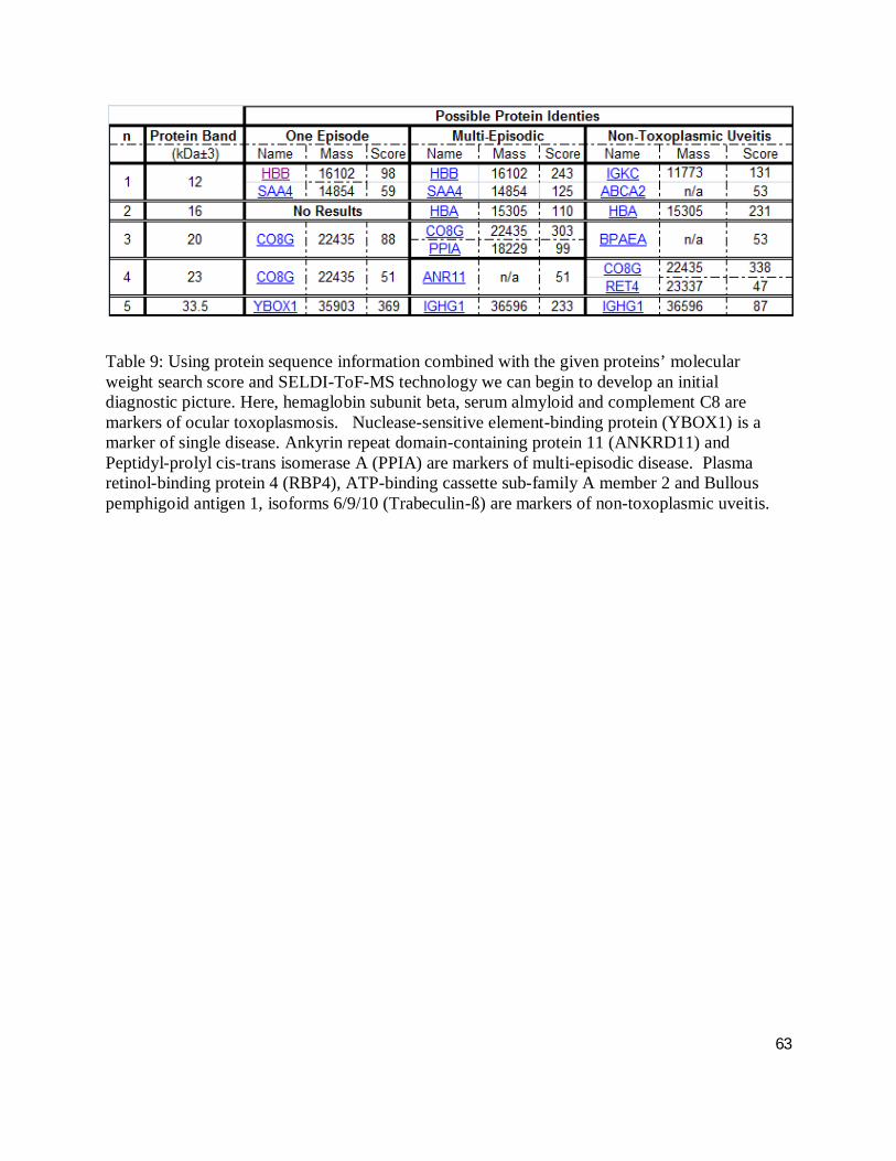
63
Table 9: Using protein sequence information combined with the given proteins’ molecular weight search score and SELDI-ToF-MS technology we can begin to develop an initial diagnostic picture. Here, hemaglobin subunit beta, serum almyloid and complement C8 are markers of ocular toxoplasmosis. Nuclease-sensitive element-binding protein (YBOX1) is a marker of single disease. Ankyrin repeat domain-containing protein 11 (ANKRD11) and Peptidyl-prolyl cis-trans isomerase A (PPIA) are markers of multi-episodic disease. Plasma retinol-binding protein 4 (RBP4), ATP-binding cassette sub-family A member 2 and Bullous pemphigoid antigen 1, isoforms 6/9/10 (Trabeculin-ß) are markers of non-toxoplasmic uveitis.
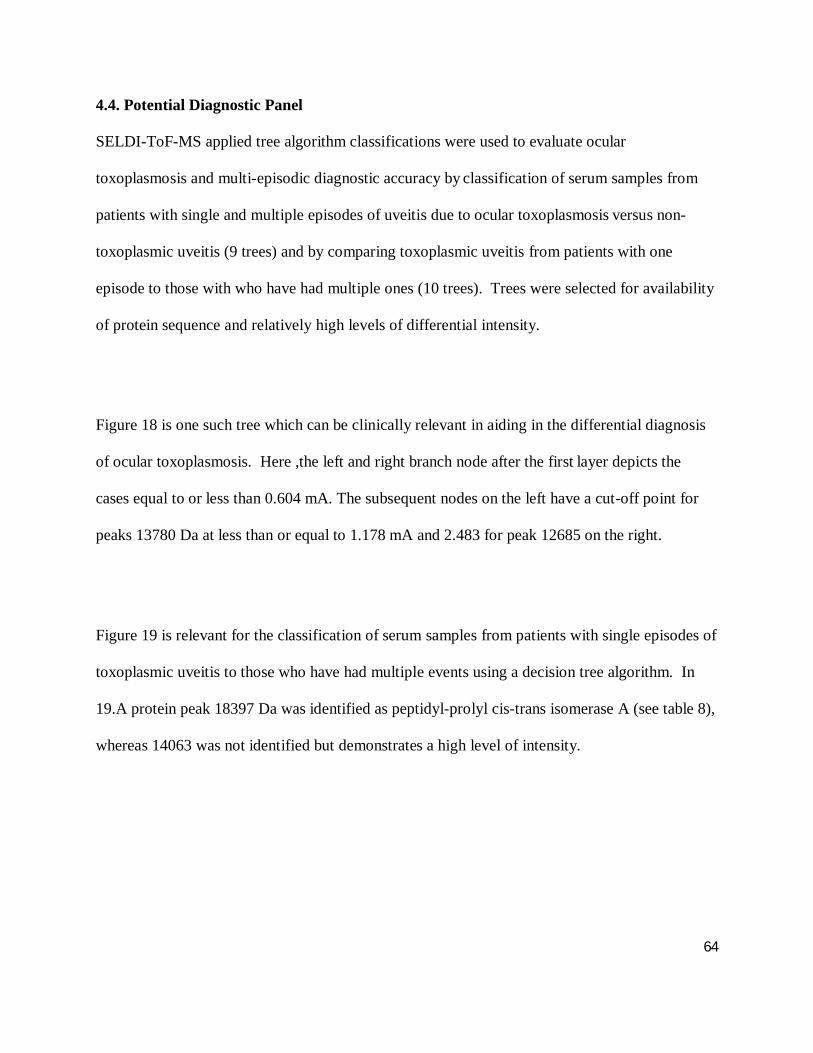
64
4.4. Potential Diagnostic Panel
SELDI-ToF-MS applied tree algorithm classifications were used to evaluate ocular
toxoplasmosis and multi-episodic diagnostic accuracy by classification of serum samples from
patients with single and multiple episodes of uveitis due to ocular toxoplasmosis versus non-
toxoplasmic uveitis (9 trees) and by comparing toxoplasmic uveitis from patients with one
episode to those with who have had multiple ones (10 trees). Trees were selected for availability
of protein sequence and relatively high levels of differential intensity.
Figure 18 is one such tree which can be clinically relevant in aiding in the differential diagnosis
of ocular toxoplasmosis. Here ,the left and right branch node after the first layer depicts the
cases equal to or less than 0.604 mA. The subsequent nodes on the left have a cut-off point for
peaks 13780 Da at less than or equal to 1.178 mA and 2.483 for peak 12685 on the right.
Figure 19 is relevant for the classification of serum samples from patients with single episodes of
toxoplasmic uveitis to those who have had multiple events using a decision tree algorithm. In
19.A protein peak 18397 Da was identified as peptidyl-prolyl cis-trans isomerase A (see table 8),
whereas 14063 was not identified but demonstrates a high level of intensity.
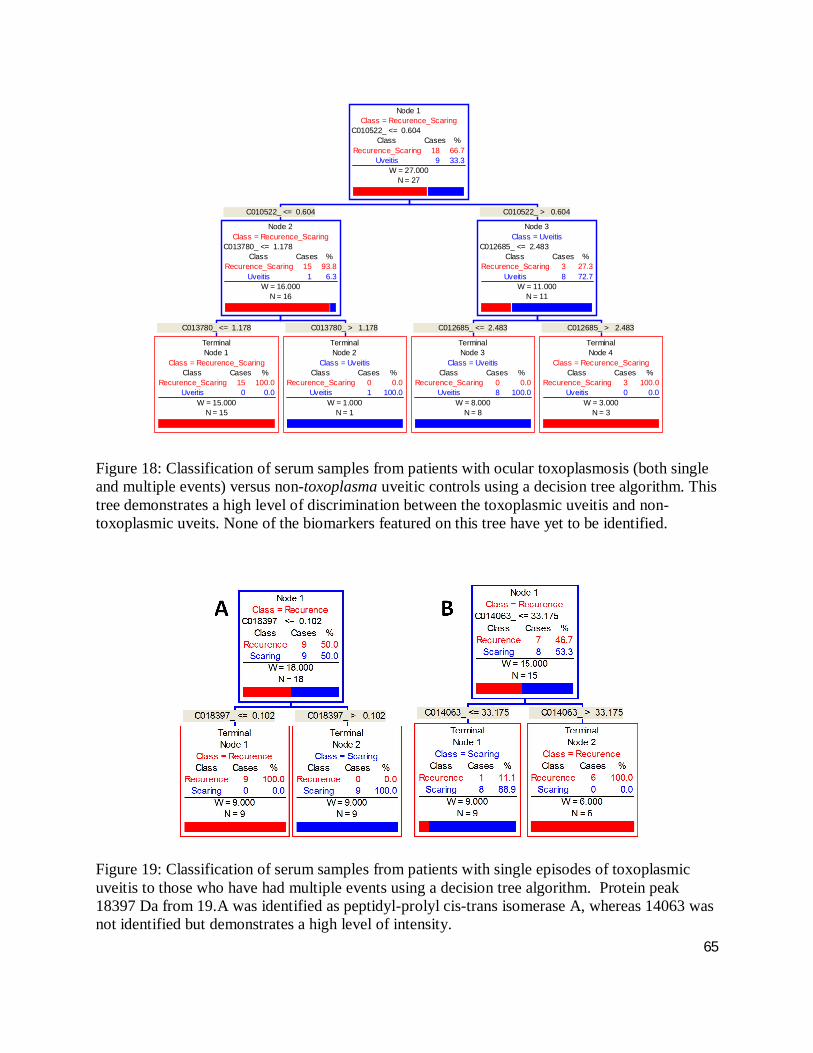
65
Figure 18: Classification of serum samples from patients with ocular toxoplasmosis (both single and multiple events) versus non-toxoplasma uveitic controls using a decision tree algorithm. This tree demonstrates a high level of discrimination between the toxoplasmic uveitis and non-toxoplasmic uveits. None of the biomarkers featured on this tree have yet to be identified.
Figure 19: Classification of serum samples from patients with single episodes of toxoplasmic uveitis to those who have had multiple events using a decision tree algorithm. Protein peak 18397 Da from 19.A was identified as peptidyl-prolyl cis-trans isomerase A, whereas 14063 was not identified but demonstrates a high level of intensity.
C013780_ <= 1.178
TerminalNode 1
Class = Recurence_ScaringClass Cases %
Recurence_Scaring 15 100.0Uveitis 0 0.0
W = 15.000N = 15
C013780_ > 1.178
TerminalNode 2
Class = UveitisClass Cases %
Recurence_Scaring 0 0.0Uveitis 1 100.0
W = 1.000N = 1
C010522_ <= 0.604
Node 2Class = Recurence_Scaring
C013780_ <= 1.178Class Cases %
Recurence_Scaring 15 93.8Uveitis 1 6.3
W = 16.000N = 16
C012685_ <= 2.483
TerminalNode 3
Class = UveitisClass Cases %
Recurence_Scaring 0 0.0Uveitis 8 100.0
W = 8.000N = 8
C012685_ > 2.483
TerminalNode 4
Class = Recurence_ScaringClass Cases %
Recurence_Scaring 3 100.0Uveitis 0 0.0
W = 3.000N = 3
C010522_ > 0.604
Node 3Class = Uveitis
C012685_ <= 2.483Class Cases %
Recurence_Scaring 3 27.3Uveitis 8 72.7
W = 11.000N = 11
Node 1Class = Recurence_Scaring
C010522_ <= 0.604Class Cases %
Recurence_Scaring 18 66.7Uveitis 9 33.3
W = 27.000N = 27
66
Chapter 5: Discussion
The diagnosis of uveitis due to ocular toxoplasmosis remains a clinical challenge. To remedy
this, novel non-invasive diagnostic tests, with high sensitivity and specificity, are needed. As
proteins represent the vast majority of biologically active molecules responsible for cellular
function and with PCR diagnostics proving to be inconclusive in disease diagnostics, protein
biomarkers could be useful in the future diagnosis of the disease.
Clinical proteomics is becoming ever more accessible due to the accumulation of DNA and
protein sequence databases, improvements in MS technologies, and the development of more
efficient computer algorithms for database searching (Graves and Haystead 2002). SELDI-ToF-
MS represents an emerging proteomic technology in biomarker discovery that allows the rapid
and sensitive analysis of complex protein mixtures and yields a compressive summary of a given
biological fluid or tissue’s proteome, without the need to first carry out protein separations, by
gels for example (Hutchens & Yip,1993; Wright Jr. and Cazares et al. 1999). Moreover, it is
well suited for biomarker discovery because of its high-throughputs, rapid analysis, and minimal
sample requirement. When samples are processed using SELDI-ToF-MS in parallel with the
latest 2-DE methodologies, “the ability of the researcher to detect a wide range of biomarkers is
sharply increased” (Boernsen 2005).
Regardless of the technology employed in the biomarker discovery process, a wide range of
biological sample types can be used including: plasma, serum, urine, saliva, cerebrospinal liquid,
67
bronchoalveolar wash out, nipple aspirate fluid, tears, amniotic fluid and tissues or cell extracts.
However, the majority of biomarker discovery studies use serum, plasma, urine, saliva and tissue
extracts (De Bock 2010). When selecting the type of biological fluid to be employed in a given
study the researcher must consider the many variables related to collection, storage, and
conditions for sample preparations. These parameters have been commented on in literature
according to the nature of the biological material. According to the Human Proteome
Organization Plasma Proteome Project Specimens Committee recommendations (HUPO 2010),
“plasma appears preferable to serum because it contains fewer peptides of degradation and
consequently presents less variability” (Tammen 2005). However, we chose serum because
proteins and peptides that “survive” the clotting procedure exhibit a stability that can be
exploited in routine clinical applications. This is a major consideration because any future
diagnostic assay will have to be able to perform in the tropics.
The human proteome is thought to be composed of more than half a million proteins (Banks et
al., 2000). The protein content of human serum is composed of a “millieu” of proteins from
most every type of cell and tissue. The serum proteome has been shown to contain information
that “directly reflects pathophysiological states and represents an invaluable source of diagnostic
information for a variety of different diseases” (Zhou 2004). Nevertheless, serum albumin
represents approximately 60 % of whole blood protein content and its presence, along with other
high abundance proteins such as immunoglobulins, complicates the detection of lower
abundance proteins which could serve as useful biomarkers. The major task we faced in the
biomarker discovery process is the fractionation of the entire proteome followed by processing
68
the massive amount of data generated into meaningful results through the use of multi-variant
statistics and various bioinformatics platforms. This is due to the extraordinarily wide dynamic
range of the serum proteome which spans more than 12 orders of magnitude (Anderson and
Anderson 2002). Protein fractionation and profiling is performed to “increase the probability of
detecting proteins with altered expression levels during disease development, progression or
treatment” (Ahmed 2005). This makes it possible to combine several of these proteins into a
discriminative protein pattern (Graves and Haystead 2002).
The potential of SELDI has been explored in various medical disciplines from cancer to
atherosclerosis and infectious diseases (Wei 2010). To date, no single, or group of, protein
biomarkers have been described for the diagnosis of either ocular or systemic toxoplasmosis, as
the technology to do so has only recently come online. This pilot study demonstrates the
potential of SELDI-ToF-MS combined with 2-DE to discover ocular toxoplasmosis biomarkers
as well as markers of the recurrent disease from patient serum.
Results presented here demonstrate that both SELDI-ToF-MS and 2-DE can be used successfully
in the search for serum biomarkers of ocular toxoplasmosis. The first goal of this study is the
identification of serum biomarkers diagnostic of ocular toxoplasmic uveitis. Second pass
analysis discovered 50 biomarkers of which eight were found to have a DI greater than one
standard of deviation. Of the eight biomarkers one was sequenced and identified to be
hemaglobin subunit beta (HBB). HBB was found to be up-regulated in toxoplasmic uveitis
69
when compared to the non-toxoplasmic variety. As was expected, following analysis by 2-DE,
HBB was also found to be expressed in both individuals with single and recurrent disease and
not in those with non-toxoplasmic uveitis. HBB is a subunit of a larger protein called
hemoglobin, which is located inside red blood cells. Hemoglobin normally consists of four
protein subunits: two subunits of beta-globin and two subunits of alpha-globin. Each of the four
protein subunits of hemoglobin carries an iron-containing molecule called heme, which is
necessary for red blood cells to bind oxygen in the lungs and deliver it to cells throughout the
body. The protein has no known role in the pathobiology of the ocular toxoplasmosis. To date
there is only one mention in the literature of HBB as a possible biomarker for any disease where
it was found to be a colon cancer biomarker, which is mostly due to its association with hypoxic
conditions within the tumour (Rho et al., 2008). However Rho and colleagues used tissue
biopsies rather than serum as the biological materials for their biomarker discovery study, which
may contain plasma and tissue specific proteins such as hemaglobin. Although, it is tempting to
hypothesize that HBB’s presence in our serum samples is due to improper removal of
erythrocytes prior to serum fractionation, this is not the case as the statistics demonstrate. Its
presence is most likely associated with free hemoglobin that is the hemoglobin outside of the red
blood cells called serum hemaglobin (Medline Plus 2010).
Eight additional biomarkers were identified and found to be related to humoral immune response
to infection. Three quarters of those markers belong to the Ig superfamily while the rest are
components of the complement system. It is important to remember that the cellular immune
response against T. gondii is critical for infection control. However, antibodies can either

70
enhance or block protective mechanisms and even mediate immunological damage, directly or
indirectly, so the Ig superfamily proteins identified can potentially serve as disease biomarkers
distinguishing either protective or pathological cellular immune events.
A 36.5 kDa band was isolated by 2-DE at a pH of between 7.0 and 9.1 and was then sequenced.
Its identity was determined to be Ig gamma-1 chain C region (IGHG1), the constant region of the
heavy chain associated with IgG1. IgG1 is the most abundant IgG subclass in humans at 66 %.
Moreover, it demonstrates the highest affinity for the Fc receptor on phagocytic cells of any of
the IgG subclasses and has a relatively high propensity to activate complement (Schroeder 2010).
In toxoplasmosis, IgG1 is up-regulated by interleukin (IL)-4 and IL-13, and is therefore
considered to be a biomarker of the chronic phase toxoplasmosis (Correa et al. 2007). This
finding is consistent with our results, where we demonstrated IGHG1 to have an expression level
three times greater in the recurrent disease when compared to non-toxoplasmic uveits, while
levels of IGHG1were unremarkable in individuals with single episodes of ocular toxoplasmosis.
Neither IGHG1 nor any of it fragments were detected by SELDI-ToF-MS in any of the 432
spectra, because chip type selected may not have bound either the protein or its fragments
(Wright Jr. 2002). However Ig lambda chain constant region along with its two isoforms of both
Ig kappa chain constant region and Ig heavy chain variable region as well as complement
components C4A and C8 gamma chain were independently detected by SELDI-ToF-MS and 2-
DE. They all represent potential immunological markers of ocular toxoplasmosis.
71
Indeed C4, a C4A precursor, is the only above mentioned immunological marker that has a
connection in the literature with toxoplasmosis. The complement system helps to clear
pathogens from the host by disrupting the target cell’s plasma membrane and it is divided into
three complimenting systems (or pathways). One of these systems is mannose-binding lectin
(MBL) pathway, which allows the host’s innate immunity to detect molecular patterns that
specifically characterise microorganisms, including T. gondii (Gadjeva 2001). Among the many
components and complexes that are involved in the MBL pathway are C4A and its precursor C4.
In fact, the C4 component of the complement had been demonstrated to be a useful indirect
marker for the prenatal diagnosis of toxoplasmosis. The use of C4 as a non-specific indicator of
congenital infection demonstrated a sensitivity and specificity of 81 % and 83 % respectively and
positive and negative predictive values of infection in 72 % and 89 %. These figures surpass the
performance of other indirect measures of infection (Cohen-Khallas 1992). Similarly, we
detected C4A to be significantly up-regulated in individuals with ocular toxoplasmosis,
suggesting that the complement-precursor was activated in these individuals, which is used in its
active form to potentiate opsonisation.
Aside from blood proteins and immunoglobins, we have identified the low abundance protein,
peptidyl-prolyl cis-trans isomerase A (PPIase) to be a biomarker of, and have a documented
connection to, ocular toxoplasmosis. The most promising of the low abundance proteins found
to be a biomarker of ocular toxoplasmosis is PPIase. PPIase has four different molecular
mechanisms associated with it that helps control the cis/trans peptide configuration: first, the
catalysis of peptide bond cis/trans isomerisation; second, as a holding function for unfolded
72
polypeptide chains; third, as a presenter protein function for an unknown number of
physiological ligands; and forth, a proline-directed binding function, which is specific to PPIases
(Fischer 2003). In terms of toxoplasmosis, we are most interested in its folding and presenter
protein function.
During invasion of host cells, T. gondii discharges the contents of small, apically located
secretory organelles called micronemes, whose proteins are known to be necessary for both
parasite motility and host cells invasion. One of the six proteins of the micronemal is TgMIC5,
which possesses a peptidyl-prolyl cis-trans isomerases sequence (Brydges 2001). This PPIase
sequence function may assist in the folding of other micronemal proteins arising in invasion of
host cells by T. gondii tachyzoites. As T. gondii modulates pro- and anti-inflammatory responses
to regulate parasite multiplication and host survival, eventually pressure from the immune
response causes the conversion of tachyzoites into slowly dividing bradyzoites (Dubey 1998).
This conversion is mediated by the production of nitric oxide in a CCR5-dependent manner
(Ibrahim 2009). The tachyzoite produces a unique chemokine mimic that activates CCR5 via
PPIase activity. As a result there is an up-regulation of downstream IL-12-dependent production
of gamma interferon (IFN-γ) in dendritic cells (DCs), which allows for host survival of acute
toxoplasmosis (Cai 2000). Conversely, T. gondii’s ability to generate such a strong protective
response though the induction of IL-12 from DCs may benefit the parasite. This in turn prevents
the protozoan from overwhelming its intermediate hosts thereby increasing the parasite’s
propensity to transmit (Aliberti 2003). Interestingly, the effect was blocked by addition of
73
cyclosporineA (CsA). As would be expected, our study found PPIA to be present only in multi-
episodic samples isolated by 2-DE.
The second goal of this pilot study was to discover a biomarker that can differentiate between
individuals with a single episode of the disease have been from those with multiple events. Thus
far, 46 markers of recurrent disease discovered by MS and 39 by 2-DE, with 18 % of the markers
found by the latter technique have been confirmed present by the former. Eight of these
biomarkers were found to have a DI greater than one standard of deviation and one was
sequenced and identified as Ig Kappa constant region. However, IgKc has no documented link
to toxoplasmosis. Thirty percent of the ocular toxoplasmosis biomarkers found are also
biomarkers of recurrent disease. These are lambda, kappa, two isoforms of Ig heavy chain
variable regions, C8-gamma, albumin and E3, which have all been discussed with the exception
of and Probable E3 ubiquitin-protein ligase (E3).
E3 ubiquitin ligase ubiquitinates histone H3 via activation of ubiquitin-like PHD and ring finger
domains 1 (UHRF1) (Karagianni 2008), UHRF1 plays a central role in confirming methylation
status of mother cells to daughter cells. It is precisely the E3-dependent UHRF1 activation of
histone ubiquitination which is known to be required for transcriptional initiation elongation,
silencing, and DNA repair (Weake 2008). Moreover UHRF1 promotes G1/S transition, which is
facilitated by T. gondii for its proliferation and plays a role in cell cycle progression (Brunet
2008). Our results show E3 to be down-regulated in recurrent disease when compared to single
74
event, suggestive of correct host response to chronic infection. Accordingly, UHRF1 is over-
expressed in various cancers essential for both cancer progression and T. gondii proliferation in
infected cells. This suggests that UHRF1 targeted therapies can be effective for treating these
diseases through the use of molecular targeted therapies and/or vaccination (Bronner 2007;
Unoki 2009).
The data sets from this pilot study were used separately to cross-validate discoveries from the
CDC/McGill T. gondii outbreak study (courtesy of Dr. Momar Ndao, McGill University) to
detect systematic biases in data such as those because of differences in pre-analytical sample
processing procedures. This differs significantly from the approaches in which data from
multiple sites are pooled together and then divided through randomization into, artificially made,
identically distributed training and test data sets. Considering the high cost associated with post-
discovery validation, however, we believe that the conservative approach that we used is similar
to the clinical environment allowing us to move more efficiently from bench to bedside given
that to date there have been no published biomarker studies of either ocular or systemic
toxoplasmosis. We were able to cross-validate almost half (47 %) of the biomarkers in this
study. Moreover, the identification of the discovered biomarkers will allow for additional
confirmation through immunoassay tests.
75
Chapter 6: Final conclusion and summary
T. gondii is an obligate intracellular protozoan parasite that infects up to a third of the world's
population. Ocular toxoplasmosis is the most common etiology of posterior uveitis, with retinal
lesions present in 20 % of those infected. The high incidence of macular scarring associated
with ocular toxoplasmosis is a leading cause of visual morbidity in the developing world causing
a highly negative social and economic impact for those affected. The ability to diagnose the
disease from a serum sample drawn from those infected would confer an advantage to patients
and their physicians.
The purpose of this study was to determine the feasibility of hypothesis-driven proteomics based
research into the diagnosis of ocular toxoplasmosis and its recurrent disease, by analysis of
peripheral blood. To that end, the study has reported potentially clinically relevant biomarkers
diagnostic of ocular toxoplasmosis. Analysis of pooled serum by MS discovered 50 markers of
ocular toxoplasmosis and 46 markers of recurrent disease. Of those biomarkers, 47% were
cross-validated within 1 % of m/z and 31 % within 0.0001 % of m/z. 14 biomarkers were
selected for validation and all were visualized by SDS-PAGE; protein sequencing is ongoing.
Protein separation by 2-DE yielded 57 differentially expressed bands, of which 20 were excised
and identified. A quarter of the identified proteins were able to be cross-validated. .
Identification and validation of all the biomarkers presented here must be completed in order to
determine their clinical relevance. As such, additional studies are required to assess the broader
76
sensitivity and specificity of the biomarkers we have discovered and to validate them. Moreover,
this study should be repeated using samples from other geographic areas as well as comparing
the biomarkers found here against biomarkers of other parasitic infections. Because the
biomarkers are of host origin, it will be necessary to determine what other pathological or non-
pathological conditions modulate the level of these proteins in the sera. Also, simpler assays will
be needed to detect the biomarkers, such as ELISA, in the future if this knowledge is to be
applied to diagnosis in low resource settings. If validated in more comprehensive studies, this
discovery will have a major impact on future disease diagnosis and treatment monitoring and the
quality of life of the patients.
An important future goal therefore is confirmation of sensitivity and specificity for the
prospective detection of ocular toxoplasmosis. Designing a trial to assess the efficacy of the
approach as a stand-alone or combined with current screening options will be important
(appendix 8.1). Once biomarkers of interest have been successfully validated, they should be
grouped together to comprise a diagnostic panel. This panel would represent the first stage in
development of an assay which would enable diagnosis the disease from a serum sample drawn
from those thought to present with atypical uveitis, with the goal of confirming or ruling out
ocular toxoplasmosis.
At the moment for hypothesis-driven research to be successful there needs to be careful selection
of the specific features of a proteome to provide information relevant for the particular
77
biomedical question. This is important largely given that the bottleneck is likely to lie not in
identifying the proteins but in their downstream characterisation. Today, proteomics is
beginning to benefit integration of genomics initiatives, such as the sequencing of the human
genome, coupled with the development of national and international proteomic funding schemes.
Now more than ever proteomics-based approaches offer the researcher the potential to move
beyond laboratory and into the clinic. Whether the identification of diagnostic markers for
various diseases including ocular toxoplasmosis results in the development of new treatment
modalities remains to be seen, but it is safe to say that the world is watching.
78
Chapter 7: Reference List
A.E. Ashcroft An Introduction to Mass Spectrometry. Leeds, Universiity of Leeds. Abreu MT, Belfort Jr R, et al. (1987). Toxoplasmose Ocular. Uveites. Oréfice F and Belfort Jr.
R. São Paulo, Roca: 211-230. Aggio FB, Muccioli C, et al. (2006). "Intravitreal triamcinolone acetonide as an adjunct in the
treatment of severe ocular toxoplasmosis." Eye. 20: 1080-1082. Ahmed, N. and G. Rice (2005). "Strategies for revealing lower abundance proteins in two-
dimensional protein maps." Journal of Chromatography B 815(1-2): 39-50. Ajzenberg, D., A. Banuls, et al. (2004). "Genetic diversity, clonality and sexuality in
Toxoplasma gondii." International journal for parasitology 34(10): 1185-1196. Ajzenberg, D., A. L. Banuls, et al. (2004). "Genetic diversity, clonality and sexuality in
Toxoplasma gondii." Int J Parasitol 34(10): 1185-1196. Ajzenberg, D., N. Cogne, et al. (2002). "Genotype of 86 Toxoplasma gondii isolates associated
with human congenital toxoplasmosis, and correlation with clinical findings." J Infect Dis 186(5): 684-689.
Aliberti, J., J. G. Valenzuela, et al. (2003). "Molecular mimicry of a CCR5 binding-domain in the microbial activation of dendritic cells." Nat Immunol 4(5): 485-490.
Anderson, N. L. and N. G. Anderson (2002). "The human plasma proteome: history, character, and diagnostic prospects." Mol Cell Proteomics 1(11): 845-867.
Antoniazzi E, Guagliano R, et al. (2008). "Ocular impaiment of toxoplasmosis." Parasitologia 50: 35-36.
Ashburn, D., A. W. Joss, et al. (1998). "Do IgA, IgE, and IgG avidity tests have any value in the diagnosis of toxoplasma infection in pregnancy?" J Clin Pathol 51(4): 312-315.
Banks, R., M. Dunn, et al. (2000). "Proteomics: new perspectives, new biomedical opportunities." The Lancet 356(9243): 1749-1756.
Belfort, R. N., S. Rasmussen, et al. (2008). "Bilateral progressive necrotizing retinichoroiditis in a immunocompromised patient: histopathologic diagnosis." Acta Ophthalmologica in press.
Belfort, R. N., B. F. Fernandes, et al. (2009). "Bilateral macular hemorrhage as a complication of drug-induced anemia: a case report." J Med Case Reports 3: 16.
R.N. Belfort, J. Isenberg, et al. (2010). Evaluating the Presence of Toxoplasma Gondii in Peripheral Blood of Patients With Uveitis. Association for Research in Vision and Ophthalmology. Fort Lauderdale, FL.
Bjellqvist, B., K. Ek, et al. (1982). "Isoelectric focusing in immobilized pH gradients: principle, methodology and some applications." J Biochem Biophys Methods 6(4): 317-339.
Boernsen, K. O., S. Gatzek, et al. (2005). "Controlled protein precipitation in combination with chip-based nanospray infusion mass spectrometry. An approach for metabolomics profiling of plasma." Anal Chem 77(22): 7255-7264.
Bonfioli AA and Orefice F (2005). "Toxoplasmosis." Semin Ophthalmol. 20: 129-141. Bosch-Driessen LH, Karimi S, et al. (2000). "Retinal detachment in ocular toxoplasmosis."
Ophthalmology. 107: 36-40. Bosch-Driessen LH, B. T., Ongkosuwito JV, Rothova A (2002). "Ocular toxoplasmosis: clinical
features and prognosis of 154 patients." Ophthalmology. 109: 869-878.
79
Bronner, C., M. Achour, et al. (2007). "The UHRF family: oncogenes that are drugable targets for cancer therapy in the near future?" Pharmacol Ther 115(3): 419-434.
Brunet, J., A. W. Pfaff, et al. (2008). "Toxoplasma gondii exploits UHRF1 and induces host cell cycle arrest at G2 to enable its proliferation." Cell Microbiol 10(4): 908-920.
Brézin AP, E. C., Burnier M Jr, Silveira C, Mahdi RM, Gazzinelli RT, Belfort R Jr, Nussenblatt RB (1990). "Identification of Toxoplasma gondii in paraffin-embedded sections by the polymerase chain reaction." Am J Ophthalmol. 110: 599-604.
Brydges, S. D., G. D. Sherman, et al. (2000). "Molecular characterization of TgMIC5, a proteolytically processed antigen secreted from the micronemes of Toxoplasma gondii." Mol Biochem Parasitol 111(1): 51-66.
Cai, G., T. Radzanowski, et al. (2000). "Identification of STAT4-dependent and independent mechanisms of resistance to Toxoplasma gondii." J Immunol 165(5): 2619-2627.
Calderaro A, P. G., Gorrini C, Peruzzi S, Zerbini L, Bommezzadri S, Dettori G, Chezzi C. (2006). "Comparison between two real-time PCR assays and a nested-PCR for the detection of Toxoplasma gondii." Acta Biomed. 77: 75-80.
Chen, R., S. Lu, et al. (2008). "Evaluation of a rapid ELISA technique for detection of circulating antigens of Toxoplasma gondii." Microbiol Immunol 52(3): 180-187.
Cohen-Khallas, Y., M. Bessières, et al. (1992). "C4 component of the complement: a new indirect marker for the prenatal diagnosis of toxoplasmosis." Presse médicale (Paris, France: 1983) 21(19): 908.
Colin J and Harie JC (1989). "Presumed toxoplasmic chorioretinitis: comparative study of treatment with pyrimethamine and sulfadiazine or clindamycin." J Fr Ophtalmol. 12: 161-165.
Contini, C., S. Seraceni, et al. (2005). "Evaluation of a Real-time PCR-based assay using the lightcycler system for detection of Toxoplasma gondii bradyzoite genes in blood specimens from patients with toxoplasmic retinochoroiditis." Int J Parasitol 35(3): 275-283.
Correa, D., I. Canedo-Solares, et al. (2007). "Congenital and acquired toxoplasmosis: diversity and role of antibodies in different compartments of the host." Parasite Immunol 29(12): 651-660.
Dao, A. and B. Fortier (2001). "[Evaluation of the Immulite 2000 Toxoplasma quantitative IgG et Toxoplasma IgM for the diagnosis of human toxoplasmosis]." Ann Biol Clin (Paris) 59(2): 157-164.
De Bock, M., D. de Seny, et al. (2010). "Challenges for biomarker discovery in body fluids using SELDI-TOF-MS." J Biomed Biotechnol 2010: 906082.
De Moura L, B.-O. L., Wada MY, Jones JL, Tuboi SH, Carmo EH, Ramalho WM, Camargo NJ, Trevisan R, Graça RM, da Silva AJ, Moura I, Dubey JP, Garrett DO. (2006). "Waterborne toxoplasmosis, Brazil, from field to gene." Emerg Infect Dis. 12: 326-329.
De Moura, L., L. M. Bahia-Oliveira, et al. (2006). "Waterborne toxoplasmosis, Brazil, from field to gene." Emerg Infect Dis 12(2): 326-329.
Dubey, J. P. (1986). "Toxoplasmosis." J Am Vet Med Assoc 189(2): 166-170. Dubey, J. P., D. S. Lindsay, et al. (1998). "Structures of Toxoplasma gondii tachyzoites,
bradyzoites, and sporozoites and biology and development of tissue cysts." Clin Microbiol Rev 11(2): 267-299.
80
Dubey JP, G. S., Labruna MB, Camargo LM, Vianna MC, Marcet PL, Lehmann T (2006). "Characterization of Toxoplasma gondii isolates in free-range chickens from Amazon, Brazil." J Parasitol. 92: 36-40.
Dworkin LL, G. T., Van Gelder RN (2002). "Real-time quantitative polymerase chain reaction diagnosis of infectious posterior uveitis." Arch Ophthalmol. 120: 1534-1539.
Eckert GU, Melamed J, et al. (2007). "Optic nerve changes in ocular toxoplasmosis." Eye. 21: 746-751.
Fardeau, C., S. Romand, et al. (2002). "Diagnosis of toxoplasmic retinochoroiditis with atypical clinical features." American journal of ophthalmology 134(2): 196-203.
Fekkar A, B. B., Touafek F, Le Hoang P, Mazier D, Paris L (2008). "Comparison of immunoblotting, calculation of the Goldmann-Witmer coefficient, and real-time PCR using aqueous humor samples for diagnosis of ocular toxoplasmosis." J Clin Microbiol 46: 1965-1967.
Fekkar, A., B. Bodaghi, et al. (2008). "Comparison of immunoblotting, calculation of the Goldmann-Witmer coefficient, and real-time PCR using aqueous humor samples for diagnosis of ocular toxoplasmosis." J Clin Microbiol 46(6): 1965-1967.
Flegr, J., J. Klose, et al. (2009). "Increased incidence of traffic accidents in Toxoplasma-infected military drivers and protective effect RhD molecule revealed by a large-scale prospective cohort study." BMC Infect Dis 9: 72.
Fischer, G. and T. Aumuller (2003). "Regulation of peptide bond cis/trans isomerization by enzyme catalysis and its implication in physiological processes." Rev Physiol Biochem Pharmacol 148: 105-150.
Fortier, B., E. Aïssi, et al. (1991). "Spontaneous abortion and reinfection by Toxoplasma gondii." The Lancet 338(8764): 444-444.
Frobel, J., S. Lehr, et al. (2009). "Mass spectrometry-based proteomics and its potential use in haematological research." Arch Physiol Biochem 115(5): 286-297.
Gadjeva, M., S. Thiel, et al. (2001). "The mannan-binding-lectin pathway of the innate immune response." Curr Opin Immunol 13(1): 74-78.
Ganesh SK, Sharma S, et al. (2004). "Fuchs' heterochromic iridocyclitis following bilateral ocular toxoplasmosis." Ocul Immunol Inflamm 12: 75-77.
Gavinet, M., F. Robert, et al. (1997). "Congenital toxoplasmosis due to maternal reinfection during pregnancy." Journal of Clinical Microbiology 35(5): 1276.
Graves, P. R. and T. A. Haystead (2002). "Molecular biologist's guide to proteomics." Microbiol Mol Biol Rev 66(1): 39-63; table of contents.
Grigg ME, G. J., Boothroyd JC, Margolis TP (2001). "Unusual abundance of atypical strains associated with human ocular toxoplasmosis." J Infect Dis. 184: 633-639.
Hanash, S. M., R. Kuick, et al. (1988). "Quantitative analysis of a new marker for common acute lymphoblastic leukemia detected by two-dimensional electrophoresis." Dis Markers 6(4): 209-220.
Hanash, S. (2003). "Disease proteomics." Nature 422(6928): 226-232. Hodgetts, A., M. Levin, et al. (2007). "Biomarker discovery in infectious diseases using SELDI."
Future Microbiol. 2(1): 35-49. Holland, G. (2003). "Ocular toxoplasmosis: a global reassessment. Part I: epidemiology and
course of disease." American journal of ophthalmology 136(6): 973-988.
81
Holland, G. N. (2000). "Ocular toxoplasmosis: new directions for clinical investigation." Ocul Immunol Inflamm 8(1): 1-7.
Holland GN (2004). "Ocular toxoplasmosis: a global reassessment. Part II: disease manifestations and management." Am J Ophthalmol. 137: 1-17.
Holliman, R. E. (1997). "Toxoplasmosis, behaviour and personality." J Infect 35(2): 105-110. Howe DK, H. S., Derouin F, Sibley LD (1997). "Determination of genotypes of Toxoplasma
gondii strains isolated from patients with toxoplasmosis." J Clin Microbiol. 35: 1411-1414.
Howe DK, S. D. (1995). "Toxoplasma gondii comprises three clonal lineages: correlation of parasite genotype with human disease." J Infect Dis. 172: 1561-1566.
HUPO (2010). "Plasma Proteome Project." Retrieved April 11, 2010, from http://www.hupo.org/research/hppp/people.asp.
Hutchens, T. and T. Yip (1993). "New desorption strategies for the mass spectrometric analysis of macromolecules." Rapid Communications in Mass Spectrometry 7(5): 1993-1905.
Jackson, M. H. and W. M. Hutchison (1989). "The prevalence and source of Toxoplasma infection in the environment." Adv Parasitol 28: 55-105.
Jenum, P. A., B. Stray-Pedersen, et al. (1998). "Incidence of Toxoplasma gondii infection in 35,940 pregnant women in Norway and pregnancy outcome for infected women." J Clin Microbiol 36(10): 2900-2906.
Jones JL, M. C., Belfort R Jr, Holland GN, Roberts JM, Silveira C (2006). "Recently Acquired Toxoplasma gondii Infection, Brazil." Emerg Infect Dis. 12: 582-587.
G. Wright Jr., L. H. Cazares, et al. (1999). "Proteinchip(R) surface enhanced laser desorption/ionization (SELDI) mass spectrometry: a novel protein biochip technology for detection of prostate cancer biomarkers in complex protein mixtures." Prostate Cancer Prostatic Dis 2(5/6): 264-276.
Ibrahim, H. M., H. Bannai, et al. (2009). "Toxoplasma gondii cyclophilin 18-mediated production of nitric oxide induces Bradyzoite conversion in a CCR5-dependent manner." Infect Immun 77(9): 3686-3695.
Karagianni, P., L. Amazit, et al. (2008). "ICBP90, a novel methyl K9 H3 binding protein linking protein ubiquitination with heterochromatin formation." Mol Cell Biol 28(2): 705-717.
Khan A, J. C., Muccioli C, Vallochi AL, Rizzo LV, Belfort R Jr, Vitor RW, Silveira C, Sibley LD. (2006). "Genetic divergence of Toxoplasma gondii strains associated with ocular toxoplasmosis, Brazil." Emerg Infect Dis. 12: 942-949.
Kishore K, C. M., Peyman GA (2001). "Intravitreal clindamycin and dexamethasone for toxoplasmic retinochoroiditis." Ophthalmic Surg Lasers 32: 183-192.
Klaren, V. N. and A. Kijlstra (2002). "Toxoplasmosis, an overview with emphasis on ocular involvement." Ocul Immunol Inflamm 10(1): 1-26.
Kodjikian L, W. M., Fleury J, Denis P, Binquet C, Peyron F, Garweg JG (2006). "Ocular manifestations in congenital toxoplasmosis." Graefes Arch Clin Exp Ophthalmol. 244: 14-21.
La Hey E and Baarsma GS (1993). "Contralateral active ocular toxoplasmosis in Fuchs' heterochromic cyclitis." Br J Ophthalmol 77: 455-456.
Lehmann, T., D. H. Graham, et al. (2004). "Variation in the structure of Toxoplasma gondii and the roles of selfing, drift, and epistatic selection in maintaining linkage disequilibria." Infect Genet Evol 4(2): 107-114.
82
Lin MH, C. T., Kuo TT, Tseng CC, Tseng CP (2000). "Real-time PCR for quantitative detection of Toxoplasma gondii." J Clin Microbiol. 38: 4121-4125.
Mahalakshmi B, T. K., Madhavan HN, Biswas J (2006). "Diagnostic value of specific local antibody production and nucleic acid amplification technique-nested polymerase chain reaction (nPCR) in clinically suspected ocular toxoplasmosis." Ocul Immunol Inflamm. 14: 105-112.
McCannel, C. A., G. N. Holland, et al. (1996). "Causes of uveitis in the general practice of ophthalmology. UCLA Community-Based Uveitis Study Group." Am J Ophthalmol 121(1): 35-46.
Medline Plus (2010). "Serum Hemoglobin." Retrieved April 8, 2010, from http://www.nlm.nih.gov/medlineplus/ency/article/003677.htm.
Mets MB and Chhabra MS (2008). "Eye manifestations of intrauterine infections and their impact on childhood blindness." Surv Ophthalmol. 53: 95-111.
Mets MB, H. E., Boyer KM, Swisher CN, Roizen N, Stein L, Stein M, Hopkins J, Withers S, Mack D, Luciano R, Patel D, Remington JS, Meier P, McLeod R. (1997). "Eye manifestations of congenital toxoplasmosis." Am J Ophthalmol. 123: 1-16.
Montoya, J. and J. Remington (1996). "Toxoplasmic chorioretinitis in the setting of acute acquired toxoplasmosis." Clin Infect Dis. 23: 277-282.
Montoya JG and Liesenfeld O (2004). "Toxoplasmosis." The Lancet. 363: 1965-1976. Montoya JG and Remington JS (2008). "Management of Toxoplasma gondii infection during
pregnancy." Clin Infect Dis. 47: 554-566. Montoya, J. G., H. B. Huffman, et al. (2004). "Evaluation of the immunoglobulin G avidity test
for diagnosis of toxoplasmic lymphadenopathy." J Clin Microbiol 42(10): 4627-4631. Montoya, J. G. and O. Liesenfeld (2004). "Toxoplasmosis." Lancet 363(9425): 1965-1976. Montoya, J. G., S. Parmley, et al. (1999). "Use of the polymerase chain reaction for diagnosis of
ocular toxoplasmosis." Ophthalmology 106(8): 1554-1563. Morhun PJ, Weisz JM, et al. (1996). "Recurrent ocular toxoplasmosis in patients treated with
systemic corticosteroids." Retina 16: 383-387. Nowakowska D, C. I., Remington JS, Grigg M, Golab E, Wilczynski J, Sibley LD. (2006).
"Genotyping of Toxoplasma gondii by multiplex PCR and peptide-based serological testing of samples from infants in Poland diagnosed with congenital toxoplasmosis." J Clin Microbiol 44: 1382-1389.
Nussenblatt RB and Belfort Jr. R (1994). "Ocular toxoplasmosis. An old disease revisited." JAMA. 271: 304-307.
O'farrell, P. (1975). "High resolution two-dimensional electrophoresis of proteins." Journal of Biological Chemistry 250(10): 4007.
Ongkosuwito JV, B.-D. E., Kijlstra A, Rothova A. (1999). "Serologic evaluation of patients with primary and recurrent ocular toxoplasmosis for evidence of recent infection." Am J Ophthalmol. 128: 407-412.
Opremcak EM, S. D., Sharpe MR (1992). "Trimethoprim-sulfamethoxazole therapy for ocular toxoplasmosis." Ophthalmology. 99: 920-925.
Payne, R. A., D. H. Joynson, et al. (1987). "Public Health Laboratory Service enzyme linked immunosorbent assay for detecting Toxoplasma specific IgM antibody." J Clin Pathol 40(3): 276-281.
83
Pena, H. F., S. M. Gennari, et al. (2008). "Population structure and mouse-virulence of Toxoplasma gondii in Brazil." Int J Parasitol 38(5): 561-569.
Peterson, P. and J. Remington, Eds. (1997). Toxoplasmosis of the central nervous system. In Defense of the Brain: Current Concepts in the Immunopathogenesis and Clinical Aspects of CNS Infections. Boston, Blackwell Scientific.
Pleyer U, T. N., Liesenfeld O (2007). "Ocular toxoplasmosis." Ophthalmologe 104: 603-615. Rao, N. A. and R. L. Font (1977). "Toxoplasmic retinochoroiditis: electron-microscopic and
immunofluorescence studies of formalin-fixed tissue." Arch Ophthalmol 95(2): 273-277. Rho, J., S. Qin, et al. (2008). "Proteomic expression analysis of surgical human colorectal cancer
tissues: up-regulation of PSB7, PRDX1, and SRP9 and hypoxic adaptation in cancer." Journal of proteome research 7(7): 2959.
Rothova A, de Boer JH, et al. (2008). "Usefulness of aqueous humor analysis for the diagnosis of posterior uveitis." Ophthalmology. 115: 306-311.
Rothova A, B. H., Meenken C, Baarsma GS, Boen-Tan TN, de Jong PT, Schweitzer CM, Timmerman Z, de Vries J, Zaal MJ, et al. (1989). "Therapy of ocular toxoplasmosis." Int Ophthalmol 12: 415-419.
Rothova A, M. C., Buitenhuis HJ, Brinkman CJ, Baarsma GS, Boen-Tan TN, de Jong PT, Klaassen-Broekema N, Schweitzer CM, Timmerman Z, et al. (1993). "Therapy for ocular toxoplasmosis." Am J Ophthalmol. 115: 517-523.
Schwab IR (1991). "The epidemiologic association of Fuchs' heterochromic iridocyclitis and ocular toxoplasmosis." Am J Ophthalmol. 111: 356-362.
Schroeder, H. W., Jr. and L. Cavacini (2010). "Structure and function of immunoglobulins." J Allergy Clin Immunol 125(2 Suppl 2): S41-52.
Scot R. Weinberger, Lee Lomas, et al., Eds. (2007). Surface-Enhanced Laser Desorption/Ionization Protein Biochip Technology for Proteomics Research and Assay Development. Spectral techniques in proteomics, Taylor and Francis Group.
Silveira C, Vallochi AL, et al. (Manuscript submitted). "Toxoplasma gondii in the peripheral blood of patients with acute and chronic toxoplasmosis." Br J Ophthalmol.
Silveira, C., R. Belfort, Jr., et al. (2001). "A follow-up study of Toxoplasma gondii infection in southern Brazil." Am J Ophthalmol 131(3): 351-354.
Silveira C, B. R. J., Muccioli C, Abreu MT, Martins MC, Victora C, Nussenblatt RB, Holland GN. (2001). "A follow-up study of Toxoplasma gondii infection in southern Brazil." Am J Ophthalmol. 131: 351-354.
Silveira C, B. R. J., Muccioli C, Holland GN, Victora CG, Horta BL, Yu F, Nussenblatt RB. (2002). "The effect of long-term intermittent trimethoprim/sulfamethoxazole treatment on recurrences of toxoplasmic retinochoroiditis." Am J Ophthalmol. 134: 41-46.
Silveira C, F. R., Muccioli C, Nussenblatt R, Belfort R Jr. (2003). "Toxoplasmosis transmitted to a newborn from the mother infected 20 years earlier." Am J Ophthalmol. 136: 370-381.
Smith JR and Cunningham Jr. ET (2002). "Atypical presentations of ocular toxoplasmosis." Curr Opin Ophthalmol. 13: 387-392.
Sobrin L, Kump LI, et al. (2007). "Intravitreal clindamycin for toxoplasmic retinochoroiditis." Retina 27: 952-957.
Soheilian M, S. M., Ghajarnia M, Dehghan MH, Yazdani S, Behboudi H, Anisian A, Peyman GA. (2005). "Prospective randomized trial of trimethoprim/sulfamethoxazole versus
84
pyrimethamine and sulfadiazine in the treatment of ocular toxoplasmosis." Ophthalmology. 112: 1876-1882.
Suzuki, Y. and A. Kobayashi (1987). "Presence of high concentrations of circulating Toxoplasma antigens during acute Toxoplasma infection in athymic nude mice." Infect Immun 55(4): 1017-1018.
Tammen, H., I. Schulte, et al. (2005). "Peptidomic analysis of human blood specimens: comparison between plasma specimens and serum by differential peptide display." Proteomics 5(13): 3414-3422.
Tan HK, S. D., Stanford M, Teär-Fahnehjelm K, Ferret N, Salt A, Gilbert R; (2007). "Risk of visual impairment in children with congenital toxoplasmic retinochoroiditis." Am J Ophthalmol. 144: 648-653.
Toledo de Abreu M, Belfort R Jr, et al. (1982). "Fuchs' heterochromic cyclitis and ocular toxoplasmosis." Am J Ophthalmol. 93: 739-744.
Unoki, M., J. Brunet, et al. (2009). "Drug discovery targeting epigenetic codes: the great potential of UHRF1, which links DNA methylation and histone modifications, as a drug target in cancers and toxoplasmosis." Biochem Pharmacol 78(10): 1279-1288.
Vallochi AL, M. C., Martins MC, Silveira C, Belfort R Jr, Rizzo LV. (2005). "The genotype of Toxoplasma gondii strains causing ocular toxoplasmosis in humans in Brazil." Am J Ophthalmol. 139: 350-361.
Vallochi, A. L., M. V. Nakamura, et al. (2002). "Ocular toxoplasmosis: more than just what meets the eye." Scand J Immunol 55(4): 324-328.
van der Puije, W. N., K. M. Bosompem, et al. (2000). "The prevalence of anti-Toxoplasma gondii antibodies in Ghanaian sheep and goats." Acta Trop 76(1): 21-26.
Wallon M, Kodjikian L, et al. (2004). "Long-term ocular prognosis in 327 children with congenital toxoplasmosis." Pediatrics 113: 1567-1572.
Weake, V. M. and J. L. Workman (2008). "Histone ubiquitination: triggering gene activity." Mol Cell 29(6): 653-663.
Wei, W., A. Martin, et al. (2010). "10 Years of SELDI: What Have we Learnt?" Current Proteomics 7(1): 15-25.
Weiss, L. M. and K. Kim (2007). Toxoplasma Gondii: The Model Apicomplexan. Perspectives and Methods. New York, Academic Press.
Wright, G. L., Jr. (2002). "SELDI proteinchip MS: a platform for biomarker discovery and cancer diagnosis." Expert Rev Mol Diagn 2(6): 549-563.
Yai, L. E., A. M. Ragozo, et al. (2009). "Genetic diversity among capybara (Hydrochaeris hydrochaeris) isolates of Toxoplasma gondii from Brazil." Vet Parasitol.
Zhou, M., D. Lucas, et al. (2004). "An investigation into the human serum “interactome”." Electrophoresis 25(9): 1289-1298.
Zhu, S. (2009). "Psychosis may be associated with toxoplasmosis." Med Hypotheses 73(5): 799-801.
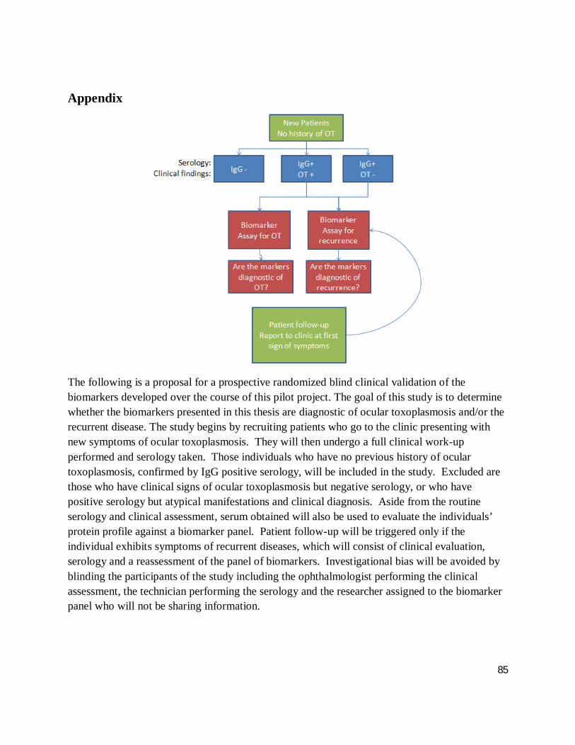
85
Appendix
The following is a proposal for a prospective randomized blind clinical validation of the biomarkers developed over the course of this pilot project. The goal of this study is to determine whether the biomarkers presented in this thesis are diagnostic of ocular toxoplasmosis and/or the recurrent disease. The study begins by recruiting patients who go to the clinic presenting with new symptoms of ocular toxoplasmosis. They will then undergo a full clinical work-up performed and serology taken. Those individuals who have no previous history of ocular toxoplasmosis, confirmed by IgG positive serology, will be included in the study. Excluded are those who have clinical signs of ocular toxoplasmosis but negative serology, or who have positive serology but atypical manifestations and clinical diagnosis. Aside from the routine serology and clinical assessment, serum obtained will also be used to evaluate the individuals’ protein profile against a biomarker panel. Patient follow-up will be triggered only if the individual exhibits symptoms of recurrent diseases, which will consist of clinical evaluation, serology and a reassessment of the panel of biomarkers. Investigational bias will be avoided by blinding the participants of the study including the ophthalmologist performing the clinical assessment, the technician performing the serology and the researcher assigned to the biomarker panel who will not be sharing information.