Embed Size (px)
Citation preview
i
Novel Ferrocenyl benzoyl peptide esters as anti-cancer agents and
Ferrocenoyl self assembled monolayers as anion sensors.
by
Alan J. Corry B.Sc. (Hons)
A thesis presented for the degree of Doctor of Philosophy
at
Dublin City University
Under the supervision of Dr. Peter T. M. Kenny
Ollscoil Chathair Bhaile Atha Cliath
School of Chemical Sciences
January 2009

ii
To my family

iii
Declaration
I hereby certify that this material, which I now submit for assessment on the programme
of study leading to the award of Ph.D is entirely my own work, that I have exercised
reasonable care to ensure that the work is original, and does not to the best of my
knowledge breach any law of copyright, and has not been taken from the work of others
save and to the extent that such work has been cited and acknowledged within the text of
my work.
Signed: ____________
Alan J. Corry
ID No.: ___________
Date: ___________
iv
Acknowledgements
I would like to thank Dr. Peter T. M. Kenny for giving me the opportunity to conduct
research under his supervision and for being supportive and patient during my three years
in the lab.
I would also like to thank:
IRCSET and the Embark Initiative for funding this research.
Dr. Norma O’Donovan, Dermot O’Sullivan and Aine Mooney for conducting the
biological studies.
Colm T. Mallon for being my electrochemistry guru and for answering all my questions
and for his help throughout, also to Dr Mary Pryce for the use of her potentiostat and PC.
To all the technical and academic staff for their help, especially John McLoughlin,
Damien McGuirk and Ambrose May.
A special thank you to all the PKRG (Peter Kenny Research Group), members, past and
present especially Brian, Paula, Aine, Will and Andy.
Also to my fellow postgraduates especially Debbie, Neil, Lorraine, Saibh, Ewa, Dan,
Brian Deegan, Liz, Emma K and Shelly, also to Sonia, Laura, Yvonne, Nicola, Emma,
Sarah, Fadi, Tom, Lynda 1&2 and Bruce. Big thanks also to Michael Ryan.
Thanks to all the lads from home Ambrose, Ronny, Mick, Ged, Brady and Warren, even
though they never really understood what I was doing.
Also, to all my family, especially Mam, Niamh, Pat, Kieran, Margaret and Ann, for all
their support in every way possible over the past 24 years. Also, to the all the members of
the Devine, Hand and Mathews families. I can start paying them back now.
v
Abstract
A series of novel N-(ferrocenyl)benzoyl peptide esters have been synthesized,
characterized and screened in vitro against the non-small cell lung cancer cell line, H1299
(cisplatin and carboplatin resistant variant). The potential production of hydroxyl radicals
would be enhanced by the benzoyl spacer as this lowers the redox potential of the
ferrocene moiety thus making the iron atom easier to oxidize. The peptide chain would
also be able to interact with biomolecules via hydrogen bonding.
A series of N-(ferrocenyl)2 and N-(ferrocenoyl)2 cystine dimethyl esters have also been
synthesized, characterized and immobilized onto gold electrodes. The electroactivity of
the ferrocene and the hydrogen bonding ability of the peptide amide bonds will be
exploited in the sensing of anions in aqueous media.
The synthesis of each series of compounds was achieved by coupling the free N-terminus
of various amino acid and peptide esters to the carboxyl group of ferrocenyl benzoic acid
(ortho, meta and para) or ferrocenecarboxylic acid using N-(3-dimethylaminopropyl)-N′-
ethylcarbodiimide hydrochloride (EDC) and 1-hydroxybenzotriazole (HOBt) coupling
protocol. All compounds were characterized by a range of spectroscopic techniques
including: 1H,
13C, DEPT 135 and HMQC NMR in addition to IR, UV-Vis, MS and CV.
The biological effects of orientation around the central benzoyl moiety, increasing
peptide chain length and lipophilicity were investigated for the N-(ferrocenyl)benzoyl
peptide esters. The most active compound was found to be N-meta-(ferrocenyl)-
benzoyl-glycine-L-alanine ethyl ester with an IC50 value of 4.0 µM while N-ortho-
(ferrocenyl)-benzoyl-glycine-L-alanine ethyl ester induced a block in the G2/M phase of
the cell cycle.
N-ortho-(ferrocenyl)-benzoyl2-L-cystine dimethyl ester displayed a linear
amperometric response to chloride anions in aqueous media while N-(ferrocenoyl)-β-
alanine2-L-cystine dimethyl ester exhibited a linear response to nitrate, dihydrogen
phosphate and adenosine nucleotides. For adenosine nucleotides N-(ferrocenoyl)-β-
alanine2-L-cystine dimethyl ester showed a nanomolar sensitivity in aqueous media.
vi
Table of Contents
Title page i
Declaration iii
Acknowledgements iv
Abstract v
Table of Contents vi
Section 1 1
Ferrocenyl benzoyl peptide esters as anti-cancer agents 1
Chapter 1 2
Biologically active ferrocene derivatives 2
1.1 Introduction. 2
1.2 Redox properties of ferrocene. 3
1.3 Ferricenium salts as anti-cancer agents. 4
1.4 Metallocene based selective estrogen receptor modulators and anti-androgens. 7
1.5 Ferrocenyl peptide conjugates as anti-cancer agents. 15
1.6 Other metallocene complexes as anti-cancer agents. 17
1.7 Ferrocene derivatives as anti-malarial agents. 20
1.8 Conclusions. 23
References. 23
Chapter 2 27
Results and Discussion 27
2.1 Introduction. 27
2.2 The synthesis of dipeptide ethyl esters. 28
2.3 The synthesis of N-(ferrocenyl)benzoyl peptide esters. 36
2.4 1H NMR studies of N-(ferrocenyl)benzoyl dipeptide esters. 39
2.5 13
C NMR and DEPT 135 studies of N-(ferrocenyl)benzoyl dipeptide esters. 46
2.6 COSY studies of N-para-(ferrocenyl)-benzoyl-γ-aminobutyric acid-L-norleucine
ethyl ester. 49
2.7 HMQC study of N-meta-(ferrocenyl)-benzoyl-β-alanine-L-2-aminobutyric acid
ethyl ester. 51
2.8 1H NMR studies of N-(ferrocenyl)benzoyl tri- and tetrapeptide ethyl esters. 53
vii
2.9 13
C NMR and DEPT 135 studies of N-(ferrocenyl)benzoyl tri- and tetrapeptide ethyl
esters. 55
2.10 Synthesis of 1, 1’-N, N’-ortho-(ferrocenyl)-bisbenzoyl amino acid and dipeptide
esters. 58
2.11 1H and
13C NMR studies of 1, 1’-N, N’-ortho-(ferrocenyl)-bisbenzoyl amino acid
and dipeptide esters. 61
2.12 Infra red spectroscopic studies of N-(ferrocenyl)benzoyl peptide esters. 63
2.13 UV-Vis spectroscopic studies of N-(ferrocenyl)benzoyl peptide esters. 65
2.14 Cyclic voltammetry of N-(ferrocenyl)benzoyl peptide esters. 66
2.15 Mass spectrometric studies of N-(ferrocenyl)benzoyl peptide esters. 68
2.16 Conclusions. 69
References 70
Chapter 3 71
In vitro anti-cancer activity of N-(ferrocenyl)benzoyl peptide esters 71
3.1 Introduction 71
3.2 Effect of redox potential and orientation around the central benzoyl moiety on cell
proliferation. 73
3.3 Effect of increasing peptide chain length on cell proliferation. 75
3.4 Effect of increasing the number of methylene groups in both the first and second
amino acids of the dipeptide chain on cell proliferation. 77
3.5 Anti-cancer activity of 1,1’-N, N’-ortho-(ferrocenyl)-bisbenzoyl-glycine-L-alanine
ethyl ester. 80
3.6 Cell cycle analysis of N-ortho-(ferrocenyl)-benzoyl-glycine-L-alanine ethyl ester.
82
3.7 Conclusions. 84
References 85
Experimental details 87
Section 2 129
Ferrocenoyl self assembled monolayers as anion sensors 129
Chapter 4 130
Anion sensors 130
viii
4.1 Introduction. 130
4.2 Ferrocenoyl and ferrocenyl based anion receptors. 131
4.3 Ion recognition by self assembled monolayers (SAMs). 140
4.4 Binding of biomolecules in water. 148
4.5 Conclusions. 149
References. 150
Chapter 5 154
Results and Discussion II 154
5.1 Introduction. 154
5.2 The preparation of N-(ferrocenyl)2 and N-(ferrocenoyl)2 cystine dimethyl esters
derivatives. 155
5.3 1H NMR studies of N-(ferrocenyl)2 and N-(ferrocenoyl)2 cystine dimethyl esters. 160
5.4 13
C NMR and DEPT 135 spectroscopic studies of N-(ferrocenyl)2 and N-
(ferrocenoyl)2 cystine dimethyl esters. 162
5.5 COSY and HMQC studies of N-(ferrocenyl)2 and N-(ferrocenoyl)2 cystine dimethyl
esters. 165
5.6 Infrared spectroscopic studies of N-(ferrocenyl)2 and N-(ferrocenoyl)2 cystine
dimethyl esters. 168
5.7 UV-Vis spectroscopic studies of N-para-(ferrocenyl)-benzoyl2-L-cystine dimethyl
ester and N-(ferrocenoyl)-β-alanine2-L-cystine dimethyl ester. 169
5.8 Cyclic voltammetry. 169
5.9 Preparation of the self assembled monolayers on gold. 171
5.10 Electrochemical characterization of self assembled monolayers. 173
5.11 Conclusions. 178
References 180
Chapter 6 181
Anion Binding Studies of N-(ferrocenyl)2 and N-(ferrocenoyl)2 cystine dimethyl esters.
181
6.1 Introduction. 181
6.2 Electrochemical anion coordination studies of monolayers of N-(ferrocenyl)2 and N-
(ferrocenoyl)2 cystine dimethyl esters in aqueous media. 183
ix
6.3 Monolayer response to changing anion concentration in aqueous media. 189
6.4 Determination of adenosine nucleotides in aqueous media. 194
6.5 Monolayer response to changing anion concentration in organic media. 197
6.6 Conclusions. 200
References. 202
Experimental details 203
Appendix 211
1
Section 1
Ferrocenyl benzoyl peptide esters as anti-cancer
agents
2
Chapter 1
Biologically active ferrocene derivatives
1.1 Introduction.
Over the past 40 years organometallic chemistry has developed into a vibrant and
important area linking organic and inorganic chemistry. Applications of organometallic
compounds are varied and numerous. They include catalysts for industrial syntheses and
anti-knocking agents for fuel. The interest in metal complexes in a biological sense was
initiated by the success of cisplatin against various types of cancer (1)
. Organometallic
compounds containing transition metals, such as Co, Cu, Fe, Ga, Ge, Mo, Pt, Sn, Rh, Ru,
Ti, and V are known to have anti-proliferative (in vitro) and anti-neoplastic (in vivo)
activities. Platinum coordination compounds, such as cisplatin, carboplatin and other
derivatives are used in the treatment of a variety of tumours (2)
. However, problems with
toxicity, harsh side effects during administration, together with acquired drug resistance
problems has prompted the search for alternative anti-cancer drugs with better
pharmacological profiles whilst retaining therapeutic efficacy. Some of the most
promising novel non-platinum anti-cancer agents are emerging from the field of
bioorganometallic chemistry. Bioorganometallic chemistry is a field devoted to the
synthesis and study of organometallic species of biological and medical interest. Notably,
the field of medicinal chemistry has benefited considerably from the incorporation of
organometallic moieties into potential drug molecules. In the wake of the success of
platinum complexes, coordination and organometallic compounds are possible alternative
therapeutics for the treatment of cancer (3)
. The resistance of some cancers to cisplatin
emphasises the need for new drugs with differing modes of action in order to overcome
this resistance. Colon and non-small-cell lung cancer are intrinsically resistant while
ovarian and small-cell lung cancers acquire resistance over time. Resistance is due to
decreased drug accumulation and an increased ability of DNA to tolerate the damage
caused by cisplatin. Deactivation of cisplatin also occurs upon binding to proteins. A
number of the severe side effects of cisplatin are attributed to this protein binding (4)
. The
ultimate aim of this research is the discovery of compounds that are active against a wide
3
range of cancers but that have fewer side effects than cisplatin (5)
. This is highlighted by
the recent entry of ruthenium compounds into Phase I clinical trials for the treatment of
colorectal carcinoma, for example imidazolium-trans-tetrachloro(dimethylsulphoxide)
imidazoleruthenium(III) (6)
.
Metallocenes, including ferrocene, are also known to have a wide range of biological
applications. Ferrocene has attracted particular attention due to its aromatic character,
stability and low toxicity. It can also be easily derivatized and the central iron atom is
also easily oxidised from Fe(II) to Fe(III). The medicinal application of ferrocene is
currently an active area of research with many reports showing its activity in vivo and in
vitro and its potential as an anti-tumour, anti-malarial and anti-fungal agent (5)
.
1.2 Redox properties of ferrocene.
The electron transfer-reactive oxygen species-oxidative stress theory (ET-ROS-OS) has
been implicated in the mechanism of action of a wide variety of biologically active
compounds, for example nitroaromatics and quinones. Therefore the development of
drugs that enhance Reactive Oxygen Species (ROS) has increased in importance. Also
the fact that cancer tissue is known to be in a state of oxidative stress further increases the
need for new drugs that can exploit this fact (7)
. Increasing the concentration of ROS may
overwhelm the cancer cells but leave normal cells unaffected. Elevated levels of ROS are
also known to induce apoptosis. Current attention is concentrated on increasing
concentration of ROS to lethal levels in cells, interfering with anti-oxidant enzymes and
the promotion of catalysts that enhance the toxicity of the ROS.
The loss of an electron from a high energy, non-bonding orbital to yield the ferricenium
cation, (Fc → Fc+), is an important aspect of the chemistry of ferrocene and is often
implicated in its cytotoxicity (2)
. This is demonstrated in Figure 1.1

4
Fe Fe- e-
+ e-
Fig. 1.1 One electron oxidation of ferrocene to yield the ferricenium ion and reverse
reduction reaction.
In biological systems ferrocene can be oxidised by hydrogen peroxide in the presence of
horseradish peroxidase. The hydroxyl radicals formed from Fc+ under physiological
conditions are proposed to act as DNA damaging agents for biologically active ferrocene
derivatives. The ferricenium cation has been shown to form charge transfer complexes
with donor groups in proteins. The reverse reaction, (Fc+ → Fc), is known to proceed
through oxidation of metalloproteins, in the presence of glutathione forming hydroxyl
radicals and through oxidation of NADH to NAD+ (2)
. The oxidation of NADH to NAD+
is a good indicator of the ferricenium cations capacity for interfering with biologically
important, enzyme controlled electron transfer reactions.
The redox status of a given biological system is vitally important as numerous processes
in living cells are mediated by redox reactions. For example, cellular respiration whereby
ATP is formed involves a series of reactions including the reduction of NAD+ and
oxygen and the oxidation of sugars. Redox activation of otherwise inactive prodrugs
coupled with further chemical modification e.g. hydrolysis, can lead to highly reactive
electrophilic compounds. A suitable bio-redox prodrug should have minimal toxicity to
healthy cells, stability to metabolism in aerobic cells and suitable bioavailability and
pharmacological properties.
1.3 Ferricenium salts as anti-cancer agents.
As early as 1984 the potential of ferricenium salts as anti-cancer candidates was observed
(8). In tests against Ehrlich ascites tumour cells in mice, ferricenium salts exhibited
remarkable anti-neoplastic potency. Ferricenium picrate 1 and trichloroacetate salts were
5
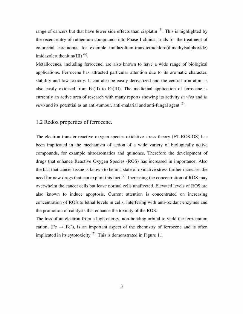
responsible for a 100% cure rate using an optimal dose of 220-300 mg/kg. The colon R85
carcinoma displayed a 60-80% inhibition of tumour growth in the presence of 1.
Fe
NO2O2N
NO2
O
1
Osella et al continued this initial work and prepared ferricenium salts, for example
[FcCOOH]+ [PF6]
-, for in vivo studies on Ehrlich ascites tumours. They observed that
ferrocenes with a Fe(II) centre were unable to inhibit cell growth but Fe(III) ferricenium
salts were cytotoxic. This cytotoxicity was independent of redox potential, at least in the
range of 175 to 330 mV. It was also observed that there was no intercalation between the
ferricenium salts and DNA. Interactions between the salts and the phosphate backbone of
DNA were proposed to be electrostatic following observations from 1H and
31P NMR
studies. Using electron spin resonance (ESR) experiments, it was proposed that the
ferricenium salts produced hydroxyl radicals under physiological conditions, which in
turn resulted in DNA damage (9)
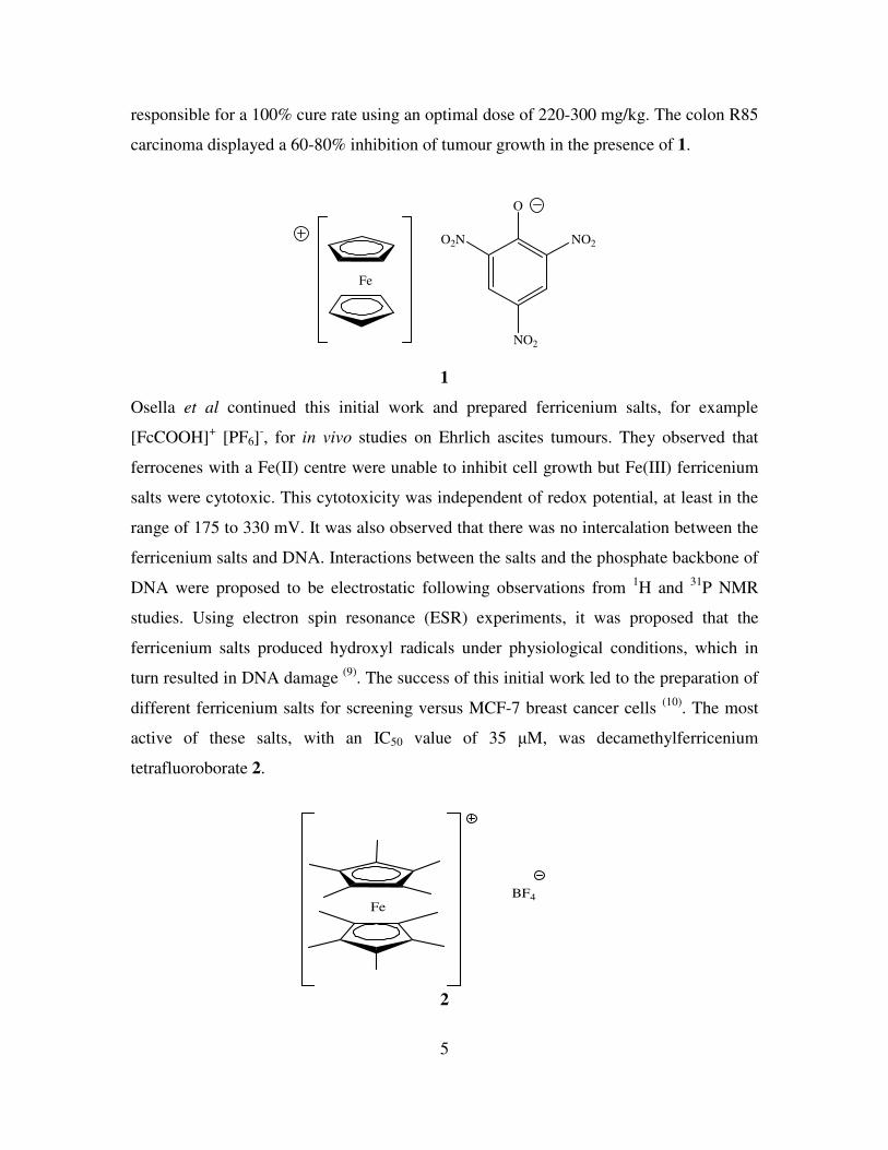
. The success of this initial work led to the preparation of
different ferricenium salts for screening versus MCF-7 breast cancer cells (10)
. The most
active of these salts, with an IC50 value of 35 µM, was decamethylferricenium
tetrafluoroborate 2.
FeBF4
2
6
ESR experiments confirmed that compound 2 is able to produce oxygen radical species
as a consequence of its degradation in aqueous media. From the ESR pattern it is
suggested that there is a Haber-Weiss like process followed by a Fenton reaction to yield
a hydroxyl radical, •OH. Compound 2 was also used in tandem with the clinically used
anti-tumour drug bleomycin. Bleomycin is known to be activated in the presence of iron.
A synergistic effect between compound 2 and bleomycin was observed. This corresponds
to the DNA damage inflicted by compound 2 and the accompanying increased level of
bleomycin activation by the Fe(II)/Fe(III) species.
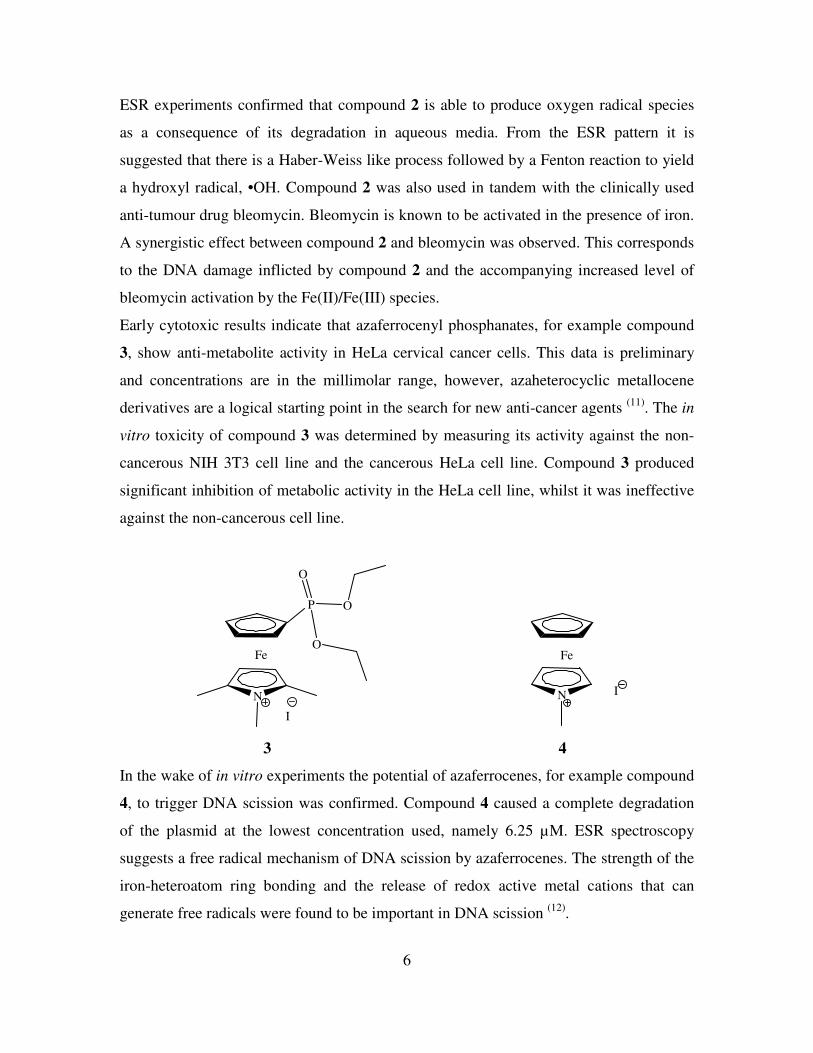
Early cytotoxic results indicate that azaferrocenyl phosphanates, for example compound
3, show anti-metabolite activity in HeLa cervical cancer cells. This data is preliminary
and concentrations are in the millimolar range, however, azaheterocyclic metallocene
derivatives are a logical starting point in the search for new anti-cancer agents (11)
. The in
vitro toxicity of compound 3 was determined by measuring its activity against the non-
cancerous NIH 3T3 cell line and the cancerous HeLa cell line. Compound 3 produced
significant inhibition of metabolic activity in the HeLa cell line, whilst it was ineffective
against the non-cancerous cell line.
I
N
Fe
P
O
O
O
N
Fe
I
3 4
In the wake of in vitro experiments the potential of azaferrocenes, for example compound
4, to trigger DNA scission was confirmed. Compound 4 caused a complete degradation
of the plasmid at the lowest concentration used, namely 6.25 µM. ESR spectroscopy
suggests a free radical mechanism of DNA scission by azaferrocenes. The strength of the
iron-heteroatom ring bonding and the release of redox active metal cations that can
generate free radicals were found to be important in DNA scission (12)
.
7
1.4 Metallocene based selective estrogen receptor modulators and anti-
androgens.
Tamoxifen is a widely prescribed selective estrogen receptor modulator (SERM). It is
used against hormone dependent breast cancer, where the estrogen receptor (ER), is
present. These are known as ER positive cells, ER(+) (13)
. SERMs are capable of
interacting with estrogen binding sites despite their non-steroidal structure.
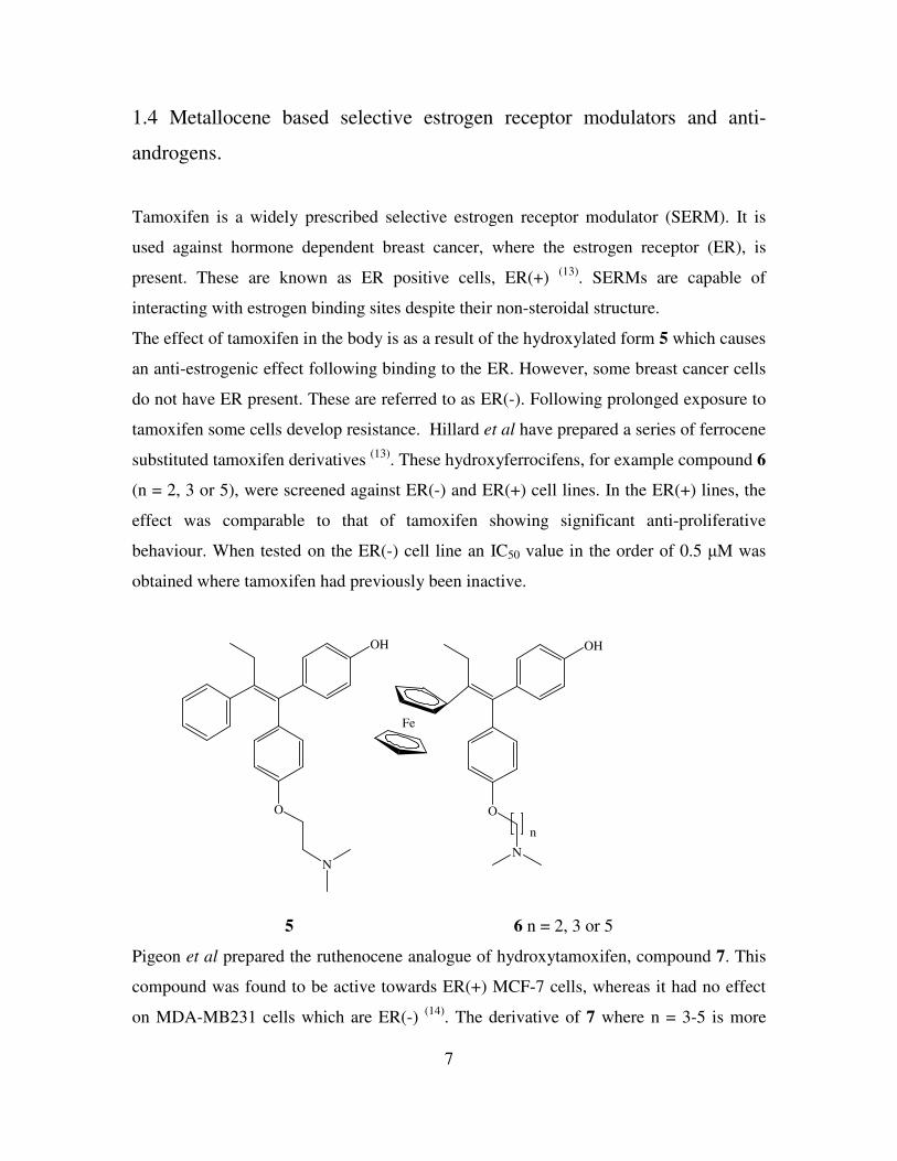
The effect of tamoxifen in the body is as a result of the hydroxylated form 5 which causes
an anti-estrogenic effect following binding to the ER. However, some breast cancer cells
do not have ER present. These are referred to as ER(-). Following prolonged exposure to
tamoxifen some cells develop resistance. Hillard et al have prepared a series of ferrocene
substituted tamoxifen derivatives (13)
. These hydroxyferrocifens, for example compound 6
(n = 2, 3 or 5), were screened against ER(-) and ER(+) cell lines. In the ER(+) lines, the
effect was comparable to that of tamoxifen showing significant anti-proliferative
behaviour. When tested on the ER(-) cell line an IC50 value in the order of 0.5 µM was
obtained where tamoxifen had previously been inactive.
O
N
OH
O
OH
Fe
N
n
5 6 n = 2, 3 or 5
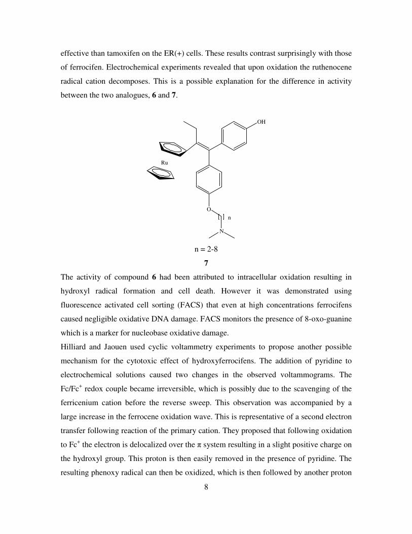
Pigeon et al prepared the ruthenocene analogue of hydroxytamoxifen, compound 7. This
compound was found to be active towards ER(+) MCF-7 cells, whereas it had no effect
on MDA-MB231 cells which are ER(-) (14)
. The derivative of 7 where n = 3-5 is more
8
effective than tamoxifen on the ER(+) cells. These results contrast surprisingly with those
of ferrocifen. Electrochemical experiments revealed that upon oxidation the ruthenocene
radical cation decomposes. This is a possible explanation for the difference in activity
between the two analogues, 6 and 7.
O
OH
Ru
N
n
n = 2-8
7
The activity of compound 6 had been attributed to intracellular oxidation resulting in
hydroxyl radical formation and cell death. However it was demonstrated using
fluorescence activated cell sorting (FACS) that even at high concentrations ferrocifens
caused negligible oxidative DNA damage. FACS monitors the presence of 8-oxo-guanine
which is a marker for nucleobase oxidative damage.
Hilliard and Jaouen used cyclic voltammetry experiments to propose another possible
mechanism for the cytotoxic effect of hydroxyferrocifens. The addition of pyridine to
electrochemical solutions caused two changes in the observed voltammograms. The
Fc/Fc+ redox couple became irreversible, which is possibly due to the scavenging of the
ferricenium cation before the reverse sweep. This observation was accompanied by a
large increase in the ferrocene oxidation wave. This is representative of a second electron
transfer following reaction of the primary cation. They proposed that following oxidation
to Fc+ the electron is delocalized over the π system resulting in a slight positive charge on
the hydroxyl group. This proton is then easily removed in the presence of pyridine. The
resulting phenoxy radical can then be oxidized, which is then followed by another proton

9
abstraction from the ethyl group furnishing a quinone methide. In the presence of a basic
species like DNA the quinone methide species 8 will be formed which in turn will lead to
cell death (13)
.
Fe
O
8
This mechanism of action is further validated electrochemically, as derivatives that were
inactive showed no electrochemical changes upon addition of pyridine to solution. The
oxidation of tamoxifen and other SERMs to quinoids is also a recognised pathway for
their cytotoxicity (15)
.
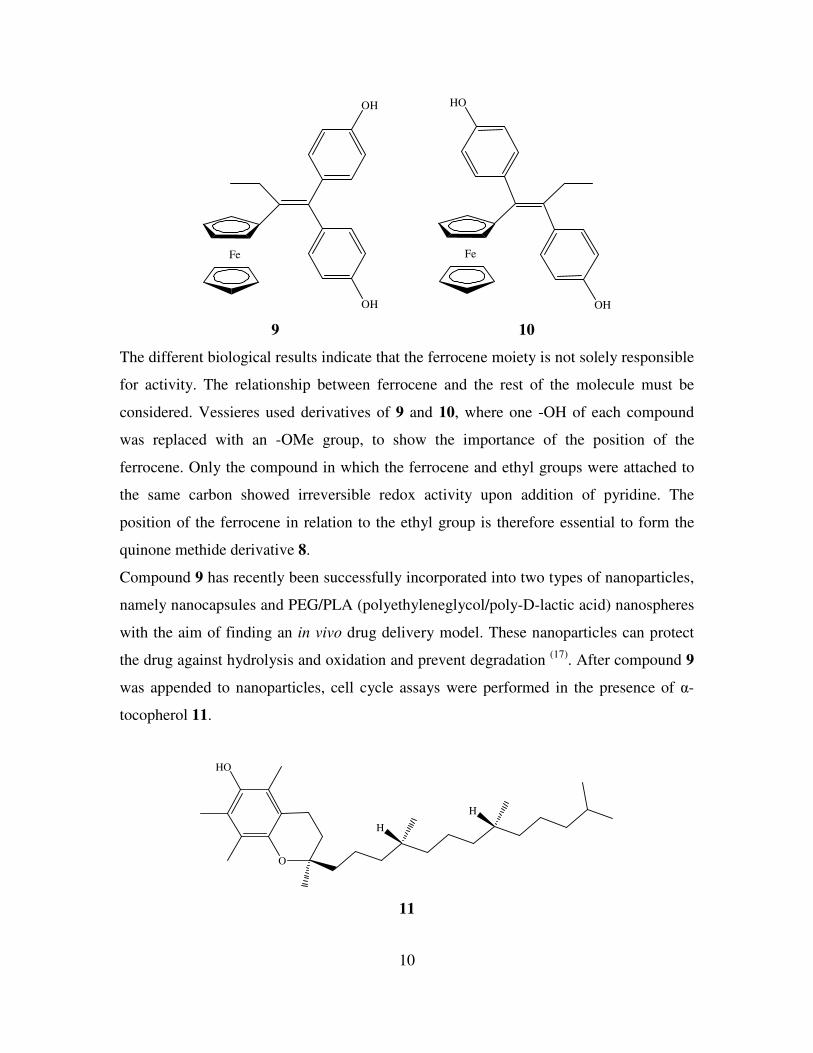
Vessieres et al have prepared a series of diphenolic compounds derivatized with
ferrocene and studied their anti-cancer activity against ER dependent and independent
breast cancer cell lines (16)
. Derivative 9 had strong anti-proliferative activity against
MCF-7 and MDA-MD231 breast cancer cells with IC50 values of 0.7 and 0.6 µM
respectively. Conversely compound 10, which is a regioisomer of compound 9, displays
only modest activity against both cell lines. There are two main differences between
compounds 9 and 10 in terms of structure. In compound 9 one of the two phenol groups
is always orientated trans to ferrocene, whereas in compound 10 there is a cis
relationship between ferrocene and the phenol group. Secondly, in compound 9 the two
phenol rings are bonded to the same carbon of the alkene, whereas in compound 10 each
carbon of the alkene is attached to a phenol ring.
10
Fe
OH
OH
Fe
OH
HO
9 10
The different biological results indicate that the ferrocene moiety is not solely responsible
for activity. The relationship between ferrocene and the rest of the molecule must be
considered. Vessieres used derivatives of 9 and 10, where one -OH of each compound
was replaced with an -OMe group, to show the importance of the position of the
ferrocene. Only the compound in which the ferrocene and ethyl groups were attached to
the same carbon showed irreversible redox activity upon addition of pyridine. The
position of the ferrocene in relation to the ethyl group is therefore essential to form the
quinone methide derivative 8.
Compound 9 has recently been successfully incorporated into two types of nanoparticles,
namely nanocapsules and PEG/PLA (polyethyleneglycol/poly-D-lactic acid) nanospheres
with the aim of finding an in vivo drug delivery model. These nanoparticles can protect
the drug against hydrolysis and oxidation and prevent degradation (17)
. After compound 9
was appended to nanoparticles, cell cycle assays were performed in the presence of α-
tocopherol 11.
HO
O
H
H
11
11
Compound 11 is the vitamin E form that is preferentially absorbed by humans and is a
well known anti-oxidant. In the presence of compound 11 the anti-proliferative effect of
compound 9 was reversed as there was a drop in the population of cells in the sub G1
phase of the cell cycle, the stage where damaged cells would be found. The presence of
an anti-oxidant may prevent oxidation of ferrocene to ferricenium and therefore prevent
the formation of compound 8 leading to a loss in anti-proliferative effect.
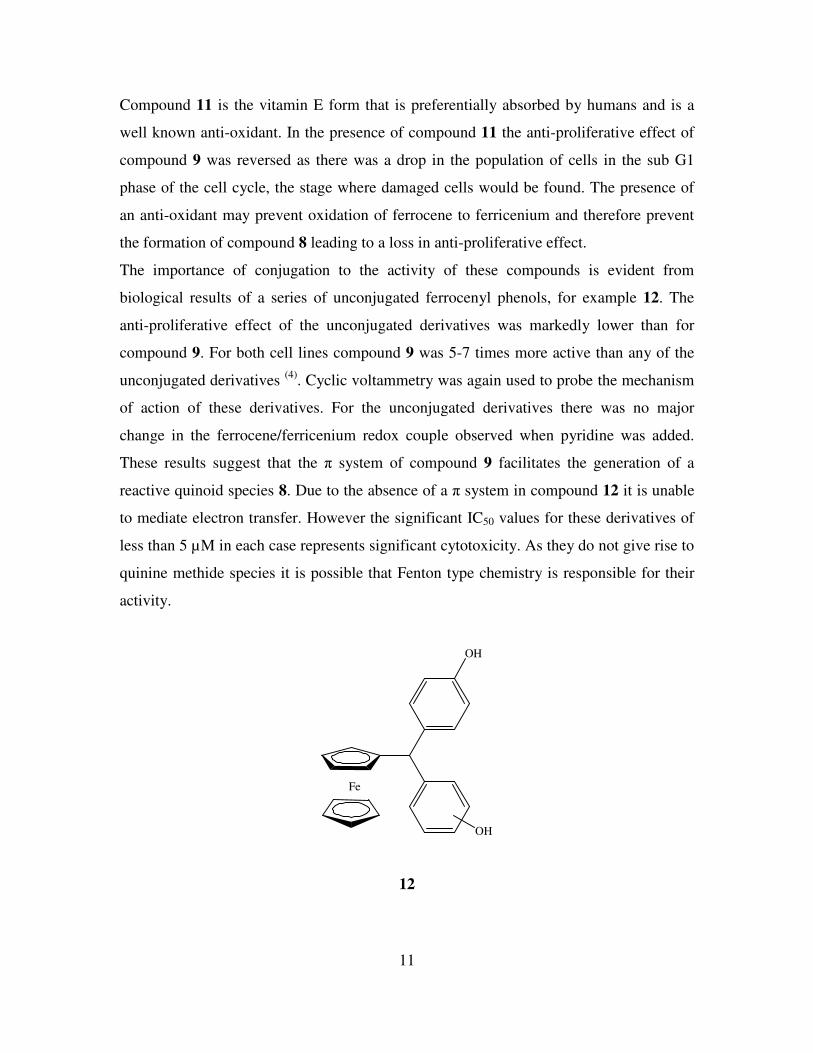
The importance of conjugation to the activity of these compounds is evident from
biological results of a series of unconjugated ferrocenyl phenols, for example 12. The
anti-proliferative effect of the unconjugated derivatives was markedly lower than for
compound 9. For both cell lines compound 9 was 5-7 times more active than any of the
unconjugated derivatives (4)
. Cyclic voltammetry was again used to probe the mechanism
of action of these derivatives. For the unconjugated derivatives there was no major
change in the ferrocene/ferricenium redox couple observed when pyridine was added.
These results suggest that the π system of compound 9 facilitates the generation of a
reactive quinoid species 8. Due to the absence of a π system in compound 12 it is unable
to mediate electron transfer. However the significant IC50 values for these derivatives of
less than 5 µM in each case represents significant cytotoxicity. As they do not give rise to
quinine methide species it is possible that Fenton type chemistry is responsible for their
activity.
Fe
OH
OH
12
12
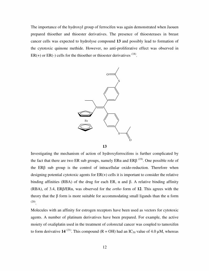
The importance of the hydroxyl group of ferrocifen was again demonstrated when Jaouen
prepared thioether and thioester derivatives. The presence of thioesterases in breast
cancer cells was expected to hydrolyse compound 13 and possibly lead to formation of
the cytotoxic quinone methide. However, no anti-proliferative effect was observed in
ER(+) or ER(-) cells for the thioether or thioester derivatives (18)
.
Fe
S
S
O
O
13
Investigating the mechanism of action of hydroxyferrocifens is further complicated by
the fact that there are two ER sub groups, namely ERα and ERβ (19)
. One possible role of
the ERβ sub group is the control of intracellular oxido-reduction. Therefore when
designing potential cytotoxic agents for ER(+) cells it is important to consider the relative
binding affinities (RBA) of the drug for each ER, α and β. A relative binding affinity
(RBA), of 3.4, ERβ/ERα, was observed for the ortho form of 12. This agrees with the
theory that the β form is more suitable for accommodating small ligands than the α form
(20).
Molecules with an affinity for estrogen receptors have been used as vectors for cytotoxic
agents. A number of platinum derivatives have been prepared. For example, the active
moiety of oxaliplatin used in the treatment of colorectal cancer was coupled to tamoxifen
to form derivative 14 (21)
. This compound (R = OH) had an IC50 value of 4.0 µM, whereas
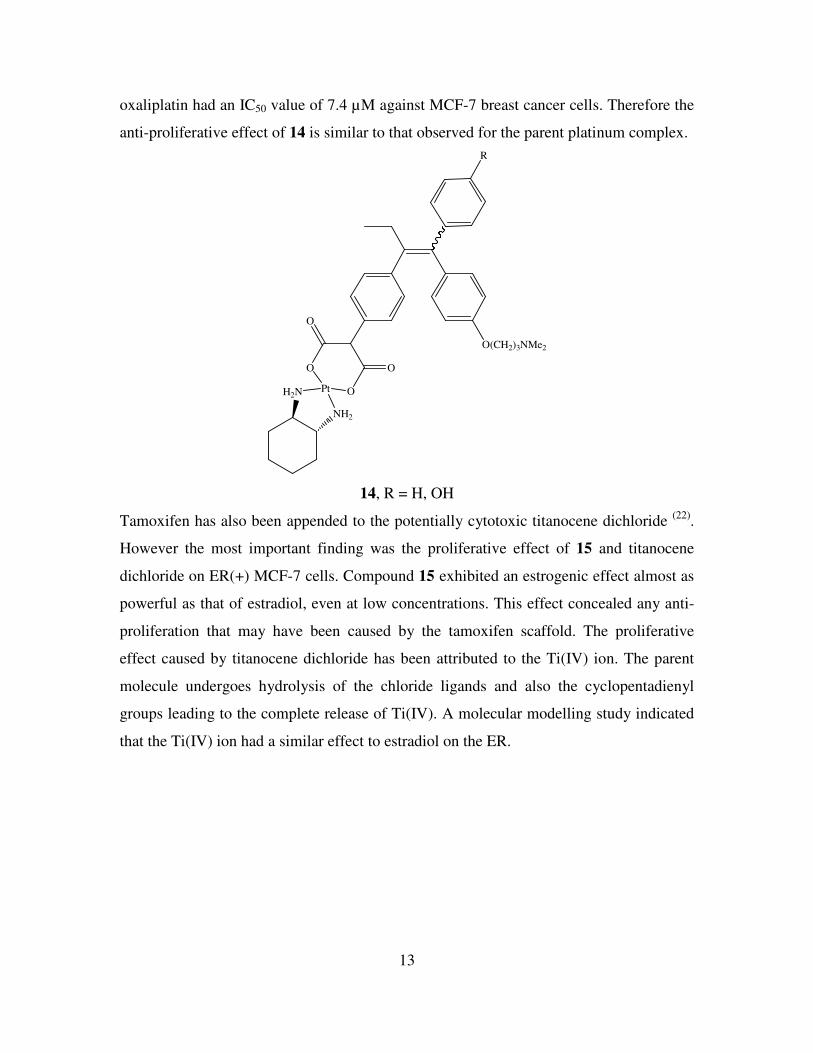
13
oxaliplatin had an IC50 value of 7.4 µM against MCF-7 breast cancer cells. Therefore the
anti-proliferative effect of 14 is similar to that observed for the parent platinum complex.
O(CH2)3NMe2
O
O
O
O
PtH2N
NH2
R
14, R = H, OH
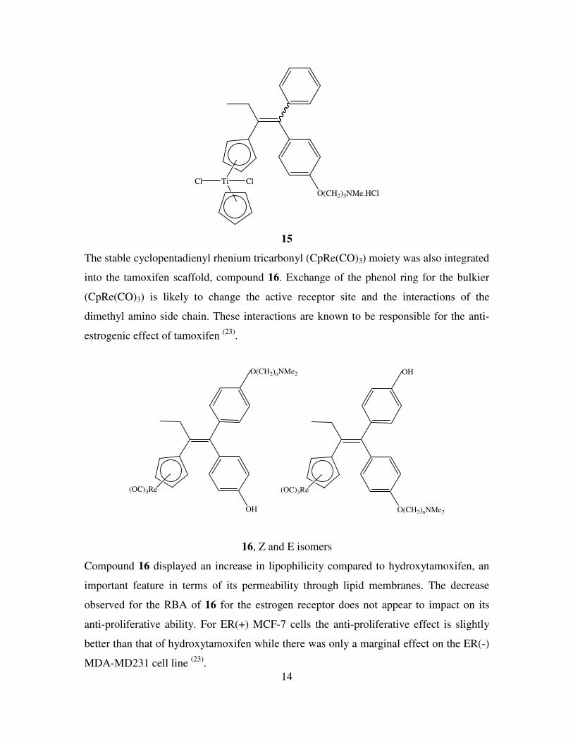
Tamoxifen has also been appended to the potentially cytotoxic titanocene dichloride (22)
.
However the most important finding was the proliferative effect of 15 and titanocene
dichloride on ER(+) MCF-7 cells. Compound 15 exhibited an estrogenic effect almost as
powerful as that of estradiol, even at low concentrations. This effect concealed any anti-
proliferation that may have been caused by the tamoxifen scaffold. The proliferative
effect caused by titanocene dichloride has been attributed to the Ti(IV) ion. The parent
molecule undergoes hydrolysis of the chloride ligands and also the cyclopentadienyl
groups leading to the complete release of Ti(IV). A molecular modelling study indicated
that the Ti(IV) ion had a similar effect to estradiol on the ER.
14
O(CH2)3NMe.HCl
TiCl Cl
15
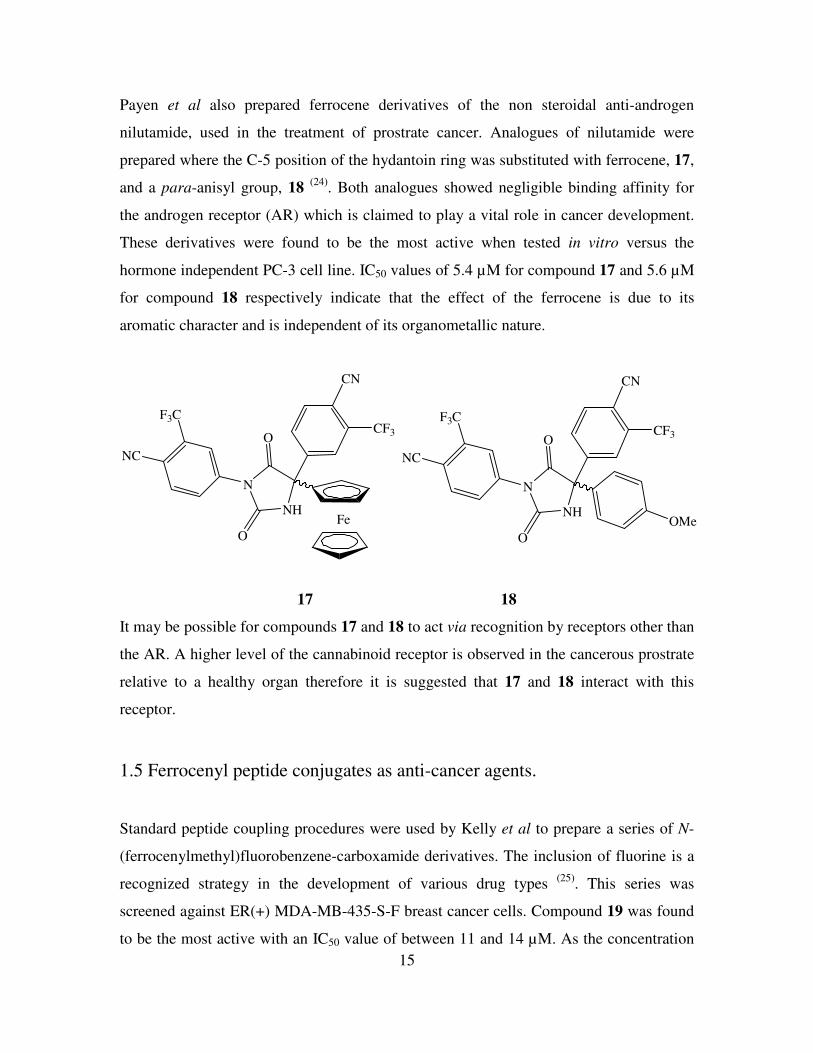
The stable cyclopentadienyl rhenium tricarbonyl (CpRe(CO)3) moiety was also integrated
into the tamoxifen scaffold, compound 16. Exchange of the phenol ring for the bulkier
(CpRe(CO)3) is likely to change the active receptor site and the interactions of the
dimethyl amino side chain. These interactions are known to be responsible for the anti-
estrogenic effect of tamoxifen (23)
.
(OC)3Re
OH
O(CH2)nNMe2
(OC)3Re
O(CH2)nNMe2
OH
16, Z and E isomers
Compound 16 displayed an increase in lipophilicity compared to hydroxytamoxifen, an
important feature in terms of its permeability through lipid membranes. The decrease
observed for the RBA of 16 for the estrogen receptor does not appear to impact on its
anti-proliferative ability. For ER(+) MCF-7 cells the anti-proliferative effect is slightly
better than that of hydroxytamoxifen while there was only a marginal effect on the ER(-)
MDA-MD231 cell line (23)
.
15
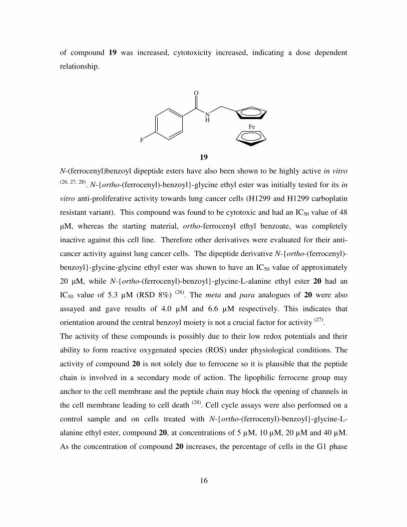
Payen et al also prepared ferrocene derivatives of the non steroidal anti-androgen
nilutamide, used in the treatment of prostrate cancer. Analogues of nilutamide were
prepared where the C-5 position of the hydantoin ring was substituted with ferrocene, 17,
and a para-anisyl group, 18 (24)
. Both analogues showed negligible binding affinity for
the androgen receptor (AR) which is claimed to play a vital role in cancer development.
These derivatives were found to be the most active when tested in vitro versus the
hormone independent PC-3 cell line. IC50 values of 5.4 µM for compound 17 and 5.6 µM
for compound 18 respectively indicate that the effect of the ferrocene is due to its
aromatic character and is independent of its organometallic nature.
FeNH
O
N
O
CN
CF3
NC
F3C
NH
O
N
O
CN
CF3
NC
F3C
OMe
17 18
It may be possible for compounds 17 and 18 to act via recognition by receptors other than
the AR. A higher level of the cannabinoid receptor is observed in the cancerous prostrate
relative to a healthy organ therefore it is suggested that 17 and 18 interact with this
receptor.
1.5 Ferrocenyl peptide conjugates as anti-cancer agents.
Standard peptide coupling procedures were used by Kelly et al to prepare a series of N-
(ferrocenylmethyl)fluorobenzene-carboxamide derivatives. The inclusion of fluorine is a
recognized strategy in the development of various drug types (25)
. This series was
screened against ER(+) MDA-MB-435-S-F breast cancer cells. Compound 19 was found
to be the most active with an IC50 value of between 11 and 14 µM. As the concentration
16
of compound 19 was increased, cytotoxicity increased, indicating a dose dependent
relationship.
Fe
NH
O
F
19
N-(ferrocenyl)benzoyl dipeptide esters have also been shown to be highly active in vitro
(26, 27, 28). N-ortho-(ferrocenyl)-benzoyl-glycine ethyl ester was initially tested for its in
vitro anti-proliferative activity towards lung cancer cells (H1299 and H1299 carboplatin
resistant variant). This compound was found to be cytotoxic and had an IC50 value of 48
µM, whereas the starting material, ortho-ferrocenyl ethyl benzoate, was completely
inactive against this cell line. Therefore other derivatives were evaluated for their anti-
cancer activity against lung cancer cells. The dipeptide derivative N-ortho-(ferrocenyl)-
benzoyl-glycine-glycine ethyl ester was shown to have an IC50 value of approximately
20 µM, while N-ortho-(ferrocenyl)-benzoyl-glycine-L-alanine ethyl ester 20 had an
IC50 value of 5.3 µM (RSD 8%) (26)
. The meta and para analogues of 20 were also
assayed and gave results of 4.0 µM and 6.6 µM respectively. This indicates that
orientation around the central benzoyl moiety is not a crucial factor for activity (27)
.
The activity of these compounds is possibly due to their low redox potentials and their
ability to form reactive oxygenated species (ROS) under physiological conditions. The
activity of compound 20 is not solely due to ferrocene so it is plausible that the peptide
chain is involved in a secondary mode of action. The lipophilic ferrocene group may
anchor to the cell membrane and the peptide chain may block the opening of channels in
the cell membrane leading to cell death (28)
. Cell cycle assays were also performed on a
control sample and on cells treated with N-ortho-(ferrocenyl)-benzoyl-glycine-L-
alanine ethyl ester, compound 20, at concentrations of 5 µM, 10 µM, 20 µM and 40 µM.
As the concentration of compound 20 increases, the percentage of cells in the G1 phase
17
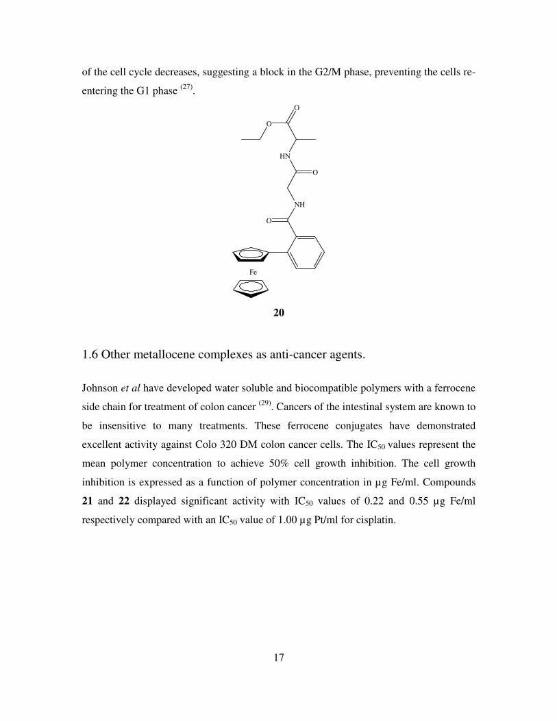
of the cell cycle decreases, suggesting a block in the G2/M phase, preventing the cells re-
entering the G1 phase (27)
.
Fe
NH
O
HN
O
O
O
20
1.6 Other metallocene complexes as anti-cancer agents.
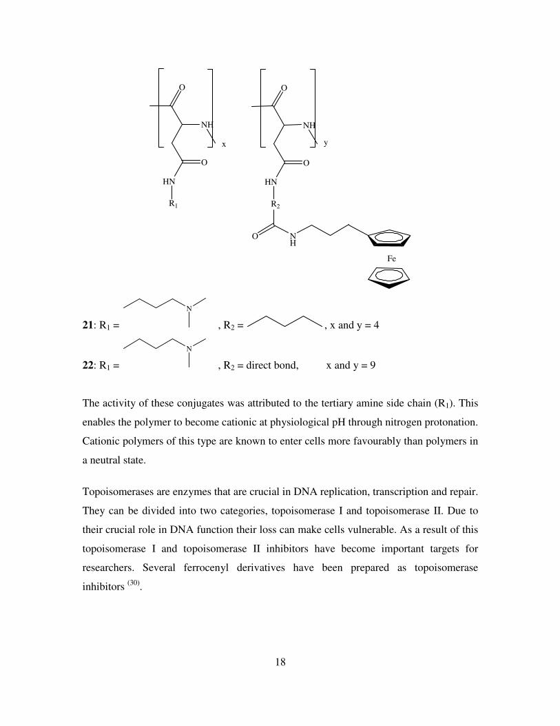
Johnson et al have developed water soluble and biocompatible polymers with a ferrocene
side chain for treatment of colon cancer (29)
. Cancers of the intestinal system are known to
be insensitive to many treatments. These ferrocene conjugates have demonstrated
excellent activity against Colo 320 DM colon cancer cells. The IC50 values represent the
mean polymer concentration to achieve 50% cell growth inhibition. The cell growth
inhibition is expressed as a function of polymer concentration in µg Fe/ml. Compounds
21 and 22 displayed significant activity with IC50 values of 0.22 and 0.55 µg Fe/ml
respectively compared with an IC50 value of 1.00 µg Pt/ml for cisplatin.
18
Fe
O
NH
HN
O
O
NH
HN
O
R1 R2
O NH
x y
21: R1 =
N
, R2 = , x and y = 4
22: R1 =
N
, R2 = direct bond, x and y = 9
The activity of these conjugates was attributed to the tertiary amine side chain (R1). This
enables the polymer to become cationic at physiological pH through nitrogen protonation.
Cationic polymers of this type are known to enter cells more favourably than polymers in
a neutral state.
Topoisomerases are enzymes that are crucial in DNA replication, transcription and repair.
They can be divided into two categories, topoisomerase I and topoisomerase II. Due to
their crucial role in DNA function their loss can make cells vulnerable. As a result of this
topoisomerase I and topoisomerase II inhibitors have become important targets for
researchers. Several ferrocenyl derivatives have been prepared as topoisomerase
inhibitors (30)
.
19
FeNHO Fe
NHON
O
O
Fe S
N
O
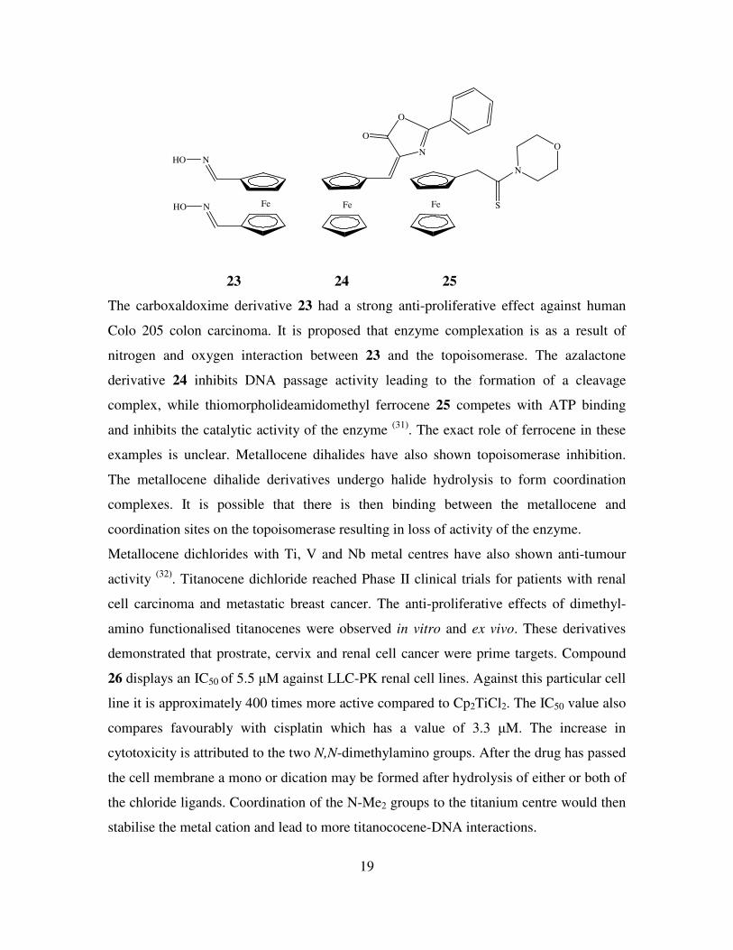
23 24 25
The carboxaldoxime derivative 23 had a strong anti-proliferative effect against human
Colo 205 colon carcinoma. It is proposed that enzyme complexation is as a result of
nitrogen and oxygen interaction between 23 and the topoisomerase. The azalactone
derivative 24 inhibits DNA passage activity leading to the formation of a cleavage
complex, while thiomorpholideamidomethyl ferrocene 25 competes with ATP binding
and inhibits the catalytic activity of the enzyme (31)
. The exact role of ferrocene in these
examples is unclear. Metallocene dihalides have also shown topoisomerase inhibition.
The metallocene dihalide derivatives undergo halide hydrolysis to form coordination
complexes. It is possible that there is then binding between the metallocene and
coordination sites on the topoisomerase resulting in loss of activity of the enzyme.
Metallocene dichlorides with Ti, V and Nb metal centres have also shown anti-tumour
activity (32)
. Titanocene dichloride reached Phase II clinical trials for patients with renal
cell carcinoma and metastatic breast cancer. The anti-proliferative effects of dimethyl-
amino functionalised titanocenes were observed in vitro and ex vivo. These derivatives
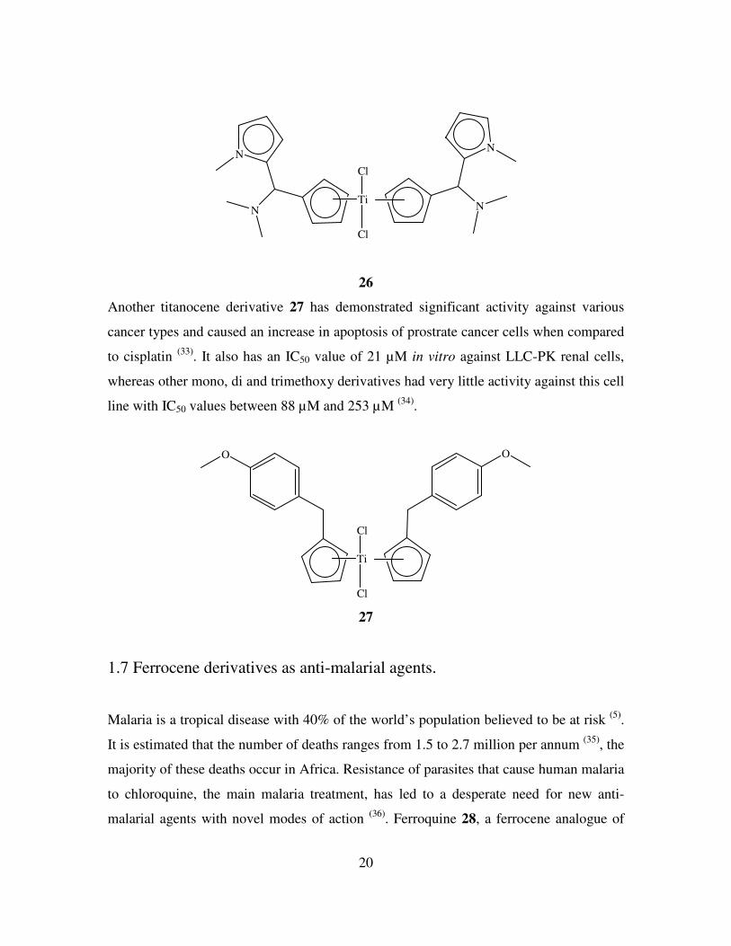
demonstrated that prostrate, cervix and renal cell cancer were prime targets. Compound
26 displays an IC50 of 5.5 µM against LLC-PK renal cell lines. Against this particular cell
line it is approximately 400 times more active compared to Cp2TiCl2. The IC50 value also
compares favourably with cisplatin which has a value of 3.3 µM. The increase in
cytotoxicity is attributed to the two N,N-dimethylamino groups. After the drug has passed
the cell membrane a mono or dication may be formed after hydrolysis of either or both of
the chloride ligands. Coordination of the N-Me2 groups to the titanium centre would then
stabilise the metal cation and lead to more titanococene-DNA interactions.
20
TiN
N
N
N
Cl
Cl
26
Another titanocene derivative 27 has demonstrated significant activity against various
cancer types and caused an increase in apoptosis of prostrate cancer cells when compared
to cisplatin (33)
. It also has an IC50 value of 21 µM in vitro against LLC-PK renal cells,
whereas other mono, di and trimethoxy derivatives had very little activity against this cell
line with IC50 values between 88 µM and 253 µM (34)
.
Ti
Cl
Cl
OO
27
1.7 Ferrocene derivatives as anti-malarial agents.
Malaria is a tropical disease with 40% of the world’s population believed to be at risk (5)
.
It is estimated that the number of deaths ranges from 1.5 to 2.7 million per annum (35)
, the
majority of these deaths occur in Africa. Resistance of parasites that cause human malaria
to chloroquine, the main malaria treatment, has led to a desperate need for new anti-
malarial agents with novel modes of action (36)
. Ferroquine 28, a ferrocene analogue of
21
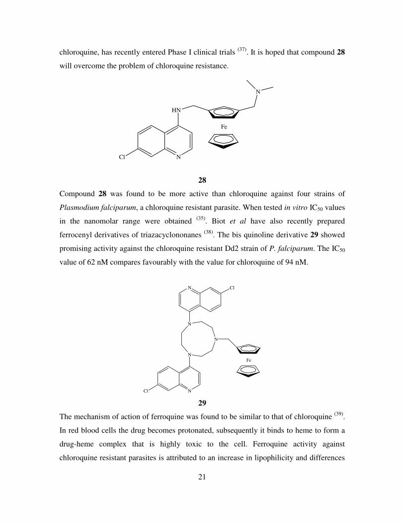
chloroquine, has recently entered Phase I clinical trials (37)
. It is hoped that compound 28
will overcome the problem of chloroquine resistance.
Fe
N
HN
NCl
28
Compound 28 was found to be more active than chloroquine against four strains of
Plasmodium falciparum, a chloroquine resistant parasite. When tested in vitro IC50 values
in the nanomolar range were obtained (35)
. Biot et al have also recently prepared
ferrocenyl derivatives of triazacyclononanes (38)
. The bis quinoline derivative 29 showed
promising activity against the chloroquine resistant Dd2 strain of P. falciparum. The IC50
value of 62 nM compares favourably with the value for chloroquine of 94 nM.
Fe
N
N
N
N
N Cl
Cl
29
The mechanism of action of ferroquine was found to be similar to that of chloroquine (39)
.
In red blood cells the drug becomes protonated, subsequently it binds to heme to form a
drug-heme complex that is highly toxic to the cell. Ferroquine activity against
chloroquine resistant parasites is attributed to an increase in lipophilicity and differences
22
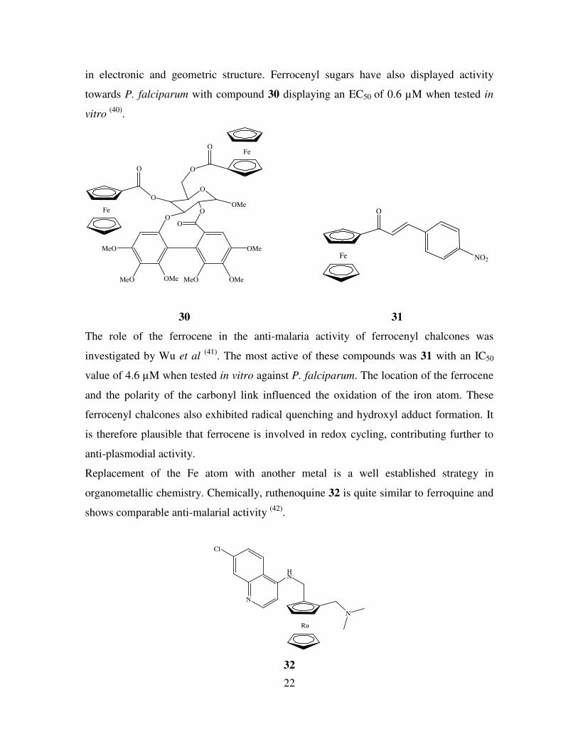
in electronic and geometric structure. Ferrocenyl sugars have also displayed activity
towards P. falciparum with compound 30 displaying an EC50 of 0.6 µM when tested in
vitro (40)
.
Fe
O
O
O
O
OMeMeO
MeO
OMe
MeO OMe
OMe
O
O
O
OFe
Fe
O
NO2
30 31
The role of the ferrocene in the anti-malaria activity of ferrocenyl chalcones was
investigated by Wu et al (41)
. The most active of these compounds was 31 with an IC50
value of 4.6 µM when tested in vitro against P. falciparum. The location of the ferrocene
and the polarity of the carbonyl link influenced the oxidation of the iron atom. These
ferrocenyl chalcones also exhibited radical quenching and hydroxyl adduct formation. It
is therefore plausible that ferrocene is involved in redox cycling, contributing further to
anti-plasmodial activity.
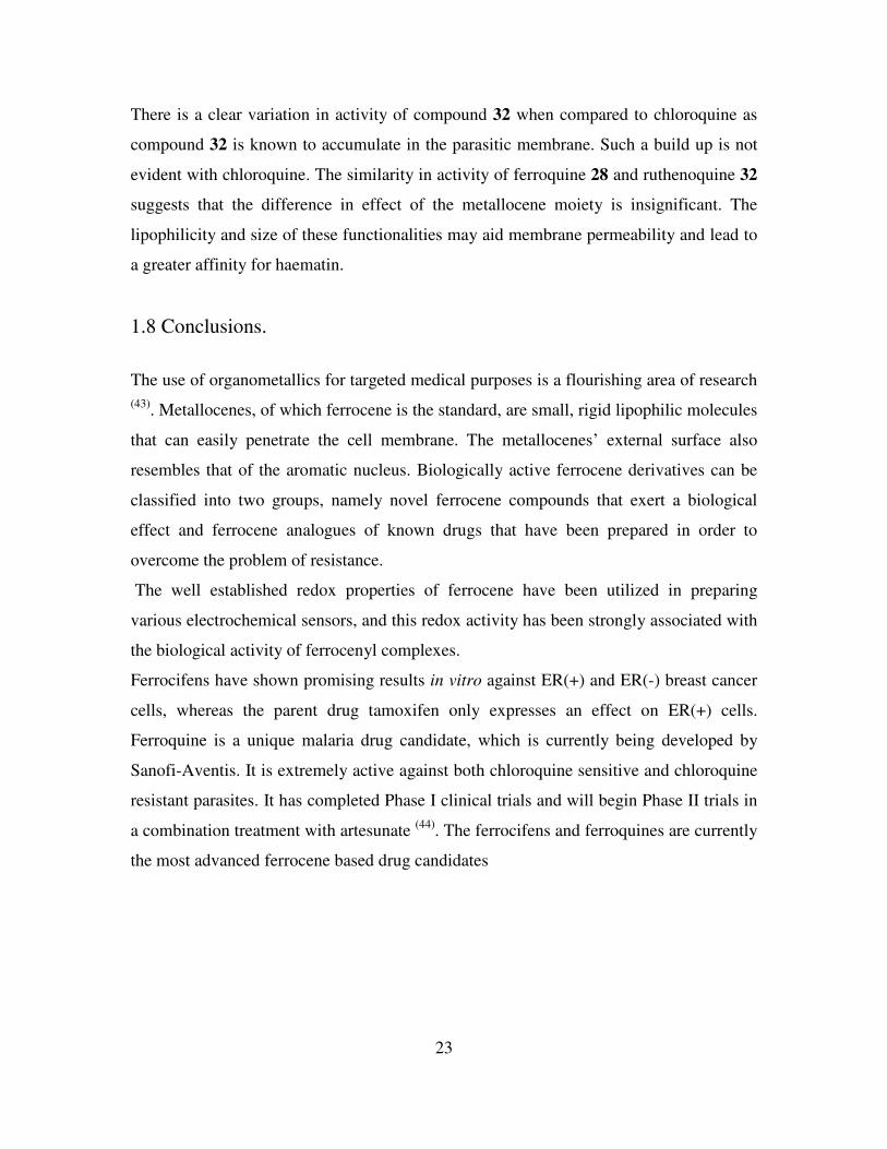
Replacement of the Fe atom with another metal is a well established strategy in
organometallic chemistry. Chemically, ruthenoquine 32 is quite similar to ferroquine and
shows comparable anti-malarial activity (42)
.
Ru
N
HN
N
Cl
32
23
There is a clear variation in activity of compound 32 when compared to chloroquine as
compound 32 is known to accumulate in the parasitic membrane. Such a build up is not
evident with chloroquine. The similarity in activity of ferroquine 28 and ruthenoquine 32
suggests that the difference in effect of the metallocene moiety is insignificant. The
lipophilicity and size of these functionalities may aid membrane permeability and lead to
a greater affinity for haematin.
1.8 Conclusions.
The use of organometallics for targeted medical purposes is a flourishing area of research
(43). Metallocenes, of which ferrocene is the standard, are small, rigid lipophilic molecules
that can easily penetrate the cell membrane. The metallocenes’ external surface also
resembles that of the aromatic nucleus. Biologically active ferrocene derivatives can be
classified into two groups, namely novel ferrocene compounds that exert a biological
effect and ferrocene analogues of known drugs that have been prepared in order to
overcome the problem of resistance.
The well established redox properties of ferrocene have been utilized in preparing
various electrochemical sensors, and this redox activity has been strongly associated with
the biological activity of ferrocenyl complexes.
Ferrocifens have shown promising results in vitro against ER(+) and ER(-) breast cancer
cells, whereas the parent drug tamoxifen only expresses an effect on ER(+) cells.
Ferroquine is a unique malaria drug candidate, which is currently being developed by
Sanofi-Aventis. It is extremely active against both chloroquine sensitive and chloroquine
resistant parasites. It has completed Phase I clinical trials and will begin Phase II trials in
a combination treatment with artesunate (44)
. The ferrocifens and ferroquines are currently
the most advanced ferrocene based drug candidates
24
References.
1. Yang, P., Guo, M., Coor. Chem. Rev., 1999, 185, 189.
2. Neuse, E. W., J. Inorg. Organomet. Polym. Mat., 2005, 15, 3.
3. Alderden, R. A., Hall, M. D., Hambley, T. W., J. Chem. Ed., 2006, 83, 728.
4. Hillard, E., Vessieres, A., Le Bideau, F., Plazuk, D., Spera, D., Huche, M.,
Jaouen, G., Chem. Med. Chem., 2006, 1, 551.
5. Fouda, M. F. R., Abd-Elzaher, M. M., Abdelsamaia, R. A., Labib, A. A., Appl.
Organomet. Chem., 2007, 21, 613.
6. Kaptiza, S., Pongratz, M., Jakupec, M. A., Heffeter, P., Berger, W., Lackinger, L.,
Keppler, B. K., Marian, B., J. Cancer Res. Clin. Oncol., 2005, 131, 101.
7. Hillard, E. A., de Abreu, F. C., Ferreira, D. C. M., Jaouen, G., Goulart, M. O. F.,
Amatore, C., Chem. Comm., 2008, 2612.
8. Kopf-Maier, P., Kopf, H., Neuse, E. W., Angew. Chem. Int. Ed. Eng., 1984, 23,
456.
9. Osella, D., Ferrali, M., Zanello, P., Laschi, F., Fontani, M., Nervi, C., Cavigiolio,
G., Inorg. Chim. Acta., 2000, 306, 42.
10. Tabbi, G., Cassino, C., Lavigiolio, G., Colangelo, D., Ghiglia, A., Viano, I.,
Osella, D., J. Med. Chem., 2002, 45, 5786.
11. Kowalski, K., Zakrzewski, J., Long, N. J., Suwaki, N., Mann, D. J., White, A. J.
P., Dalton Trans., 2006, 571.
12. Kowalski, K., Suwaki, N., Zakrzewski, J., White, A. J. P., Long, N. J., Mann, D.
J., Dalton Trans., 2007, 743.
13. Hillard, E., Vessieres, A., Thouin, L., Jaouen, G., Amatore, C., Angew. Chem. Int.
Ed., 2006, 45, 285.
14. Pigeon, P., Top, S., Vessieres, A., Huche, M., Hillard, E. A., Salomon, E., Jaouen,
G., J. Med. Chem., 2005, 48, 2814.
15. Fan, P. W., Zhang, F., Bolton, J. L., Chem. Res. Toxicol., 2000, 13, 45.
16. Vessieres, A., Top, S., Pigeon, P., Hillard, E., Boubeker, L., Spera, D., Jaouen, G.,
J. Med. Chem., 2005, 48, 3937.
25
17. Nguyen, A., Marsaud, V., Bouclier, C., Top, S., Vessieres, A., Pigeon, P., Gref,
R., Legrand, P., Jaouen, G., Renoir, J-M., Inter. J. of Pharmaceutics, 2008, 347,
128.
18. Heilmann, J. B., Hillard, E. A., Plamont, M-A., Pigeon, P., Bolte, M., Jaouen, G.
Vessieres, A., J. Organomet. Chem., 2008, 693, 1716.
19. Osella, D., Mahboobi, H., Colangelo, D., Cavigiolio, G., Vessieres, A., Jaouen,
G., Inorg. Chim. Acta., 2005, 358, 1993.
20. Plazuk, D., Vessieres, A., Le Bideau, F., Jaouen, G., Zakrzewski, J., Tett. Lett.,
2004, 45, 5425.
21. Vessieres, A., Top, S., Beck, W., Hillard, E., Jaouen, G., Dalton Trans., 2006,
529.
22. Top, S., Kaloun, E. B., Vessieres, A., Laios, I., Leclerq, G., Jaouen, G., J.
Organomet. Chem. 2002, 643-644, 350.
23. Top, S., Vessieres, A., Pigeon, P., Rager, M-N., Huche, M., Salomon, E.,
Cabestaing, C., Vaissermann, J., Jaouen, G., Chem. Bio. Chem., 2004, 5, 1104.
24. Payen, O., Top, S., Vessieres, A., Brule, E., Plamont, M-A., McGlinchey, M. J.,
Muller-Bunz, H., Jaouen, G., J. Med. Chem., 2008, 51, 1791
25. Kelly, P. N., Pretre, A., Devoy, S., O’Reilly, J., Devery, R., Goel, A., Gallagher,
J. F., Lough, A. J., Kenny, P. T. M., J. Organomet. Chem., 2007, 692, 1327.
26. Corry, A. J., Goel, A., Alley, S. R., Kelly, P. N., O’Sullivan, D., Savage, D.,
Kenny, P. T. M., J. Organomet. Chem., 2007, 692, 1405.
27. Corry, A. J., O’Donovan, N., Mooney, A., O’Sullivan, D., Rai, D. K., Kenny, P.
T. M., J. Organomet. Chem., 2008, doi: 10.1016/j.jorganchem.2008.09.072.
28. Goel, A., Savage, D., Alley, S. R., Kelly, P. N., O’Sullivan, D., Mueller-Bunz, H.,
Kenny, P. T. M., J. Organomet. Chem., 2007, 692, 1292.
29. Johnson, M. T., Kreft, E., N’Da, D. D., Neuse, E. W., van Resberg, C. E. J., J.
Inorg. Organomet. Polym., 2003, 13, 255.
30. Basnet, A., Thapa, P., Karki, R., Na, Y., Jahng, Y., Jeong, B. S., Jeong, T. C.,
Lee, C. S., Lee, E. S., Biorg. Med. Chem., 2007, 15, 4351.
31. Sai Krishna, A. D., Panda, G., Kondapi, A. K., Arch. Biochem. Biophys., 2005,
438, 206.

26
32. Pampillon, C., Sweeney, N. J., Strohfeldt, K., Tacke, M., J. Organomet. Chem.,
2007, 692, 2153.
33. O’Connor, K., Gill, C., Tacke, M., Rehmann, F. J. K., Strohfeldt, K., Sweeney,
N., Fitzpatrick, J. M., Watson, R. W. G., Apoptosis, 2006, 11, 1205.
34. Claffey, J., Hogan, M., Muller-Bunz, H., Pampillon, C., Tacke, M., J. Organomet.
Chem., 2008, 693, 526.
35. Biot, C., Delhaes, L., Abessolo, H., Domarle, O., Maciejewski, L.A., Mortuaire,
M., Delcourt, P., Deloron, P., Camus, D., Dive, D., Brocard, J. S., J. Organomet.
Chem., 1999, 589, 59.
36. Ciu, X., Vlahakis, J. Z., Crandall, I. E., Szarek, W. A., Bioorg. Med. Chem., 2008,
16, 1927.
37. Daher, W., Biot, C., Fandeur, T., Jouin, H., Pelinski, L., Viscogliosi, E., Fraisse,
L., Pradines, B., Brocard, J., Khalife, J., Dive, D., Malaria J., 2006, 5, 11.
38. Biot, C., Dessolin, J., Ricard, I., Dive, D., J. Organomet. Chem., 2004, 689, 4678.
39. Biot, C., Taramelli, D., Forfar-Bares, I., Maciejewski, L. A., Boyce, M.,
Nowogrocki, G., Brocard, J. S., Basilico, N., Olliaro, P., Egan, T. J., Mol.
Pharaceut., 2005, 2, 185.
40. Itoh, T., Shirakami, S., Ishida, N., Yamashita, Y., Yoshida, T., Kim, H-S.,
Wataga, Y., Bioorg. Med. Chem. Lett., 2000, 10, 1657.
41. Wu, X., Wilairat, P., Go, M-L., Bioorg. Med. Chem. Lett., 2002, 12, 2299.
42. Blackie, M. A. L., Beagley, P., Croft, S. L., Kendrick, H., Moss, J. R., Chibale,
K., Bioorg. Med. Chem., 2007, 15, 6510.
43. Jaouen, G., ‘Bioorganometallics’, Wiley-VCH 2006.
44. Dive, D., Biot, C., Chem. Med. Chem., 2008, 3, 383.
27
Chapter 2
Results and Discussion
2.1 Introduction.
Organometallic compounds have been successfully incorporated in a wide variety of
materials with diverse applications. Ferrocene is one such compound that is recognised as
a promising candidate for use in novel materials due to its ease of use and
electrochemical and spectroscopic properties (1)
. As a result of this, ferrocene research
has received an increased level of attention over the past decade. The ultimate goal of this
research is the development of novel sensor compounds, peptide mimetic models and
unnatural drugs (2)
. N-(ferrocenyl)benzoyl amino acid and dipeptide esters were originally
prepared as potential anion sensing agents (3)
however they demonstrated cytotoxicity
following in vitro screening. The compounds are composed of three key moieties,
namely, (i) an electroactive core, (ii) a conjugated aromatic linker and (iii) an amino acid
or peptide derivative that can interact with other molecules via hydrogen bonding. N-
ortho-(ferrocenyl)-benzoyl-glycine ethyl ester was initially tested for its in vitro anti-
proliferative activity towards lung cancer cells (H1299 and H1299 carboplatin and
cisplatin resistant variants). This compound was found to be cytotoxic and had an IC50
value of 48 µM, whereas the starting material, ortho-ferrocenyl ethyl benzoate, was
completely inactive against this cell line. Therefore other derivatives were evaluated for
their anti-cancer activity against lung cancer cell lines. Initial results showed that the
cytotoxicity of the meta dipeptide, N-meta-(ferrocenyl)-benzoyl-L-alanine-glycine
ethyl ester is ca. 2 times higher than the ortho-glycine derivative, the IC50 value being 26
µM (RSD 20%) whilst the corresponding ortho analogue, N-ortho-(ferrocenyl)-
benzoyl-L-alanine-glycine ethyl ester has an IC50 value of 21 µM (RSD 20%). The
dipeptide derivative N-ortho-(ferrocenyl)-benzoyl-glycine-glycine ethyl ester was
shown to have an IC50 value of approximately 20 µM, also N-ortho-(ferrocenyl)-
benzoyl-glycine-L-alanine ethyl ester is more active than N-ortho-(ferrocenyl)-
benzoyl-L-alanine-glycine ethyl ester with an IC50 value of 5.3 µM (RSD 8%). From
28
this it may be assumed that the glycine residue of the dipeptide that is attached to the
benzoyl group is important for activity. The larger amino acid alanine as the second
residue also increased activity. To assess the effects of lipophilicity, the alanine group
can be replaced with residues that differ by a methylene (CH2) unit. By incorporating the
amino acid 2-aminobutyric acid (Abu) the methyl group of alanine (CH3) is transformed
to an ethyl group (C2H5). This process can be extended by using norvaline (Nva) and
norleucine (Nle) in the synthesis to introduce propyl (C3H7) and butyl (C4H9) groups
respectively. The number of methylene groups in the first amino acid of the dipeptide
chain was also extended using β-alanine and γ-aminobutyric acid. As the glycine
dipeptide derivative was more active than the glycine amino acid derivative the study was
therefore extended to longer peptide chains with additional glycine residues. 1,1’-N, N’-
ortho-(ferrocenyl)-bisbenzoyl amino acid and dipeptide esters were also prepared in
order to assess the effect of a disubstituted ferrocene molecule on activity.
Coupling reactions were used in the preparation of the dipeptide esters and also to
facilitate the introduction of the ferrocenyl benzoyl group onto the peptide esters.
Ferrocenyl benzoic acids, ortho, meta and para, were prepared and were treated with 1-
hydroxybenzotriazole (HOBt), N-(3-dimethylaminopropyl)-N’-ethylcarbodiimide
hydrochloride (EDC), and triethylamine (TEA) in dichloromethane at 0 oC in the
presence of the peptide esters.
The primary objective was to prepare analogues of the lead compound N-ortho-
(ferrocenyl)-benzoyl-glycine-L-alanine ethyl ester 20 and compare their in vitro
biological activity. This was achieved by varying the sequence and size of the peptide
chain and by altering their orientation around the central benzoyl moiety.
2.2 The synthesis of dipeptide ethyl esters.
The dipeptides required in this study were not available commercially and therefore had
to be prepared. Conventional peptide chemistry was employed where BOC protected
glycine, β-alanine and γ-aminobutyric acid were reacted with the ethyl esters of L-
alanine, L-2-aminobutyric acid, L-norvaline and L-norleucine via the EDC/HOBt
coupling protocol. Subsequent deprotection of the BOC group using trifluoroacetic acid
29
allows for reaction at the N-terminus between the free amino group and ferrocenyl
benzoic acids.
In the formation of the peptide bond between two amino acids, protection of the N-
terminus of one amino acid and the C-terminus of the other amino acid is required to
ensure regiospecific coupling. All compounds were subsequently characterized using a
variety of NMR and spectroscopic techniques. As they were intermediates in the
synthesis of N-(ferrocenyl)benzoyl dipeptide esters they will not be included in the
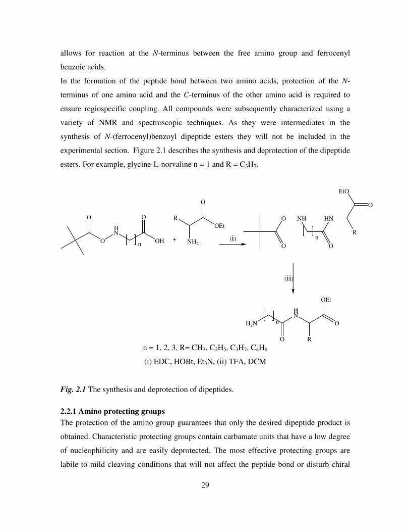
experimental section. Figure 2.1 describes the synthesis and deprotection of the dipeptide
esters. For example, glycine-L-norvaline n = 1 and R = C3H7.
O
HN
O O
OHn
+ NH2
R
OEt
O
NH
O
HN
R
O
EtO
n
O
O
H2N
O
HN
R
O
OEt
n
(i)
(ii)
n = 1, 2, 3, R= CH3, C2H5, C3H7, C4H9
(i) EDC, HOBt, Et3N, (ii) TFA, DCM
Fig. 2.1 The synthesis and deprotection of dipeptides.
2.2.1 Amino protecting groups
The protection of the amino group guarantees that only the desired dipeptide product is
obtained. Characteristic protecting groups contain carbamate units that have a low degree
of nucleophilicity and are easily deprotected. The most effective protecting groups are
labile to mild cleaving conditions that will not affect the peptide bond or disturb chiral
30
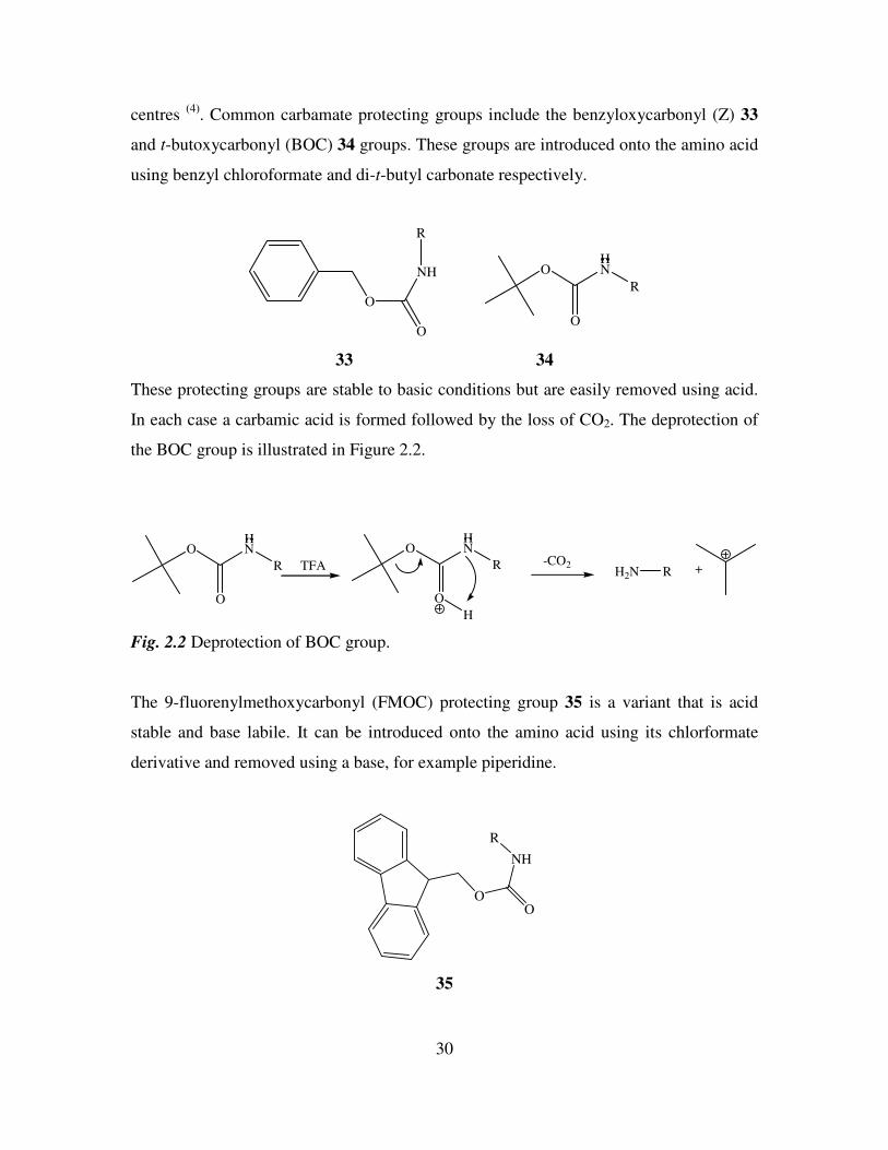
centres (4)
. Common carbamate protecting groups include the benzyloxycarbonyl (Z) 33
and t-butoxycarbonyl (BOC) 34 groups. These groups are introduced onto the amino acid
using benzyl chloroformate and di-t-butyl carbonate respectively.
O
O
NH
R
O
O
HN
R
33 34
These protecting groups are stable to basic conditions but are easily removed using acid.
In each case a carbamic acid is formed followed by the loss of CO2. The deprotection of
the BOC group is illustrated in Figure 2.2.
O
O
HN
R
O
O
HN
R
H
-CO2H2N R +TFA
Fig. 2.2 Deprotection of BOC group.
The 9-fluorenylmethoxycarbonyl (FMOC) protecting group 35 is a variant that is acid
stable and base labile. It can be introduced onto the amino acid using its chlorformate
derivative and removed using a base, for example piperidine.
OO
NH
R
35
31
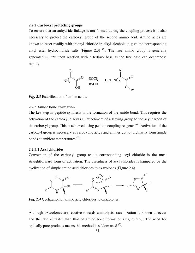
2.2.2 Carboxyl protecting groups
To ensure that an anhydride linkage is not formed during the coupling process it is also
necessary to protect the carboxyl group of the second amino acid. Amino acids are
known to react readily with thionyl chloride in alkyl alcohols to give the corresponding
alkyl ester hydrochloride salts (Figure 2.3) (5)
. The free amino group is generally
generated in situ upon reaction with a tertiary base as the free base can decompose
rapidly.
NH2
R
O
OH
SOCl2
R'-OHNH2
R
O
OR'
HCl.
Fig. 2.3 Esterification of amino acids.
2.2.3 Amide bond formation.
The key step in peptide synthesis is the formation of the amide bond. This requires the
activation of the carboxylic acid i.e., attachment of a leaving group to the acyl carbon of
the carboxyl group. This is achieved using peptide coupling reagents (6)
. Activation of the
carboxyl group is necessary as carboxylic acids and amines do not ordinarily form amide
bonds at ambient temperatures (7)
.
2.2.3.1 Acyl chlorides
Conversion of the carboxyl group to its corresponding acyl chloride is the most
straightforward form of activation. The usefulness of acyl chlorides is hampered by the
cyclization of simple amino acid chlorides to oxazolones (Figure 2.4).
NH
O
R
H
OCl
N
O
R
H
OCl
N
O
R
H
O
R
RR
Fig. 2.4 Cyclization of amino acid chlorides to oxazolones.
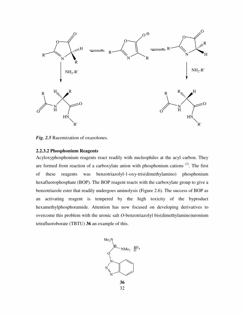
Although oxazolones are reactive towards aminolysis, racemization is known to occur
and the rate is faster than that of amide bond formation (Figure 2.5). The need for
optically pure products means this method is seldom used (7)
.
32
N
O
R
H
O
N
O
R
O
N
O
O
H
R
NH
H R
RR
R
O
HN
R'
NH
R H
O
HN
R'
NH2-R' NH2-R'
O
R
O
R
Fig. 2.5 Racemization of oxazolones.
2.2.3.2 Phosphonium Reagents
Acyloxyphosphonium reagents react readily with nucleophiles at the acyl carbon. They
are formed from reaction of a carboxylate anion with phosphonium cations (7)
. The first
of these reagents was benzotriazolyl-1-oxy-tris(dimethylamino) phosphonium
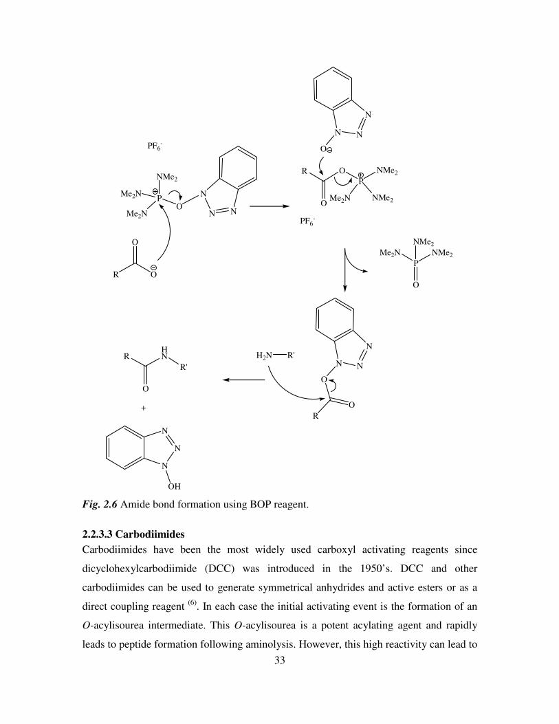
hexafluorophosphate (BOP). The BOP reagent reacts with the carboxylate group to give a
benzotriazole ester that readily undergoes aminolysis (Figure 2.6). The success of BOP as
an activating reagent is tempered by the high toxicity of the byproduct
hexamethylphosphoramide. Attention has now focused on developing derivatives to
overcome this problem with the uronic salt O-benzotriazolyl bis(dimethylamino)uronium
tetrafluoroborate (TBTU) 36 an example of this.
N
N
N
O
Me2N
NMe2BF4
36
33
N
N
N
P
Me2N
Me2N
NMe2
PF6-
R O
O
O
P
NMe2
Me2N NMe2
O
O
R
PF6-
N
N
N
O
N
N
N
O
O
R
P
O
Me2N
NMe2
NMe2
H2N R'HN
R'
O
R
+
N
N
N
OH
Fig. 2.6 Amide bond formation using BOP reagent.
2.2.3.3 Carbodiimides
Carbodiimides have been the most widely used carboxyl activating reagents since
dicyclohexylcarbodiimide (DCC) was introduced in the 1950’s. DCC and other
carbodiimides can be used to generate symmetrical anhydrides and active esters or as a
direct coupling reagent (6)
. In each case the initial activating event is the formation of an
O-acylisourea intermediate. This O-acylisourea is a potent acylating agent and rapidly
leads to peptide formation following aminolysis. However, this high reactivity can lead to
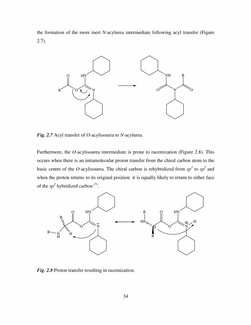
34
the formation of the more inert N-acylurea intermediate following acyl transfer (Figure
2.7).
N
HN
O
O
R N
NH
O O
R
Fig. 2.7 Acyl transfer of O-acylisourea to N-acylurea.
Furthermore, the O-acylisourea intermediate is prone to racemization (Figure 2.8). This
occurs when there is an intramolecular proton transfer from the chiral carbon atom to the
basic centre of the O-acylisourea. The chiral carbon is rehybridized from sp3 to sp
2 and
when the proton returns to its original position it is equally likely to return to either face
of the sp2 hybridized carbon
(7).
N
HN
O
O
NH
R
HR
N
HN
O
O
HN
R
R
H
Fig. 2.8 Proton transfer resulting in racemization.
35
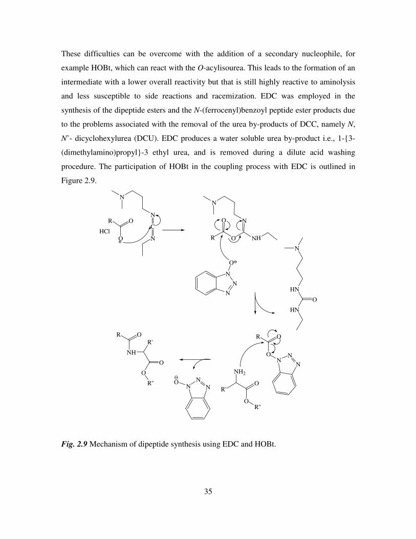
These difficulties can be overcome with the addition of a secondary nucleophile, for
example HOBt, which can react with the O-acylisourea. This leads to the formation of an
intermediate with a lower overall reactivity but that is still highly reactive to aminolysis
and less susceptible to side reactions and racemization. EDC was employed in the
synthesis of the dipeptide esters and the N-(ferrocenyl)benzoyl peptide ester products due
to the problems associated with the removal of the urea by-products of DCC, namely N,
N’- dicyclohexylurea (DCU). EDC produces a water soluble urea by-product i.e., 1-3-
(dimethylamino)propyl-3 ethyl urea, and is removed during a dilute acid washing
procedure. The participation of HOBt in the coupling process with EDC is outlined in
Figure 2.9.
R O
O N
C
N
R
O
O
N
HN
N
N
N
O
HN
HN
O
R O
ON
N
NNH2
R O
NH
R'
O
O
N
NN
R'O
O
R''
R''
NN
NO
HCl
Fig. 2.9 Mechanism of dipeptide synthesis using EDC and HOBt.
36
2.3 The synthesis of N-(ferrocenyl)benzoyl peptide esters.
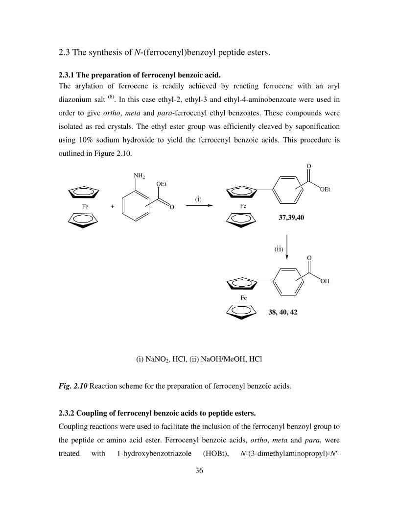
2.3.1 The preparation of ferrocenyl benzoic acid.
The arylation of ferrocene is readily achieved by reacting ferrocene with an aryl
diazonium salt (8)
. In this case ethyl-2, ethyl-3 and ethyl-4-aminobenzoate were used in
order to give ortho, meta and para-ferrocenyl ethyl benzoates. These compounds were
isolated as red crystals. The ethyl ester group was efficiently cleaved by saponification
using 10% sodium hydroxide to yield the ferrocenyl benzoic acids. This procedure is
outlined in Figure 2.10.
Fe +
NH2
O
OEt
Fe
O
OEt
Fe
O
OH
(i)
(ii)
37,39,40
38, 40, 42
(i) NaNO2, HCl, (ii) NaOH/MeOH, HCl
Fig. 2.10 Reaction scheme for the preparation of ferrocenyl benzoic acids.
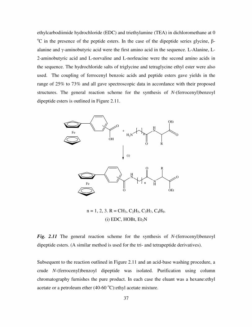
2.3.2 Coupling of ferrocenyl benzoic acids to peptide esters.
Coupling reactions were used to facilitate the inclusion of the ferrocenyl benzoyl group to
the peptide or amino acid ester. Ferrocenyl benzoic acids, ortho, meta and para, were
treated with 1-hydroxybenzotriazole (HOBt), N-(3-dimethylaminopropyl)-N′-
37
ethylcarbodiimide hydrochloride (EDC) and triethylamine (TEA) in dichloromethane at 0
oC in the presence of the peptide esters. In the case of the dipeptide series glycine, β-
alanine and γ-aminobutyric acid were the first amino acid in the sequence. L-Alanine, L-
2-aminobutyric acid and L-norvaline and L-norleucine were the second amino acids in
the sequence. The hydrochloride salts of triglycine and tetraglycine ethyl ester were also
used. The coupling of ferrocenyl benzoic acids and peptide esters gave yields in the
range of 25% to 73% and all gave spectroscopic data in accordance with their proposed
structures. The general reaction scheme for the synthesis of N-(ferrocenyl)benzoyl
dipeptide esters is outlined in Figure 2.11.
Fe
O
OH
+
H2N
O
HN
R
O
OEt
n
Fe
O
HN
O
NHn
(i)
R
O
OEt
n = 1, 2, 3. R = CH3, C2H5, C3H7, C4H9.
(i) EDC, HOBt, Et3N
Fig. 2.11 The general reaction scheme for the synthesis of N-(ferrocenyl)benzoyl
dipeptide esters. (A similar method is used for the tri- and tetrapeptide derivatives).
Subsequent to the reaction outlined in Figure 2.11 and an acid-base washing procedure, a
crude N-(ferrocenyl)benzoyl dipeptide was isolated. Purification using column
chromatography furnishes the pure product. In each case the eluant was a hexane:ethyl
acetate or a petroleum ether (40-60 oC):ethyl acetate mixture.
38
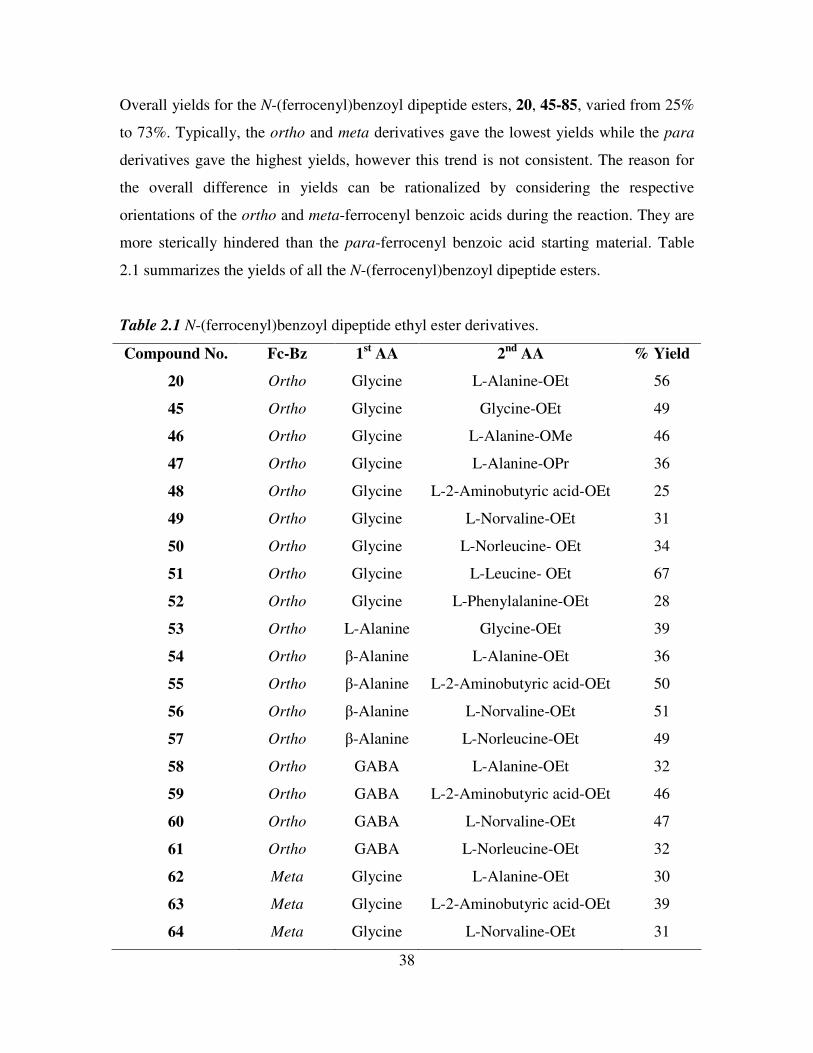
Overall yields for the N-(ferrocenyl)benzoyl dipeptide esters, 20, 45-85, varied from 25%
to 73%. Typically, the ortho and meta derivatives gave the lowest yields while the para
derivatives gave the highest yields, however this trend is not consistent. The reason for
the overall difference in yields can be rationalized by considering the respective
orientations of the ortho and meta-ferrocenyl benzoic acids during the reaction. They are
more sterically hindered than the para-ferrocenyl benzoic acid starting material. Table
2.1 summarizes the yields of all the N-(ferrocenyl)benzoyl dipeptide esters.
Table 2.1 N-(ferrocenyl)benzoyl dipeptide ethyl ester derivatives.
Compound No. Fc-Bz 1st AA 2
nd AA % Yield
20 Ortho Glycine L-Alanine-OEt 56
45 Ortho Glycine Glycine-OEt 49
46 Ortho Glycine L-Alanine-OMe 46
47 Ortho Glycine L-Alanine-OPr 36
48 Ortho Glycine L-2-Aminobutyric acid-OEt 25
49 Ortho Glycine L-Norvaline-OEt 31
50 Ortho Glycine L-Norleucine- OEt 34
51 Ortho Glycine L-Leucine- OEt 67
52 Ortho Glycine L-Phenylalanine-OEt 28
53 Ortho L-Alanine Glycine-OEt 39
54 Ortho β-Alanine L-Alanine-OEt 36
55 Ortho β-Alanine L-2-Aminobutyric acid-OEt 50
56 Ortho β-Alanine L-Norvaline-OEt 51
57 Ortho β-Alanine L-Norleucine-OEt 49
58 Ortho GABA L-Alanine-OEt 32
59 Ortho GABA L-2-Aminobutyric acid-OEt 46
60 Ortho GABA L-Norvaline-OEt 47
61 Ortho GABA L-Norleucine-OEt 32
62 Meta Glycine L-Alanine-OEt 30
63 Meta Glycine L-2-Aminobutyric acid-OEt 39
64 Meta Glycine L-Norvaline-OEt 31
39
65 Meta Glycine L-Norleucine-OEt 34
66 Meta β-Alanine L-Alanine-OEt 28
67 Meta β-Alanine L-2-Aminobutyric acid-OEt 55
68 Meta β-Alanine L-Norvaline-OEt 44
69 Meta β-Alanine L-Norleucine-OEt 42
70 Meta GABA L-Alanine-OEt 32
71 Meta GABA L-2-Aminobutyric acid-OEt 36
72 Meta GABA L-Norvaline-OEt 59
73 Meta GABA L-Norleucine-OEt 55
74 Para Glycine L-Alanine-OEt 33
75 Para Glycine L-2-Aminobutyric acid-OEt 45
76 Para Glycine L-Norvaline-OEt 73
77 Para Glycine L-Norleucine-OEt 40
78 Para β-Alanine L-Alanine-OEt 52
79 Para β-Alanine L-2-Aminobutyric acid-OEt 50
80 Para β-Alanine L-Norvaline-OEt 51
81 Para β-Alanine L-Norleucine-OEt 54
82 Para GABA L-Alanine-OEt 47
83 Para GABA L-2-Aminobutyric acid-OEt 39
84 Para GABA L-Norvaline-OEt 47
85 Para GABA L-Norleucine-OEt 48
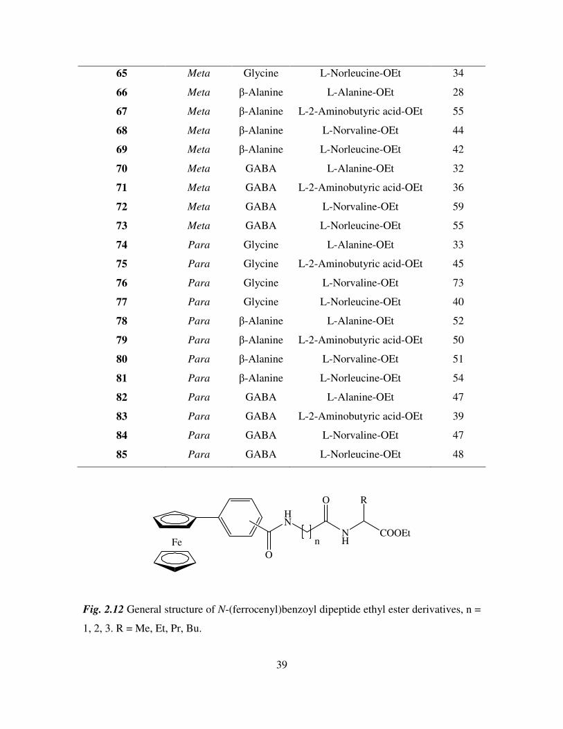
Fe
O
HN
NH
O
COOEt
R
n
Fig. 2.12 General structure of N-(ferrocenyl)benzoyl dipeptide ethyl ester derivatives, n =
1, 2, 3. R = Me, Et, Pr, Bu.
40
2.4 1H NMR studies of N-(ferrocenyl)benzoyl dipeptide esters.
All the 1H NMR experiments were performed in d6-DMSO as the N-(ferrocenyl)benzoyl
dipeptide esters showed limited solubility in other deuterated solvents. In d6-DMSO the
amide protons of the peptide chain appear between δ 8.89 and δ 8.14. The spectra have
three signals in the ferrocenyl region which are typical of the mono-substituted ferrocene
splitting pattern. The protons of the substituted ring appear as fine triplets or singlets
between δ 4.95 and δ 4.38, while the unsubstituted cyclopentadiene ring appears as a
singlet at approximately δ 4.0.
The aromatic splitting pattern present in the 1H NMR spectra of N-(ferrocenyl)benzoyl
dipeptide esters varied depending on whether ortho, meta or para ferrocenyl benzoic acid
was used in the final coupling step. The ortho derivatives have a doublet, triplet, triplet,
doublet splitting pattern in the majority of cases, with each peak integrating for one
proton. The meta derivatives give rise to a singlet, multiplet, triplet splitting pattern
where the multiplet integrates for two protons. The para derivatives give the archetypal
para disubstituted aromatic splitting pattern with two doublets that both integrate for two
protons with coupling constants ranging from 8.4 Hz to 8.8 Hz.
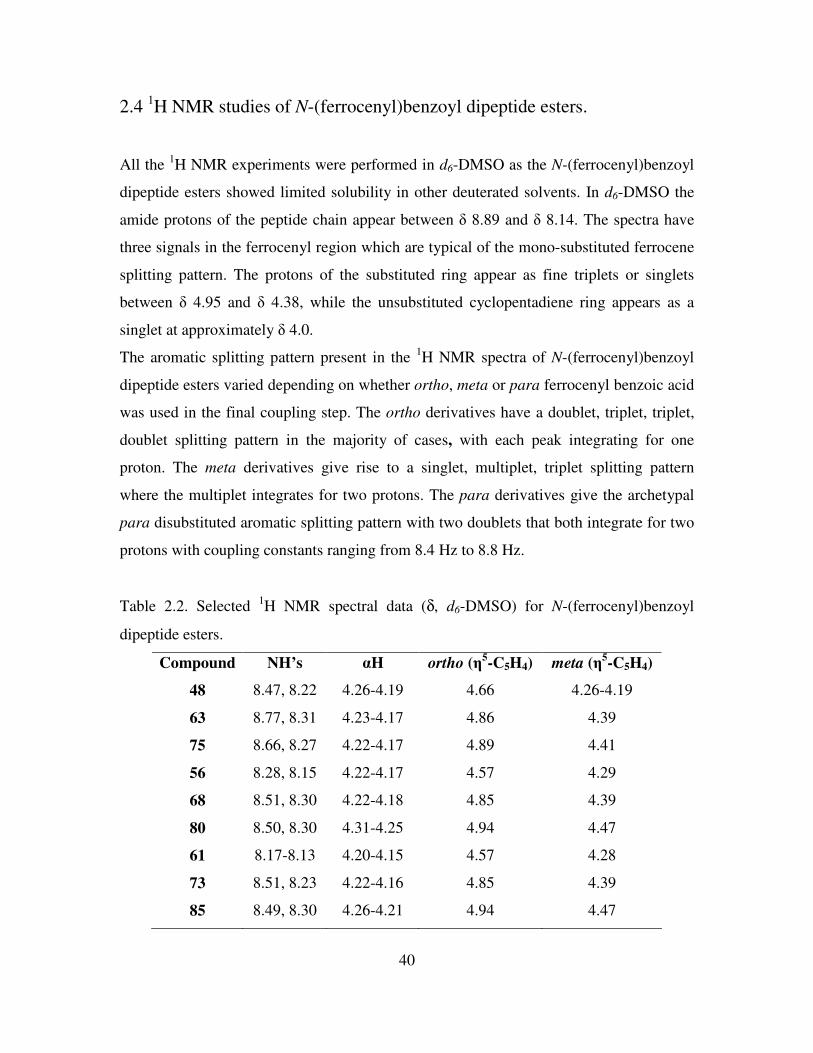
Table 2.2. Selected 1H NMR spectral data (δ, d6-DMSO) for N-(ferrocenyl)benzoyl
dipeptide esters.
Compound NH’s αH ortho (η5-C5H4)
meta (η
5-C5H4)
48 8.47, 8.22 4.26-4.19 4.66 4.26-4.19
63 8.77, 8.31 4.23-4.17 4.86 4.39
75 8.66, 8.27 4.22-4.17 4.89 4.41
56 8.28, 8.15 4.22-4.17 4.57 4.29
68 8.51, 8.30 4.22-4.18 4.85 4.39
80 8.50, 8.30 4.31-4.25 4.94 4.47
61 8.17-8.13 4.20-4.15 4.57 4.28
73 8.51, 8.23 4.22-4.16 4.85 4.39
85 8.49, 8.30 4.26-4.21 4.94 4.47
41
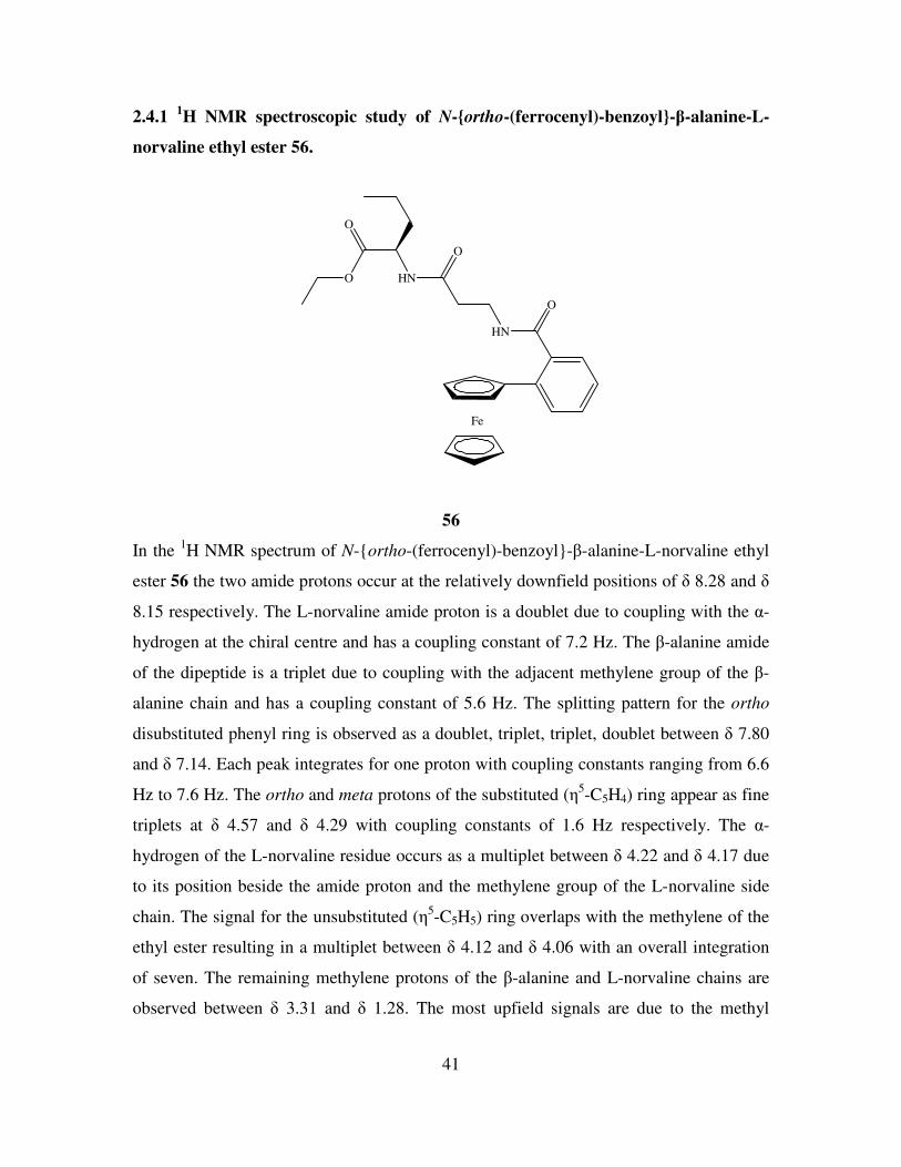
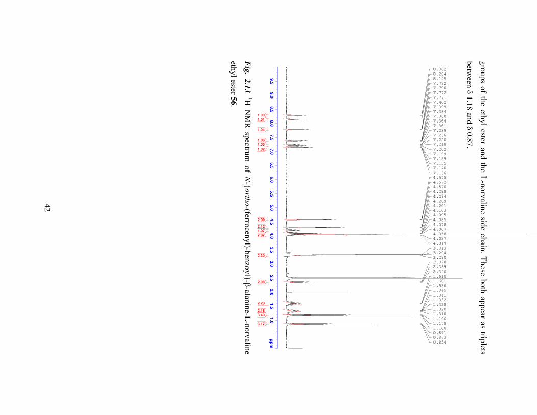
2.4.1 1H NMR spectroscopic study of N-ortho-(ferrocenyl)-benzoyl-β-alanine-L-
norvaline ethyl ester 56.
Fe
O
HN
O
HN
O
O
56
In the 1H NMR spectrum of N-ortho-(ferrocenyl)-benzoyl-β-alanine-L-norvaline ethyl
ester 56 the two amide protons occur at the relatively downfield positions of δ 8.28 and δ
8.15 respectively. The L-norvaline amide proton is a doublet due to coupling with the α-
hydrogen at the chiral centre and has a coupling constant of 7.2 Hz. The β-alanine amide
of the dipeptide is a triplet due to coupling with the adjacent methylene group of the β-
alanine chain and has a coupling constant of 5.6 Hz. The splitting pattern for the ortho
disubstituted phenyl ring is observed as a doublet, triplet, triplet, doublet between δ 7.80
and δ 7.14. Each peak integrates for one proton with coupling constants ranging from 6.6
Hz to 7.6 Hz. The ortho and meta protons of the substituted (η5-C5H4) ring appear as fine
triplets at δ 4.57 and δ 4.29 with coupling constants of 1.6 Hz respectively. The α-
hydrogen of the L-norvaline residue occurs as a multiplet between δ 4.22 and δ 4.17 due
to its position beside the amide proton and the methylene group of the L-norvaline side
chain. The signal for the unsubstituted (η5-C5H5) ring overlaps with the methylene of the
ethyl ester resulting in a multiplet between δ 4.12 and δ 4.06 with an overall integration
of seven. The remaining methylene protons of the β-alanine and L-norvaline chains are
observed between δ 3.31 and δ 1.28. The most upfield signals are due to the methyl
42
gro
ups o
f the eth
yl ester an
d th
e L-n
orv
aline sid
e chain
. Th
ese both
app
ear as triplets
betw
een δ
1.1
8 an
d δ
0.8
7.
9.5
9.0
8.5
8.0
7.5
7.0
6.5
6.0
5.5
5.0
4.5
4.0
3.5
3.0
2.5
2.0
1.5
1.0
pp
m 0.854
0.873
0.891
1.160
1.178
1.196
1.310
1.320
1.328
1.332
1.341
1.345
1.586
1.601
1.610
2.340
2.359
2.378
3.290
3.294
3.313
4.019
4.037
4.058
4.067
4.078
4.085
4.095
4.103
4.201
4.289
4.294
4.298
4.570
4.572
4.575
7.136
7.140
7.155
7.159
7.199
7.202
7.218
7.220
7.236
7.239
7.361
7.364
7.380
7.384
7.399
7.402
7.771
7.772
7.790
7.792
8.145
8.284
8.302
3.17
3.49
2.18
2.20
2.08
2.30
7.87
1.07
2.12
2.09
1.02
1.05
1.06
1.04
1.01
1.00
Fig
. 2.1
3
1H N
MR
sp
ectrum
of
N-
orth
o-(ferro
cenyl)-b
enzo
yl
-β-alan
ine-L
-norv
aline
ethyl ester 5
6.
43
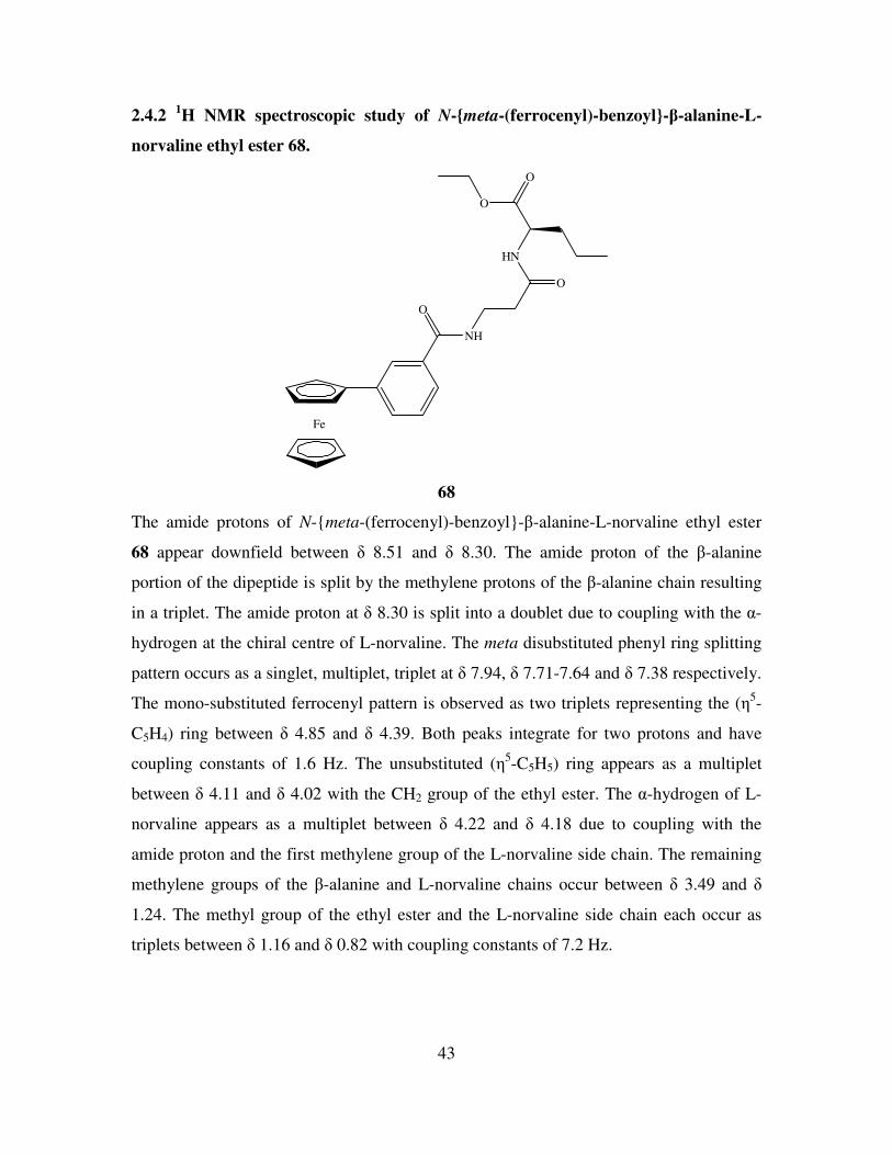
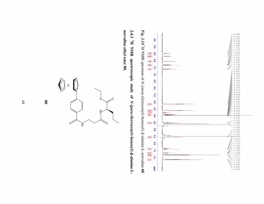
2.4.2 1H NMR spectroscopic study of N-meta-(ferrocenyl)-benzoyl-β-alanine-L-
norvaline ethyl ester 68.
Fe
O
NH
O
HN
O
O
68
The amide protons of N-meta-(ferrocenyl)-benzoyl-β-alanine-L-norvaline ethyl ester
68 appear downfield between δ 8.51 and δ 8.30. The amide proton of the β-alanine
portion of the dipeptide is split by the methylene protons of the β-alanine chain resulting
in a triplet. The amide proton at δ 8.30 is split into a doublet due to coupling with the α-
hydrogen at the chiral centre of L-norvaline. The meta disubstituted phenyl ring splitting
pattern occurs as a singlet, multiplet, triplet at δ 7.94, δ 7.71-7.64 and δ 7.38 respectively.
The mono-substituted ferrocenyl pattern is observed as two triplets representing the (η5-
C5H4) ring between δ 4.85 and δ 4.39. Both peaks integrate for two protons and have
coupling constants of 1.6 Hz. The unsubstituted (η5-C5H5) ring appears as a multiplet
between δ 4.11 and δ 4.02 with the CH2 group of the ethyl ester. The α-hydrogen of L-
norvaline appears as a multiplet between δ 4.22 and δ 4.18 due to coupling with the
amide proton and the first methylene group of the L-norvaline side chain. The remaining
methylene groups of the β-alanine and L-norvaline chains occur between δ 3.49 and δ
1.24. The methyl group of the ethyl ester and the L-norvaline side chain each occur as
triplets between δ 1.16 and δ 0.82 with coupling constants of 7.2 Hz.
44
9.5
9.0
8.5
8.0
7.5
7.0
6.5
6.0
5.5
5.0
4.5
4.0
3.5
3.0
2.5
2.0
1.5
1.0
pp
m 0.802
0.821
0.839
1.138
1.156
1.173
1.277
1.286
1.294
1.298
1.306
1.312
1.572
1.587
1.609
1.621
2.462
2.466
3.459
3.471
3.486
3.501
4.013
4.024
4.035
4.045
4.054
4.063
4.072
4.081
4.090
4.095
4.099
4.210
4.215
4.218
4.229
4.237
4.382
4.387
4.391
4.842
4.847
4.851
7.359
7.378
7.397
7.638
7.641
7.657
7.692
7.707
7.711
7.940
7.944
8.306
8.325
8.511
8.524
3.11
3.08
2.14
2.12
2.34
2.18
7.01
1.10
2.02
2.03
1.01
2.08
1.02
0.99
1.00
Fig
. 2.1
4 1H
NM
R sp
ectrum
of N
-m
eta-(ferro
cenyl)-b
enzo
yl
-β-alan
ine-L
-norv
aline 6
8.
2.4
.3
1H
NM
R
spectro
scop
ic stu
dy
of
N-p
ara
-(ferrocen
yl)-b
enzo
yl-β
-ala
nin
e-L-
norv
alin
e ethyl ester 8
0.
Fe
O NH
O
HN
OO
80
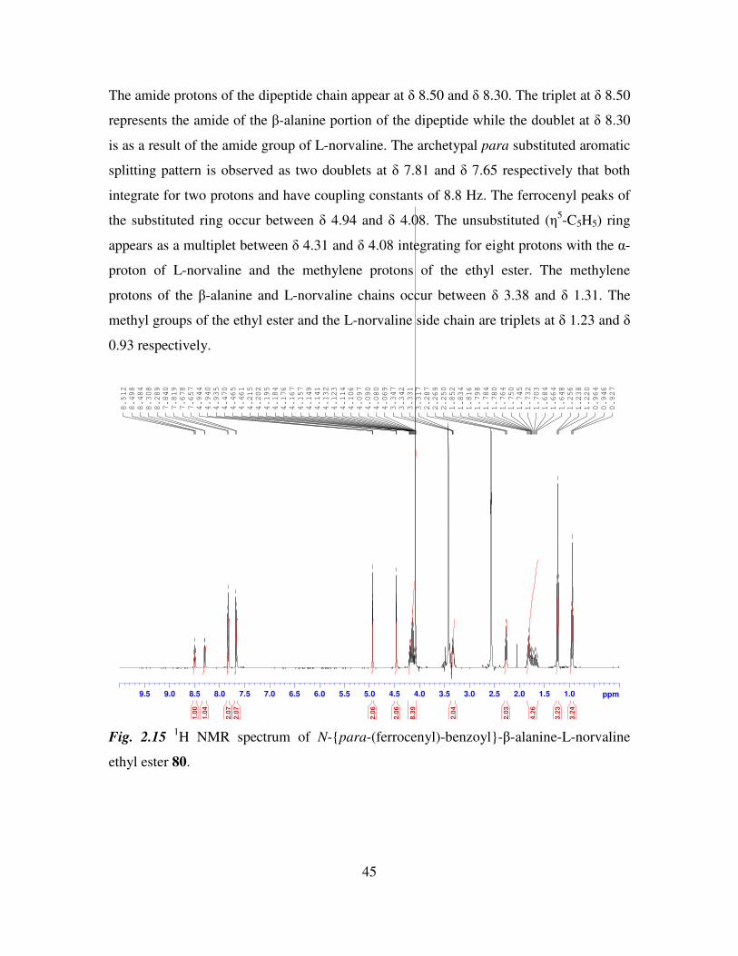
45
The amide protons of the dipeptide chain appear at δ 8.50 and δ 8.30. The triplet at δ 8.50
represents the amide of the β-alanine portion of the dipeptide while the doublet at δ 8.30
is as a result of the amide group of L-norvaline. The archetypal para substituted aromatic
splitting pattern is observed as two doublets at δ 7.81 and δ 7.65 respectively that both
integrate for two protons and have coupling constants of 8.8 Hz. The ferrocenyl peaks of
the substituted ring occur between δ 4.94 and δ 4.08. The unsubstituted (η5-C5H5) ring
appears as a multiplet between δ 4.31 and δ 4.08 integrating for eight protons with the α-
proton of L-norvaline and the methylene protons of the ethyl ester. The methylene
protons of the β-alanine and L-norvaline chains occur between δ 3.38 and δ 1.31. The
methyl groups of the ethyl ester and the L-norvaline side chain are triplets at δ 1.23 and δ
0.93 respectively.
9.5 9.0 8.5 8.0 7.5 7.0 6.5 6.0 5.5 5.0 4.5 4.0 3.5 3.0 2.5 2.0 1.5 1.0 ppm
0.927
0.946
0.964
1.220
1.238
1.256
1.648
1.664
1.684
1.703
1.732
1.745
1.750
1.764
1.780
1.784
1.798
1.816
1.834
1.852
2.250
2.269
2.287
3.317
3.331
3.342
3.347
4.069
4.080
4.090
4.097
4.106
4.114
4.123
4.132
4.141
4.149
4.157
4.167
4.176
4.184
4.195
4.202
4.215
4.461
4.465
4.470
4.935
4.940
4.944
7.657
7.678
7.819
7.840
8.289
8.308
8.484
8.498
8.512
3.2
4
3.2
3
4.2
6
2.0
3
2.0
4
8.3
9
2.0
6
2.0
6
2.0
7
2.0
7
1.0
4
1.0
0
Fig. 2.15 1H NMR spectrum of N-para-(ferrocenyl)-benzoyl-β-alanine-L-norvaline
ethyl ester 80.
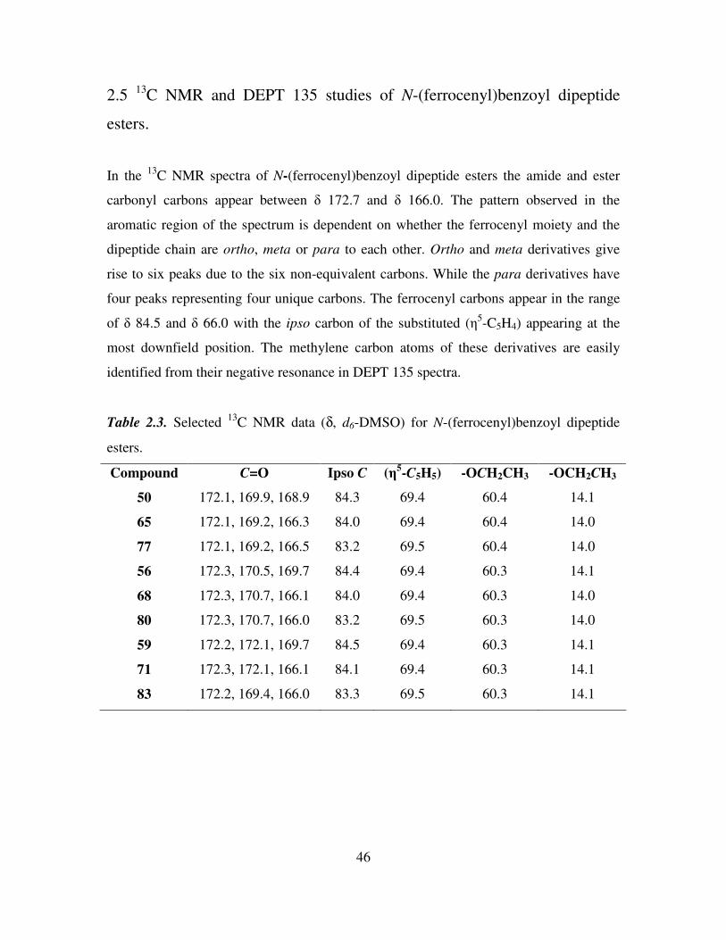
46
2.5 13
C NMR and DEPT 135 studies of N-(ferrocenyl)benzoyl dipeptide
esters.
In the 13
C NMR spectra of N-(ferrocenyl)benzoyl dipeptide esters the amide and ester
carbonyl carbons appear between δ 172.7 and δ 166.0. The pattern observed in the
aromatic region of the spectrum is dependent on whether the ferrocenyl moiety and the
dipeptide chain are ortho, meta or para to each other. Ortho and meta derivatives give
rise to six peaks due to the six non-equivalent carbons. While the para derivatives have
four peaks representing four unique carbons. The ferrocenyl carbons appear in the range
of δ 84.5 and δ 66.0 with the ipso carbon of the substituted (η5-C5H4) appearing at the
most downfield position. The methylene carbon atoms of these derivatives are easily
identified from their negative resonance in DEPT 135 spectra.
Table 2.3. Selected 13
C NMR data (δ, d6-DMSO) for N-(ferrocenyl)benzoyl dipeptide
esters.
Compound C=O Ipso C (η5-C5H5) -OCH2CH3 -OCH2CH3
50 172.1, 169.9, 168.9 84.3 69.4 60.4 14.1
65 172.1, 169.2, 166.3 84.0 69.4 60.4 14.0
77 172.1, 169.2, 166.5 83.2 69.5 60.4 14.0
56 172.3, 170.5, 169.7 84.4 69.4 60.3 14.1
68 172.3, 170.7, 166.1 84.0 69.4 60.3 14.0
80 172.3, 170.7, 166.0 83.2 69.5 60.3 14.0
59 172.2, 172.1, 169.7 84.5 69.4 60.3 14.1
71 172.3, 172.1, 166.1 84.1 69.4 60.3 14.1
83 172.2, 169.4, 166.0 83.3 69.5 60.3 14.1
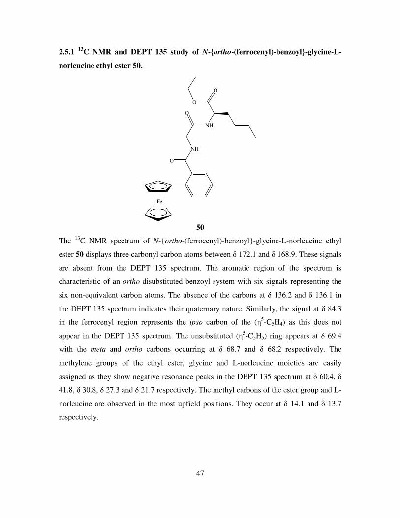
47
2.5.1 13
C NMR and DEPT 135 study of N-ortho-(ferrocenyl)-benzoyl-glycine-L-
norleucine ethyl ester 50.
Fe
NH
O
O
NH
O
O
50
The 13
C NMR spectrum of N-ortho-(ferrocenyl)-benzoyl-glycine-L-norleucine ethyl
ester 50 displays three carbonyl carbon atoms between δ 172.1 and δ 168.9. These signals
are absent from the DEPT 135 spectrum. The aromatic region of the spectrum is
characteristic of an ortho disubstituted benzoyl system with six signals representing the
six non-equivalent carbon atoms. The absence of the carbons at δ 136.2 and δ 136.1 in
the DEPT 135 spectrum indicates their quaternary nature. Similarly, the signal at δ 84.3
in the ferrocenyl region represents the ipso carbon of the (η5-C5H4) as this does not
appear in the DEPT 135 spectrum. The unsubstituted (η5-C5H5) ring appears at δ 69.4
with the meta and ortho carbons occurring at δ 68.7 and δ 68.2 respectively. The
methylene groups of the ethyl ester, glycine and L-norleucine moieties are easily
assigned as they show negative resonance peaks in the DEPT 135 spectrum at δ 60.4, δ
41.8, δ 30.8, δ 27.3 and δ 21.7 respectively. The methyl carbons of the ester group and L-
norleucine are observed in the most upfield positions. They occur at δ 14.1 and δ 13.7
respectively.
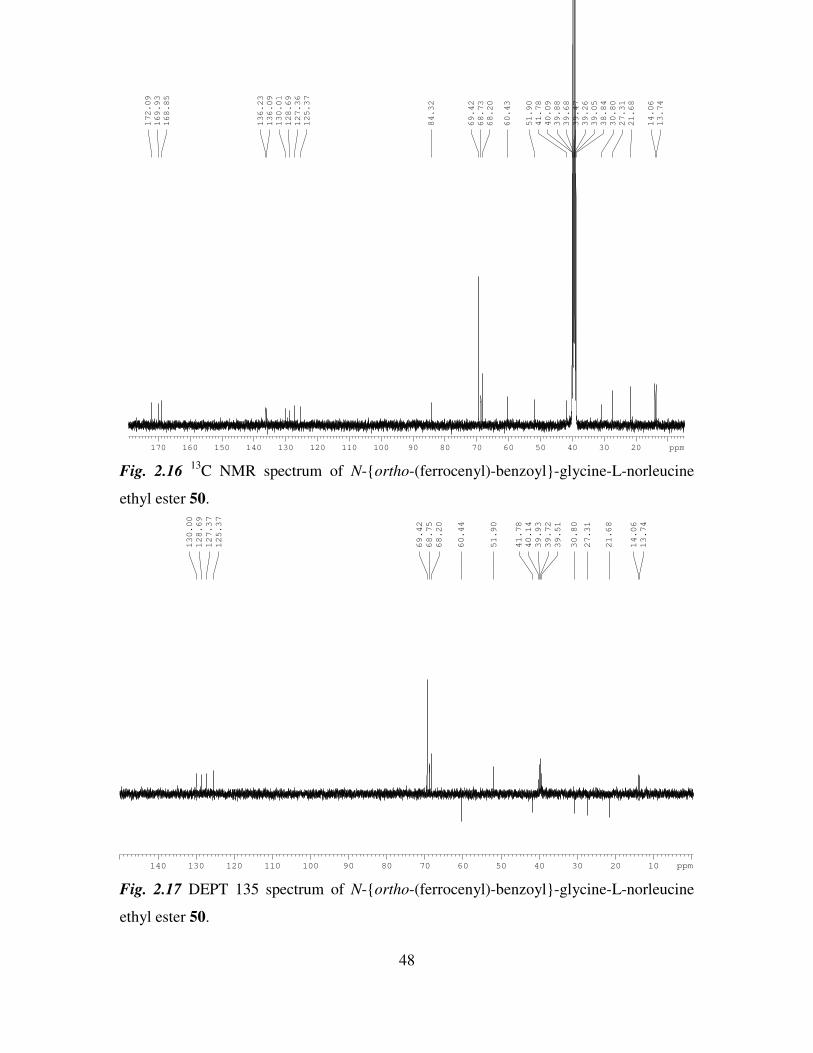
48
2030405060708090100110120130140150160170 ppm
13.74
14.06
21.68
27.31
30.80
38.84
39.05
39.26
39.47
39.68
39.88
40.09
41.78
51.90
60.43
68.20
68.73
69.42
84.32
125.37
127.36
128.69
130.01
136.09
136.23
168.85
169.93
172.09
Fig. 2.16 13
C NMR spectrum of N-ortho-(ferrocenyl)-benzoyl-glycine-L-norleucine
ethyl ester 50.
140 130 120 110 100 90 80 70 60 50 40 30 20 10 ppm
13.74
14.06
21.68
27.31
30.80
39.51
39.72
39.93
40.14
41.78
51.90
60.44
68.20
68.75
69.42
125.37
127.37
128.69
130.00
Fig. 2.17 DEPT 135 spectrum of N-ortho-(ferrocenyl)-benzoyl-glycine-L-norleucine
ethyl ester 50.
49
2.6 COSY studies of N-para-(ferrocenyl)-benzoyl-γ-aminobutyric acid-L-
norleucine ethyl ester.
COSY (Correlation Spectroscopy) is the simplest example of a two-dimensional NMR
technique. There are two coordinate axes, each representing a chemical shift range. The
data is plotted as a grid with both chemical shift ranges and the third dimension shows the
intensity of the observed signal (9)
. In the COSY spectrum of N-para-(ferrocenyl)-
benzoyl-γ-aminobutyric acid-L-norleucine ethyl ester 85 the proton spectrum is plotted
along each axis. The spectrum shows distinct spots on the diagonal with each spot
corresponding to the same peak on each coordinate axis. It is clear that the amide proton
of the γ-aminobutyric acid a (δ 8.49) correlates with the methylene of the γ-aminobutyric
acid chain b (δ 3.36-3.30), while the amide proton of L-norleucine e (δ 8.30) couples
with the methine proton f (δ 4.21) at the chiral centre of L-norleucine. Correlation is also
present between the ortho and meta protons of the substituted (η5-C5H4), l and k. The first
methylene group of the L-norleucine side chain g (δ 1.75-1.60) is identified from its
coupling with the methine proton f. The first methylene group of L-norleucine g (δ 1.75-
1.60) also couples with the second methylene group h, while the second group h also
couples with the third methylene group i. The third methylene group i in turn couples
with the methyl group of the chain j. The middle methylene group of the γ-aminobutyric
acid c is easily identified due to its coupling with both remaining methylene groups, b
and d. The methylene m and methyl protons n of the ethyl ester only show coupling with
each other, as do the aromatic protons of the benzoyl group, o and p.
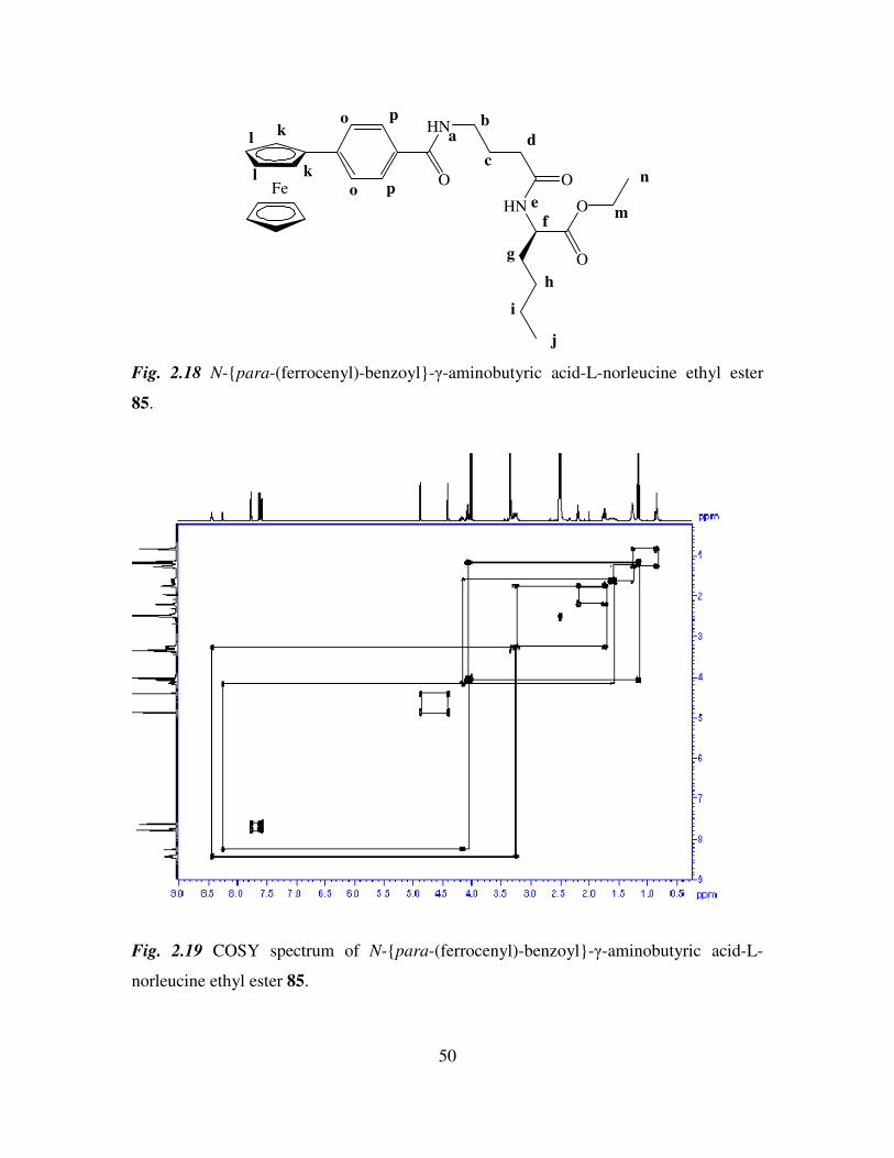
50
FeO
HN
O
HN
O
O
ab
c
d
e
f
g
h
i
j
k
k
l
l
m
no
o
p
p
Fig. 2.18 N-para-(ferrocenyl)-benzoyl-γ-aminobutyric acid-L-norleucine ethyl ester
85.
Fig. 2.19 COSY spectrum of N-para-(ferrocenyl)-benzoyl-γ-aminobutyric acid-L-
norleucine ethyl ester 85.
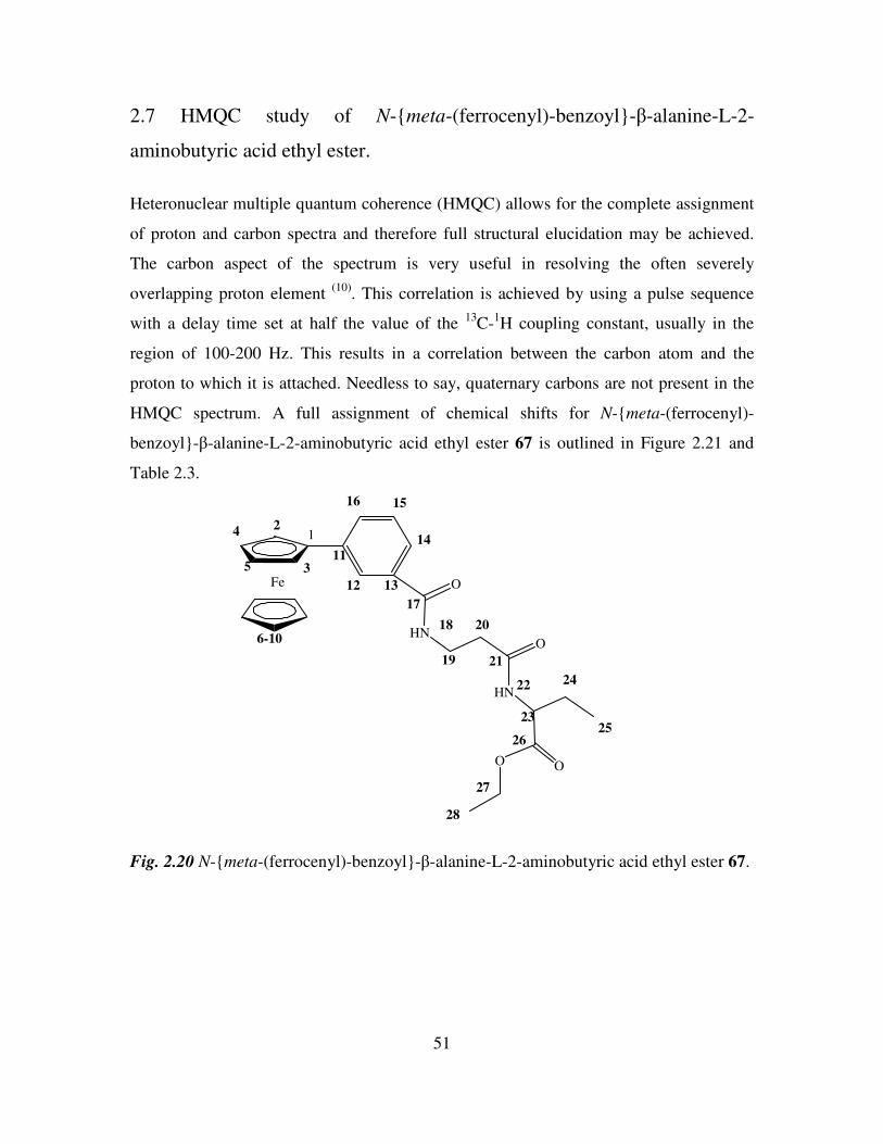
51
2.7 HMQC study of N-meta-(ferrocenyl)-benzoyl-β-alanine-L-2-
aminobutyric acid ethyl ester.
Heteronuclear multiple quantum coherence (HMQC) allows for the complete assignment
of proton and carbon spectra and therefore full structural elucidation may be achieved.
The carbon aspect of the spectrum is very useful in resolving the often severely
overlapping proton element (10)
. This correlation is achieved by using a pulse sequence
with a delay time set at half the value of the 13
C-1H coupling constant, usually in the
region of 100-200 Hz. This results in a correlation between the carbon atom and the
proton to which it is attached. Needless to say, quaternary carbons are not present in the
HMQC spectrum. A full assignment of chemical shifts for N-meta-(ferrocenyl)-
benzoyl-β-alanine-L-2-aminobutyric acid ethyl ester 67 is outlined in Figure 2.21 and
Table 2.3.
Fe O
HNO
HN
OO
12
3
4
5
6-10
11
12 13
14
1516
17
18
19
20
21
22
23
24
2526
27
28
Fig. 2.20 N-meta-(ferrocenyl)-benzoyl-β-alanine-L-2-aminobutyric acid ethyl ester 67.
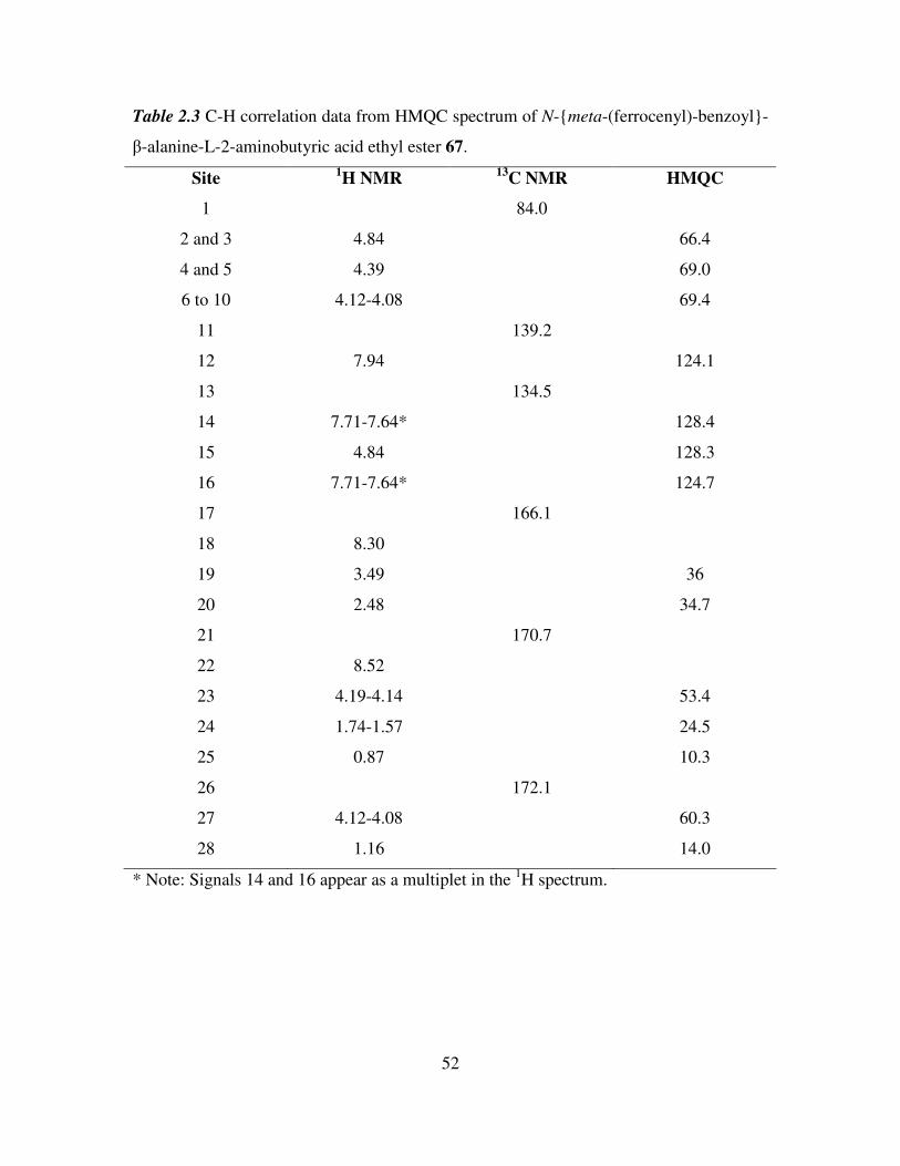
52
Table 2.3 C-H correlation data from HMQC spectrum of N-meta-(ferrocenyl)-benzoyl-
β-alanine-L-2-aminobutyric acid ethyl ester 67.
Site 1H NMR
13C NMR HMQC
1 84.0
2 and 3 4.84 66.4
4 and 5 4.39 69.0
6 to 10 4.12-4.08 69.4
11 139.2
12 7.94 124.1
13 134.5
14 7.71-7.64* 128.4
15 4.84 128.3
16 7.71-7.64* 124.7
17 166.1
18 8.30
19 3.49 36
20 2.48 34.7
21 170.7
22 8.52
23 4.19-4.14 53.4
24 1.74-1.57 24.5
25 0.87 10.3
26 172.1
27 4.12-4.08 60.3
28 1.16 14.0
* Note: Signals 14 and 16 appear as a multiplet in the 1H spectrum.
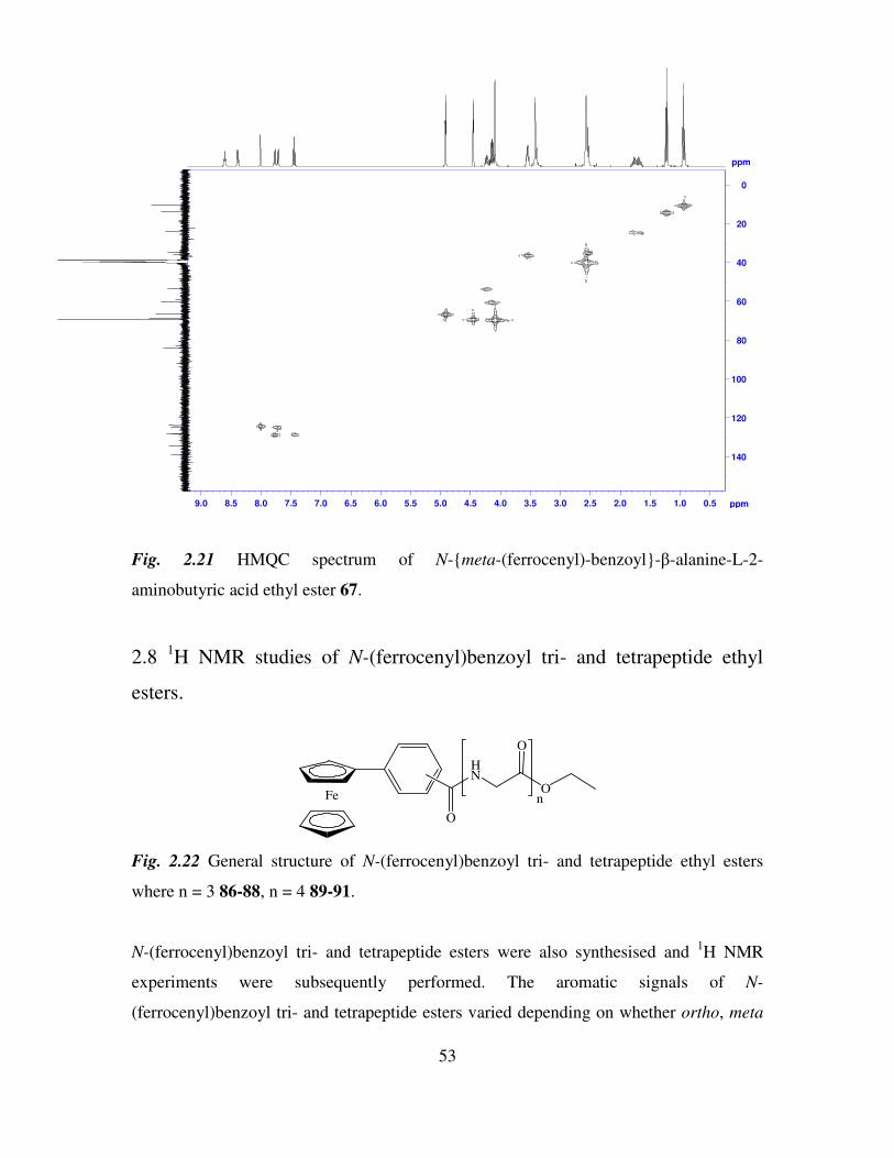
53
ppm
0.51.01.52.02.53.03.54.04.55.05.56.06.57.07.58.08.59.0 ppm
140
120
100
80
60
40
20
0
Fig. 2.21 HMQC spectrum of N-meta-(ferrocenyl)-benzoyl-β-alanine-L-2-
aminobutyric acid ethyl ester 67.
2.8 1H NMR studies of N-(ferrocenyl)benzoyl tri- and tetrapeptide ethyl
esters.
Fe
HN
O
O
O
n
Fig. 2.22 General structure of N-(ferrocenyl)benzoyl tri- and tetrapeptide ethyl esters
where n = 3 86-88, n = 4 89-91.
N-(ferrocenyl)benzoyl tri- and tetrapeptide esters were also synthesised and 1H NMR
experiments were subsequently performed. The aromatic signals of N-
(ferrocenyl)benzoyl tri- and tetrapeptide esters varied depending on whether ortho, meta
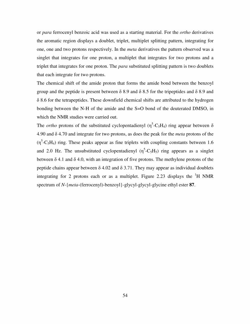
54
or para ferrocenyl benzoic acid was used as a starting material. For the ortho derivatives
the aromatic region displays a doublet, triplet, multiplet splitting pattern, integrating for
one, one and two protons respectively. In the meta derivatives the pattern observed was a
singlet that integrates for one proton, a multiplet that integrates for two protons and a
triplet that integrates for one proton. The para substituted splitting pattern is two doublets
that each integrate for two protons.
The chemical shift of the amide proton that forms the amide bond between the benzoyl
group and the peptide is present between δ 8.9 and δ 8.5 for the tripeptides and δ 8.9 and
δ 8.6 for the tetrapeptides. These downfield chemical shifts are attributed to the hydrogen
bonding between the N-H of the amide and the S=O bond of the deuterated DMSO, in
which the NMR studies were carried out.
The ortho protons of the substituted cyclopentadienyl (η5-C5H4) ring appear between δ
4.90 and δ 4.70 and integrate for two protons, as does the peak for the meta protons of the
(η5-C5H4) ring. These peaks appear as fine triplets with coupling constants between 1.6
and 2.0 Hz. The unsubstituted cyclopentadienyl (η5-C5H5) ring appears as a singlet
between δ 4.1 and δ 4.0, with an integration of five protons. The methylene protons of the
peptide chains appear between δ 4.02 and δ 3.71. They may appear as individual doublets
integrating for 2 protons each or as a multiplet. Figure 2.23 displays the 1H NMR
spectrum of N-meta-(ferrocenyl)-benzoyl-glycyl-glycyl-glycine ethyl ester 87.
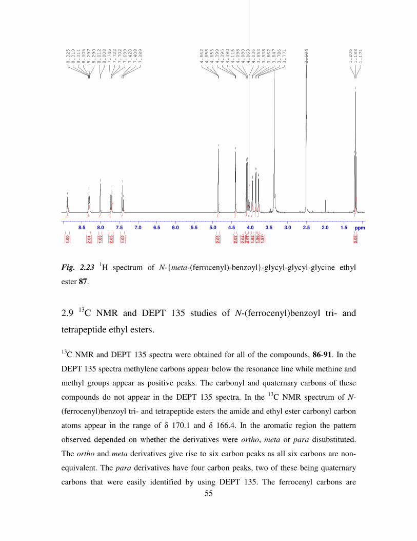
55
1.52.02.53.03.54.04.55.05.56.06.57.07.58.08.5 ppm
1.171
1.188
1.206
2.504
3.771
3.786
3.847
3.862
3.938
3.953
4.036
4.063
4.080
4.098
4.116
4.390
4.395
4.399
4.853
4.858
4.862
7.389
7.408
7.428
7.699
7.702
7.722
7.745
8.008
8.012
8.290
8.297
8.305
8.311
8.319
8.325
3.0
8
1.9
7
1.9
6
1.9
2
4.9
7
2.0
4
2.0
2
2.0
3
1.0
2
2.0
5
1.0
3
2.0
1
1.0
0
Fig. 2.23 1H spectrum of N-meta-(ferrocenyl)-benzoyl-glycyl-glycyl-glycine ethyl
ester 87.
2.9 13
C NMR and DEPT 135 studies of N-(ferrocenyl)benzoyl tri- and
tetrapeptide ethyl esters.
13
C NMR and DEPT 135 spectra were obtained for all of the compounds, 86-91. In the
DEPT 135 spectra methylene carbons appear below the resonance line while methine and
methyl groups appear as positive peaks. The carbonyl and quaternary carbons of these
compounds do not appear in the DEPT 135 spectra. In the 13
C NMR spectrum of N-
(ferrocenyl)benzoyl tri- and tetrapeptide esters the amide and ethyl ester carbonyl carbon
atoms appear in the range of δ 170.1 and δ 166.4. In the aromatic region the pattern
observed depended on whether the derivatives were ortho, meta or para disubstituted.
The ortho and meta derivatives give rise to six carbon peaks as all six carbons are non-
equivalent. The para derivatives have four carbon peaks, two of these being quaternary
carbons that were easily identified by using DEPT 135. The ferrocenyl carbons are
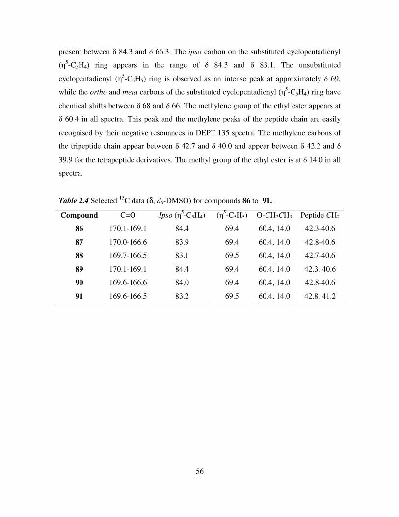
56
present between δ 84.3 and δ 66.3. The ipso carbon on the substituted cyclopentadienyl
(η5-C5H4) ring appears in the range of δ 84.3 and δ 83.1. The unsubstituted
cyclopentadienyl (η5-C5H5) ring is observed as an intense peak at approximately δ 69,
while the ortho and meta carbons of the substituted cyclopentadienyl (η5-C5H4) ring have
chemical shifts between δ 68 and δ 66. The methylene group of the ethyl ester appears at
δ 60.4 in all spectra. This peak and the methylene peaks of the peptide chain are easily
recognised by their negative resonances in DEPT 135 spectra. The methylene carbons of
the tripeptide chain appear between δ 42.7 and δ 40.0 and appear between δ 42.2 and δ
39.9 for the tetrapeptide derivatives. The methyl group of the ethyl ester is at δ 14.0 in all
spectra.
Table 2.4 Selected 13
C data (δ, d6-DMSO) for compounds 86 to 91.
Compound C=O Ipso (η5-C5H4) (η
5-C5H5) O-CH2CH3 Peptide CH2
86 170.1-169.1 84.4 69.4 60.4, 14.0 42.3-40.6
87 170.0-166.6 83.9 69.4 60.4, 14.0 42.8-40.6
88 169.7-166.5 83.1 69.5 60.4, 14.0 42.7-40.6
89 170.1-169.1 84.4 69.4 60.4, 14.0 42.3, 40.6
90 169.6-166.6 84.0 69.4 60.4, 14.0 42.8-40.6
91 169.6-166.5 83.2 69.5 60.4, 14.0 42.8, 41.2
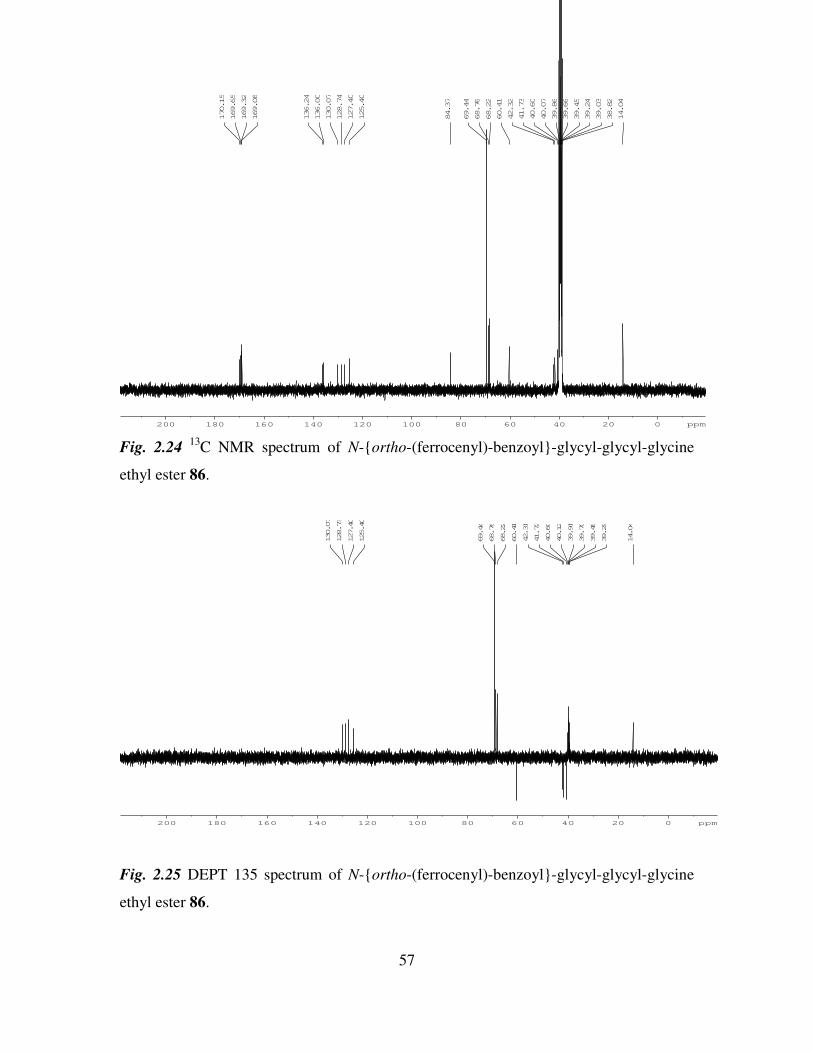
57
200 180 160 140 120 100 80 60 40 20 0 ppm
14.04
38.82
39.03
39.24
39.45
39.66
39.86
40.07
40.60
41.73
42.32
60.41
68.22
68.76
69.44
84.37
125.40
127.40
128.74
130.07
136.00
136.24
169.08
169.32
169.65
170.15
Fig. 2.24 13
C NMR spectrum of N-ortho-(ferrocenyl)-benzoyl-glycyl-glycyl-glycine
ethyl ester 86.
200 180 160 140 120 100 80 60 40 20 0 ppm
14.04
39.29
39.49
39.70
39.91
40.12
40.60
41.72
42.31
60.41
68.22
68.76
69.44
125.40
127.40
128.73
130.07
Fig. 2.25 DEPT 135 spectrum of N-ortho-(ferrocenyl)-benzoyl-glycyl-glycyl-glycine
ethyl ester 86.
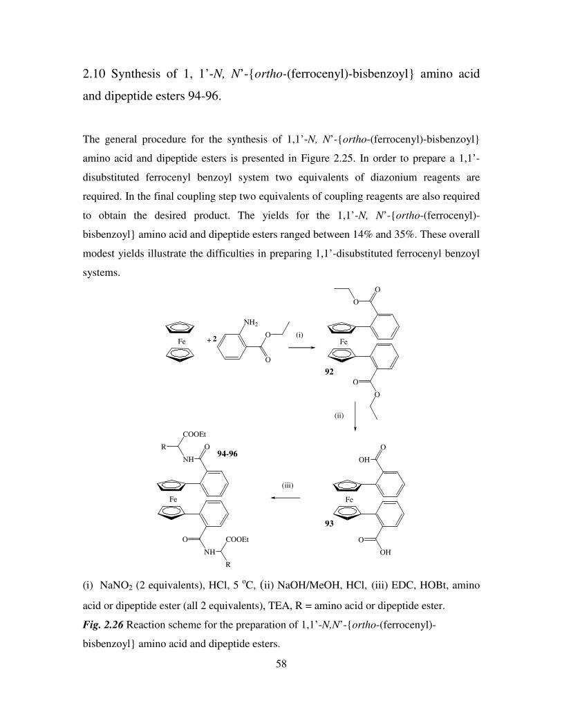
58
2.10 Synthesis of 1, 1’-N, N’-ortho-(ferrocenyl)-bisbenzoyl amino acid
and dipeptide esters 94-96.
The general procedure for the synthesis of 1,1’-N, N’-ortho-(ferrocenyl)-bisbenzoyl
amino acid and dipeptide esters is presented in Figure 2.25. In order to prepare a 1,1’-
disubstituted ferrocenyl benzoyl system two equivalents of diazonium reagents are
required. In the final coupling step two equivalents of coupling reagents are also required
to obtain the desired product. The yields for the 1,1’-N, N’-ortho-(ferrocenyl)-
bisbenzoyl amino acid and dipeptide esters ranged between 14% and 35%. These overall
modest yields illustrate the difficulties in preparing 1,1’-disubstituted ferrocenyl benzoyl
systems.
Fe +
NH2
O
O2 Fe
O
O
O
O
Fe
O
OH
O
OH
Fe
O
NH
O
NH
COOEt
R
R
COOEt
(i)
(ii)
(iii)
92
93
94-96
(i) NaNO2 (2 equivalents), HCl, 5 oC, (ii) NaOH/MeOH, HCl, (iii) EDC, HOBt, amino
acid or dipeptide ester (all 2 equivalents), TEA, R = amino acid or dipeptide ester.
Fig. 2.26 Reaction scheme for the preparation of 1,1’-N,N’-ortho-(ferrocenyl)-
bisbenzoyl amino acid and dipeptide esters.
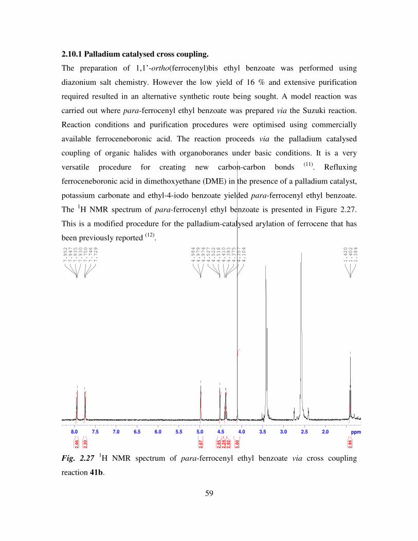
59
2.10.1 Palladium catalysed cross coupling.
The preparation of 1,1’-ortho(ferrocenyl)bis ethyl benzoate was performed using
diazonium salt chemistry. However the low yield of 16 % and extensive purification
required resulted in an alternative synthetic route being sought. A model reaction was
carried out where para-ferrocenyl ethyl benzoate was prepared via the Suzuki reaction.
Reaction conditions and purification procedures were optimised using commercially
available ferroceneboronic acid. The reaction proceeds via the palladium catalysed
coupling of organic halides with organoboranes under basic conditions. It is a very
versatile procedure for creating new carbon-carbon bonds (11)
. Refluxing
ferroceneboronic acid in dimethoxyethane (DME) in the presence of a palladium catalyst,
potassium carbonate and ethyl-4-iodo benzoate yielded para-ferrocenyl ethyl benzoate.
The 1H NMR spectrum of para-ferrocenyl ethyl benzoate is presented in Figure 2.27.
This is a modified procedure for the palladium-catalysed arylation of ferrocene that has
been previously reported (12)
.
2.02.53.03.54.04.55.05.56.06.57.07.58.0 ppm
1.384
1.402
1.420
4.104
4.357
4.375
4.393
4.410
4.518
4.522
4.527
4.974
4.979
4.984
7.729
7.746
7.750
7.930
7.935
7.947
7.952
2.9
5
5.0
0
2.0
22
.24
2.0
5
2.0
7
2.2
0
2.0
6
Fig. 2.27 1H NMR spectrum of para-ferrocenyl ethyl benzoate via cross coupling
reaction 41b.
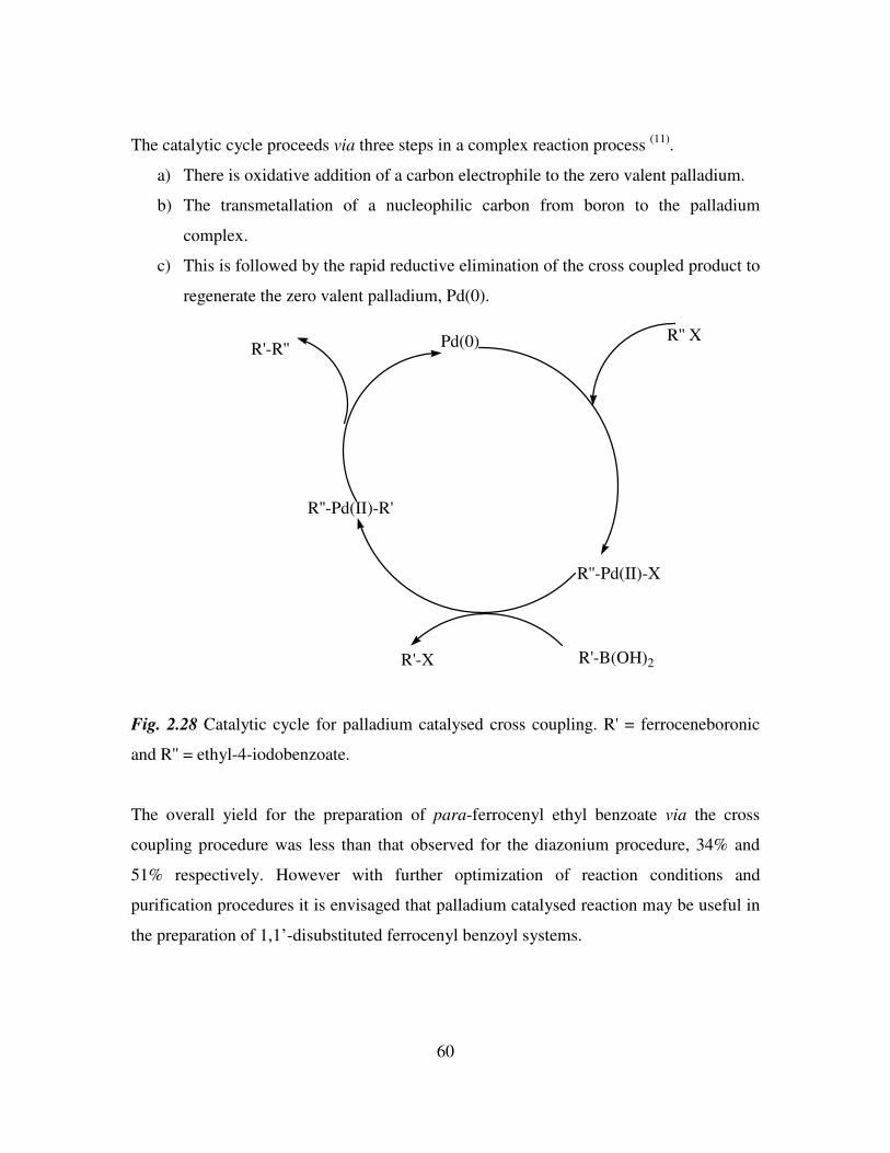
60
The catalytic cycle proceeds via three steps in a complex reaction process (11)
.
a) There is oxidative addition of a carbon electrophile to the zero valent palladium.
b) The transmetallation of a nucleophilic carbon from boron to the palladium
complex.
c) This is followed by the rapid reductive elimination of the cross coupled product to
regenerate the zero valent palladium, Pd(0).
Pd(0) R'' X
R''-Pd(II)-X
R''-Pd(II)-R'
R'-R''
R'-B(OH)2R'-X
Fig. 2.28 Catalytic cycle for palladium catalysed cross coupling. R' = ferroceneboronic
and R'' = ethyl-4-iodobenzoate.
The overall yield for the preparation of para-ferrocenyl ethyl benzoate via the cross
coupling procedure was less than that observed for the diazonium procedure, 34% and
51% respectively. However with further optimization of reaction conditions and
purification procedures it is envisaged that palladium catalysed reaction may be useful in
the preparation of 1,1’-disubstituted ferrocenyl benzoyl systems.
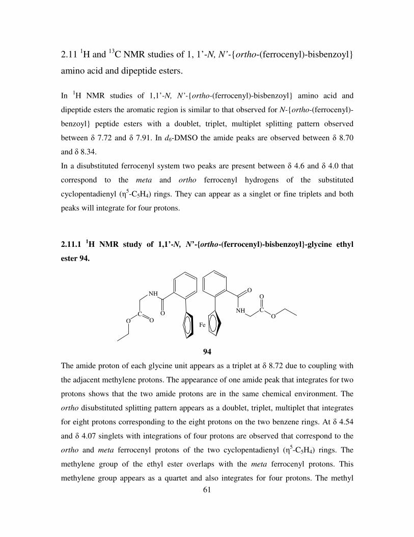
61
2.11 1H and
13C NMR studies of 1, 1’-N, N’-ortho-(ferrocenyl)-bisbenzoyl
amino acid and dipeptide esters.
In 1H NMR studies of 1,1’-N, N’-ortho-(ferrocenyl)-bisbenzoyl amino acid and
dipeptide esters the aromatic region is similar to that observed for N-ortho-(ferrocenyl)-
benzoyl peptide esters with a doublet, triplet, multiplet splitting pattern observed
between δ 7.72 and δ 7.91. In d6-DMSO the amide peaks are observed between δ 8.70
and δ 8.34.
In a disubstituted ferrocenyl system two peaks are present between δ 4.6 and δ 4.0 that
correspond to the meta and ortho ferrocenyl hydrogens of the substituted
cyclopentadienyl (η5-C5H4) rings. They can appear as a singlet or fine triplets and both
peaks will integrate for four protons.
2.11.1 1H NMR study of 1,1’-N, N’-ortho-(ferrocenyl)-bisbenzoyl-glycine ethyl
ester 94.
O
NH
CO O
Fe
O
NH CO
O
94
The amide proton of each glycine unit appears as a triplet at δ 8.72 due to coupling with
the adjacent methylene protons. The appearance of one amide peak that integrates for two
protons shows that the two amide protons are in the same chemical environment. The
ortho disubstituted splitting pattern appears as a doublet, triplet, multiplet that integrates
for eight protons corresponding to the eight protons on the two benzene rings. At δ 4.54
and δ 4.07 singlets with integrations of four protons are observed that correspond to the
ortho and meta ferrocenyl protons of the two cyclopentadienyl (η5-C5H4) rings. The
methylene group of the ethyl ester overlaps with the meta ferrocenyl protons. This
methylene group appears as a quartet and also integrates for four protons. The methyl
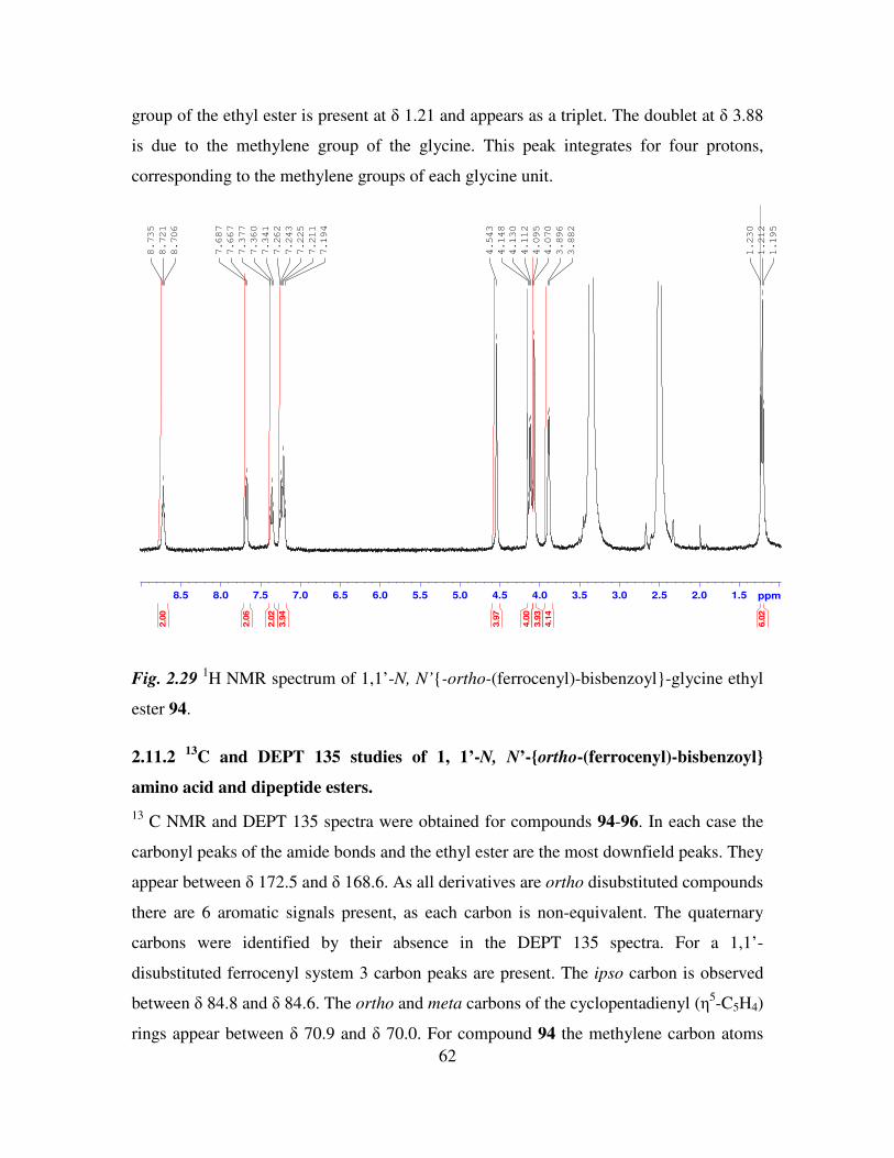
62
group of the ethyl ester is present at δ 1.21 and appears as a triplet. The doublet at δ 3.88
is due to the methylene group of the glycine. This peak integrates for four protons,
corresponding to the methylene groups of each glycine unit.
1.52.02.53.03.54.04.55.05.56.06.57.07.58.08.5 ppm
1.195
1.212
1.230
3.882
3.896
4.070
4.095
4.112
4.130
4.148
4.543
7.194
7.211
7.225
7.243
7.262
7.341
7.360
7.377
7.667
7.687
8.706
8.721
8.735
6.0
2
4.1
4
3.9
3
4.0
0
3.9
7
3.9
4
2.0
2
2.0
6
2.0
0
Fig. 2.29 1H NMR spectrum of 1,1’-N, N’-ortho-(ferrocenyl)-bisbenzoyl-glycine ethyl
ester 94.
2.11.2 13
C and DEPT 135 studies of 1, 1’-N, N’-ortho-(ferrocenyl)-bisbenzoyl
amino acid and dipeptide esters.
13 C NMR and DEPT 135 spectra were obtained for compounds 94-96. In each case the
carbonyl peaks of the amide bonds and the ethyl ester are the most downfield peaks. They
appear between δ 172.5 and δ 168.6. As all derivatives are ortho disubstituted compounds
there are 6 aromatic signals present, as each carbon is non-equivalent. The quaternary
carbons were identified by their absence in the DEPT 135 spectra. For a 1,1’-
disubstituted ferrocenyl system 3 carbon peaks are present. The ipso carbon is observed
between δ 84.8 and δ 84.6. The ortho and meta carbons of the cyclopentadienyl (η5-C5H4)
rings appear between δ 70.9 and δ 70.0. For compound 94 the methylene carbon atoms
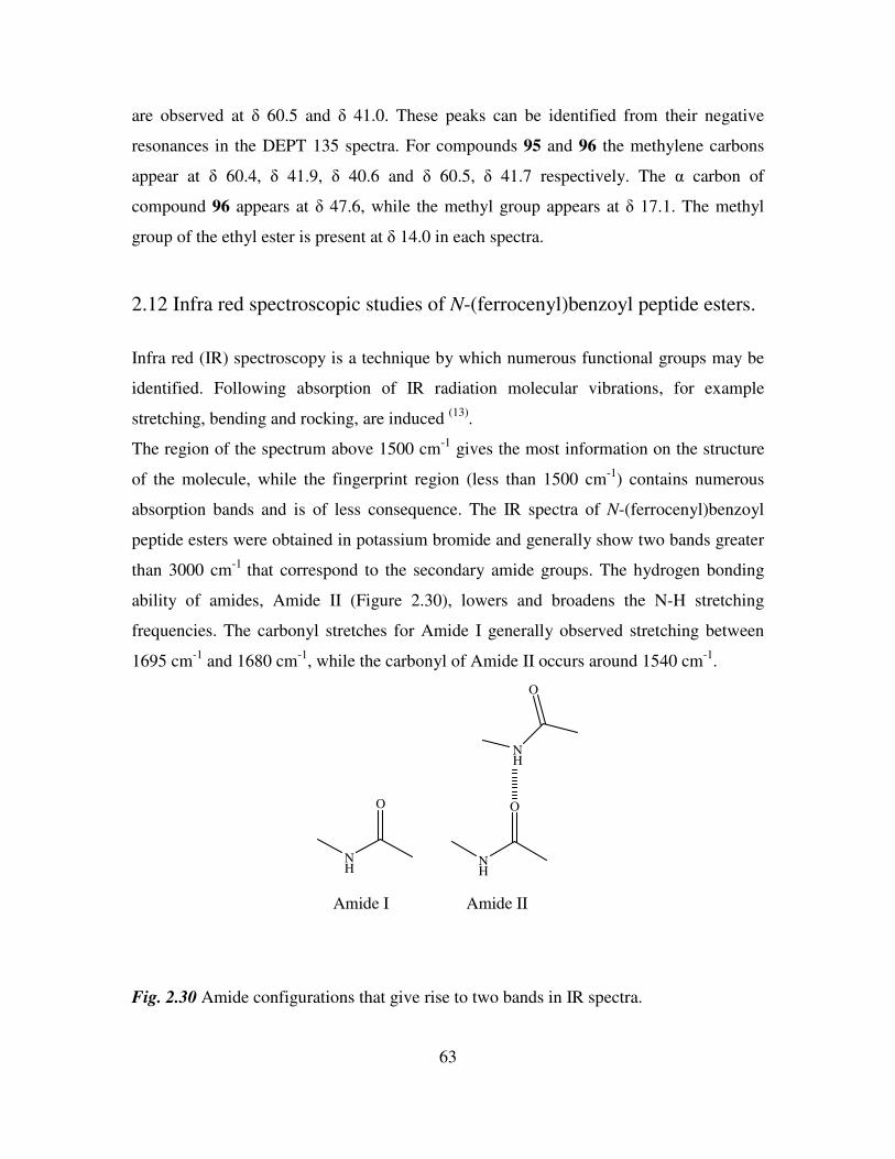
63
are observed at δ 60.5 and δ 41.0. These peaks can be identified from their negative
resonances in the DEPT 135 spectra. For compounds 95 and 96 the methylene carbons
appear at δ 60.4, δ 41.9, δ 40.6 and δ 60.5, δ 41.7 respectively. The α carbon of
compound 96 appears at δ 47.6, while the methyl group appears at δ 17.1. The methyl
group of the ethyl ester is present at δ 14.0 in each spectra.
2.12 Infra red spectroscopic studies of N-(ferrocenyl)benzoyl peptide esters.
Infra red (IR) spectroscopy is a technique by which numerous functional groups may be
identified. Following absorption of IR radiation molecular vibrations, for example
stretching, bending and rocking, are induced (13)
.
The region of the spectrum above 1500 cm-1
gives the most information on the structure
of the molecule, while the fingerprint region (less than 1500 cm-1
) contains numerous
absorption bands and is of less consequence. The IR spectra of N-(ferrocenyl)benzoyl
peptide esters were obtained in potassium bromide and generally show two bands greater
than 3000 cm-1
that correspond to the secondary amide groups. The hydrogen bonding
ability of amides, Amide II (Figure 2.30), lowers and broadens the N-H stretching
frequencies. The carbonyl stretches for Amide I generally observed stretching between
1695 cm-1
and 1680 cm-1
, while the carbonyl of Amide II occurs around 1540 cm-1
.
O
NH
O
NH
NH
O
Amide I Amide II
Fig. 2.30 Amide configurations that give rise to two bands in IR spectra.
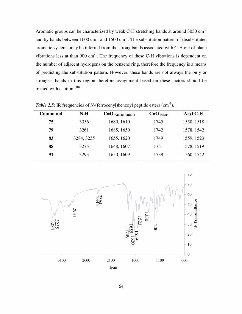
64
Aromatic groups can be characterized by weak C-H stretching bands at around 3030 cm-1
and by bands between 1600 cm-1
and 1500 cm-1
. The substitution pattern of disubstituted
aromatic systems may be inferred from the strong bands associated with C-H out of plane
vibrations less at than 900 cm-1
. The frequency of these C-H vibrations is dependent on
the number of adjacent hydrogens on the benzene ring, therefore the frequency is a means
of predicting the substitution pattern. However, these bands are not always the only or
strongest bands in this region therefore assignment based on these factors should be
treated with caution (10)
.
Table 2.5: IR frequencies of N-(ferrocenyl)benzoyl peptide esters (cm-1
).
Compound N-H C=O Amide I and II C=O Ester Aryl C-H
75 3356 1680, 1610 1745 1558, 1518
79 3261 1685, 1650 1742 1578, 1542
83 3284, 3235 1655, 1620 1749 1559, 1523
88 3275 1648, 1607 1751 1578, 1519
91 3293 1650, 1609 1739 1560, 1542
0
10
20
30
40
50
60
70
80
60011001600210026003100
1/cm
% T
ran
smit
tan
ce
32
84
32
35
29
31
23
45
23
64
17
49
16
55
16
20
15
59
15
23
13
30
12
00
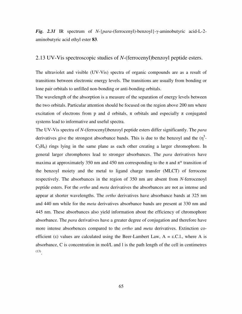
65
Fig. 2.31 IR spectrum of N-para-(ferrocenyl)-benzoyl-γ-aminobutyric acid-L-2-
aminobutyric acid ethyl ester 83.
2.13 UV-Vis spectroscopic studies of N-(ferrocenyl)benzoyl peptide esters.
The ultraviolet and visible (UV-Vis) spectra of organic compounds are as a result of
transitions between electronic energy levels. The transitions are usually from bonding or
lone pair orbitals to unfilled non-bonding or anti-bonding orbitals.
The wavelength of the absorption is a measure of the separation of energy levels between
the two orbitals. Particular attention should be focused on the region above 200 nm where
excitation of electrons from p and d orbitals, π orbitals and especially π conjugated
systems lead to informative and useful spectra.
The UV-Vis spectra of N-(ferrocenyl)benzoyl peptide esters differ significantly. The para
derivatives give the strongest absorbance bands. This is due to the benzoyl and the (η5-
C5H4) rings lying in the same plane as each other creating a larger chromophore. In
general larger chromphores lead to stronger absorbances. The para derivatives have
maxima at approximately 350 nm and 450 nm corresponding to the π and π* transition of
the benzoyl moiety and the metal to ligand charge transfer (MLCT) of ferrocene
respectively. The absorbances in the region of 350 nm are absent from N-ferrocenoyl
peptide esters. For the ortho and meta derivatives the absorbances are not as intense and
appear at shorter wavelengths. The ortho derivatives have absorbance bands at 325 nm
and 440 nm while for the meta derivatives absorbance bands are present at 330 nm and
445 nm. These absorbances also yield information about the efficiency of chromophore
absorbance. The para derivatives have a greater degree of conjugation and therefore have
more intense absorbences compared to the ortho and meta derivatives. Extinction co-
efficient (ε) values are calculated using the Beer-Lambert Law, A = ε.C.l., where A is
absorbance, C is concentration in mol/L and l is the path length of the cell in centimetres
(13).
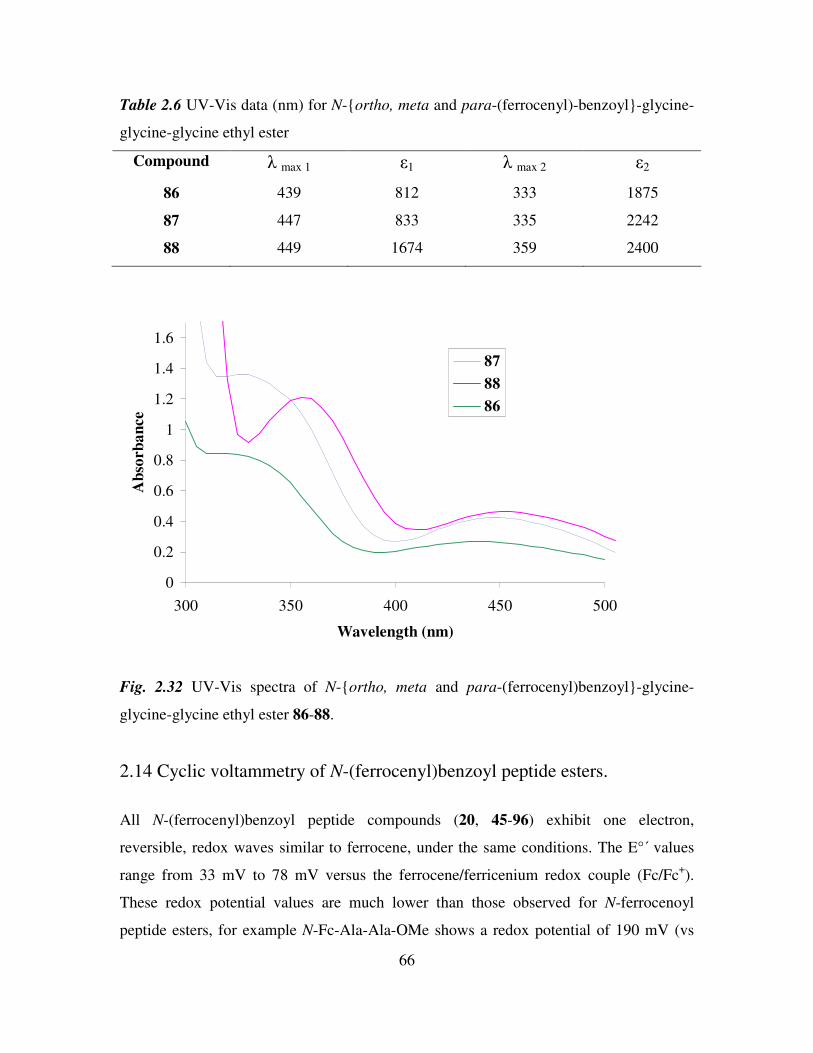
66
Table 2.6 UV-Vis data (nm) for N-ortho, meta and para-(ferrocenyl)-benzoyl-glycine-
glycine-glycine ethyl ester
Compound λ max 1 ε1 λ max 2 ε2
86 439 812 333 1875
87 447 833 335 2242
88 449 1674 359 2400
0
0.2
0.4
0.6
0.8
1
1.2
1.4
1.6
300 350 400 450 500
Wavelength (nm)
Ab
sorb
an
ce
87
88
86
Fig. 2.32 UV-Vis spectra of N-ortho, meta and para-(ferrocenyl)benzoyl-glycine-
glycine-glycine ethyl ester 86-88.
2.14 Cyclic voltammetry of N-(ferrocenyl)benzoyl peptide esters.
All N-(ferrocenyl)benzoyl peptide compounds (20, 45-96) exhibit one electron,
reversible, redox waves similar to ferrocene, under the same conditions. The E°΄ values
range from 33 mV to 78 mV versus the ferrocene/ferricenium redox couple (Fc/Fc+).
These redox potential values are much lower than those observed for N-ferrocenoyl
peptide esters, for example N-Fc-Ala-Ala-OMe shows a redox potential of 190 mV (vs
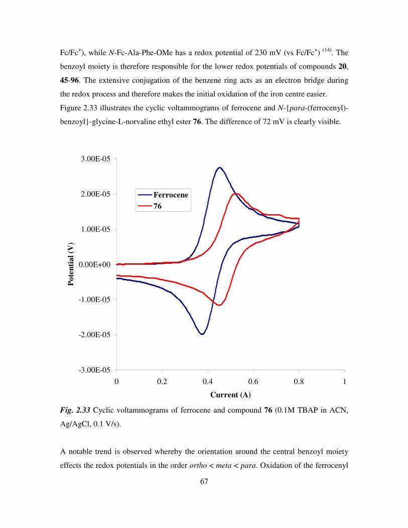
67
Fc/Fc+), while N-Fc-Ala-Phe-OMe has a redox potential of 230 mV (vs Fc/Fc
+)
(14). The
benzoyl moiety is therefore responsible for the lower redox potentials of compounds 20,
45-96. The extensive conjugation of the benzene ring acts as an electron bridge during
the redox process and therefore makes the initial oxidation of the iron centre easier.
Figure 2.33 illustrates the cyclic voltammograms of ferrocene and N-para-(ferrocenyl)-
benzoyl-glycine-L-norvaline ethyl ester 76. The difference of 72 mV is clearly visible.
-3.00E-05
-2.00E-05
-1.00E-05
0.00E+00
1.00E-05
2.00E-05
3.00E-05
0 0.2 0.4 0.6 0.8 1
Current (A)
Po
ten
tia
l (V
)
Ferrocene
76
Fig. 2.33 Cyclic voltammograms of ferrocene and compound 76 (0.1M TBAP in ACN,
Ag/AgCl, 0.1 V/s).
A notable trend is observed whereby the orientation around the central benzoyl moiety
effects the redox potentials in the order ortho < meta < para. Oxidation of the ferrocenyl
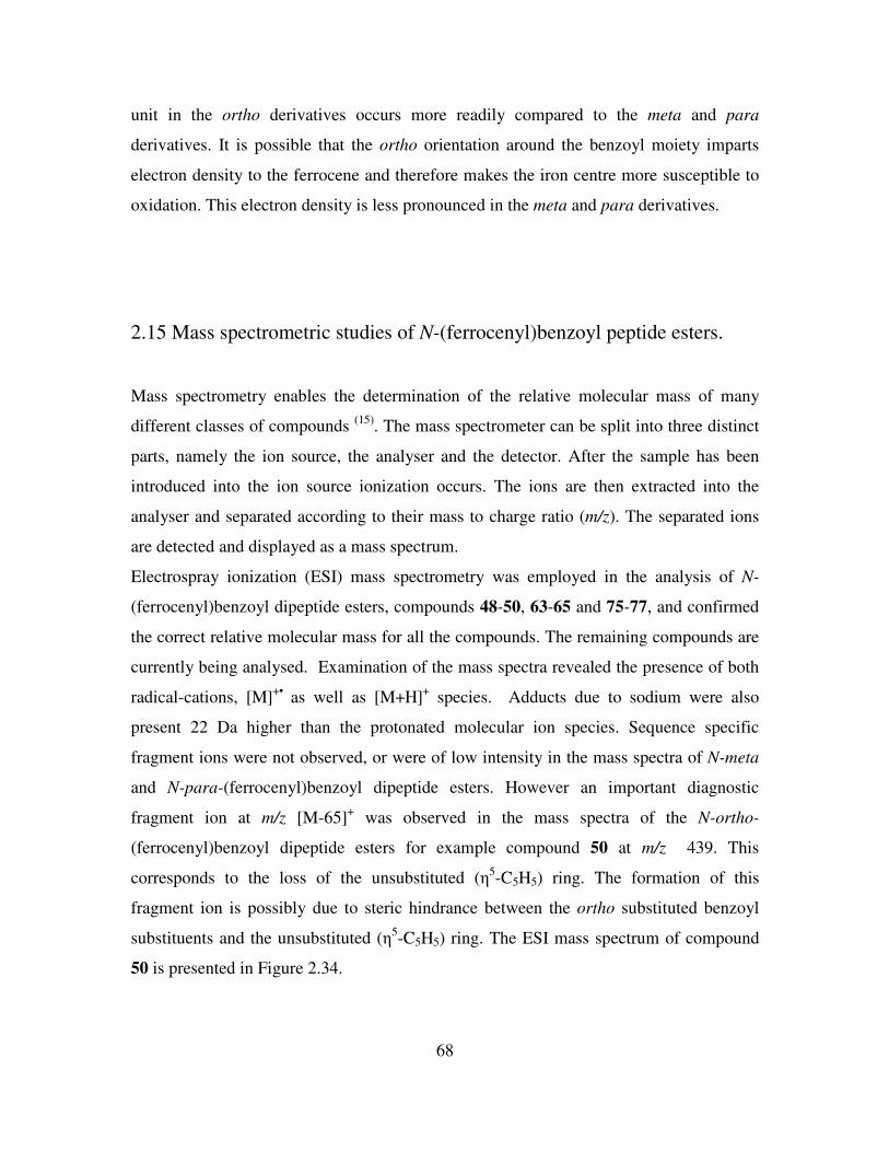
68
unit in the ortho derivatives occurs more readily compared to the meta and para
derivatives. It is possible that the ortho orientation around the benzoyl moiety imparts
electron density to the ferrocene and therefore makes the iron centre more susceptible to
oxidation. This electron density is less pronounced in the meta and para derivatives.
2.15 Mass spectrometric studies of N-(ferrocenyl)benzoyl peptide esters.
Mass spectrometry enables the determination of the relative molecular mass of many
different classes of compounds (15)
. The mass spectrometer can be split into three distinct
parts, namely the ion source, the analyser and the detector. After the sample has been
introduced into the ion source ionization occurs. The ions are then extracted into the
analyser and separated according to their mass to charge ratio (m/z). The separated ions
are detected and displayed as a mass spectrum.
Electrospray ionization (ESI) mass spectrometry was employed in the analysis of N-
(ferrocenyl)benzoyl dipeptide esters, compounds 48-50, 63-65 and 75-77, and confirmed
the correct relative molecular mass for all the compounds. The remaining compounds are
currently being analysed. Examination of the mass spectra revealed the presence of both
radical-cations, [M]+•
as well as [M+H]+ species. Adducts due to sodium were also
present 22 Da higher than the protonated molecular ion species. Sequence specific
fragment ions were not observed, or were of low intensity in the mass spectra of N-meta
and N-para-(ferrocenyl)benzoyl dipeptide esters. However an important diagnostic
fragment ion at m/z [M-65]+ was observed in the mass spectra of the N-ortho-
(ferrocenyl)benzoyl dipeptide esters for example compound 50 at m/z 439. This
corresponds to the loss of the unsubstituted (η5-C5H5) ring. The formation of this
fragment ion is possibly due to steric hindrance between the ortho substituted benzoyl
substituents and the unsubstituted (η5-C5H5) ring. The ESI mass spectrum of compound
50 is presented in Figure 2.34.
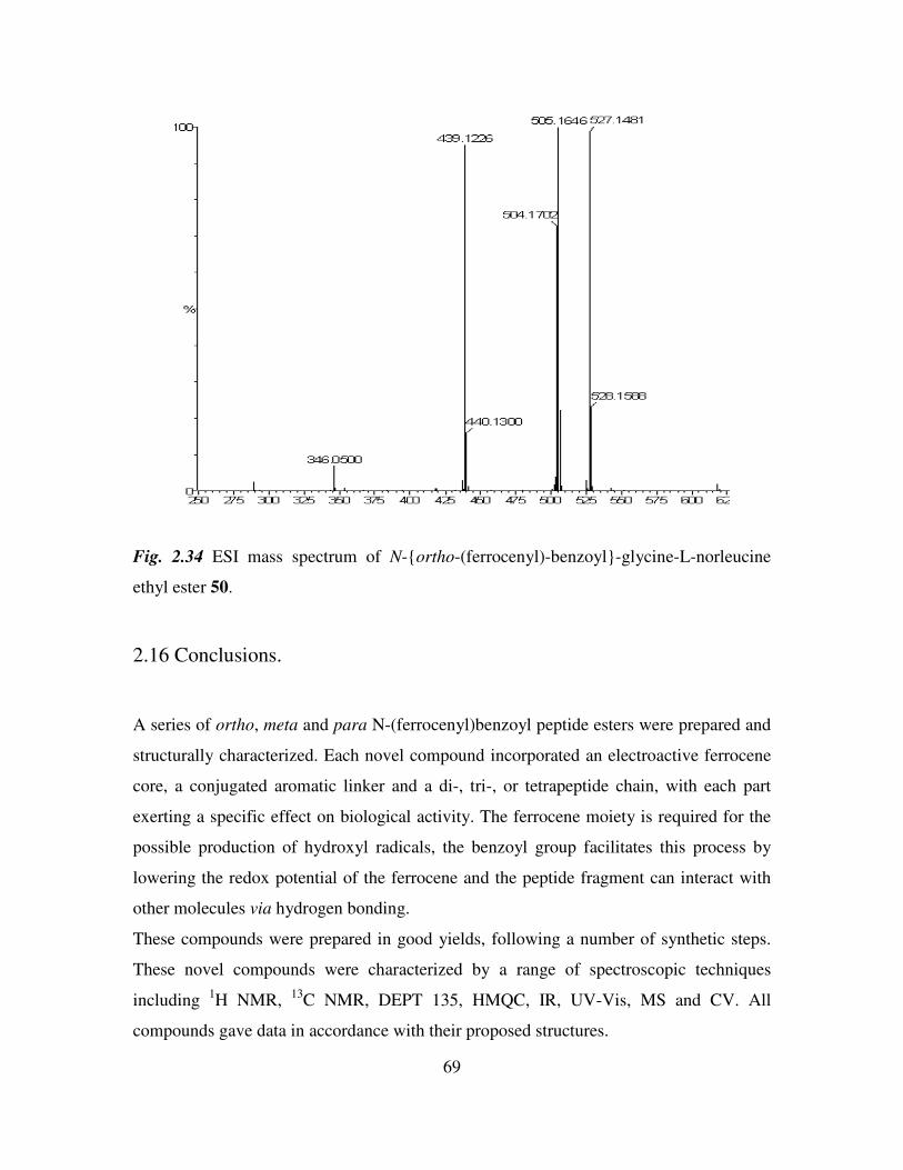
69
Fig. 2.34 ESI mass spectrum of N-ortho-(ferrocenyl)-benzoyl-glycine-L-norleucine
ethyl ester 50.
2.16 Conclusions.
A series of ortho, meta and para N-(ferrocenyl)benzoyl peptide esters were prepared and
structurally characterized. Each novel compound incorporated an electroactive ferrocene
core, a conjugated aromatic linker and a di-, tri-, or tetrapeptide chain, with each part
exerting a specific effect on biological activity. The ferrocene moiety is required for the
possible production of hydroxyl radicals, the benzoyl group facilitates this process by
lowering the redox potential of the ferrocene and the peptide fragment can interact with
other molecules via hydrogen bonding.
These compounds were prepared in good yields, following a number of synthetic steps.
These novel compounds were characterized by a range of spectroscopic techniques
including 1H NMR,
13C NMR, DEPT 135, HMQC, IR, UV-Vis, MS and CV. All
compounds gave data in accordance with their proposed structures.
70
References
1. Togni, A., Hayashi, T., Ferrocenes, 1994, Wiley, VCH publications.
2. Savage, D., Gallagher, J. F., Ida, Y., Kenny, P. T. M., Inorg. Chem. Comm., 2002,
5, 1034.
3. Savage, D. M., ‘ The Synthesis and Characterization of Novel N-Ferrocenyl
Benzoyl Amino Acid and Dipeptide Derivatives’, DCU, Ph.D, Thesis, 2003.
4. Hanson, J. R., ‘Organic Synthetic Methods’, 2002, RSC.
5. White, A., J. Biochem., 1959, 71, 217.
6. Marder, O., Alberico, F., Chemistry Today Peptides, 2003, 35.
7. Jones, J., ‘Amino Acid and Peptide Synthesis’, Oxford University Press, 1992.
8. Rosenblum, M., Glenn-Howells, W., Banerjee, A. K., Bennett, C., J. Am. Chem.
Soc., 1962, 84, 2726.
9. Breitmaier, E., ‘Structure Elucidation by NMR in Organic Chemistry’, Wiley,
1993.
10. Williams, D. H., Fleming, I., ‘Spectroscopic Methods in Organic Chemistry’,
McGraw-Hill, 1995 5th
edition.
11. Matos, K., Soderquist, J. A., J. Org. Chem., 1998, 63, 461.
12. Knapp, R., Rehahn, M., J. Organomet. Chem., 1993, 452, 235.
13. McMurry, J., ‘Organic Chemistry’, Brooks/Cole, 2000 5th
edition.
14. van Staveren, D. R., Weyhermuller, T., Metzler Nolte, N., Dalton Trans., 2003,
210.
15. Silverstein, R. M., Webster, F. X., Kiemle, D. J., ‘Spectrometric Identification of
Organic Compounds’, John Wiley and Sons, 2005, 7th
edition.
71
Chapter 3
In vitro anti-cancer activity of N-(ferrocenyl)benzoyl peptide esters
3.1 Introduction
The term cancer is used to describe diseases characterized by uncontrolled, abnormal cell
division and that are capable of spreading to other parts of the body through the blood
and lymph systems. It may be found in many forms including solid tissue formations and
leukaemias. Cancers result from a reduction or loss of control of the growth of cells (1)
. In
the early stages the cells formed resemble the parent cells but as the cancer advances they
lose the appearance and function of the parent cell. If this loss of function is not halted
the consequences will be life threatening. Both external (chemicals and radiation) and
internal (hormones and inherited genes) forces acting alone or in tandem may be
responsible for the initiation and propagation of cancer. In addition, many years can pass
between cause and detection and in some types of cancer the early detection of malignant
growths is a major problem (2)
.
Lung cancer is the most prevalent cancer worldwide, with 1.5 million cases diagnosed in
2003. This is greater than the diagnoses of breast, colon and prostrate cancer combined.
The five year survival rate in the United States is 16.8% while in Great Britain the Figure
is half that (3)
. Lung cancer is the leading cause of cancer mortality in Ireland. It is
responsible for approximately 20% of all cancer deaths (4)
. Smoking is responsible for
about 87% of cases, with chemical exposure also a contributory factor. Despite being one
of the easier diseases to prevent it remains one of the most difficult to treat. This is
primarily due to the late presentation of symptoms, the high level of metastasis and the
development of resistance to chemotherapeutic agents. Radiation and chemotherapy
treatments are crucial in lung cancer treatment as surgery is only effective in the early
stages of lung cancer. 75-85% of all cases of lung cancer are non–small cell lung cancers
(NSCLC). The chemotherapy drugs used are dependant on whether the cancer is
classified as small cell lung cancer (SCLC) or NSCLC. The primary treatments for SCLC
are cisplatin, carboplatin, cyclophosphamide or a combination of cisplatin and etoposide.
Current therapy for NSCLC is surgery and adjuvant chemotherapy for early-stage disease
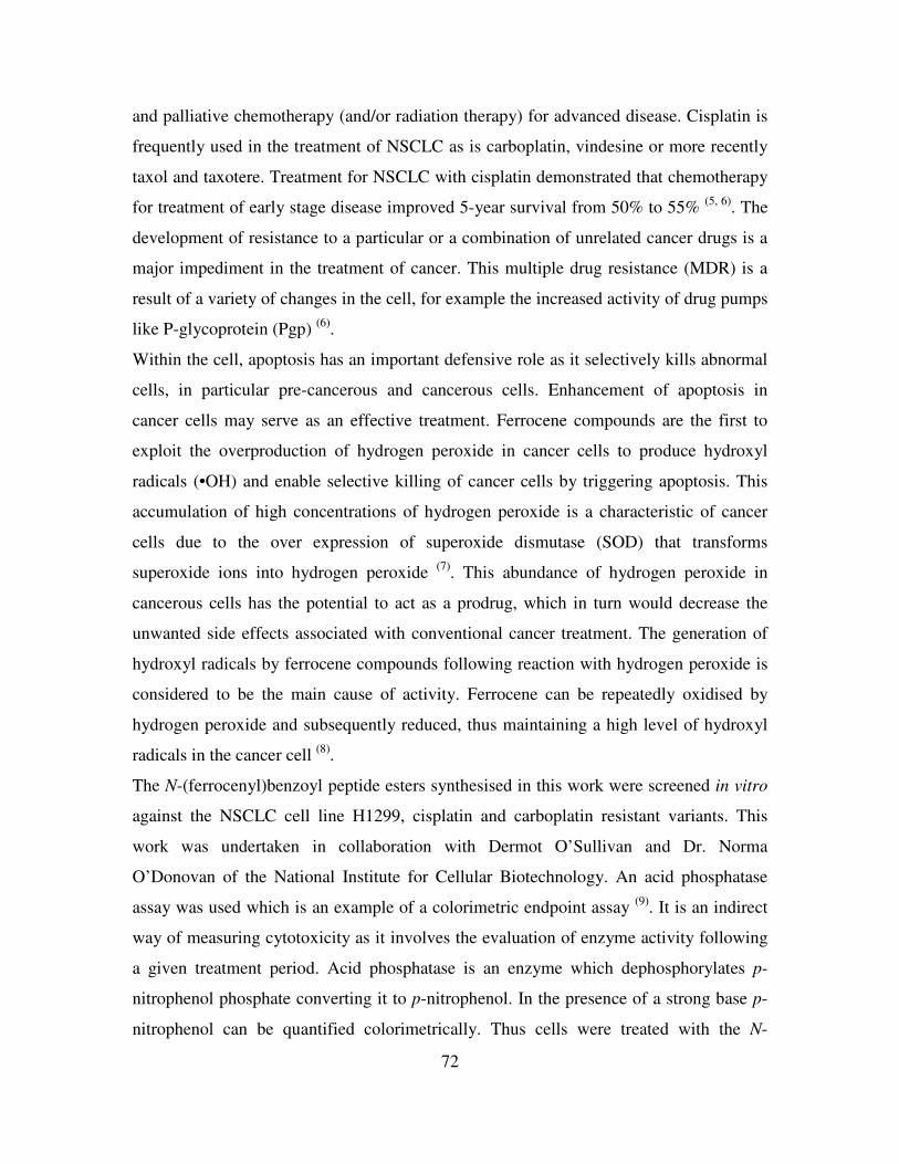
72
and palliative chemotherapy (and/or radiation therapy) for advanced disease. Cisplatin is
frequently used in the treatment of NSCLC as is carboplatin, vindesine or more recently
taxol and taxotere. Treatment for NSCLC with cisplatin demonstrated that chemotherapy
for treatment of early stage disease improved 5-year survival from 50% to 55% (5, 6)
. The
development of resistance to a particular or a combination of unrelated cancer drugs is a
major impediment in the treatment of cancer. This multiple drug resistance (MDR) is a
result of a variety of changes in the cell, for example the increased activity of drug pumps
like P-glycoprotein (Pgp) (6)
.
Within the cell, apoptosis has an important defensive role as it selectively kills abnormal
cells, in particular pre-cancerous and cancerous cells. Enhancement of apoptosis in
cancer cells may serve as an effective treatment. Ferrocene compounds are the first to
exploit the overproduction of hydrogen peroxide in cancer cells to produce hydroxyl
radicals (•OH) and enable selective killing of cancer cells by triggering apoptosis. This
accumulation of high concentrations of hydrogen peroxide is a characteristic of cancer
cells due to the over expression of superoxide dismutase (SOD) that transforms
superoxide ions into hydrogen peroxide (7)
. This abundance of hydrogen peroxide in
cancerous cells has the potential to act as a prodrug, which in turn would decrease the
unwanted side effects associated with conventional cancer treatment. The generation of
hydroxyl radicals by ferrocene compounds following reaction with hydrogen peroxide is
considered to be the main cause of activity. Ferrocene can be repeatedly oxidised by
hydrogen peroxide and subsequently reduced, thus maintaining a high level of hydroxyl
radicals in the cancer cell (8)
.
The N-(ferrocenyl)benzoyl peptide esters synthesised in this work were screened in vitro
against the NSCLC cell line H1299, cisplatin and carboplatin resistant variants. This
work was undertaken in collaboration with Dermot O’Sullivan and Dr. Norma
O’Donovan of the National Institute for Cellular Biotechnology. An acid phosphatase
assay was used which is an example of a colorimetric endpoint assay (9)
. It is an indirect
way of measuring cytotoxicity as it involves the evaluation of enzyme activity following
a given treatment period. Acid phosphatase is an enzyme which dephosphorylates p-
nitrophenol phosphate converting it to p-nitrophenol. In the presence of a strong base p-
nitrophenol can be quantified colorimetrically. Thus cells were treated with the N-
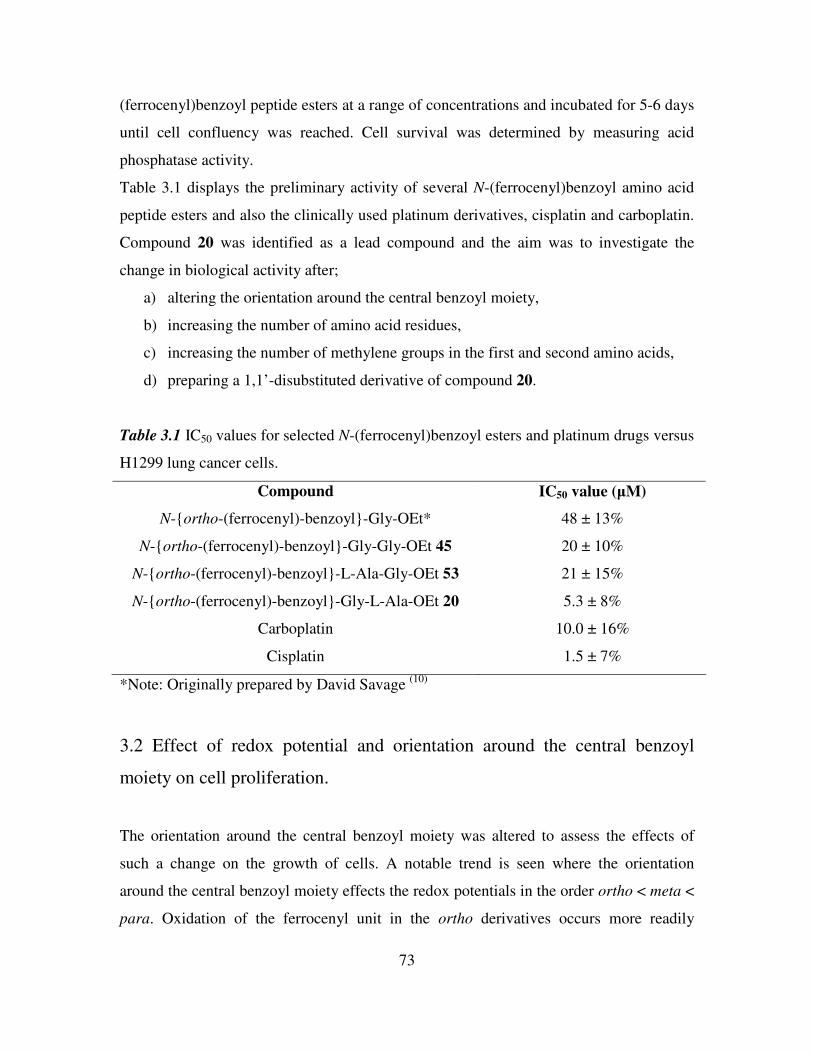
73
(ferrocenyl)benzoyl peptide esters at a range of concentrations and incubated for 5-6 days
until cell confluency was reached. Cell survival was determined by measuring acid
phosphatase activity.
Table 3.1 displays the preliminary activity of several N-(ferrocenyl)benzoyl amino acid
peptide esters and also the clinically used platinum derivatives, cisplatin and carboplatin.
Compound 20 was identified as a lead compound and the aim was to investigate the
change in biological activity after;
a) altering the orientation around the central benzoyl moiety,
b) increasing the number of amino acid residues,
c) increasing the number of methylene groups in the first and second amino acids,
d) preparing a 1,1’-disubstituted derivative of compound 20.
Table 3.1 IC50 values for selected N-(ferrocenyl)benzoyl esters and platinum drugs versus
H1299 lung cancer cells.
Compound IC50 value (µM)
N-ortho-(ferrocenyl)-benzoyl-Gly-OEt* 48 ± 13%
N-ortho-(ferrocenyl)-benzoyl-Gly-Gly-OEt 45 20 ± 10%
N-ortho-(ferrocenyl)-benzoyl-L-Ala-Gly-OEt 53 21 ± 15%
N-ortho-(ferrocenyl)-benzoyl-Gly-L-Ala-OEt 20 5.3 ± 8%
Carboplatin 10.0 ± 16%
Cisplatin 1.5 ± 7%
*Note: Originally prepared by David Savage (10)
3.2 Effect of redox potential and orientation around the central benzoyl
moiety on cell proliferation.
The orientation around the central benzoyl moiety was altered to assess the effects of
such a change on the growth of cells. A notable trend is seen where the orientation
around the central benzoyl moiety effects the redox potentials in the order ortho < meta <
para. Oxidation of the ferrocenyl unit in the ortho derivatives occurs more readily
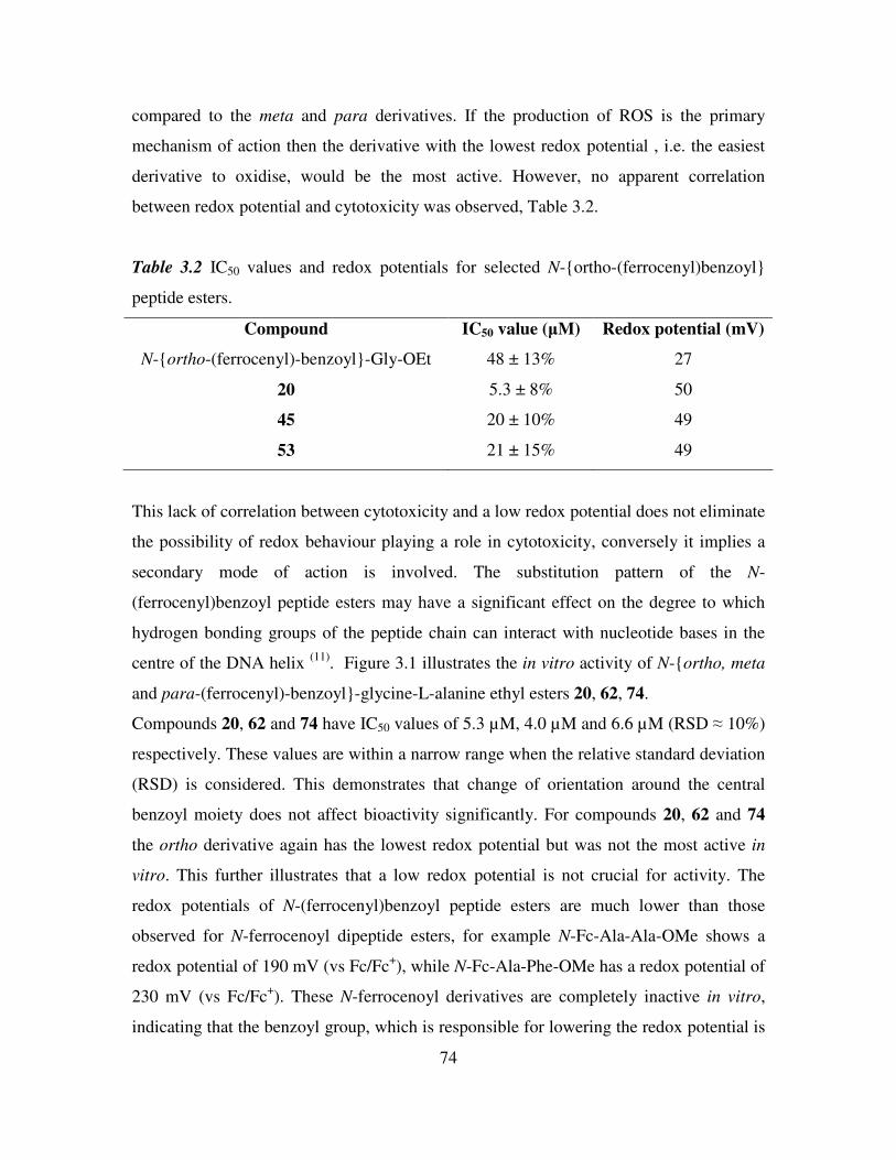
74
compared to the meta and para derivatives. If the production of ROS is the primary
mechanism of action then the derivative with the lowest redox potential , i.e. the easiest
derivative to oxidise, would be the most active. However, no apparent correlation
between redox potential and cytotoxicity was observed, Table 3.2.
Table 3.2 IC50 values and redox potentials for selected N-ortho-(ferrocenyl)benzoyl
peptide esters.
Compound IC50 value (µM) Redox potential (mV)
N-ortho-(ferrocenyl)-benzoyl-Gly-OEt 48 ± 13% 27
20 5.3 ± 8% 50
45 20 ± 10% 49
53 21 ± 15% 49
This lack of correlation between cytotoxicity and a low redox potential does not eliminate
the possibility of redox behaviour playing a role in cytotoxicity, conversely it implies a
secondary mode of action is involved. The substitution pattern of the N-
(ferrocenyl)benzoyl peptide esters may have a significant effect on the degree to which
hydrogen bonding groups of the peptide chain can interact with nucleotide bases in the
centre of the DNA helix (11)
. Figure 3.1 illustrates the in vitro activity of N-ortho, meta
and para-(ferrocenyl)-benzoyl-glycine-L-alanine ethyl esters 20, 62, 74.
Compounds 20, 62 and 74 have IC50 values of 5.3 µM, 4.0 µM and 6.6 µM (RSD ≈ 10%)
respectively. These values are within a narrow range when the relative standard deviation
(RSD) is considered. This demonstrates that change of orientation around the central
benzoyl moiety does not affect bioactivity significantly. For compounds 20, 62 and 74
the ortho derivative again has the lowest redox potential but was not the most active in
vitro. This further illustrates that a low redox potential is not crucial for activity. The
redox potentials of N-(ferrocenyl)benzoyl peptide esters are much lower than those
observed for N-ferrocenoyl dipeptide esters, for example N-Fc-Ala-Ala-OMe shows a
redox potential of 190 mV (vs Fc/Fc+), while N-Fc-Ala-Phe-OMe has a redox potential of
230 mV (vs Fc/Fc+). These N-ferrocenoyl derivatives are completely inactive in vitro,
indicating that the benzoyl group, which is responsible for lowering the redox potential is
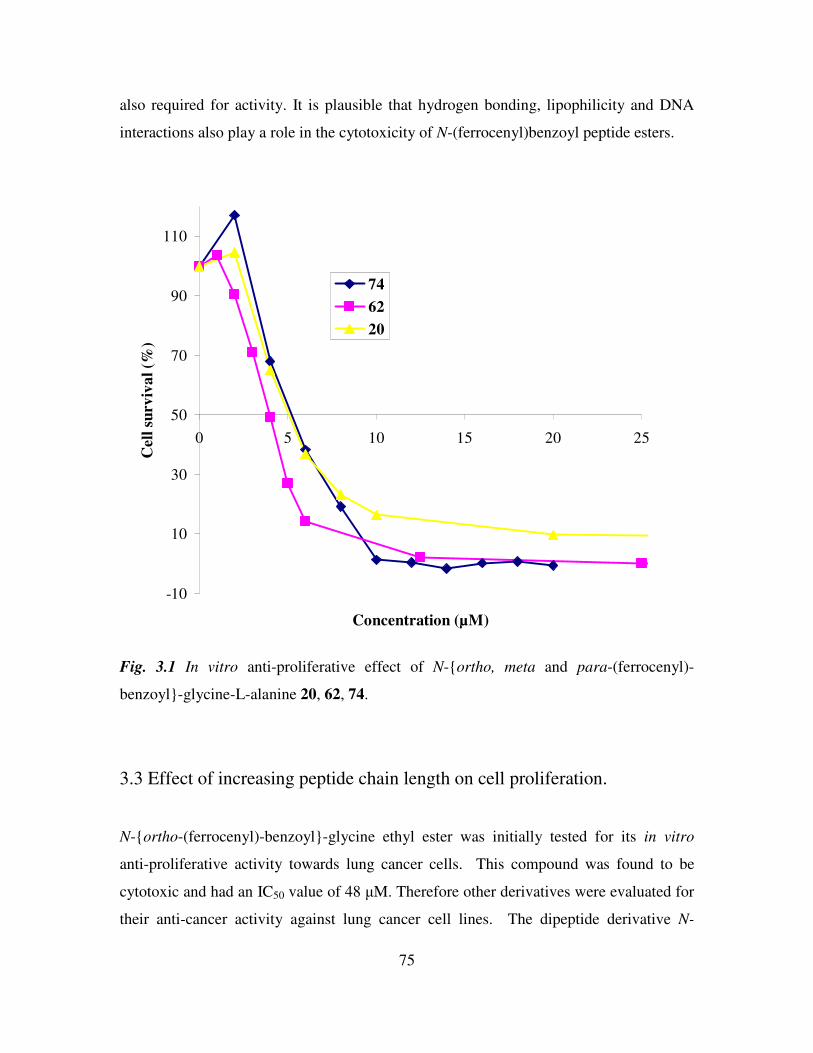
75
also required for activity. It is plausible that hydrogen bonding, lipophilicity and DNA
interactions also play a role in the cytotoxicity of N-(ferrocenyl)benzoyl peptide esters.
-10
10
30
50
70
90
110
0 5 10 15 20 25
Concentration (µM)
Cell
su
rv
iva
l (%
)
74
62
20
Fig. 3.1 In vitro anti-proliferative effect of N-ortho, meta and para-(ferrocenyl)-
benzoyl-glycine-L-alanine 20, 62, 74.
3.3 Effect of increasing peptide chain length on cell proliferation.
N-ortho-(ferrocenyl)-benzoyl-glycine ethyl ester was initially tested for its in vitro
anti-proliferative activity towards lung cancer cells. This compound was found to be
cytotoxic and had an IC50 value of 48 µM. Therefore other derivatives were evaluated for
their anti-cancer activity against lung cancer cell lines. The dipeptide derivative N-
76
ortho-(ferrocenyl)-benzoyl-glycine-glycine ethyl ester was shown to have an IC50
value of 20 µM (RSD 10%). As the dipeptide derivative was more active than the amino
acid derivative, a logical extension of this study was the preparation of longer peptide
chains. Therefore the peptide moiety was extended by the addition of additional glycine
residues.
A plot of cell survival versus compound concentration for the compounds N-ortho-
(ferrocenyl)-benzoyl-glycine ethyl ester, 45, 86 and 89 is presented in Figure 3.2.
Compound 48 had an IC50 value of 20 µM while compound 86 and 89 both had IC50
values greater than 50 µM. Thus increasing the length of the peptide chain from dipeptide
to tri- and tetrapeptide has a negative impact on the anti-proliferative effect of the
ferrocenyl derivatives. Compound 86 had an IC50 value of 63 µM (RSD 8%), whereas
compound 89 did not register an IC50 value in the concentration range used. The
increasing peptide chain length may make the compound too polar and prevent it from
permeating the lipophilic cell membrane. It can therefore be concluded that a dipeptide
chain is required for optimum activity, Figure 3.2. Derivatives 86 and 89 have very
similar redox potentials compared to N-(ferrocenyl)benzoyl dipeptide esters which are
highly active in vitro, this confirms that the peptide chain imparts a secondary mechanism
of action to these compounds.
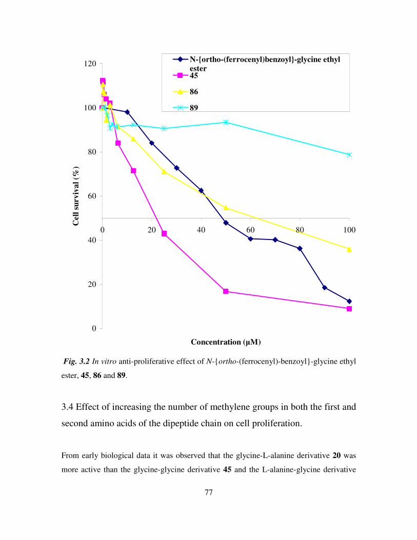
77
0
20
40
60
80
100
120
0 20 40 60 80 100
Concentration (µM)
Cell
su
rv
iva
l (%
)
N-ortho-(ferrocenyl)benzoyl-glycine ethylester45
86
89
Fig. 3.2 In vitro anti-proliferative effect of N-ortho-(ferrocenyl)-benzoyl-glycine ethyl
ester, 45, 86 and 89.
3.4 Effect of increasing the number of methylene groups in both the first and
second amino acids of the dipeptide chain on cell proliferation.
From early biological data it was observed that the glycine-L-alanine derivative 20 was
more active than the glycine-glycine derivative 45 and the L-alanine-glycine derivative
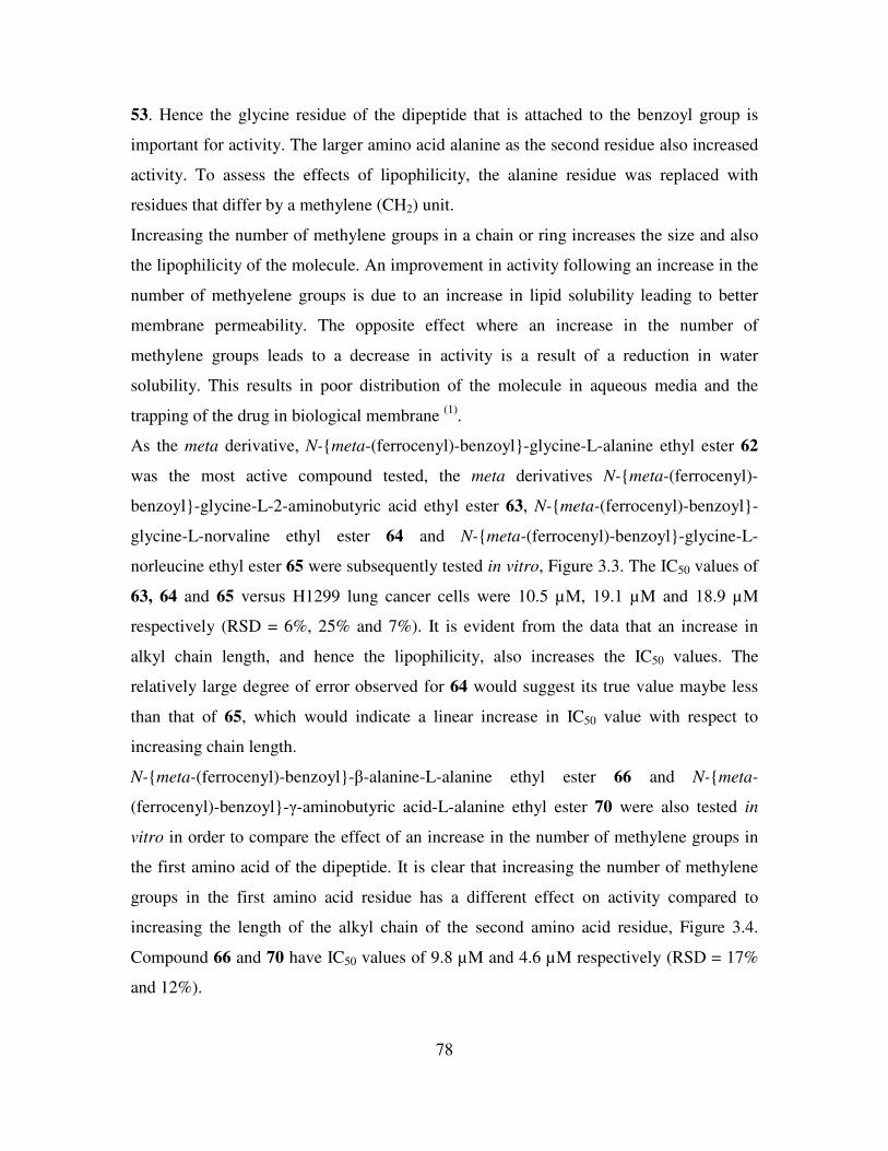
78
53. Hence the glycine residue of the dipeptide that is attached to the benzoyl group is
important for activity. The larger amino acid alanine as the second residue also increased
activity. To assess the effects of lipophilicity, the alanine residue was replaced with
residues that differ by a methylene (CH2) unit.
Increasing the number of methylene groups in a chain or ring increases the size and also
the lipophilicity of the molecule. An improvement in activity following an increase in the
number of methyelene groups is due to an increase in lipid solubility leading to better
membrane permeability. The opposite effect where an increase in the number of
methylene groups leads to a decrease in activity is a result of a reduction in water
solubility. This results in poor distribution of the molecule in aqueous media and the
trapping of the drug in biological membrane (1)
.
As the meta derivative, N-meta-(ferrocenyl)-benzoyl-glycine-L-alanine ethyl ester 62
was the most active compound tested, the meta derivatives N-meta-(ferrocenyl)-
benzoyl-glycine-L-2-aminobutyric acid ethyl ester 63, N-meta-(ferrocenyl)-benzoyl-
glycine-L-norvaline ethyl ester 64 and N-meta-(ferrocenyl)-benzoyl-glycine-L-
norleucine ethyl ester 65 were subsequently tested in vitro, Figure 3.3. The IC50 values of
63, 64 and 65 versus H1299 lung cancer cells were 10.5 µM, 19.1 µM and 18.9 µM
respectively (RSD = 6%, 25% and 7%). It is evident from the data that an increase in
alkyl chain length, and hence the lipophilicity, also increases the IC50 values. The
relatively large degree of error observed for 64 would suggest its true value maybe less
than that of 65, which would indicate a linear increase in IC50 value with respect to
increasing chain length.
N-meta-(ferrocenyl)-benzoyl-β-alanine-L-alanine ethyl ester 66 and N-meta-
(ferrocenyl)-benzoyl-γ-aminobutyric acid-L-alanine ethyl ester 70 were also tested in
vitro in order to compare the effect of an increase in the number of methylene groups in
the first amino acid of the dipeptide. It is clear that increasing the number of methylene
groups in the first amino acid residue has a different effect on activity compared to
increasing the length of the alkyl chain of the second amino acid residue, Figure 3.4.
Compound 66 and 70 have IC50 values of 9.8 µM and 4.6 µM respectively (RSD = 17%
and 12%).
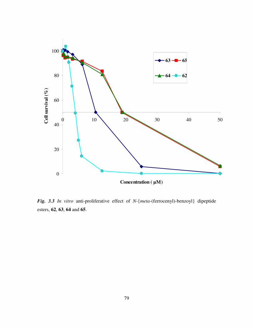
79
0
20
40
60
80
100
0 10 20 30 40 50
Concentration ( µM)
Cel
l su
rviv
al (%
)
63 65
64 62
Fig. 3.3 In vitro anti-proliferative effect of N-meta-(ferrocenyl)-benzoyl dipeptide
esters, 62, 63, 64 and 65.
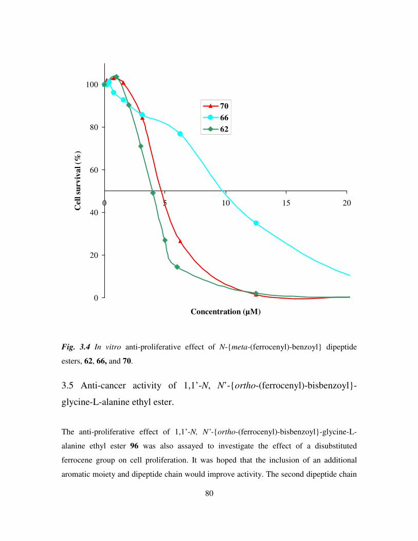
80
0
20
40
60
80
100
0 5 10 15 20
Concentration (µM)
Cell
su
rv
iva
l (%
)
70
66
62
Fig. 3.4 In vitro anti-proliferative effect of N-meta-(ferrocenyl)-benzoyl dipeptide
esters, 62, 66, and 70.
3.5 Anti-cancer activity of 1,1’-N, N’-ortho-(ferrocenyl)-bisbenzoyl-
glycine-L-alanine ethyl ester.
The anti-proliferative effect of 1,1’-N, N’-ortho-(ferrocenyl)-bisbenzoyl-glycine-L-
alanine ethyl ester 96 was also assayed to investigate the effect of a disubstituted
ferrocene group on cell proliferation. It was hoped that the inclusion of an additional
aromatic moiety and dipeptide chain would improve activity. The second dipeptide chain
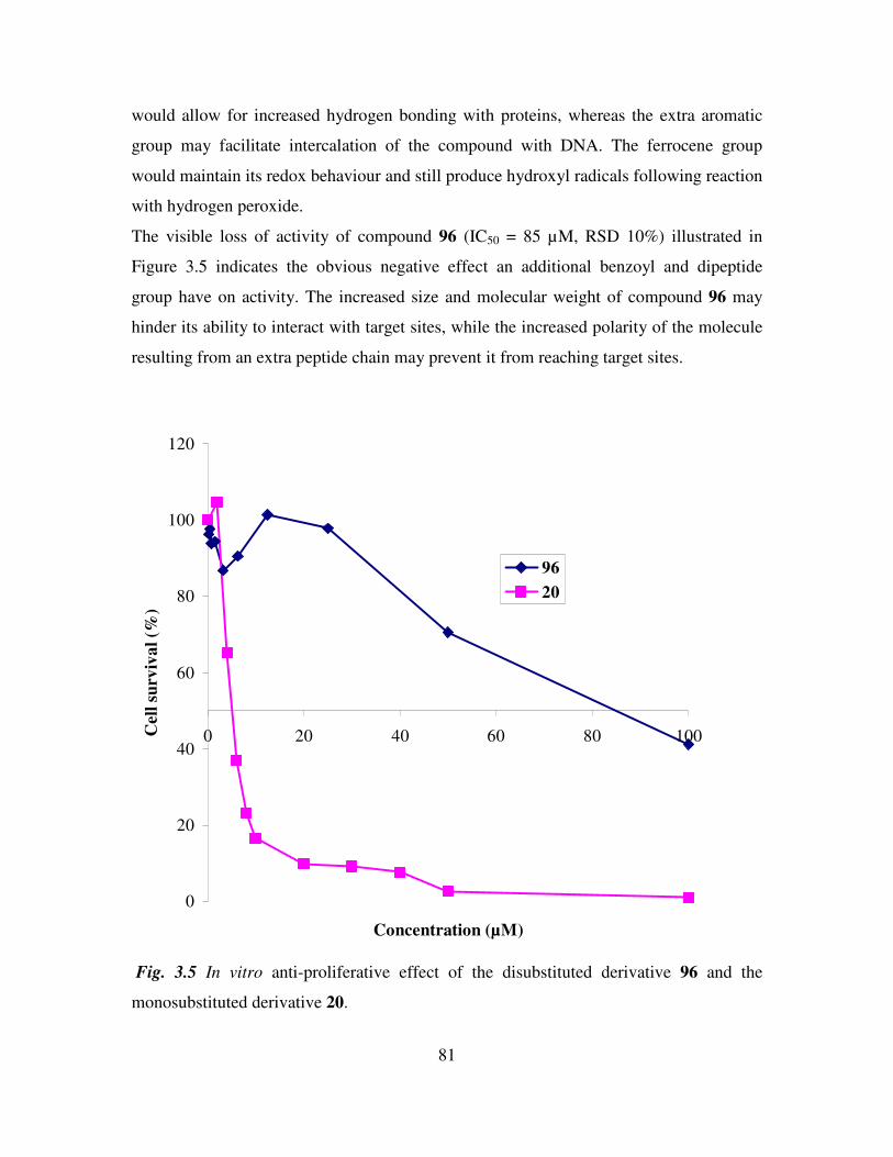
81
would allow for increased hydrogen bonding with proteins, whereas the extra aromatic
group may facilitate intercalation of the compound with DNA. The ferrocene group
would maintain its redox behaviour and still produce hydroxyl radicals following reaction
with hydrogen peroxide.
The visible loss of activity of compound 96 (IC50 = 85 µM, RSD 10%) illustrated in
Figure 3.5 indicates the obvious negative effect an additional benzoyl and dipeptide
group have on activity. The increased size and molecular weight of compound 96 may
hinder its ability to interact with target sites, while the increased polarity of the molecule
resulting from an extra peptide chain may prevent it from reaching target sites.
0
20
40
60
80
100
120
0 20 40 60 80 100
Concentration (µM)
Cell
su
rviv
al
(%)
96
20
Fig. 3.5 In vitro anti-proliferative effect of the disubstituted derivative 96 and the
monosubstituted derivative 20.
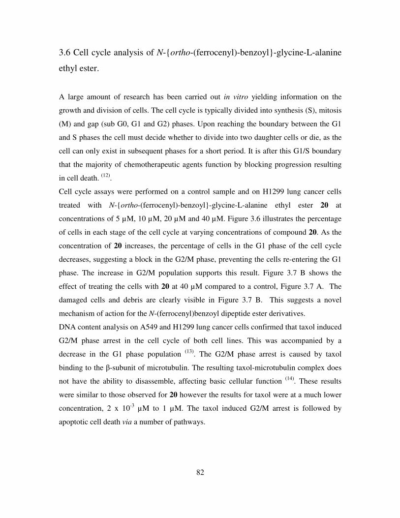
82
3.6 Cell cycle analysis of N-ortho-(ferrocenyl)-benzoyl-glycine-L-alanine
ethyl ester.
A large amount of research has been carried out in vitro yielding information on the
growth and division of cells. The cell cycle is typically divided into synthesis (S), mitosis
(M) and gap (sub G0, G1 and G2) phases. Upon reaching the boundary between the G1
and S phases the cell must decide whether to divide into two daughter cells or die, as the
cell can only exist in subsequent phases for a short period. It is after this G1/S boundary
that the majority of chemotherapeutic agents function by blocking progression resulting
in cell death. (12)
.
Cell cycle assays were performed on a control sample and on H1299 lung cancer cells
treated with N-ortho-(ferrocenyl)-benzoyl-glycine-L-alanine ethyl ester 20 at
concentrations of 5 µM, 10 µM, 20 µM and 40 µM. Figure 3.6 illustrates the percentage
of cells in each stage of the cell cycle at varying concentrations of compound 20. As the
concentration of 20 increases, the percentage of cells in the G1 phase of the cell cycle
decreases, suggesting a block in the G2/M phase, preventing the cells re-entering the G1
phase. The increase in G2/M population supports this result. Figure 3.7 B shows the
effect of treating the cells with 20 at 40 µM compared to a control, Figure 3.7 A. The
damaged cells and debris are clearly visible in Figure 3.7 B. This suggests a novel
mechanism of action for the N-(ferrocenyl)benzoyl dipeptide ester derivatives.
DNA content analysis on A549 and H1299 lung cancer cells confirmed that taxol induced
G2/M phase arrest in the cell cycle of both cell lines. This was accompanied by a
decrease in the G1 phase population (13)
. The G2/M phase arrest is caused by taxol
binding to the β-subunit of microtubulin. The resulting taxol-microtubulin complex does
not have the ability to disassemble, affecting basic cellular function (14)
. These results
were similar to those observed for 20 however the results for taxol were at a much lower
concentration, 2 x 10⁻³
µM to 1 µM. The taxol induced G2/M arrest is followed by
apoptotic cell death via a number of pathways.
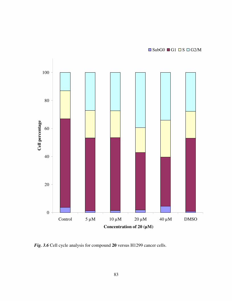
83
0
20
40
60
80
100
Control 5 µM 10 µM 20 µM 40 µM DMSO
Concentration of 20 (µM)
Cell
percen
tag
e
SubG0 G1 S G2/M
Fig. 3.6 Cell cycle analysis for compound 20 versus H1299 cancer cells.
84
(A) (B)
Fig. 3.7 Cell cycle analysis of compound 20 at 40 µM (B) and a control sample (A)
versus H1299 lung cancer cells.
3.7 Conclusions.
In summary, various N-(ferrocenyl)benzoyl peptide esters were tested in vitro to assess
their anti-proliferative effect on the non-small cell lung cancer cell line H1299.
Analogues of the lead compound N-ortho-(ferrocenyl)-benzoyl-glycine-L-alanine ethyl
ester 20 were prepared with the aim of improving its biological activity.
The derivatives that were tested had various substitution patterns around the central
benzoyl group. The derivatives also had varying degrees of lipophilicity and
hydrophilicity with respect to the lead compound 20. The substitution pattern was found
not to be critical for activity with the meta and para derivatives of compound 20 showing
comparable activity. N-meta-(ferrocenyl)-benzoyl-glycine-L-alanine ethyl ester 62 was
the most active compound in the entire study with an IC50 value of 4.0 µM (± 10%).
Biological activity decreased when longer peptide chains were added to the ferrocenyl
benzoyl group. When additional methylene groups were added to the side chain of the
second amino acid residue a loss in activity was observed. This result was not repeated
when extra methylene groups were appended to the first amino acid residue of the
dipeptide derivatives. The γ-aminobutyric acid derivative displayed comparable activity
to the compounds with glycine as the first amino acid. The biological activity of 1,1’-N,
Damaged
cells/debris
85
N’-ortho-(ferrocenyl)-bisbenzoyl-glycine-L-alanine ethyl ester 96 was also assayed to
investigate the effect of a disubstituted ferrocene group on cell proliferation. A significant
decrease in activity was observed for this compound. N-ortho-(ferrocenyl)-benzoyl-
glycine-L-alanine ethyl ester 20 was shown to cause arrest in the G2/M phase of the cell
cycle. This represents a possible mode of action for N-(ferrocenyl)benzoyl dipeptide
esters.
86
References
1. Thomas, G., ‘Medicinal Chemistry, An Introduction’, John Wiley and Sons, 2000.
2. Nicolaou, K. C., Montagnon, T., ‘Molecules that changed the world’, Weinheim,
2008.
3. Khuri, F. R., Cohen, V., Clin. Can. Res., 2004, 10, 4249.
4. http://www.who.int/research/en/.
5. Non-small cell lung cancer collaborative group, Chemotherapy for non-small cell
lung cancer, Cochrane Database of Systematic Reviews, 2000, 2, CD002131.
6. Breen, L., Murphy, L., Keenan, J., Clynes, M., Toxicology in vitro, 2008, 22,
1234.
7. Kinnula, V. L., Crapo, J. D., Free Rad. Bio. Med., 2004, 36, 718.
8. Wlassoff, W. A., Albright, C. D., Sivashinski, S., Ivanova, A., Appelbaum, J. G.,
Salganik, R. I., J. Pharma. Pharmacol., 2007, 59, 1549.
9. Kelly, P. N., Pretre, A., Devoy, S., O’Reilly, J., Devery, R., Goel, A., Gallagher,
J. F., Lough, A. J., Kenny, P. T. M., J. Organomet. Chem., 2007, 692, 1327.
10. Savage, D. M., ‘The Synthesis and Characterization of Novel N-Ferrocenyl
Benzoyl Amino Acid and Dipeptide Derivatives’, DCU, Ph.D, Thesis, 2003.
11. Mooney, A., Corry, A. J., O’Sullivan, D., Rai, D. K., Kenny, P. T. M., J.
Organomet. Chem. 2008, doi:10.1016/j.organchem.2008.09.064.
12. Reddy, M. V. R., Mallireddigari, M. R., Cosenza, S. C., Palella, V. R., Iqbal, N.
M., Robell, K. A., Kang, A. D., Reddy, E. P., J. Med Chem., 2008, 51, 86.
13. Gas, G. C., Holiday, D., Gallardo, R., Haas, C., Cancer Letters, 2001, 165, 147.
14. Kumar, N., J. Biol. Chem., 1981, 256, 10435.
87
Experimental details
Experimental Note
All chemicals were purchased from Sigma-Aldrich and used as received. Commercial
grade reagents were used without further purification. Riedel de Haën silica gel was used
for flash and thin layer chromatography. Where necessary solvents were purified prior to
use, dichloromethane was distilled from calcium hydride and triethylamine was distilled
and stored over potassium hydroxide pellets. Melting points were determined using a
Griffin melting point apparatus and are uncorrected. Infrared spectra were recorded on a
Nicolet 405 FT-IR spectrometer and UV-Vis spectra on a Hewlett-Packard 8452A diode
array UV-Vis spectrophotometer. NMR spectra were obtained on a Bruker AC 400 NMR
spectrometer operating at 400 MHz for 1H NMR and 100 MHz for
13C NMR. The
1H and
13C NMR chemical shifts (ppm) are relative to TMS and all coupling constants (J) are in
Hertz. Electrospray ionization mass spectra were obtained on a Micromass LCT mass
spectrometer. Elemental Analysis was carried out by the Microanalytical Laboratory at
University College Dublin. Cyclic voltammograms were recorded in acetonitrile (Sigma-
Aldrich), with 0.1 M tetrabutylammonium perchlorate (TBAP) as a supporting
electrolyte, using a CH Instruments 600a electrochemical analyzer (Pico-Amp Booster
and Faraday Cage). The experiments were carried out at room temperature. A three-
electrode cell consisting of a glassy carbon working-electrode, a platinum wire counter-
electrode and an Ag/AgCl reference electrode was used. The Eº´ values obtained for the
test samples were referenced relative to the ferrocene/ferricenium redox couple.
88
General procedures for the synthesis of starting materials for N-(ferrocenyl)benzoyl
peptide esters.
ortho-Ferrocenyl ethyl benzoate 37.
Concentrated hydrochloric acid (4 mls) was added to a solution of ethyl-2-aminobenzoate
(1.90 g, 11.5 mmol) in 15 mls of deionized water. Sodium nitrite (0.90 g, 13 mmol) in 15
mls of deionized water was then added to this solution with stirring at a temperature of
less than 5 oC. The resulting pale yellow diazonium salt was added to ferrocene (2.14 g,
11.5 mmol) in 50 mls of diethyl ether and allowed to react for 12 hours. The reaction was
washed with water and the organic layer was dried over MgSO4. The solvent was
removed in vacuo. Column chromatography eluant 3:2 petroleum ether (40-60 oC)
:diethyl ether yielded the desired product as a red solid (1.69 g, 44%). m.p. 65-67 oC.
1H NMR (400 MHz) δ (d6-DMSO): 7.88 (1H, d, J = 8 Hz, ArH), 7.52 (1H, t, J = 6 Hz,
ArH), 7.41 (1H, d, J = 7.6 Hz, ArH), 7.32 (1H, t, J = 7.6 Hz, ArH), 4.47 2H, t, J = 1.6
Hz, ortho on (η5-C5H4), 4.33 2H, t, J = 1.6 Hz, meta on (η5
-C5H4), 4.17 (2H, q, J =
7.2 Hz, -OCH2CH3), 4.09 5H, s, (η5-C5H5), 1.14 (3H, t, J = 7.2 Hz, -OCH2CH3).
13C NMR (100 MHz) δ (d6-DMSO): 169.0, 137.4, 131.8, 131.0, 130.1, 127.8, 125.9,
85.1, 70.1, 69.5, 68.7, 60.7 (-ve DEPT), 13.8.
ortho-Ferrocenyl benzoic acid 38.
ortho-Ferrocenyl ethyl benzoate (1.0 g, 3 mmol) in 15 mls of methanol was added to 15
mls of a 10% sodium hydroxide solution and refluxed for 3 hours. The solution was
cooled in ice and concentrated HCl was added until pH 2 was reached. The product was
isolated by filtration as an orange solid (0.51 g, 56%). m.p. 124-126 °C.
1H NMR (400 MHz) δ (d6-DMSO: 13.0 (1H, s, -COOH), 7.82 (1H, d, J = 8 Hz, ArH),
7.44 (1H, t, J = 8 Hz, ArH), 7.37 (1H, d, J = 8 Hz, ArH), 7.27 (1H, t, J = 8 Hz, ArH),
4.55 2H, s, ortho on (η5-C5H4), 4.32 2H, s, meta on (η5
-C5H4), 4.08 5H, s, (η5-
C5H5).
13C NMR (100 MHz) δ (d6-DMSO): 171.2, 137.0, 133.3, 131.1, 129.8, 127.9, 126.1,
85.5, 69.9, 69.1, 68.5.
89
meta-Ferrocenyl ethyl benzoate 39.
Concentrated hydrochloric acid (4 mls) was added to a solution of ethyl-3-aminobenzoate
(1.90 g, 11.5 mmol) in 15 mls of deionized water. Sodium nitrite (0.90 g, 13 mmol) in 15
mls of deionized water was then added to this solution with stirring at a temperature of
less than 5 oC. The resulting pale yellow diazonium salt was added to ferrocene (2.14 g,
11.5 mmol) in 50 mls of diethyl ether and allowed to react for 12 hours. The reaction was
washed with water and the organic layer was dried over MgSO4. The solvent was
removed in vacuo. Column chromatography eluant 3:2 petroleum ether (40-60 oC)
:diethyl ether yielded the desired product as a red solid (1.81 g, 46 %). m.p. 74-76 oC.
1H NMR (400 MHz) δ (d6-DMSO): 8.03 (1H, s, ArH), 7.85 (1H, d, J = 8 Hz, ArH), 7.75
(1H, d, J = 8 Hz, ArH), 7.45 (1H, t, J = 8 Hz, ArH), 4.84 2H, t, J = 2 Hz, ortho on (η5-
C5H4), 4.39 2H, t, J = 2 Hz, meta on (η5-C5H4), 4.34 (2H, q, J = 7.2 Hz, -OCH2CH3),
4.03 5H, s, (η5-C5H5), 1.35 (3H, t, J = 8 Hz, -OCH2CH3).
13C NMR (100 MHz) δ (d6-DMSO): 165.8, 139.8, 130.6, 130.0, 129.0, 128.7, 125.8,
83.4, 69.4, 69.3, 66.4, 60.7 (-ve DEPT), 14.2.
meta-Ferrocenyl benzoic acid 40.
meta-Ferrocenyl ethyl benzoate (1.0 g, 3 mmol) in 15 mls of methanol was added to 15
mls of a 10% sodium hydroxide solution and refluxed for 3 hours. The solution was
cooled in ice and concentrated HCl was added until pH 2 was reached. The product was
isolated by filtration as an orange solid (0.58 g, 63%). m.p. 160-162 oC.
1H NMR (400 MHz) δ (d6-DMSO): 12.9 (1H, s, -COOH), 8.03 (1H, s, ArH), 7.85 (1H, d,
J = 7.2 Hz, ArH), 7.77 (1H, d, J = 8 Hz, ArH), 7.50 (1H, t, J = 8 Hz, ArH), 4.83 2H, t, J
= 1.6 Hz, ortho on (η5-C5H4), 4.39 2H, t, J = 1.6 Hz, meta on (η5
-C5H4), 4.03 5H, s,
(η5-C5H5).
13C NMR (100 MHz) δ (d6-DMSO): 168.2, 139.5, 131.2, 130.2, 128.7, 126.6, 126.1,
83.7, 69.4, 69.2, 66.4.
para-Ferrocenyl ethyl benzoate 41a.
90
Concentrated hydrochloric acid (4 mls) was added to a solution of ethyl-4-aminobenzoate
(1.90 g, 11.5 mmol) in 15 mls of deionized water. Sodium nitrite (0.90 g, 13 mmol) in 15
mls of deionized water was then added to this solution with stirring at a temperature of
less than 5 oC. The resulting pale yellow diazonium salt was added to ferrocene (2.14 g,
11.5 mmol) in 50 mls of diethyl ether and allowed to react for 12 hours. The reaction was
washed with water and the organic layer was dried over MgSO4. The solvent was
removed in vacuo. Column chromatography eluant 3:2 petroleum ether (40-60 oC)
:diethyl ether yielded the desired product as a red solid (1.91 g, 51 %).
para-Ferrocenyl ethyl benzoate via cross coupling 41b.
Ferroceneboronic acid (0.50 g, 2.17 mmol) was dissolved in 40 mls of dimethoxyethane.
Ethyl-4-iodobenzoate (0.60 g, 2.17 mmol), 1,1′-bis-(diphenylphosphino)
ferrocenedichloropalladium(II) (0.075 g) and 5 mls of 3 M K2CO3 were then added. The
reaction was refluxed for 72 hours. Water (30 mls) was added and the reaction was
extracted with chloroform (3 x 50 mls). The combined organic layers were then washed
with water (3 x 50 mls) and dried over MgSO4. The solvent was removed in vacuo.
Column chromatography eluant 3:2 petroleum ether (40-60 oC):diethyl ether yielded
the desired product as a red solid (0.12 g, 34 %). m.p. 92-94 oC.
1H NMR (400 MHz) δ (d6-DMSO): 7.86 (2H, d, J = 6.8 Hz, ArH), 7.67 (2H, d, J = 6.8
Hz, ArH), 4.90 2H, t, J = 2 Hz, ortho on (η5-C5H4), 4.45 2H, t, J = 2 Hz, meta on
(η5-C5H4), 4.32 (2H, q, J = 7.2 Hz, -OCH2CH3), 4.03 5H, s, (η5
-C5H5), 1.33 (3H, t, J
= 7.2 Hz, -OCH2CH3).
13C NMR (100 MHz) δ (d6-DMSO): 165.7, 145.1, 130.1 129.2, 125.7, 82.6, 69.8, 69.6,
66.6, 60.5 (-ve DEPT), 14.2.
para-Ferrocenyl benzoic acid 42.
para-Ferrocenyl ethyl benzoate (1.0 g, 3 mmol) in 15 mls of methanol was added to 15
mls of a 10% sodium hydroxide solution and refluxed for 3 hours. The solution was
cooled in ice and concentrated HCl was added until pH 2 was reached. The product was
isolated by filtration as an orange solid (0.53 g, 58%). m.p. lit (decomp.) at 203 oC.
91
1H NMR (400 MHz) δ (d6-DMSO): 12.7 (1H, s, -COOH), 7.87 (2H, d, J = 7.2 Hz, ArH),
7.64 (2H, d, J = 7.2 Hz, ArH), 4.90 2H, t, J = 1.6 Hz, ortho on (η5-C5H4), 4.41 2H, t,
J = 1.6 Hz, meta on (η5-C5H4), 4.04 5H, s, (η5
-C5H5).
13C NMR (100 MHz) δ (d6-DMSO): 167.3, 144.6, 129.4, 127.8, 125.6, 82.9, 69.7, 69.5,
66.7.
Glycyl-glycyl-glycine ethyl ester hydrochloride 43.
Glycyl-glycyl-glycine (0.42 g, 2.22 mmol) was added slowly to a solution of ethanol (30
mls) and thionyl chloride (5 mls) at 0 oC. The solution was stirred at room temperature
for 72 hours. Evaporation of solvent furnished the desired product as a white powder
(0.51 g, 90%). m.p. (decomp.) at 200 oC.
1H NMR (400 MHz) δ (d6-DMSO): 8.96 (1H, t, J = 6 Hz, -CONH-), 8.68 (1H, t, J = 6
Hz, -CONH-), 8.46 (3H, br s, -NH3+Cl
-), 4.17 (2H, q, J = 7.2 Hz, -OCH2CH3), 3.90 (4H,
d, J = 6.8 Hz, -NHCH2CO-), 3.68, (2H, s, -NHCH2CO-), 1.12 (3H, t, J = 6.8 Hz, -
OCH2CH3)
13C NMR (100 MHz) δ (d6-DMSO): 169.6, 168.8, 168.3, 60.4 (-ve DEPT), 41.7 (-ve
DEPT), 40.53 (-ve DEPT), 40.1 (-ve DEPT), 14.0.
Glycyl-glycyl-glycyl-glycine ethyl ester hydrochloride 44.
Glycyl-glycyl-glycyl-glycine (0.178 g, 0.72 mmol) was added slowly to a solution of
ethanol (30 mls) and thionyl chloride (5 mls) at 0 oC. The solution was stirred at room
temperature for 72 hours. Evaporation of solvent furnished the desired product as a white
powder (0.21g, 94%). m.p. (decomp.) at 210 oC.
1H NMR (400 MHz) δ (d6-DMSO): 8.72 (1H, t, J = 5.6 Hz -CONH-), 8.37 (2H, q, J = 6
Hz -CONH-), 8.13 (3H, br s, -NH3+Cl
-), 4.09 (2H J = 7.2 -OCH2CH3), 3.84 (4H, t, J = 6
Hz, -NHCH2CO-), 3.76 (2H, d, J = 6 Hz -NHCH2CO-), 3.60 (2H, s, -NHCH2CO-), 1.19
(3H , t, J = 7.2 Hz -OCH2CH3).
13C NMR (100 MHz) δ (d6-DMSO): 169.7, 169.2, 168.6, 166.3, 60.4 (-ve DEPT), 41.9 (-
ve DEPT), 41.6 (-ve DEPT), 40.6 (-ve DEPT), 40.1 (-ve DEPT), 14.0.
92
General procedure for the synthesis of N-ortho-(ferrocenyl)-benzoyl dipeptide esters.
N-ortho-(ferrocenyl)-benzoyl-glycine-glycine ethyl ester 45.
1-Hydroxybenzotriazole (0.14 g, 1 mmol) was added to a solution of ortho-ferrocenyl
benzoic acid (0.27 g, 0.9 mmol), N-(3-dimethylaminopropyl)-N’-ethylcarbodiimide
hydrochloride (0.2 g, 1 mmol) and triethylamine (2 mls) in dichloromethane (40 mls) at 0
oC. After 30 minutes glycyl-glycine ethyl ester (0.2 g, 1 mmol) was added and the
reaction was stirred at room temperature for 48 hours. The reaction mixture was washed
with water, 10% potassium hydrogen carbonate and 5% citric acid. The organic layer was
dried over MgSO4 and the solvent was removed in vacuo. The product was purified by
column chromatography (eluant 1:1 hexane:ethyl acetate). Recrystallization from hexane:
ethyl acetate furnished the title compound as orange needles (0.198 g, 49%). m.p. 69-71
°C; E°΄ = 49 mV (vs Fc/Fc
+).
UV-Vis λmax ACN: 326, 439 nm.
I.R. υmax (KBr): 3397, 2983, 1737, 1657, 1650, 1524, 1379, 1202 cm-1
.
1H NMR (400 MHz) δ (d6-DMSO): 8.50 (1H, t, J = 6Hz, -CONH-), 8.20 (1H, t, J = 6 Hz,
-CONH-), 7.79 (1H, d, J = 7.6 Hz, ArH), 7.40 (1H, t, J = 7.6 Hz, ArH), 7.29-7.24 (2H, m,
ArH), 4.64 2H, t, J = 2 Hz, ortho on (η5-C5H4), 4.27 2H, t, J = 2 Hz, meta on
(η5-C5H4), 4.22 (2H, q, J = 7.2 Hz, -OCH2CH3), 4.06 5H, s, (η5
-C5H5), 3.87 (2H, d, J
= 5.6 Hz, -NHCH2CO-), 3.83 (2H, d, J = 5.6 Hz, -NHCH2CO-), 1.28 (3H, t, J = 7.2 Hz, -
OCH2CH3).
13C NMR (100 MHz) δ (d6-DMSO): 170.5, 170.1, 169.7, 136.6, 136.4, 130.4, 129.1,
127.8, 125.8, 84.8, 69.8, 69.1, 68.6, 60.9 (-ve DEPT), 42.3(-ve DEPT), 41.0(-ve DEPT),
14.4.
N-ortho-(ferrocenyl)-benzoyl-glycine-L-alanine methyl ester 46.
For compound 46 glycine-L-alanine methyl ester (0.15 g, 0.75 mmol) was used as a
starting material. The product was purified by column chromatography (eluant 1:1
hexane:ethyl acetate) and isolated as an orange oil (0.12 g, 46%). Eº´ = 47 mV (vs
Fc/Fc+); [α]D
20 = -50
o (c 2, EtOH).
UV-Vis λmax ACN: 331, 439 nm.
93
I.R. νmax (KBr): 3550, 2916, 1771, 1650, 1560 cm-1
.
1H NMR (400 MHz) δ (d6-DMSO): 8.48 (1H, t, J = 6 Hz, -CONH-), 8.31 (1H, d, J = 6.8
Hz, -CONH-), 7.78 (1H, d, J = 8 Hz, ArH), 7.42-7.38 (1H, m, ArH), 7.24 (2H, d, J = 4.4
Hz, ArH), 4.66 2H, s, ortho on (η5-C5H4), 4.33 1H, quint, J = 7.2 Hz, -CH(CH3),
4.26 2H, s, meta on (η5-C5H4), 4.05 5H, s, (η5
-C5H5) 3.90-3.75 (2H, m, -NHCH2CO-
), 3.64 (3H, s, -OCH3), 1.29 3H, d, J = 7.2 Hz, -CH(CH3).
13C NMR (100 MHz) δ (d6-DMSO): 173.0, 170.3, 168.6, 136.3, 136.1, 130.0, 128.7,
127.4, 125.4, 84.3, 69.4, 68.8, 68.2, 51.9, 47.5, 41.7 (-ve DEPT), 17.1.
N-ortho-(ferrocenyl)-benzoyl-glycine-L-alanine ethyl ester 20.
For compound 20 glycine-L-alanine ethyl ester (0.13 g, 0.75 mmol) was used as a
starting material. The product was purified by column chromatography (eluant 1:1
hexane:ethyl acetate) and isolated as an orange oil (0.15 g, 56%). Eº´ = 50 mV (vs
Fc/Fc+); [α]D
20 = -21
o (c 2, EtOH).
UV-Vis λmax ACN: 324, 445 nm.
I.R. νmax (KBr): 3577, 3281, 1793, 1657, 1560, 1293 cm-1
.
1H NMR (400 MHz) δ (d6-DMSO): 8.47 (1H, t, J = 6 Hz, -CONH-), 8.29 (1H, d, J = 6.8
Hz, -CONH-), 7.78 (1H, d, J = 7.6 Hz, ArH), 7.42-7.37 (1H, m, ArH), 7.23 (2H, d, J = 4
Hz, ArH), 4.66-4.64 2H, m, ortho on (η5-C5H4), 4.32-4.25 3H, m, meta on (η5
-C5H4),
-CH(CH3), 4.10 (2H, q, J = 6.8 Hz, -OCH2CH3), 4.04 5H, s, (η5-C5H5), 3.89-3.75
(2H, m, -NHCH2CO-), 1.28 3H, d, J = 7.2 Hz, -CH(CH3), 1.19 (3H, t, J = 7.2 Hz, -
OCH2CH3).
13C NMR (100 MHz) δ (d6-DMSO): 172.5, 170.0, 168.6, 138.2, 136.2, 130.0, 128.7,
127.4, 125.4, 84.3, 69.4, 68.8, 68.2, 60.5 (-ve DEPT), 47.6, 41.7 (-ve DEPT), 17.1, 14.0.
N-ortho-(ferrocenyl)-benzoyl-glycine-L-alanine propyl ester 47.
For compound 47 glycine-L-alanine propyl ester (0.21 g, 0.95 mmol) was used as a
starting material. The product was purified by column chromatography (eluant 1:1
hexane:ethyl acetate) and isolated as an orange oil (0.14 g, 36%). Eº´ = 48 mV (vs
Fc/Fc+); [α]D
20 = -39
o (c 2, EtOH).
94
UV-Vis λmax ACN: 331, 442 nm.
I.R. νmax (KBr): 3560, 2920, 1789, 1661, 1556 cm-1
.
1H NMR (400 MHz) δ (d6-DMSO): 8.77 (1H, t, J = 5.6 Hz, -CONH-), 8.29 (1H, d, J =
6.8 Hz, -CONH-), 7.78 (1H, d, J = 7.6 Hz, ArH), 7.42-7.38 (1H, m, ArH), 7.23 (2H, d, J
= 4.4 Hz, ArH), 4.65 2H, s, ortho on (η5-C5H4), 4.32-4.26 3H, m, meta on (η5
-C5H4),
-CH(CH3), 4.05-4.01 7H, m, (η5-C5H5), -OCH2CH2CH3, 3.89-3.75 (2H, m, -
NHCH2CO-), 1.61 (2H, sx, J = 7.2 Hz, -OCH2CH2CH3), 1.29 3H, d, J = 7.2 Hz, -
CH(CH3), 0.89 (3H, t, J = 7.2 Hz, -OCH2CH2CH3).
13C NMR (100 MHz) δ (d6-DMSO): 172.6, 169.9, 168.6, 136.2, 136.1, 130.0, 128.9,
127.4, 125.4, 84.3, 69.4, 68.7, 68.2, 65.9 (-ve DEPT), 47.7, 41.7 (-ve DEPT), 21.5 (-ve
DEPT), 17.1, 10.2.
N-ortho-(ferrocenyl)-benzoyl-glycine-L-2-aminobutyric acid ethyl ester 48.
For compound 48 glycine-L-2-aminobutyric acid ethyl ester (0.14 g, 0.75 mmol) was
used as a starting material. The product was purified by column chromatography (eluant
1:1 hexane:ethyl acetate) and isolated as an orange oil (0.07 g, 25%). Eº´ = 38 mV (vs
Fc/Fc+); [α]D
20 = +4
o (c 2, MeOH).
UV-Vis λmax ACN: 330, 440 nm.
I.R. νmax (KBr): 3568, 2927, 1793, 1654, 1560, 1420, 1120 cm-1
.
1H NMR (400 MHz) δ (d6-DMSO): 8.47 (1H, t, J = 6 Hz, -CONH-), 8.22 (1H, d, J = 7.2
Hz, -CONH-), 7.80 (1H, d, J = 8 Hz, ArH), 7.42-7.37 (1H, m, ArH), 7.24 (2H, d, J = 4
Hz, ArH), 4.66 2H, s, ortho on (η5-C5H4), 4.26-4.19 3H, m, meta on (η5
-C5H4), -
CH(C2H5), 4.15-4.07 (2H, m, -OCH2CH3), 4.04 5H, s, (η5-C5H5), 3.92-3.78 (2H, m, -
NHCH2CO-), 1.79-1.60 2H, m, -CH(CH2CH3), 1.20 (3H, t, J = 7.2 Hz, -OCH2CH3),
0.89 3H, t, J = 7.2 Hz, -CH(CH2CH3).
13C NMR (100 MHz) δ (d6-DMSO): 171.9, 169.9, 168.9, 136.3, 136.0, 130.0, 128.7,
127.4, 125.4, 84.3, 69.4, 68.7, 68.2, 60.4 (-ve DEPT), 53.3, 41.8 (-ve DEPT), 24.5 (-ve
DEPT), 14.1, 10.1.
Analysis: found: C, 62.60; H, 5.95; N, 5.80,
C25H28FeN2O4 requires: C, 63.04; H, 5.92; N, 5.88.
95
Mass spectrum: found: [M]+•
476.1413,
C25H28FeN2O4 requires: 476.1398.
N-ortho-(ferrocenyl)-benzoyl-glycine-L-norvaline ethyl ester 49.
For compound 49 glycine-L-norvaline ethyl ester (0.15 g, 0.75 mmol) was used as a
starting material. The product was purified by column chromatography (eluant 1:1
hexane:ethyl acetate) and isolated as an orange oil (0.09 g, 31%). Eº´ = 43 mV (vs
Fc/Fc+); [α]D
20 = +19
o (c 2, MeOH).
UV-Vis λmax ACN: 329, 445 nm.
I.R. νmax (KBr): 3569, 3423, 2963, 1735, 1560, 1261, 1105 cm-1
.
1H NMR (400 MHz) δ (d6-DMSO): 8.46 (1H, t, J = 6 Hz, -CONH-), 8.20 (1H, d, J = 7.6
Hz, -CONH-), 7.80 (1H, d, J = 7.6 Hz, ArH), 7.40-7.37 (1H, m, ArH), 7.24 (2H, d, J = 4
Hz, ArH), 4.66-4.64 2H, m, ortho on (η5-C5H4), 4.27-4.20 3H, m, meta on (η5
-C5H4),
-CH(C3H7), 4.14-4.06 (2H, m, -OCH2CH3), 4.02 5H, s, (η5-C5H5), 3.91- 3.77 (2H, m,
-NHCH2CO-), 1.72-1.55 4H, m, -CH(CH2CH2CH3), 1.21 (3H, t, J = 6.8 Hz, -
OCH2CH3), 0.90 3H, t, J = 7.2 Hz, -CH(CH2CH2CH3).
13C NMR (100 MHz) δ (d6-DMSO): 172.1, 170.0, 168.8, 136.0, 130.0, 129.9, 128.7,
127.4, 125.4, 84.3, 69.4, 68.7, 68.2, 60.4 (-ve DEPT), 51.7, 41.8 (-ve DEPT), 33.2 (-ve
DEPT), 18.5 (-ve DEPT), 14.1, 13.5.
Analysis: found: C, 64.16; H, 6.35; N, 5.80,
C26H30FeN2O4 requires: C, 63.68; H, 6.17; N, 5.71.
Mass spectrum: found: [M]+•
490.1575,
C26H30FeN2O4 requires: 490.1555.
N-ortho-(ferrocenyl)-benzoyl-glycine-L-norleucine ethyl ester 50.
For compound 50 glycine-L-norleucine ethyl ester (0.16 g, 0.75 mmol) was used as a
starting material. The product was purified by column chromatography (eluant 1:1
hexane:ethyl acetate) and isolated as an orange oil (0.10 g, 34%). Eº´ = 38 mV (vs
Fc/Fc+); [α]D
20 = +14
o (c 2, MeOH).
UV-Vis λmax ACN: 329, 440 nm.
96
I.R. νmax (KBr): 3314, 2961, 1840, 1735, 1560 1261, 1105 cm-1
.
1H NMR (400 MHz) δ (d6-DMSO): 8.46 (1H, t, J = 6 Hz, -CONH-), 8.20 (1H, d, J = 7.6
Hz, -CONH-), 7.80 (1H, d, J = 7.6 Hz, ArH), 7.42-7.37 (1H, m, ArH), 7.24 (2H, d, J = 4
Hz, ArH), 4.65 2H, q, J = 1.6 Hz, ortho on (η5-C5H4), 4.28-4.23 3H, m, meta on (η5
-
C5H4), -CH(C4H9), 4.13-4.06 (2H, m, -OCH2CH3), 4.06 5H, s, (η5-C5H5), 3.91-3.77
(2H, m, -NHCH2CO-), 1.73-1.58 2H, m, -CH(CH2CH2CH2CH3), 1.32-1.25 4H, m, -
CH(CH2CH2CH2CH3), 1.20 (3H, t, J = 6.8 Hz, -OCH2CH3), 0.87 3H, t, J = 7.6 Hz -
CH(CH2CH2CH2CH3.
13C NMR (100 MHz) δ (d6-DMSO): 172.1, 169.9, 168.9, 136.2, 136.1, 130.0, 128.7,
127.4, 125.4, 84.3, 69.4, 68.7, 68.2, 60.4 (-ve DEPT), 51.9, 41.8 (-ve DEPT), 30.8 (-ve
DEPT), 27.3 (-ve DEPT), 21.7 (-ve DEPT) 14.1 13.7.
Analysis: found: C, 63.81; H, 6.45; N, 5.32,
C26H30FeN2O4 requires: C, 64.29; H, 6.39; N, 5.55.
Mass spectrum: found: [M]+•
504.1702,
C27H32FeN2O4 requires: 504.1711.
N-ortho-(ferrocenyl)-benzoyl-glycine-L-leucine ethyl ester 51.
For compound 51 glycine-L-leucine ethyl ester hydrochloride (0.25 g, 1 mmol) was used
as a starting material. The product was purified by column chromatography (eluant 1:1
hexane:ethyl acetate) and isolated as an orange oil (0.19 g, 67%). Eº´ = 40 mV (vs
Fc/Fc+); [α]D
20 = -47
o (c 2, EtOH).
1H NMR (400 MHz) δ (d6-DMSO): 8.47 (1H, t, J = 6 Hz, -CONH-), 8.24 (1H, d, J = 8
Hz, -CONH-), 7.78 (1H, d, J = 7.6 Hz, ArH), 7.42-7.37 (1H, m, ArH), 7.23 (2H, d, J = 4
Hz, ArH), 4.66-4.64 2H, m, ortho on (η5-C5H4), 4.35-4.26 1H, m, -
CH(CH2CH(CH3)2), 4.25 2H, t, J = 2 Hz, meta on (η5-C5H4), 4.11 (2H, q, J = 7.2 Hz,
-OCH2CH3), 4.05 5H, s, (η5-C5H5), 3.90-3.77 (2H, m, -NHCH2CO-), 1.63-1.46 3H,
m, -CH(CH2CH(CH3)2), 1.20 (3H, t, J = 6.8 Hz, -OCH2CH3), 0.90 3H, d, J = 6.4 Hz, -
CH2CH(CH3)2, 0.86 3H, d, J = 6.4 Hz, -CH2CH(CH3)2.
97
13C NMR (100 MHz) δ (d6-DMSO): 172.3, 169.9, 168.9, 136.3, 136.0, 130.0, 128.7,
127.4, 125.4, 84.3, 69.4, 68.7, 68.2, 60.5 (-ve DEPT), 50.3, 41.7 (-ve DEPT), 39.7 (-ve
DEPT), 24.1, 22.7, 21.4, 14.0.
N-ortho-(ferrocenyl)-benzoyl-glycine-L-phenylalanine ethyl ester 52.
For compound 52 glycine-L-phenylalanine ethyl ester hydrochloride (0.2 g, 0.7 mmol)
was used as a starting material. The product was purified by column chromatography
(eluant 1:1 hexane:ethyl acetate) and isolated as an orange solid (0.11 g, 28%). m.p. 51-
53 °C; E°΄ = 51 mV (vs Fc/Fc
+); [α]D
20= +2° (c 2.1, EtOH).
UV-Vis λmax ACN: 323, 440 nm.
I.R. υmax (KBr): 3331, 2933, 1735, 1654, 1648, 1523, 1376, 1216 cm-1
.
1H NMR (400 MHz) δ (d6-DMSO): 8.45 (1H, t, J = 6 Hz, -CONH-), 8.34 (1H, d, J = 7.6
Hz, -CONH-), 7.77 (1H, d, J = 8 Hz, ArH), 7.40 (1H, t, J = 8 Hz, ArH), 7.31-7.18 (7H,
m, ArH), 4.65 2H, s, ortho on (η5-C5H4), 4.52-4.46 1H, m, J = 6.4 Hz, -CH(CH2Ph),
4.25 2H, s, meta on (η5-C5H4), 4.08-4.02 7H, m, (η5
-C5H5), -OCH2CH3, 3.80 (2H, t,
J = 5.2 Hz, -NHCH2CO-), 3.04-2.92 2H, m, -CH(CH2Ph), 1.11 (3H, t, J = 7.2 Hz, -
OCH2CH3).
13C NMR (100 MHz) δ (d6-DMSO): 171.7, 170.3, 169.2, 137.3, 136.6, 136.4, 130.7,
129.5, 129.1, 128.6, 127.7, 127.0, 125.7, 84.3, 69.4, 68.7, 68.2, 60.9 (-ve DEPT), 54.1,
42.2 (-ve DEPT), 37.3 (-ve DEPT), 14.3.
N-ortho-(ferrocenyl)-benzoyl-L-alanine-glycine ethyl ester 53.
For compound 53 L-alanine-glycine ethyl ester hydrochloride (0.2 g, 1.0 mmol) was used
as a starting material. The product was purified by column chromatography (eluant 1:1
hexane:ethyl acetate). Recrystallization from hexane:ethyl acetate furnished the title
compound as orange needles (0.181 g, 39%). m.p 55-57 °C; E°΄ = 49 mV (vs Fc/Fc
+);
[α]D20
= +0.3° (c 2.05, EtOH).
UV-Vis λmax ACN: 321, 441 nm.
I.R. υmax (KBr): 3326, 2933, 1752, 1655, 1638, 1509, 1200 cm-1
.
98
1H NMR (400 MHz) δ (d6-DMSO): 8.35 (1H, d, J = 7.2 Hz, -CONH-), 8.18 (1H, t, J =
7.2 Hz, -CONH-), 7.80 (1H, d, J = 8 Hz, ArH), 7.41 (1H, t, J = 8 Hz, ArH), 7.26-7.24
(2H, m, ArH), 4.59-4.57 2H, m, ortho on (η5-C5H4), 4.41 1H, quint, J = 7.6 Hz, -
CH(CH3), 4.29-4.25 2H, m, meta on (η5-C5H4), 4.13-4.01 7H, m, -OCH2CH3,
(η5-C5H5), 3.93-3.78 (2H, m, -NHCH2CO-), 1.25 3H, d, J = 7.6 Hz, -CH(CH3), 1.17
(3H, t, J = 7.2 Hz, -OCH2CH3)
13C NMR (100 MHz) δ (d6-DMSO):173.0, 170.1, 169.7, 136.6, 136.5, 130.5, 129.0,
127.8, 125.8, 84.6, 69.4, 69.1, 68.4, 60.8 (-ve DEPT), 48.7, 41.1 (-ve DEPT), 18.0, 14.4.
N-ortho-(ferrocenyl)-benzoyl-β-alanine-L-alanine ethyl ester 54.
For compound 54 β-alanine-L-alanine ethyl ester (0.26 g, 0.95 mmol) was used as a
starting material. The product was purified by column chromatography (eluant 1:1
hexane:ethyl acetate) and isolated as an orange solid (0.14 g, 36%). m.p. 99-101 oC; Eº´ =
33 mV (vs Fc/Fc+); [α]D
20 = -21
o (c 2, EtOH).
UV-Vis λmax ACN: 328, 445 nm.
I.R. νmax (KBr): 3276, 3081, 2346, 1736, 1542, 1449, 1307, 1195, 1048, 822 cm-1
.
1H NMR (400 MHz) δ (d6-DMSO): 8.42 (1H, d, J = 6.8 Hz, -CONH-), 8.23 (1H, t, J =
5.6 Hz, -CONH-), 7.84 (1H, d, J = 8 Hz, ArH), 7.45 (1H, t, J = 8 Hz, ArH), 7.29 (1H, t, J
= 7.6 Hz, ArH), 7.21 (1H, d, J = 6.8 Hz, ArH), 4.68 2H, s, ortho on (η5-C5H4), 4.36
2H, t, J = 1.6 Hz, meta on (η5-C5H4), 4.27 1H, quint, J = 7.2 Hz, -CH(CH3), 4.18-
4.08 7H, m, (η5-C5H5), -OCH2CH3, 3.34-3.32 (2H, m, -NHCH2CH2CO-), 2.40 (2H, t, J
= 6.8 Hz, -NHCH2CH2CO-), 1.32 3H, d, J = 7.2 Hz, -CH(CH3), 1.24 (3H, t, J = 7.6
Hz, -OCH2CH3).
13C NMR (100 MHz) δ (d6-DMSO): 172.7, 170.2, 170.0, 136.7, 135.8, 129.9, 128.5,
127.1, 125.4, 84.4, 69.4, 68.6, 68.2, 60.3 (-ve DEPT), 47.6, 35.6 (-ve DEPT), 34.5 (-ve
DEPT), 16.9, 14.0.
N-ortho-(ferrocenyl)-benzoyl-β-alanine-L-2-aminobutyric acid ethyl ester 55.
For compound 55 β-alanine-L-2-aminobutyric acid ethyl ester (0.32 g, 1.3 mmol) was
used as a starting material. The product was purified by column chromatography (eluant
99
1:1 hexane:ethyl acetate), and isolated as an orange solid (0.20 g, 50%). m.p. 132-134 oC;
Eº´ = 52 mV (vs Fc/Fc+); [α]D
20 = -20
o (c 2, EtOH).
UV-Vis λmax ACN: 326, 445 nm.
I.R. νmax (KBr): 3255, 3082, 2346, 1741, 1542, 1432, 1311, 1206, 1151, 1074, 891 cm-1
.
1H NMR (400 MHz) δ (d6-DMSO): 8.28 (1H, d, J = 7.6 Hz, -CONH-), 8.15 (1H, t, J =
5.6 Hz, -CONH-), 7.78 (1H, d, J = 7.2 Hz, ArH), 7.37 (1H, t, J = 7.6 Hz, ArH), 7.22 (1H,
t, J = 7.6 Hz, ArH), 7.14 (1H, d, J = 7.6 Hz, ArH), 4.64 2H, t, J = 1.2 Hz, ortho on (η5-
C5H4), 4.36 2H, t, J = 1.6 Hz, meta on (η5-C5H4), 4.14-4.02 8H, m, (η5
-C5H5), -
OCH2CH3, -CH(C2H5), 3.38-3.35 (2H, m, -NHCH2CH2CO-), 2.43 (2H, t, J = 7.6 Hz, -
NHCH2CH2CO-), 1.80-1.63 2H, m, -CH(CH2CH3), 1.25 (3H, t, J = 6.8 Hz, -
OCH2CH3), 0.96 3H, t, J = 7.6 Hz, -CH(CH2CH3).
13C NMR (100 MHz) δ (d6-DMSO): 172.1, 170.5, 169.7, 136.7, 135.8, 129.9, 128.5,
127.1, 125.4, 84.4, 69.4, 68.6, 68.2, 60.3 (-ve DEPT), 53.4, 35.7 (-ve DEPT), 34.5 (-ve
DEPT), 24.2 (-ve DEPT), 14.1, 10.3.
N-ortho-(ferrocenyl)-benzoyl-β-alanine-L-norvaline ethyl ester 56.
For compound 56 β-alanine-L-norvaline ethyl ester (0.33 g, 1.3 mmol) was used as a
starting material. The product was purified by column chromatography (eluant 1:1
hexane:ethyl acetate) and isolated as an orange solid (0.21 g, 51%). m.p. 116-118 oC; Eº´
= 49 mV (vs Fc/Fc+); [α]D
20 = -16
o (c 2, EtOH).
UV-Vis λmax ACN: 326, 445 nm.
I.R. νmax (KBr): 3260, 3083, 2876, 1743, 1663, 1542, 1389, 1310, 1268, 1196, 1069 cm-1
.
1H NMR (400 MHz) δ (d6-DMSO): 8.28 (1H, d, J = 7.2 Hz, -CONH-), 8.15 (1H, t, J =
5.6 Hz, -CONH-), 7.80 (1H, d, J = 7.2 Hz, ArH), 7.38 (1H, t, J = 7.4 Hz, ArH), 7.22
(1H, t, J = 6.6 Hz, ArH), 7.14 (1H, d, J = 7.6 Hz, ArH), 4.57 2H, t, J = 1.6 Hz, ortho on
(η5-C5H4), 4.29 2H, t, J = 1.6 Hz, meta on (η5
-C5H4), 4.22-4.17 1H, m, -CH(C3H7),
4.12-4.06 7H, m, (η5-C5H5), -OCH2CH3, 3.31-3.29 (2H, m, -NHCH2CH2CO-), 2.36
(2H, t, J = 7.6 Hz, -NHCH2CH2CO-), 1.67-1.53 2H, m, -CH(CH2CH2CH3), 1.38-1.28
2H, m, -CH(CH2CH2CH3), 1.18 (3H, t, J = 7.2 Hz, -OCH2CH3), 0.87 3H, t, J = 7.2
Hz, -CH(CH2CH2CH3).

100
13C NMR (100 MHz) δ (d6-DMSO): 172.3, 170.5, 169.7, 136.7, 135.8, 129.9, 128.5,
127.1, 125.4, 84.4, 69.4, 68.6, 68.2, 60.3 (-ve DEPT), 51.7, 35.7 (-ve DEPT), 34.5 (-ve
DEPT), 32.9 (-ve DEPT), 18.6 (-ve DEPT), 14.1, 13.5.
N-ortho-(ferrocenyl)-benzoyl-β-alanine-L-norleucine ethyl ester 57.
For compound 57 β-alanine-L-norleucine ethyl ester (0.33 g, 1.2 mmol) was used as a
starting material. The product was purified by column chromatography (eluant 1:1
hexane:ethyl acetate) and isolated as an orange solid (0.21 g, 49%). m.p. 93-95 oC; Eº´ =
51 mV (vs Fc/Fc+); [α]D
20 = -51
o (c 2, EtOH).
UV-Vis λmax ACN: 329, 445 nm.
I.R. νmax (KBr): 3270, 2930, 2346, 1794, 1654, 1459, 1376, 1192, 1026 cm-1
.
1H NMR (400 MHz) δ (d6-DMSO): 8.27 (1H, d, J = 7.2 Hz, -CONH-), 8.14 (1H, t, J =
5.6 Hz, -CONH-), 7.77 (1H, d, J = 7.2 Hz, ArH), 7.38 (1H, t, J = 7.8 Hz, ArH), 7.22 (1H,
t, J = 7.4 Hz, ArH), 7.14 (1H, d, J = 7.6 Hz, ArH), 4.57 2H, t, J = 1.6 Hz, ortho on (η5-
C5H4), 4.29 2H, t, J = 1.6 Hz, meta on (η5-C5H4), 4.20-4.15 1H, m, -CH(C4H9),
4.12-4.08 7H, m, (η5-C5H5), -OCH2CH3, 3.32-3.29 (2H, m, -NHCH2CH2CO-), 2.36
(2H, t, J = 7.6 Hz, -NHCH2CH2CO-), 1.69-1.56 2H, m, -CH(CH2CH2CH2CH3), 1.32-
1.24 4H, m, -CH(CH2CH2CH2CH3), 1.18 (3H, t, J = 6.8 Hz, -OCH2CH3), 0.86 3H, t,
J = 7.2 Hz, -CH(CH2CH2CH2CH3).
13C NMR (100 MHz) δ (d6-DMSO): 172.2, 170.4, 169.6, 136.6, 135.7, 129.8, 128.4,
127.0, 125.3, 84.4, 69.3, 68.5, 68.1, 60.2 (-ve DEPT), 51.9, 35.7 (-ve DEPT), 34.5 (-ve
DEPT), 30.5 (-ve DEPT), 27.5 (-ve DEPT), 21.7 (-ve DEPT), 14.1, 13.7.
N-ortho-(ferrocenyl)-benzoyl-γ-aminobutyric acid-L-alanine ethyl ester 58.
For compound 58 γ-aminobutyric acid-L-alanine ethyl ester (0.19 g, 0.95 mmol) was
used as a starting material. The product was purified by column chromatography (eluant
1:1 hexane:ethyl acetate) and isolated as an orange oil (0.13g, 32%). Eº´ = 46 mV (vs
Fc/Fc+); [α]D
20 = -41
o (c 2, EtOH).
UV-Vis λmax ACN: 327, 445 nm.
I.R. νmax (KBr): 3284, 3081, 2935, 1548, 1458, 1376, 1304, 1266, 1157, 1005, 890 cm-1
.
101
1H NMR (400 MHz) δ (d6-DMSO): 8.22 (1H, d, J = 7.2 Hz, -CONH-), 8.16 (1H, t, J =
5.6 Hz, -CONH-), 7.80 (1H, d, J = 8 Hz, -ArH), 7.39 (1H, t, J = 7.2 Hz, ArH), 7.23 (1H,
t, J = 7.2 Hz, ArH), 7.17 (1H, d, J = 7.6 Hz, ArH), 4.58 2H, t, J = 2 Hz, ortho on (η5-
C5H4), 4.28 2H, t, J = 2 Hz, meta on (η5-C5H4), 4.22 1H, quint, J = 7.6 Hz, -
CH(CH3), 4.10-4.02 7H, m, (η5-C5H5), -OCH2CH3, 3.14 (2H, q, J = 6.8 Hz, -
NHCH2CH2CH2CO-), 2.11 (2H, t, J = 7.2 Hz, -NHCH2CH2CH2CO-), 1.66 (2H, quint, J
= 7.2 Hz, -NHCH2CH2CH2CO-), 1.25 3H, d, J = 7.2 Hz, -CH(CH3), 1.18 (3H, t, J =
7.2 Hz, -OCH2CH3).
13C NMR (100 MHz) δ (d6-DMSO): 172.7, 171.8, 169.7, 137.0, 135.7, 129.9, 128.4,
127.0, 125.5, 84.5, 69.4, 68.6, 68.2, 60.3 (-ve DEPT), 47.6, 38.5 (-ve DEPT), 32.5 (-ve
DEPT), 25.0 (-ve DEPT), 16.9, 14.0.
N-ortho-(ferrocenyl)-benzoyl-γ-aminobutyric acid-L-2-aminobutyric acid ethyl
ester 59.
For compound 59 γ-aminobutyric acid-L-2-aminobutyric acid ethyl ester (0.22 g, 1
mmol) was used as a starting material. The product was purified by column
chromatography (eluant 1:1 hexane:ethyl acetate) and isolated as an orange oil (0.19 g,
46%). Eº´ = 45 mV (vs Fc/Fc+); [α]D
20 = -19
o (c 2, EtOH).
UV-Vis λmax ACN: 328, 445 nm.
I.R. νmax (KBr): 3284, 3082, 2876, 1736, 1528, 1459, 1375, 1299, 1105, 1026 cm-1
.
1H NMR (400 MHz) δ (d6-DMSO): 8.23-8.20 (2H, m, 2-CONH-), 7.86 (1H, d, J = 8 Hz,
ArH), 7.45 (1H, t, J = 7.2 Hz, ArH), 7.29 (1H, t, J = 7.6 Hz, ArH) 7.23 (1H, d, J = 7.6
Hz, ArH), 4.64 2H, t, J = 2 Hz, ortho on (η5-C5H4), 4.31 2H, t, J = 2 Hz, meta on (η5
-
C5H4), 4.22-4.08 8H, m, (η5-C5H5), -OCH2CH3, -CH(C2H5), 3.21-3.16 (2H, m, -
NHCH2CH2CH2CO-), 2.20 (2H, t, J = 7.2 Hz, -NHCH2CH2CH2CO-), 1.80-1.61 4H, m,
-NHCH2CH2CH2CO-, -CH(CH2CH3), 1.24 (3H, t, J = 7.2 Hz, -OCH2CH3), 0.95 3H, t,
J = 7.6 Hz, -CH(CH2CH3).
13C NMR (100 MHz) δ (d6-DMSO): 172.2, 172.1, 169.7, 137.0, 135.7, 129.9, 128.4,
127.0, 125.5, 84.5, 69.4, 68.6, 68.2, 60.3 (-ve DEPT), 53.3, 38.4 (-ve DEPT), 32.6 (-ve
DEPT), 25.1 (-ve DEPT), 24.2 (-ve DEPT), 14.1, 10.3.
102
N-ortho-(ferrocenyl)-benzoyl- γ -aminobutyric acid-L-norvaline ethyl ester 60.
For compound 60 γ -aminobutyric acid-L-norvaline ethyl ester (0.25 g, 1 mmol) was used
as a starting material. The product was purified by column chromatography (eluant 1:1
hexane:ethyl acetate) and isolated as an orange oil (0.20 g, 47%). Eº´ = 43 mV (vs
Fc/Fc+); [α]D
20 = -25
o (c 2, EtOH).
UV-Vis λmax ACN: 327, 440 nm.
I.R. νmax (KBr): 3280, 2928, 2364, 1719, 1637, 1560, 1542, 1262, 1213, 1107, 1032 cm-1
.
1H NMR (400 MHz) δ (d6-DMSO): 8.17-8.13 (2H, m, 2-CONH-), 7.79 (1H, d, J = 7.2
Hz, ArH), 7.38 (1H, t, J = 7.4 Hz, ArH), 7.23 (1H, t, J = 7.4 Hz, ArH), 7.15 (1H, d, J =
7.4 Hz, ArH), 4.57 2H, t, J = 2 Hz, ortho on (η5-C5H4), 4.28 2H, t, J = 2 Hz, meta on
(η5-C5H4), 4.22-4.16 1H, m, -CH(C3H7), 4.11-4.07 7H, m, (η5
-C5H5), -OCH2CH3),
3.13-3.11 (2H, m, -NHCH2CH2CH2CO-), 2.12 (2H, t, J = 7.6 Hz, -NHCH2CH2CH2CO-),
1.67-1.58 4H, m, -NHCH2CH2CH2CO-, -CH(CH2CH2CH3), 1.34-1.30 2H, m, -
CH(CH2CH2CH3), 1.17 (3H, t, J = 7.2 Hz, -OCH2CH3), 0.87 3H, t, J = 7.6 Hz, -
CH(CH2CH2CH3).
13C NMR (100 MHz) δ (d6-DMSO): 172.4, 170.0, 169.7, 137.0, 135.7, 129.9, 128.4,
127.0, 125.5, 84.5, 69.4, 68.6, 68.1, 60.3 (-ve DEPT), 51.6, 38.5 (-ve DEPT), 32.9 (-ve
DEPT), 32.6 (-ve DEPT), 25.1 (-ve DEPT), 18.6 (-ve DEPT), 14.1, 13.4.
N-ortho-(ferrocenyl)-benzoyl-γ-aminobutyric acid-L-norleucine ethyl ester 61.
For compound 61 γ-aminobutyric acid-L-norleucine ethyl ester (0.23 g, 1 mmol) was
used as a starting material. The product was purified by column chromatography (eluant
1:1 hexane:ethyl acetate) and isolated as an orange oil (0.14 g, 32%). Eº´ = 48 mV (vs
Fc/Fc+); [α]D
20 = -44
o (c 2, EtOH).
UV-Vis λmax ACN: 327, 445 nm.
I.R. νmax (KBr): 3293, 3082, 2957, 1735, 1542, 1375, 1268, 1190, 1105, 1029 cm-1
.
1H NMR (400 MHz) δ (d6-DMSO): 8.17-8.13 (2H, m, 2-CONH-), 7.78 (1H, d, J = 8 Hz,
ArH), 7.38 (1H, t, J = 7.6 Hz, ArH), 7.23 (1H, t, J = 7.6 Hz, ArH), 7.16 (1H, d, J = 7.6
Hz, ArH) 4.57 2H, t, J = 1.6 Hz, ortho on (η5-C5H4), 4.28 2H, t, J = 1.6 Hz, meta on
103
(η5-C5H4), 4.20-4.15 1H, m, -CH(C4H9), 4.12-4.04 7H, m, (η5
-C5H5), -OCH2CH3),
3.13-3.09 (2H, m -NHCH2CH2CH2CO-), 2.12 (2H, t, J = 7.2 Hz, -NHCH2CH2CH2CO-),
1.68-1.54 4H, m, -NHCH2CH2CH2CO-, -CH(CH2CH2CH2CH3), 1.28-1.20 4H, m, -
CH(CH2CH2CH2CH3), 1.18 (3H, t, J = 7.2 Hz, -OCH2CH3), 0.86 3H, t, J = 6.8 Hz, -
CH(CH2CH2CH2CH3).
13C NMR (100 MHz) δ (d6-DMSO): 172.3, 172.0, 169.7, 137.0, 135.7, 129.9, 128.4,
127.0, 125.4, 84.5, 69.4, 68.6, 68.1, 60.3 (-ve DEPT), 51.8, 38.5 (-ve DEPT), 32.6 (-ve
DEPT), 30.5 (-ve DEPT), 27.5 (-ve DEPT), 25.1 (-ve DEPT), 21.6 (-ve DEPT), 14.1,
13.7.
General procedure for the preparation of N-meta-(ferrocenyl)-benzoyl dipeptide
esters.
N-meta-(ferrocenyl)-benzoyl-glycine-L-alanine ethyl ester 62.
1-Hydroxybenzotriazole (0.11 g, 0.83 mmol) was added to a solution of meta-ferrocenyl
benzoic acid (0.20 g, 0.65 mmol), N-(3-dimethylaminopropyl)-N’-ethylcarbodiimide
hydrochloride (0.16 g 0.83 mmol) and triethylamine (2 mls) in dichloromethane (40 mls)
at 0 oC. After 30 minutes glycine-L-alanine ethyl ester (0.16 g, 0.83 mmol) was added
and the reaction was stirred at room temperature for 48 hours. The reaction mixture was
washed with water, 10% potassium hydrogen carbonate and 5% citric acid. The organic
layer was dried over MgSO4 and the solvent was removed in vacuo. The product was
purified by column chromatography (eluant 1:1 hexane:ethyl acetate). Recrystallization
from ethyl acetate furnished the product as an orange solid (0.14 g, 30%). m.p. 94-96 oC;
Eº´ = 66 mV (vs Fc/Fc+); [α]D
20 = -22
o (c 2, EtOH).
UV-Vis λmax ACN: 326, 443 nm.
I.R. νmax (KBr): 3550, 2917, 1792, 1651, 1563 cm-1
.
1H NMR (400 MHz) δ (d6-DMSO): 8.79 (1H, t, J = 5.6 Hz, -CONH-), 8.41 (1H, d, J =
7.2 Hz, -CONH-), 8.00 (1H, s, ArH), 7.73-7.68 (2H, m, ArH), 7.40 (1H, t, J = 7.6 Hz,
ArH), 4.85 2H, t, J = 1.6 Hz, ortho on (η5-C5H4), 4.39 2H, t, J = 2 Hz, meta on (η5
-
C5H4), 4.28 1H, quint, J = 7.2 Hz, -CH(CH3), 4.09 (2H, q, J = 7.2 Hz, -OCH2CH3),
104
4.03-3.87 7 H, m, (η5-C5H5), -NHCH2CO-, 1.29 3H, d, J = 7.2 Hz, -CH(CH3), 1.18
(3H, t, J = 7.2 Hz, -OCH2CH3).
13C NMR (100 MHz) δ (d6-DMSO): 172.6, 169.0, 166.4, 139.2, 134.0, 128.7, 128.4,
124.9, 124.2, 84.0, 69.4, 69.1, 66.3, 60.5 (-ve DEPT), 47.7, 42.1 (-ve DEPT), 17.0, 14.0.
N-meta-(ferrocenyl)-benzoyl-glycine-L-2-aminobutyric acid ethyl ester 63.
For compound 63 glycine-L-2-aminobutyric acid ethyl ester (0.14 g, 0.83 mmol) was
used as a starting material. The product was purified by column chromatography (eluant
1:1 hexane:ethyl acetate). Recrystallization from ethyl acetate furnished the product as an
orange solid (0.11 g, 39%). m.p. 124-126 oC; Eº´ = 58 mV (vs Fc/Fc
+); [α]D
20 = +1
o (c 2,
MeOH).
UV-Vis λmax ACN: 328, 445 nm.
I.R. νmax (KBr): 3569, 2965, 1735, 1560, 1261, 1105 cm-1
.
1H NMR (400 MHz) δ (d6-DMSO): 8.77 (1H, t, J = 6 Hz, -CONH-), 8.31 (1H, d, J = 7.6
Hz, -CONH-), 8.00 (1H, s, ArH), 7.74-7.69 (2H, m, ArH), 7.40 (1H, t, J = 7.6 Hz, ArH),
4.86 2H, t, J = 2 Hz, ortho on (η5-C5H4), 4.39 2H, t, J = 2 Hz, meta on (η5
-C5H4),
4.23-4.17 1H, m, -CH(C2H5), 4.12-4.08 (2H, m, -OCH2CH3), 4.03-3.90 7H, m, (η5-
C5H5), -NHCH2CO-), 1.78-1.62 2H, m, -CH(CH2CH3), 1.19 (3H, t, J = 7.2 Hz, -
OCH2CH3), 0.90 3H, t, J = 7.2 Hz, -CH(CH2CH3).
13C NMR (100 MHz) δ (d6-DMSO): 171.9, 169.2, 166.3, 139.2, 134.0, 128.7, 128.4,
124.9, 124.2, 84.0, 69.4, 69.1, 66.4, 60.4 (-ve DEPT), 53.4, 42.1 (-ve DEPT), 24.3 (-ve
DEPT), 14.1, 10.2.
Analysis: found: C, 62.54; H, 5.95; N, 5.73,
C25H28FeN2O4 requires: C, 63.04; H, 5.92; N, 5.88.
Mass spectrum: found: [M]+•
476.1380,
C25H28FeN2O4 requires: 476.1398.
N-meta-(ferrocenyl)-benzoyl-glycine-L-norvaline ethyl ester 64.
For compound 64 glycine-L-norvaline ethyl ester (0.15 g, 0.75 mmol) was used as a
starting material. The product was purified by column chromatography (eluant 1:1
105
hexane:ethyl acetate). Recrystallization from ethyl acetate furnished the product as an
orange solid (0.10 g, 31%). m.p. 138-140 oC; Eº´ = 52 mV (vs Fc/Fc
+); [α]D
20 = -9
o (c 2,
MeOH).
UV-Vis λmax ACN: 330, 445 nm.
I.R. νmax (KBr): 3651, 2961, 1840, 1735, 1560, 1261, 1105 cm-1
.
1H NMR (400 MHz) δ (d6-DMSO): 8.75 (1H, t, J = 6 Hz, -CONH-), 8.30 (1H, d, J = 7.6
Hz, -CONH-), 8.00 (1H, s, ArH), 7.73-7.68 (2H, m, ArH), 7.40 (1H, t, J = 7.6 Hz, ArH),
4.85 2H, t, J = 1.6 Hz, ortho on (η5-C5H4), 4.40 2H, t, J = 1.6 Hz, meta on (η5
-
C5H4), 4.29-4.23 1H, m, -CH(C3H7), 4.12-4.08 (2H, m, -OCH2CH3), 4.06-3.84 7H,
m, (η5-C5H5), -NHCH2CO-, 1.71-1.57 2H, m, -CH(CH2CH2CH3), 1.38-1.36 2H, m, -
CH(CH2CH2CH3), 1.19 (3H, t, J = 7.2 Hz, -OCH2CH3), 0.88 3H, t, J = 7.6 Hz, -
CH(CH2CH2CH3).
13C NMR (100 MHz) δ (d6-DMSO): 172.2, 169.2, 166.3, 139.2, 134.0, 128.7, 128.4,
124.9, 124.2, 84.0, 69.4, 69.0, 66.4, 60.4 (-ve DEPT), 51.7, 42.0 (-ve DEPT), 33.0 (-ve
DEPT), 18.5 (-ve DEPT), 14.0, 13.5.
Analysis: found: C, 64.11; H, 6.47; N, 5.89,
C26H30FeN2O4 requires: C, 63.68; H, 6.17; N, 5.71.
Mass spectrum: found: [M]+•
490.1573,
C26H30FeN2O4 requires: 490.1555.
N-meta-(ferrocenyl)-benzoyl-glycine-L-norleucine ethyl ester 65.
For compound 65 glycine-L-norleucine ethyl ester (0.19 g, 0.86 mmol) was used as a
starting material. The product was purified by column chromatography (eluant 1:1
hexane:ethyl acetate). Recrystallization from ethyl acetate furnished the product as an
orange solid (0.14 g, 34%). m.p. 141-143 oC; Eº´ = 54 mV (vs Fc/Fc
+); [α]D
20 = +19
o (c 2,
MeOH).
UV-Vis λmax ACN: 328, 445 nm.
I.R. νmax (KBr): 3558, 2958, 1774, 1605, 1206, 1151 cm-1
.
1H NMR (400 MHz) δ (d6-DMSO): 8.84 (1H, t, J = 5.6 Hz, -CONH-), 8.39 (1H, d, J =
7.6 Hz, -CONH-), 8.07 (1H, s, ArH), 7.80-7.75 (2H, m, ArH), 7.46 (1H, t, J = 7.6 Hz,
106
ArH), 4.92 2H, t, J = 1.6 Hz, ortho on (η5-C5H4), 4.45 2H, t, J = 1.6 Hz, meta on (η5
-
C5H4), 4.33-4.28 1H, m, -CH(C4H9), 4.19-4.12 (2H, m, -OCH2CH3), 4.09-3.95 7H,
m, (η5-C5H5), -NHCH2CO-), 1.79-1.64 2H, m, -CH(CH2CH2CH2CH3), 1.36-1.33 4H,
m, -CH(CH2CH2CH2CH3), 1.25 (3H, t, J = 7.2 Hz, -OCH2CH3), 0.93 3H, t, J = 7.2
Hz, -CH(CH2CH2CH2CH3).
13C NMR (100 MHz) δ (d6-DMSO): 172.1, 169.2, 166.3, 139.2, 134.0, 128.7, 128.4,
124.9, 124.2, 84.0, 69.4, 69.1, 66.3, 60.4 (-ve DEPT), 51.9, 42.1 (-ve DEPT), 30.7 (-ve
DEPT), 27.4 (-ve DEPT), 21.7 (-ve DEPT), 14.0, 13.8.
Analysis: found: C, 64.01; H, 6.36; N, 5.26,
C26H30FeN2O4 requires: C, 64.29; H, 6.39; N, 5.55.
Mass spectrum: found: [M]+•
504.1704,
C27H32FeN2O4 requires: 504.1711.
N-meta-(ferrocenyl)-benzoyl-β-alanine-L-alanine ethyl ester 66.
For compound 66 β-alanine-L-alanine ethyl ester (0.18 g, 1 mmol) was used as a starting
material. The product was purified by column chromatography (eluant 1:1 hexane:ethyl
acetate). Recrystallization from ethyl acetate furnished the product as an orange solid
(0.11 g, 28%). m.p. 108-110 oC; Eº´ = 52 mV (vs Fc/Fc
+); [α]D
20 = +21
o (c 2, EtOH).
UV-Vis λmax ACN: 328, 445 nm.
I.R. νmax (KBr): 3276, 3079, 2345, 1736, 1579, 1364, 1323, 1154, 1104, 1054 cm-1
.
1H NMR (400 MHz) δ (d6-DMSO): 8.53 (1H, t, J = 5.2 Hz, -CONH-), 8.38 (1H, d, J =
6.8 Hz, -CONH-), 7.94 (1H, s, ArH), 7.69 (1H, d, J = 7.6 Hz, ArH), 7.64 (1H, d, J = 8.0
Hz, ArH), 7.38 (1H, t, J = 8.0 Hz, ArH), 4.85 2H, t, J = 1.6 Hz, ortho on (η5-C5H4),
4.39 2H, t, J = 1.6 Hz, meta on (η5-C5H4), 4.23 1H, quint, J = 7.2 Hz, -CH(CH3),
4.19-4.03 7H, m, (η5-C5H5), -OCH2CH3, 3.49 (2H, q, J = 6.8 Hz, -NHCH2CH2CO-),
2.44 (2H, t, J = 7.2 Hz, -NHCH2CH2CO-), 1.26 3H, d, J = 7.6 Hz, -CH(CH3), 1.16
(3H, t, J = 6.8 Hz, -OCH2CH3).
13C NMR (100 MHz) δ (d6-DMSO): 172.7, 170.4, 166.1, 139.2, 134.5, 128.5, 128.3,
124.7, 124.1, 84.0, 69.4, 69.1, 66.4, 60.3 (-ve DEPT), 47.6, 36.0 (-ve DEPT), 34.9 (-ve
DEPT), 16.9, 14.0.
107
N-meta-(ferrocenyl)-benzoyl-β-alanine-L-2-aminobutyric acid ethyl ester 67.
For compound 67 β-alanine-L-2-aminobutyric acid ethyl ester (0.19 g, 0.95 mmol) was
used as a starting material. The product was purified by column chromatography (eluant
1:1 hexane:ethyl acetate). Recrystallization from ethyl acetate furnished the product as an
orange solid (0.22 g, 55%). m.p. 126-128 oC; Eº´ = 65 mV (vs Fc/Fc
+); [α]D
20 = -31
o (c 2,
EtOH).
UV-Vis λmax ACN: 329, 445 nm.
I.R. νmax (KBr): 3261, 3081, 2345, 1712, 1685, 1578, 1376, 1189, 1031 cm-1
.
1H NMR (400 MHz) δ (d6-DMSO): 8.52 (1H, t, J = 5.6 Hz, -CONH-), 8.30 (1H, d, J =
7.6 Hz, -CONH-), 7.94 (1H, s, ArH), 7.71-7.64 (2H, m, ArH), 7.38 (1H, t, J = 7.6 Hz,
ArH), 4.84 2H, t, J = 1.6 Hz, ortho on (η5-C5H4), 4.39 2H, t, J = 2 Hz, meta on (η5
-
C5H4), 4.19-4.14 1H, m, -CH(C2H5), 4.12-4.08 7H, m, (η5-C5H5), -OCH2CH3, 3.49
(2H, q, J = 6.8 Hz, -NHCH2CH2CO-), 2.48-2.46 (2H, m, -NHCH2CH2CO-), 1.74-1.57
2H, m, -CH(CH2CH3), 1.16 (3H, t, J = 7.2 Hz, -OCH2CH3), 0.87 3H, t, J = 7.2 Hz, -
CH(CH2CH3).
13C NMR (100 MHz) δ (d6-DMSO): 172.1, 170.7, 166.1, 139.2, 134.5, 128.4, 128.3,
124.7, 124.1, 84.0, 69.4, 69.0, 66.4, 60.3 (-ve DEPT), 53.4, 36.0 (-ve DEPT), 34.9 (-ve
DEPT), 24.2 (-ve DEPT), 14.0, 10.3.
N-meta-(ferrocenyl)-benzoyl-β-alanine-L-norvaline ethyl ester 68.
For compound 68 β-alanine-L-norvaline ethyl ester (0.20 g, 0.95 mmol) was used as a
starting material. The product was purified by column chromatography (eluant 1:1
hexane:ethyl acetate). Recrystallization from ethyl acetate furnished the product as an
orange powder (0.18 g, 44%). m.p. 146-148 oC; Eº´ = 58 mV (vs Fc/Fc
+); [α]D
20 = -20
o (c
2, EtOH).
UV-Vis λmax ACN: 329, 445 nm.
I.R. νmax (KBr): 3267, 2930, 1741, 1685, 1579, 1459, 1321, 1188, 1104, 1032 cm-1
.
1H NMR (400 MHz) δ (d6-DMSO): 8.51 (1H, t, J = 5.6 Hz, -CONH-), 8.30 (1H, d, J =
7.6 Hz, -CONH-), 7.94 (1H, s, ArH), 7.71-7.64 (2H, m, ArH), 7.38 (1H, t, J = 7.6 Hz,
108
ArH), 4.85 2H, t, J = 1.6 Hz, ortho on (η5-C5H4), 4.39 2H, t, J = 1.6 Hz, meta on (η5
-
C5H4), 4.22-4.18 1H, m, -CH(C3H7) 4.11-4.02 7H, m, (η5-C5H5), -OCH2CH3, 3.49
(2H, q, J = 7.2 Hz, -NHCH2CH2CO-), 2.47-2.40 (2H, m, -NHCH2CH2CO-), 1.68-1.52
2H, m, -CH(CH2CH2CH3), 1.35-1.24 2H, m, -CH(CH2CH2CH3), 1.16 (3H, t, J = 7.2
Hz, -OCH2CH3), 0.82 3H, t, J = 7.2 Hz, -CH(CH2CH2CH3).
13C NMR (100 MHz) δ (d6-DMSO): 172.3, 170.7, 166.1, 139.2, 134.5, 128.5, 128.3,
124.7, 124.1, 84.0, 69.4, 69.0, 66.4, 60.3 (-ve DEPT), 51.7, 36.0 (-ve DEPT), 35.0 (-ve
DEPT), 32.9 (-ve DEPT), 18.6 (-ve DEPT), 14.0, 13.4.
N-meta-(ferrocenyl)-benzoyl-β-alanine-L-norleucine ethyl ester 69.
For compound 69 β-alanine-L-norleucine ethyl ester (0.22 g, 0.95 mmol) was used as a
starting material. The product was purified by column chromatography (eluant 1:1
hexane:ethyl acetate). Recrystallization from ethyl acetate furnished the product as an
orange powder (0.18 g, 42%). m.p. 142-144 oC; Eº´ = 68 mV (vs Fc/Fc
+); [α]D
20 = -65
o (c
2, EtOH).
UV-Vis λmax ACN: 328, 445 nm.
I.R. νmax (KBr): 3261, 2933, 2860, 2346, 1741, 1605, 1542, 1377, 1271, 1184 cm-1
.
1H NMR (400 MHz) δ (d6-DMSO): 8.51 (1H, t, J = 5.6 Hz, -CONH-), 8.30 (1H, d, J =
7.6 Hz, -CONH-), 7.94 (1H, s, ArH), 7.71-7.64 (2H, m, ArH), 7.37 (1H, t, J = 8 Hz,
ArH), 4.84 2H, t, J = 1.6 Hz, ortho on (η5-C5H4), 4.38 2H, t, J = 2 Hz, meta on (η5
-
C5H4), 4.22-4.18 1H, m, -CH(C4H9), 4.13-4.01 7H, m, (η5-C5H5), -OCH2CH3, 3.49
(2H, q, J = 7.2 Hz, -NHCH2CH2CO-), 2.47-2.45 (2H, m, -NHCH2CH2CO-), 1.69-1.54
2H, m, -CH(CH2CH2CH2CH3), 1.25-1.20 4H, m, -CH(CH2CH2CH2CH3), 1.16 (3H,
t, J = 6.8 Hz, -OCH2CH3), 0.80 3H, t, J = 7.2 Hz, -CH(CH2CH2CH2CH3).
13C NMR (100 MHz) δ (d6-DMSO): 172.3, 170.7, 166.1, 139.2, 134.5, 128.5, 128.3,
124.7, 124.1, 84.0, 69.4, 69.0, 66.4, 60.3 (-ve DEPT), 51.9, 36.0 (-ve DEPT), 34.9 (-ve
DEPT), 30.5 (-ve DEPT), 27.4 (-ve DEPT), 21.6 (-ve DEPT), 14.0, 13.7.
N-meta-(ferrocenyl)-benzoyl-γ-aminobutyric acid-L-alanine ethyl ester 70.
109
For compound 70 γ-aminobutyric acid-L-alanine ethyl ester (0.19 g, 0.95 mmol) was
used as a starting material. The product was purified by column chromatography (eluant
1:1 hexane:ethyl acetate). Recrystallization from ethyl acetate furnished the product as an
orange powder (0.13 g, 32%). m.p. 128-130 oC; Eº´ = 62 mV (vs Fc/Fc
+); [α]D
20 = -71
o (c
2, EtOH).
UV-Vis λmax ACN: 330, 439 nm.
I.R. νmax (KBr): 3331, 2929, 2346, 1629, 1459, 1376, 1199, 1104, 1055 cm-1
.
1H NMR (400 MHz) δ (d6-DMSO): 8.51 (1H, t, J = 5.6 Hz, -CONH-), 8.29 (1H, d, J =
6.8 Hz, -CONH-), 7.95 (1H, s, ArH), 7.71-7.65 (2H, m, ArH), 7.38 (1H, t, J = 8 Hz,
ArH), 4.85 2H, t, J = 2 Hz, ortho on (η5-C5H4), 4.39 2H, t, J = 2 Hz, meta on (η5
-
C5H4), 4.22 1H, quint, J = 7.2 Hz, -CH(CH3), 4.10-4.02 7H, m, (η5-C5H5), -
OCH2CH3, 3.32-3.26 (2H, m -NHCH2CH2CH2CO-), 2.20 (2H, t, J = 7.2 Hz, -
NHCH2CH2CH2CO-), 1.81-1.74 (2H, m, -NHCH2CH2CH2CO-), 1.25 3H, d, J = 7.2 Hz,
-CH(CH3), 1.18 (3H, t, J = 6.8 Hz, -OCH2CH3).
13C NMR (100 MHz) δ (d6-DMSO): 172.7, 172.9, 166.1, 139.2, 134.6, 128.5, 128.3,
124.7, 124.1, 84.1, 69.4, 69.1, 66.4, 60.3 (-ve DEPT), 47.6, 38.9 (-ve DEPT), 32.6 (-ve
DEPT), 25.3 (-ve DEPT), 16.9, 14.0.
N-meta-(ferrocenyl)-benzoyl-γ-aminobutyric acid-L-2-aminobutyric acid ethyl
ester 71.
For compound 71 γ-aminobutyric acid-L-2-aminobutyric acid ethyl ester (0.20 g, 0.95
mmol) was used as a starting material. The product was purified by column
chromatography (eluant 1:1 hexane:ethyl acetate). Recrystallization from ethyl acetate
furnished the product as an orange powder (0.15 g, 36%). m.p. 97-99 oC; Eº´ = 56 mV (vs
Fc/Fc+); [α]D
20 = +16
o (c 2, EtOH).
UV-Vis λmax ACN: 327, 445 nm.
I.R. νmax (KBr): 3278, 3080, 2988, 2345 1740, 1604, 1368, 1206, 1104, 1031 cm-1
.
1H NMR (400 MHz) δ (d6-DMSO): 8.51 (1H, t, J = 5.6 Hz, -CONH-), 8.23 (1H, d, J =
7.2 Hz, -CONH-), 7.94 (1H, s, ArH), 7.71-7.65 (2H, m, ArH), 7.38 (1H, t, J = 8 Hz,
ArH), 4.85 2H, t, J = 1.6 Hz, ortho on (η5-C5H4), 4.39 2H, t, J = 1.6 Hz, meta on
110
(η5-C5H4), 4.17-4.05 8H, m, (η5
-C5H5), -OCH2CH3, -CH(C2H5), 3.32-3.28 (2H, m, -
NHCH2CH2CH2CO-), 2.24 (2H, t, J = 7.6 Hz, -NHCH2CH2CH2CO-), 1.82-1.57 4H, m,
-NHCH2CH2CH2CO-, -CH(CH2CH3), 1.18 (3H, t, J = 7.2 Hz, -OCH2CH3), 0.88 3H, t,
J = 7.6 Hz, -CH(CH2CH3).
13C NMR (100 MHz) δ (d6-DMSO): 172.3, 172.1, 166.1, 139.2, 134.6, 128.5, 128.3,
124.7, 124.1, 84.1, 69.4, 69.0, 66.4, 60.3 (-ve DEPT), 53.4, 38.9 (-ve DEPT), 32.6 (-ve
DEPT), 25.4 (-ve DEPT), 24.7 (-ve DEPT), 14.1, 10.3.
N-meta-(ferrocenyl)-benzoyl-γ-aminobutyric acid-L-norvaline ethyl ester 72.
For compound 72 γ-aminobutyric acid-L-norvaline ethyl ester (0.22 g, 0.95 mmol) was
used as a starting material. The product was purified by column chromatography (eluant
1:1 hexane:ethyl acetate). Recrystallization from ethyl acetate furnished the product as an
orange powder (0.25 g, 59%). m.p. 102-104 oC; Eº´ = 64 mV (vs Fc/Fc
+); [α]D
20 = -51
o
(c 2, EtOH).
UV-Vis λmax ACN: 331, 445 nm.
I.R. νmax (KBr): 3301, 3104, 2345, 1794, 1606, 1458, 1375, 1203, 1146, 1020 cm-1
.
1H NMR (400 MHz) δ (d6-DMSO): 8.51 (1H, t, J = 5.6 Hz, -CONH-), 8.23 (1H, d, J =
7.6 Hz, -CONH-), 7.95 (1H, s, ArH), 7.71-7.65 (2H, m, ArH), 7.38 (1H, t, J = 8 Hz,
ArH), 4.85 2H, t, J = 2 Hz, ortho on (η5-C5H4), 4.39 2H, t, J = 2 Hz, meta on (η5
-
C5H4), 4.23-4.18 1H, m, -CH(CH3), 4.14-4.07 7H, m, (η5-C5H5), -OCH2CH3, 3.31-
3.26 (2H, m, -NHCH2CH2CH2CO-), 2.22 (2H, t, J = 7.6 Hz, -NHCH2CH2CH2CO-), 1.80
(2H, quint, J = 5.6 Hz, -NHCH2CH2CH2CO-), 1.65-1.57 2H, m, -CH(CH2CH2CH3),
1.34-1.30 2H, m, -CH(CH2CH2CH3), 1.18 (3H, t, J = 6.8 Hz, -OCH2CH3), 0.86 3H, t,
J = 7.2 Hz, -CH(CH2CH2CH3).
13C NMR (100 MHz) δ (d6-DMSO): 172.4, 172.2, 166.1, 139.2, 134.6, 128.5, 128.3,
124.7, 124.1, 84.1, 69.4, 69.1, 66.4, 60.3 (-ve DEPT), 51.7, 38.9 (-ve DEPT), 32.9 (-ve
DEPT), 32.6 (-ve DEPT), 25.4 (-ve DEPT), 18.6 (-ve DEPT), 14.0, 13.4.
N-meta-(ferrocenyl)-benzoyl-γ-aminobutyric acid-L-norleucine ethyl ester 73.
111
For compound 73 γ-aminobutyric acid-L-norleucine ethyl ester (0.23 g, 0.95 mmol) was
used as a starting material. The product was purified by column chromatography (eluant
1:1 hexane:ethyl acetate). Recrystallization from ethyl acetate furnished the product as an
orange powder (0.24 g, 55%). m.p. 112-114 oC; Eº´ = 61 mV (vs Fc/Fc
+); [α]D
20 = +29
o (c
2, EtOH).
UV-Vis λmax ACN: 330, 445 nm.
I.R. νmax (KBr): 3312, 2924, 1751, 1604, 1541, 1458, 1319, 1104, 1000 cm-1
.
1H NMR (400 MHz) δ (d6-DMSO): 8.51 (1H, t, J = 5.6 Hz, -CONH-), 8.23 (1H, d, J =
7.6 Hz, -CONH-), 7.95 (1H, s, ArH), 7.71-7.65 (2H, m, ArH), 7.38 (1H, t, J = 7.6 Hz,
ArH), 4.85 2H, t, J = 2 Hz, ortho on (η5-C5H4), 4.39 2H, t, J = 2 Hz, meta on (η5
-
C5H4), 4.22-4.16 1H, m, -CH(C4H9), 4.12-4.03 7H, m, (η5-C5H5), -OCH2CH3),
3.32-3.26 (2H, m, -NHCH2CH2CH2CO-), 2.22 (2H, t, J = 7.2 Hz, -NHCH2CH2CH2CO-),
1.78 (2H, quint, J = 7.2 Hz, -NHCH2CH2CH2CO-), 1.66-1.57 2H, m, -
CH(CH2CH2CH2CH3), 1.30-1.27 4H, m, -CH(CH2CH2CH2CH3), 1.17 (3H, t, J = 7.2
Hz, -OCH2CH3), 0.85 3H, t, J = 7.2 Hz, -CH(CH2CH2CH2CH3).
13C NMR (100 MHz) δ (d6-DMSO): 172.3, 172.2, 166.1, 139.2, 134.6, 128.5, 128.3,
124.7, 124.1, 84.1, 69.4, 69.0, 66.4, 60.3 (-ve DEPT), 51.9, 38.9 (-ve DEPT), 32.6 (-ve
DEPT), 30.5 (-ve DEPT), 27.5 (-ve DEPT), 25.4 (-ve DEPT), 21.7 (-ve DEPT), 14.0,
13.7.
General procedure for the preparation of N-para-(ferrocenyl)-benzoyl dipeptide
esters.
N-para-(ferrocenyl)-benzoyl-glycine-L-alanine ethyl ester 74.
1-Hydroxybenzotriazole (0.11 g, 0.83 mmol) was added to a solution of para-ferrocenyl
benzoic acid (0.20 g, 0.65 mmol), N-(3-dimethylaminopropyl)-N’-ethylcarbodiimide
hydrochloride (0.16 g 0.83 mmol) and triethylamine (2 mls) in dichloromethane (40 mls)
at 0 oC. After 30 minutes glycine-L-alanine ethyl ester (0.14 g, 0.83 mmol) was added
and the reaction was stirred at room temperature for 48 hours. The reaction mixture was
washed with water, 10% potassium hydrogen carbonate and 5% citric acid. The organic
layer was dried over MgSO4 and the solvent was removed in vacuo. The product was
112
purified by column chromatography (eluant 1:1 hexane:ethyl acetate). Recrystallization
from ethyl acetate furnished the product as an orange solid (0.16 g, 33%). m.p. 170-172
oC; Eº´ = 73 mV (vs Fc/Fc
+); [α]D
20 = +9
o (c 2, EtOH).
UV-Vis λmax ACN: 350, 452 nm.
I.R. νmax (KBr): 3569, 2925, 1780, 1650, 1555 cm-1
.
1H NMR (400 MHz) δ (d6-DMSO): 8.68 (1H, t, J = 5.6 Hz, -CONH-), 8.38 (1H, d, J =
6.8 Hz, -CONH-), 7.79 (2H, d, J = 8.4 Hz, ArH), 7.61 (2H, d, J = 8.4 Hz, ArH), 4.89
2H, t, J = 1.6 Hz, ortho on (η5-C5H4), 4.41 2H, t, J = 1.6 Hz, meta on (η5
-C5H4),
4.27 1H, quint, J = 7.2 Hz, -CH(CH3), 4.09 (2H, q, J = 6.4 Hz, -OCH2CH3), 4.02 5H,
s, (η5-C5H5), 3.98-3.84 (2H, m, -NHCH2CO-), 1.30 (3H, d, J = 7.2 Hz, -CH(CH3), 1.18
(3H, t, J = 7.2 Hz, -OCH2CH3).
13C NMR (100 MHz) δ (d6-DMSO): 172.5, 169.4, 168.0, 143.2, 131.2, 127.5, 125.3,
82.5, 69.4, 66.5, 66.2, 60.5 (-ve DEPT), 47.6, 42.0 (-ve DEPT), 17.0, 14.0
N-para-(ferrocenyl)-benzoyl-glycine-L-2-aminobutyric acid ethyl ester 75.
For compound 75 glycine-L-2-aminobutyric acid ethyl ester (0.16 g, 0.83 mmol) was
used as a starting material. The product was purified by column chromatography (eluant
1:1 hexane:ethyl acetate). Recrystallization from ethyl acetate furnished the product as an
orange powder (0.14 g, 45%). m.p. 110-112 oC; Eº´ = 77 mV (vs Fc/Fc
+); [α]D
20 = +10
o (c
2, MeOH).
UV-Vis λmax ACN: 355, 450 nm.
I.R. νmax (KBr): 3569, 2957, 1639, 1605, 1506, 1401, 1104 cm-1
.
1H NMR (400 MHz) δ (d6-DMSO): 8.66 (1H, t, J = 6 Hz, -CONH-), 8.27 (1H, d, J = 7.6
Hz, -CONH-), 7.81 (2H, d, J = 8.4 Hz, ArH), 7.64 (2H, d, J = 8.4 Hz, ArH), 4.89 2H, t,
J = 2 Hz, ortho on (η5-C5H4), 4.41 2H, t, J = 2 Hz, meta on (η5
-C5H4), 4.22-4.17
1H, m, -CH(C2H5), 4.17-4.07 (2H, m, -OCH2CH3), 4.04-3.87 7H, m, (η5-C5H5), -
NHCH2CO-, 1.78-1.61 2H, m, -CH(CH2CH3), 1.19 (3H, t, J = 7.2 Hz, -OCH2CH3),
0.90 3H, t, J = 7.6 Hz, -CH(CH2CH3).
113
13C NMR (100 MHz) δ (d6-DMSO): 171.9, 169.2, 166.2, 142.7, 131.1, 127.4, 125.3,
83.2, 69.5, 66.5, 66.4, 60.4 (-ve DEPT), 53.3, 42.2 (-ve DEPT), 24.4 (-ve DEPT), 14.1,
10.2.
Analysis: found: C, 62.55; H, 5.95; N, 5.76,
C25H28FeN2O4 requires: C, 63.04; H, 5.92; N, 5.88.
Mass spectrum: found: [M]+•
476.1391,
C25H28FeN2O4 requires: 476.1398.
N-para-(ferrocenyl)-benzoyl-glycine-L-norvaline ethyl ester 76.
For compound 76 glycine-L-norvaline ethyl ester (0.15 g, 0.75 mmol) was used as a
starting material. The product was purified by column chromatography (eluant 1:1
hexane:ethyl acetate). Recrystallization from ethyl acetate furnished the product as an
orange solid (0.21 g, 73%). m.p. 122-124 oC; Eº´ = 72 mV (vs Fc/Fc
+); [α]D
20 = -5
o (c 2,
MeOH).
UV-Vis λmax ACN: 355, 450 nm.
I.R. νmax (KBr): 3355, 2957, 1639, 1543, 1198, 1104 cm-1
.
1H NMR (400 MHz) δ (d6-DMSO): 8.67 (1H, t, J = 6 Hz, -CONH-), 8.30 (1H, d, J = 7.6
Hz, -CONH-), 7.81 (2H, d, J = 8.4 Hz, ArH), 7.64 (2H, d, J = 8.4 Hz, ArH), 4.90 2H, t,
J = 2 Hz, ortho on (η5-C5H4), 4.51 2H, t, J = 2 Hz, meta on (η5
-C5H4), 4.28-4.23
1H, m, -CH(C3H7), 4.13-4.05 (2H, m, -OCH2CH3), 4.02 5H, s, (η5-C5H5), 3.99-3.86
(2H, m, -NHCH2CO-), 1.71-1.56 2H, m, -CH(CH2CH2CH3), 1.39-1.28 2H, m, -
CH(CH2CH2CH3), 1.20 (3H, t, J = 7.2 Hz, -OCH2CH3), 0.90 3H, t, J = 7.2 Hz, -
CH(CH2CH2CH3).
13C NMR (100 MHz) δ (d6-DMSO): 172.2, 169.2, 166.2, 142.7, 131.1, 127.4, 125.3,
83.2, 69.5, 66.6, 66.5, 60.4 (-ve DEPT), 51.7, 42.1 (-ve DEPT), 33.0 (-ve DEPT), 18.5 (-
ve DEPT), 14.0, 13.5.
Analysis: found: C, 64.06; H, 6.39; N, 5.90,
C26H30FeN2O4 requires: C, 63.68; H, 6.17; N, 5.71.
Mass spectrum: found: [M]+•
490.1551,
C26H30FeN2O4 requires: 490.1555.
114
N-para-(ferrocenyl)-benzoyl-glycine-L-norleucine ethyl ester 77.
For compound 77 glycine-L-norleucine ethyl ester (0.18 g, 0.86 mmol) was used as a
starting material. The product was purified by column chromatography (eluant 1:1
hexane:ethyl acetate). Recrystallization from ethyl acetate furnished the product as an
orange solid (0.13 g, 40%). m.p. 135-137 oC; Eº´ = 69 mV (vs Fc/Fc
+); [α]D
20 = -6
o (c 2,
MeOH).
UV-Vis λmax ACN: 355, 450 nm.
I.R. νmax (KBr): 3413, 2974, 1745, 1680, 1413, 1149 cm-1
.
1H NMR (400 MHz) δ (d6-DMSO): 8.66 (1H, t, J = 6 Hz, -CONH-), 8.27 (1H, d, J = 7.6
Hz, -CONH-), 7.80 (2H, d, J = 8.4 Hz, ArH), 7.62 (2H, d, J = 8.8 Hz, ArH), 4.89 2H, t,
J = 2 Hz, ortho on (η5-C5H4), 4.41 2H, t, J = 2 Hz, meta on (η5
-C5H4), 4.27-4.21
1H, m, -CH(C4H9), 4.11-4.07 (2H, m, -OCH2CH3), 4.02 5H, s, (η5-C5H5), 3.99-3.90
(2H, m, -NHCH2CO-), 1.73-1.61 2H, m, -CH(CH2CH2CH2CH3), 1.32-1.24 4H, m, -
CH(CH2CH2CH2CH3), 1.18 (3H, t, J = 7.2 Hz, -OCH2CH3), 0.86 3H, t, J = 6.4 Hz, -
CH(CH2CH2CH2CH3).
13C NMR (100 MHz) δ (d6-DMSO): 172.1, 169.2, 166.5, 142.7, 131.1, 127.4, 125.3,
83.2, 69.5, 66.6, 66.5, 60.4 (-ve DEPT), 51.9, 42.1 (-ve DEPT), 30.7 (-ve DEPT), 27.4 (-
ve DEPT), 21.7 (-ve DEPT), 14.0, 13.7.
Analysis: found: C, 63.96; H, 6.50; N, 5.41;
C26H30FeN2O4 requires: C, 64.29; H, 6.39; N, 5.55;
Mass spectrum: found: [M]+•
504.1702
C27H32FeN2O4 requires: 504.1711
N-para-(ferrocenyl)-benzoyl-β-alanine-L-alanine ethyl ester 78.
For compound 78 β-alanine-L-alanine ethyl ester (0.18 g, 0.95 mmol) was used as a
starting material. The product was purified by column chromatography (eluant 1:1
hexane:ethyl acetate). Recrystallization from ethyl acetate furnished the product as an
orange powder (0.19 g, 52%). m.p. 167-169 oC; Eº´ = 70 mV (vs Fc/Fc
+); [α]D
20 = -23
o (c
2, EtOH).
115
UV-Vis λmax ACN: 349, 450 nm.
I.R. νmax (KBr): 3255, 3080, 2980, 2869, 1655, 1518, 1428, 1197, 885 cm-1
.
1H NMR (400 MHz) δ (d6-DMSO): 8.44 (1H, t, J = 5.6 Hz, -CONH-), 8.36 (1H, d, J =
6.8 Hz, -CONH-), 7.71 (2H, d, J = 8.4 Hz, ArH), 7.59 (2H, d, J = 8.4 Hz, ArH), 4.88
2H, t, J = 1.6 Hz, ortho on (η5-C5H4), 4.41 2H, t, J = 2 Hz, meta on (η5
-C5H4), 4.24
1H, quint, J = 7.2 Hz, -CH(CH3), 4.13-4.04 (2H, m, -OCH2CH3), 4.02 5H, s, (η5-
C5H5), 3.46 (2H, q, J = 7.2 Hz, -NHCH2CH2CO-), 2.43 (2H, t, J = 7.6 Hz, -
NHCH2CH2CO-), 1.26 3H, d, J = 7.2 Hz, -CH(CH3), 1.16 (3H, t, J = 6.8 Hz, -
OCH2CH3).
13C NMR (100 MHz) δ (d6-DMSO): 172.7, 170.4, 166.0, 142.5, 131.5, 127.3, 125.3,
83.2, 69.5, 69.4, 66.6, 60.4 (-ve DEPT), 47.6, 35.9 (-ve DEPT), 34.9 (-ve DEPT), 16.9,
14.0.
N-para-(ferrocenyl)-benzoyl-β-alanine- L-2-aminobutyric acid ethyl ester 79.
For compound 79 β-alanine- L-2-aminobutyric acid ethyl ester (0.20 g, 1 mmol) was used
as a starting material. The product was purified by column chromatography (eluant 1:1
hexane:ethyl acetate). Recrystallization from ethyl acetate furnished the product as an
orange powder (0.19 g, 50%). m.p. 156-158 oC; Eº´ = 71 mV (vs Fc/Fc
+); [α]D
20 = -42
o (c
2, EtOH).
UV-Vis λmax ACN: 349, 450 nm.
I.R. νmax (KBr): 3261, 3081, 2971, 1742, 1578, 1376, 1322, 1189, 1104, 914 cm-1
.
1H NMR (400 MHz) δ (d6-DMSO): 8.52 (1H, t, J = 5.2 Hz, -CONH-), 8.36 (1H, d, J =
7.6 Hz, -CONH-), 7.81 (2H, d, J = 8.4 Hz, ArH), 7.66 (2H, d, J = 8.4 Hz, ArH), 4.95
2H, t, J = 1.6 Hz, ortho on (η5-C5H4), 4.47 2H, t, J = 1.6 Hz, meta on (η5
-C5H4),
4.24 1H, m, -CH(C2H5), 4.17-4.09 (2H, m, -OCH2CH3), 4.08 5H, s, (η5-C5H5), 3.53
(2H, q, J = 7.2 Hz, -NHCH2CH2CO-), 2.52 (2H, t, J = 7.2 Hz, -NHCH2CH2CO-), 1.79-
1.65 2H, m, -CH(CH2CH3), 1.23 (3H, t, J = 7.2 Hz, -OCH2CH3), 0.94 3H, t, J = 7.2
Hz, -CH(CH2CH3).
116
13C NMR (100 MHz) δ (d6-DMSO): 172.1, 170.7, 166.0, 142.5, 131.5, 127.3, 125.3,
83.2, 69.5, 69.4, 66.6, 60.2 (-ve DEPT), 53.4, 35.9 (-ve DEPT), 34.9 (-ve DEPT), 24.2 (-
ve DEPT), 14.0, 10.3.
N-para-(ferrocenyl)-benzoyl-β-alanine- L-norvaline ethyl ester 80.
For compound 80 β-alanine-L-norvaline ethyl ester (0.21 g, 0.95 mmol) was used as a
starting material. The product was purified by column chromatography (eluant 1:1
hexane:ethyl acetate). Recrystallization from ethyl acetate furnished the product as an
orange powder (0.20 g, 51%). m.p. 137-139 oC; Eº´ = 73 mV (vs Fc/Fc
+); [α]D
20 = +23
o (c
2, EtOH).
UV-Vis λmax ACN: 349, 450 nm.
I.R. νmax (KBr): 3310, 3254, 3078, 1741, 1655, 1521, 1389, 1303, 1106 cm-1
.
1H NMR (400 MHz) δ (d6-DMSO): 8.50 (1H, t, J = 5.6 Hz, -CONH-), 8.36 (1H, d, J =
7.6 Hz, -CONH-), 7.84 (2H, d, J = 8.4 Hz, ArH), 7.65 (2H, d, J = 8.4 Hz, ArH), 4.94
2H, t, J = 2 Hz, ortho on (η5-C5H4), 4.47 2H, t, J = 1.6 Hz, meta on (η5
-C5H4), 4.31-
4.08 8H, m, -CH(C3H7), -OCH2CH3, (η5-C5H5), 3.52 (2H, q, J = 7.2 Hz, -
NHCH2CH2CO-), 2.53-2.49 (2H, m, -NHCH2CH2CO-), 1.72-1.59 (2H, m, -
CH(CH2CH2CH3), 1.39-1.31 2H, m, -CH(CH2CH2CH3), 1.23 (3H, t, J = 6.8 Hz, -
OCH2CH3), 0.93 (3H, t, J = 7.2 Hz, -CH(CH2CH2CH3).
13C NMR (100 MHz) δ (d6-DMSO): 172.3, 170.7, 166.0, 142.5, 131.5, 127.3, 125.3,
83.2, 69.5, 69.4, 66.0, 60.3 (-ve DEPT), 51.7, 36.0 (-ve DEPT), 34.9 (-ve DEPT), 32.9
(-ve DEPT), 18.6 (-ve DEPT), 14.0, 13.4.
N-para-(ferrocenyl)-benzoyl-β-alanine-L-norleucine ethyl ester 81.
For compound 81 β-alanine-L-norleucine ethyl ester (0.22 g, 0.95 mmol) was used as a
starting material. The product was purified by column chromatography (eluant 1:1
hexane:ethyl acetate). Recrystallization from ethyl acetate furnished the product as an
orange powder (0.24 g, 54%). m.p. 142-144 oC; Eº´ = 78 mV (vs Fc/Fc
+); [α]D
20 = +17
o
(c 2, EtOH).
UV-Vis λmax ACN: 349, 450 nm.
117
I.R. νmax (KBr): 3260, 2759, 2345, 1648, 1542, 1300, 1104, 1032 cm-1
.
1H NMR (400 MHz) δ (d6-DMSO): 8.50 (1H, t, J = 5.6 Hz, -CONH-), 8.36 (1H, d, J =
7.6 Hz, -CONH-), 7.81 (2H, d, J = 8.4 Hz, ArH), 7.65 (2H, d, J = 8.4 Hz, ArH), 4.94
2H, t, J = 1.6 Hz, ortho on (η5-C5H4), 4.47 2H, t, J = 1.6 Hz, meta on (η5
-C5H4),
4.29-4.24 1H, m, -CH(C4H9), 4.18-4.10 (2H, m, -OCH2CH3), 4.08 5H, s, (η5-C5H5),
3.53 (2H, q, J = 7.2 Hz, -NHCH2CH2CO-), 2.53-2.49 (2H, m, -NHCH2CH2CO-), 1.73-
1.61 2H, m, -CH(CH2CH2CH2CH3), 1.35-1.27 4H, m, -CH(CH2CH2CH2CH3), 1.23
(3H, t, J = 7.2 Hz, -OCH2CH3), 0.89 3H, t, J = 7.2 Hz, -CH(CH2CH2CH2CH3).
13C NMR (100 MHz) δ (d6-DMSO): 172.3, 170.7, 166.0, 142.5, 131.5, 127.2, 125.3,
83.2, 69.5, 69.4, 66.6, 60.3 (-ve DEPT), 51.9, 36.0 (-ve DEPT), 35.0 (-ve DEPT), 30.5 (-
ve DEPT), 27.5(-ve DEPT), 21.7 (-ve DEPT), 14.0, 13.7.
N-para-(ferrocenyl)-benzoyl-γ-aminobutyric acid-L-alanine ethyl ester 82.
For compound 82 γ-aminobutyric acid-L-alanine ethyl ester (0.19 g, 0.95 mmol) was
used as a starting material. The product was purified by column chromatography (eluant
1:1 hexane:ethyl acetate). Recrystallization from ethyl acetate furnished the product as an
orange powder (0.19 g, 47%). m.p. 144-146 oC; Eº´ = 74 mV (vs Fc/Fc
+); [α]D
20 = -39
o (c
2, EtOH).
UV-Vis λmax ACN: 349, 450 nm.
I.R. νmax (KBr): 3292, 3058, 1746, 1559, 1458, 1320, 1206, 1103 cm-1
.
1H NMR (400 MHz) δ (d6-DMSO): 8.43 (1H, t, J = 5.2 Hz, -CONH-), 8.28 (1H, d, J =
6.8 Hz, -CONH-), 7.76 (2H, d, J = 8.4 Hz, ArH), 7.60 (2H, d, J = 8.4 Hz, ArH), 4.88
2H, t, J = 2 Hz, ortho on (η5-C5H4), 4.41 2H, t, J = 2 Hz, meta on (η5
-C5H4), 4.23
1H, quint, J = 7.2 Hz, -CH(CH3), 4.10-4.00 7H, m, (η5-C5H5), -OCH2CH3, 3.30-
3.26 (2H, m, -NHCH2CH2CH2CO-), 2.18 (2H, t, J = 7.2 Hz, -NHCH2CH2CH2CO-), 1.75
(2H, quint, J = 7.2 Hz, NHCH2CH2CH2CO-), 1.25 3H, d, J = 7.2 Hz, -CH(CH3), 1.18
(3H, t, J = 6.8 Hz, -OCH2CH3).
13C NMR (100 MHz) δ (d6-DMSO): 172.7, 171.9, 166.1, 142.5, 131.7, 127.3, 125.3,
83.3, 69.5, 69.4, 66.6, 60.3 (-ve DEPT), 47.6, 38.8 (-ve DEPT), 32.6 (-ve DEPT), 25.3 (-
ve DEPT), 16.9, 14.0.
118
N-para-(ferrocenyl)-benzoyl-γ-aminobutyric acid-L-2-aminobutyric acid ethyl
ester 83.
For compound 83 γ-aminobutyric acid-L-2-aminobutyric acid ethyl ester (0.20 g, 1mmol)
was used as a starting material. The product was purified by column chromatography
(eluant 1:1 hexane:ethyl acetate). Recrystallization from ethyl acetate furnished the
product as an orange powder (0.16 g, 39%). m.p. 171-173 oC; Eº´ = 71 mV (vs Fc/Fc
+);
[α]D20
= -12o (c 2, EtOH).
UV-Vis λmax ACN: 349, 450 nm.
I.R. νmax (KBr): 3284, 3058, 2345, 1749, 1655, 1655, 1559, 1330, 1160, 1018 cm-1
.
1H NMR (400 MHz) δ (d6-DMSO): 8.50 (1H, t, J = 5.6 Hz, -CONH-), 8.29 (1H, d, J =
7.6 Hz, -CONH-), 7.82 (2H, d, J = 8.4 Hz, ArH), 7.64 (2H, d, J = 8.4 Hz, ArH), 4.94
2H, t, J = 1.6 Hz, ortho on (η5-C5H4), 4.47 2H, t, J = 2 Hz, meta on (η5
-C5H4), 4.22-
4.08 8H, m, (η5-C5H5), -OCH2CH3, -CH(C2H5), 3.35-3.32 (2H, m, -
NHCH2CH2CH2CO-), 2.27 (2H, t, J = 7.2 Hz, -NHCH2CH2CH2CO-), 1.85-1.63 4H, m,
-NHCH2CH2CH2CO-, -CH(CH2CH3), 1.24 (3H, t, J = 7.2 Hz, -OCH2CH3), 0.95 3H, t,
J = 7.2 Hz, -CH(CH2CH3).
13C NMR (100 MHz) δ (d6-DMSO): 172.2, 169.4, 166.0, 142.4, 131.6, 127.3, 125.3,
83.3, 69.5, 69.4, 66.6, 60.3 (-ve DEPT), 53.4, 38.8 (-ve DEPT), 32.6 (-ve DEPT), 25.4 (-
ve DEPT), 24.2 (-ve DEPT), 14.1 10.3.
N-para-(ferrocenyl)-benzoyl-γ-aminobutyric acid-L-norvaline ethyl ester 84.
For compound 84 γ-aminobutyric acid-L-norvaline ethyl ester (0.20g, 0.95 mmol) was
used as a starting material. The product was purified by column chromatography (eluant
1:1 hexane:ethyl acetate). Recrystallization from ethyl acetate furnished the product as an
orange powder (0.20 g, 47%). m.p. 188-190 oC; Eº´ = 69 mV (vs Fc/Fc
+); [α]D
20 = +14
o (c
2, EtOH).
UV-Vis λmax ACN: 349, 450 nm.
I.R. νmax (KBr): 3293, 3065, 2870, 1910, 1663, 1380, 1199, 1161, 1103 cm-1
.
1H NMR (400 MHz) δ (d6-DMSO): 8.50 (1H, t, J = 5.6 Hz, -CONH-), 8.29 (1H, d, J =
7.6 Hz, -CONH-), 7.82 (2H, d, J = 8.4 Hz, ArH), 7.66 (2H, d, J = 8.4 Hz, ArH), 4.94
119
2H, t, J = 2 Hz, ortho on (η5-C5H4), 4.47 2H, t, J = 2 Hz, meta on (η5
-C5H4), 4.28-
4.23 1H, m, -CH(C3H7), 4.18-4.08 7H, m, (η5-C5H5), -OCH2CH3, 3.37-3.30 (2H, m,
-NHCH2CH2CH2CO-), 2.26 (2H, t, J = 7.6 Hz, -NHCH2CH2CH2CO-), 1.82 (2H, quint, J
= 6.8 Hz, NHCH2CH2CH2CO-), 1.73-1.59 2H, m, -CH(CH2CH2CH3), 1.44-1.33 2H,
m, -CH(CH2CH2CH3), 1.24 (3H, t, J = 7.2 Hz, -OCH2CH3), 0.93 3H, t, J = 7.2 Hz, -
CH(CH2CH2CH3).
13C NMR (100 MHz) δ (d6-DMSO): 172.4, 169.5, 166.0, 142.4, 131.7, 127.3, 125.3,
83.3, 69.5, 69.4, 66.6, 60.3 (-ve DEPT), 51.7, 38.8 (-ve DEPT), 32.9 (-ve DEPT), 32.6
(-ve DEPT), 25.4 (-ve DEPT), 18.6 (-ve DEPT), 14.1, 13.4.
N-para-(ferrocenyl)-benzoyl-γ-aminobutyric acid-L-norleucine ethyl ester 85.
For compound 85 γ-aminobutyric acid-L-norleucine ethyl ester (0.22 g, 0.95 mmol) was
used as a starting material. The product was purified by column chromatography (eluant
1:1 hexane:ethyl acetate). Recrystallization from ethyl acetate furnished the product as an
orange powder (0.21 g, 48%). m.p. 166-168 oC; Eº´ = 70 mV (vs Fc/Fc
+); [α]D
20 = -11
o (c
2, EtOH).
UV-Vis λmax ACN: 349, 451 nm.
I.R. νmax (KBr): 3315, 2931, 1751, 1522, 1509, 1376, 1220, 1191, 1018 cm-1
.
1H NMR (400 MHz) δ (d6-DMSO): 8.49 (1H, t, J = 5.6 Hz, -CONH-), 8.30 (1H, d, J =
7.6 Hz, -CONH-), 7.82 (2H, d, J = 8.4 Hz, ArH), 7.66 (2H, d, J = 8.8 Hz, ArH), 4.94
2H, t, J = 2 Hz, ortho on (η5-C5H4), 4.47 2H, t, J = 2 Hz, meta on (η5
-C5H4), 4.26-
4.21 1H, m, -CH(C4H9), 4.18-4.11 (2H, m, -OCH2CH3), 4.08 5H, s, (η5-C5H5), 3.36-
3.30 (2H, m, -NHCH2CH2CH2CO-), 2.26 (2H, t, J = 7.2 Hz, -NHCH2CH2CH2CO-), 1.80
(2H, quint, J = 7.2 Hz, -NHCH2CH2CH2CO-), 1.75-1.60 2H, m, -
CH(CH2CH2CH2CH3), 1.39-1.30 4H, m, -CH(CH2CH2CH2CH3), 1.24 (3H, t, J = 7.2
Hz, -OCH2CH3), 0.92 3H, t, J = 6.8 Hz, -CH(CH2CH2CH2CH3).
13C NMR (100 MHz) δ (d6-DMSO): 172.4, 169.5, 166.0, 142.2, 131.7, 127.3, 125.3,
83.3, 69.5, 69.4, 66.6, 60.3 (-ve DEPT), 51.9, 38.8 (-ve DEPT), 32.6 (-ve DEPT), 30.5 (-
ve DEPT), 27.5 (-ve DEPT), 25.4 (-ve DEPT), 21.7 (-ve DEPT), 14.1, 13.7.
120
General procedure for the preparation of N-(ferrocenyl)benzoyl tri- and tetrapeptide
esters.
N-ortho-(ferrocenyl)-benzoyl-glycyl-glycyl-glycine ethyl ester 86.
1-Hydroxybenzotriazole (0.189 g, 1.4 mmol) was added to a solution of ortho-ferrocenyl
benzoic acid (0.245 g, 0.8 mmol), N-(3-dimethylaminopropyl)-N’-ethylcarbodiimide
hydrochloride (0.267 g 1.4 mmol) and triethylamine (2 mls) in dichloromethane (40 mls)
at 0 oC. After 30 minutes glycyl-glycyl-glycine ethyl ester hydrochloride (0.23 g, 0.9
mmol) was added and the reaction was stirred at room temperature for 48 hours. The
reaction mixture was washed with water, 10% potassium hydrogen carbonate and 5%
citric acid. The organic layer was dried over MgSO4 and the solvent was removed in
vacuo. The product was purified by column chromatography (eluant: ethyl acetate).
Recrystallization from ethyl acetate furnished the product as an orange solid (0.24 g,
59%). m.p. 138-140 o
C; Eº´ = 39 mV (vs Fc/Fc+).
UV-Vis λmax ACN: 335, 450 nm.
I.R. νmax (KBr): 3293, 2927, 2851, 1737, 1691, 1516, 1426, 1277, 1104 cm-1
.
1H NMR (400 MHz) δ (d6-DMSO): 8.54 (1H, t, J = 6 Hz, -CONH-), 8.32 (1H, t, J = 5.6
Hz, -CONH-), 8.13 (1H, t, J = 6 Hz, -CONH-), 7.85 (1H, d, J = 7.6 Hz, ArH), 7.42 (1H,
t, J = 4.8 Hz, ArH), 7.38-7.22 (2H, m, ArH), 4.65 2H, t, J = 1.6 Hz, ortho on (η5-
C5H4), 4.26 2H, t, J = 1.6 Hz, meta on (η5-C5H4), 4.05 7H, m, (η5
-C5H5), -
OCH2CH3, 3.83-3.78 (6H, m, -NHCH2CO-), 1.17 (3H, t, J = 7.2 Hz, -OCH2CH3)
13C NMR (100 MHz) δ (d6-DMSO): 170.1, 169.6, 169.3, 169.1, 136.2, 136.0, 130.6,
128.7, 127.4, 125.4, 84.4, 69.4, 68.8, 68.2, 60.4 (-ve DEPT), 42.3 (-ve DEPT), 41.7 (-ve
DEPT), 40.6 (-ve DEPT), 14.0.
Analysis: found: C, 59.72; H, 5.56; N, 8.75,
C25H27FeN3O5 requires: C, 59.42; H, 5.39; N, 8.32.
Mass spectrum: found: [M+Na]+
528.20,
C25H27N3O5FeNa requires: 528.12.
121
N-meta-(ferrocenyl)-benzoyl-glycyl-glycyl-glycine ethyl ester 87.
For compound 87 meta-ferrocenyl benzoic acid (0.24 g, 0.8 mmol) was used as a starting
material. Recrystallization from ethyl acetate furnished the product as a yellow solid
(0.26 g, 64%). m.p. 166-168 oC; Eº´ = 55 mV (vs Fc/Fc
+).
UV-Vis λmax ACN: 325, 450 nm.
I.R. νmax (KBr): 3229, 3079, 1831, 1725, 1740, 1603, 1335, 1118, 1105 cm-1
.
1H NMR (400 MHz) δ (d6-DMSO): 8.89 (1H, t, J = 6 Hz, -CONH-), 8.32-8.29 (2H, m, -
CONH-), 8.01 (1H, s, ArH), 7.75-7.70 (2H, m, ArH), 7.43 (1H, t, J = 8 Hz, ArH), 4.86
2H, t, J = 2 Hz, ortho on (η5-C5H4), 4.40, 2H, t, J = 1.6 Hz, meta on (η5
-C5H4),
4.12, (2H, q, J = 7.2 Hz, -OCH2CH3), 4.04 5H, s, (η5-C5H5), 3.95 (2H, d, J = 6 Hz, -
NHCH2CO-), 3.86 (2H, d, J = 6 Hz, -NHCH2CO-), 3.77 (2H, d, J = 6 Hz, -NHCH2CO-),
1.20 (3H, t, J = 7.2 Hz -OCH2CH3).
13C NMR (100 MHz) δ (d6-DMSO): 170.0, 169.7, 169.4, 166.6, 139.2, 133.9, 128.8,
128.4, 124.9, 124.3, 83.9, 69.4, 69.1, 66.4, 60.4 (-ve DEPT), 42.8 (-ve DEPT), 41.7 (-ve
DEPT), 40.6 (-ve DEPT), 14.0.
Analysis: found: C, 59.08; H, 5.22; N, 8.64;
C25H27FeN3O5 requires: C, 59.42; H, 5.39; N, 8.32;
Mass spectrum: found: [M+Na]+
528.20
C25H27N3O5FeNa requires: 528.12
N-para-(ferrocenyl)-benzoyl-glycyl-glycyl-glycine ethyl ester 88.
For compound 88 para-ferrocenyl benzoic acid (0.21g, 0.7 mmol) was used as a starting
material. Recrystallization from ethyl acetate furnished the product as an orange solid.
(0.22 g, 62%). m.p. 206-208 oC; Eº´ = 73 mV (vs Fc/Fc
+).
UV-Vis λmax ACN: 355, 450 nm.
I.R. νmax (KBr): 3275, 3090, 2987, 2345, 1751, 1607, 1519, 1378, 1249, 1028, 993 cm-1
.
1H NMR (400 MHz) δ (d6-DMSO): 8.79 (1H, t, J = 6 Hz, -CONH-), 8.30 (2H, t, J = 6.4
Hz, -CONH-), 7.83 (2H, d, J = 8.4 Hz, ArH), 7.65 (2H, d, J = 8.4 Hz, ArH), 4.90 2H, t,
J = 2 Hz, ortho on (η5-C5H4), 4.41 2H, t, J = 1.6 Hz, meta on (η5
-C5H4), 4.10 (2H, q,
J = 7.2 Hz, -OCH2CH3), 4.02 5H, s, (η5-C5H5), 3.92 (2H, d, J = 5.6 Hz, -NHCH2CO-),
122
3.86 (2H, d, J = 6 Hz, -NHCH2CO-), 3.76 (2H, d, J = 6 Hz, -NHCH2CO-), 1.19 (3H, t, J
= 6.8 Hz -OCH2CH3).
13C NMR (100 MHz) δ (d6-DMSO): 169.7, 169.5, 169.4, 166.5, 142.8, 130.9, 127.5,
125.3, 83.1, 69.5, 66.6, 66.4, 60.4 (-ve DEPT), 42.7 (-ve DEPT), 41.7 (-ve DEPT), 40.6 (-
ve DEPT), 14.0.
Analysis: found: C, 59.32; H, 5.47; N, 7.91,
C25H27FeN3O5 requires: C, 59.42; H, 5.39; N, 8.32.
Mass spectrum: found: [M+Na]+
528.20,
C25H27N3O5FeNa requires: 528.12.
N-ortho-(ferrocenyl)-benzoyl-glycyl-glycyl-glycyl-glycine ethyl ester 89.
For compound 89 ortho-ferrocenyl benzoic acid (0.18 g, 0.6 mmol) and glycyl-glycyl-
glycyl-glycine ethyl ester hydrochloride (0.18 g, 0.6 mmol) were used as starting
materials. The product was purified by column chromatography (eluant 9:1 ethyl
acetate:methanol). Recrystallization from ethyl acetate furnished the product as an orange
solid (0.10 g, 30%). m.p. 168-170 oC; Eº´ = 44 mV (vs Fc/Fc
+).
UV-Vis λmax ACN: 332, 445 nm.
I.R. νmax (KBr): 3293, 3083, 2346, 1522, 1430, 1407, 1211, 1105, 1011 cm-1
.
1H NMR (400 MHz) δ (d6-DMSO): 8.50 (1H, t, J = 6 Hz, -CONH-), 8.35-8.29 (2H, m, -
CONH-), 8.17 (1H, t, J = 5.6 Hz, -CONH-), 7.87 (1H, d, J = 7.6 Hz, ArH), 7.47 (1H, t, J
= 4.8 Hz, ArH), 7.35-7.31 (2H, m, ArH), 4.71 2H, J = 2 Hz, ortho on (η5-C5H4), 4.33
2H, t, J = 2 Hz, meta on (η5-C5H4), 4.17-4.11 7H, m, (η5
-C5H5), -OCH2CH3, 3.91
(4H, t, J = 4.8 Hz, -NHCH2CO-), 3.86 (2H, d, J = 5.6 Hz, -NHCH2CO-), 3.81 (2H, d, J =
6 Hz, -NHCH2CO-), 1.25 (3H, t, J = 7.2 Hz, -OCH2CH3).
13C NMR (100 MHz) δ (d6-DMSO): 170.1, 169.6, 169.3, 169.2, 169.1, 136.2, 136.0,
130.0, 128.7, 127.4, 125.4, 84.4, 69.4, 68.7, 68.2, 60.4 (-ve DEPT), 42.3 (-ve DEPT),
42.1 (-ve DEPT), 41.7 (-ve DEPT), 40.6 (-ve DEPT), 14.0.
Analysis: found: C, 57.18; H, 5.53; N, 9.75,
C27H30FeN4O6 requires: C, 57.66; H, 5.38; N, 9.96.
Mass spectrum: found: [M+Na]+
585.20,
123
C27H30N4O6FeNa requires: 585.12.
N-meta-(ferrocenyl)-benzoyl-glycyl-glycyl-glycyl-glycine ethyl ester 90.
For compound 90 meta-ferrocenyl benzoic acid (0.18 g, 0.6 mmol) was used as a starting
material. Recrystallization from ethyl acetate furnished the product as a yellow solid
(0.09 g, 27%). m.p. 171-173 oC; Eº´ = 58 mV (vs Fc/Fc
+).
UV-Vis λmax ACN: 330, 450 nm.
I.R. νmax (KBr): 3280, 3084, 2366, 1735, 1559, 1458, 1376, 1283, 1204, 1148 cm-1
.
1H NMR (400 MHz) δ (d6-DMSO): 8.95 (1H, t, J = 4.4 Hz, -CONH-), 8.34 (2H, q, J =
3.2 Hz, -CONH-), 8.29 (2H, t, J = 6 Hz, -CONH-), 8.01 (1H, s, ArH), 7.87-7.79 (2H, m,
ArH), 7.47 (1H, t, J = 7.6 Hz, ArH), 4.93 2H, t, J = 1.6 Hz, ortho on (η5-C5H4), 4.46
2H, t, J = 1.6 Hz, meta on (η5-C5H4), 4.15 (2H, q, J = 7.2 Hz, -OCH2CH3), 4.10 5H,
s, (η5-C5H5), 4.02 (2H, d, J = 5.6 Hz, -NHCH2CO-), 3.89-3.82 (6H, m, -NHCH2CO-),
1.25 (3H, t, J = 7.2 Hz, -OCH2CH3).
13C NMR (100 MHz) δ (d6-DMSO): 169.6, 169.5, 169.3, 169.1, 166.6, 139.2, 133.9,
128.8, 128.4, 124.9, 124.3, 84.0, 69.4, 69.1, 66.4, 60.4 (-ve DEPT), 42.8 (-ve DEPT),
42.1 (-ve DEPT), 41.7 (-ve DEPT), 40.6 (-ve DEPT), 14.0.
Analysis: found: C, 57.57; H, 5.74; N, 10.09,
C27H30FeN4O6 requires: C, 57.66; H, 5.38; N, 9.96.
Mass spectrum: found: [M+Na]+
585.20,
C27H30N4O6FeNa requires: 585.12.
N-para-(ferrocenyl)benzoyl-glycyl-glycyl-glycyl-glycine ethyl ester 91.
For compound 91 para-ferrocenyl benzoic acid (0.18g, 0.6 mmol) was used as a starting
material. Recrystallization from ethyl acetate furnished the product as an orange solid
(0.10 g, 30%). m.p. 160-162 oC; Eº´ = 75 mV (vs Fc/Fc
+).
UV-Vis λmax ACN: 349, 450 nm.
I.R. νmax (KBr): 3270, 2937, 2739, 2345, 1719, 1542, 1474, 1283, 1120, 1035 cm-1
.
1H NMR (400 MHz) δ (d6-DMSO): 8.83 (1H, t, J = 5.6 Hz, -CONH-), 8.29-8.21 (3H, m,
-CONH-), 7.83 (2H, d, J = 8.4 Hz ArH), 7.64 (2H, d, J = 8.4 Hz ArH), 4.90 2H, t, J =
124
1.6 Hz, ortho on (η5-C5H4), 4.41 2H, t, J = 1.6 Hz, meta on (η5
-C5H4), 4.01 (2H, q, J
= 6.8 Hz, -OCH2CH3), 3.93 (2H, d, J = 5.6 Hz -NHCH2CO-), 3.82 (2H, d, J = 6 Hz, -
NHCH2CO-), 3.76 (4H, t, J = 6.4 Hz, -NHCH2CO-), 1.18 (3H, t, J = 7.2 Hz, -
OCH2CH3).
13C NMR (100 MHz) δ (d6-DMSO): 169.60, 169.57, 169.3, 169.2, 166.5, 142.8, 131.1,
127.6, 125.3, 83.2, 69.53, 69.47, 66.6, 60.4 (-ve DEPT), 42.8 (-ve DEPT), 42.1 (-ve
DEPT), 41.7 (-ve DEPT), 41.2 (-ve DEPT), 14.0.
Analysis: found: C, 57.96; H, 5.77; N, 10.18,
C27H30FeN4O6 requires: C, 57.66; H, 5.38; N, 9.96.
Mass spectrum: found: [M+Na]+
585.20,
C27H30N4O6FeNa requires: 585.12.
General procedure for the preparation of starting materials for 1,1’-N,N’-ortho-
(ferrocenyl)-bisbenzoyl amino acid and dipeptide esters.
1,1’-N,N’-ortho-(ferrocenyl)bisethyl benzoate 92.
Concentrated hydrochloric acid (5 mls) was added to a solution of ethyl-2-aminobenzoate
(3.90 g, 23.6 mmol) in 15 mls of deionized water. Sodium nitrite (1.78 g, 25.8 mmol) in
15 mls of deionized water was then added to this solution with stirring at a temperature of
less than 5 oC. The resulting pale yellow diazonium salt was added to ferrocene (2.0 g,
10.8 mmol) in 50 mls of diethyl ether and allowed to react for 36 hours. The reaction was
then washed with water and the organic layer was dried over MgSO4. The solvent was
removed in vacuo. Column chromatography eluant 3:2 petroleum ether (40-60 oC)
:diethyl ether yielded the desired product as a red solid. (0.84 g, 16%).
UV-Vis λmax ACN: 338, 445 nm.
1H NMR (400 MHz) δ (d6-DMSO): 7.68 (2H, d, J = 4.8 Hz, ArH), 7.41-7.39 (4H, m,
ArH), 7.30 (2H, t, J = 7.8 Hz, ArH) 4.33 4H, t, J = 2 Hz, ortho on (η5-C5H4), 4.27
4H, t, J = 2 Hz, meta on (η5-C5H4), 4.12 (4H, q, J = 7.2 Hz, -OCH2CH3), 1.10 (6H, t, J
= 7.2 Hz, -OCH2CH3).
125
13C NMR (100 MHz) δ (d6-DMSO): 169.0, 136.5, 131.8, 130.8, 130.1, 127.8, 125.9,
85.7, 71.5, 70.5, 60.9 (-ve DEPT), 13.7.
1,1’-N,N’-ortho-(ferrocenyl)bisbenzoic acid 93.
1,1’-N,N’-ortho-(ferrocenyl)bisethyl benzoate (0.5 g, 1.17 mmol) in 15 mls of methanol
was added to 15 mls of a 10% sodium hydroxide solution and refluxed for 3 hours. The
solution was cooled in ice and concentrated HCl was added until pH 2 was reached. The
product was isolated by filtration as an orange solid (0.24 g, 48%).
UV-Vis λmax ACN: 339, 440 nm.
1H NMR (400 MHz) δ (d6-DMSO): 12.84 (2H, br s, -COOH), 7.68 (2H, d, J = 7.6 ArH),
7.38-7.33 (4H, m, ArH), 7.29 (2H, t, J = 6.4 Hz, ArH), 4.41 4H, t, J = 2 Hz, ortho on
(η5-C5H4), 4.20 4H, t, J = 2 Hz, meta on (η5
-C5H4).
13C NMR (100 MHz) δ (d6-DMSO): 170.7, 136.0, 133.0, 130.7, 129.5, 127.5, 125.8,
85.6, 70.6, 70.5.
General procedure for preparation of 1,1’-N,N’-ortho-(ferrocenyl)-bisbenzoyl amino
acid and dipeptide esters.
1,1’-N,N’-ortho-(ferrocenyl)-bisbenzoyl-glycine ethyl ester 94.
1-Hydroxybenzotriazole (0.05 g, 0.37 mmol) was added to a solution of 1,1’-N,N’-ortho-
(ferrocenyl) benzoic acid (0.08 g, 0.18 mmol), N-(3-dimethylaminopropyl)-N’-
ethylcarbodiimide hydrochloride (0.07 g 0.36 mmol) and triethylamine (2 mls) in
dichloromethane (40 mls) at 0 oC. After 30 minutes glycine ethyl ester hydrochloride
(0.05 g, 0.4 mmol) was added and the reaction was stirred at room temperature for 48
hours. The reaction mixture was washed with water, 10% potassium hydrogen carbonate
and 5% citric acid. The organic layer was dried over MgSO4 and the solvent was
removed in vacuo. The product was purified by column chromatography eluant 1:1
petroleum ether (40-60 oC):ethyl acetate and isolated as an orange oil (0.015 g, 14%).
Eº´ = 59 mV (vs Fc/Fc+).
UV-Vis λmax ACN: 330, 442 nm.
126
I.R. νmax (KBr): 3568, 2921, 1790, 1658, 1568 cm-1
.
1H NMR (400 MHz) δ (d6-DMSO): 8.72 (2H, t, J = 5.6 Hz, -CONH-), 7.69 (2H, d, J = 8
Hz, ArH), 7.37 (2H, t, J = 6.8 Hz ArH), 7.26-7.19 (4H, m, ArH), 4.54 4H, s, ortho on
(η5-C5H4), 4.13 (4H, q, J = 7.2 Hz, -OCH2CH3), 4.07 4H, s, meta on (η5
-C5H4), 3.89
(4H, d, J = 5.6 Hz -NHCH2CO-), 1.21 (6H, t, J = 7.2 Hz -OCH2CH3).
13C NMR (100 MHz) δ (d6-DMSO): 170.2, 169.7, 136.2, 135.7, 130.1, 128.9, 127.3,
125.4, 84.6, 70.9, 70.0, 60.5 (-ve DEPT), 41.0 (-ve DEPT), 14.0
1,1’-N,N’-ortho-(ferrocenyl)-bisbenzoyl-glycyl-glycine ethyl ester 95.
For compound 95 glycyl-glycine ethyl ester hydrochloride (0.09 g, 0.46 mmol) was used
as a starting material. The product was purified by column chromatography eluant 1:1
petroleum ether (40-60 oC):ethyl acetateand isolated as an orange oil (0.05 g, 31%). Eº´
= 61 mV (vs Fc/Fc+).
UV-Vis λmax ACN: 328, 440 nm.
I.R. νmax (KBr): 3565, 2925, 1790, 1651, 1560 cm-1
.
1H NMR (400 MHz) δ (d6-DMSO): 8.50 (2H, t, J = 6 Hz, -CONH-), 8.22 (2H, t, J = 6
Hz, -CONH-), 7.68 (2H, d, J = 8 Hz, ArH), 7.36 (2H, t, J = 7.2 Hz, ArH), 7.28-7.21 (4H,
m, ArH), 4.53 4H, t, J = 2 Hz, ortho on (η5-C5H4), 4.13-4.07 8H, m, meta on (η5
-
C5H4), -OCH2CH3, 3.88 (4H, d, J = 5.6 Hz, -NHCH2CO-), 3.81 (4H, d, J = 5.6 Hz, -
NHCH2CO-), 1.22 (6H, t, J = 7.2 Hz, -OCH2CH3).
13C NMR (100 MHz) δ (d6-DMSO): 170.3, 169.7, 169.3, 136.0, 135.5, 130.0, 128.7,
127.3, 125.3, 84.8, 70.9, 70.0, 60.4 (-ve DEPT), 41.9 (-ve DEPT), 40.6 (-ve DEPT), 14.0.
1,1’-N,N’-ortho-(ferrocenyl)-bisbenzoyl-glycyl-L-alanine ethyl ester 96.
For compound 96 glycyl-L-alanine ethyl ester hydrochloride (0.09 g, 0.46 mmol) was
used as a starting material. The product was purified by column chromatography eluant
1:1 petroleum ether (40-60 oC):ethyl acetate and isolated as an orange oil (0.06 g, 35%).
Eº´ = 59 mV (vs Fc/Fc+); [α]D
20 = -23
o (c 2, EtOH).
UV-Vis λmax ACN: 338, 440 nm.
I.R. νmax (KBr): 3566, 2920, 1790, 1660, 1562 cm-1
.
127
1H NMR (400 MHz) δ (d6-DMSO): 8.52 (2H, t, J = 6 Hz, -CONH-), 8.36 (2H, d, J = 7.2
Hz, -CONH-), 7.75 (2H, d, J = 7.6 Hz, ArH), 7.43-7.39 (2H, m, ArH), 7.30 (4H, d, J = 4
Hz, ArH), 4.62-4.59 4H, m, ortho on (η5-C5H4), 4.35 2H, quint, J = 7.2 Hz, -
CH(CH3), 4.19-4.10 8H, m, meta on (η5-C5H4), -OCH2CH3, 3.94-3.83 (4H, m, -
NHCH2CO-), 1.36 6H, d, J = 7.2 Hz, -CH(CH3), 1.26 (6H, t, J = 7.2 Hz, -OCH2CH3).
13C NMR (100 MHz) δ (d6-DMSO): 172.5, 170.0, 168.6, 136.1, 135.6, 130.0, 128.7,
127.3, 125.4, 84.8 70.9, 70.0, 60.5 (-ve DEPT), 47.8, 41.8 (-ve DEPT), 17.1, 14.0.
General in vitro anti-proliferation assay procedure.
The compound was dissolved in 100 µl of DMSO and dilutions at two times their final
concentrations were prepared in cell culture media. 100 µl of these compound dilutions
were added to plates containing cells that had been incubated for 24 hours in a 37 oC, 5%
CO2 incubator. The plates were then incubated in the same conditions for 6-7 days or
until cell confluency reached 80-90%. The assessment of cell survival in the presence of
the drug was then measured using an acid phosphatase assay. Media was removed from
the plates and each well on the plate was washed with 100 µl PBS. This was removed and
100 µl of freshly prepared phosphatase substrate in 0.1 M sodium acetate was added to
each well. The plates were then incubated in the dark for 2 hours at 37 o
C. Colour
development was monitored during this time then the enzymatic reaction was stopped by
adding 50 µl of 1N NaOH. The fluorescence of the plate was measured at 405 nm with a
reference wavelength of 620 nm. The IC50 value was determined by plotting cell survival
percentage (relative to control cells) against drug concentration.
General cell cycle analysis procedure.
2.5 x 10⁴ cells were plated in wells of 24-well plates, in RPMI 1640 media (Gibco)
containing 10% foetal calf serum (Harlan). After 24 hours, the cells were treated with
compound 20. Dimethyl sulfoxide (DMSO) control wells were included in each assay.
After 72 hours the media was collected into microcentrifuge tubes and the wells were
washed with PBS, which was also collected. Cells were trypsinised and added to the
media collected for each sample. The tubes were centrifuged at 300 x g for 5 minutes and
the media was aspirated. The cell pellets were re-suspended in PBS, and each cell
128
suspension was transferred to a well of a round bottomed 96 well plate. The plate was
centrifuged at 450 x g for 5 minutes and the supernatant aspirated leaving approximately
15 µl in each well. The remaining volume was used to re-suspend the cells and 200 µl of
ice cold 70 % ethanol was added gradually to each well. The plates were then stored at 4
oC overnight. After fixing, the cells were stained according to the protocol for the Guava
PCA-96 cell cycle assay and analysed on the Guava EasyCyte (Guava Technologies).
129
Section 2
Ferrocenoyl self assembled monolayers as anion
sensors
130
Chapter 4
Anion sensors
4.1 Introduction.
The design of molecular and ionic receptors and the study of their potential sensing
abilities have been subject to increased scrutiny in recent years (1)
. This interest stems
from the importance of ionic species in biological and chemical processes. The overall
goal of this research is the creation of responsive or intelligent materials and new sensory
devices. In due course it is hoped this will lead to an improved understanding of the
intermolecular forces that govern the binding process (2)
. Photochemical, chemical and
electrical measurements are all reasonable methods to impart control on a host-guest
system. Electrochemistry is the most attractive of these methods as electron transfer
represents the most basic form of chemical reaction. Although electron transfer is
relatively simple, the gain or loss of electrons in the host or guest can lead to large
changes in the magnitude of intermolecular interactions. Three methods have been
applied to the electrochemical detection of host-guest interactions (3)
.
1) Ion selective electrodes (ISE’s), involve the extraction of a charged guest species
into a membrane by an electro-inactive host and the subsequent measurement of
the membrane potential.
2) Measurement of potential changes of a redox active host molecule upon formation
of a complex using, for example, cyclic voltammetry.
3) Production of an electropolymerizable monomer that contains an ion binding site
and polymerization of this monomer onto an electrode surface to yield an ion-
selective chemically modified electrode (CME).
Considerable attention has focused on a new generation of molecules that contain
signalling or responsive functional groups as a central part of the host framework. The
incorporation of such functional groups in close proximity to a binding site enables the
host to be used as a model sensor for a target guest species. Binding of a guest species
will alter the electrochemical behaviour of the host molecule (4)
.
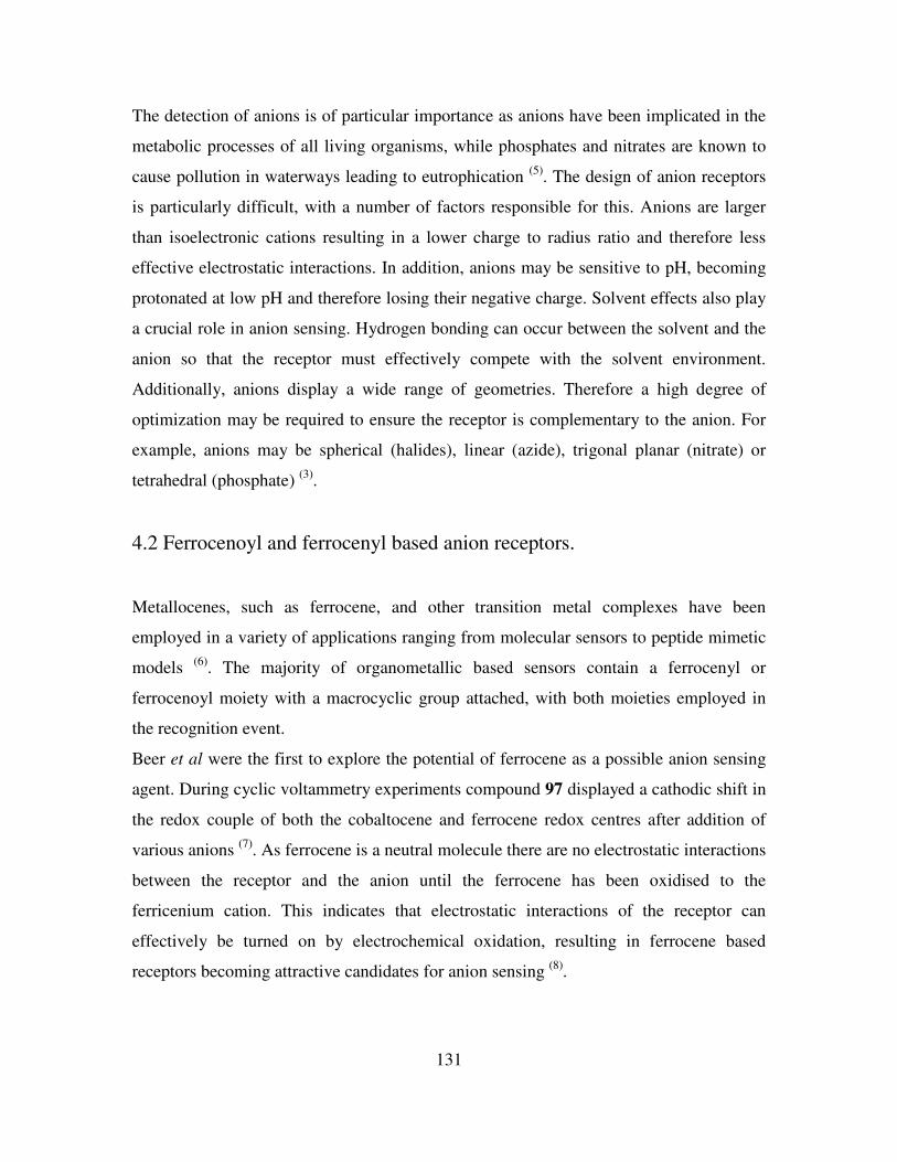
131
The detection of anions is of particular importance as anions have been implicated in the
metabolic processes of all living organisms, while phosphates and nitrates are known to
cause pollution in waterways leading to eutrophication (5)
. The design of anion receptors
is particularly difficult, with a number of factors responsible for this. Anions are larger
than isoelectronic cations resulting in a lower charge to radius ratio and therefore less
effective electrostatic interactions. In addition, anions may be sensitive to pH, becoming
protonated at low pH and therefore losing their negative charge. Solvent effects also play
a crucial role in anion sensing. Hydrogen bonding can occur between the solvent and the
anion so that the receptor must effectively compete with the solvent environment.
Additionally, anions display a wide range of geometries. Therefore a high degree of
optimization may be required to ensure the receptor is complementary to the anion. For
example, anions may be spherical (halides), linear (azide), trigonal planar (nitrate) or
tetrahedral (phosphate) (3)
.
4.2 Ferrocenoyl and ferrocenyl based anion receptors.
Metallocenes, such as ferrocene, and other transition metal complexes have been
employed in a variety of applications ranging from molecular sensors to peptide mimetic
models (6)
. The majority of organometallic based sensors contain a ferrocenyl or
ferrocenoyl moiety with a macrocyclic group attached, with both moieties employed in
the recognition event.
Beer et al were the first to explore the potential of ferrocene as a possible anion sensing
agent. During cyclic voltammetry experiments compound 97 displayed a cathodic shift in
the redox couple of both the cobaltocene and ferrocene redox centres after addition of
various anions (7)
. As ferrocene is a neutral molecule there are no electrostatic interactions
between the receptor and the anion until the ferrocene has been oxidised to the
ferricenium cation. This indicates that electrostatic interactions of the receptor can
effectively be turned on by electrochemical oxidation, resulting in ferrocene based
receptors becoming attractive candidates for anion sensing (8)
.
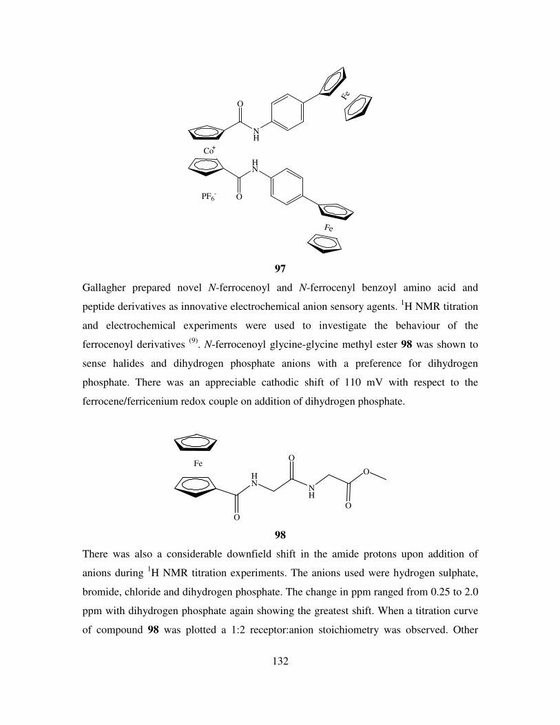
132
Co
O
O
HN
NH
PF6-
Fe
Fe
97
Gallagher prepared novel N-ferrocenoyl and N-ferrocenyl benzoyl amino acid and
peptide derivatives as innovative electrochemical anion sensory agents. 1H NMR titration
and electrochemical experiments were used to investigate the behaviour of the
ferrocenoyl derivatives (9)
. N-ferrocenoyl glycine-glycine methyl ester 98 was shown to
sense halides and dihydrogen phosphate anions with a preference for dihydrogen
phosphate. There was an appreciable cathodic shift of 110 mV with respect to the
ferrocene/ferricenium redox couple on addition of dihydrogen phosphate.
Fe
O
HN
O
NH
O
O
98
There was also a considerable downfield shift in the amide protons upon addition of
anions during 1H NMR titration experiments. The anions used were hydrogen sulphate,
bromide, chloride and dihydrogen phosphate. The change in ppm ranged from 0.25 to 2.0
ppm with dihydrogen phosphate again showing the greatest shift. When a titration curve
of compound 98 was plotted a 1:2 receptor:anion stoichiometry was observed. Other
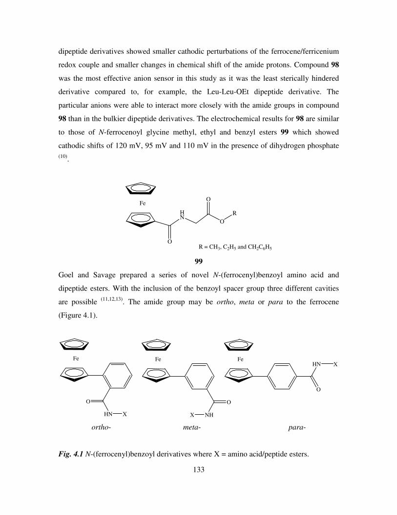
133
dipeptide derivatives showed smaller cathodic perturbations of the ferrocene/ferricenium
redox couple and smaller changes in chemical shift of the amide protons. Compound 98
was the most effective anion sensor in this study as it was the least sterically hindered
derivative compared to, for example, the Leu-Leu-OEt dipeptide derivative. The
particular anions were able to interact more closely with the amide groups in compound
98 than in the bulkier dipeptide derivatives. The electrochemical results for 98 are similar
to those of N-ferrocenoyl glycine methyl, ethyl and benzyl esters 99 which showed
cathodic shifts of 120 mV, 95 mV and 110 mV in the presence of dihydrogen phosphate
(10).
Fe
O
HN
O
O
R
R = CH3, C2H5 and CH2C6H5
99
Goel and Savage prepared a series of novel N-(ferrocenyl)benzoyl amino acid and
dipeptide esters. With the inclusion of the benzoyl spacer group three different cavities
are possible (11,12,13)
. The amide group may be ortho, meta or para to the ferrocene
(Figure 4.1).
Fe
O
HN X
Fe
O
NHX
Fe
O
HN X
ortho- meta- para-
Fig. 4.1 N-(ferrocenyl)benzoyl derivatives where X = amino acid/peptide esters.
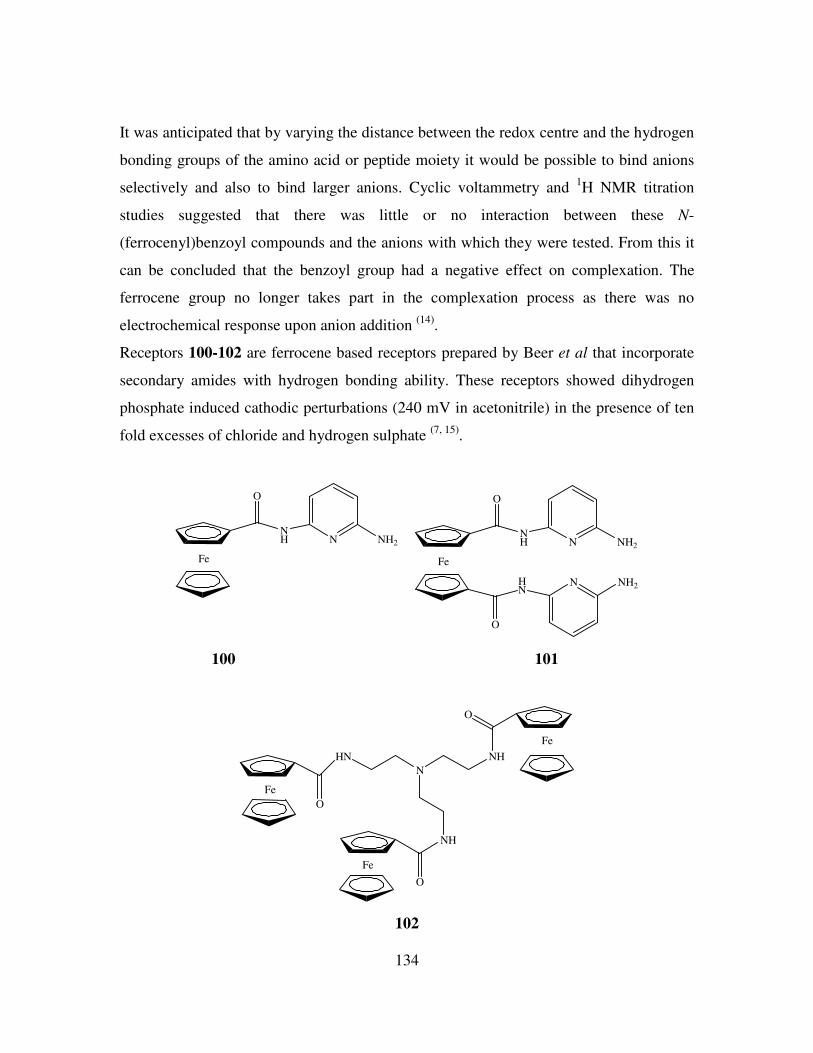
134
It was anticipated that by varying the distance between the redox centre and the hydrogen
bonding groups of the amino acid or peptide moiety it would be possible to bind anions
selectively and also to bind larger anions. Cyclic voltammetry and 1H NMR titration
studies suggested that there was little or no interaction between these N-
(ferrocenyl)benzoyl compounds and the anions with which they were tested. From this it
can be concluded that the benzoyl group had a negative effect on complexation. The
ferrocene group no longer takes part in the complexation process as there was no
electrochemical response upon anion addition (14)
.
Receptors 100-102 are ferrocene based receptors prepared by Beer et al that incorporate
secondary amides with hydrogen bonding ability. These receptors showed dihydrogen
phosphate induced cathodic perturbations (240 mV in acetonitrile) in the presence of ten
fold excesses of chloride and hydrogen sulphate (7, 15)
.
Fe
O
NH N NH2
Fe
O
NH N NH2
O
HN
N NH2
100 101
N
NHHN
NH
O
O
O
Fe
Fe
Fe
102
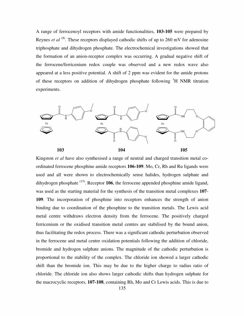
135
A range of ferrocenoyl receptors with amide functionalities, 103-105 were prepared by
Reynes et al (8)
. These receptors displayed cathodic shifts of up to 260 mV for adenosine
triphosphate and dihydrogen phosphate. The electrochemical investigations showed that
the formation of an anion-receptor complex was occurring. A gradual negative shift of
the ferrocene/ferricenium redox couple was observed and a new redox wave also
appeared at a less positive potential. A shift of 2 ppm was evident for the amide protons
of these receptors on addition of dihydrogen phosphate following 1H NMR titration
experiments.
Fe
O
NH
O
O
HN
O
Fe
O
NH
O
NH
O
O
Fe
O
NH
O
103 104 105
Kingston et al have also synthesised a range of neutral and charged transition metal co-
ordinated ferrocene phosphine amide receptors 106-109. Mo, Cr, Rh and Ru ligands were
used and all were shown to electrochemically sense halides, hydrogen sulphate and
dihydrogen phosphate (15)
. Receptor 106, the ferrocene appended phosphine amide ligand,
was used as the starting material for the synthesis of the transition metal complexes 107-
109. The incorporation of phosphine into receptors enhances the strength of anion
binding due to coordination of the phosphine to the transition metals. The Lewis acid
metal centre withdraws electron density from the ferrocene. The positively charged
ferricenium or the oxidised transition metal centres are stabilised by the bound anion,
thus facilitating the redox process. There was a significant cathodic perturbation observed
in the ferrocene and metal centre oxidation potentials following the addition of chloride,
bromide and hydrogen sulphate anions. The magnitude of the cathodic perturbation is
proportional to the stability of the complex. The chloride ion showed a larger cathodic
shift than the bromide ion. This may be due to the higher charge to radius ratio of
chloride. The chloride ion also shows larger cathodic shifts than hydrogen sulphate for
the macrocyclic receptors, 107-108, containing Rh, Mo and Cr Lewis acids. This is due to
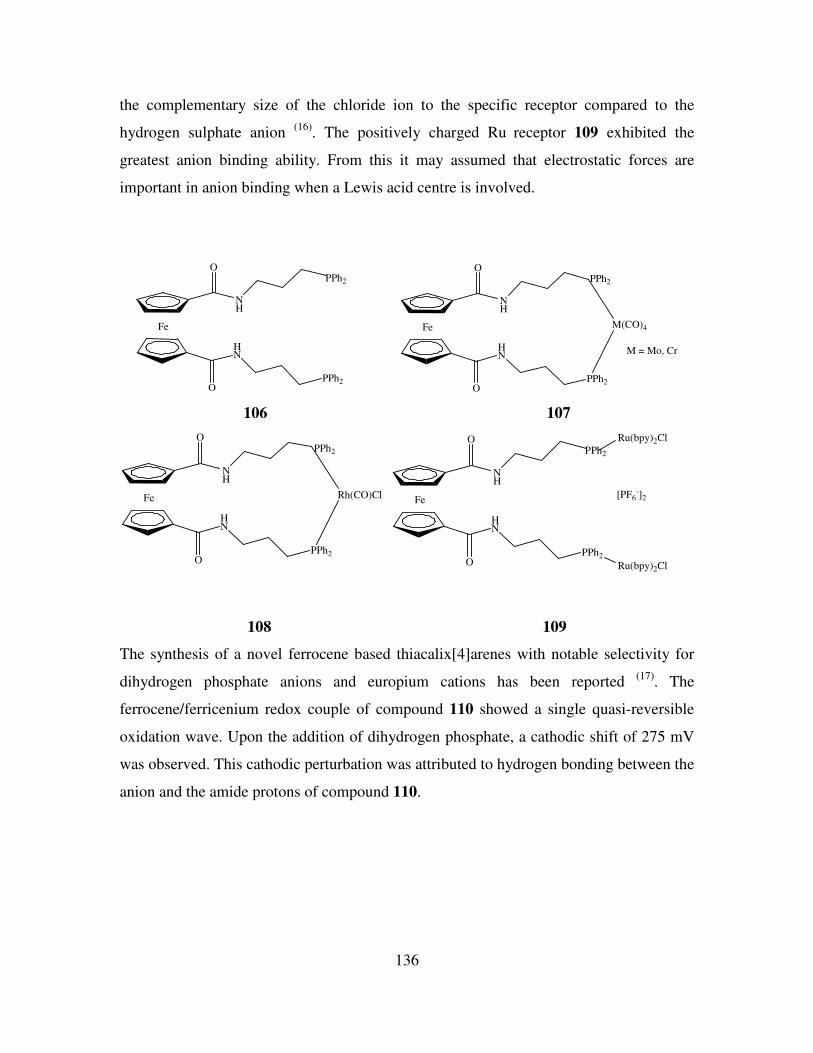
136
the complementary size of the chloride ion to the specific receptor compared to the
hydrogen sulphate anion (16)
. The positively charged Ru receptor 109 exhibited the
greatest anion binding ability. From this it may assumed that electrostatic forces are
important in anion binding when a Lewis acid centre is involved.
Fe
O
O
NH
HN
PPh2
PPh2
Fe
O
O
NH
HN
PPh2
PPh2
M(CO)4
M = Mo, Cr
106 107
Fe
O
O
NH
HN
PPh2
PPh2
Rh(CO)ClFe
O
O
NH
HN
PPh2
PPh2
Ru(bpy)2Cl
Ru(bpy)2Cl
[PF6-]2
108 109
The synthesis of a novel ferrocene based thiacalix[4]arenes with notable selectivity for
dihydrogen phosphate anions and europium cations has been reported (17)
. The
ferrocene/ferricenium redox couple of compound 110 showed a single quasi-reversible
oxidation wave. Upon the addition of dihydrogen phosphate, a cathodic shift of 275 mV
was observed. This cathodic perturbation was attributed to hydrogen bonding between the
anion and the amide protons of compound 110.
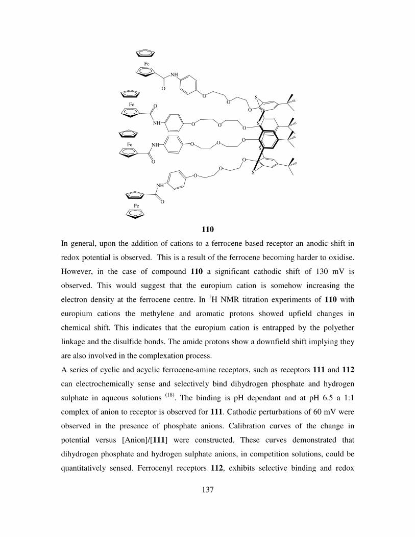
137
O
O
O
S
O
S
S
S
O
O
NH
O
OONH
O
OONH
O
O
O
NH
O
Fe
Fe
Fe
Fe
110
In general, upon the addition of cations to a ferrocene based receptor an anodic shift in
redox potential is observed. This is a result of the ferrocene becoming harder to oxidise.
However, in the case of compound 110 a significant cathodic shift of 130 mV is
observed. This would suggest that the europium cation is somehow increasing the
electron density at the ferrocene centre. In 1H NMR titration experiments of 110 with
europium cations the methylene and aromatic protons showed upfield changes in
chemical shift. This indicates that the europium cation is entrapped by the polyether
linkage and the disulfide bonds. The amide protons show a downfield shift implying they
are also involved in the complexation process.
A series of cyclic and acyclic ferrocene-amine receptors, such as receptors 111 and 112
can electrochemically sense and selectively bind dihydrogen phosphate and hydrogen
sulphate in aqueous solutions (18)
. The binding is pH dependant and at pH 6.5 a 1:1
complex of anion to receptor is observed for 111. Cathodic perturbations of 60 mV were
observed in the presence of phosphate anions. Calibration curves of the change in
potential versus [Anion]/[111] were constructed. These curves demonstrated that
dihydrogen phosphate and hydrogen sulphate anions, in competition solutions, could be
quantitatively sensed. Ferrocenyl receptors 112, exhibits selective binding and redox
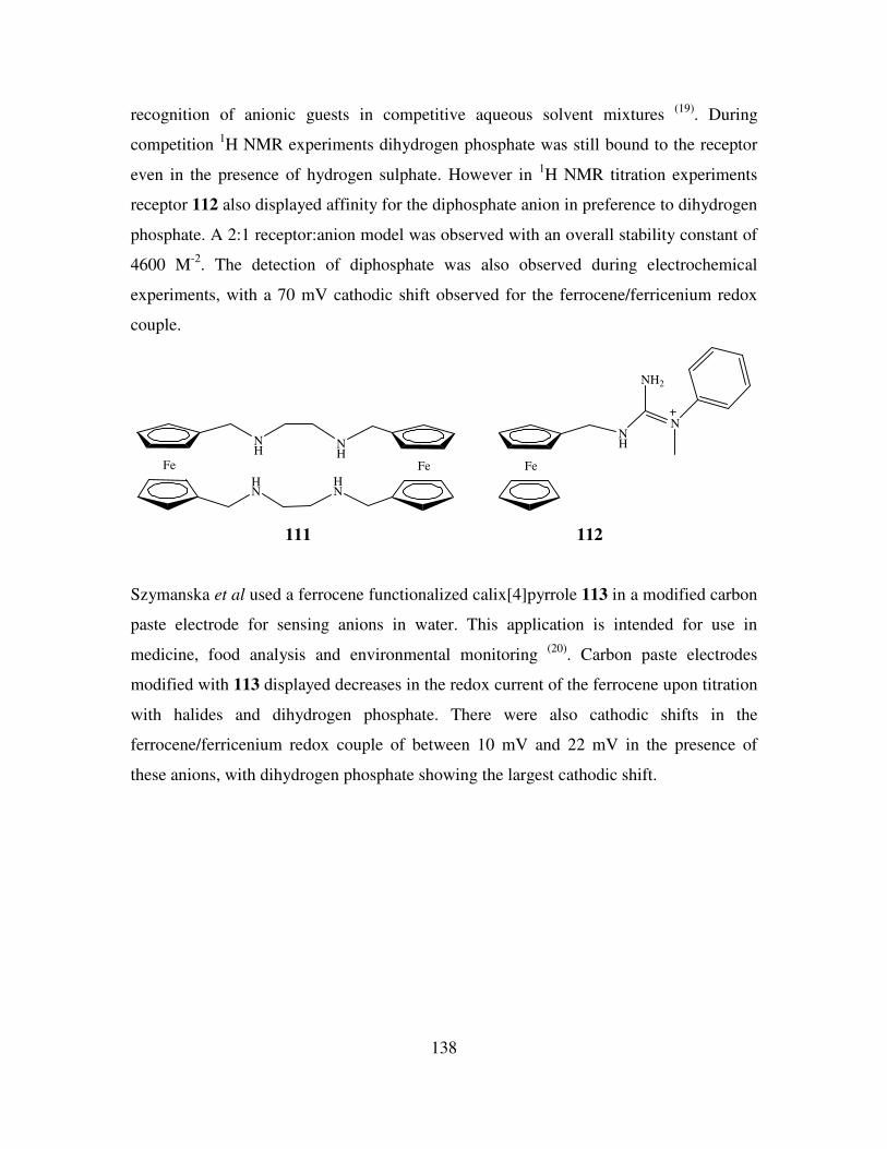
138
recognition of anionic guests in competitive aqueous solvent mixtures (19)
. During
competition 1H NMR experiments dihydrogen phosphate was still bound to the receptor
even in the presence of hydrogen sulphate. However in 1H NMR titration experiments
receptor 112 also displayed affinity for the diphosphate anion in preference to dihydrogen
phosphate. A 2:1 receptor:anion model was observed with an overall stability constant of
4600 M-2
. The detection of diphosphate was also observed during electrochemical
experiments, with a 70 mV cathodic shift observed for the ferrocene/ferricenium redox
couple.
Fe
HN
NH
HN
NH
Fe Fe
NH
NH2
N
111 112
Szymanska et al used a ferrocene functionalized calix[4]pyrrole 113 in a modified carbon
paste electrode for sensing anions in water. This application is intended for use in
medicine, food analysis and environmental monitoring (20)
. Carbon paste electrodes
modified with 113 displayed decreases in the redox current of the ferrocene upon titration
with halides and dihydrogen phosphate. There were also cathodic shifts in the
ferrocene/ferricenium redox couple of between 10 mV and 22 mV in the presence of
these anions, with dihydrogen phosphate showing the largest cathodic shift.
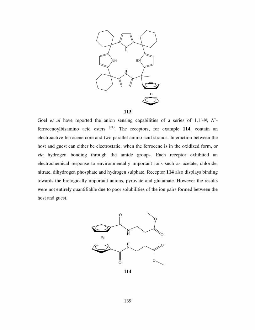
139
HN
HN
NH
NH
Fe
113
Goel et al have reported the anion sensing capabilities of a series of 1,1’-N, N’-
ferrocenoylbisamino acid esters (21)
. The receptors, for example 114, contain an
electroactive ferrocene core and two parallel amino acid strands. Interaction between the
host and guest can either be electrostatic, when the ferrocene is in the oxidized form, or
via hydrogen bonding through the amide groups. Each receptor exhibited an
electrochemical response to environmentally important ions such as acetate, chloride,
nitrate, dihydrogen phosphate and hydrogen sulphate. Receptor 114 also displays binding
towards the biologically important anions, pyruvate and glutamate. However the results
were not entirely quantifiable due to poor solubilities of the ion pairs formed between the
host and guest.
Fe
O
HN
O
O
O
NH O
O
114
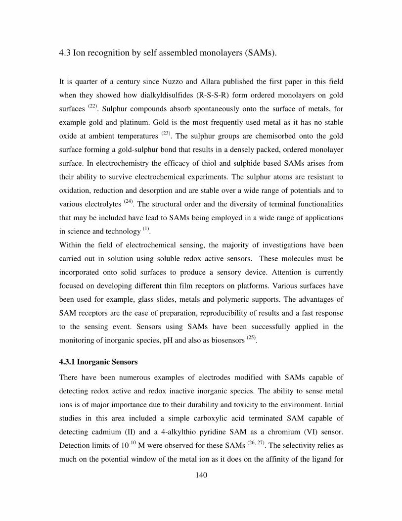
140
4.3 Ion recognition by self assembled monolayers (SAMs).
It is quarter of a century since Nuzzo and Allara published the first paper in this field
when they showed how dialkyldisulfides (R-S-S-R) form ordered monolayers on gold
surfaces (22)
. Sulphur compounds absorb spontaneously onto the surface of metals, for
example gold and platinum. Gold is the most frequently used metal as it has no stable
oxide at ambient temperatures (23)
. The sulphur groups are chemisorbed onto the gold
surface forming a gold-sulphur bond that results in a densely packed, ordered monolayer
surface. In electrochemistry the efficacy of thiol and sulphide based SAMs arises from
their ability to survive electrochemical experiments. The sulphur atoms are resistant to
oxidation, reduction and desorption and are stable over a wide range of potentials and to
various electrolytes (24)
. The structural order and the diversity of terminal functionalities
that may be included have lead to SAMs being employed in a wide range of applications
in science and technology (1)
.
Within the field of electrochemical sensing, the majority of investigations have been
carried out in solution using soluble redox active sensors. These molecules must be
incorporated onto solid surfaces to produce a sensory device. Attention is currently
focused on developing different thin film receptors on platforms. Various surfaces have
been used for example, glass slides, metals and polymeric supports. The advantages of
SAM receptors are the ease of preparation, reproducibility of results and a fast response
to the sensing event. Sensors using SAMs have been successfully applied in the
monitoring of inorganic species, pH and also as biosensors (25)
.
4.3.1 Inorganic Sensors
There have been numerous examples of electrodes modified with SAMs capable of
detecting redox active and redox inactive inorganic species. The ability to sense metal
ions is of major importance due to their durability and toxicity to the environment. Initial
studies in this area included a simple carboxylic acid terminated SAM capable of
detecting cadmium (II) and a 4-alkylthio pyridine SAM as a chromium (VI) sensor.
Detection limits of 10-10
M were observed for these SAMs (26, 27)
. The selectivity relies as
much on the potential window of the metal ion as it does on the affinity of the ligand for
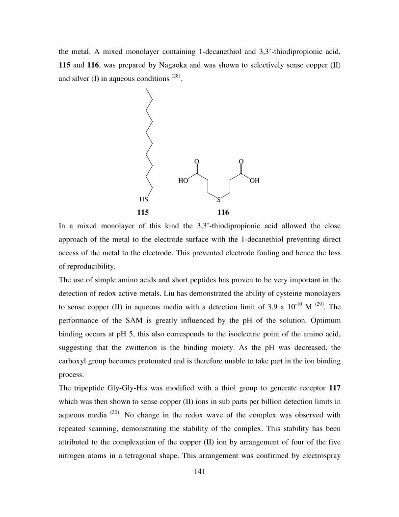
141
the metal. A mixed monolayer containing 1-decanethiol and 3,3’-thiodipropionic acid,
115 and 116, was prepared by Nagaoka and was shown to selectively sense copper (II)
and silver (I) in aqueous conditions (28)
.
HS S
O
OH
O
HO
115 116
In a mixed monolayer of this kind the 3,3’-thiodipropionic acid allowed the close
approach of the metal to the electrode surface with the 1-decanethiol preventing direct
access of the metal to the electrode. This prevented electrode fouling and hence the loss
of reproducibility.
The use of simple amino acids and short peptides has proven to be very important in the
detection of redox active metals. Liu has demonstrated the ability of cysteine monolayers
to sense copper (II) in aqueous media with a detection limit of 3.9 x 10⁻¹⁰
M (29)
. The
performance of the SAM is greatly influenced by the pH of the solution. Optimum
binding occurs at pH 5, this also corresponds to the isoelectric point of the amino acid,
suggesting that the zwitterion is the binding moiety. As the pH was decreased, the
carboxyl group becomes protonated and is therefore unable to take part in the ion binding
process.
The tripeptide Gly-Gly-His was modified with a thiol group to generate receptor 117
which was then shown to sense copper (II) ions in sub parts per billion detection limits in
aqueous media (30)
. No change in the redox wave of the complex was observed with
repeated scanning, demonstrating the stability of the complex. This stability has been
attributed to the complexation of the copper (II) ion by arrangement of four of the five
nitrogen atoms in a tetragonal shape. This arrangement was confirmed by electrospray
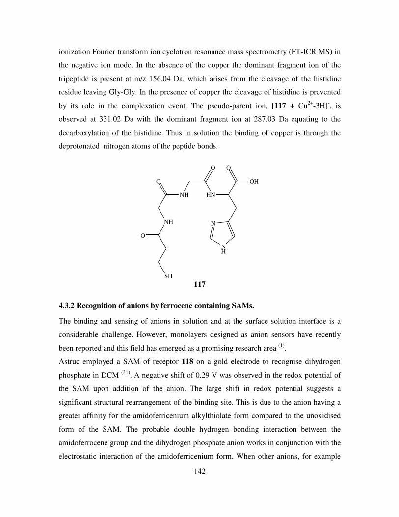
142
ionization Fourier transform ion cyclotron resonance mass spectrometry (FT-ICR MS) in
the negative ion mode. In the absence of the copper the dominant fragment ion of the
tripeptide is present at m/z 156.04 Da, which arises from the cleavage of the histidine
residue leaving Gly-Gly. In the presence of copper the cleavage of histidine is prevented
by its role in the complexation event. The pseudo-parent ion, [117 + Cu2+
-3H]-, is
observed at 331.02 Da with the dominant fragment ion at 287.03 Da equating to the
decarboxylation of the histidine. Thus in solution the binding of copper is through the
deprotonated nitrogen atoms of the peptide bonds.
SH
O
NH
O
NH
O
HN
O
OH
N
NH
117
4.3.2 Recognition of anions by ferrocene containing SAMs.
The binding and sensing of anions in solution and at the surface solution interface is a
considerable challenge. However, monolayers designed as anion sensors have recently
been reported and this field has emerged as a promising research area (1)
.
Astruc employed a SAM of receptor 118 on a gold electrode to recognise dihydrogen
phosphate in DCM (31)
. A negative shift of 0.29 V was observed in the redox potential of
the SAM upon addition of the anion. The large shift in redox potential suggests a
significant structural rearrangement of the binding site. This is due to the anion having a
greater affinity for the amidoferricenium alkylthiolate form compared to the unoxidised
form of the SAM. The probable double hydrogen bonding interaction between the
amidoferrocene group and the dihydrogen phosphate anion works in conjunction with the
electrostatic interaction of the amidoferricenium form. When other anions, for example
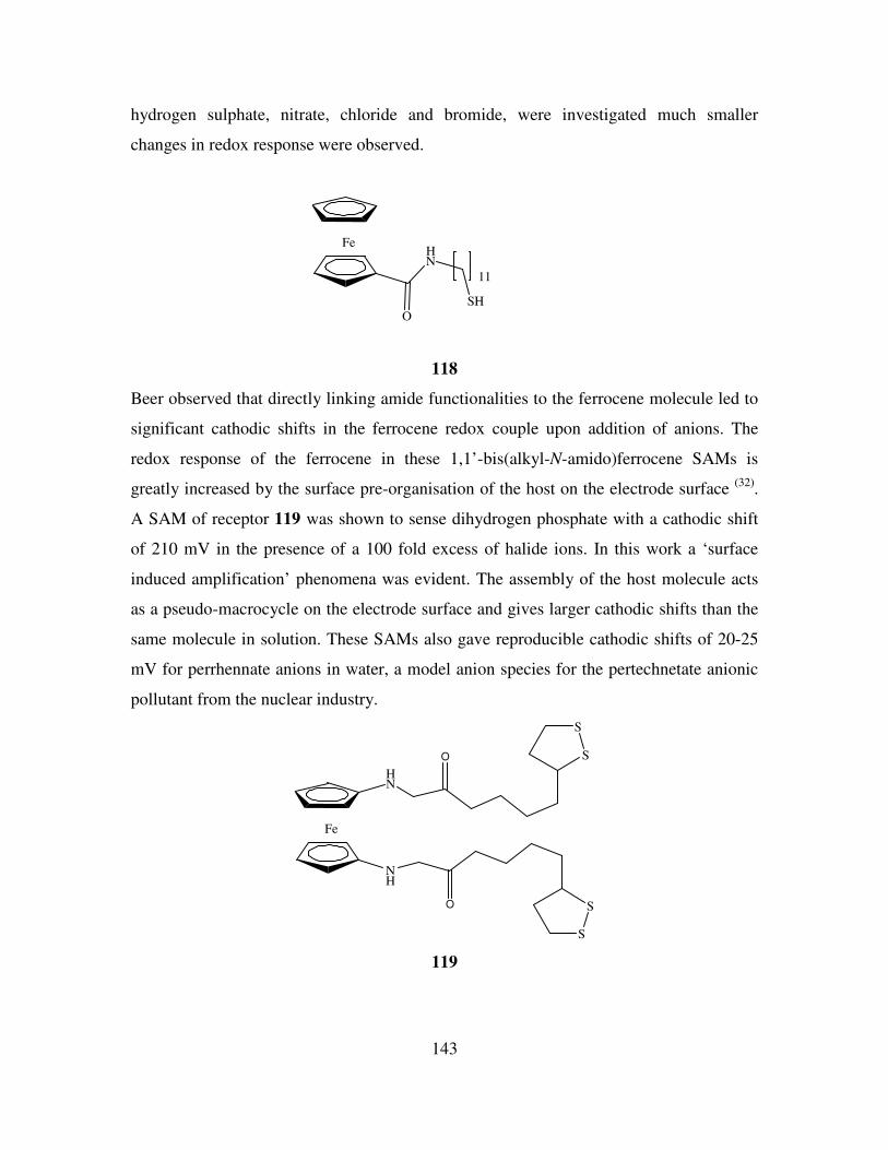
143
hydrogen sulphate, nitrate, chloride and bromide, were investigated much smaller
changes in redox response were observed.
Fe
O
HN
SH
11
118
Beer observed that directly linking amide functionalities to the ferrocene molecule led to
significant cathodic shifts in the ferrocene redox couple upon addition of anions. The
redox response of the ferrocene in these 1,1’-bis(alkyl-N-amido)ferrocene SAMs is
greatly increased by the surface pre-organisation of the host on the electrode surface (32)
.
A SAM of receptor 119 was shown to sense dihydrogen phosphate with a cathodic shift
of 210 mV in the presence of a 100 fold excess of halide ions. In this work a ‘surface
induced amplification’ phenomena was evident. The assembly of the host molecule acts
as a pseudo-macrocycle on the electrode surface and gives larger cathodic shifts than the
same molecule in solution. These SAMs also gave reproducible cathodic shifts of 20-25
mV for perrhennate anions in water, a model anion species for the pertechnetate anionic
pollutant from the nuclear industry.
Fe
HN
NH
O
O
S
S
S
S
119
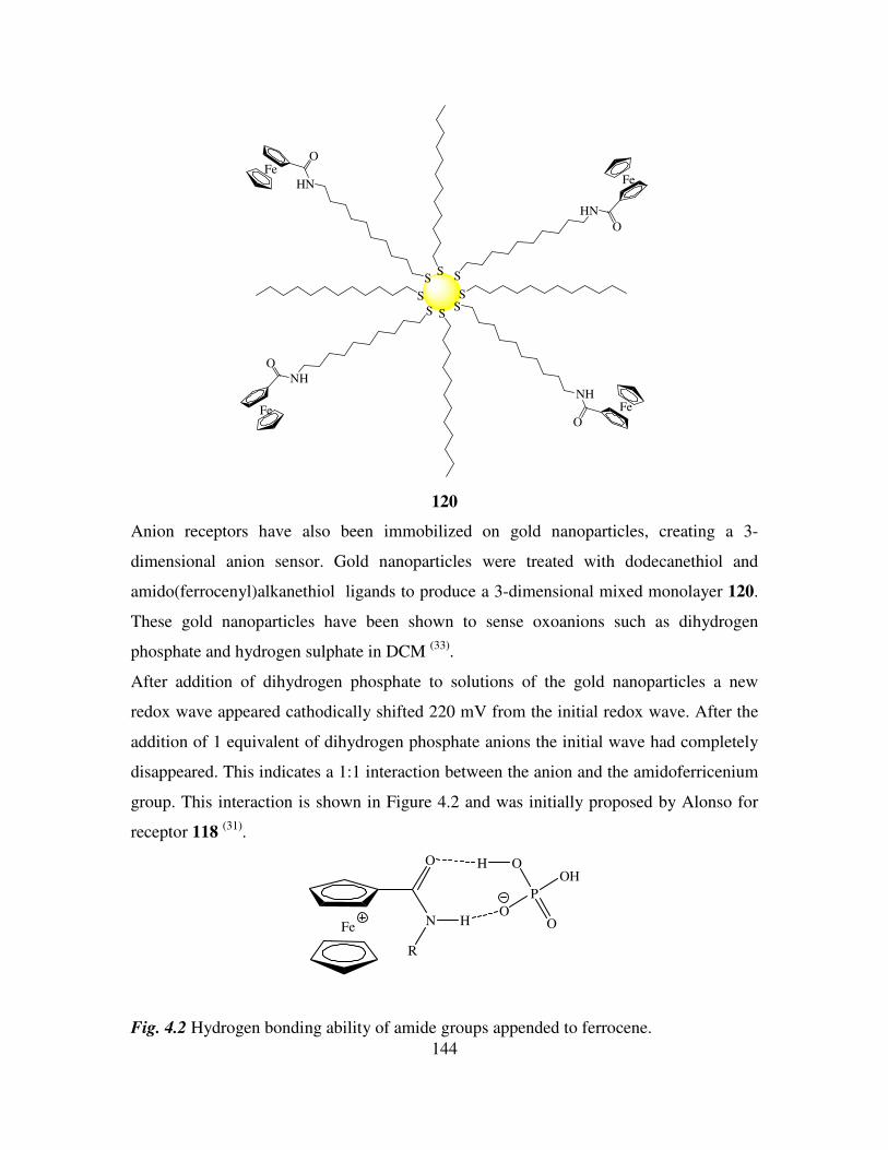
144
SS
S
S
Fe
O
NH
S
FeO
HN
S
Fe
O
HN
S
Fe
O
NH
S
120
Anion receptors have also been immobilized on gold nanoparticles, creating a 3-
dimensional anion sensor. Gold nanoparticles were treated with dodecanethiol and
amido(ferrocenyl)alkanethiol ligands to produce a 3-dimensional mixed monolayer 120.
These gold nanoparticles have been shown to sense oxoanions such as dihydrogen
phosphate and hydrogen sulphate in DCM (33)
.
After addition of dihydrogen phosphate to solutions of the gold nanoparticles a new
redox wave appeared cathodically shifted 220 mV from the initial redox wave. After the
addition of 1 equivalent of dihydrogen phosphate anions the initial wave had completely
disappeared. This indicates a 1:1 interaction between the anion and the amidoferricenium
group. This interaction is shown in Figure 4.2 and was initially proposed by Alonso for
receptor 118 (31)
.
Fe
O
N
R
H
P
OOH
OO
H
Fig. 4.2 Hydrogen bonding ability of amide groups appended to ferrocene.
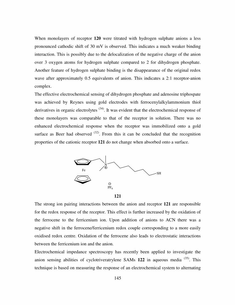
145
When monolayers of receptor 120 were titrated with hydrogen sulphate anions a less
pronounced cathodic shift of 30 mV is observed. This indicates a much weaker binding
interaction. This is possibly due to the delocalization of the negative charge of the anion
over 3 oxygen atoms for hydrogen sulphate compared to 2 for dihydrogen phosphate.
Another feature of hydrogen sulphate binding is the disappearance of the original redox
wave after approximately 0.5 equivalents of anion. This indicates a 2:1 receptor-anion
complex.
The effective electrochemical sensing of dihydrogen phosphate and adenosine triphospate
was achieved by Reynes using gold electrodes with ferrocenylalkylammonium thiol
derivatives in organic electrolytes (34)
. It was evident that the electrochemical response of
these monolayers was comparable to that of the receptor in solution. There was no
enhanced electrochemical response when the receptor was immobilized onto a gold
surface as Beer had observed (32)
. From this it can be concluded that the recognition
properties of the cationic receptor 121 do not change when absorbed onto a surface.
Fe
N
SH
PF6
121
The strong ion pairing interactions between the anion and receptor 121 are responsible
for the redox response of the receptor. This effect is further increased by the oxidation of
the ferrocene to the ferricenium ion. Upon addition of anions to ACN there was a
negative shift in the ferrocene/ferricenium redox couple corresponding to a more easily
oxidised redox centre. Oxidation of the ferrocene also leads to electrostatic interactions
between the ferricenium ion and the anion.
Electrochemical impedance spectroscopy has recently been applied to investigate the
anion sensing abilities of cyclotriveratrylene SAMs 122 in aqueous media (35)
. This
technique is based on measuring the response of an electrochemical system to alternating
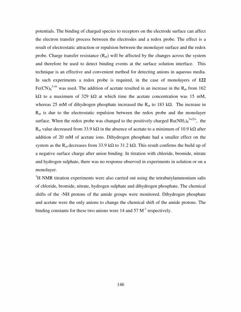
146
potentials. The binding of charged species to receptors on the electrode surface can affect
the electron transfer process between the electrodes and a redox probe. The effect is a
result of electrostatic attraction or repulsion between the monolayer surface and the redox
probe. Charge transfer resistance (Ret) will be affected by the changes across the system
and therefore be used to detect binding events at the surface solution interface. This
technique is an effective and convenient method for detecting anions in aqueous media.
In such experiments a redox probe is required, in the case of monolayers of 122
Fe(CN)63-/4-
was used. The addition of acetate resulted in an increase in the Ret from 162
kΩ to a maximum of 329 kΩ at which time the acetate concentration was 15 mM,
whereas 25 mM of dihydrogen phosphate increased the Ret to 183 kΩ. The increase in
Ret is due to the electrostatic repulsion between the redox probe and the monolayer
surface. When the redox probe was changed to the positively charged Ru(NH3)63+/2+
, the
Ret value decreased from 33.9 kΩ in the absence of acetate to a minimum of 10.9 kΩ after
addition of 20 mM of acetate ions. Dihydrogen phosphate had a smaller effect on the
system as the Ret decreases from 33.9 kΩ to 31.2 kΩ. This result confirms the build up of
a negative surface charge after anion binding. In titration with chloride, bromide, nitrate
and hydrogen sulphate, there was no response observed in experiments in solution or on a
monolayer.
1H NMR titration experiments were also carried out using the tetrabutylammonium salts
of chloride, bromide, nitrate, hydrogen sulphate and dihydrogen phosphate. The chemical
shifts of the -NH protons of the amide groups were monitored. Dihydrogen phosphate
and acetate were the only anions to change the chemical shift of the amide protons. The
binding constants for these two anions were 14 and 57 M-1
respectively.
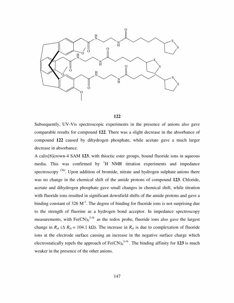
147
O
HN
NH
O
S
S
O
O
HN
NH
O
S
S
O
O
HN
NH
O
S
S
O
122
Subsequently, UV-Vis spectroscopic experiments in the presence of anions also gave
comparable results for compound 122. There was a slight decrease in the absorbance of
compound 122 caused by dihydrogen phosphate, while acetate gave a much larger
decrease in absorbance.
A calix[6]crown-4 SAM 123, with thioctic ester groups, bound fluoride ions in aqueous
media. This was confirmed by 1H NMR titration experiments and impedance
spectroscopy (36)
. Upon addition of bromide, nitrate and hydrogen sulphate anions there
was no change in the chemical shift of the amide protons of compound 123. Chloride,
acetate and dihydrogen phosphate gave small changes in chemical shift, while titration
with fluoride ions resulted in significant downfield shifts of the amide protons and gave a
binding constant of 326 M-1
. The degree of binding for fluoride ions is not surprising due
to the strength of fluorine as a hydrogen bond acceptor. In impedance spectroscopy
measurements, with Fe(CN)63-/4-
as the redox probe, fluoride ions also gave the largest
change in Ret (∆ Ret = 104.1 kΩ). The increase in Ret is due to complexation of fluoride
ions at the electrode surface causing an increase in the negative surface charge which
electrostatically repels the approach of Fe(CN)63-/4-
. The binding affinity for 123 is much
weaker in the presence of the other anions.
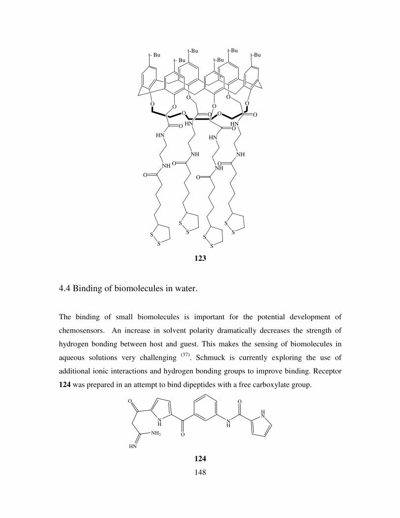
148
O
t-But- Bu
OO
t- Bu
O
t-Bu
O
t-But-Bu
O
O O
O
HN
NH
O
S
S
O
HN
NH
O
S
S
O
HN
NH
O
S
S
O
HN
NH
O
S
S
123
4.4 Binding of biomolecules in water.
The binding of small biomolecules is important for the potential development of
chemosensors. An increase in solvent polarity dramatically decreases the strength of
hydrogen bonding between host and guest. This makes the sensing of biomolecules in
aqueous solutions very challenging (37)
. Schmuck is currently exploring the use of
additional ionic interactions and hydrogen bonding groups to improve binding. Receptor
124 was prepared in an attempt to bind dipeptides with a free carboxylate group.
HN
O
NH
O
NH
O
HN
NH2
124
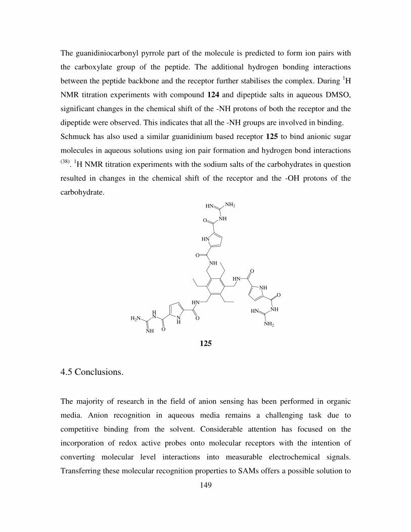
149
The guanidiniocarbonyl pyrrole part of the molecule is predicted to form ion pairs with
the carboxylate group of the peptide. The additional hydrogen bonding interactions
between the peptide backbone and the receptor further stabilises the complex. During 1H
NMR titration experiments with compound 124 and dipeptide salts in aqueous DMSO,
significant changes in the chemical shift of the -NH protons of both the receptor and the
dipeptide were observed. This indicates that all the -NH groups are involved in binding.
Schmuck has also used a similar guanidinium based receptor 125 to bind anionic sugar
molecules in aqueous solutions using ion pair formation and hydrogen bond interactions
(38).
1H NMR titration experiments with the sodium salts of the carbohydrates in question
resulted in changes in the chemical shift of the receptor and the -OH protons of the
carbohydrate.
HN
O
NH
O
NHHN
NH2
HN
ONH
O
HNH2N
NH
NH
O
HN
O NH
NH2HN
125
4.5 Conclusions.
The majority of research in the field of anion sensing has been performed in organic
media. Anion recognition in aqueous media remains a challenging task due to
competitive binding from the solvent. Considerable attention has focused on the
incorporation of redox active probes onto molecular receptors with the intention of
converting molecular level interactions into measurable electrochemical signals.
Transferring these molecular recognition properties to SAMs offers a possible solution to
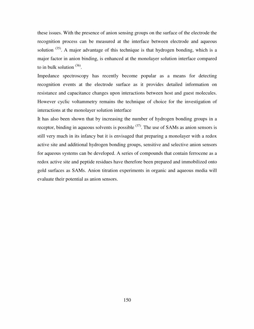
150
these issues. With the presence of anion sensing groups on the surface of the electrode the
recognition process can be measured at the interface between electrode and aqueous
solution (35)
. A major advantage of this technique is that hydrogen bonding, which is a
major factor in anion binding, is enhanced at the monolayer solution interface compared
to in bulk solution (36)
.
Impedance spectroscopy has recently become popular as a means for detecting
recognition events at the electrode surface as it provides detailed information on
resistance and capacitance changes upon interactions between host and guest molecules.
However cyclic voltammetry remains the technique of choice for the investigation of
interactions at the monolayer solution interface
It has also been shown that by increasing the number of hydrogen bonding groups in a
receptor, binding in aqueous solvents is possible (37)
. The use of SAMs as anion sensors is
still very much in its infancy but it is envisaged that preparing a monolayer with a redox
active site and additional hydrogen bonding groups, sensitive and selective anion sensors
for aqueous systems can be developed. A series of compounds that contain ferrocene as a
redox active site and peptide residues have therefore been prepared and immobilized onto
gold surfaces as SAMs. Anion titration experiments in organic and aqueous media will
evaluate their potential as anion sensors.
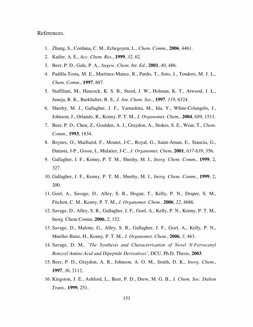
151
References.
1. Zhang, S., Cordana, C. M., Echegoyen, L., Chem. Comm., 2006, 4461.
2. Kaifer, A. E., Acc. Chem. Res., 1999, 32, 62.
3. Beer, P. D., Gale, P. A., Angew. Chem. Int. Ed., 2001, 40, 486.
4. Padilla-Tosta, M. E., Martinez-Manez, R., Pardo, T., Soto, J., Tendero, M. J. L.,
Chem. Comm., 1997, 887.
5. Staffilani, M., Hancock, K. S. B., Steed, J. W., Holman, K. T., Atwood, J. L.,
Juneja, R. K., Burkhalter, R. S., J. Am. Chem. Soc., 1997, 119, 6324.
6. Sheehy, M. J., Gallagher, J. F., Yamashita, M., Ida, Y., White-Colangelo, J.,
Johnson, J., Orlando, R., Kenny, P. T. M., J. Organomet. Chem., 2004, 689, 1511.
7. Beer, P. D., Chen, Z., Goulden, A. J., Graydon, A., Stokes, S. E., Wear, T., Chem.
Comm., 1993, 1834.
8. Reynes, O., Maillaird, F., Moutet, J-C., Royal, G., Saint-Aman, E., Stanciu, G.,
Dutasta, J-P., Gosse, I., Mulatier, J-C., J. Organomet. Chem, 2001, 637-639, 356.
9. Gallagher, J. F., Kenny, P. T. M., Sheehy, M. J., Inorg. Chem. Comm., 1999, 2,
327.
10. Gallagher, J. F., Kenny, P. T. M., Sheehy, M. J., Inorg. Chem. Comm., 1999, 2,
200.
11. Goel, A., Savage, D., Alley, S. R., Hogan, T., Kelly, P. N., Draper, S. M.,
Fitchett, C. M., Kenny, P. T. M., J. Organomet. Chem., 2006, 22, 4686.
12. Savage, D., Alley, S. R., Gallagher, J. F., Goel, A., Kelly, P. N., Kenny, P. T. M.,
Inorg. Chem Comm, 2006, 2, 152.
13. Savage, D., Malone, G., Alley, S. R., Gallagher, J. F., Goel, A., Kelly, P. N.,
Mueller-Bunz, H., Kenny, P. T. M., J. Organomet. Chem., 2006, 3, 463.
14. Savage, D. M., ‘The Synthesis and Characterization of Novel N-Ferrocenyl
Bonzoyl Amino Acid and Dipeptide Derivatives’, DCU, Ph.D, Thesis, 2003.
15. Beer, P. D., Graydon, A. R., Johnson, A. O. M., Smith, D. K., Inorg. Chem.,
1997, 36, 2112.
16. Kingston, J. E., Ashford, L., Beer, P. D., Drew, M. G. B., J. Chem. Soc. Dalton
Trans., 1999, 251.
152
17. Guo, D. S., Liu, Z. P., Ma, J. P., Huang, R. Q., Tett. Lett., 2007, 48, 1221.
18. Beer, P. D., Cadman, J., Lloris, J. M., Martinez-Manez, R., Padilla, M. E., Pardo,
T., Smith, D. K., Soto, J., Dalton Trans., 1999, 127.
19. Beer, P.D., Drew, M. G. B., Smith, D. K., J. Organomet. Chem., 1997, 543, 259.
20. Szymanska, I., Radecka, H., Radecki, J., Gale, P. A., Warriner, C. N., J.
Electroanal. Chem, 2006, 591, 223.
21. Goel, A., Brennan, N., Brady, N., Kenny, P. T. M., Biosens. Bioelectron., 2007,
22, 2047.
22. Nuzzo, R. G., Allara, D. L., J. Am. Chem. Soc., 1983, 105, 4481.
23. Gooding, J. J., Mearns, F., Yang, W., Liu, J., Electroanalysis, 2003, 15, 81.
24. Finklea, H. O., ‘Self-assembled Monolayers on Electrodes’, Encyclopedia of
Analytical Chemistry, John Wiley and Sons.
25. Shervedani, R. K., Babadi, M. K., Talanta, 2006, 69, 741.
26. Turyan, I., Mandler, D., Anal. Chem., 1994, 66, 58.
27. Shen, S., Mark, J. E., Seliskar, C. J., Mark, H. B., Heineman, W. R., J. Solid State
Electrochem., 1997, 1, 148.
28. Nagaoka, T., Chen, Z., Okuno, H., Nakayama, N., Ogura, K., Anal. Sci., 1999, 15,
857.
29. Liu, A. C., Chen, D. C., Lin, C. C., Chou, H. H., Chen, C. H., Anal. Chem., 1999,
71, 1549.
30. Yang, W., Jaramillo, D., Gooding, J. J., Hibbert, D. B., Zhang, R., Willett, G. D.,
Kisher, K. J., Chem. Comm., 2001, 1982.
31. Alonso, E., Labande, A., Raehm, L., Kern, J. M., Astruc, D., C. R. Acad. Sci.,
1999, 209.
32. Beer, P. D., Davis, J. J., Drillsma-Milgrom, D. A., Szemes, F., Chem. Comm.,
2002, 1716.
33. Labande, A., Ruiz, J., Astruc, D., J. Am Chem. Soc., 2002, 124, 1782.
34. Reynes, O., Bucher, C., Moutet, J-C., Royal, G., Saint-Aman, E., Inorg. Chim.
Acta, 2008, 361, 1784.
35. Zhang, S., Echegoyen, L., J. Am Chem. Soc., 2005, 127, 2006.
36. Zhang, S., Palkar, A., Echegoyen, L., Langmuir, 2006, 22, 10732.
153
37. Schmuck, C., Schwegmann, M., Org. Lett., 2005, 7, 3517.
38. Schmuck, C., Geiger, L., J. Am Chem. Soc., 2004, 126, 8898.

154
Chapter 5
Results and Discussion II
5.1 Introduction.
The development of electrodes modified with monolayers for ion sensing has led to the
successful detection of ionic species at the solution electrode interface. The detection of
redox active and inactive metal ions was freely achieved with monolayers of redox active
and inactive molecules, for example, pyridines and crown ethers (1)
.
Anion recognition in aqueous media remains a challenging task due to competitive
binding from the solvent. The use of cationic anion sensors which employ electrostatic
interactions can overcome the problem of solvent competition. However their counter
ions have been known to interfere with the recognition process (2)
.
Transferring these molecular recognition properties to SAMs offers a possible solution to
these issues. With the presence of anion sensing groups on the surface of the electrode
the recognition process can be measured at the interface between electrode and aqueous
solution. A major advantage of this technique is that hydrogen bonding, which is a key
factor in anion binding, is enhanced at the monolayer solution interface compared to in
bulk solution (3)
.
The primary aim of this project is the preparation of self assembled monolayers (SAMs)
on gold electrodes that incorporate three main structural components.
a) An electroactive ferrocene core.
b) An amino acid or peptide moiety with hydrogen bonding groups.
c) Sulphur atoms that would facilitate covalent bonding to gold.
An example of such a SAM is illustrated in Figure 5.1.
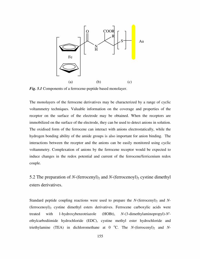
155
Fe
O
NH
COOR
R
S Au
(a) (b) (c)
Fig. 5.1 Components of a ferrocene-peptide based monolayer.
The monolayers of the ferrocene derivatives may be characterized by a range of cyclic
voltammetry techniques. Valuable information on the coverage and properties of the
receptor on the surface of the electrode may be obtained. When the receptors are
immobilized on the surface of the electrode, they can be used to detect anions in solution.
The oxidised form of the ferrocene can interact with anions electrostatically, while the
hydrogen bonding ability of the amide groups is also important for anion binding. The
interactions between the receptor and the anions can be easily monitored using cyclic
voltammetry. Complexation of anions by the ferrocene receptor would be expected to
induce changes in the redox potential and current of the ferrocene/ferricenium redox
couple.
5.2 The preparation of N-(ferrocenyl)2 and N-(ferrocenoyl)2 cystine dimethyl
esters derivatives.
Standard peptide coupling reactions were used to prepare the N-(ferrocenyl)2 and N-
(ferrocenoyl)2 cystine dimethyl esters derivatives. Ferrocene carboxylic acids were
treated with 1-hydroxybenzotriazole (HOBt), N-(3-dimethylaminopropyl)-N′-
ethylcarbodiimide hydrochloride (EDC), cystine methyl ester hydrochloride and
triethylamine (TEA) in dichloromethane at 0 oC. The N-(ferrocenyl)2 and N-
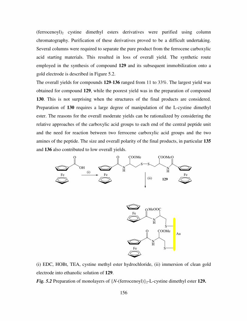
156
(ferrocenoyl)2 cystine dimethyl esters derivatives were purified using column
chromatography. Purification of these derivatives proved to be a difficult undertaking.
Several columns were required to separate the pure product from the ferrocene carboxylic
acid starting materials. This resulted in loss of overall yield. The synthetic route
employed in the synthesis of compound 129 and its subsequent immobilization onto a
gold electrode is described in Figure 5.2.
The overall yields for compounds 129-136 ranged from 11 to 33%. The largest yield was
obtained for compound 129, while the poorest yield was in the preparation of compound
130. This is not surprising when the structures of the final products are considered.
Preparation of 130 requires a large degree of manipulation of the L-cystine dimethyl
ester. The reasons for the overall moderate yields can be rationalized by considering the
relative approaches of the carboxylic acid groups to each end of the central peptide unit
and the need for reaction between two ferrocene carboxylic acid groups and the two
amines of the peptide. The size and overall polarity of the final products, in particular 135
and 136 also contributed to low overall yields.
Fe
O
OH
Fe
O
NH
COOMe
S S
NH
COOMeO
Fe
S
MeOOC
NH
OFe
Au
S
COOMe
NH
O
Fe
(i)
(ii) 129
(i) EDC, HOBt, TEA, cystine methyl ester hydrochloride, (ii) immersion of clean gold
electrode into ethanolic solution of 129.
Fig. 5.2 Preparation of monolayers of N-(ferrocenoyl)2-L-cystine dimethyl ester 129.
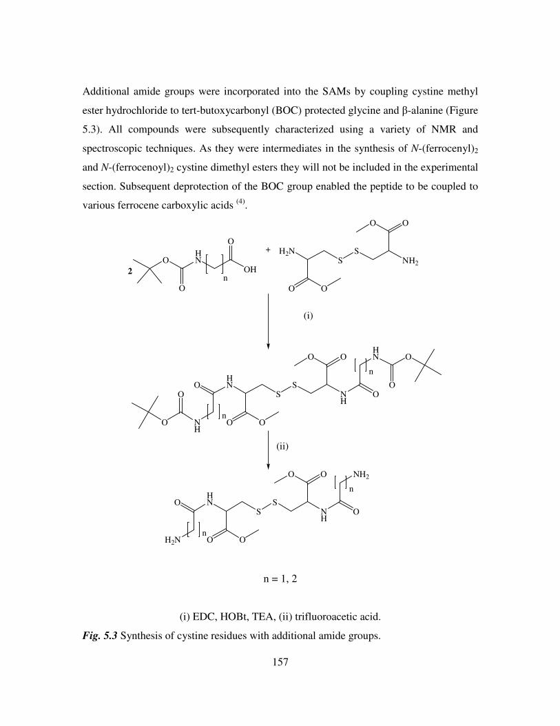
157
Additional amide groups were incorporated into the SAMs by coupling cystine methyl
ester hydrochloride to tert-butoxycarbonyl (BOC) protected glycine and β-alanine (Figure
5.3). All compounds were subsequently characterized using a variety of NMR and
spectroscopic techniques. As they were intermediates in the synthesis of N-(ferrocenyl)2
and N-(ferrocenoyl)2 cystine dimethyl esters they will not be included in the experimental
section. Subsequent deprotection of the BOC group enabled the peptide to be coupled to
various ferrocene carboxylic acids (4)
.
HN
OO
SS
NH
OO
OHN
O
OH
O
n2
+
(i)
O
HN O
OO
NH
O
O
H2N
OO
SS
NH2
OO
n
n
(ii)
HN
OO
SS
NH
OO
O
NH2
O
H2Nn
n
n = 1, 2
(i) EDC, HOBt, TEA, (ii) trifluoroacetic acid.
Fig. 5.3 Synthesis of cystine residues with additional amide groups.
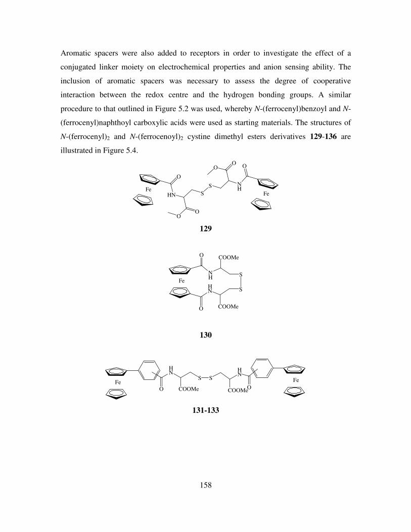
158
Aromatic spacers were also added to receptors in order to investigate the effect of a
conjugated linker moiety on electrochemical properties and anion sensing ability. The
inclusion of aromatic spacers was necessary to assess the degree of cooperative
interaction between the redox centre and the hydrogen bonding groups. A similar
procedure to that outlined in Figure 5.2 was used, whereby N-(ferrocenyl)benzoyl and N-
(ferrocenyl)naphthoyl carboxylic acids were used as starting materials. The structures of
N-(ferrocenyl)2 and N-(ferrocenoyl)2 cystine dimethyl esters derivatives 129-136 are
illustrated in Figure 5.4.
Fe
O
HN
OO
S
S NH
OO O
Fe
129
Fe
O
O
NH
HN
COOMe
COOMe
S
S
130
Fe
O
HN
COOMe
S S
COOMe
HN
O
Fe
131-133
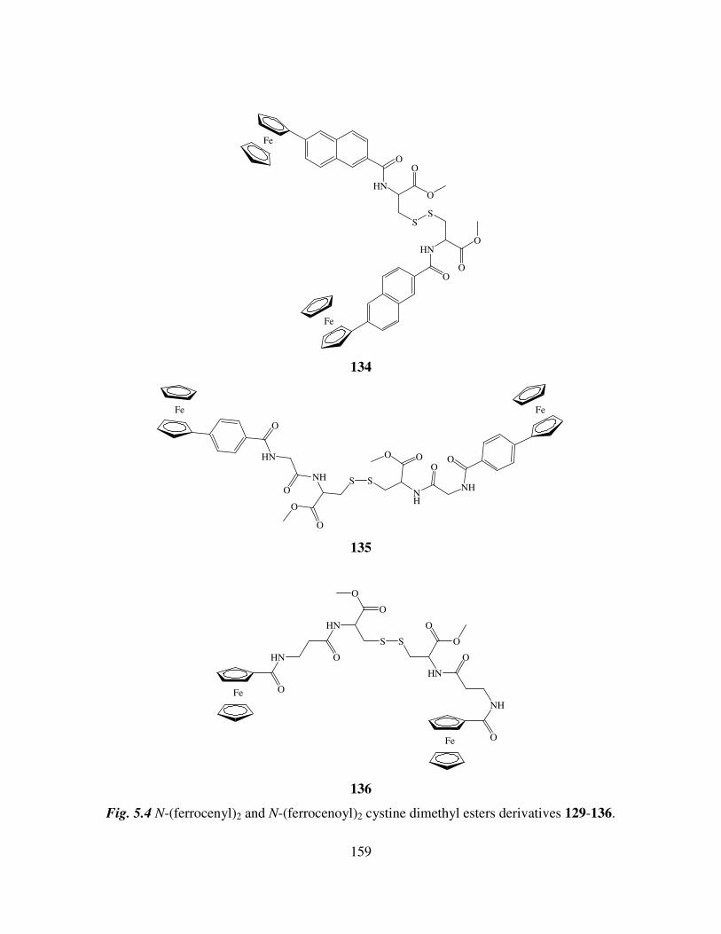
159
Fe
O
HN
O
O
SS
HN
O
O
O
Fe
134
Fe
O
HN
O
NH
O
O
S S
NH
OO
O
NH
O
Fe
135
Fe O
HN O
HN
O
S
O
S
HN
O
O
O
NH
OFe
136
Fig. 5.4 N-(ferrocenyl)2 and N-(ferrocenoyl)2 cystine dimethyl esters derivatives 129-136.
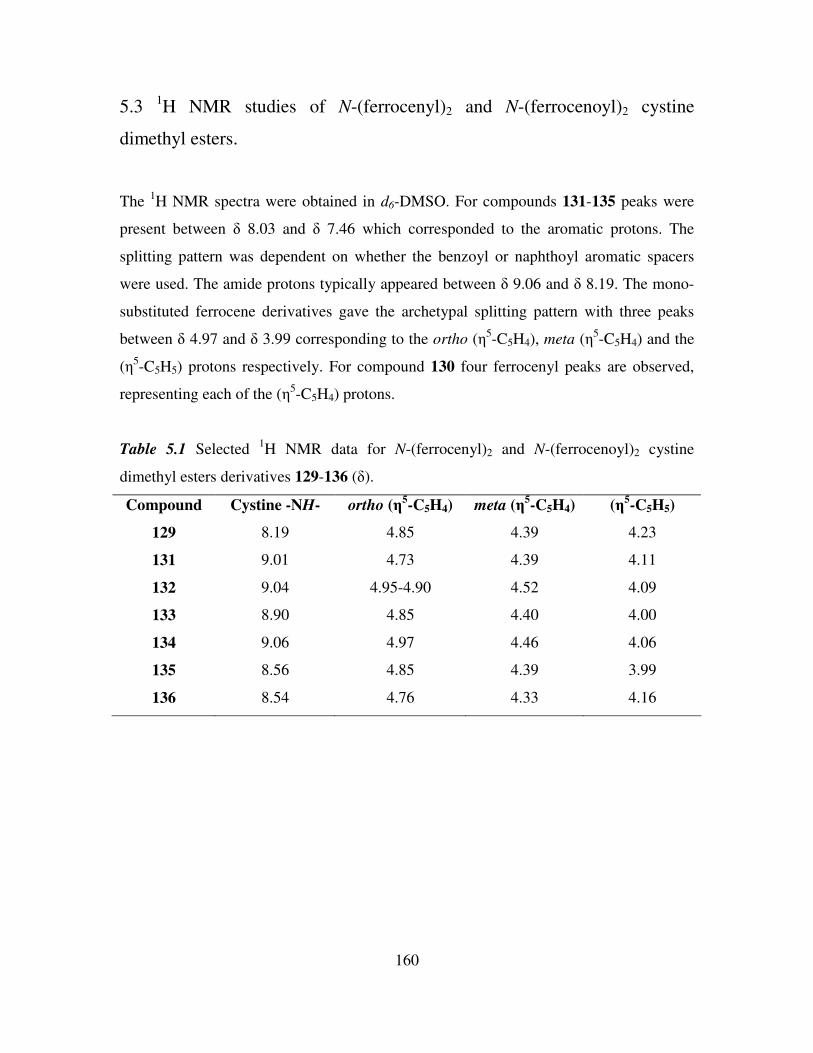
160
5.3 1H NMR studies of N-(ferrocenyl)2 and N-(ferrocenoyl)2 cystine
dimethyl esters.
The 1H NMR spectra were obtained in d6-DMSO. For compounds 131-135 peaks were
present between δ 8.03 and δ 7.46 which corresponded to the aromatic protons. The
splitting pattern was dependent on whether the benzoyl or naphthoyl aromatic spacers
were used. The amide protons typically appeared between δ 9.06 and δ 8.19. The mono-
substituted ferrocene derivatives gave the archetypal splitting pattern with three peaks
between δ 4.97 and δ 3.99 corresponding to the ortho (η5-C5H4), meta (η
5-C5H4) and the
(η5-C5H5) protons respectively. For compound 130 four ferrocenyl peaks are observed,
representing each of the (η5-C5H4) protons.
Table 5.1 Selected 1H NMR data for N-(ferrocenyl)2 and N-(ferrocenoyl)2 cystine
dimethyl esters derivatives 129-136 (δ).
Compound Cystine -NH- ortho (η5-C5H4) meta (η
5-C5H4) (η
5-C5H5)
129 8.19 4.85 4.39 4.23
131 9.01 4.73 4.39 4.11
132 9.04 4.95-4.90 4.52 4.09
133 8.90 4.85 4.40 4.00
134 9.06 4.97 4.46 4.06
135 8.56 4.85 4.39 3.99
136 8.54 4.76 4.33 4.16
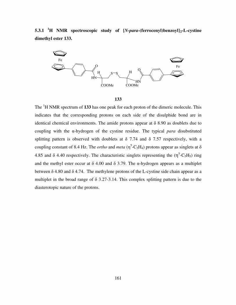
161
5.3.1 1H NMR spectroscopic study of N-para-(ferrocenyl)benzoyl2-L-cystine
dimethyl ester 133.
Fe
O
HNHN
OFe
COOMe
H S S
COOMe
H
133
The 1H NMR spectrum of 133 has one peak for each proton of the dimeric molecule. This
indicates that the corresponding protons on each side of the disulphide bond are in
identical chemical environments. The amide protons appear at δ 8.90 as doublets due to
coupling with the α-hydrogen of the cystine residue. The typical para disubstituted
splitting pattern is observed with doublets at δ 7.74 and δ 7.57 respectively, with a
coupling constant of 8.4 Hz. The ortho and meta (η5-C5H4) protons appear as singlets at δ
4.85 and δ 4.40 respectively. The characteristic singlets representing the (η5-C5H5) ring
and the methyl ester occur at δ 4.00 and δ 3.79. The α-hydrogen appears as a multiplet
between δ 4.80 and δ 4.74. The methylene protons of the L-cystine side chain appear as a
multiplet in the broad range of δ 3.27-3.14. This complex splitting pattern is due to the
diasterotopic nature of the protons.
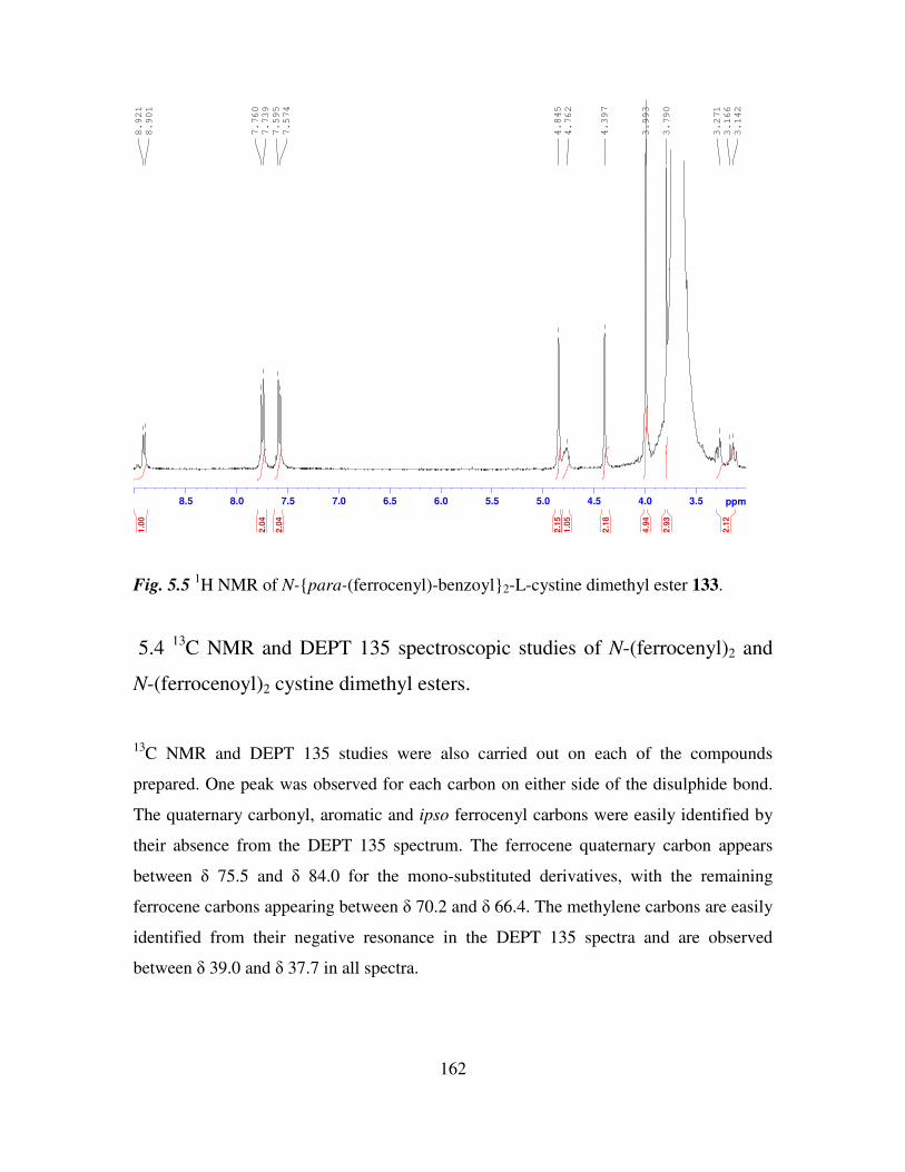
162
3.54.04.55.05.56.06.57.07.58.08.5 ppm
3.142
3.166
3.271
3.790
3.993
4.397
4.762
4.845
7.574
7.595
7.739
7.760
8.901
8.921
2.1
2
2.9
3
4.9
4
2.1
8
1.0
5
2.1
5
2.0
4
2.0
4
1.0
0
Fig. 5.5 1H NMR of N-para-(ferrocenyl)-benzoyl2-L-cystine dimethyl ester 133.
5.4 13
C NMR and DEPT 135 spectroscopic studies of N-(ferrocenyl)2 and
N-(ferrocenoyl)2 cystine dimethyl esters.
13C NMR and DEPT 135 studies were also carried out on each of the compounds
prepared. One peak was observed for each carbon on either side of the disulphide bond.
The quaternary carbonyl, aromatic and ipso ferrocenyl carbons were easily identified by
their absence from the DEPT 135 spectrum. The ferrocene quaternary carbon appears
between δ 75.5 and δ 84.0 for the mono-substituted derivatives, with the remaining
ferrocene carbons appearing between δ 70.2 and δ 66.4. The methylene carbons are easily
identified from their negative resonance in the DEPT 135 spectra and are observed
between δ 39.0 and δ 37.7 in all spectra.
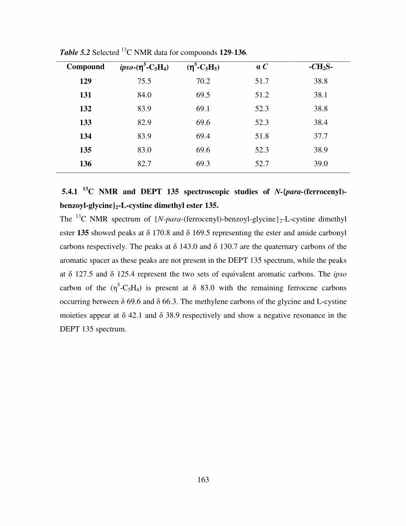
163
Table 5.2 Selected 13
C NMR data for compounds 129-136.
Compound ipso-(ηηηη5-C5H4) (ηηηη5
-C5H5) α C -CH2S-
129 75.5 70.2 51.7 38.8
131 84.0 69.5 51.2 38.1
132 83.9 69.1 52.3 38.8
133 82.9 69.6 52.3 38.4
134 83.9 69.4 51.8 37.7
135 83.0 69.6 52.3 38.9
136 82.7 69.3 52.7 39.0
5.4.1 13
C NMR and DEPT 135 spectroscopic studies of N-para-(ferrocenyl)-
benzoyl-glycine2-L-cystine dimethyl ester 135.
The 13
C NMR spectrum of N-para-(ferrocenyl)-benzoyl-glycine2-L-cystine dimethyl
ester 135 showed peaks at δ 170.8 and δ 169.5 representing the ester and amide carbonyl
carbons respectively. The peaks at δ 143.0 and δ 130.7 are the quaternary carbons of the
aromatic spacer as these peaks are not present in the DEPT 135 spectrum, while the peaks
at δ 127.5 and δ 125.4 represent the two sets of equivalent aromatic carbons. The ipso
carbon of the (η5-C5H4) is present at δ 83.0 with the remaining ferrocene carbons
occurring between δ 69.6 and δ 66.3. The methylene carbons of the glycine and L-cystine
moieties appear at δ 42.1 and δ 38.9 respectively and show a negative resonance in the
DEPT 135 spectrum.
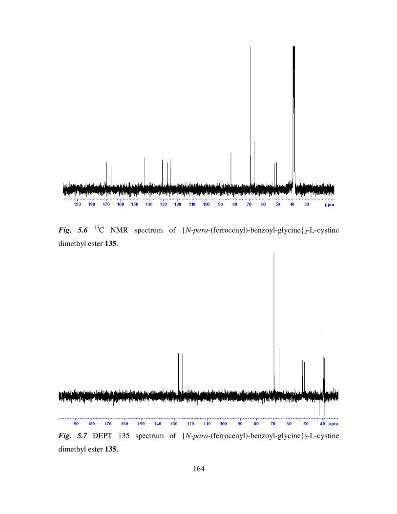
164
Fig. 5.6 13
C NMR spectrum of N-para-(ferrocenyl)-benzoyl-glycine2-L-cystine
dimethyl ester 135.
Fig. 5.7 DEPT 135 spectrum of N-para-(ferrocenyl)-benzoyl-glycine2-L-cystine
dimethyl ester 135.
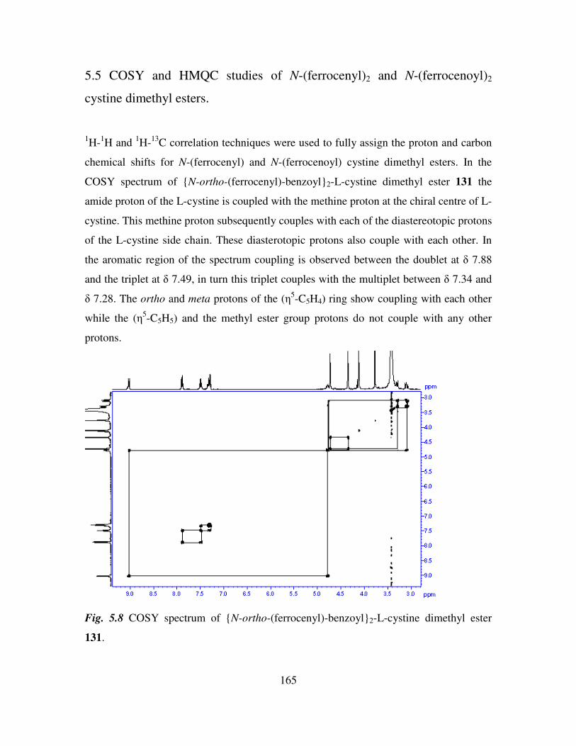
165
5.5 COSY and HMQC studies of N-(ferrocenyl)2 and N-(ferrocenoyl)2
cystine dimethyl esters.
1H-
1H and
1H-
13C correlation techniques were used to fully assign the proton and carbon
chemical shifts for N-(ferrocenyl) and N-(ferrocenoyl) cystine dimethyl esters. In the
COSY spectrum of N-ortho-(ferrocenyl)-benzoyl2-L-cystine dimethyl ester 131 the
amide proton of the L-cystine is coupled with the methine proton at the chiral centre of L-
cystine. This methine proton subsequently couples with each of the diastereotopic protons
of the L-cystine side chain. These diasterotopic protons also couple with each other. In
the aromatic region of the spectrum coupling is observed between the doublet at δ 7.88
and the triplet at δ 7.49, in turn this triplet couples with the multiplet between δ 7.34 and
δ 7.28. The ortho and meta protons of the (η5-C5H4) ring show coupling with each other
while the (η5-C5H5) and the methyl ester group protons do not couple with any other
protons.
Fig. 5.8 COSY spectrum of N-ortho-(ferrocenyl)-benzoyl2-L-cystine dimethyl ester
131.
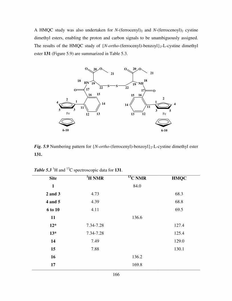
166
A HMQC study was also undertaken for N-(ferrocenyl)2 and N-(ferrocenoyl)2 cystine
dimethyl esters, enabling the proton and carbon signals to be unambiguously assigned.
The results of the HMQC study of N-ortho-(ferrocenyl)-benzoyl2-L-cystine dimethyl
ester 131 (Figure 5.9) are summarized in Table 5.3.
Fe
HN
O
O O
SNH
O O
O
Fe
S
6-10 6-10
12
3
4
511
12 13
14
1516
17
1819
20
21
22 22
20
21
19
18
17
1615
14
13 12
11
12
3
4
5
Fig. 5.9 Numbering pattern for N-ortho-(ferrocenyl)-benzoyl2-L-cystine dimethyl ester
131.
Table 5.3 1H and
13C spectroscopic data for 131.
Site 1H NMR
13C NMR HMQC
1 84.0
2 and 3 4.73 68.3
4 and 5 4.39 68.8
6 to 10 4.11 69.5
11 136.6
12* 7.34-7.28 127.4
13* 7.34-7.28 125.4
14 7.49 129.0
15 7.88 130.1
16 136.2
17 169.8
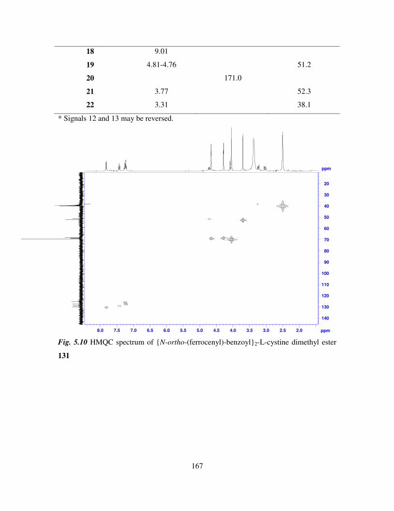
167
18 9.01
19 4.81-4.76 51.2
20 171.0
21 3.77 52.3
22 3.31 38.1
* Signals 12 and 13 may be reversed.
ppm
2.02.53.03.54.04.55.05.56.06.57.07.58.0 ppm
20
30
40
50
60
70
80
90
100
110
120
130
140
Fig. 5.10 HMQC spectrum of N-ortho-(ferrocenyl)-benzoyl2-L-cystine dimethyl ester
131
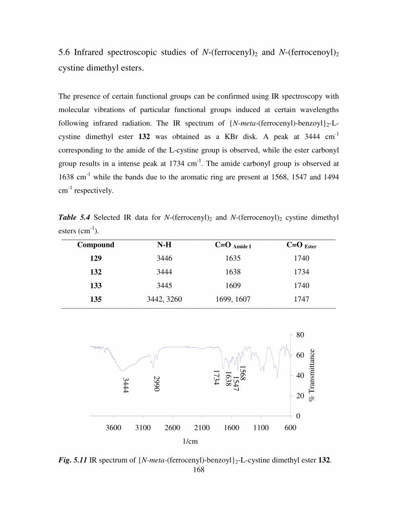
168
5.6 Infrared spectroscopic studies of N-(ferrocenyl)2 and N-(ferrocenoyl)2
cystine dimethyl esters.
The presence of certain functional groups can be confirmed using IR spectroscopy with
molecular vibrations of particular functional groups induced at certain wavelengths
following infrared radiation. The IR spectrum of N-meta-(ferrocenyl)-benzoyl2-L-
cystine dimethyl ester 132 was obtained as a KBr disk. A peak at 3444 cm-1
corresponding to the amide of the L-cystine group is observed, while the ester carbonyl
group results in a intense peak at 1734 cm-1
. The amide carbonyl group is observed at
1638 cm-1
while the bands due to the aromatic ring are present at 1568, 1547 and 1494
cm-1
respectively.
Table 5.4 Selected IR data for N-(ferrocenyl)2 and N-(ferrocenoyl)2 cystine dimethyl
esters (cm-1
).
Compound N-H C=O Amide I C=O Ester
129 3446 1635 1740
132 3444 1638 1734
133 3445 1609 1740
135 3442, 3260 1699, 1607 1747
0
20
40
60
80
600110016002100260031003600
1/cm
% T
ransm
itta
nce
3444
1734
2990
1638
15681547
Fig. 5.11 IR spectrum of N-meta-(ferrocenyl)-benzoyl2-L-cystine dimethyl ester 132.
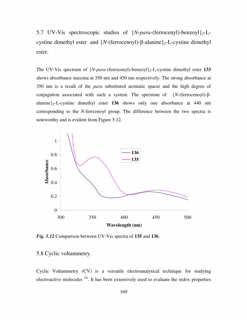
169
5.7 UV-Vis spectroscopic studies of N-para-(ferrocenyl)-benzoyl2-L-
cystine dimethyl ester and N-(ferrocenoyl)-β-alanine2-L-cystine dimethyl
ester.
The UV-Vis spectrum of N-para-(ferrocenyl)-benzoyl2-L-cystine dimethyl ester 133
shows absorbance maxima at 350 nm and 450 nm respectively. The strong absorbance at
350 nm is a result of the para substituted aromatic spacer and the high degree of
conjugation associated with such a system. The spectrum of N-(ferrocenoyl)-β-
alanine2-L-cystine dimethyl ester 136 shows only one absorbance at 440 nm
corresponding to the N-ferrcenoyl group. The difference between the two spectra is
noteworthy and is evident from Figure 5.12.
0
0.2
0.4
0.6
0.8
1
300 350 400 450 500
Wavelength (nm)
Ab
sorb
an
ce
136
135
Fig. 5.12 Comparison between UV-Vis spectra of 135 and 136.
5.8 Cyclic voltammetry.
Cyclic Voltammetry (CV) is a versatile electroanalytical technique for studying
electroactive molecules (5)
. It has been extensively used to evaluate the redox properties
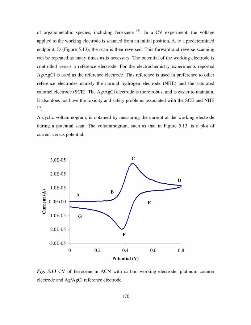
170
of organometallic species, including ferrocene (6)
. In a CV experiment, the voltage
applied to the working electrode is scanned from an initial position, A, to a predetermined
endpoint, D (Figure 5.13), the scan is then reversed. This forward and reverse scanning
can be repeated as many times as is necessary. The potential of the working electrode is
controlled versus a reference electrode. For the electrochemistry experiments reported
Ag/AgCl is used as the reference electrode. This reference is used in preference to other
reference electrodes namely the normal hydrogen electrode (NHE) and the saturated
calomel electrode (SCE). The Ag/AgCl electrode is more robust and is easier to maintain.
It also does not have the toxicity and safety problems associated with the SCE and NHE
(7).
A cyclic voltammogram, is obtained by measuring the current at the working electrode
during a potential scan. The voltammogram, such as that in Figure 5.13, is a plot of
current versus potential.
-3.0E-05
-2.0E-05
-1.0E-05
0.0E+00
1.0E-05
2.0E-05
3.0E-05
0 0.2 0.4 0.6 0.8
Potential (V)
Cu
rren
t (A
)
AB
C
D
E
F
G
Fig. 5.13 CV of ferrocene in ACN with carbon working electrode, platinum counter
electrode and Ag/AgCl reference electrode.
171
A. The initial potential is applied at 0 V. The potential is then scanned in a positive
direction.
B. When the potential is sufficiently positive, the ferrocenyl compound is oxidised to
the ferricenium compound, Fc → Fc+. The anodic current then begins to increase.
C. The anodic current continues to increase until the system surrounding the
electrode is depleted of ferrocenyl compound due to its conversion to ferricenium
species.
D-G. At this stage the ferricenium species begins to get reduced back to the ferrocenyl
species, thus the cathodic current begins. As is the case with a redox system that
behaves in an ideal fashion, the reverse scan roughly mirrors the forward scan.
The reverse scan ends when the current reaches the start point, 0 V.
5.9 Preparation of the self assembled monolayers on gold.
Gold is the substrate of choice for sulphur containing SAMs, as gold electrodes can be
handled in air without oxide formation and they can also tolerate harsh chemical
procedures for the removal of organic contaminants (8)
.
Cleaning protocols form an important part of monolayer preparation. Polishing with 0.05
µm alumina and subsequent electrochemical cycling in 0.5 M H2SO4 acts as both a
cleaning and an annealing process. Cyclic voltammograms resulting from redox cycling
in the gold oxidation region evaluates the cleanliness and the crystallinity of the gold
surface and also gives a true value of the gold surface area (9)
.
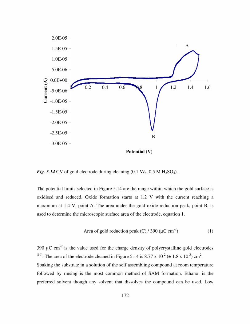
172
-3.0E-05
-2.5E-05
-2.0E-05
-1.5E-05
-1.0E-05
-5.0E-06
0.0E+00
5.0E-06
1.0E-05
1.5E-05
2.0E-05
0 0.2 0.4 0.6 0.8 1 1.2 1.4 1.6
Potential (V)
Cu
rre
nt
(A)
A A
B
Fig. 5.14 CV of gold electrode during cleaning (0.1 V/s, 0.5 M H2SO4).
The potential limits selected in Figure 5.14 are the range within which the gold surface is
oxidised and reduced. Oxide formation starts at 1.2 V with the current reaching a
maximum at 1.4 V, point A. The area under the gold oxide reduction peak, point B, is
used to determine the microscopic surface area of the electrode, equation 1.
Area of gold reduction peak (C) / 390 (µC cm-2
) (1)
390 µC cm-2
is the value used for the charge density of polycrystalline gold electrodes
(10). The area of the electrode cleaned in Figure 5.14 is 8.77 x 10
-2 (± 1.8 x 10
-3) cm
2.
Soaking the substrate in a solution of the self assembling compound at room temperature
followed by rinsing is the most common method of SAM formation. Ethanol is the
preferred solvent though any solvent that dissolves the compound can be used. Low
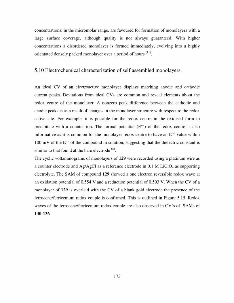
173
concentrations, in the micromolar range, are favoured for formation of monolayers with a
large surface coverage, although quality is not always guaranteed. With higher
concentrations a disordered monolayer is formed immediately, evolving into a highly
orientated densely packed monolayer over a period of hours (11)
.
5.10 Electrochemical characterization of self assembled monolayers.
An ideal CV of an electroactive monolayer displays matching anodic and cathodic
current peaks. Deviations from ideal CVs are common and reveal elements about the
redox centre of the monolayer. A nonzero peak difference between the cathodic and
anodic peaks is as a result of changes in the monolayer structure with respect to the redox
active site. For example, it is possible for the redox centre in the oxidised form to
precipitate with a counter ion. The formal potential (E°΄) of the redox centre is also
informative as it is common for the monolayer redox centre to have an E°΄ value within
100 mV of the E°΄ of the compound in solution, suggesting that the dielectric constant is
similar to that found at the bare electrode (8)
.
The cyclic voltammograms of monolayers of 129 were recorded using a platinum wire as
a counter electrode and Ag/AgCl as a reference electrode in 0.1 M LiClO4 as supporting
electrolyte. The SAM of compound 129 showed a one electron reversible redox wave at
an oxidation potential of 0.554 V and a reduction potential of 0.503 V. When the CV of a
monolayer of 129 is overlaid with the CV of a blank gold electrode the presence of the
ferrocene/ferricenium redox couple is confirmed. This is outlined in Figure 5.15. Redox
waves of the ferrocene/ferricenium redox couple are also observed in CV’s of SAMs of
130-136.
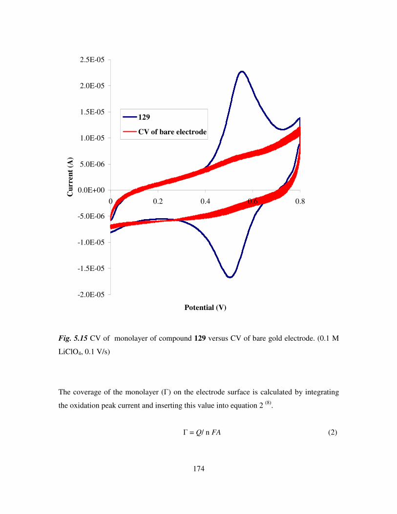
174
-2.0E-05
-1.5E-05
-1.0E-05
-5.0E-06
0.0E+00
5.0E-06
1.0E-05
1.5E-05
2.0E-05
2.5E-05
0 0.2 0.4 0.6 0.8
Potential (V)
Cu
rren
t (A
)
129
CV of bare electrode
Fig. 5.15 CV of monolayer of compound 129 versus CV of bare gold electrode. (0.1 M
LiClO4, 0.1 V/s)
The coverage of the monolayer (Γ) on the electrode surface is calculated by integrating
the oxidation peak current and inserting this value into equation 2 (8)
.
Γ = Q/ n FA (2)
175
Where Γ represents the surface coverage (mol/cm2), Q is the area of peak (C), n is the
number of electrons, F is the Faraday constant and A is the area of the electrode (cm2)
calculated from equation 1. A summary of surface coverage for the prepared monolayers
in aqueous media are presented in Table 5.5.
Monolayers of thiols, sulphides and disulphides are stable over a wide range of potentials
but at very negative potentials in strongly basic electrolytes they are desorbed
quantitatively. The half reaction is outlined in equation 3 (12)
.
AuS-R + e- → Au (0) +
- S-R (3)
During desorption experiments electrodes were immersed in 0.5 M KOH and the
potential was scanned cathodically from 0 to -1.20 V at a scan rate of 0.1 V/s. Figure 5.16
was recorded for a gold electrode with a monolayer of 136 and shows a desorption wave
at -0.90 V versus Ag/AgCl which is due to reductive desorption of the cystine derivatives
attached to the surface.
-1.4E-05
-1.2E-05
-1.0E-05
-8.0E-06
-6.0E-06
-4.0E-06
-2.0E-06
0.0E+00
2.0E-06
-1.4 -1.2 -1 -0.8 -0.6 -0.4 -0.2 0
Potential (V)
Cu
rren
t (A
)
A
Fig. 5.16 Reductive desorption of a monolayer of 136.
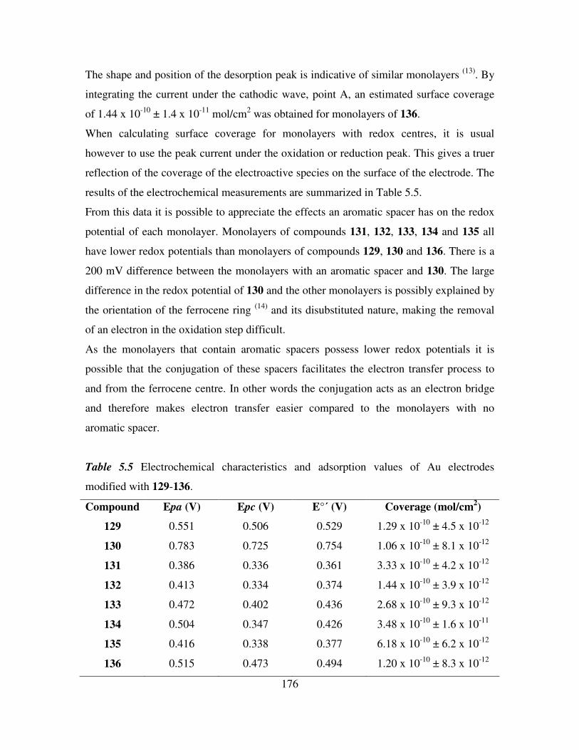
176
The shape and position of the desorption peak is indicative of similar monolayers (13)
. By
integrating the current under the cathodic wave, point A, an estimated surface coverage
of 1.44 x 10⁻¹⁰
± 1.4 x 10⁻¹¹
mol/cm2 was obtained for monolayers of 136.
When calculating surface coverage for monolayers with redox centres, it is usual
however to use the peak current under the oxidation or reduction peak. This gives a truer
reflection of the coverage of the electroactive species on the surface of the electrode. The
results of the electrochemical measurements are summarized in Table 5.5.
From this data it is possible to appreciate the effects an aromatic spacer has on the redox
potential of each monolayer. Monolayers of compounds 131, 132, 133, 134 and 135 all
have lower redox potentials than monolayers of compounds 129, 130 and 136. There is a
200 mV difference between the monolayers with an aromatic spacer and 130. The large
difference in the redox potential of 130 and the other monolayers is possibly explained by
the orientation of the ferrocene ring (14)
and its disubstituted nature, making the removal
of an electron in the oxidation step difficult.
As the monolayers that contain aromatic spacers possess lower redox potentials it is
possible that the conjugation of these spacers facilitates the electron transfer process to
and from the ferrocene centre. In other words the conjugation acts as an electron bridge
and therefore makes electron transfer easier compared to the monolayers with no
aromatic spacer.
Table 5.5 Electrochemical characteristics and adsorption values of Au electrodes
modified with 129-136.
Compound Epa (V) Epc (V) E°΄ (V) Coverage (mol/cm2)
129 0.551 0.506 0.529 1.29 x 10⁻¹⁰
± 4.5 x 10⁻¹²
130 0.783 0.725 0.754 1.06 x 10⁻¹⁰
± 8.1 x 10⁻¹²
131 0.386 0.336 0.361 3.33 x 10⁻¹⁰
± 4.2 x 10⁻¹²
132 0.413 0.334 0.374 1.44 x 10⁻¹⁰
± 3.9 x 10⁻¹²
133 0.472 0.402 0.436 2.68 x 10⁻¹⁰
± 9.3 x 10⁻¹²
134 0.504 0.347 0.426 3.48 x 10⁻¹⁰
± 1.6 x 10⁻¹¹
135 0.416 0.338 0.377 6.18 x 10⁻¹⁰
± 6.2 x 10⁻¹²
136 0.515 0.473 0.494 1.20 x 10⁻¹⁰
± 8.3 x 10⁻¹²
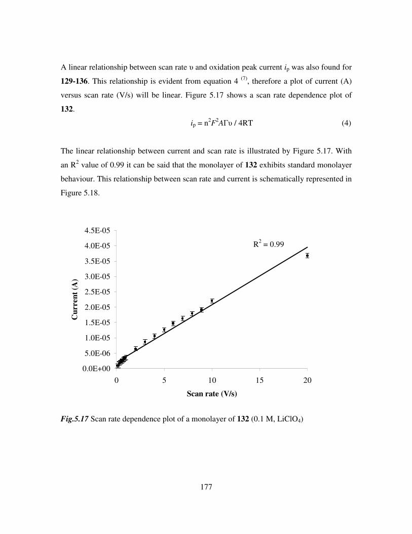
177
A linear relationship between scan rate υ and oxidation peak current ip was also found for
129-136. This relationship is evident from equation 4 (7)
, therefore a plot of current (A)
versus scan rate (V/s) will be linear. Figure 5.17 shows a scan rate dependence plot of
132.
ip = n2F
2AΓυ / 4RT (4)
The linear relationship between current and scan rate is illustrated by Figure 5.17. With
an R2 value of 0.99 it can be said that the monolayer of 132 exhibits standard monolayer
behaviour. This relationship between scan rate and current is schematically represented in
Figure 5.18.
R2 = 0.99
0.0E+00
5.0E-06
1.0E-05
1.5E-05
2.0E-05
2.5E-05
3.0E-05
3.5E-05
4.0E-05
4.5E-05
0 5 10 15 20
Scan rate (V/s)
Cu
rren
t (A
)
Fig.5.17 Scan rate dependence plot of a monolayer of 132 (0.1 M, LiClO4)
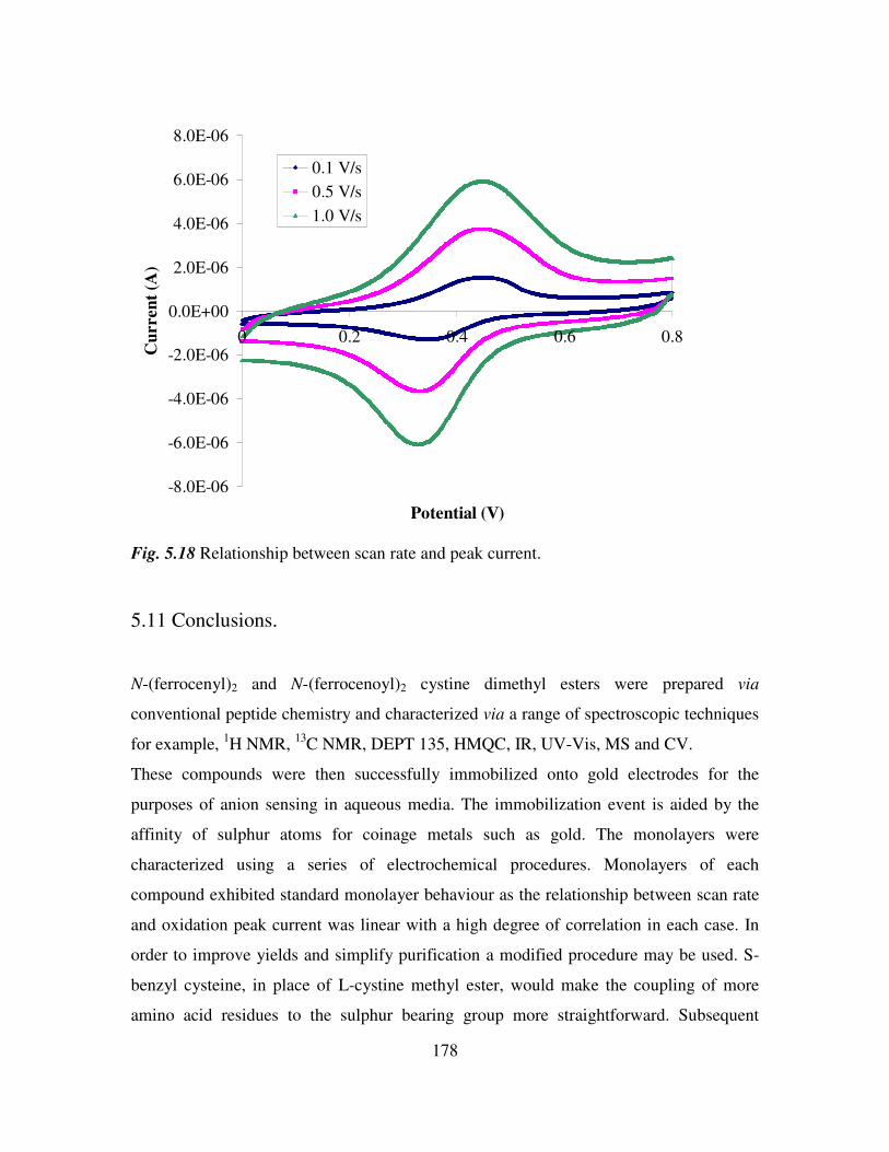
178
-8.0E-06
-6.0E-06
-4.0E-06
-2.0E-06
0.0E+00
2.0E-06
4.0E-06
6.0E-06
8.0E-06
0 0.2 0.4 0.6 0.8
Potential (V)
Cu
rren
t (A
)0.1 V/s
0.5 V/s
1.0 V/s
Fig. 5.18 Relationship between scan rate and peak current.
5.11 Conclusions.
N-(ferrocenyl)2 and N-(ferrocenoyl)2 cystine dimethyl esters were prepared via
conventional peptide chemistry and characterized via a range of spectroscopic techniques
for example, 1H NMR,
13C NMR, DEPT 135, HMQC, IR, UV-Vis, MS and CV.
These compounds were then successfully immobilized onto gold electrodes for the
purposes of anion sensing in aqueous media. The immobilization event is aided by the
affinity of sulphur atoms for coinage metals such as gold. The monolayers were
characterized using a series of electrochemical procedures. Monolayers of each
compound exhibited standard monolayer behaviour as the relationship between scan rate
and oxidation peak current was linear with a high degree of correlation in each case. In
order to improve yields and simplify purification a modified procedure may be used. S-
benzyl cysteine, in place of L-cystine methyl ester, would make the coupling of more
amino acid residues to the sulphur bearing group more straightforward. Subsequent
179
removal of the benzyl group would allow for the immobilization of the receptor on gold
surfaces. Such an approach may result in an easier synthetic route for the receptor
molecules. With the judicious choice of starting materials cavities of varying sizes would
result, increasing the selectivity of these receptors.
180
References
1. Zhang, S., Cardona, C. M., Echegoyen, L., Chem. Comm., 2006, 4461.
2. Beer, P. D., Gale, P. A., Angew. Chem. Int. Ed., 2001, 40, 486.
3. Zhang, S., Palkar, A., Echegoyen, L., Langmuir, 2006, 22, 10732.
4. Bodanzsky, M., ‘Principles of Peptide Synthesis’, Oxford Science Publications,
1992.
5. Chen, Z., Graydon, A. R., Beer, P. D., J. Chem. Soc., Faraday Trans., 1996, 92,
97.
6. Mabbott, G. A., J. Chem. Ed., 1983, 60, 697.
7. Bagotsky, V. S., ‘Fundamentals of Electrochemistry’, John Wiley and Sons,
2006.
8. Finklea, H. O., ‘Self-assembled Monolayers on Electrodes’, Encyclopedia of
Analytical Chemistry, John Wiley and Sons.
9. Hoogvliet, J. C., Dijksma, M., Kamp, B., van Bennekom, W. P., Anal. Chem.,
2000, 72, 2016.
10. Trasatti, S., Petrii, O. A., Pure & Appl. Chem., 1991, 63, 711.
11. Bain, C. D., Troughton, E. B., Tao, Y-T., Evall, J., Whitesides, G. M., Nuzzo, R.
G., J. Am. Chem. Soc., 1989, 111, 321.
12. Walczak, M. M., Popenoe, D. D., Deinhammer, R. S., Lamp, B. D.,Chung, C.,
Porter, M. D., Langmuir, 1991, 7, 2687.
13. Zhang, S., Echegoyen, L., J. Am. Chem. Soc., 2005, 127, 2006.
14. Kondo, T., Takechi, M., Sato, Y., Uosaki, K., J. Electroanal. Chem., 1995, 381,
203.
181
Chapter 6
Anion Binding Studies of N-(ferrocenyl)2 and N-(ferrocenoyl)2
cystine dimethyl esters.
6.1 Introduction.
Redox active receptors have great potential as chemical sensors for the detection of non-
electroactive ionic species. It is possible to convert molecular level interactions into
electrochemically detectable signals. Particular attention has therefore focussed on the
immobilization of redox active groups onto receptors to facilitate the measurement of
host-guest interactions (1)
. Receptors that contain ferrocene are of particular interest as ion
pairing interactions, an important sensing mode, can be switched on by ferrocene
oxidation. Ion-pairing interactions are of particular importance in sensing negatively
charged species. The detection and quantitation of anions is of crucial importance due to
their roles in every day life. They are implicated in various biological processes, the
eutrophication of waterways and the pollution of seas and oceans by pertechnetate
following the reprocessing of nuclear fuel (2)
. Anion receptor design is a particular
challenge as they are larger than cations and therefore have a lower charge to radius ratio
meaning electrostatic interactions are less effective. Anions are also sensitive to pH,
therefore the receptor must act within the pH range of the anion. They also display a wide
range of geometries making receptor design more challenging.
The majority of results in the field of anion sensing have been reported in bulk organic
solvent. It is important to transfer these recognition events to solid surfaces as the
recognition event is greatly enhanced at the surface solution interface. This will therefore
aid the sensing of anions in aqueous media. This laboratory has recently published the
anion sensing ability of ferrocenyl receptors in organic solvent (3, 4)
. It was anticipated
that by confining the receptor molecules to the electrode surface it would facilitate the
binding of various anions in aqueous media. The peptide residues would utilise the
hydrogen bonding ability of the amide bond, while the ferrocene moiety would impart
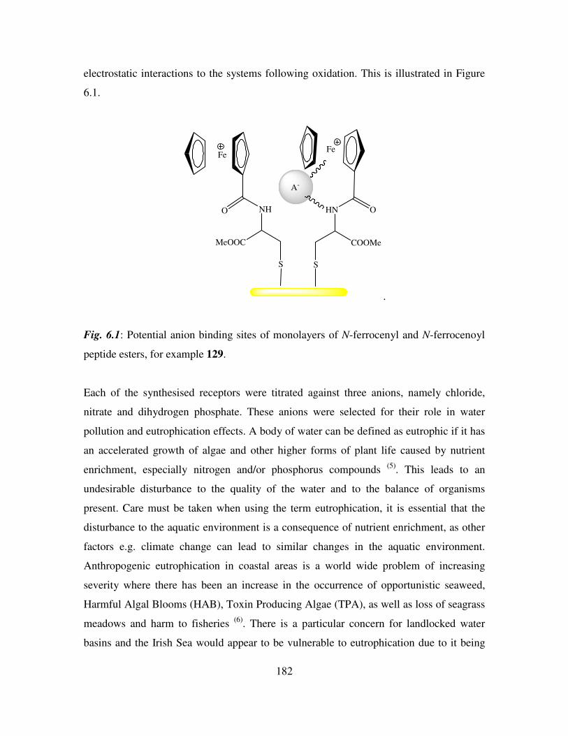
182
electrostatic interactions to the systems following oxidation. This is illustrated in Figure
6.1.
Fe
O NH
MeOOC
S S
COOMe
HN O
Fe
A-
.
Fig. 6.1: Potential anion binding sites of monolayers of N-ferrocenyl and N-ferrocenoyl
peptide esters, for example 129.
Each of the synthesised receptors were titrated against three anions, namely chloride,
nitrate and dihydrogen phosphate. These anions were selected for their role in water
pollution and eutrophication effects. A body of water can be defined as eutrophic if it has
an accelerated growth of algae and other higher forms of plant life caused by nutrient
enrichment, especially nitrogen and/or phosphorus compounds (5)
. This leads to an
undesirable disturbance to the quality of the water and to the balance of organisms
present. Care must be taken when using the term eutrophication, it is essential that the
disturbance to the aquatic environment is a consequence of nutrient enrichment, as other
factors e.g. climate change can lead to similar changes in the aquatic environment.
Anthropogenic eutrophication in coastal areas is a world wide problem of increasing
severity where there has been an increase in the occurrence of opportunistic seaweed,
Harmful Algal Blooms (HAB), Toxin Producing Algae (TPA), as well as loss of seagrass
meadows and harm to fisheries (6)
. There is a particular concern for landlocked water
basins and the Irish Sea would appear to be vulnerable to eutrophication due to it being
183
an inner sea and it receiving large quantities of anthropogenic nutrients from river
discharges.
Eutrophication is therefore the major threat to water quality in Ireland (7)
. Nutrient losses
from agricultural practices have been implicated directly and indirectly for the
eutrophication of surface water in Ireland and the UK. The main point source pollution
from agriculture is inappropriately managed farmyard dirty water. This generally consists
of farmyard run off, parlour washings, silage and farmyard manure effluents. Land
spreading is the most common practice for managing dairy farm dirty water, however,
this can lead to the degradation of surface and ground water.
The over fertilisation of grassland has also been identified as a common problem in
Ireland. It has recently been reported that 70% of the anthropogenic load of phosphorus
in Irish freshwater is from agricultural sources. Nitrogen is also an important contributory
factor to eutrophication and has human health implications when present in high
concentrations (8)
. Therefore, the development of novel sensory devices for the detection
of nitrates, phosphates and other anionic pollutants is of considerable importance to the
environment and the future of our waterways. These anions were also selected as they
display an array or geometries namely, spherical, trigonal planar and tetrahedral.
6.2 Electrochemical anion coordination studies of monolayers of N-
(ferrocenyl)2 and N-(ferrocenoyl)2 cystine dimethyl esters in aqueous media.
The anion sensing ability of monolayers of N-ferrocenyl and N-ferrocenoyl peptide esters
were investigated by cyclic voltammetry experiments. All the electrochemistry
experiments were carried out on equipment kindly supplied by Dr. Mary Pryce. A
standard three-electrode setup, Figure 6.2, was employed in all experiments where gold
was the working electrode, platinum wire was the counter electrode and Ag/AgCl was the
reference electrode. The CVs were measured in deionized water with lithium perchlorate
(LiClO4) as supporting electrolyte. The measurements were carried out at room
temperature and at a scan rate of 0.1 V/s.
Important features of CVs include the position of the anodic peak potential (Epa) and the
cathodic peak potential (Epc) (9)
. The formal oxidation potential (E°΄) for an

184
electrochemically reversible couple, in which electrons are rapidly exchanged, is centred
between Epa and Epc and given by equation 1.
E°΄ = Epa + Epc (1)
2
The magnitude of oxidation and reduction peak current should also be noted as changes
in the peak current provide information on the surface coverage of redox molecules on
the electrode surface.
Electrolyte (LiClO4)
Counter Electrode (Pt Wire)
Working Electrode (Gold)Reference Electrode (Ag/AgCl)
Fig. 6.2 Schematic diagram of an electrochemical cell as used in anion titration
experiments.
The monolayers of each compound 129-136 were preliminarily titrated with 2 ml of a
0.01 M solution of chloride, nitrate and dihydrogen phosphate anions, as their
tetrabutylammonium salts. It was anticipated that the CVs would display a negative shift
in the Fc/Fc+ redox couple. This is due to the redox centre becoming more easily oxidised
following an increase in electron density upon anion binding. However in each case the
change in redox potential was minimal but a noticeable change in the peak current of the
oxidation and reduction peaks was observed. It is reasonable to suggest that this decrease
in peak current is as a result of irreversible binding between the receptor and the anion.
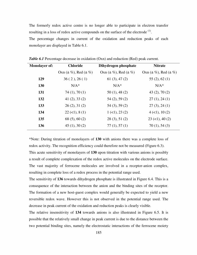
185
The formerly redox active centre is no longer able to participate in electron transfer
resulting in a loss of redox active compounds on the surface of the electrode (1)
.
The percentage changes in current of the oxidation and reduction peaks of each
monolayer are displayed in Table 6.1.
Table 6.1 Percentage decrease in oxidation (Oxn) and reduction (Red) peak current.
Monolayer of: Chloride Dihydrogen phosphate Nitrate
Oxn (± %), Red (± %) Oxn (± %), Red (± %) Oxn (± %), Red (± %)
129 36 ( 2 ), 26 ( 1) 61 (3), 47 (2) 55 (2), 62 (1)
130 N/A* N/A* N/A*
131 74 (1), 70 (1) 50 (1), 48 (2) 43 (2), 70 (2)
132 41 (2), 33 (2) 54 (2), 59 (2) 27 (1), 24 (1)
133 26 (2), 31 (2) 54 (3), 59 (2) 27 (3), 24 (1)
134 22 (<1), 8 (1) 1 (<1), 23 (2) 4 (<1), 10 (2)
135 68 (5), 60 (2) 28 (3), 51 (2) 23 (<1), 40 (2)
136 45 (1), 30 (2) 77 (1), 57 (1) 70 (1), 54 (3)
*Note: During titration of monolayers of 130 with anions there was a complete loss of
redox activity. The recognition efficiency could therefore not be measured (Figure 6.3).
This acute sensitivity of monolayers of 130 upon titration with various anions is possibly
a result of complete complexation of the redox active molecules on the electrode surface.
The vast majority of ferrocene molecules are involved in a receptor-anion complex,
resulting in complete loss of a redox process in the potential range used.
The sensitivity of 136 towards dihydrogen phosphate is illustrated in Figure 6.4. This is a
consequence of the interaction between the anion and the binding sites of the receptor.
The formation of a new host-guest complex would generally be expected to yield a new
reversible redox wave. However this is not observed in the potential range used. The
decrease in peak current of the oxidation and reduction peaks is clearly visible.
The relative insensitivity of 134 towards anions is also illustrated in Figure 6.5. It is
possible that the relatively small change in peak current is due to the distance between the
two potential binding sites, namely the electrostatic interactions of the ferrocene moiety
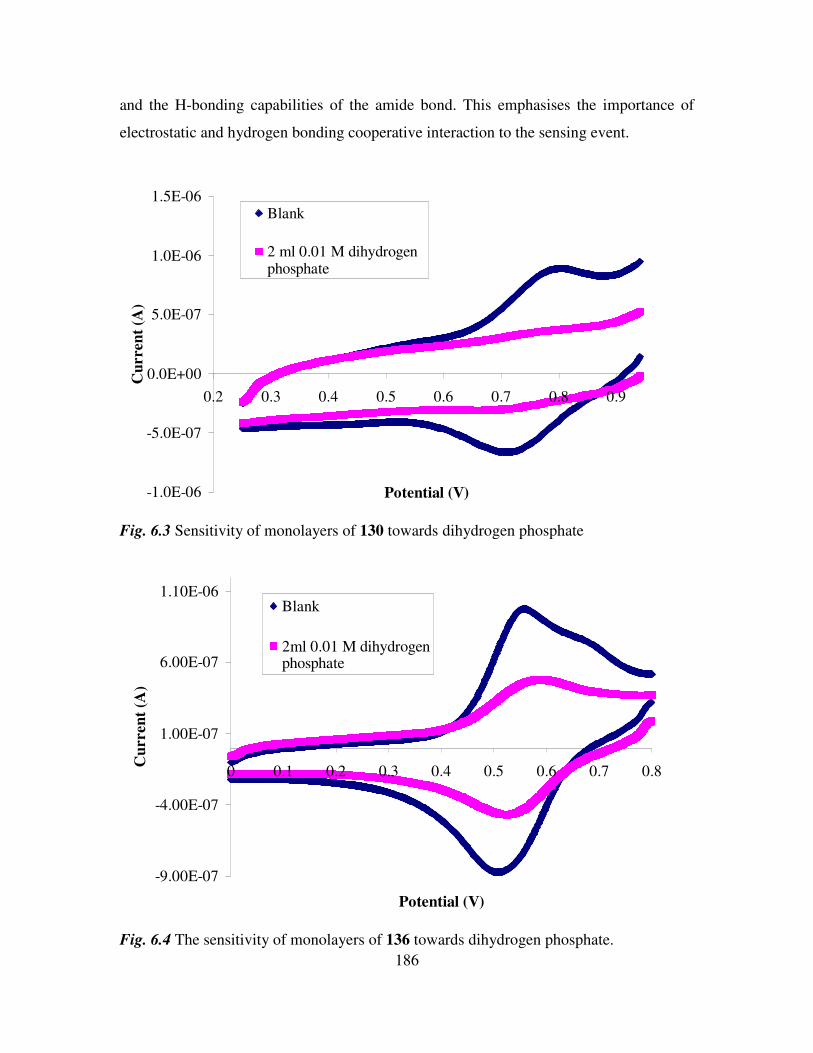
186
and the H-bonding capabilities of the amide bond. This emphasises the importance of
electrostatic and hydrogen bonding cooperative interaction to the sensing event.
-1.0E-06
-5.0E-07
0.0E+00
5.0E-07
1.0E-06
1.5E-06
0.2 0.3 0.4 0.5 0.6 0.7 0.8 0.9
Potential (V)
Cu
rre
nt
(A)
Blank
2 ml 0.01 M dihydrogenphosphate
Fig. 6.3 Sensitivity of monolayers of 130 towards dihydrogen phosphate
-9.00E-07
-4.00E-07
1.00E-07
6.00E-07
1.10E-06
0 0.1 0.2 0.3 0.4 0.5 0.6 0.7 0.8
Potential (V)
Cu
rren
t (A
)
Blank
2ml 0.01 M dihydrogenphosphate
Fig. 6.4 The sensitivity of monolayers of 136 towards dihydrogen phosphate.
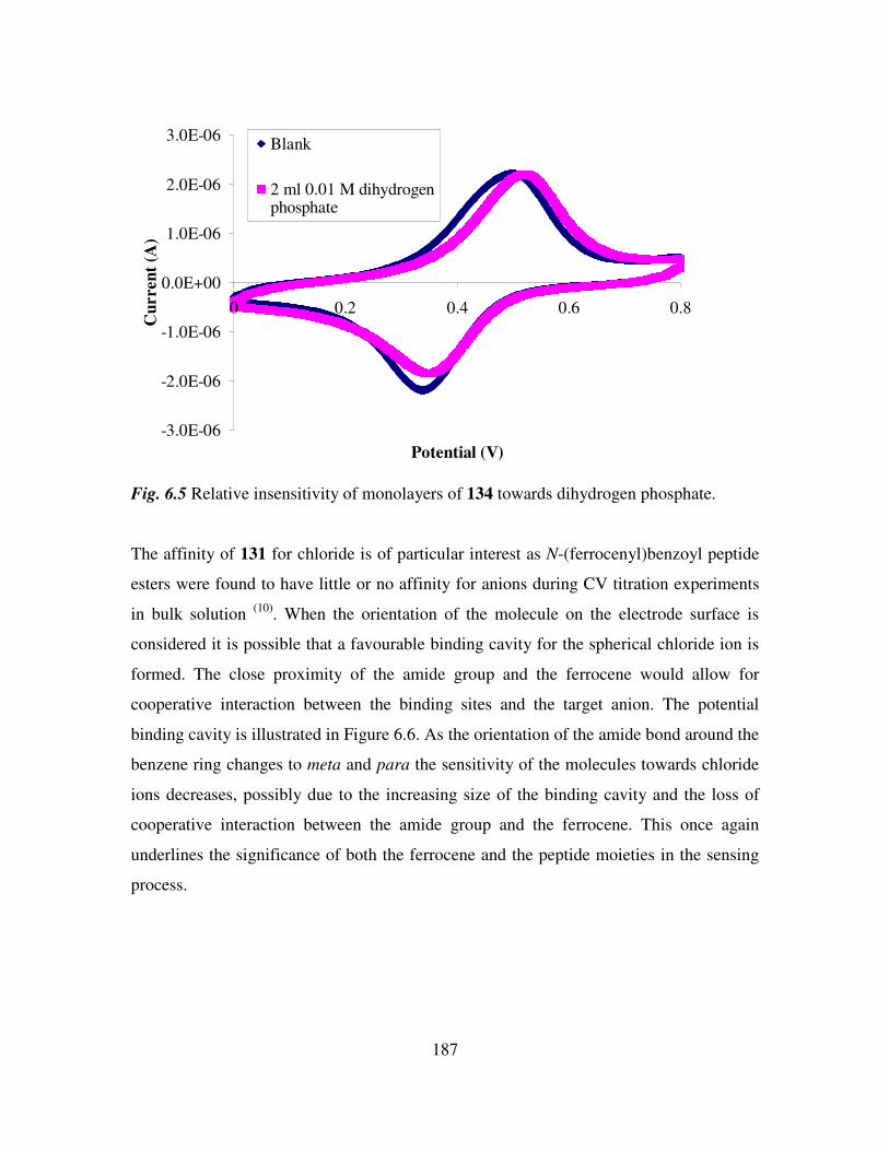
187
-3.0E-06
-2.0E-06
-1.0E-06
0.0E+00
1.0E-06
2.0E-06
3.0E-06
0 0.2 0.4 0.6 0.8
Potential (V)
Cu
rre
nt
(A)
Blank
2 ml 0.01 M dihydrogenphosphate
Fig. 6.5 Relative insensitivity of monolayers of 134 towards dihydrogen phosphate.
The affinity of 131 for chloride is of particular interest as N-(ferrocenyl)benzoyl peptide
esters were found to have little or no affinity for anions during CV titration experiments
in bulk solution (10)
. When the orientation of the molecule on the electrode surface is
considered it is possible that a favourable binding cavity for the spherical chloride ion is
formed. The close proximity of the amide group and the ferrocene would allow for
cooperative interaction between the binding sites and the target anion. The potential
binding cavity is illustrated in Figure 6.6. As the orientation of the amide bond around the
benzene ring changes to meta and para the sensitivity of the molecules towards chloride
ions decreases, possibly due to the increasing size of the binding cavity and the loss of
cooperative interaction between the amide group and the ferrocene. This once again
underlines the significance of both the ferrocene and the peptide moieties in the sensing
process.
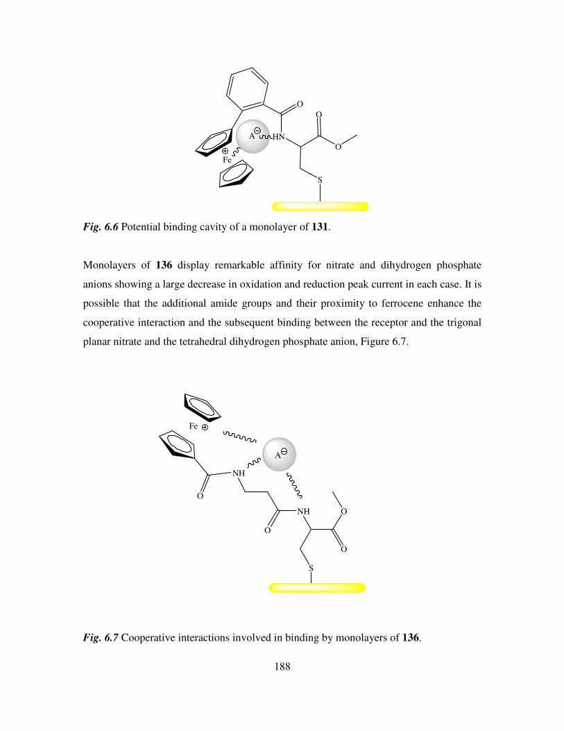
188
Fe
O
HN
O
O
S
A
Fig. 6.6 Potential binding cavity of a monolayer of 131.
Monolayers of 136 display remarkable affinity for nitrate and dihydrogen phosphate
anions showing a large decrease in oxidation and reduction peak current in each case. It is
possible that the additional amide groups and their proximity to ferrocene enhance the
cooperative interaction and the subsequent binding between the receptor and the trigonal
planar nitrate and the tetrahedral dihydrogen phosphate anion, Figure 6.7.
Fe
O
NH
O
NH
O
S
O
A
Fig. 6.7 Cooperative interactions involved in binding by monolayers of 136.
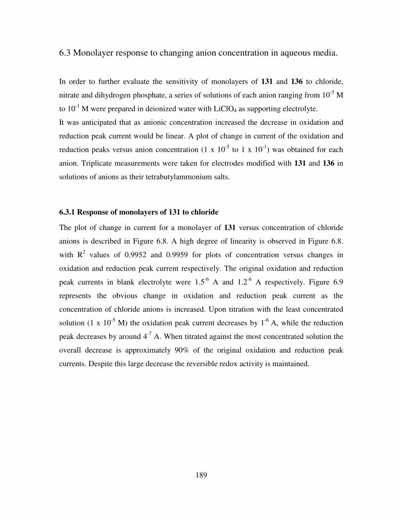
189
6.3 Monolayer response to changing anion concentration in aqueous media.
In order to further evaluate the sensitivity of monolayers of 131 and 136 to chloride,
nitrate and dihydrogen phosphate, a series of solutions of each anion ranging from 10-5
M
to 10-1
M were prepared in deionized water with LiClO4 as supporting electrolyte.
It was anticipated that as anionic concentration increased the decrease in oxidation and
reduction peak current would be linear. A plot of change in current of the oxidation and
reduction peaks versus anion concentration (1 x 10⁻⁵
to 1 x 10⁻¹
) was obtained for each
anion. Triplicate measurements were taken for electrodes modified with 131 and 136 in
solutions of anions as their tetrabutylammonium salts.
6.3.1 Response of monolayers of 131 to chloride
The plot of change in current for a monolayer of 131 versus concentration of chloride
anions is described in Figure 6.8. A high degree of linearity is observed in Figure 6.8.
with R2 values of 0.9952 and 0.9959 for plots of concentration versus changes in
oxidation and reduction peak current respectively. The original oxidation and reduction
peak currents in blank electrolyte were 1.5-6
A and 1.2-6
A respectively. Figure 6.9
represents the obvious change in oxidation and reduction peak current as the
concentration of chloride anions is increased. Upon titration with the least concentrated
solution (1 x 10⁻⁵
M) the oxidation peak current decreases by 1-6
A, while the reduction
peak decreases by around 4-7
A. When titrated against the most concentrated solution the
overall decrease is approximately 90% of the original oxidation and reduction peak
currents. Despite this large decrease the reversible redox activity is maintained.
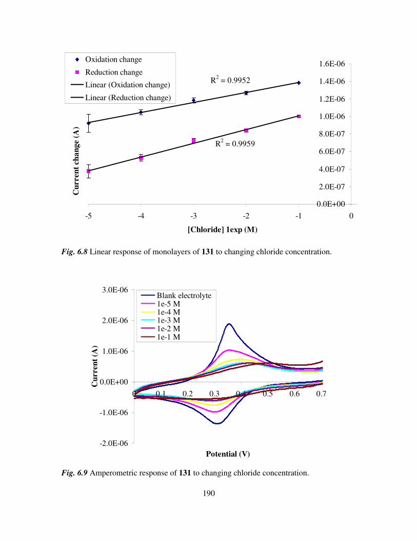
190
R2 = 0.9952
R2 = 0.9959
0.0E+00
2.0E-07
4.0E-07
6.0E-07
8.0E-07
1.0E-06
1.2E-06
1.4E-06
1.6E-06
-5 -4 -3 -2 -1 0
[Chloride] 1exp (M)
Cu
rren
t ch
an
ge (
A)
Oxidation change
Reduction change
Linear (Oxidation change)
Linear (Reduction change)
Fig. 6.8 Linear response of monolayers of 131 to changing chloride concentration.
-2.0E-06
-1.0E-06
0.0E+00
1.0E-06
2.0E-06
3.0E-06
0 0.1 0.2 0.3 0.4 0.5 0.6 0.7
Potential (V)
Cu
rren
t (A
)
Blank electrolyte 1e-5 M1e-4 M1e-3 M1e-2 M1e-1 M
Fig. 6.9 Amperometric response of 131 to changing chloride concentration.
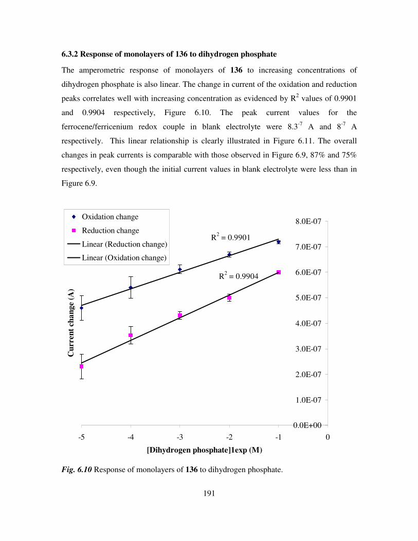
191
6.3.2 Response of monolayers of 136 to dihydrogen phosphate
The amperometric response of monolayers of 136 to increasing concentrations of
dihydrogen phosphate is also linear. The change in current of the oxidation and reduction
peaks correlates well with increasing concentration as evidenced by R2 values of 0.9901
and 0.9904 respectively, Figure 6.10. The peak current values for the
ferrocene/ferricenium redox couple in blank electrolyte were 8.3-7
A and 8-7
A
respectively. This linear relationship is clearly illustrated in Figure 6.11. The overall
changes in peak currents is comparable with those observed in Figure 6.9, 87% and 75%
respectively, even though the initial current values in blank electrolyte were less than in
Figure 6.9.
R2 = 0.9904
R2 = 0.9901
0.0E+00
1.0E-07
2.0E-07
3.0E-07
4.0E-07
5.0E-07
6.0E-07
7.0E-07
8.0E-07
-5 -4 -3 -2 -1 0
[Dihydrogen phosphate]1exp (M)
Cu
rren
t ch
an
ge (
A)
Oxidation change
Reduction change
Linear (Reduction change)
Linear (Oxidation change)
Fig. 6.10 Response of monolayers of 136 to dihydrogen phosphate.
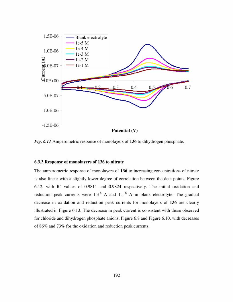
192
-1.5E-06
-1.0E-06
-5.0E-07
0.0E+00
5.0E-07
1.0E-06
1.5E-06
0 0.1 0.2 0.3 0.4 0.5 0.6 0.7
Potential (V)
Cu
rren
t (A
)
Blank electrolyte
1e-5 M
1e-4 M
1e-3 M
1e-2 M
1e-1 M
Fig. 6.11 Amperometric response of monolayers of 136 to dihydrogen phosphate.
6.3.3 Response of monolayers of 136 to nitrate
The amperometric response of monolayers of 136 to increasing concentrations of nitrate
is also linear with a slightly lower degree of correlation between the data points, Figure
6.12, with R2 values of 0.9811 and 0.9824 respectively. The initial oxidation and
reduction peak currents were 1.3-6
A and 1.1-6
A in blank electrolyte. The gradual
decrease in oxidation and reduction peak currents for monolayers of 136 are clearly
illustrated in Figure 6.13. The decrease in peak current is consistent with those observed
for chloride and dihydrogen phosphate anions, Figure 6.8 and Figure 6.10, with decreases
of 86% and 73% for the oxidation and reduction peak currents.
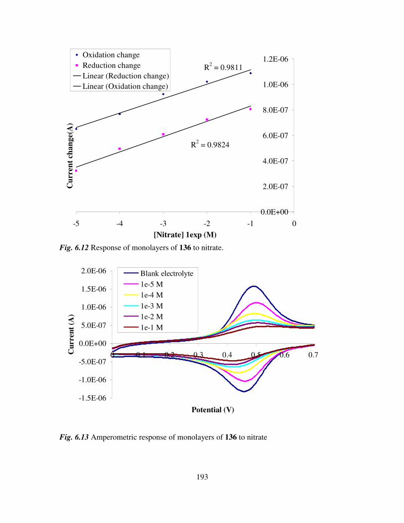
193
R2 = 0.9824
R2 = 0.9811
0.0E+00
2.0E-07
4.0E-07
6.0E-07
8.0E-07
1.0E-06
1.2E-06
-5 -4 -3 -2 -1 0
[Nitrate] 1exp (M)
Cu
rren
t ch
an
ge(
A)
Oxidation change
Reduction change
Linear (Reduction change)
Linear (Oxidation change)
Fig. 6.12 Response of monolayers of 136 to nitrate.
-1.5E-06
-1.0E-06
-5.0E-07
0.0E+00
5.0E-07
1.0E-06
1.5E-06
2.0E-06
0 0.1 0.2 0.3 0.4 0.5 0.6 0.7
Potential (V)
Cu
rren
t (A
)
Blank electrolyte
1e-5 M
1e-4 M
1e-3 M
1e-2 M
1e-1 M
Fig. 6.13 Amperometric response of monolayers of 136 to nitrate
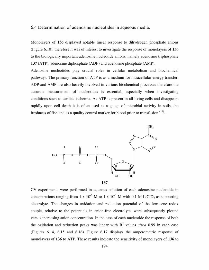
194
6.4 Determination of adenosine nucleotides in aqueous media.
Monolayers of 136 displayed notable linear response to dihydrogen phosphate anions
(Figure 6.10), therefore it was of interest to investigate the response of monolayers of 136
to the biologically important adenosine nucleotide anions, namely adenosine triphosphate
137 (ATP), adenosine diphosphate (ADP) and adenosine phosphate (AMP).
Adenosine nucleotides play crucial roles in cellular metabolism and biochemical
pathways. The primary function of ATP is as a medium for intracellular energy transfer.
ADP and AMP are also heavily involved in various biochemical processes therefore the
accurate measurement of nucleotides is essential, especially when investigating
conditions such as cardiac ischemia. As ATP is present in all living cells and disappears
rapidly upon cell death it is often used as a gauge of microbial activity in soils, the
freshness of fish and as a quality control marker for blood prior to transfusion (11)
.
N
NN
N
NH2
O
OHOH
HH
HH
OP
O
O-
OP
O-
O
OP
O-
O
HO
137
CV experiments were performed in aqueous solution of each adenosine nucleotide in
concentrations ranging from 1 x 10⁻⁹
M to 1 x 10⁻¹
M with 0.1 M LiClO4 as supporting
electrolyte. The changes in oxidation and reduction potential of the ferrocene redox
couple, relative to the potentials in anion-free electrolyte, were subsequently plotted
versus increasing anion concentration. In the case of each nucleotide the response of both
the oxidation and reduction peaks was linear with R2 values circa 0.99 in each case
(Figures 6.14, 6.15 and 6.16). Figure 6.17 displays the amperometric response of
monolayers of 136 to ATP. These results indicate the sensitivity of monolayers of 136 to

195
adenosine nucleotides in aqueous media. Sensitivity in the nanomolar range was observed
with the electroactivity of the ferrocene/ferricenium redox couple maintained throughout.
There were minimal cathodic or anodic shifts in the position of the redox couple. No new
reversible redox couples corresponding to a 136-anion complex was observed in the
potential range used.
In titration with AMP the overall decrease in the current of the oxidation and reduction
peaks was 96% and 90% respectively. When monolayers of 136 were titrated against
ADP, the overall response was similar to that of AMP titrations with a decrease of 89%
and 93% in the oxidation and reduction peaks of the ferrocene/ferricenium redox couple
compared to in blank electrolyte. Similarly during experiments with ATP the overall
decreases were 84% and 79% for the oxidation and reduction peaks respectively. These
large decreases in peak currents demonstrate the sensitivity of such monolayers to
adenosine nucleotides, however redox activity was maintained throughout, thus
facilitating the accurate measurement of changes in redox activity.
R2 = 0.9892
R2 = 0.9810
0.0E+00
5.0E-07
1.0E-06
1.5E-06
2.0E-06
2.5E-06
3.0E-06
3.5E-06
4.0E-06
4.5E-06
-9 -8 -7 -6 -5 -4 -3 -2 -1 0[AMP] 1exp (M)
Cu
rren
t ch
an
ge (
A)
Oxidation change
Reduction change
Linear (Oxidation change)
Linear (Reduction change)
Fig. 6.14 Response of monolayers of 136 to AMP.
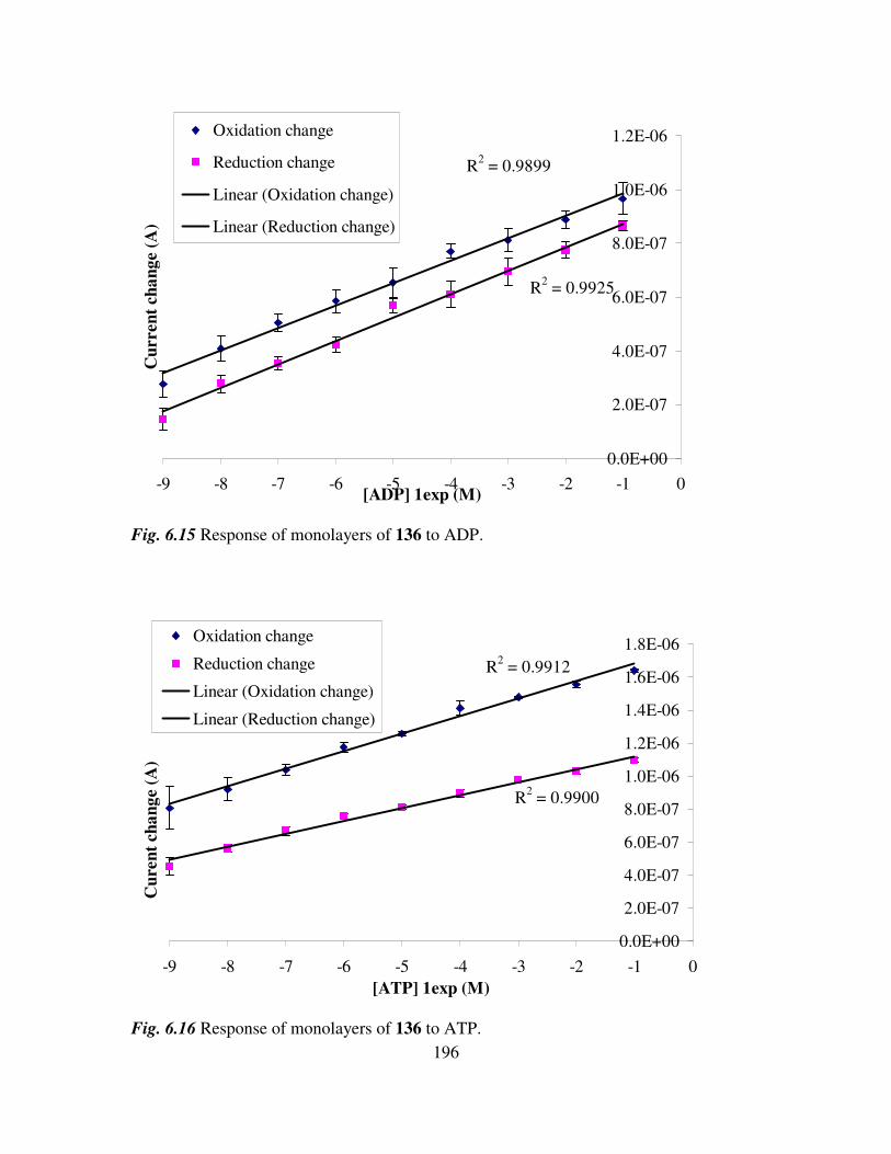
196
R2 = 0.9899
R2 = 0.9925
0.0E+00
2.0E-07
4.0E-07
6.0E-07
8.0E-07
1.0E-06
1.2E-06
-9 -8 -7 -6 -5 -4 -3 -2 -1 0[ADP] 1exp (M)
Cu
rren
t ch
an
ge (
A)
Oxidation change
Reduction change
Linear (Oxidation change)
Linear (Reduction change)
Fig. 6.15 Response of monolayers of 136 to ADP.
R2 = 0.9912
R2 = 0.9900
0.0E+00
2.0E-07
4.0E-07
6.0E-07
8.0E-07
1.0E-06
1.2E-06
1.4E-06
1.6E-06
1.8E-06
-9 -8 -7 -6 -5 -4 -3 -2 -1 0
[ATP] 1exp (M)
Cu
ren
t ch
an
ge (
A)
Oxidation change
Reduction change
Linear (Oxidation change)
Linear (Reduction change)
Fig. 6.16 Response of monolayers of 136 to ATP.
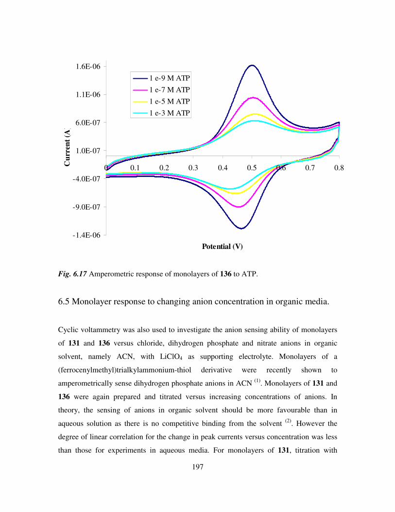
197
-1.4E-06
-9.0E-07
-4.0E-07
1.0E-07
6.0E-07
1.1E-06
1.6E-06
0 0.1 0.2 0.3 0.4 0.5 0.6 0.7 0.8
Potential (V)
Cu
rren
t (A
)
1 e-9 M ATP
1 e-7 M ATP
1 e-5 M ATP
1 e-3 M ATP
Fig. 6.17 Amperometric response of monolayers of 136 to ATP.
6.5 Monolayer response to changing anion concentration in organic media.
Cyclic voltammetry was also used to investigate the anion sensing ability of monolayers
of 131 and 136 versus chloride, dihydrogen phosphate and nitrate anions in organic
solvent, namely ACN, with LiClO4 as supporting electrolyte. Monolayers of a
(ferrocenylmethyl)trialkylammonium-thiol derivative were recently shown to
amperometrically sense dihydrogen phosphate anions in ACN (1)
. Monolayers of 131 and
136 were again prepared and titrated versus increasing concentrations of anions. In
theory, the sensing of anions in organic solvent should be more favourable than in
aqueous solution as there is no competitive binding from the solvent (2)
. However the
degree of linear correlation for the change in peak currents versus concentration was less
than those for experiments in aqueous media. For monolayers of 131, titration with
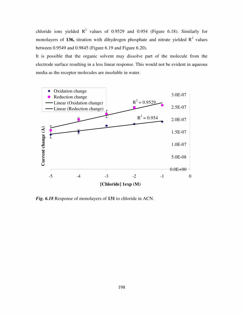
198
chloride ions yielded R2 values of 0.9529 and 0.954 (Figure 6.18). Similarly for
monolayers of 136, titration with dihydrogen phosphate and nitrate yielded R2 values
between 0.9549 and 0.9845 (Figure 6.19 and Figure 6.20).
It is possible that the organic solvent may dissolve part of the molecule from the
electrode surface resulting in a less linear response. This would not be evident in aqueous
media as the receptor molecules are insoluble in water.
R2 = 0.954
R2 = 0.9529
0.0E+00
5.0E-08
1.0E-07
1.5E-07
2.0E-07
2.5E-07
3.0E-07
-5 -4 -3 -2 -1 0
[Chloride] 1exp (M)
Cu
rren
t ch
an
ge (
A)
Oxidation change
Reduction change
Linear (Oxidation change)
Linear (Reduction change)
Fig. 6.18 Response of monolayers of 131 to chloride in ACN.
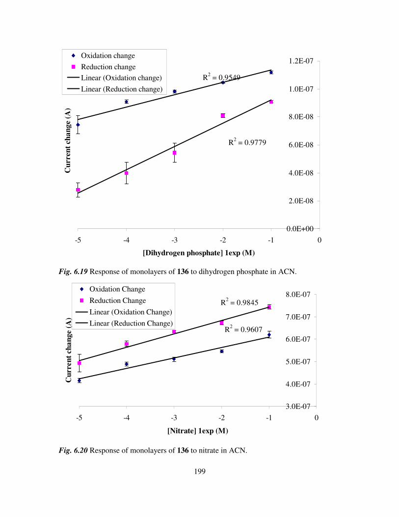
199
R2 = 0.9549
R2 = 0.9779
0.0E+00
2.0E-08
4.0E-08
6.0E-08
8.0E-08
1.0E-07
1.2E-07
-5 -4 -3 -2 -1 0
[Dihydrogen phosphate] 1exp (M)
Cu
rren
t ch
an
ge
(A)
Oxidation change
Reduction change
Linear (Oxidation change)
Linear (Reduction change)
Fig. 6.19 Response of monolayers of 136 to dihydrogen phosphate in ACN.
R2 = 0.9607
R2 = 0.9845
3.0E-07
4.0E-07
5.0E-07
6.0E-07
7.0E-07
8.0E-07
-5 -4 -3 -2 -1 0
[Nitrate] 1exp (M)
Cu
rren
t ch
an
ge (
A)
Oxidation Change
Reduction Change
Linear (Oxidation Change)
Linear (Reduction Change)
Fig. 6.20 Response of monolayers of 136 to nitrate in ACN.
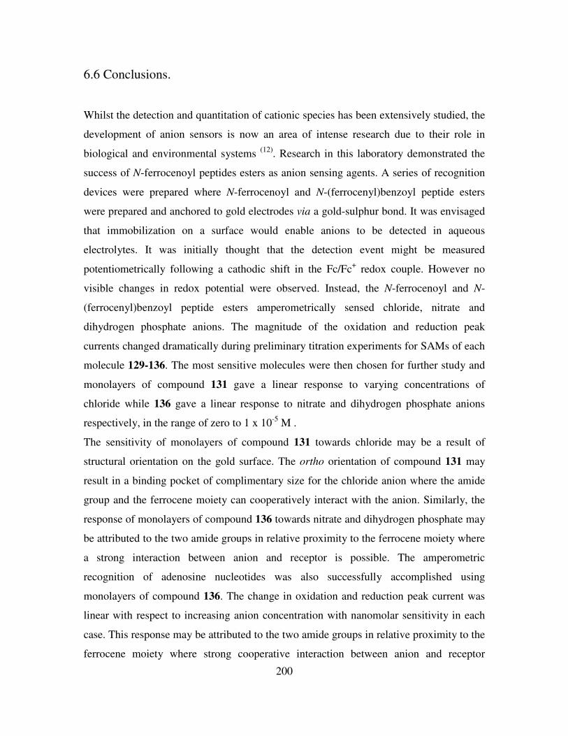
200
6.6 Conclusions.
Whilst the detection and quantitation of cationic species has been extensively studied, the
development of anion sensors is now an area of intense research due to their role in
biological and environmental systems (12)
. Research in this laboratory demonstrated the
success of N-ferrocenoyl peptides esters as anion sensing agents. A series of recognition
devices were prepared where N-ferrocenoyl and N-(ferrocenyl)benzoyl peptide esters
were prepared and anchored to gold electrodes via a gold-sulphur bond. It was envisaged
that immobilization on a surface would enable anions to be detected in aqueous
electrolytes. It was initially thought that the detection event might be measured
potentiometrically following a cathodic shift in the Fc/Fc+ redox couple. However no
visible changes in redox potential were observed. Instead, the N-ferrocenoyl and N-
(ferrocenyl)benzoyl peptide esters amperometrically sensed chloride, nitrate and
dihydrogen phosphate anions. The magnitude of the oxidation and reduction peak
currents changed dramatically during preliminary titration experiments for SAMs of each
molecule 129-136. The most sensitive molecules were then chosen for further study and
monolayers of compound 131 gave a linear response to varying concentrations of
chloride while 136 gave a linear response to nitrate and dihydrogen phosphate anions
respectively, in the range of zero to 1 x 10⁻⁵
M .
The sensitivity of monolayers of compound 131 towards chloride may be a result of
structural orientation on the gold surface. The ortho orientation of compound 131 may
result in a binding pocket of complimentary size for the chloride anion where the amide
group and the ferrocene moiety can cooperatively interact with the anion. Similarly, the
response of monolayers of compound 136 towards nitrate and dihydrogen phosphate may
be attributed to the two amide groups in relative proximity to the ferrocene moiety where
a strong interaction between anion and receptor is possible. The amperometric
recognition of adenosine nucleotides was also successfully accomplished using
monolayers of compound 136. The change in oxidation and reduction peak current was
linear with respect to increasing anion concentration with nanomolar sensitivity in each
case. This response may be attributed to the two amide groups in relative proximity to the
ferrocene moiety where strong cooperative interaction between anion and receptor
201
binding sites is possible. The small degree of error following triplicate measurements
indicates a high degree of reproducibility. To the best of our knowledge monolayers of
compound 136 are the first ferrocene based monolayers that can amperometrically sense
dihydrogen phosphate and adenosine nucleotides in aqueous media. These findings have
major potential for the development of novel sensory devices capable of detecting various
anionic species in water.
In future, 1,1’-N,N’-ferrocenoylbispeptide esters could be immobilized onto gold
electrodes. This would create a large binding cavity and the additional amide groups
would aid complexation. Also, the inclusion of a pyrrole or pyridine as a spacer groups
between the ferrocene and peptide moieties would also provide additional hydrogen
bonding groups and may increase sensitivity. In addition, the detection of neurologically
important anions such as lactate (CH3CH(OH)COO−), pyruvate (CH3COCOO
−) and
glutamate (HOOC–CH(NH2)CH2CH2COO−) may also be investigated.
202
References.
1. Reynes, O., Bucher, C., Moutet, J-C., Royal, G., Saint-Aman, E., Inorg. Chim.
Acta., 2008, 261, 1784.
2. Beer, P. D., Gale, P. A., Angew. Chem. Int. Ed., 2001, 40, 486.
3. Goel, A., Brennan, N., Brady, N., Kenny, P. T. M., Biosens. Bioelectron., 2007,
22, 2047.
4. Gallagher, J. F., Kenny, P. T. M., Sheehy, M. J., Inorg. Chem. Comm., 1999, 2,
327.
5. Gowan, R. J., Tett, P., Kennington, K., Mills, D. K., Shammon, T. M., Stewart, B.
M., Greenwood, N., Flanagan, C., Devlin, M., Wither, A., Estuarine, Coastal and
Shelf Sci., 2008, 76, 239.
6. Mc Intyre, A. D., Marine Pollution Bulletin, 1995, 31, 147.
7. Dunne, E. J., Culleton, N., O’Donovan, G., Harrington, R., Olsen, A. E., Ecol.
Eng., 2005, 24, 221.
8. Clenaghan, C., Collins, C., Crowe, C., 2001, Phosphorous Regulation National
Implementation Report, EPA.
9. Bard, A. J., Faulkner, L.R., Electrochemical Methods: Fundamentals and
Applications, 2001, 2nd
Edition, Wiley.
10. Savage, D. M., ‘ The Synthesis and Characterization of Novel N-Ferrocenyl
Benzoyl Amino Acid and Dipeptide Derivatives’, DCU, Ph.D, Thesis, 2003.
11. Perez-Ruiz, T., Martinez-Lozano, C., Tomas, V., Martin, J., Anal. Bioanal. Chem,
2003, 377, 189.
12. Zhang, S., Palkar, A., Echegoyen, L., Langmuir, 2006, 22, 10732.
203
Experimental details
Experimental Note
All chemicals were purchased from Sigma-Aldrich and used as received. Commercial
grade reagents were used without further purification. Riedel de Haën silica gel was used
for flash and thin layer chromatography. Where necessary solvents were purified prior to
use, dichloromethane was distilled from calcium hydride and triethylamine was distilled
and stored over potassium hydroxide pellets. Melting points were determined using a
Griffin melting point apparatus and are uncorrected. Infrared spectra were recorded on a
Nicolet 405 FT-IR spectrometer and UV-Vis spectra on a Hewlett-Packard 8452A diode
array UV-Vis spectrophotometer. NMR spectra were obtained on a Bruker AC 400 NMR
spectrometer operating at 400 MHz for 1H NMR and 100 MHz for
13C NMR. The
1H and
13C NMR chemical shifts (ppm) are relative to TMS and all coupling constants (J) are in
Hertz. Electrospray ionization mass spectra were obtained on a Micromass LCT mass
spectrometer.
Electrochemistry was performed in a standard three electrode cell, at room temperature,
with a polycrystalline macro-gold working electrode, Ag/AgCl reference electrode
saturated with KCl and a platinum wire as a counter electrode. All solutions were
degassed with N2 and a stream of N2 was maintained over the solution during
experiments. Cyclic voltammetry experiments were performed on a CH Instruments 600a
electrochemical workstation. Working electrodes were polished prior to monolayer
formation by polishing with alumina (0.05 µm) followed by sonication in deionized
water. The electrodes were electrochemically cleaned by scanning in 0.5 M H2SO4
between -0.25 and 1.45 V until stable voltammograms were obtained. The reference
electrode was separated from the solution by a salt bridge filled with 0.5 M H2SO4 to
prevent contamination of the working electrolyte with chloride ions. The area of the
working electrode was determined by integration of the gold oxide reduction peak, taking
the charge per square centimetre area as 390 µC for gold. Monolayers of compounds 129-
136 were formed by immersing the working electrode in a solution of 0.1 mM of

204
compounds 129-136 in ethanol overnight. Cyclic voltammetry experiments were
performed in 0.1 M LiClO4. All experiments were performed in triplicate.
General procedures for the preparation of starting materials for N-(ferrocenyl) and N-
(ferrocenoyl) cystine dimethyl esters.
1,1’-Ferrocenedicarboxylic acid 126.
1,1’-Diacetylferrocene (1.06 g, 3.9 mmol) was added to a 10% sodium hypochlorite
solution (20 mls) and stirred at 45-50 oC for 1.5 hours. After 1.5, 2 and 3 hours sodium
hypochlorite (10 mls) was added. Following the final addition of the 10% sodium
hypochlorite solution the reaction was stirred for a further 2 hours. The reaction mixture
was then filtered while hot and treated with bisulfite solution (5%). Dilute HCl was added
and the crude product was isolated by filtration. The crude product was redissolved in
sodium bicarbonate solution (5%) and refiltered. The filtrate was treated with dilute HCl
and the pure product was collected by filtration as an orange solid (0.67g, 62%) m.p.
(decomp.) at 260 oC.
1H NMR (400 MHz) δ (d6-DMSO): 12.32 (2H, s, -COOH), 4.69 4H, s, ortho on (η5
-
C5H4), 4.45 4H, s, meta on (η5-C5H4).
13C NMR (100 MHz) δ (d6-DMSO): 171.0, 73.4, 72.6, 71.2.
Methyl-6-ferrocenylnaphthalene-2-carboxylate 127.
Concentrated hydrochloric acid (4 mls) was added to a solution of methyl-6-
aminonaphthalene-2-carboxylate (0.58 g, 2.7 mmol) in 15 mls of deionized water.
Sodium nitrite (0.18 g, 2.7 mmol) in 15 mls of deionized water was then added to this
solution with stirring at a temperature of less that 5 oC. The resulting pale yellow
diazonium salt was added to ferrocene (0.50 g, 2.7 mmol) in 50 mls of diethyl ether and
allowed to react for 12 hours. The reaction was then washed with water and the organic
layer was dried over MgSO4. The solvent was removed in vacuo. Column
chromatography eluant 3:2 petroleum ether (40-60 oC):diethyl ether yielded the desired
product as a red solid (0.12 g, 12%). m.p. 156-158 oC.
205
1H NMR (400 MHz) δ (d6-DMSO): 8.55 (1H, s, ArH), 8.11 (1H, s, ArH), 8.08 (1H, d, J =
8.8 Hz, ArH), 7.99-7.95 (2H, m, ArH), 7.81 (1H, dd, J = 1.6 Hz, J = 8.8 Hz, ArH), 4.91
2H, t, J = 1.6 Hz, ortho on (η5-C5H4), 4.46 2H, t, J = 1.6 Hz, meta on (η5
-C5H4),
4.04 5H, s, (η5-C5H5), 3.91 (3H, s, -OCH3).
13C NMR (100 MHz) δ (d6-DMSO): 166.4, 140.0, 135.4, 130.6, 130.4, 129.2, 127.8,
126.1, 125.8, 125.1, 122.7, 83.7, 69.6, 69.5, 66.8, 52.2.
6-Ferrocenylnaphthalene-2-carboxylic acid 128.
Methyl-6-ferrocenylnaphthalene-2-carboxylate (0.12 g, 0.32 mmol) in 15 mls of
methanol was added to 15 mls of a 10% sodium hydroxide solution and refluxed for 3
hours. The solution was cooled in ice and concentrated HCl was added until pH 2 was
reached. The product was isolated by filtration as an orange solid (0.07 g, 61%). m.p.
(decomp.) at 200 oC.
1H NMR (400 MHz) δ (d6-DMSO): 12.7 (1H, s, -COOH), 8.55 (1H, s, ArH), 8.04 (1H, s,
ArH), 8.03 (1H, d, J = 8.4 Hz, ArH), 7.92 (2H, s, ArH), 7.81 (1H, dd, J = 1.6 Hz, J = 8.4
Hz, ArH), 4.99 2H, t, J = 1.6 Hz, ortho on (η5-C5H4), 4.47 2H, t, J = 1.6 Hz, meta on
(η5-C5H4), 4.01 5H, s, (η5
-C5H5).
13C NMR (100 MHz) δ (d6-DMSO): 167.5, 139.7, 135.3, 130.6, 130.4, 129.1, 127.6,
127.0, 125.9, 125.5, 122.7, 83.8, 69.6, 69.5, 66.7.
Procedures for the preparation of N-(ferrocenyl)2 and N-(ferrocenoyl)2 cystine
dimethyl esters.
N-(ferrocenoyl)2-L-cystine dimethyl ester 129.
N-(3-dimethylaminopropyl)-N′-ethylcarbodiimide hydrochloride (0.16 g 0.84 mmol),
triethylamine (2 mls) and 1-hydroxybenzotriazole (0.12 g, 0.88 mmol) were added to a
solution of ferrocenecarboxylic acid (0.20 g, 0.87 mmol) in dichloromethane at 0 oC.
After 30 minutes L-cystine dimethyl ester hydrochloride (0.15 g, 0.44 mmol) was added,
the solution was raised to room temperature and was stirred for 48 hours. The solvent was
206
removed in vacuo and the product was purified by column chromatography (eluant ethyl
acetate) and isolated as a yellow powder (0.10 g, 33%). m.p. 177-179 oC.
I.R. νmax (KBr): 3446, 2920, 1740, 1635, 1490, 1049 cm-1
.
UV-Vis λmax ACN: 435 nm.
1H NMR (400 MHz) δ (d6-DMSO): 8.19 (2H, d, J = 7.6 Hz, -CONH-), 4.84-4.82 4H, m,
ortho on (η5-C5H4), 4.72-4.66 2H, m, -CH(CH2S-), 4.39 4H, s, meta on (η5
-C5H4),
4.23 10H, s, (η5-C5H5), 3.68 (6H, s, -OCH3), 3.22-3.11 4H, m, -CH(CH2S-).
13C NMR (100 MHz) δ (d6-DMSO): 171.3, 169.1, 75.5, 70.2, 69.4, 68.3, 52.2, 51.7, 38.8
(-ve DEPT).
Mass spectrum: found: [M+H]+
693.0472,
C30H33N2O6S2Fe2 requires: 693.0479.
1,1’-Ferrocenoyl-L-cystine dimethyl ester 130.
N-(3-dimethylaminopropyl)-N′-ethylcarbodiimide hydrochloride (0.34 g 1.8 mmol),
triethylamine (2 mls) and 1-hydroxybenzotriazole (0.24 g, 1.8 mmol) were added to a
solution of 1,1’-ferrocenedicarboxylic acid (0.20 g, 0.7 mmol) in dichloromethane at 0
oC. After 30 minutes L-cystine dimethyl ester hydrochloride (0.25 g, 0.7 mmol) was
added, the solution was raised to room temperature and was stirred for 48 hours. The
solvent was removed in vacuo and the product was purified by column chromatography
(eluant ethyl acetate) and isolated as a yellow powder (0.04 g, 11%). m.p. 207-209 oC.
I.R. νmax (KBr): 3438, 2921, 1747, 1645, 1494, 1451, 1050 cm-1
.
UV-Vis λmax ACN: 431 nm.
1H NMR (400 MHz) δ (d6-DMSO): 8.28 (2H, d, J = 8 Hz, -CONH-), 4.82-4.77 4H, m, -
CH(CH2S-), (η5-C5H4), 4.65 2H, s, (η5
-C5H4), 4.53 2H, s, (η5-C5H4) 4.38 2H, s,
(η5-C5H4), 3.63 (6H, s, -OCH3), 3.03-2.98 4H, m, -CH(CH2S-)
13C NMR (100 MHz) δ (d6-DMSO): 171.0, 168.4, 78.1, 71.0, 70.7, 70.2, 70.0, 52.2, 51.7,
38.9 (-ve DEPT).
Mass spectrum: found: [M+H]+
507.0324,
C20H23N2O6S2Fe requires: 507.0347.
207
N-ortho-(ferrocenyl)-benzoyl2-L-cystine dimethyl ester 131.
N-(3-dimethylaminopropyl)-N′-ethylcarbodiimide hydrochloride (0.16 g 0.84 mmol),
triethylamine (2 mls) and 1-hydroxybenzotriazole (0.12 g, 0.88 mmol) were added to a
solution of ortho-ferrocenyl benzoic acid (0.26 g, 0.84 mmol) in dichloromethane at 0 oC.
After 30 minutes L-cystine dimethyl ester hydrochloride (0.15 g, 0.44 mmol) was added,
the solution was raised to room temperature and was stirred for 48 hours. The solvent was
removed in vacuo and the product was purified by column chromatography (eluant ethyl
acetate) and isolated as a yellow powder (0.05 g, 13%). m.p. 180-182 oC.
I.R. νmax (KBr): 3444, 2925, 1747, 1646, 1493, 1051 cm-1
.
UV-Vis λmax ACN: 340, 445 nm.
1H NMR (400 MHz) δ (d6-DMSO): 9.01 (2H, d, J = 8 Hz, -CONH-), 7.88 (2H, d, J = 7.6
Hz, ArH), 7.49 (2H, t, J = 7.6 Hz, ArH), 7.34-7.28 (4H, m, ArH), 4.81-4.76 2H, m, -
CH(CH2S-), 4.73 4H, s, ortho on (η5-C5H4), 4.39 4H, s, meta on (η5
-C5H4), 4.11
10H, s, (η5-C5H5), 3.77 (6H, s, -OCH3), 3.31, 2H, dd, J = 4.8 Hz, J = 13.6 Hz, -
CH(CH2S-), 3.13-3.07 2H, m, -CH(CH2S-).
13C NMR (100 MHz) δ (d6-DMSO): 171.0, 169.8, 136.6, 136.2, 130.1, 129.0, 127.4,
125.4, 84.0, 69.5, 68.8, 68.3, 52.3, 51.2, 38.1 (-ve DEPT).
Mass spectrum: found: [M+H]+
845.1089,
C42H41N2O6S2Fe2 requires: 845.1105.
N-meta-(ferrocenyl)-benzoyl2-L-cystine dimethyl ester 132.
N-(3-dimethylaminopropyl)-N′-ethylcarbodiimide hydrochloride (0.16 g 0.84 mmol),
triethylamine (2 mls) and 1-hydroxybenzotriazole (0.12 g, 0.88 mmol) were added to a
solution of meta-ferrocenyl benzoic acid (0.26 g, 0.84 mmol) in dichloromethane at 0 oC.
After 30 minutes L-cystine dimethyl ester hydrochloride (0.15 g, 0.44 mmol) was added,
the solution was raised to room temperature and was stirred for 48 hours. The solvent was
removed in vacuo and the product was purified by column chromatography (eluant ethyl
acetate) and isolated as a yellow powder (0.11 g, 30%). m.p. 190-192 oC.
I.R. νmax (KBr): 3444, 2990, 1734, 1568, 1547, 1638, 1494 cm-1
.
UV-Vis λmax ACN: 325, 438 nm.
208
1H NMR (400 MHz) δ (d6-DMSO): 9.04 (2H, d, J = 7.6 Hz, -CONH-), 8.03 (2H, s, ArH),
7.50 (4H, t, J = 6.4 Hz, ArH), 7.46 (2H, q, J = 8 Hz, ArH), 4.95-4.90 6H, m, ortho on
(η5-C5H4), -CH(CH2S-), 4.52 4H, s, meta on (η5
-C5H4), 4.09 10H, s, (η5-C5H5),
3.75 (6H, s, -OCH3), 3.52-3.30 4H, m, -CH(CH2S-).
13C NMR (100 MHz) δ (d6-DMSO): 171.1, 166.4, 139.4, 133.4, 129.2, 128.5, 124.9,
124.5, 83.9, 69.1, 66.5, 66.4, 52.3, 51.8, 38.8 (-ve DEPT).
Mass spectrum: found: [M+H]+
845.1068,
C42H41N2O6S2Fe2 requires: 845.1105.
N-para-(ferrocenyl)-benzoyl2-L-cystine dimethyl ester 133.
N-(3-dimethylaminopropyl)-N′-ethylcarbodiimide hydrochloride (0.16 g 0.84 mmol),
triethylamine (2 mls) and 1-hydroxybenzotriazole (0.12 g, 0.88 mmol) were added to a
solution of para-ferrocenyl benzoic acid (0.26 g, 0.84 mmol) in dichloromethane at 0 oC.
After 30 minutes L-cystine dimethyl ester hydrochloride (0.15 g, 0.44 mmol) was added,
the solution was raised to room temperature and was stirred for 48 hours. The solvent was
removed in vacuo and the product was purified by column chromatography (eluant ethyl
acetate) and isolated as an orange powder (0.12 g, 32%). m.p. 193-195 oC
I.R. νmax (KBr):3445, 2926, 1740, 1609, 1541, 1494, 1452 cm-1
.
UV-Vis λmax ACN: 350, 450 nm.
1H NMR (400 MHz) δ (d6-DMSO): 8.90 (2H, d, J = 7.6 Hz, -CONH-), 7.74 (4H, d, J =
8.4 Hz, ArH), 7.57 (4H, d, J = 8.4 Hz, ArH), 4.85 4H, s, ortho on (η5-C5H4), 4.80-4.74
2H, m, -CH(CH2S-), 4.40 4H, s, meta on (η5-C5H4), 4.00 10H, s, (η5
-C5H5), 3.79
(6H, s, -OCH3), 3.27-3.14 4H, m, -CH(CH2S-).
13C NMR (100 MHz) δ (d6-DMSO): 171.2, 166.6, 143.3, 130.3, 127.6, 125.4, 82.9, 69.6,
69.5, 66.6, 52.3, 51.8, 38.4 (-ve DEPT).
Mass spectrum: found: [M+H]+
845.1122,
C42H41N2O6S2Fe2 requires: 845.1105.
N-6-ferrocenyl-2-naphthoyl2-L-cystine dimethyl ester 134.
209
N-(3-dimethylaminopropyl)-N′-ethylcarbodiimide hydrochloride (0.05 g 0.2 mmol),
triethylamine (2 mls) and 1-hydroxybenzotriazole (0.03 g, 0.2 mmol) were added to a
solution of 6-ferrocenylnaphthalene-2-carboxylic acid (0.07 g, 0.2 mmol) in
dichloromethane at 0 oC. After 30 minutes L-cystine dimethyl ester hydrochloride (0.4 g,
0.1 mmol) was added, the solution was raised to room temperature and was stirred for 48
hours. The solvent was removed in vacuo and the product was purified by column
chromatography (eluant ethyl acetate) and isolated as an orange powder (0.12 g, 32%).
m.p. 125-127 oC.
I.R. νmax (KBr): 3444, 2926, 1745, 1620 1494, 1050 cm-1
.
UV-Vis λmax ACN: 376, 442 nm.
1H NMR (400 MHz) δ (d6-DMSO): 9.06 (2H, d, J = 7.6 Hz, -CONH-), 8.44 (2H, s, ArH),
8.08-7.79 (10H, m, ArH), 4.97 4H, t, J = 2 Hz, ortho on (η5-C5H4), 4.87-4.80 2H, m, -
CH(CH2S-), 4.46 4H, t, J = 2 Hz, meta on (η5-C5H4), 4.06 10H, s, (η5
-C5H5), 3.70
(3H, s, -OCH3), 3.07-2.93 4H, m, -CH(CH2S-).
13C NMR (100 MHz) δ (d6-DMSO): 170.7, 166.5, 139.3, 137.8, 134.3, 131.6, 128.7,
127.7, 127.4, 126.0, 125.5, 123.8, 83.9, 69.4, 66.6, 66.5, 51.8, 50.5, 37.7 (-ve DEPT).
Mass spectrum: found: [M+H]+
945.1404,
C50H45N2O6S2Fe2 requires: 945.1418.
N-para-(ferrocenyl)-benzoyl-glycine2-L-cystine dimethyl ester 135.
N-(3-dimethylaminopropyl)-N′-ethylcarbodiimide hydrochloride (0.30 g 1.6 mmol),
triethylamine (2 mls) and 1-hydroxybenzotriazole (0.22 g, 1.6 mmol) were added to a
solution of para-(ferrocenyl) benzoic acid (0.52 g, 1.6 mmol) in dichloromethane at 0 oC.
After 30 minutes (glycine)2-L-cystine dimethyl ester (0.28 g, 0.88 mmol) was added, the
solution was raised to room temperature and was stirred for 48 hours. The solvent was
removed in vacuo and the product was purified by column chromatography (eluant ethyl
acetate) and isolated as an orange powder (0.17 g, 20%). m.p. 144-146 oC.
I.R. νmax (KBr): 3442, 2926, 1747, 1700, 1607, 1494, 1450 cm-1
.
UV-Vis λmax ACN: 355, 450 nm.
210
1H NMR (400 MHz) δ (d6-DMSO): 8.70 (2H, t, J = 6 Hz, -CONH-), 8.55 (2H, d, J = 7.6
Hz, -CONH-), 7.77 (4H, d, J = 8.4 Hz, ArH), 7.59 (4H, d, J = 8.4 Hz, ArH), 4.85 4H, s,
ortho on (η5-C5H4), 4.63 2H, q, J = 8 Hz, -CH(CH2S-), 4.39 4H, s, meta on (η5
-
C5H4), 3.99 10H, s, (η5-C5H5), 3.93 (4H, d, J = 5.2 Hz, -NHCH2CO-), 3.77 (6H, s, -
OCH3), 3.17-3.12 2H, m, -CH(CH2S-), 2.99-2.94 2H, m, -CH(CH2S-).
13C NMR (100 MHz) δ (d6-DMSO): 170.8, 169.5, 166.5, 143.0, 130.7, 127.5, 125.4,
83.0, 69.6, 69.5, 66.5, 52.3, 51.2, 42.1 (-ve DEPT), 38.9 (-ve DEPT).
Mass spectrum: found: [M+H]+
959.1550,
C46H47N4O8S2Fe2 requires: 959.1534.
N-(ferrocenoyl)-β-alanine2-L-cystine dimethyl ester 136.
N-(3-dimethylaminopropyl)-N′-ethylcarbodiimide hydrochloride (0.46 g 2.42 mmol),
triethylamine (2 mls) and 1-hydroxybenzotriazole (0.33 g, 2.42 mmol) were added to a
solution of ferrocenecarboxylic acid (0.56 g, 2.42 mmol), in dichloromethane at 0 oC.
After 30 minutes (β-alanine)2-L-cystine dimethyl ester (0.46 g, 1.21 mmol) was added,
the solution was raised to room temperature and was stirred for 48 hours. The solvent was
removed in vacuo and the product was purified by column chromatography (eluant ethyl
acetate) and isolated as a yellow powder (0.23 g, 23%). m.p. 136-138 oC.
I.R. νmax (KBr): 3444, 2926, 1740, 1622, 1547, 1494, 1452 cm-1
.
UV-Vis λmax ACN: 440 nm.
1H NMR (400 MHz) δ (d6-DMSO): 8.54 (2H, d, J = 7.6 Hz, -CONH-), 7.80 (2H, t, J =
5.6 Hz, -CONH-), 4.76 4H, s, ortho on (η5-C5H4), 4.61-4.57 2H, m, -CH(CH2S-),
4.33 4H, s, meta on (η5-C5H4), 4.16 10H, s, (η5
-C5H5), 3.64 (6H, s, -OCH3), 3.42-
3.36 (4H, m, -NHCH2CH2CO-), 3.12 2H, dd, J = 5.2 Hz, J = 13.8 Hz, -CH(CH2S-),
2.98-2.94 2H, m, -CH(CH2S-) 2.46 (4H, t, J = 7.2 Hz, -NHCH2CH2CO-).
13C NMR (100 MHz) δ (d6-DMSO): 170.9, 170.7, 169.0, 82.7, 69.3, 68.1, 68.0, 52.7,
51.6, 39.0 (-ve DEPT), 35.5 (-ve DEPT), 35.3 (-ve DEPT).
Mass spectrum: found: [M+H]+
835.1234,
C36H43N4O8S2Fe2 requires: 835.1221.

211
Appendix
212
Abbreviations
µl Microlitre.
µM Micromolar.
oC Degrees Celsius.
Ala Alanine.
ADP Adenosine diphosphate.
AMP Adenosine monophosphate.
ATP Adenosine triphosphate.
Abu 2-aminobutyric acid.
ACN Acetonitrile.
AR Androgen receptor.
BOC Tert-butoxycarbonyl.
BOP Benzotriazolyl-1-oxy-tris-(dimethylamino)-phosphonium
hexafluorophosphate.
CDCl3 Deuterated chloroform.
CME Chemically modified electrode.
CO2 Carbon dioxide.
CV Cyclic Voltammetry.
Da Dalton.
DCC Dicyclohexylcarbodiimide.
DCM Dichloromethane.
DCU N, N’-Dicyclohexylurea.
DEPT Distortionless enhancement by polarization transfer.
DME Dimethoxyethane.
DMSO Dimethyl sulfoxide.
DNA Deoxyribonucleic acid.
EC50 Half maximal effective concentration.
EDC N-(3-dimethylaminopropyl)-N′-ethylcarbodiimide hydrochloride.
E°΄ Formal redox potential.
Epa Anodic peak potential.
Epc Cathodic peak potential.
213
ER Estrogen receptor.
ESI Electrospray ionization.
ESR Electron spin resonance.
FACS Fluorescence activated cell sorting.
Fc/Fc+ Ferrocene/ferricenium redox couple.
FMOC 9-Fluorenylmethoxycarbonyl.
HAB Harmful Algal Bloom.
HCl Hydrochloric acid.
HMQC Heteronuclear multiple quantum coherence.
HOBt 1-Hydroxybenzotriazole.
Hz Hertz.
IC50 Half maximal inhibitory concentration.
IR Infrared.
ISE Ion selective electrode.
LiClO4 Lithium Perchlorate.
M Molar.
MDR Multi-drug resistance.
MeOH Methanol.
MgSO4 Magnesium sulphate.
MHz Mega hertz.
ml Millilitre.
MLCT Metal-ligand charge transfer.
mmol Millimole.
MS Mass spectrometry.
mV Millivolt.
NADH Nicotinamide adenine dinucleotide.
NaNO2 Sodium nitrite.
Nle Norleucine.
nM Nanomolar.
NMR Nuclear magnetic resonance.
NSCLC Non-small cell lung cancer.
214
Nva Norvaline.
Pgp P-glycoprotein.
ppm Parts per million.
RBA Relative binding affinity.
ROS Reactive oxygen species.
RSD Relative standard deviation.
SAM Self assembled monolayer.
SCLC Small cell lung cancer.
SERM Specific estrogen receptor modulator.
SOCl2 Thionyl chloride.
SOD Superoxide dismutase.
TBAP Tetrabutylammonium perchlorate.
TBTU O-benzotriazolyl bis(dimethylamino)uronium tetrafluoroborate.
TEA Triethylamine.
TFA Trifluoroacetic acid.
TMS Tetramethylsilane
TPA Toxin producing algae
UV-Vis Ultraviolet-Visible
V Volt.
Z/CBz Benzyloxycarbonyl.