Embed Size (px)
Citation preview
Occult sentinel lymph node metastases & breast Ca
- a review of the most recent data from NSABP-B32
Bruce J. Youngson MSc MD FCRPC U.H.N. Dept. of Pathology
U. of T. Pathology Update Nov. 11, 2011
U. of T. CME mandatory conflict of interest disclosure:
• I have no conflicts of interest, affiliations, financial interests, or other relationships relevant to the following presentation.
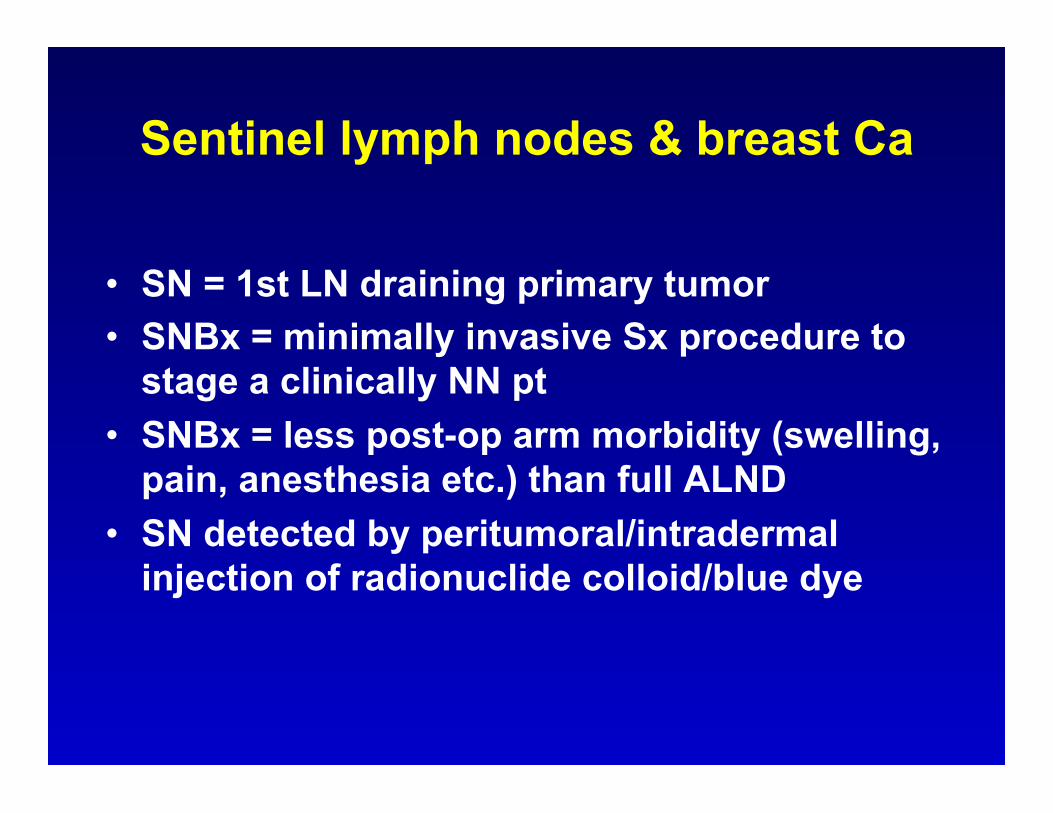
Sentinel lymph nodes & breast Ca
• SN = 1st LN draining primary tumor • SNBx = minimally invasive Sx procedure to
stage a clinically NN pt • SNBx = less post-op arm morbidity (swelling,
pain, anesthesia etc.) than full ALND • SN detected by peritumoral/intradermal
injection of radionuclide colloid/blue dye
L
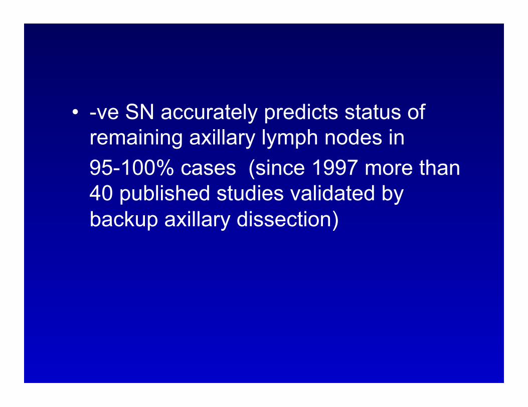
• -ve SN accurately predicts status of remaining axillary lymph nodes in
95-100% cases (since 1997 more than 40 published studies validated by backup axillary dissection)
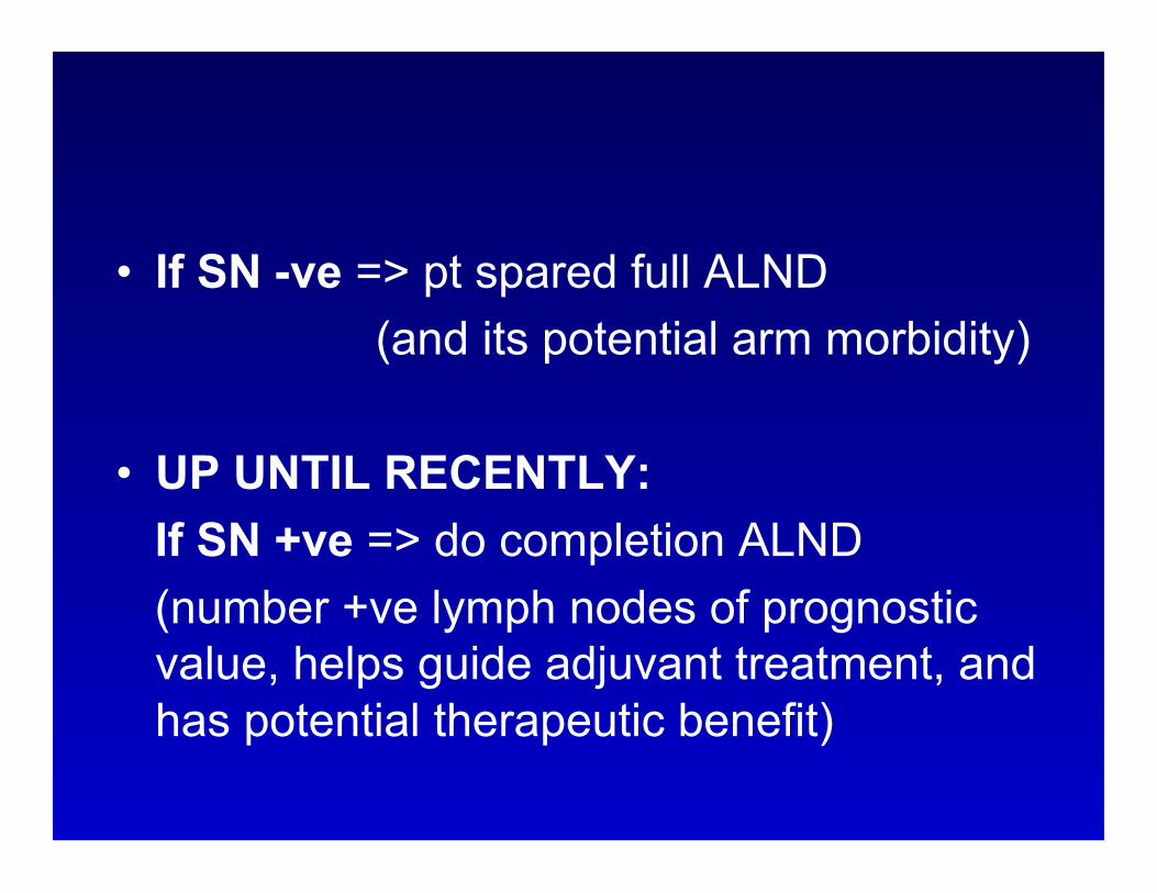
• If SN -ve => pt spared full ALND (and its potential arm morbidity)
• UP UNTIL RECENTLY: If SN +ve => do completion ALND (number +ve lymph nodes of prognostic
value, helps guide adjuvant treatment, and has potential therapeutic benefit)
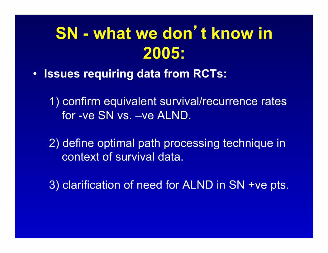
SN - what we don’t know in 2005:
• Issues requiring data from RCTs: 1) confirm equivalent survival/recurrence rates for -ve SN vs. –ve ALND. 2) define optimal path processing technique in context of survival data. 3) clarification of need for ALND in SN +ve pts.
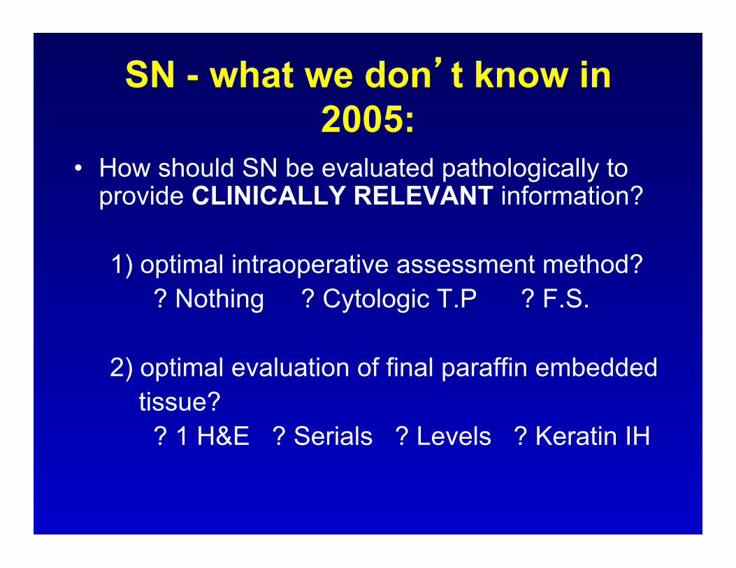
SN - what we don’t know in 2005:
• How should SN be evaluated pathologically to provide CLINICALLY RELEVANT information?
1) optimal intraoperative assessment method? ? Nothing ? Cytologic T.P ? F.S. 2) optimal evaluation of final paraffin embedded tissue? ? 1 H&E ? Serials ? Levels ? Keratin IH

Pathologic examination of lymph nodes
• 2 components: a) gross examination & sectioning b) histologic examination of slides a) Gross examination: - grossly ID & isolate all lymph nodes present - serially slice and submit each LN in toto (slicing LN at <=2 mm levels ensures all macromets are ID histologically)
Pathologic examination of lymph nodes
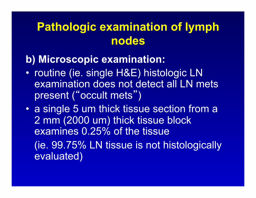
b) Microscopic examination: • routine (ie. single H&E) histologic LN
examination does not detect all LN mets present (“occult mets”)
• a single 5 um thick tissue section from a 2 mm (2000 um) thick tissue block examines 0.25% of the tissue
(ie. 99.75% LN tissue is not histologically evaluated)
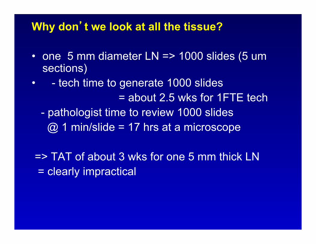
Why don’t we look at all the tissue?
• one 5 mm diameter LN => 1000 slides (5 um sections)
• - tech time to generate 1000 slides = about 2.5 wks for 1FTE tech - pathologist time to review 1000 slides @ 1 min/slide = 17 hrs at a microscope => TAT of about 3 wks for one 5 mm thick LN = clearly impractical
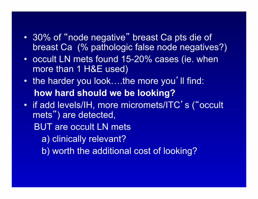
• 30% of “node negative” breast Ca pts die of breast Ca (% pathologic false node negatives?)
• occult LN mets found 15-20% cases (ie. when more than 1 H&E used)
• the harder you look….the more you’ll find: how hard should we be looking? • if add levels/IH, more micromets/ITC’s (“occult
mets”) are detected, BUT are occult LN mets a) clinically relevant? b) worth the additional cost of looking?
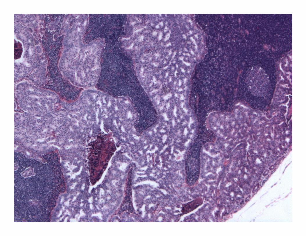
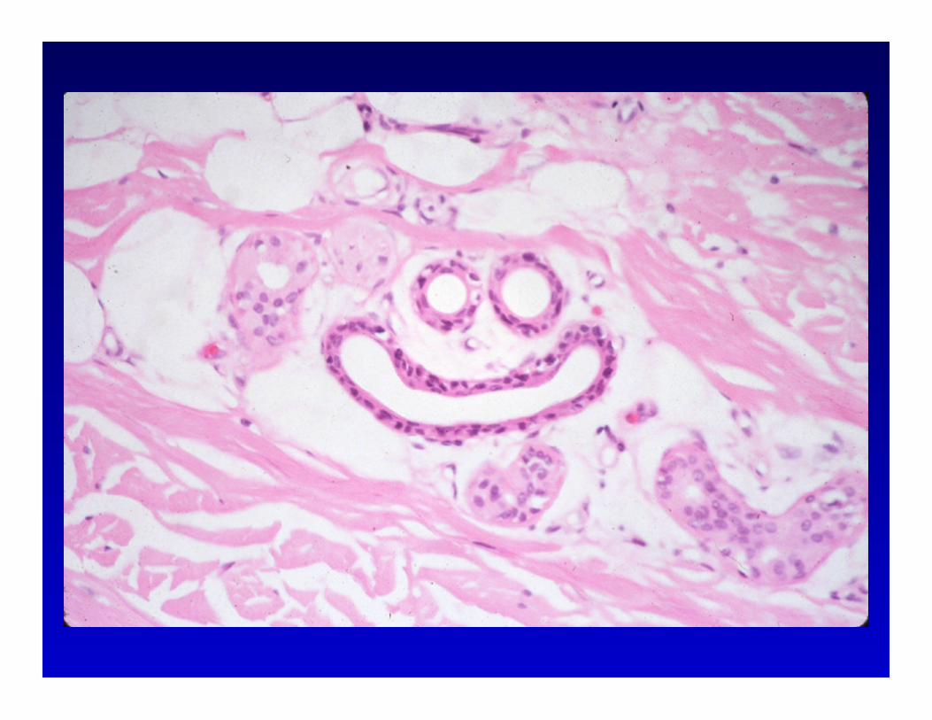
Histologic LN metastases can be easy……………….

or not so easy…….
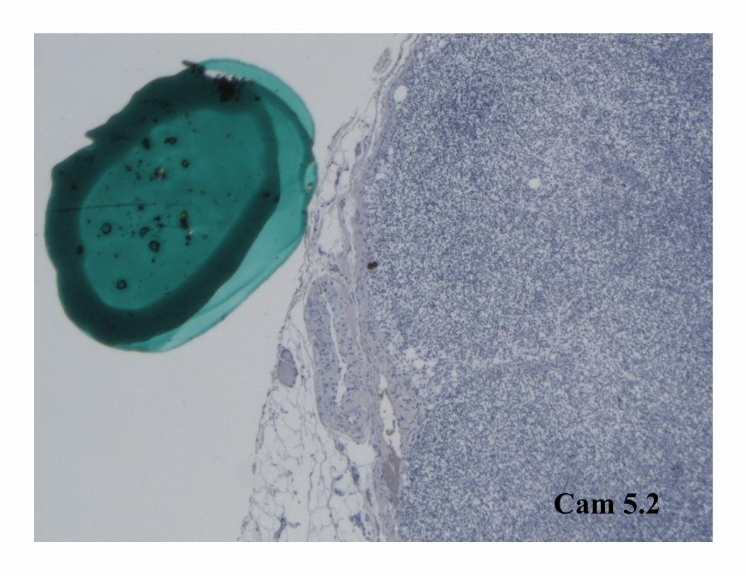
Cam 5.2

Cam 5.2

Cam 5.2
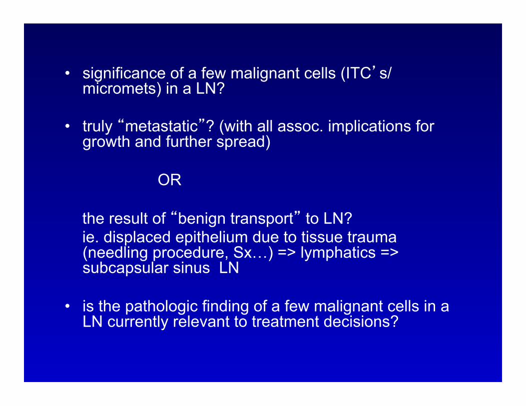
• significance of a few malignant cells (ITC’s/micromets) in a LN?
• truly “metastatic”? (with all assoc. implications for growth and further spread)
OR the result of “benign transport” to LN? ie. displaced epithelium due to tissue trauma
(needling procedure, Sx…) => lymphatics => subcapsular sinus LN
• is the pathologic finding of a few malignant cells in a
LN currently relevant to treatment decisions?

• some retrospective/observational studies suggest occult LN mets are a prognostic factor for dz recurrence or survival in breast cancer,
HOWEVER, data from prospective, randomized clinical
trials has been lacking …….. until recently…
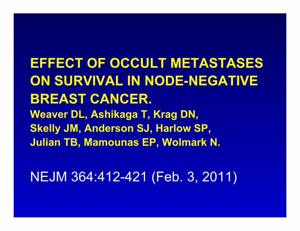
EFFECT OF OCCULT METASTASES ON SURVIVAL IN NODE-NEGATIVE BREAST CANCER. Weaver DL, Ashikaga T, Krag DN, Skelly JM, Anderson SJ, Harlow SP, Julian TB, Mamounas EP, Wolmark N. NEJM 364:412-421 (Feb. 3, 2011)

NSABP B-32 • multicenter (80 sites in USA/Canada), prospective, RCT
• eligibility: operable, clinically NN invasive breast Ca
• Hypothesis: pt with -ve SN will have equivalent Dz free & overall survival rates whether treated by SN bx alone or SN + completion ALND
(SN morbidity less than SN plus ALND)
• intensive study of -ve SN serially sliced in long axis at 2 mm intervals (ie. further H&E and CK IH at 0.45 mm & 0.96 mm) to look at incidence of occult mets & correlate with pt outcome in a blinded fashion
ie. ? clinical significance of occult mets in SN
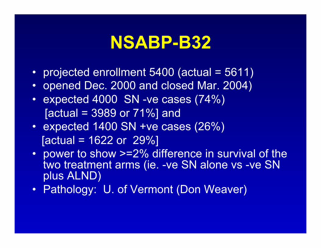
NSABP-B32 • projected enrollment 5400 (actual = 5611) • opened Dec. 2000 and closed Mar. 2004) • expected 4000 SN -ve cases (74%) [actual = 3989 or 71%] and • expected 1400 SN +ve cases (26%) [actual = 1622 or 29%] • power to show >=2% difference in survival of the
two treatment arms (ie. -ve SN alone vs -ve SN plus ALND)
• Pathology: U. of Vermont (Don Weaver)
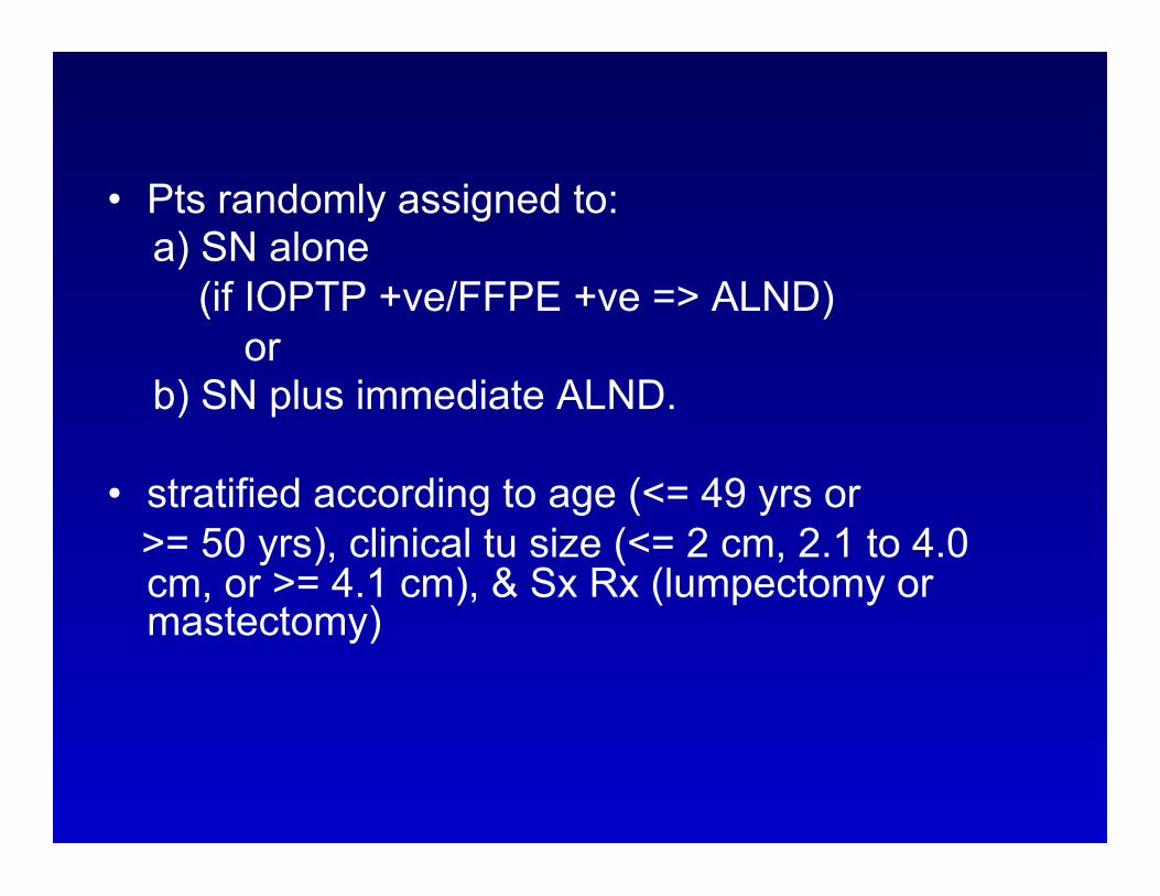
• Pts randomly assigned to: a) SN alone (if IOPTP +ve/FFPE +ve => ALND) or b) SN plus immediate ALND. • stratified according to age (<= 49 yrs or >= 50 yrs), clinical tu size (<= 2 cm, 2.1 to 4.0
cm, or >= 4.1 cm), & Sx Rx (lumpectomy or mastectomy)
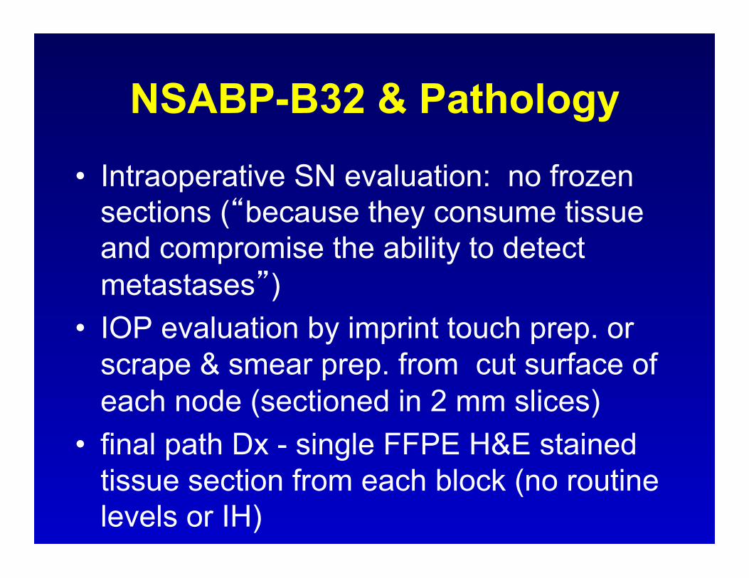
NSABP-B32 & Pathology
• Intraoperative SN evaluation: no frozen sections (“because they consume tissue and compromise the ability to detect metastases”)
• IOP evaluation by imprint touch prep. or scrape & smear prep. from cut surface of each node (sectioned in 2 mm slices)
• final path Dx - single FFPE H&E stained tissue section from each block (no routine levels or IH)
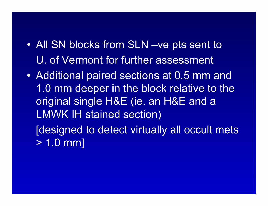
• All SN blocks from SLN –ve pts sent to U. of Vermont for further assessment • Additional paired sections at 0.5 mm and
1.0 mm deeper in the block relative to the original single H&E (ie. an H&E and a LMWK IH stained section)
[designed to detect virtually all occult mets > 1.0 mm]
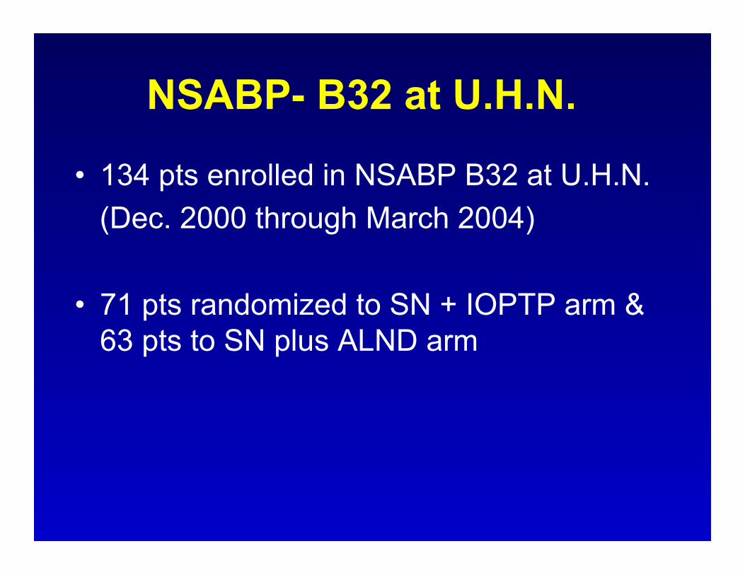
NSABP- B32 at U.H.N.
• 134 pts enrolled in NSABP B32 at U.H.N. (Dec. 2000 through March 2004) • 71 pts randomized to SN + IOPTP arm &
63 pts to SN plus ALND arm
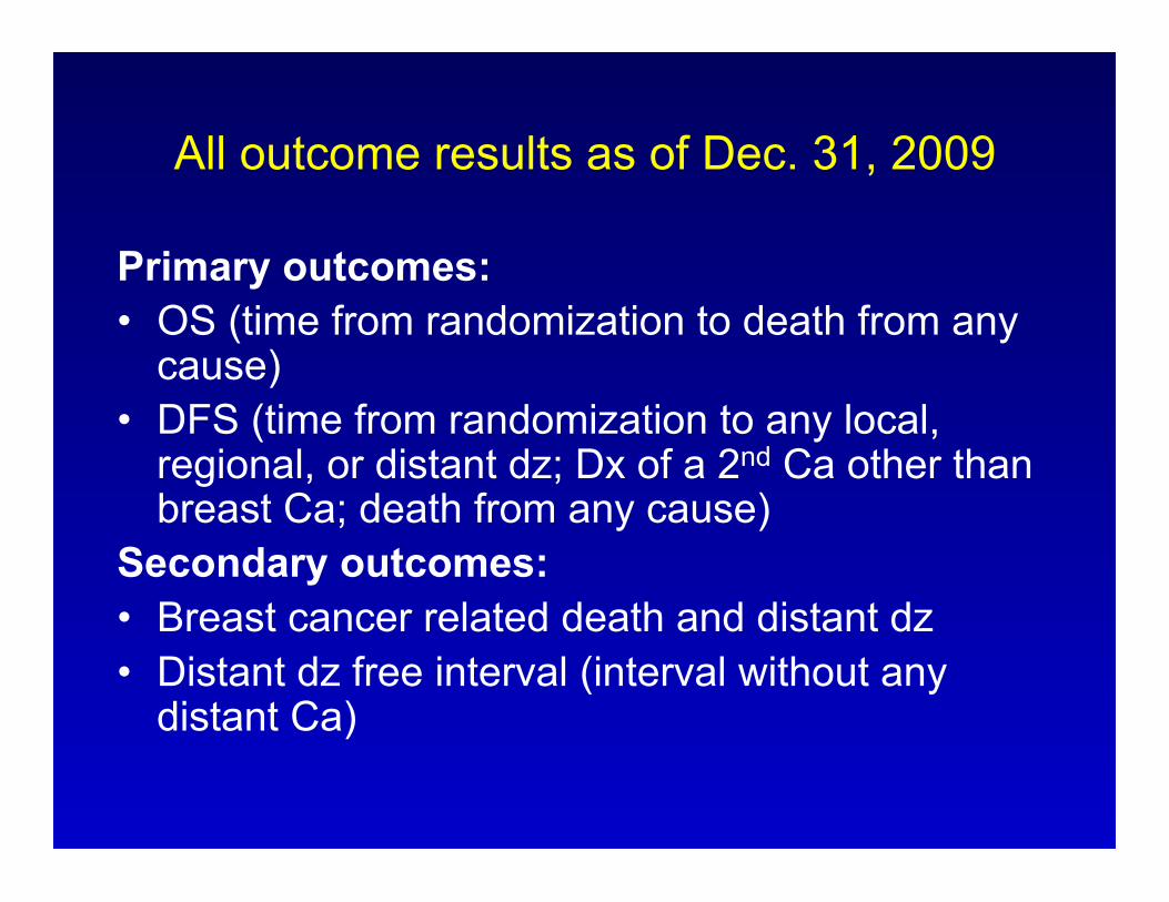
All outcome results as of Dec. 31, 2009
Primary outcomes: • OS (time from randomization to death from any
cause) • DFS (time from randomization to any local,
regional, or distant dz; Dx of a 2nd Ca other than breast Ca; death from any cause)
Secondary outcomes: • Breast cancer related death and distant dz • Distant dz free interval (interval without any
distant Ca)
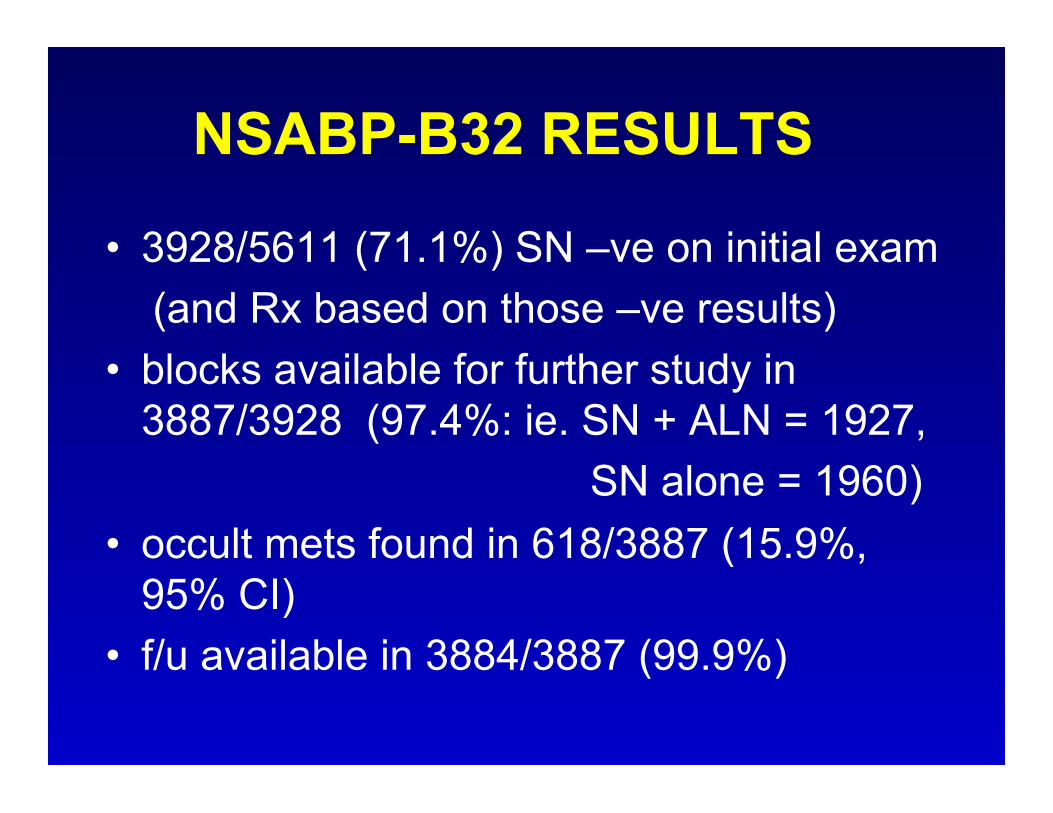
NSABP-B32 RESULTS
• 3928/5611 (71.1%) SN –ve on initial exam (and Rx based on those –ve results) • blocks available for further study in
3887/3928 (97.4%: ie. SN + ALN = 1927, SN alone = 1960) • occult mets found in 618/3887 (15.9%,
95% CI) • f/u available in 3884/3887 (99.9%)

• 637 had outcome events (302 died, 120 died from breast Ca)
• median f/u = 95.3 months (7.9 yrs) • systemic therapy in 82% each Rx arm • radiotherapy 84% each Rx arm
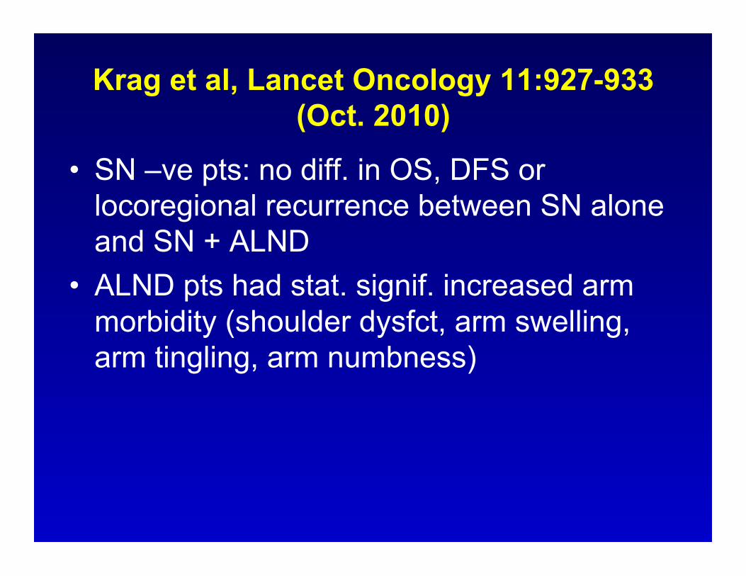
Krag et al, Lancet Oncology 11:927-933 (Oct. 2010)
• SN –ve pts: no diff. in OS, DFS or locoregional recurrence between SN alone and SN + ALND
• ALND pts had stat. signif. increased arm morbidity (shoulder dysfct, arm swelling, arm tingling, arm numbness)

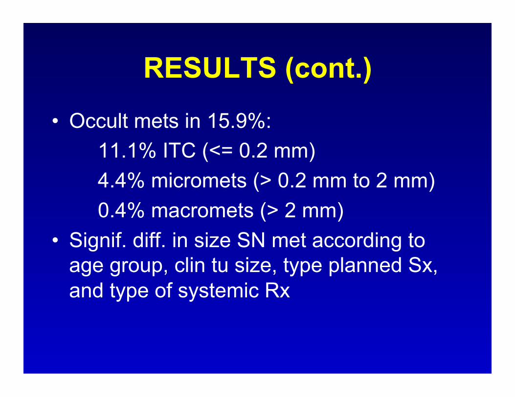
RESULTS (cont.)
• Occult mets in 15.9%: 11.1% ITC (<= 0.2 mm) 4.4% micromets (> 0.2 mm to 2 mm) 0.4% macromets (> 2 mm) • Signif. diff. in size SN met according to
age group, clin tu size, type planned Sx, and type of systemic Rx
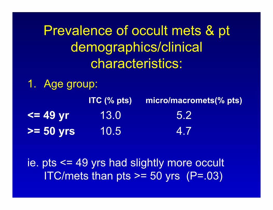
Prevalence of occult mets & pt demographics/clinical
characteristics: 1. Age group: ITC (% pts) micro/macromets(% pts)
<= 49 yr 13.0 5.2 >= 50 yrs 10.5 4.7 ie. pts <= 49 yrs had slightly more occult
ITC/mets than pts >= 50 yrs (P=.03)

Prevalence of occult mets & pt demographics/clinical
characteristics: 2. Clinical tumor size: ITC (% pts) micro/macromets(% pts) <= 2.0 cm 10.2 4.5 2.1-4.0 cm 15.3 6.3 >= 4.1 cm 16.7 5.0 ie. as tumor size increases beyond 2 cm,
prevalence of occult ITC/mets increases (P < .001)
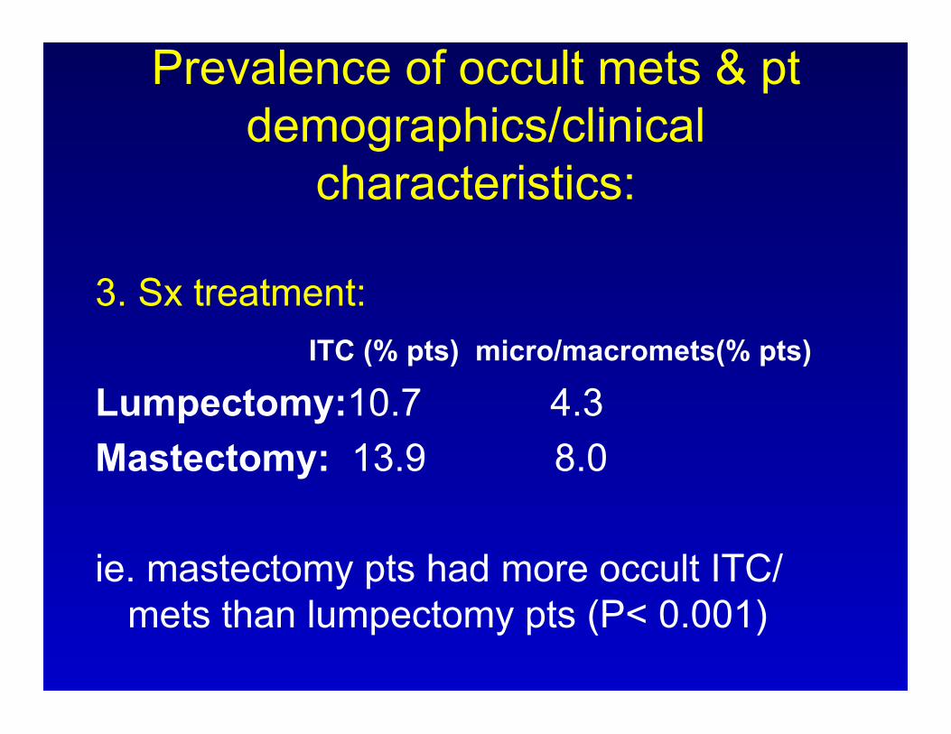
Prevalence of occult mets & pt demographics/clinical
characteristics:
3. Sx treatment: ITC (% pts) micro/macromets(% pts) Lumpectomy:10.7 4.3 Mastectomy: 13.9 8.0 ie. mastectomy pts had more occult ITC/
mets than lumpectomy pts (P< 0.001)
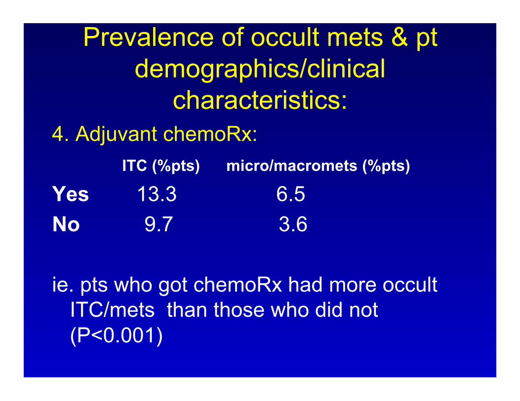
Prevalence of occult mets & pt demographics/clinical
characteristics: 4. Adjuvant chemoRx: ITC (%pts) micro/macromets (%pts)
Yes 13.3 6.5 No 9.7 3.6 ie. pts who got chemoRx had more occult
ITC/mets than those who did not (P<0.001)
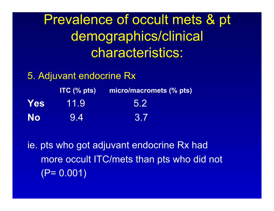
Prevalence of occult mets & pt demographics/clinical
characteristics: 5. Adjuvant endocrine Rx ITC (% pts) micro/macromets (% pts)
Yes 11.9 5.2 No 9.4 3.7 ie. pts who got adjuvant endocrine Rx had more occult ITC/mets than pts who did not (P= 0.001)
Prevalence of occult mets & pt demographics/clinical
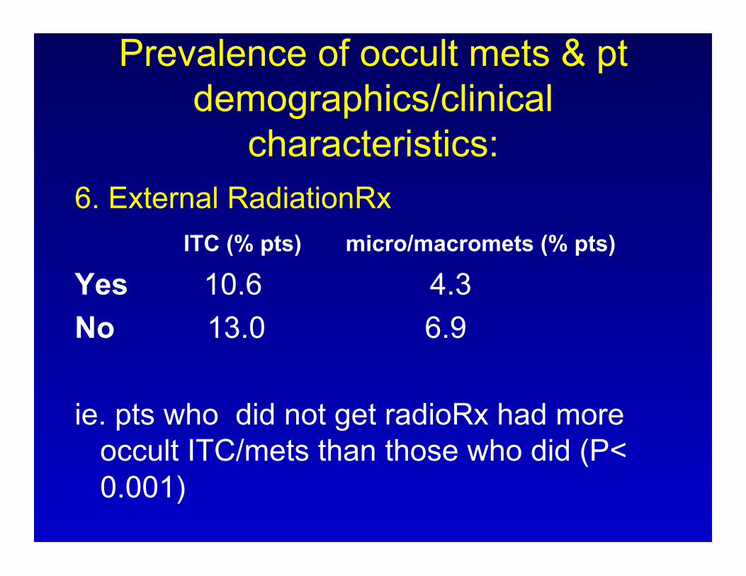
characteristics: 6. External RadiationRx ITC (% pts) micro/macromets (% pts)
Yes 10.6 4.3 No 13.0 6.9 ie. pts who did not get radioRx had more
occult ITC/mets than those who did (P< 0.001)
Prevalence of occult mets & pt demographics/clinical
characteristics:
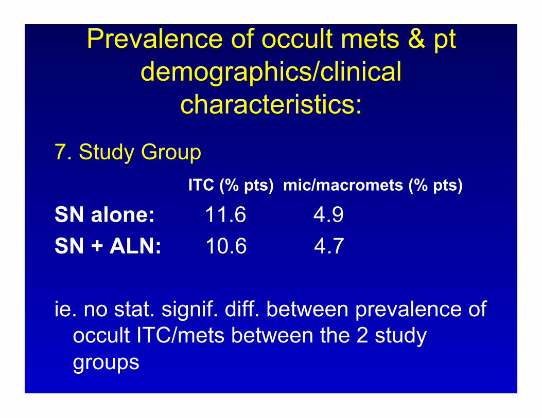
7. Study Group ITC (% pts) mic/macromets (% pts)
SN alone: 11.6 4.9 SN + ALN: 10.6 4.7 ie. no stat. signif. diff. between prevalence of
occult ITC/mets between the 2 study groups
Prevalence of occult mets & pt demographics/clinical
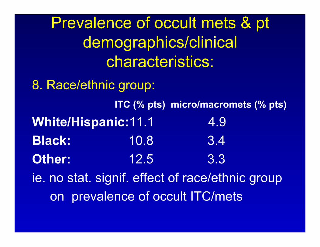
characteristics: 8. Race/ethnic group: ITC (% pts) micro/macromets (% pts) White/Hispanic:11.1 4.9 Black: 10.8 3.4 Other: 12.5 3.3 ie. no stat. signif. effect of race/ethnic group on prevalence of occult ITC/mets
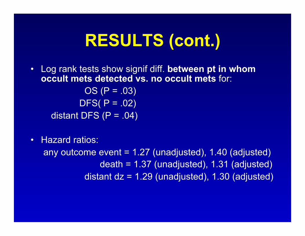
RESULTS (cont.) • Log rank tests show signif diff. between pt in whom
occult mets detected vs. no occult mets for: OS (P = .03) DFS( P = .02) distant DFS (P = .04) • Hazard ratios: any outcome event = 1.27 (unadjusted), 1.40 (adjusted) death = 1.37 (unadjusted), 1.31 (adjusted) distant dz = 1.29 (unadjusted), 1.30 (adjusted)
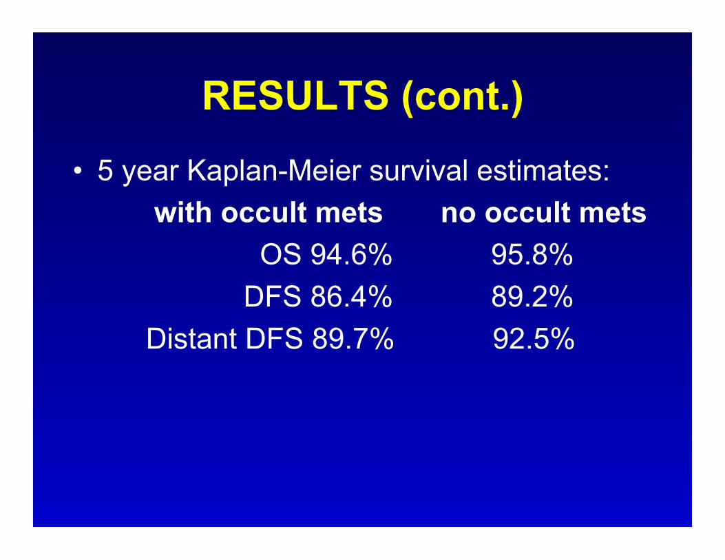
RESULTS (cont.)
• 5 year Kaplan-Meier survival estimates: with occult mets no occult mets OS 94.6% 95.8% DFS 86.4% 89.2% Distant DFS 89.7% 92.5%
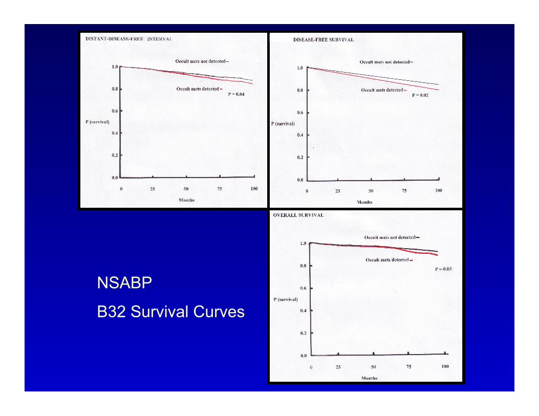
NSABP
B32 Survival Curves
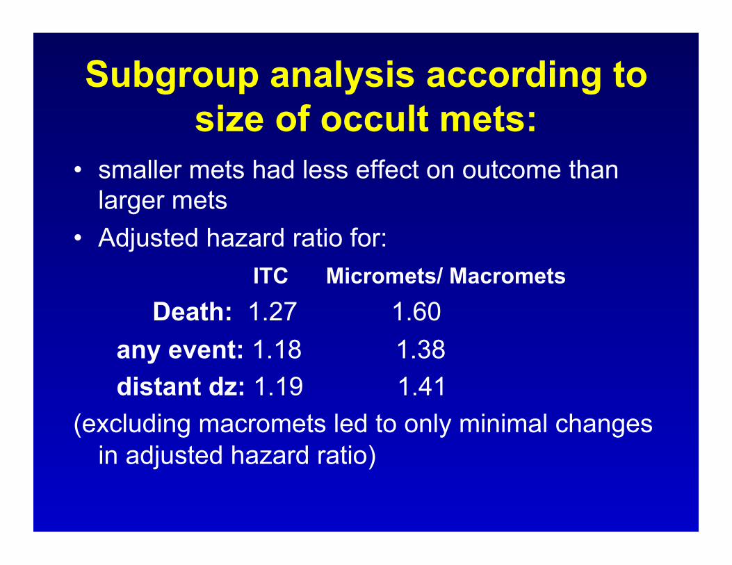
Subgroup analysis according to size of occult mets:
• smaller mets had less effect on outcome than larger mets
• Adjusted hazard ratio for: ITC Micromets/ Macromets
Death: 1.27 1.60 any event: 1.18 1.38 distant dz: 1.19 1.41 (excluding macromets led to only minimal changes
in adjusted hazard ratio)
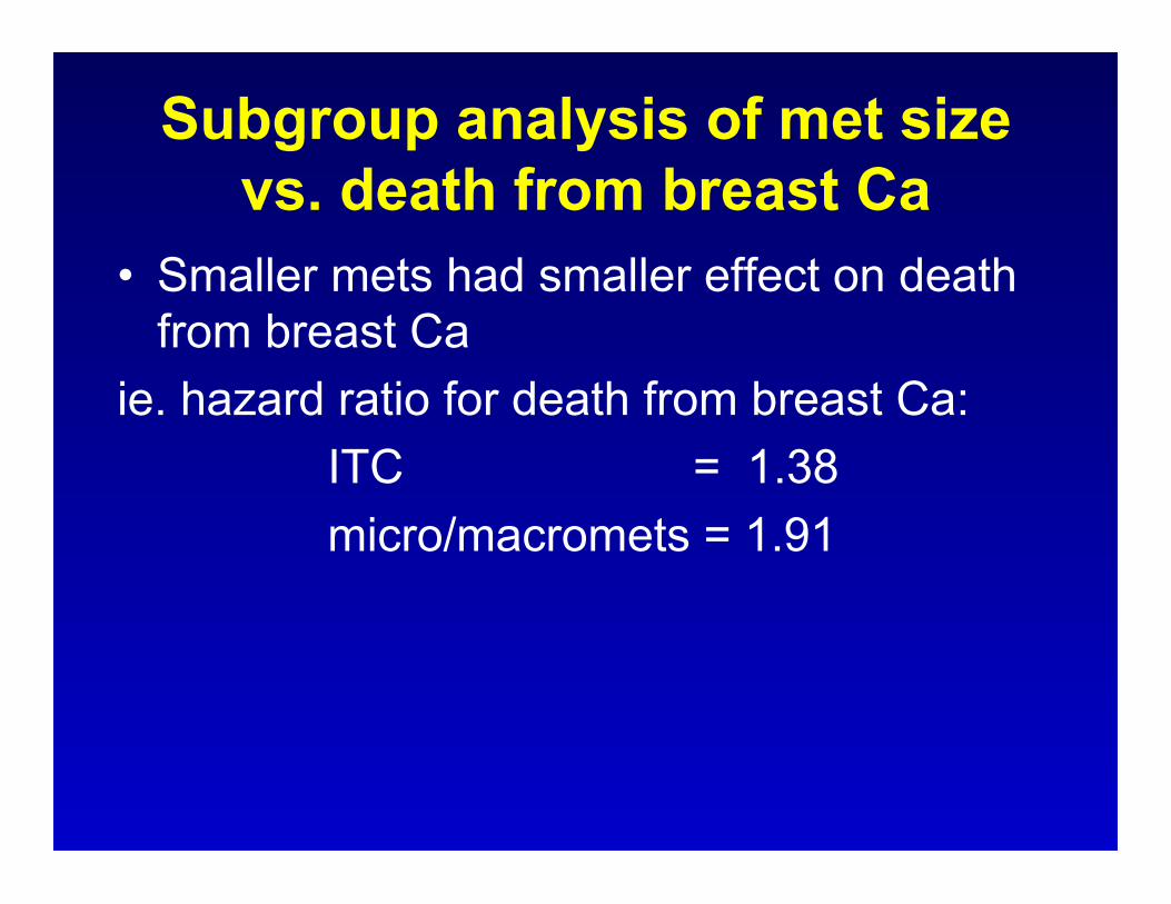
Subgroup analysis of met size vs. death from breast Ca
• Smaller mets had smaller effect on death from breast Ca
ie. hazard ratio for death from breast Ca: ITC = 1.38 micro/macromets = 1.91

RESULTS (cont.) • 5 yr Kaplan-Meier estimates of % pt who did not
die of breast Ca:
no occult mets: 98.4% ITC: 97.8% micromets/macromets: 96.0% (caveat: “confidence in these estimates is limited
by the small number of outcome events”)
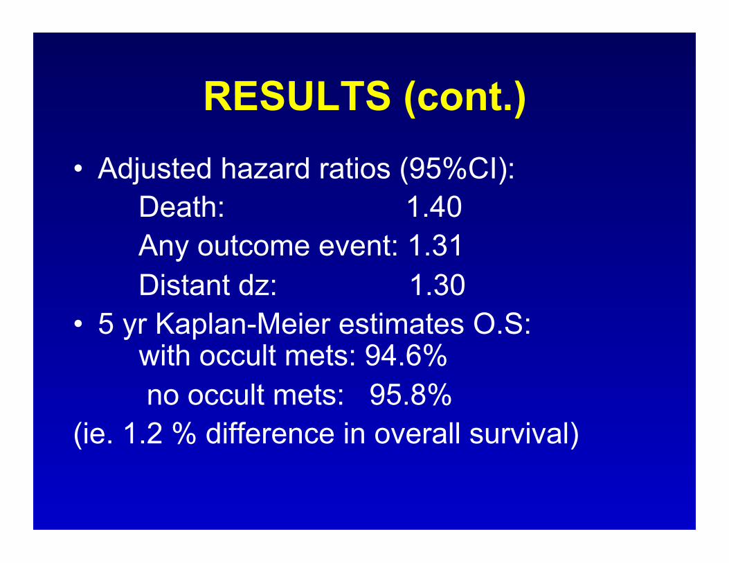
RESULTS (cont.) • Adjusted hazard ratios (95%CI): Death: 1.40 Any outcome event: 1.31 Distant dz: 1.30 • 5 yr Kaplan-Meier estimates O.S:
with occult mets: 94.6% no occult mets: 95.8% (ie. 1.2 % difference in overall survival)
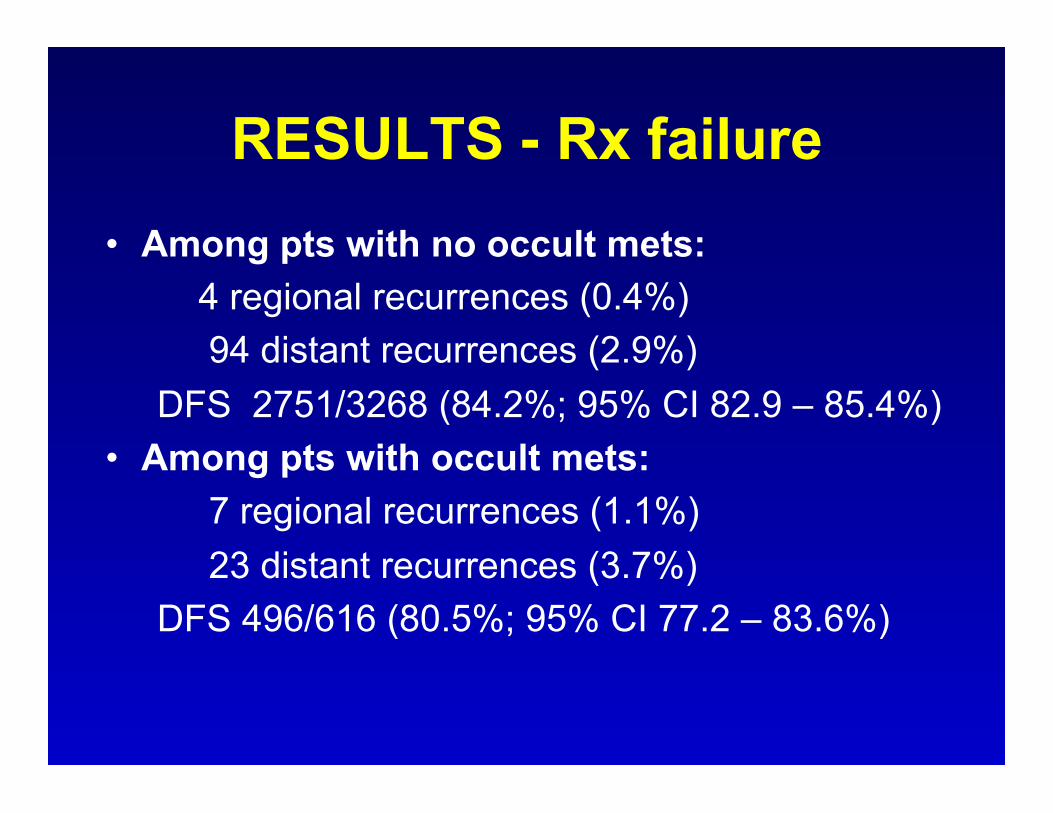
RESULTS - Rx failure • Among pts with no occult mets: 4 regional recurrences (0.4%) 94 distant recurrences (2.9%) DFS 2751/3268 (84.2%; 95% CI 82.9 – 85.4%) • Among pts with occult mets: 7 regional recurrences (1.1%) 23 distant recurrences (3.7%) DFS 496/616 (80.5%; 95% CI 77.2 – 83.6%)
Conclusions:
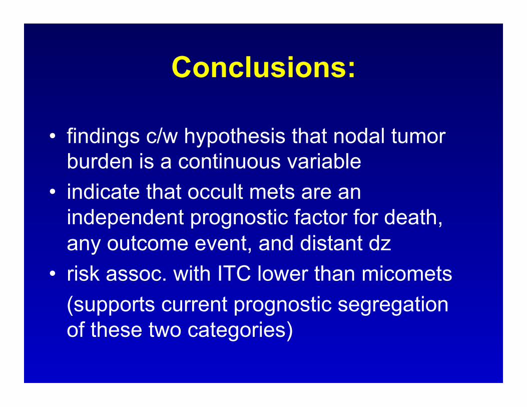
• findings c/w hypothesis that nodal tumor burden is a continuous variable
• indicate that occult mets are an independent prognostic factor for death, any outcome event, and distant dz
• risk assoc. with ITC lower than micomets (supports current prognostic segregation
of these two categories)
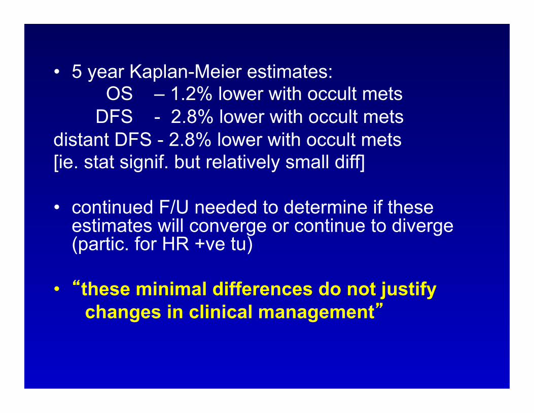
• 5 year Kaplan-Meier estimates: OS – 1.2% lower with occult mets DFS - 2.8% lower with occult mets distant DFS - 2.8% lower with occult mets [ie. stat signif. but relatively small diff] • continued F/U needed to determine if these
estimates will converge or continue to diverge (partic. for HR +ve tu)
• “these minimal differences do not justify changes in clinical management”
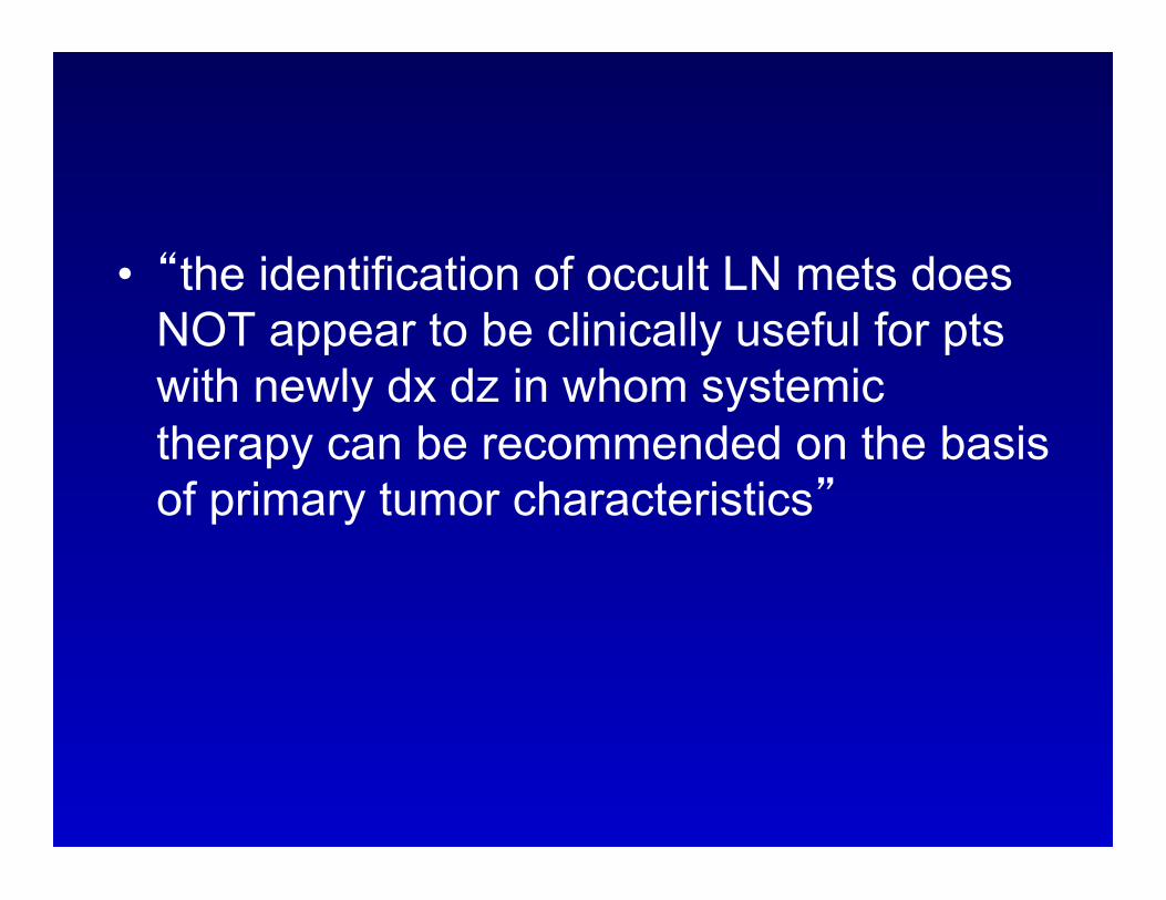
• “the identification of occult LN mets does NOT appear to be clinically useful for pts with newly dx dz in whom systemic therapy can be recommended on the basis of primary tumor characteristics”
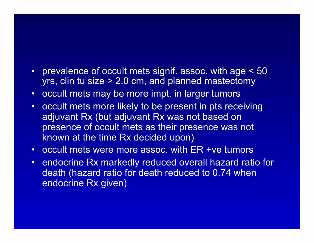
• prevalence of occult mets signif. assoc. with age < 50 yrs, clin tu size > 2.0 cm, and planned mastectomy
• occult mets may be more impt. in larger tumors • occult mets more likely to be present in pts receiving
adjuvant Rx (but adjuvant Rx was not based on presence of occult mets as their presence was not known at the time Rx decided upon)
• occult mets were more assoc. with ER +ve tumors • endocrine Rx markedly reduced overall hazard ratio for
death (hazard ratio for death reduced to 0.74 when endocrine Rx given)
CONCLUSIONS
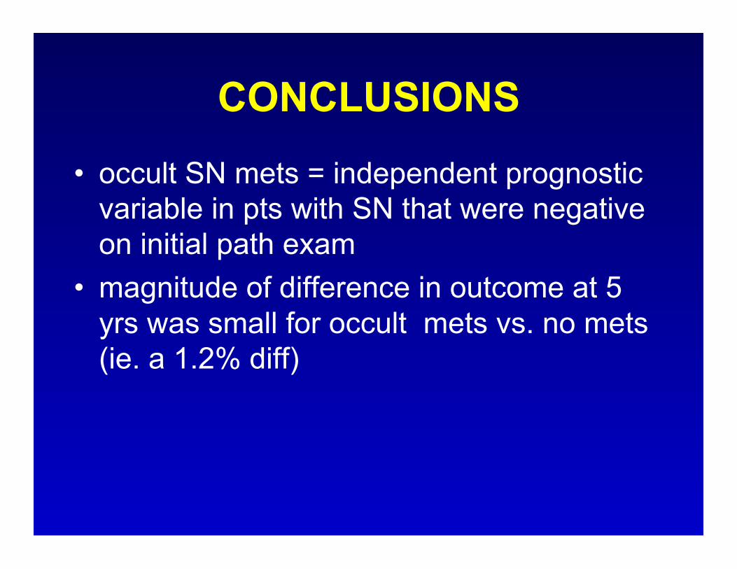
• occult SN mets = independent prognostic variable in pts with SN that were negative on initial path exam
• magnitude of difference in outcome at 5 yrs was small for occult mets vs. no mets (ie. a 1.2% diff)
CONCLUSIONS (cont.)
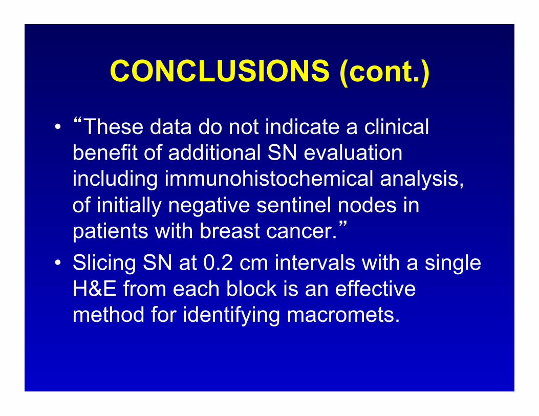
• “These data do not indicate a clinical benefit of additional SN evaluation including immunohistochemical analysis, of initially negative sentinel nodes in patients with breast cancer.”
• Slicing SN at 0.2 cm intervals with a single H&E from each block is an effective method for identifying macromets.

CONCLUSIONS (cont.)
• ITC had smaller effect on every outcome evaluated (OS, DFS, DDFS, death from breast Ca) than micromets., regardless whether occult macromets were included or excluded
• magnitude of difference in Kaplan-Meier estimates for death due to breast carcinoma was small for SN ITC vs
SN –ve (ie. 0.6%) compared to SN micromets vs no micromets (ie. 2.4%)

CONCLUSIONS • Regardless of whether ITC/micromets are
detected in initial sections or deepers/IH, they have less prognostic significance than macromets and should be classified separately
• Local radiation therapy and adjuvant systemic therapy, particularly endocrine therapy appear to attenuate the unfavourable effect of occult mets

CONCLUSIONS
• The small outcome differences between those with and without occult mets was small, but statistically significant (1-3%) at 5 years, but warrants continued f/u and analysis
• “findings argue against analysis of additional tissue levels or routine IH analysis for routine SN evaluation”.
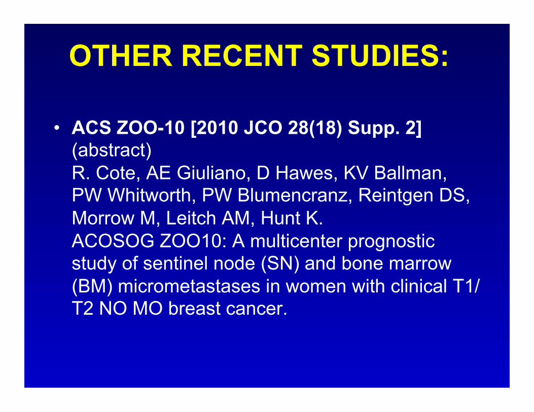
OTHER RECENT STUDIES:
• ACS ZOO-10 [2010 JCO 28(18) Supp. 2] (abstract) R. Cote, AE Giuliano, D Hawes, KV Ballman, PW Whitworth, PW Blumencranz, Reintgen DS, Morrow M, Leitch AM, Hunt K. ACOSOG ZOO10: A multicenter prognostic study of sentinel node (SN) and bone marrow (BM) micrometastases in women with clinical T1/T2 NO MO breast cancer.

ACOSOG ZOO10 (cont.) • multicenter, prospective observational study • n = 5539 • 5 yr survival diff. between pts in whom occult
mets detected by IH vs pts in whom occult mets not detected by IH was not statistically significant (SN IHC +ve 95.1% 5 yr survival vs. SN IHC –ve 95.8%; diff. of 0.7% P=0.53)
• Note: prevalence of occult mets lower in ZOO-10 (10.5%) than in NSABP-B32 (15.9%)
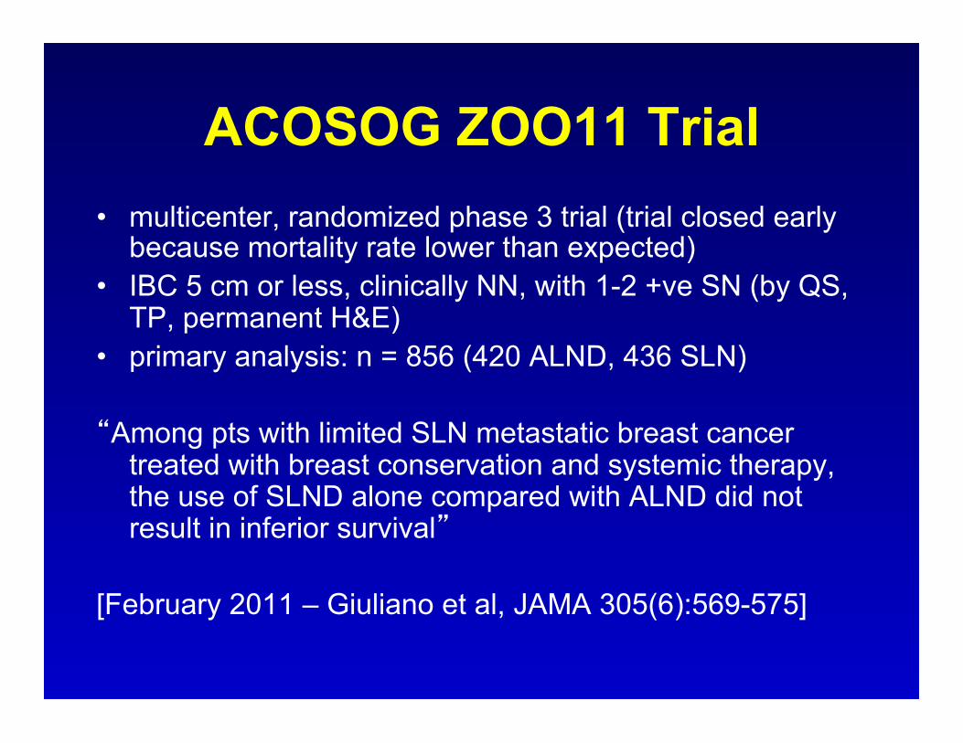
ACOSOG ZOO11 Trial • multicenter, randomized phase 3 trial (trial closed early
because mortality rate lower than expected) • IBC 5 cm or less, clinically NN, with 1-2 +ve SN (by QS,
TP, permanent H&E) • primary analysis: n = 856 (420 ALND, 436 SLN) “Among pts with limited SLN metastatic breast cancer
treated with breast conservation and systemic therapy, the use of SLND alone compared with ALND did not result in inferior survival”
[February 2011 – Giuliano et al, JAMA 305(6):569-575]
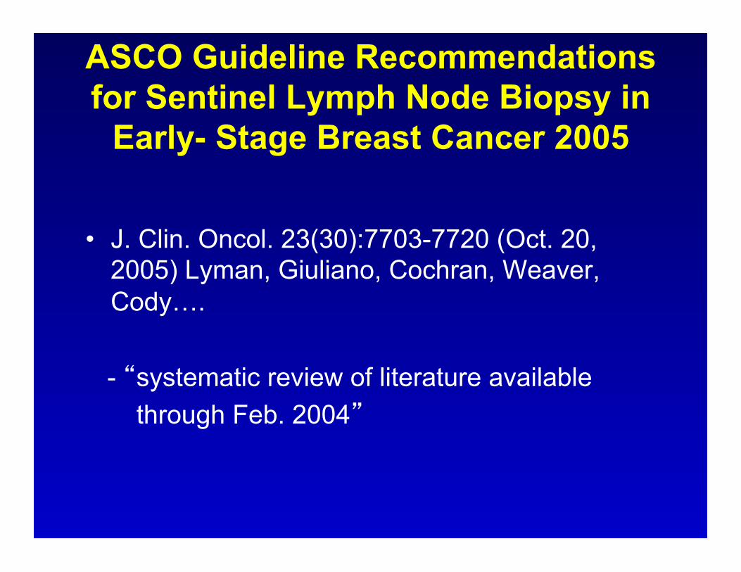
ASCO Guideline Recommendations for Sentinel Lymph Node Biopsy in
Early- Stage Breast Cancer 2005
• J. Clin. Oncol. 23(30):7703-7720 (Oct. 20, 2005) Lyman, Giuliano, Cochran, Weaver, Cody….
- “systematic review of literature available through Feb. 2004”
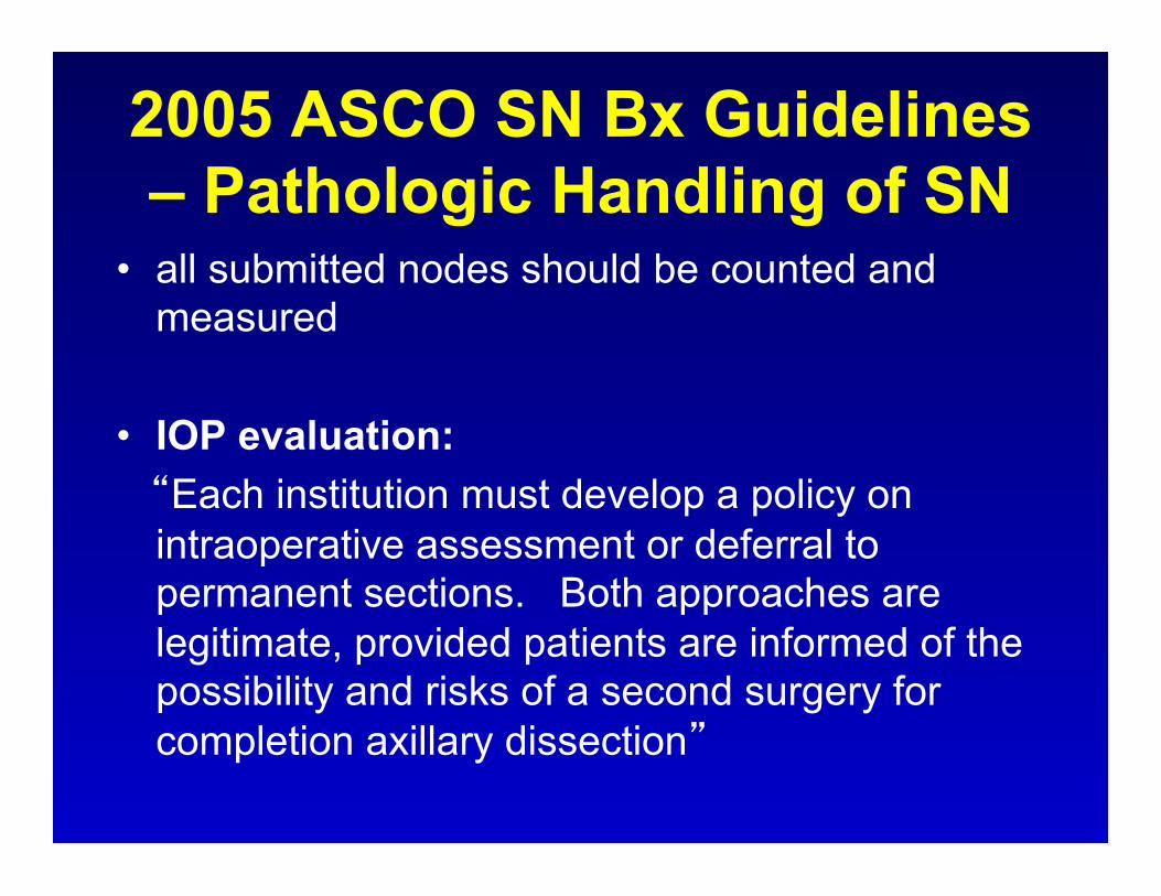
2005 ASCO SN Bx Guidelines – Pathologic Handling of SN
• all submitted nodes should be counted and measured
• IOP evaluation: “Each institution must develop a policy on
intraoperative assessment or deferral to permanent sections. Both approaches are legitimate, provided patients are informed of the possibility and risks of a second surgery for completion axillary dissection”
2005 ASCO SN Bx Guidelines
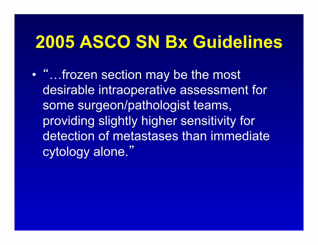
• “…frozen section may be the most desirable intraoperative assessment for some surgeon/pathologist teams, providing slightly higher sensitivity for detection of metastases than immediate cytology alone.”
2005 ASCO SN Bx Guidelines
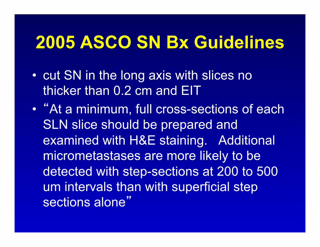
• cut SN in the long axis with slices no thicker than 0.2 cm and EIT
• “At a minimum, full cross-sections of each SLN slice should be prepared and examined with H&E staining. Additional micrometastases are more likely to be detected with step-sections at 200 to 500 um intervals than with superficial step sections alone”
2005 ASCO SN Bx Guidelines
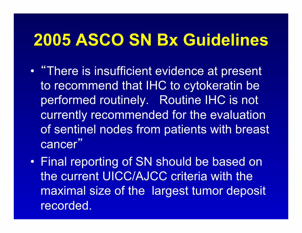
• “There is insufficient evidence at present to recommend that IHC to cytokeratin be performed routinely. Routine IHC is not currently recommended for the evaluation of sentinel nodes from patients with breast cancer”
• Final reporting of SN should be based on the current UICC/AJCC criteria with the maximal size of the largest tumor deposit recorded.

2005 ASCO SN Bx Guidelines
• “The decision to utilize IHC analysis and act on the results remains for now a matter of discussion among individual surgeons, oncologists, and pathologists, based on a determination of the best course for their patients, assessed from their own experience and review of the available literature”
Patterns of practice in pathologic handling of SN.
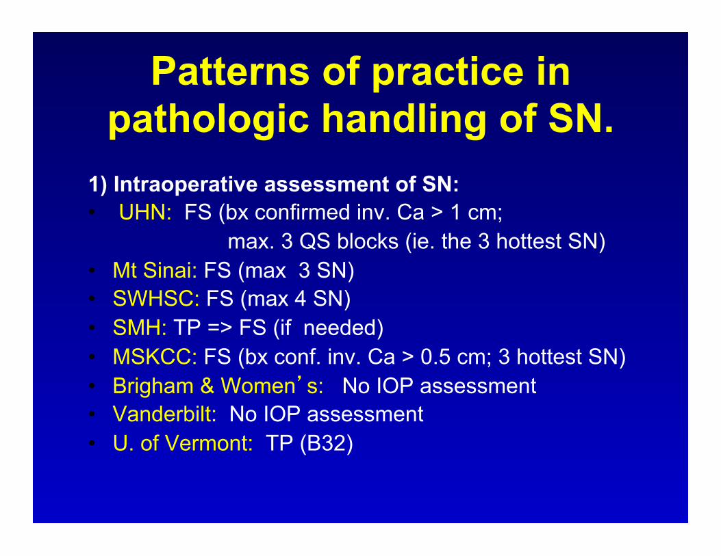
1) Intraoperative assessment of SN: • UHN: FS (bx confirmed inv. Ca > 1 cm; max. 3 QS blocks (ie. the 3 hottest SN) • Mt Sinai: FS (max 3 SN) • SWHSC: FS (max 4 SN) • SMH: TP => FS (if needed) • MSKCC: FS (bx conf. inv. Ca > 0.5 cm; 3 hottest SN) • Brigham & Women’s: No IOP assessment • Vanderbilt: No IOP assessment • U. of Vermont: TP (B32)
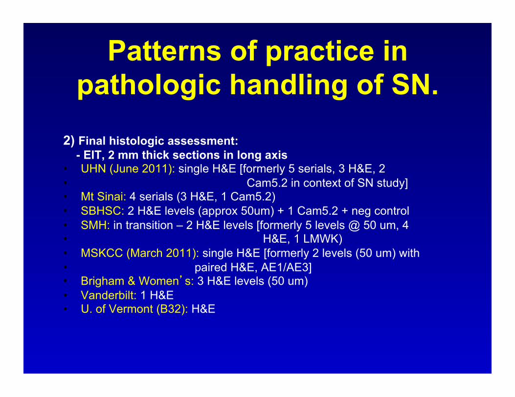
Patterns of practice in pathologic handling of SN.
2) Final histologic assessment: - EIT, 2 mm thick sections in long axis • UHN (June 2011): single H&E [formerly 5 serials, 3 H&E, 2 • Cam5.2 in context of SN study] • Mt Sinai: 4 serials (3 H&E, 1 Cam5.2) • SBHSC: 2 H&E levels (approx 50um) + 1 Cam5.2 + neg control • SMH: in transition – 2 H&E levels [formerly 5 levels @ 50 um, 4 • H&E, 1 LMWK) • MSKCC (March 2011): single H&E [formerly 2 levels (50 um) with • paired H&E, AE1/AE3] • Brigham & Women’s: 3 H&E levels (50 um) • Vanderbilt: 1 H&E • U. of Vermont (B32): H&E
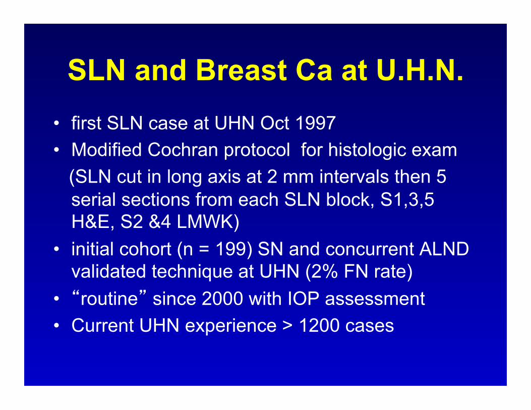
SLN and Breast Ca at U.H.N. • first SLN case at UHN Oct 1997 • Modified Cochran protocol for histologic exam (SLN cut in long axis at 2 mm intervals then 5
serial sections from each SLN block, S1,3,5 H&E, S2 &4 LMWK)
• initial cohort (n = 199) SN and concurrent ALND validated technique at UHN (2% FN rate)
• “routine” since 2000 with IOP assessment • Current UHN experience > 1200 cases

• McCready D.R., Yong W.S., Ng A. K.T., Miller N., Done S., Youngson B. (2004). Influence of the new AJCC Breast Cancer Staging System on Sentinel Lymph Node Positivity and False Negative Rates. Journal National Cancer Institute 96(11):873-875.
• McCready D.R., Yong W.S., Ng A.K.T., Miller N., Done S., Youngson B. (2004) AJCC Breast Cancer Staging System and Sentinel Lymph Nodes - response to letter to the editor. JNCI 96(21):1639-1640.
• Boileau J.F., Easson A., Escallon J.M., Leong W. L., Reedijk M., Youngson B.J., McCready D.R. (2008). Sentinel lymph nodes in breast cancer: relevance of axillary level II nodes and optimal number of nodes that need to be removed. Ann Surg Oncol 15 (6):1710-16.
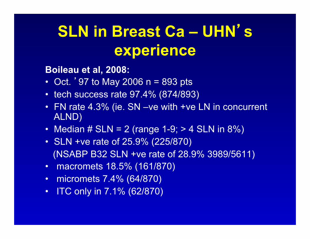
SLN in Breast Ca – UHN’s experience
Boileau et al, 2008: • Oct. ’97 to May 2006 n = 893 pts • tech success rate 97.4% (874/893) • FN rate 4.3% (ie. SN –ve with +ve LN in concurrent
ALND) • Median # SLN = 2 (range 1-9; > 4 SLN in 8%) • SLN +ve rate of 25.9% (225/870) (NSABP B32 SLN +ve rate of 28.9% 3989/5611) • macromets 18.5% (161/870) • micromets 7.4% (64/870) • ITC only in 7.1% (62/870)
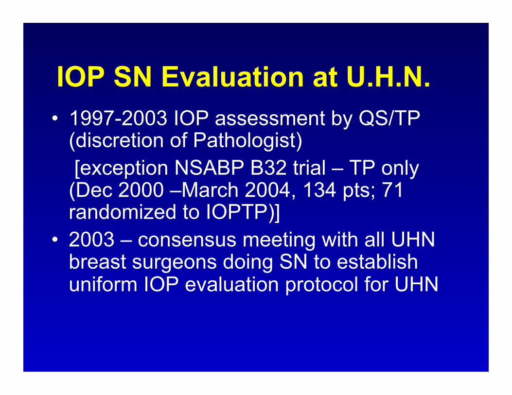
IOP SN Evaluation at U.H.N. • 1997-2003 IOP assessment by QS/TP
(discretion of Pathologist) [exception NSABP B32 trial – TP only
(Dec 2000 –March 2004, 134 pts; 71 randomized to IOPTP)]
• 2003 – consensus meeting with all UHN breast surgeons doing SN to establish uniform IOP evaluation protocol for UHN
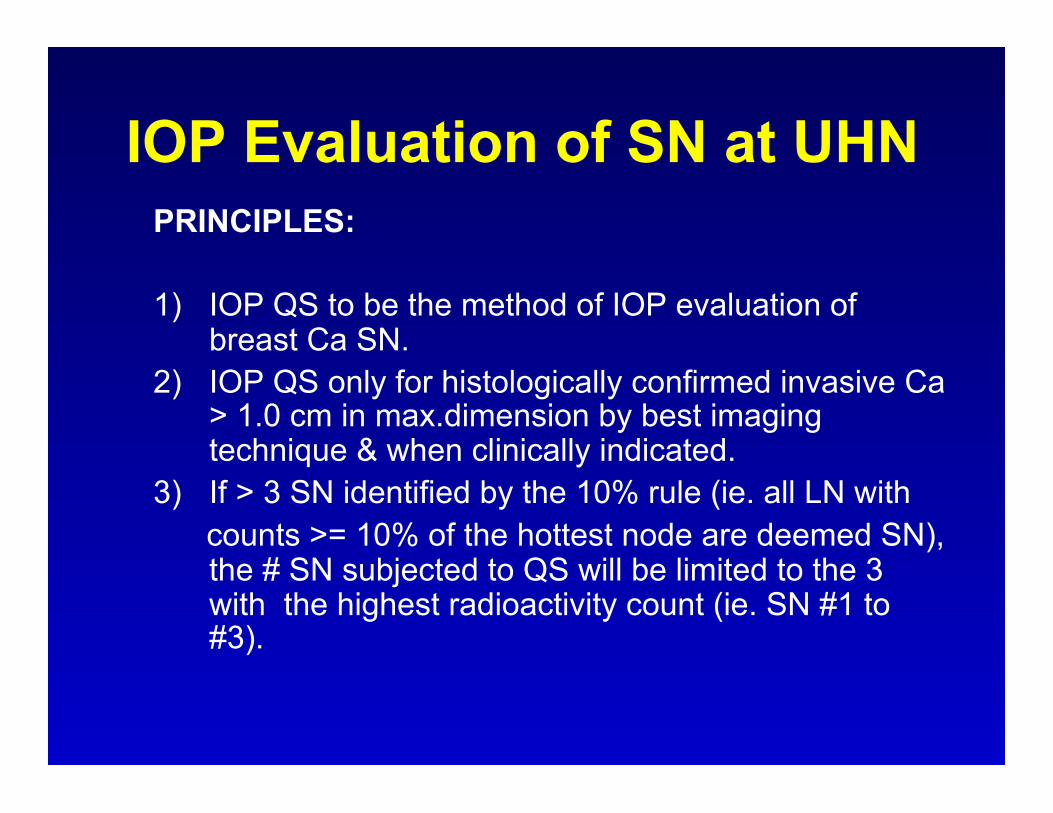
IOP Evaluation of SN at UHN PRINCIPLES: 1) IOP QS to be the method of IOP evaluation of
breast Ca SN. 2) IOP QS only for histologically confirmed invasive Ca
> 1.0 cm in max.dimension by best imaging technique & when clinically indicated.
3) If > 3 SN identified by the 10% rule (ie. all LN with counts >= 10% of the hottest node are deemed SN),
the # SN subjected to QS will be limited to the 3 with the highest radioactivity count (ie. SN #1 to #3).

Principles of IOP Evaluation of SN (cont.)
4) Where a specimen labeled “SN #x” contains > 1 grossly identifiable LN, QS will be done on only one of these LNs, ie. the largest/most grossly suspicious LN. 5) Maximum of 3 QS blocks per case (at Pathologist
discretion). 6) This protocol will be re-evaluated/modified as new data
becomes available.

IOP SN QS – UHN’s Experience
• 20-30 IOP SLN breast Ca cases/month @ PMH (BY, NM, AH, DG, TVDK, SS).
• 1-2 IOP SLN breast Ca cases/month @ TGH (medically complex/immed. reconst. cases).
IOP SN QS – UHN’s Experience
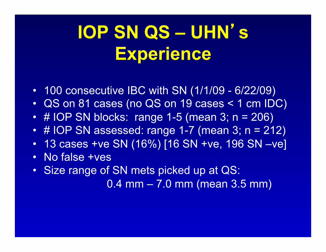
• 100 consecutive IBC with SN (1/1/09 - 6/22/09) • QS on 81 cases (no QS on 19 cases < 1 cm IDC) • # IOP SN blocks: range 1-5 (mean 3; n = 206) • # IOP SN assessed: range 1-7 (mean 3; n = 212) • 13 cases +ve SN (16%) [16 SN +ve, 196 SN –ve] • No false +ves • Size range of SN mets picked up at QS: 0.4 mm – 7.0 mm (mean 3.5 mm)
IOP SN QS – UHN’s Experience (cont.)
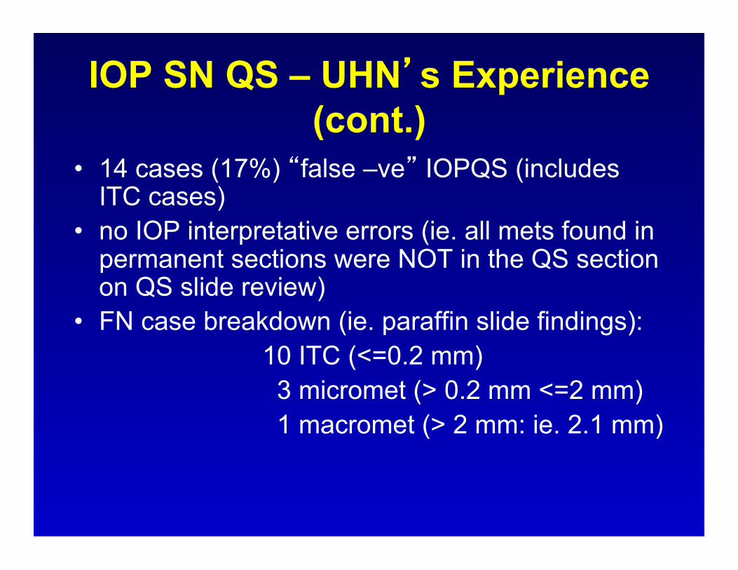
• 14 cases (17%) “false –ve” IOPQS (includes ITC cases)
• no IOP interpretative errors (ie. all mets found in permanent sections were NOT in the QS section on QS slide review)
• FN case breakdown (ie. paraffin slide findings): 10 ITC (<=0.2 mm) 3 micromet (> 0.2 mm <=2 mm) 1 macromet (> 2 mm: ie. 2.1 mm)
Impact of recent studies on SLN evaluation IBC
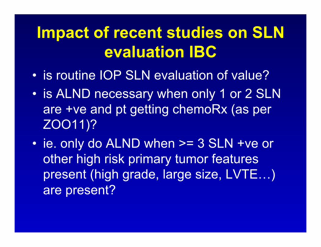
• is routine IOP SLN evaluation of value? • is ALND necessary when only 1 or 2 SLN
are +ve and pt getting chemoRx (as per ZOO11)?
• ie. only do ALND when >= 3 SLN +ve or other high risk primary tumor features present (high grade, large size, LVTE…) are present?
Impact of recent studies on SLN evaluation IBC
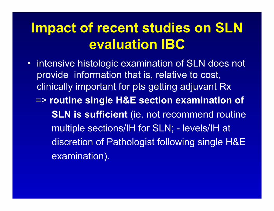
• intensive histologic examination of SLN does not provide information that is, relative to cost, clinically important for pts getting adjuvant Rx
=> routine single H&E section examination of SLN is sufficient (ie. not recommend routine multiple sections/IH for SLN; - levels/IH at discretion of Pathologist following single H&E examination).
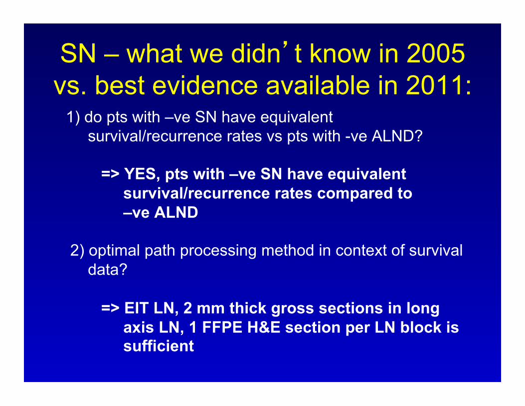
SN – what we didn’t know in 2005 vs. best evidence available in 2011: 1) do pts with –ve SN have equivalent survival/recurrence rates vs pts with -ve ALND? => YES, pts with –ve SN have equivalent survival/recurrence rates compared to –ve ALND 2) optimal path processing method in context of survival data? => EIT LN, 2 mm thick gross sections in long axis LN, 1 FFPE H&E section per LN block is sufficient
SN – what we didn’t know in 2005 vs. best evidence available in 2011
(cont.):
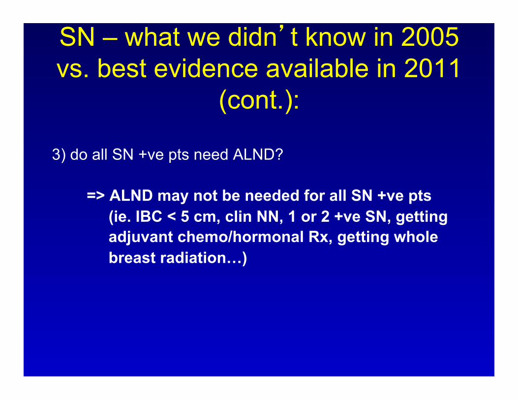
3) do all SN +ve pts need ALND? => ALND may not be needed for all SN +ve pts (ie. IBC < 5 cm, clin NN, 1 or 2 +ve SN, getting adjuvant chemo/hormonal Rx, getting whole breast radiation…)


![Screening recurrence and lymph node metastases in head and … · 2012-12-07 · first two years [3,4]. The status of the lymph nodes appears to be one of the most important prognostic](https://img.pdfslide.net/doc/110x75/5e9dad3661435c7e0e0c9ee4/screening-recurrence-and-lymph-node-metastases-in-head-and-2012-12-07-first-two.jpg)


![Journal of Cancer · Web viewadenocarcinoma with concomitant gastric, duodenal, bone, and mediastinal lymph node metastases, even gastrointestinal metastases [8]. The eye is a rare](https://img.pdfslide.net/doc/110x75/6003379640e0301f0a60f79e/journal-of-web-view-adenocarcinoma-with-concomitant-gastric-duodenal-bone-and.jpg)