Embed Size (px)
Citation preview
Ontogenetic Expression of TrkNeurotrophin Receptors in the Chick
Auditory System
SARAH L. COCHRAN,1 JENNIFER S. STONE,1
OLIVIA BERMINGHAM-MCDONOGH,1,2 SCOTT R. AKERS,1 FRANCES LEFCORT,3
AND EDWIN W RUBEL1*1Virginia Merrill Bloedel Hearing Research Center, Department of Otolaryngology/Head
and Neck Surgery, University of Washington, Seattle, Washington 98195-79232Department of Biological Structure, University of Washington,
Seattle, Washington 98195-79233Department of Biology and WWAMI Medical Program, Montana State University,
Bozeman, Montana 59717
ABSTRACTNeurotrophins and their cognate receptors are critical to normal nervous system
development. Trk receptors are high-affinity receptors for nerve-growth factor (trkA),brain-derived neurotrophic factor and neurotrophin-4/5 (trkB), and neurotrophin-3 (trkC). Weexamine the expression of these three neurotrophin tyrosine kinase receptors in the chickauditory system throughout most of development. Trks were localized in the auditorybrainstem, the cochlear ganglion, and the basilar papilla of chicks from embryonic (E) day 5 toE21, by using antibodies and standard immunocytochemical methods. TrkB mRNA waslocalized in brainstem nuclei by in situ hybridization.TrkB and trkC are highly expressed inthe embryonic auditory brainstem, and their patterns of expression are both spatially andtemporally dynamic. During early brainstem development, trkB and trkC are localized in theneuronal cell bodies and in the surrounding neuropil of nucleus magnocellularis (NM) andnucleus laminaris (NL). During later development, trkC is expressed in the cell bodies of NMand NL, whereas trkB is expressed in the nerve calyces surrounding NM neurons and in theventral, but not the dorsal, dendrites of NL. In the periphery, trkB and trkC are located in thecochlear ganglion neurons and in peripheral fibers innervating the basilar papilla andsynapsing at the base of hair cells.The protracted expression of trks seen in our materials isconsistent with the hypothesis that the neurotrophins/tyrosine kinase receptors play one orseveral roles in the development of auditory circuitry. In particular, the polarized expressionof trkB in NL is coincident with refinement of NM terminal arborizations on NL. J. Comp.Neurol. 413:271–288, 1999. r 1999 Wiley-Liss, Inc.
Indexing terms: cellular protein sorting; nucleus laminaris; nucleus magnocellularis; basilar
papilla; brain-derived neurotrophin factor; neurotrophin-3
Classically, neurotrophins and their cognate receptorswere considered mediators of target innervation and selec-tive neuronal survival (Levi-Montalcini and Angeletti,1968). Since the discovery of the first neurotrophin, nervegrowth factor (NGF), three other neurotrophins (brain-derived neurotrophic factor (BDNF), neurotrophin-4/5 (NT-4/5), and neurotrophin-3 (NT-3) have been discovered, andour understanding of neurotrophins has broadened suffi-ciently such that we are beginning to identify the numer-ous aspects of neuronal development, maintenance, andrepair that neurotrophins can mediate. These varied as-pects include cell differentiation (reviewed in Chao, 1992;
Lachyankar et al., 1997), cell survival (reviewed in Levi-Montalcini, 1987), axonal growth and morphology (Segalet al., 1995; San Jose et al., 1997), dendritic morphology(Cohen-Cory and Fraser, 1994, 1995; McAllister et al.,1995, 1997), synaptic efficacy (Patterson et al., 1996; Suen
Grant sponsor: National Institutes of Health; Grant numbers: DC00395,DC00299.
*Correspondence to: Dr. Edwin W Rubel, Virginia Merrill Bloedel Hear-ing Research Center, CHDD, Box 357923, University of Washington,Seattle, WA 98195-7923. E-mail: [email protected]
Received 4 March 1999; Revised 19 May 1999; Accepted 24 June 1999
THE JOURNAL OF COMPARATIVE NEUROLOGY 413:271–288 (1999)
r 1999 WILEY-LISS, INC.
et al., 1997), ion channel expression (Jimenez et al., 1997),neuronal plasticity (Lindholm et al., 1994), and cell death(Carter et al., 1996; Frade et al., 1996; Bredesen andRabizadeh, 1997; for review: Frade and Barde, 1998).Moreover, the presence of neurotrophins and their recep-tors has been demonstrated in the adult brain (Friedmanet al., 1991; Wu et al., 1996; Conner et al., 1997; Yan et al.,1997), suggesting that neurotrophins are necessary bothfor the initiation and for the maintenance of synapticcircuitry.
Studies of developing sensory systems have suggested aubiquitous involvement of neurotrophins in the establish-ment of the intricate patterns of connectivity that underliesensory function. Neurotrophins and their transmem-brane protein tyrosine kinase receptors (trkA: NGF; trkB:BDNF and NT-4/5; trkC: NT-3) have been observed in thedeveloping olfactory (Holcomb et al., 1995; Roskams et al.,1996), gustatory (Nosrat et al., 1996, 1997; Fritzsch et al.,1997b; Zhang et al., 1997), somatosensory (Nosrat et al.,1997), and visual (Allendoerfer et al., 1994; Cohen-Coryand Fraser, 1994, 1995; Lauterborn et al., 1994; vonBartheld et al., 1996a,b; Cabelli et al., 1997) systems.Studies of the auditory system have demonstrated thepresence of neurotrophins and trks in amphibians (Don etal., 1997), avians (Bernd and Represa, 1989; Represa et al.,1991; von Bartheld et al., 1991; Represa et al., 1993; Berndet al., 1994; Hallbook and Fritzsch, 1997; Pirvola et al.,1997), and mammals (Despres et al., 1991; von Bartheld etal., 1991; Pirvola et al., 1992, 1994; Ylikoski et al., 1993;Schecterson and Bothwell, 1994; Wheeler et al., 1994;Hafidi et al., 1996; Knipper et al., 1996; Vazquez et al.,1996; Wiechers et al., 1999). Furthermore, the exigency ofneurotrophins and trks in the stabilization of inner earinnervation of mammals has been established (Ernfors etal., 1994, 1995; Shimmang et al., 1995; Fritzsch et al.,1997a; and for review, see Fritzsch et al., 1997c).
In contrast to the abundance of studies describing theexpression and the biological significance of neurotrophinsand their associated receptors in the mammalian auditorysystem, fewer studies to date have addressed these samequestions in the avian auditory system. The present studyexamines the distribution of the three trk receptors in thecochlear ganglion, basilar papilla (cochlea), and auditorybrainstem nuclei of the chick during embryonic develop-ment. We describe the dynamic changes in the patterns oftrk protein expression throughout most of the embryonicperiod and correlate trk expression with established onto-genetic events.
MATERIALS AND METHODS
Tissue preparation
Fertilized White Leghorn chicken (Gallus domesticus)eggs were purchased from H & N International (Redmond,WA). Eggs were incubated at 37–38°C and a relative 80%humidity. Over 150 embryos were used for this study. TheAnimal Care and Use Committee of the University ofWashington approved all procedures described below.
The expression of trk receptors in White Leghorn chick-ens was followed from embryonic day 5 (E5) until just priorto hatching (E21). Embryonic age was determined bystaging according to Hamburger and Hamilton (1951) andis expressed in embryonic days. All data reported wereconfirmed on a minimum of four animals at each agereported. For tissue processing, the embryo was removed
from the egg, decapitated, and the tissue (brainstem ortemporal bone encasing the cochlear ganglion and basilarpapilla) was quickly dissected in Hank’s buffered saltsolution (Gibco BRL, Life Technologies, Inc., Gaithers-burg, MD). The tissue was immersion-fixed overnight inmethacarn fixative (60% methanol, 30% chloroform, and10% glacial acetic acid) in preparation for immunocyto-chemical or histological processing. In some cases, thetissue was immersion-fixed with 4% paraformaldehyde. Inboth the hindbrain and the periphery, the methacarnfixation increased the overall intensity of staining whileequally preserving the anatomical structure of the tissue.Paraformaldehyde-fixed tissue was rinsed in phosphate-buffered saline (PBS, 0.01 M, pH 7.4). The methacarn-fixed tissue was rehydrated through a series of decreasingconcentrations of methanol, then rinsed in PBS. Hind-brain tissue was embedded in agar and sectioned on avibratome into three alternating sets of 50 µm sections.Initially, in each case, these three sets were simulta-neously processed for trkA, trkB, and trkC immunoreactiv-ity. In later experiments, tissue was not processed for trkAimmunoreactivity. Instead, the third set was reserved forNissl staining with thionin. The isolated temporal bonewas cryoprotected in 30% sucrose in PBS until it sank. Thetissue was flash-frozen in dry ice-cooled heptane, coveredin OCT compound (Sakura Finetek USA Inc., Torrance,CA), cryostat-sectioned at 14 µm, and thaw-mounted ontochrome alum-subbed slides, again generating three sets ofalternating sections. The sections were stored at 220°Cuntil processed.
Trk antibodies
The trk antibodies used in this study were kindlyprovided by Drs. Frances Lefcort (Montana State Univer-sity, Bozeman, MT) and Louis Reichardt (Howard HughesMedical Institute, University of California, San Francisco,CA). The antibodies are rabbit polyclonal antibodies specifi-cally directed against the entire extracellular domain oftrkA, trkB, or trkC chick tyrosine kinase neurotrophinreceptors. They have been previously characterized (Lef-cort et al., 1996; Oakley et al., 1997).
Immunocytochemistry
Standard immunocytochemical methods were used forimmunolabeling trk receptors in the chick inner ear andauditory brainstem. The brain tissue was processed asfree-floating sections in multiwell plates. The temporalbone tissue was processed on slides because it was thaw-mounted during the cutting procedure. Although the proto-col for both was similar, longer incubation times for thethicker brain tissue were used; these times are detailedbelow. All steps were performed at room temperature, withthe exception of the primary antibody incubation.
To quench endogenous peroxidase activity, the sectionswere initially incubated for 1.5 hours in Tris-bufferedsaline (TBS: 25 mM Tris, pH 7.4, 150 mM NaCl) containing3% hydrogen peroxide and 10% methanol. The sectionswere rinsed in a series of 25 mM Tris buffer washes, thenpreincubated for 3 hours in a blocking solution to preventnonspecific IgG binding (TBS, 0.03% Triton-X-100, 10%normal goat serum [Vector Laboratories, Burlingame, CA],and 0.1% bovine serum albumin [Sigma, St. Louis, MO]).The sections were subsequently incubated overnight inprimary antibody (1:3,000) at 4°C. Negative control sec-tions were incubated in the same solution with the pri-
272 S. L. COCHRAN ET AL.
mary antibody omitted. Sections were rinsed in a series ofbuffer washes, then incubated in biotinylated goat anti-rabbit secondary antibody (Vector; 1:200) for 4 hours. Aftera series of washes in buffer, tissue was incubated for 1.5hours in ABC solution from a Vectastain Elite ABC-peroxidase kit (Vector) in order to amplify the immunoreac-tivity. The tissue was rinsed in several 50 mM Tris/HClbuffer washes (pH 7.6), then antibody labeling was visual-ized by using diaminobenzidine tetrahydrochloride (DAB)as the chromagen (0.05% DAB, 0.003% hydrogen peroxide,50 mM Tris). After DAB processing, sections were washedin a series of 50 mM Tris washes and given a final rinse indistilled water. The brain sections were mounted ontochrome alum-subbed slides and allowed to dry overnight.All tissue sections were dehydrated in a series of increas-ing ethanols and xylene and coverslipped with Permount(Fisher Scientific, Fair Lawn, NJ).
Cryostat sections through the dorsal root ganglia (DRG)of an E8 chick were routinely included in the immunolabel-ing experiments as positive controls. In each case, wefound restricted patterns of trk protein expression in theDRG similar to that described previously (Lefcort et al.,1996; Oakley et al., 1997).
TrkB digoxigenin-labeled RNA probes
TrkB full-length cDNA cloned into pBluescript wasgenerously provided by Drs. Kristin Boeshore and ThomasLarge (Case Western University, Cleveland, OH). Theplasmids were linearized and cRNA probes synthesizedusing T3 and T7 RNA polymerase. To enhance probepenetration into the tissue sections, probes were hydro-lyzed to an average of 250 bp in length.
In situ hybridization
E12 embryos were removed from the egg, decapitated,and the brainstem was quickly dissected in cold PBS with2 mM EGTA; 400 µm coronal sections generated with an‘‘egg-slicer’’ guillotine (Katz, 1987) were fixed in 4% formal-dehyde in PBS/2 mM EGTA for 2 hours. Sections werewashed in PBS containing 0.1% Tween 20 (PTW), then in50% MeOH/PTW, and then stored in 100% MeOH at -70°Cuntil processed. The in situ hybridization protocol wefollowed was based on Henrique et al. (1995). All stepswere performed at room temperature unless otherwisenoted. The sections were rehydrated through a methanolseries, rinsed in PTW, and treated with 10 µg/ml protein-ase K in PTW for 15–20 minutes. The sections were thenwashed in PTW and postfixed for 20 minutes in PTWcontaining 4% formaldehyde and 0.1% glutaraldehyde.Following several washes in PTW, the sections were rinsedin 1:1 hybridization mix/PTW. The hybridization mixcontains 50% formamide, 1.33 saline/sodium citrate buffer,5 mM EDTA, 50 µg/ml yeast RNA, 0.2% Tween-20, 0.5%CHAPS (3-[(3-cholamidopropyl)-dimethylammonio]-1-pro-panesulfonate), 100 µg/ml heparin. Sections were rinsed inhybridization mix, then incubated in fresh hybridizationmix for 1 hour at 65°C. After 1 hour, prewarmed hybridiza-tion mix containing the digoxigenin-labeled RNA probewas added and the tissue was incubated overnight at 65°C.The tissue was washed for 1 hour in several changes ofprewarmed hybridization mix, washed for 10 minutes inprewarmed 1:1 hybridization mix/ MABT buffer (100 mMmaleic acid disodium salt, 150 mM NaCl, 0.1% Tween-20,pH 7.5), and finally, washed in MABT alone. The sectionswere then incubated for 1 hour in MABT with 2% Boeh-
ringer blocking reagent (BBR: Boehringer Mannheim Bio-chemicals Inc., Indianapolis, IN) added. In a second block-ing step, sections were incubated for 1 hour in 20%heat-treated goat serum in MABT/2% BBR. The tissue wasincubated overnight at 4°C in an anti-digoxigenin anti-body (Boehringer Mannheim) diluted 1/2,000 in MABT/2%BBR/20% serum. The next day, three postantibody rinseswere followed by three 1-hour washes in MABT. The tissuewas then rinsed twice in NTMT buffer (5 M NaCl, 2 M TrisHCl, pH 9.5, 2 M MgCl2, 10% Tween-20) before incubatingthe tissue in the color reagent containing 3-nitro bluetetrazolium salt, 5-bromo-4-chloro-3-indolyl phosphate, andlevamisole in NTMT. The sections were allowed to react atroom temperature for up to 24 hours, then washed severaltimes in PTW. The tissue was refixed in 4% formalin/PTWfor 2 hours, rinsed several times in PTW, and stored at 4°Cin PTW/0.1% sodium azide.
Analysis and imaging
The tissue was visualized under light microscopy on aLeitz Aristoplan microscope by using standard brightfieldand Nomarski optics. Sections were photographed withVelvia color slide film (Fuji Photo Film Co., Ltd., Tokyo,Japan). Digital photomicrographs were generated by scan-ning the slides into Adobe Photoshop 4.0 (Adobe, MountainView, CA) with a Nikon LS-1000 film scanner. DigitizedPhotoshop images were enhanced for contrast and weattempted to make parallel enhancement to plates thatwere compared within the same figure. Images wereprinted with a dye-sublimation Phaser printer (Tektronix,Beaverton, OR).
RESULTS
The specificity of the polyclonal antibodies for the chicktrk neurotrophin receptors has been demonstrated previ-ously using several approaches (see Lefcort et al., 1996;Oakley et al., 1997). In the present studies, we routinelyran both positive and negative controls to assay thespecificity of the trk antibodies (see Fig. 1). Beyond thesecontrols, however, the spatially and temporally distinctpatterns of immunolabeling of each trk lend confidence tothe specificity of the antibodies.
Trk protein immunolabeling is present in the auditorysystem of the chick throughout the embryonic period weassessed and into posthatch development, though thepattern of labeling differs remarkably among receptors,among tissues, and across ages. In general, trkB (theprimary receptor for brain-derived neurotrophic factor andneurotrophin 4/5) and trkC (the primary receptor forneurotrophin-3) immunolabeling is prevalent at all agesboth peripherally and centrally. In contrast, trkA (theprimary receptor for nerve growth factor) immunolabelingis distinctly absent in all tissues at the ages studied.
Trk immunolabeling in the cochlear ganglionand basilar papilla
E5– 8. The peripheral processes of cochlear ganglioncells (afferent fibers) invade the sensory epithelium (thebasilar papilla) predominantly during the period betweenE5 to E7 (Whitehead and Morest, 1985a). TrkA immunola-bel is not detectable in the auditory periphery at this age(Fig. 2A), nor in successively older animals. TrkB stainingin the cochlear ganglion is very light and is distributedthroughout the neurons of the cochlear ganglion (Fig. 2B).
Trk RECEPTORS IN EMBRYONIC CHICK AUDITORY SYSTEM 273
In addition, there is light immunolabeling along thesuperior edge of the basilar papilla (arrow, Fig. 2B), whichis the portion of the sensory epithelium that will beoccupied primarily by tall hair cells once hair cell differen-tiation has taken place (Fischer, 1992). This light labelingappears fibrous, and therefore, we suggest that it is
present in a handful of trkB-labeled fibers that havepenetrated the basilar papilla at its superior edge. ThesetrkB-labeled fibers course radially through the basilarpapilla toward the lumenal edge of the epithelium. Incontrast, trkC appears to be the most strongly expressedhigh-affinity neurotrophin tyrosine kinase receptor in the
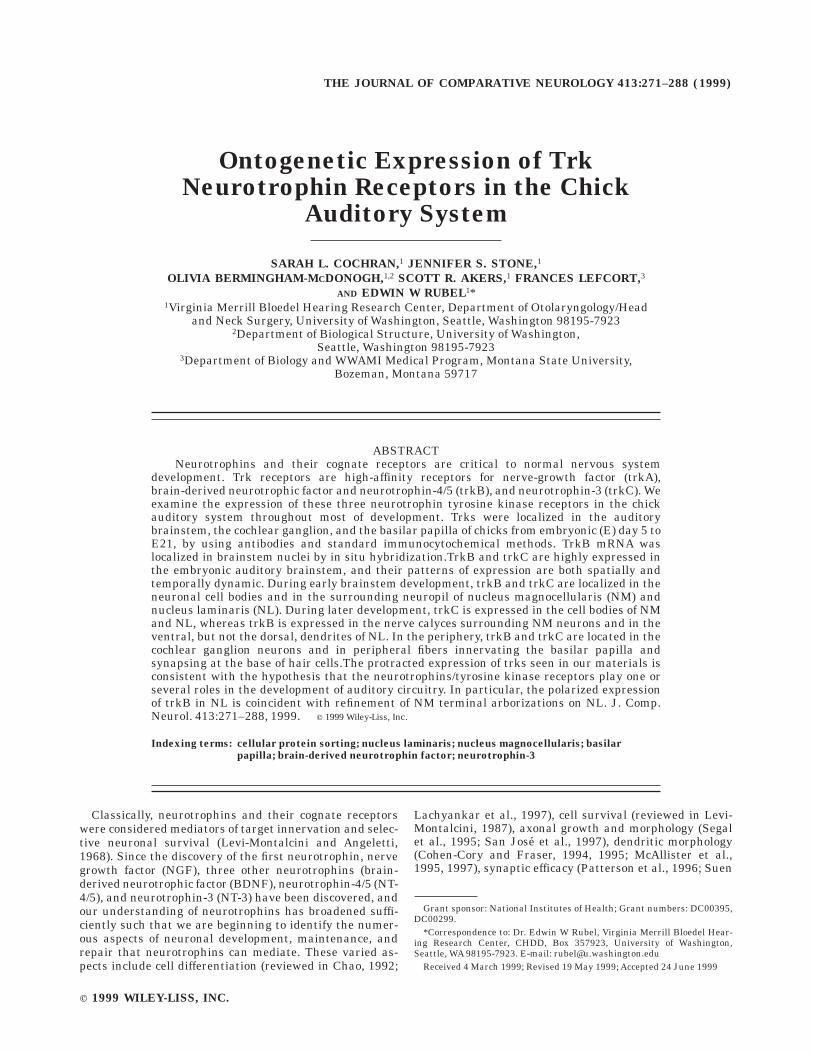
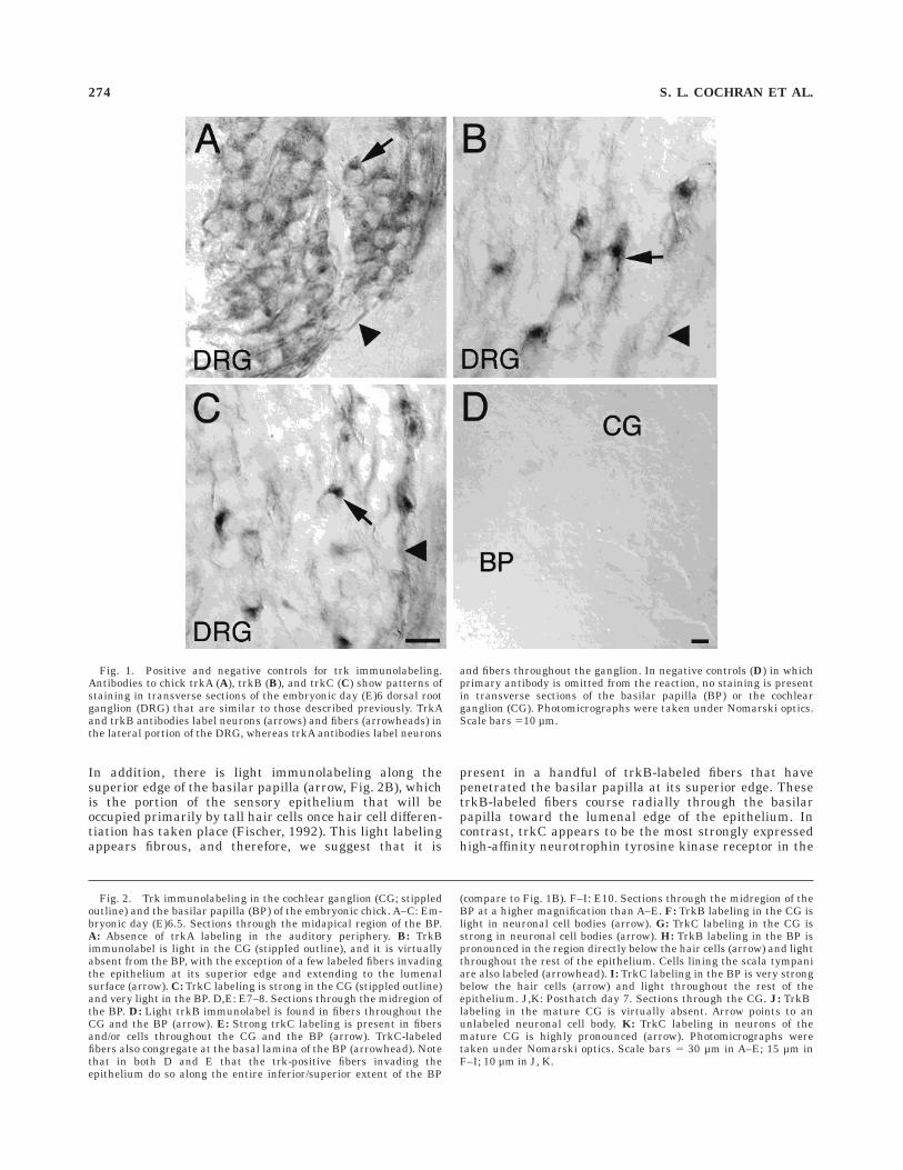
Fig. 1. Positive and negative controls for trk immunolabeling.Antibodies to chick trkA (A), trkB (B), and trkC (C) show patterns ofstaining in transverse sections of the embryonic day (E)6 dorsal rootganglion (DRG) that are similar to those described previously. TrkAand trkB antibodies label neurons (arrows) and fibers (arrowheads) inthe lateral portion of the DRG, whereas trkA antibodies label neurons
and fibers throughout the ganglion. In negative controls (D) in whichprimary antibody is omitted from the reaction, no staining is presentin transverse sections of the basilar papilla (BP) or the cochlearganglion (CG). Photomicrographs were taken under Nomarski optics.Scale bars 510 µm.
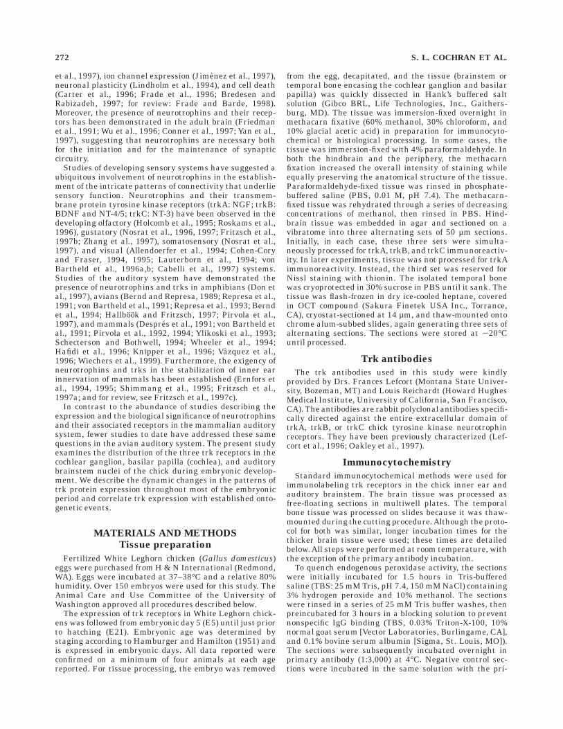
Fig. 2. Trk immunolabeling in the cochlear ganglion (CG; stippledoutline) and the basilar papilla (BP) of the embryonic chick. A–C: Em-bryonic day (E)6.5. Sections through the midapical region of the BP.A: Absence of trkA labeling in the auditory periphery. B: TrkBimmunolabel is light in the CG (stippled outline), and it is virtuallyabsent from the BP, with the exception of a few labeled fibers invadingthe epithelium at its superior edge and extending to the lumenalsurface (arrow). C: TrkC labeling is strong in the CG (stippled outline)and very light in the BP. D,E: E7–8. Sections through the midregion ofthe BP. D: Light trkB immunolabel is found in fibers throughout theCG and the BP (arrow). E: Strong trkC labeling is present in fibersand/or cells throughout the CG and the BP (arrow). TrkC-labeledfibers also congregate at the basal lamina of the BP (arrowhead). Notethat in both D and E that the trk-positive fibers invading theepithelium do so along the entire inferior/superior extent of the BP
(compare to Fig. 1B). F–I: E10. Sections through the midregion of theBP at a higher magnification than A–E. F: TrkB labeling in the CG islight in neuronal cell bodies (arrow). G: TrkC labeling in the CG isstrong in neuronal cell bodies (arrow). H: TrkB labeling in the BP ispronounced in the region directly below the hair cells (arrow) and lightthroughout the rest of the epithelium. Cells lining the scala tympaniare also labeled (arrowhead). I: TrkC labeling in the BP is very strongbelow the hair cells (arrow) and light throughout the rest of theepithelium. J,K: Posthatch day 7. Sections through the CG. J: TrkBlabeling in the mature CG is virtually absent. Arrow points to anunlabeled neuronal cell body. K: TrkC labeling in neurons of themature CG is highly pronounced (arrow). Photomicrographs weretaken under Nomarski optics. Scale bars 5 30 µm in A–E; 15 µm inF–I; 10 µm in J, K.
274 S. L. COCHRAN ET AL.
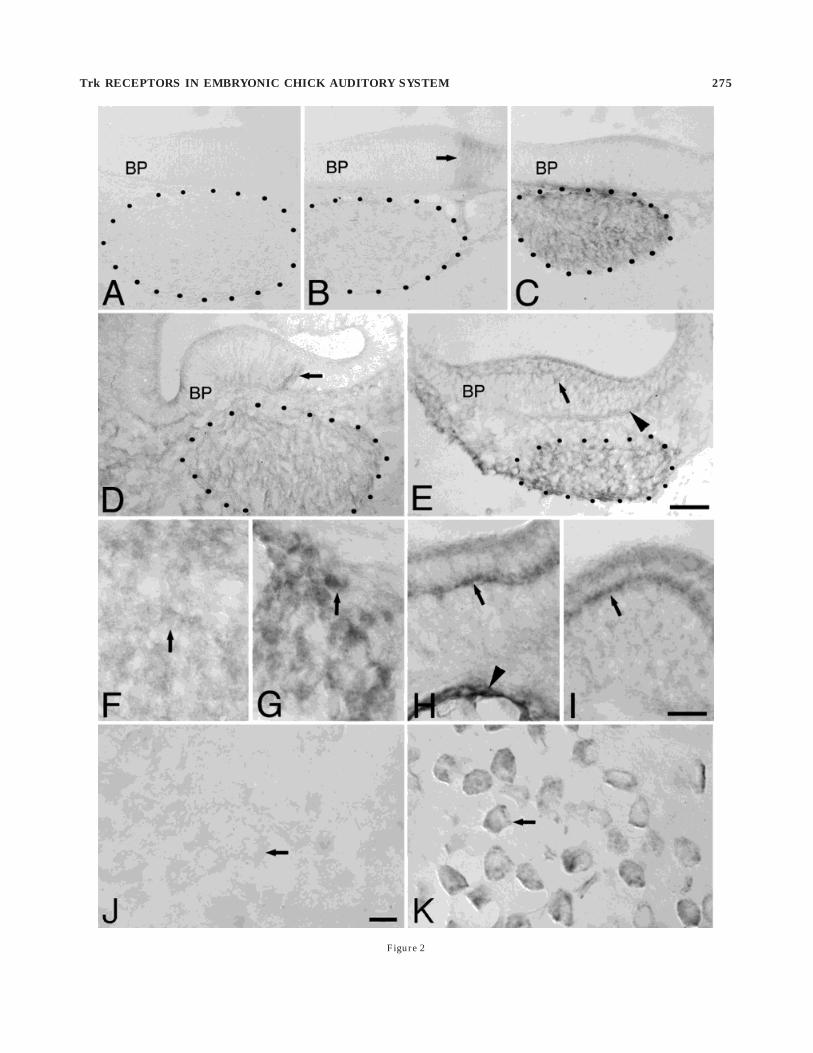
Figure 2
Trk RECEPTORS IN EMBRYONIC CHICK AUDITORY SYSTEM 275
developing auditory periphery, and this pattern is alreadyevident in the E6.5 chick. TrkC immunolabel is abundantin the cochlear ganglion (Fig. 2C). At this age, heavy trkCimmunolabel is distributed in and around the neurons ofthe cochlear ganglion, suggesting that it is present withinneural cell bodies as well as the neural processes of theganglion. Although the cochlear ganglion lies in closeapposition to the sensory epithelium at this age, the heavytrkC labeling does not appear to extend into the epithe-lium. Rather, there is light immunolabel within the epithe-lium that may be due to its presence in the immatureepithelial cells or the neural processes, or both.
By E8, efferent projections from the brainstem begininvading the basilar papilla (Fritzsch et al., 1993), andcells within the basilar papilla have begun to differentiateinto either sensory (hair) cells or support cells (Cotancheand Sulik, 1984; Fermin and Cohen, 1984; Whitehead andMorest, 1985b; Molea et al., 1999). Figure 2D and E depictstypical trkB and trkC staining, respectively, in the E8auditory periphery. The patterns of trkB and trkC immuno-label at E8 are similar to those seen at E6.5, with somenotable exceptions. The distribution of trkB label in co-chlear ganglion neurons is consistent with that seenpreviously, i.e., it is somatic and is distributed uniformlythroughout the ganglion, though the signal at E8 appearsto be slightly darker. The distribution of trkB label in thebasilar papilla, however, is quite different at E8 than thatseen earlier. It is still fibrous, but it is distributed through-out the superior/inferior axis of the sensory epithelium(Fig. 2D). Some labeled fibers appear to span the entirewidth of the basilar papilla, whereas others stop short ofthe lumen.
At E8, trkC patterns of staining in the auditory periph-ery are similar to patterns of trkB staining, with someexceptions. Figure 2E demonstrates the trkC stainingtypical of the cochlear ganglion and basilar papilla. TrkCstaining is pronounced in neurons throughout the extent ofthe cochlear ganglion, and it appears more intense thanthe trkB staining. In contrast to staining seen at E6.5,there appears to be less extrasomatic labeling in theganglion at this time. In the basilar papilla, trkC immuno-label appears more pronounced and more specific thanthat seen in younger tissue. TrkC labeling is present inneural fibers within the epithelium and in processes nearthe basal lamina. In addition, many trkC-labeled fibersextend radially through the epithelium; some emerge atthe lumenal edge, whereas others stop short of the lumen.Like the trkB-labeled fibers, trkC-labeled fibers are distrib-uted along the entire superior/inferior axis of the papilla.Some trkC labeling appears to be in the hair cells orsupport cells of the basilar papilla as well as in the neuralfibers.
We should note that despite our close scrutiny, anygradients in trkB or trkC labeling in the cochlear ganglionor in the basilar papilla (i.e., along the inferior/superior orthe proximal/distal axes) have escaped our detection.Careful analyses of older embryos have similarly resultedin a lack of staining gradients within labeled structures.The single exception to this is the appearance of thehandful of trkB-labeled fibers confined to the superior edgeof the basilar papilla at E6.5 mentioned previously (andshown in Fig. 2B). At E8, trkB- and trkC-labeled fibers canbe seen entering and leaving the ganglion in sagittalsections (data not shown). No trkB labeling was present inthe ventral region of the medulla, where the somata of the
efferent tracts are located (Whitehead and Morest, 1981;Fritzsch et al., 1993), nor in efferent fibers emanating fromthat region (data not shown). Therefore, we believe thatthe trkB-labeled fibers in the receptor epithelium areafferent fibers derived from cochlear ganglion rather thanefferent fibers.
E9–10. By E9–10, synapses between cochlear ganglioncells and hair cells in the basilar papilla have begun toform (Rebillard and Pujol, 1983; Whitehead and Morest,1985a) and hair cell differentiation is still underway(Cotanche and Sulik, 1984; Whitehead and Morest, 1985b).Figure 2F–I illustrates the distribution of trkB and trkCimmunolabeling in the auditory periphery of the chick atE9–10. In the cochlear ganglion, the general patterns oftrkB (Fig. 2F) and trkC (Fig. 2G) staining resemble thoseseen at E8. The trkC labeling remains considerably heavierthan the trkB labeling. However, there is a markeddifference in the distribution of the trk-labeled fiberswithin the basilar papilla at this time. Both trkB (Fig. 2H)and trkC (Fig. 2I) immunolabeling is localized to a horizon-tal stripe near the lumenal surface of the epithelium. Atthis point in development, the cells within the receptorepithelium have begun segregating into two layers, withnewly differentiated hair cells tightly packed in an upperlayer, and support cells loosely organized in a lower layer(Whitehead and Morest, 1985b). Thus, the trkB and trkClabeling in the basilar papilla appears to be present inneural fibers that have coalesced at the base of the newlydifferentiated hair cells, but the label may also be localizedwithin the hair cells themselves.
We noted strong trkB immunolabeling in cells lining thescala tympani that was highly reproducible (Fig. 2H). Thisstaining was not seen in negative controls in which theprimary antibody was omitted. Although it is possible thatthis labeling actually represents detection of the trkBantigen, it is more plausible that: (1) these cells have anoncovalent affinity for the primary antibody; or (2) theycontain an antigen that resembles trkB and this labelingrepresents cross-reactivity.
Posthatch. In the posthatch chick, trk labeling in thebasilar papilla has disappeared (data not shown). In thecochlear ganglion, trkB labeling has abated as well (Fig.2J). However, trkC labeling in the ganglion cell bodies(Fig. 2K) persists at levels comparable to those seen inyounger animals (see Fig. 2C, E, G). Our analyses indicatethat this mature trk labeling pattern is achieved by E14 inall tissues of the auditory periphery. Although a consider-able portion of ontogenetic development has occurred inthe chick auditory periphery by E11–12, synapse forma-tion (afferent and efferent) continues in the basilar papillauntil around E17 or so (Rebillard and Pujol, 1983; Ferminand Cohen, 1984; Whitehead and Morest, 1985a), andcentral cochlear ganglion terminations only begin to con-dense into their characteristic calyces at E13–15 (Jhaveriand Morest, 1982). Thus, it appears that both trkB andtrkC may be essential for developmental events occurringin the chick auditory periphery during the first two-thirdsof development, whereas trk C alone may play a role inevents occurring during the final week in ovo and in theposthatch animal.
Trk Immunolabeling in the auditorybrainstem
E5–8. The anlage of the auditory brainstem nuclei inthe chick first appears as a C-shaped structure at the
276 S. L. COCHRAN ET AL.
lateral edge of the brainstem, where the three principalnuclei, nucleus magnocellularis (NM), nucleus laminaris(NL), and nucleus angularis (NA), are indistinguishablefrom one another. Immunolabeling for the neurotrophintyrosine kinase trkB and trkC receptors is visible in theanlage of the auditory brainstem as early as the anlage canbe clearly identified (E6–7). In contrast, trkA immunolabelis not apparent in the auditory brainstem anlage, nor is itapparent in the auditory brainstem nuclei of successivelyolder animals. This absence of trkA in the central auditorysystem (at least at lower levels) is commensurate to theabsence of trkA noted peripherally. Our observations sug-gest that trkA does not participate at all in the embryonicdevelopment of the peripheral or brainstem auditory sys-tem in chick.
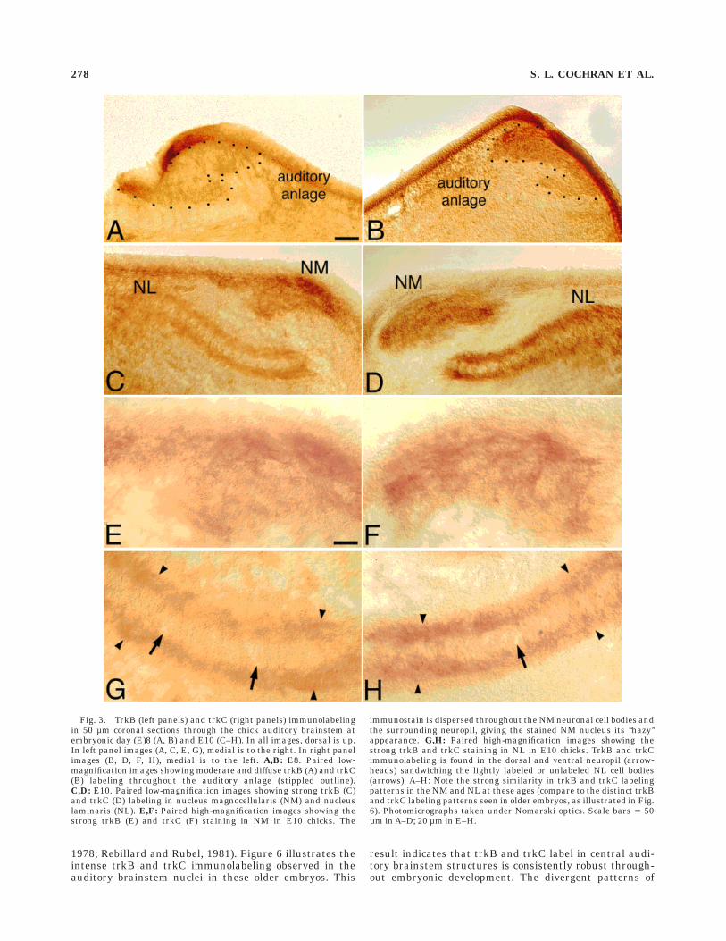
At E7–8, the distribution and intensity of trkB and trkCimmunostaining in the auditory brainstem anlage areindistinguishable from one another. The staining is moder-ate and diffuse, appearing somewhat ‘‘hazy,’’ as illustratedby Figure 3A and B. Trk-labeled fibers are not apparenteither within or surrounding the labeled anlage. Althoughdiffuse, trkB and trkC immunolabel is distributed uni-formly throughout the anlage, both in the mediolateraland dorsoventral extents of the structure, as illustrated inthe coronal sections in Figure 3A and B, and throughoutthe rostrocaudal extent. Although the label is specific, trkBand trkC immunolabel in the auditory anlage is notremarkably different than that found in the surroundingbrainstem structures. That is, trkB and trkC labeling iseminent throughout many of the hindbrain structures atmore or less equal intensities at this age.
E9–10. As the surrounding brainstem is developingand expanding in all three dimensions, the relative posi-tions of the auditory brainstem nuclei shift mediallywithin the brainstem. By E9–10, NM, NL, and NA areeasily distinguished in Nissl-stained tissue or in tissuestained by Bodian’s protargol method (Rubel et al., 1976;Jhaveri and Morest, 1982; Young and Rubel, 1986). At thistime, the refinement of NM axonal terminations on NL hasbegun (Young and Rubel, 1986). Trk immunolabel in theE10 auditory brainstem nuclei is moderate to heavy andsomewhat diffuse, as illustrated in Figure 3C–H. TrkB andtrkC immunolabeling patterns are indistinguishable fromone another in the E10 brainstem, just as in the E8 tissue(Fig. 3A and B). In NM, trkB, and trkC immunolabel islocated in the neuronal cell bodies throughout NM and inthe neuropil surrounding NM proper (Fig. 3C–F). It islikely that the hazy appearance of trkB and trkC immuno-staining in and around NM at this age is due to theprofusion of somatic processes (or dendrites) that charac-terize NM from about E7 to E11 and which will eventuallyretract (Jackson and Parks, 1982; Jhaveri and Morest,1982; Young and Rubel, 1986). In NL, trkB and trkCimmunolabel is located most densely in the dorsal andventral neuropil. Little immunolabel is apparent in theneuronal cell bodies of NL, which may be due to the ratherscant amount of somatic cytoplasm present at this stage ofdevelopment. The labeling found in the dorsal and in theventral neuropil of NL appears symmetrical at this age(Fig. 3C, D, G, and H). In nucleus angularis, trkB and trkClabel at this stage of embryonic development is moderatelyintense and is distributed throughout the extent of thenucleus (not shown). Typical of the entire region, trkB andtrkC staining patterns in nucleus angularis are indistin-guishable from each other in E9–10 animals. We are
unable to detect gradients in trkB or trkC immunolabel inany of the auditory brainstem nuclei along the mediolat-eral or rostrocaudal axes at this age, nor can we detectthem in successively older embryonic chicks.
E11–12. The first time physiological responses can beobserved in NM and NL is at about E11 (Jackson et al.,1982). In parallel, NM and NL neurons begin to undergo aperiod of exuberant growth of dendrites (Jackson andParks, 1982; Jhaveri and Morest, 1982; Smith, 1981) and aperiod of dramatic cell death in NL (Rubel et al., 1976).Whereas both the intensity and the distribution of trkBand trkC immunolabel are remarkably similar in animalsprior to E11, from E11 onward, the distributions of trkBand trkC in the auditory brainstem are markedly distinct.
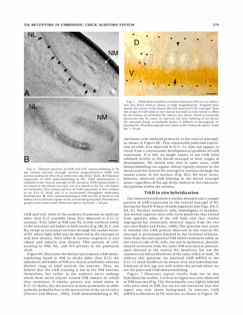
By far, the most striking change from earlier timepointsis the pattern of trkB labeling now observed in NL. Figure4A illustrates this unique pattern in an E12 embryo. Atthis time, trkB label is predominantly confined to theneuropil, as seen in younger animals. However, in starkcontrast to that seen at younger ages, the trkB label inE11–12 animals (and in older embryos) is uniquely polar-ized. TrkB immunolabel is apparent only (or predomi-nantly) in the ventral neuropil of NL (Fig. 4A). TrkBimmunolabel is not apparent in the dorsal neuropil of NL,nor is it abundant in the neuronal cell bodies of NL. Thisresult is more evident at higher magnification, whereunlabeled NL neurons appear to be perched upon a shelf oftrkB label in the ventral neuropil (Fig. 5). This pattern willbe discussed in more detail in a following section.
TrkB immunolabeling in NM is distributed throughoutthe nucleus proper (Fig. 4A). Although the ‘‘hazy’’ appear-ance of label is still predominant, often the trkB labelappears to be condensed around the periphery of theneuronal cell bodies, i.e., perisomatically, and less con-densed within NM cells. At this age, the auditory nerveendings terminating on NM form immature, highly arbo-rized endings (Jhaveri and Morest, 1982; Whitehead andMorest, 1985a), rather than the nerve calyces characteris-tic of older animals. Thus, the heterogeneous trkB label inNM suggests that trkB is located in the auditory nerveendings contacting NM cells rather than in the NM cellsthemselves.
TrkC immunolabeling in NM, in contrast, is becomingincreasingly confined to the neuronal cell bodies of NM, asillustrated in Figure 4B. The trkC label in the cell bodies isstrong, whereas the trkC labeling in the surrounding NMneuropil is becoming light and diffuse. In NL, trkC immu-nolabeling is quite robust, equal in intensity to that foundin NM. As illustrated in Figure 4B, it is now located in boththe cell bodies of NL neurons and in the neuropil, wherebefore it had been located almost exclusively in the neuro-pil. In some caudal sections through NL, light to moderatetrkC labeling is observed in the NL dendrites.
GE12. The last one-third of embryonic development ischaracterized by several prominent events: the concomi-tant retraction of NM dendrites (Jackson and Parks, 1982;Jhaveri and Morest, 1982; Young and Rubel, 1986) withthe formation of true auditory nerve ‘‘calyces’’ surroundingNM cell bodies on which they terminate (Jhaveri andMorest, 1982); the continued refinement of NM axonalarborizations on NL (Young and Rubel, 1986); the stabiliza-tion of dendrite number and length and of cell number inNL (Rubel et al., 1976; Smith and Rubel, 1979; Smith,1981); and the development of acoustically driven re-sponses (Saunders et al., 1973, 1974; Jackson and Rubel,
Trk RECEPTORS IN EMBRYONIC CHICK AUDITORY SYSTEM 277
1978; Rebillard and Rubel, 1981). Figure 6 illustrates theintense trkB and trkC immunolabeling observed in theauditory brainstem nuclei in these older embryos. This
result indicates that trkB and trkC label in central audi-tory brainstem structures is consistently robust through-out embryonic development. The divergent patterns of
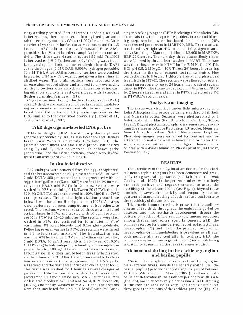
Fig. 3. TrkB (left panels) and trkC (right panels) immunolabelingin 50 µm coronal sections through the chick auditory brainstem atembryonic day (E)8 (A, B) and E10 (C–H). In all images, dorsal is up.In left panel images (A, C, E, G), medial is to the right. In right panelimages (B, D, F, H), medial is to the left. A,B: E8. Paired low-magnification images showing moderate and diffuse trkB (A) and trkC(B) labeling throughout the auditory anlage (stippled outline).C,D: E10. Paired low-magnification images showing strong trkB (C)and trkC (D) labeling in nucleus magnocellularis (NM) and nucleuslaminaris (NL). E,F: Paired high-magnification images showing thestrong trkB (E) and trkC (F) staining in NM in E10 chicks. The
immunostain is dispersed throughout the NM neuronal cell bodies andthe surrounding neuropil, giving the stained NM nucleus its ‘‘hazy’’appearance. G,H: Paired high-magnification images showing thestrong trkB and trkC staining in NL in E10 chicks. TrkB and trkCimmunolabeling is found in the dorsal and ventral neuropil (arrow-heads) sandwiching the lightly labeled or unlabeled NL cell bodies(arrows). A–H: Note the strong similarity in trkB and trkC labelingpatterns in the NM and NL at these ages (compare to the distinct trkBand trkC labeling patterns seen in older embryos, as illustrated in Fig.6). Photomicrographs taken under Nomarski optics. Scale bars 5 50µm in A–D; 20 µm in E–H.
278 S. L. COCHRAN ET AL.
trkB and trkC label in the auditory brainstem in embryosolder than E12 resemble those first observed in E11–12animals. TrkC label in NM and NL is now confined solelyto the neuronal cell bodies in both nuclei (Fig. 6B, D, F, andH), except in occasional sections through the caudal extentof NL where light label may be observed in the neuropil aswell (not shown). TrkC label in nucleus angularis is alsorobust and somatic (not shown). This pattern of trkCstaining in NM, NL, and NA persists in the posthatchchick.
Figure 6C illustrates the distinct perisomatic trkB immu-nolabeling found in NM in chicks older than E12; theadendritic cell bodies of NM are clearly unlabeled, whereasdistinct rings of label encircle the neurons. Thus, webelieve that the trkB staining is not in the NM neuronsthemselves, but rather in the auditory nerve endingswhich form nerve calyces around NM somata on whichthey terminate. A similar pattern was noted above inE11–12 chicks, but the pattern is more prominent in olderanimals, probably due to the maturation of the nerve calyx(Jhaveri and Morest, 1982). TrkB immunolabeling in NL
continues to be confined primarily to the ventral neuropil,as shown in Figure 6E. This remarkable polarized expres-sion of trkB, first observed in E11–12, does not appear toresult from a rostrocaudal developmental gradient of trkBexpression. If it did, we might expect to see trkB labelconfined strictly to the dorsal neuropil at later stages ofdevelopment. We should note that in some cases, trkBimmunolabeling can appear almost equally intense in thedorsal and the ventral NL neuropil in sections through thecaudal extent of the nucleus (Fig. 6G). We have never,however, observed trkB labeling in the dorsal neuropilalone, regardless of the age of the embryo or the rostrocau-dal position within the nucleus.
TrkB in situ hybridization
Our immunocytochemistry studies demonstrate a uniquepattern of trkB expression in the ventral neuropil of NLduring the final 8–9 days of embryogenesis (see Figs. 4A; 5;6A, E). Nucleus laminaris cells, homologous to mamma-lian medial superior olive cells, have dendrites that extendfrom opposite sides of the cell body and that receivesegregated but essentially identical inputs from the twoears (see Rubel and Parks, 1988). The question that arisesis whether the trkB protein observed in the ventral NLneuropil is presynaptic (located in the terminal arboriza-tions from the contralateral NM which terminate solely onthe ventral side of NL cells, but not in ipsilateral, dorsallylocated terminals from the same NM neurons) or postsyn-aptic (located in the ventral NL dendrites, but not thesymmetrical dorsal dendrites of the same cells), or both. Toaddress this question, we localized trkB mRNA in theE12–13 chick hindbrain by means of in situ hybridization.Embryos of this age are well within the period where wesee the polarized trkB immunolabeling.
Figure 7 illustrates typical results from our in situhybridization studies. Little or no signal was ever observedin NM neurons (Fig. 7A). Occasionally, very lightly labeledcells were seen in NM, but we are not convinced that thissignal was ever above background. In contrast, trkBmRNA is abundant in NL neurons, as shown in Figure 7B.
Fig. 4. Distinct patterns of trkB and trkC immunolabeling in 50µm coronal sections through nucleus magnocellularis (NM) andnucleus laminaris (NL) of an embryonic day (E)12 chick. A: Polarizedexpression of trkB immunolabeling in NL. TrkB immunostain isconfined to the ventral neuropil of NL (arrows). TrkB immunolabel isnot found in the dorsal neuropil, nor is it found in the NL cell bodies(arrowheads). This unique pattern of trkB expression is first evidentin an E11–12 chick and it is maintained throughout embryonicdevelopment. B: TrkC immunolabeling in NM and NL is found in cellbodies and in limited regions of the surrounding neuropil. Photomicro-graphs were taken under Nomarski optics. Scale bar 5 20 µm.
Fig. 5. TrkB immunolabel in nucleus laminaris (NL) in an embry-onic day (E)12 embryo shown at high magnification. Stippled linesdenote the extent of the dorsal (D) and ventral (V) NL neuropil. Notethe stripe of trkB label in the ventral neuropil (arrow) which is offsetby the lamina of unlabeled NL somata just above. Small arrowheadsdemarcate one NL soma. In contrast, the faint labeling of the dorsalNL neuropil (large arrowhead) makes it difficult to distinguish itsboundaries. Photomicrograph was taken under Nomarski optics. Scalebar 5 10 µm.
Trk RECEPTORS IN EMBRYONIC CHICK AUDITORY SYSTEM 279
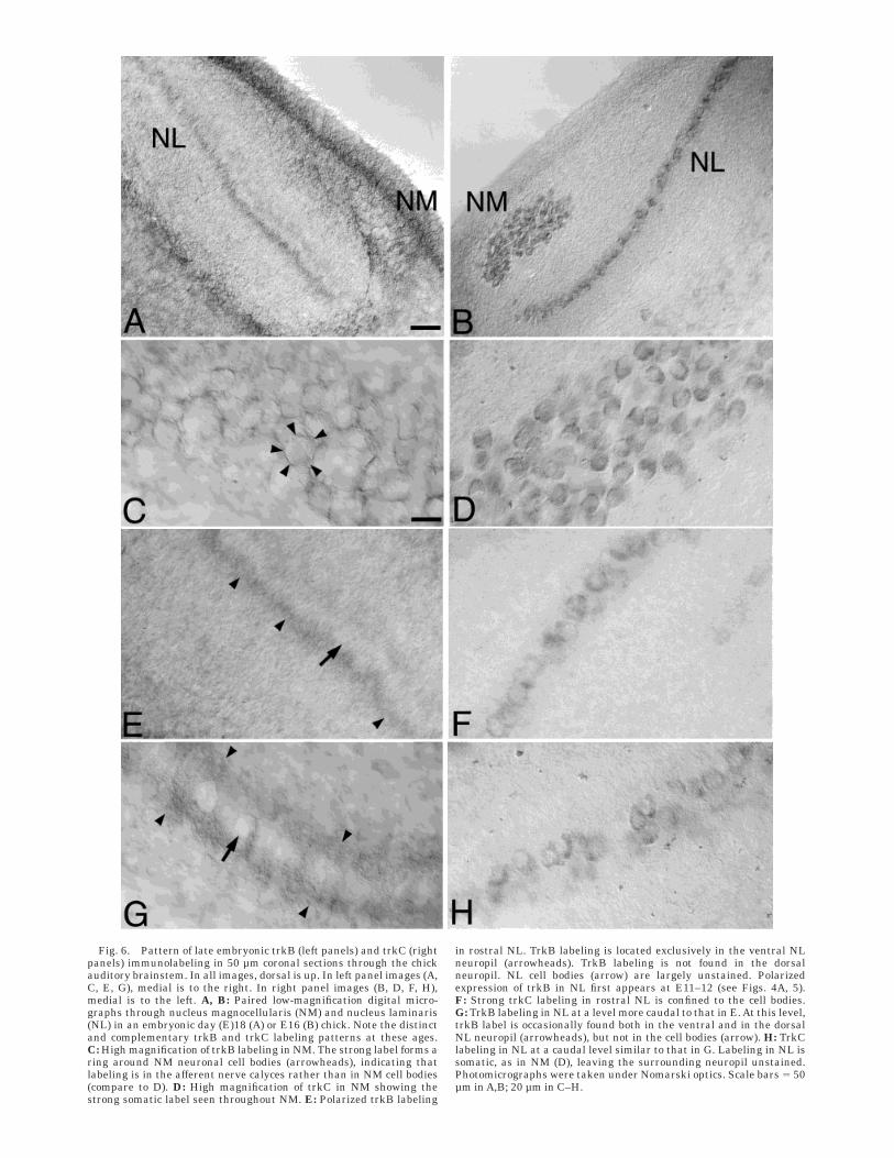
Fig. 6. Pattern of late embryonic trkB (left panels) and trkC (rightpanels) immunolabeling in 50 µm coronal sections through the chickauditory brainstem. In all images, dorsal is up. In left panel images (A,C, E, G), medial is to the right. In right panel images (B, D, F, H),medial is to the left. A, B: Paired low-magnification digital micro-graphs through nucleus magnocellularis (NM) and nucleus laminaris(NL) in an embryonic day (E)18 (A) or E16 (B) chick. Note the distinctand complementary trkB and trkC labeling patterns at these ages.C: High magnification of trkB labeling in NM. The strong label forms aring around NM neuronal cell bodies (arrowheads), indicating thatlabeling is in the afferent nerve calyces rather than in NM cell bodies(compare to D). D: High magnification of trkC in NM showing thestrong somatic label seen throughout NM. E: Polarized trkB labeling
in rostral NL. TrkB labeling is located exclusively in the ventral NLneuropil (arrowheads). TrkB labeling is not found in the dorsalneuropil. NL cell bodies (arrow) are largely unstained. Polarizedexpression of trkB in NL first appears at E11–12 (see Figs. 4A, 5).F: Strong trkC labeling in rostral NL is confined to the cell bodies.G: TrkB labeling in NL at a level more caudal to that in E. At this level,trkB label is occasionally found both in the ventral and in the dorsalNL neuropil (arrowheads), but not in the cell bodies (arrow). H: TrkClabeling in NL at a caudal level similar to that in G. Labeling in NL issomatic, as in NM (D), leaving the surrounding neuropil unstained.Photomicrographs were taken under Nomarski optics. Scale bars 5 50µm in A,B; 20 µm in C–H.
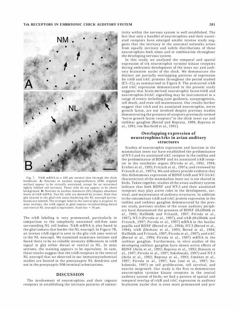
The trkB labeling is very pronounced, particularly incomparison to the completely unstained cell-free zonesurrounding NL cell bodies. TrkB mRNA is also found inthe glial somata that border the NL neuropil. In Figure 7B,an intense trkB signal is seen in the glia rich zone ventralto the NL neuropil. We examined numerous sections andfound there to be no reliable intensity differences in trkBsignal in glia either dorsal or ventral to NL. In mostsections, the staining appears to be equivalent. In sum,these results suggest that the trkB receptors in the ventralNL neuropil that we observed in our immunocytochemicalstudies are located in the postsynaptic NL dendrites andnot in the presynaptic NM terminal arborizations.
DISCUSSION
The involvement of neurotrophins and their cognatereceptors in establishing the intricate patterns of connec-
tivity within the nervous system is well established. Thefact that only a handful of neurotrophins and their associ-ated receptors have emerged amidst intense study sug-gests that the intricacy in the neuronal networks arisesfrom equally intricate and subtle distributions of theseneurotrophins both alone and in combination throughoutthe developing nervous system.
In this study, we analyzed the temporal and spatialexpression of trk neurotrophin tyrosine kinase receptorsduring embryonic development of the inner ear and audi-tory brainstem nuclei of the chick. We demonstrate thedistinct yet partially overlapping patterns of expressionfor trkB and trkC proteins throughout the period studied(E5–21), as summarized in Figure 8. The protracted trkBand trkC expression demonstrated in the present studysuggests that brain-derived neurotrophic factor/trkB andneurotrophin-3/trkC signalling may be instrumental in arange of events including axon guidance, synaptogenesis,cell death, and even cell maintenance. Our results furthersuggest that trkA and its associated neurotrophin, nervegrowth factor, are not involved despite previous studiesdemonstrating the presence of receptors previously termed‘‘nerve growth factor receptors’’ in the chick inner ear andcochlear ganglion (Bernd and Represa, 1989; Represa etal., 1991; von Bartheld et al., 1991).
Overlapping expression ofneurotrophins/trks in avian auditory
structures
Studies of neurotrophin expression and function in themammalian inner ear have established the predominanceof NT-3 and its associated trkC receptor in the cochlea andthe predominance of BDNF and its associated trkB recep-tor in the vestibular organs (Pirvola et al., 1992, 1994;Ernfors et al., 1995; Fritzsch et al., 1997a; and reviewed byFritzsch et al., 1997c). We and others provide evidence thatthis dichotomous expression of BDNF/trkB and NT-3/trkCcharacteristic of the mammalian inner ear is not shared inbirds. Taken together, studies of the avian auditory systemindicate that both BDNF and NT-3 and their associatedreceptors may play active roles in the development, sur-vival, and maintenance of auditory structures. In additionto the concomitant trkB and trkC protein expression in thecochlea and cochlear ganglion demonstrated by the pres-ent study, previous studies of the avian auditory periph-ery have documented the presence of BDNF (Hallbook etal., 1993; Hallbook and Fritzsch, 1997; Pirvola et al.,1997), NT-3 (Pirvola et al., 1997), and trkB (Hallbook andFritzsch, 1997; Pirvola et al., 1997) mRNA in the basilarpapilla and BDNF (Bernd et al., 1994), NT-3 (Bernd et al.,1994), trkB (Dechant et al., 1993; Bernd et al., 1994;Hallbook and Fritzsch, 1997; Pirvola et al., 1997), and trkC(Bernd et al., 1994; Pirvola et al., 1997) mRNA in thecochlear ganglion. Furthermore, in vitro studies of thedeveloping cochlear ganglion have shown active effects ofBDNF (Avila et al., 1993; Represa et al., 1993; Hossain etal., 1997; Pirvola et al., 1997; Sokolowski, 1997) and NT-3(Avila et al., 1993; Represa et al., 1993; Jimenez et al.,1997; Pirvola et al., 1997; San Jose et al., 1997; So-kolowski, 1997) on cell proliferation, cell survival, andneurite outgrowth. Our study is the first to demonstrateneurotrophin tyrosine kinase receptors in the centralauditory system of birds; we find a pattern of spatial andtemporal overlap of trkB and trkC expression in auditorybrainstem nuclei that is even more pronounced and pro-
Fig. 7. TrkB mRNA in a 100 µm coronal slice through the chickhindbrain. A: Neurons in nucleus magnocellularis (NM; stippledoutline) appear to be virtually unstained, except for an occasionallightly labeled cell (arrows). These cells do not appear to be abovebackground. B: Neurons in nucleus laminaris (NL) display abundantlevels of trkB mRNA. Two NL cells are denoted by arrows. Note thatglia located in the glial-rich zones bordering the NL neuropil (arrow-heads) are labeled. The stronger label in the ventral glia is atypical. Inmost sections, the trkB signal in glial somata circumscribing dorsaland ventral NL neuropil is equivalent. Scale bar 5 30 µm.
Trk RECEPTORS IN EMBRYONIC CHICK AUDITORY SYSTEM 281
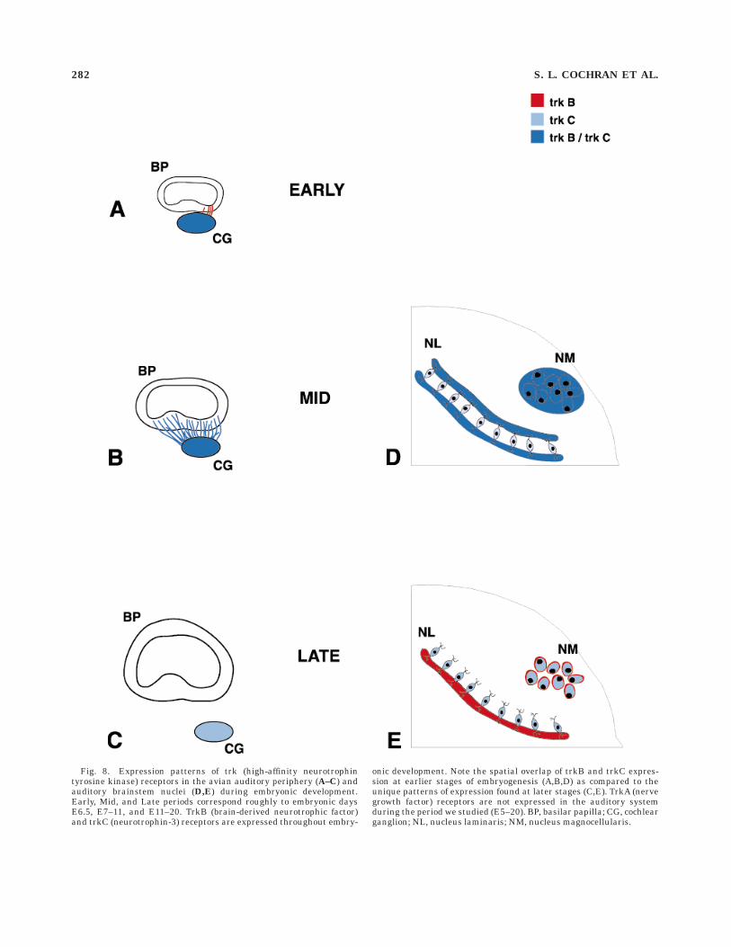
Fig. 8. Expression patterns of trk (high-affinity neurotrophintyrosine kinase) receptors in the avian auditory periphery (A–C) andauditory brainstem nuclei (D,E) during embryonic development.Early, Mid, and Late periods correspond roughly to embryonic daysE6.5, E7–11, and E11–20. TrkB (brain-derived neurotrophic factor)and trkC (neurotrophin-3) receptors are expressed throughout embry-
onic development. Note the spatial overlap of trkB and trkC expres-sion at earlier stages of embryogenesis (A,B,D) as compared to theunique patterns of expression found at later stages (C,E). TrkA (nervegrowth factor) receptors are not expressed in the auditory systemduring the period we studied (E5–20). BP, basilar papilla; CG, cochlearganglion; NL, nucleus laminaris; NM, nucleus magnocellularis.
282 S. L. COCHRAN ET AL.
tracted than in the periphery. As yet, it is unknownwhether the overlap in trk expression in central auditorystructures is accompanied by an overlap in neurotrophinexpression as it is in the periphery.
Previous studies of neurotrophin receptorsin the developing avian auditory system
NGF receptors. The present study describes the gen-eral lack of trkA receptors, receptors with specific affinityfor the neurotrophin NGF, in chick in any of the auditorystructures at any embryonic stage examined. Severalprevious studies of nerve growth factor receptor expres-sion in avians had demonstrated the presence of putativeNGF receptors during auditory ontogeny. In situ hybridiza-tion studies by von Bartheld et al. (1991) demonstrated thepresence of low-affinity nerve growth factor receptors(which they termed NGFR) in the chick otocyst epitheliumbeginning at E3. These receptors were observed in thecochlear ganglion at moderate levels from E4 to E13 and atlower levels from E13 to posthatch. The NGF receptorswere not observed in the cochlea at any age (von Bartheldet al., 1991). The NGF receptors were not observed eitherin the auditory brainstem (von Bartheld et al., personalcommunication). In other studies, NGFRs were noted inthe otic vesicle in another avian species, the quail (Berndand Represa, 1989; Represa et al., 1991). In these studies,radioactively labeled NGF was used to localize putativenerve growth factor receptors. These studies found NGFreceptors in the medial half of the otic vesicle (Bernd andRepresa, 1989) and in the cochlear portion of the cochleo-vestibular ganglion (Bernd and Represa, 1989; Represa etal., 1991). Our current understanding of neurotrophinreceptors lends insight to the fact that these studies were,in fact, characterizing the expression and distribution ofp75 receptors, receptors which have equal affinities for allneurotrophins (Rodriguez-Tebar et al., 1990; Squinto etal., 1991), rather than characterizing that of trkA recep-tors, receptors with specific affinity for NGF. At the time ofthese studies, trk receptors, neurotrophin receptors acti-vated differentially by different neurotrophins, were justbeing discovered (reviewed in Bothwell, 1995). The p75receptors lack the tyrosine-kinase signalling domain andmay facilitate or compete with trk receptors (e.g., Davies etal., 1993; Hantzopoulos et al., 1994; Chao and Hempstead,1995; Kaplan and Miller, 1997; and see Ibanez, 1998 for areview of the distinctions between trk and p75 receptors).In sum, our finding trkB and trkC receptors in the cochlearganglion and the inner ear suggests that BDNF and NT-3are released in the vicinity and are available to bind tolocal trk and p75 receptors. Thus, our results are notinconsistent with previous literature, but rather they posethe intriguing question as to what the combined expres-sion of p75 receptors and trkB and trkC receptors may beeffecting in the cochlear ganglion that is not occurringelsewhere through this mechanism.
NT-3 receptors. In an initial study of neurotrophin-3in the chick, Hallbook et al. (1993) reported that NT-3mRNA is not found in the chick otic vesicle during embry-onic days 4–6. Because we find clear evidence for trkCreceptors in the acoustic ganglion when we first look (atE6), it appears that trkC receptors are present before NT-3is available to bind them. However, it is possible that theexpression of NT-3 turns on rapidly just around the timethat we first see trkC receptors in the ganglion. In supportof this, Pirvola et al. (1997) first note NT-3 mRNA labeling
in the basilar papilla at E6, though labeling appears weakand by E12 it is absent. Interestingly, neurotrophin-3mRNA is found in the quail cochlear ganglion by E4, and itis still present at E7 and E10 (Bernd et al., 1994). In anearly study of trkC expression in the developing chick,Williams et al. (1993) reported the lack of trkC mRNA inthe chick cochlear ganglion at E3, E9, and E19. We andothers have ample data that suggest that trkC is ex-pressed in the avian cochlear ganglion from E5 onward.Besides the present study, Pirvola et al. (1997) describesrobust levels of trkC mRNA in the cochlear ganglion ofyounger chicks (E5–8), with reduced levels in increasinglyolder animals. Likewise, Bernd et al. (1994) describes trkCmRNA expression in the cochlear ganglion of the quail atstages 26, 31, and 36 (,E5, E7, and E10 in the chick). Inaddition, the in vitro effects of NT-3 on cultured ganglionneurons of the chick have been demonstrated repeatedly(Avila et al., 1993; Represa et al., 1993; Jimenez et al.,1997; San Jose et al., 1997). It is possible that thediscrepant results of Williams et al. (1993) arise becausetrkC mRNA levels in the developing cochlear ganglion fallbelow the detection limit of the oligonucleotide probe usedin their study. Finally, we are intrigued by the decreases intrkC mRNA signal reported by Pirvola et al. (1997) be-cause trkC expression in the cochlear ganglion, as demon-strated by immunocytochemistry, appears to remain ro-bust throughout embryogenesis and into the posthatchperiod. Again, we propose that perhaps the trkC mRNAlevels in older embryos fall below the detection limit of thein situ hybridization technique used. As a final note, weadd that it is puzzling that trkC receptors are expressed inthe cochlear ganglion throughout embryonic developmentand even into the posthatch period, whereas peripherallevels of NT-3 in the chick appear to abate during midem-bryogenesis (Pirvola et al., 1997) or are entirely undetect-able (Hallbook et al., 1993). The presence of trkC receptorsin the absence of the only ligand known to signal throughthem suggests that a ligand as yet unidentified mayinitiate trkC signalling. Because the pattern of NT-3distribution in the brain is not known, it is also possiblethat cochlear ganglion cells in older embryos maintaintrkC receptors because they are enjoying NT-3 supportfrom central sources (e.g., nucleus magnocellularis andnucleus angularis).
BDNF receptors. Prior studies have localized trkBmRNA in the cochlear ganglion and basilar papilla of theembryonic chick (Dechant et al., 1993; Hallbook andFritzsch, 1997; Pirvola et al., 1997) and quail (Bernd et al.,1994). An early study of BDNF expression during avianontogeny localized BDNF mRNA in the cochlear ganglion(Bernd et al., 1994), whereas more recent studies havefailed to see BDNF mRNA in the cochlear ganglion (Hall-book and Fritzsch, 1997; Pirvola et al., 1997), but havenoted BDNF mRNA in hair cells of the basilar papilla(Pirvola et al., 1997). In vitro studies have reported effectsof BDNF on cochlear ganglion cells in culture (Avila et al.,1993; Represa et al., 1993; Hossain et al., 1997; Pirvola etal., 1997; Sokolowski et al., 1997). The present resultsdescribe trkB receptors in the afferent fibers of the basilarpapilla, but not in the hair cells or support cells of thesensory epithelium. Interestingly, light levels of trkBmRNA have been reported in some non-neuronal cells(tympanic border cells, support cells, and hyaline cells) ofthe E18 basilar papilla (Pirvola et al., 1997). These cellswere labeled lightly when the hybridization probe used in
Trk RECEPTORS IN EMBRYONIC CHICK AUDITORY SYSTEM 283
the studies encoded the extracellular domain of the trkBreceptor. Other studies in which the hybridization probeencoded the functional tyrosine kinase domain yieldedresults in which trkB mRNA label was not found in thebasilar papilla, but rather was confined to the cochlearganglion (Pirvola et al., 1997). The trkB antibody used inthe present study was directed against the extracellulardomain, so we might have expected to see some cells in thebasilar papilla labeled as Pirvola et al. (1997) described.We did not. It is possible that the truncated trkB messagein the non-neuronal cells is of such low levels that the levelof trkB receptor expression is little to none and thereforeescaped our detection. We note that one report of trkBexpression in the mammalian auditory periphery de-scribes trkB immunoreactivity in sensory hair cells in therat during the period of synaptic innervation (Knipper etal., 1996). A more puzzling finding is that Pirvola et al.(1997) continue to see high levels of trkB mRNA in theneurons of the cochlear ganglion in the mature basilarpapilla, whereas we find that trkB immunolabeling in thecochlear ganglion abates after E12. There is no obviousreason for the discrepant results from these two studies.We can only posit that trkB message in the cochlearganglion is not translated into trkB protein after E12.
Lack of gradients in ontogenetic expressionof trks within individual auditory structures
A common feature of a developing tissue is an orderlyprogression of development that proceeds along an identi-fiable anatomical axis. In the embryonic chick basilarpapilla, for instance, hair cell differentiation occurs in adistal (apical) to proximal (basal) direction (Cotanche andSulik, 1984), whereas synaptogenesis proceeds in theopposite (proximal to distal) direction (Fermin and Cohen,1984). As Cotanche and Sulik (1984) observe, this anomalyimplies that hair cells differentiate independently andthat newly differentiated hair cells may themselves guidedeveloping afferent nerve processes by providing targetsites for the formation of synapses. In the brainstem,development proceeds along the tonotopic axis, from rostro-medial to caudolateral (for review, see Rubel and Parks,1988). Some of these processes take up to a week (orone-third of embryonic development) to complete. Thus,marked differences within the basilar papilla itself, orwithin neurons in nucleus magnocellularis or nucleuslaminaris, exist at any given time during ontogeny. Itseemed likely, then, that we would find spatial/temporalgradients or waves of neurotrophin receptor expressionwithin individual tissues. For instance, we might haveexpected to see a proximal to distal wave of neurotrophinreceptor expression in the fibers invading the basilarpapilla that would accompany the wave of synaptogenesis.However, despite efforts to identify developmental gradi-ents of neurotrophin receptor expression within individualauditory tissues, we observed none, with the exception ofthe small set of lightly labeled trkB fibers found in thesuperior basilar papilla which preceded the robust fiberlabeling found throughout the papilla 1 day later.
Interestingly, Pirvola et al. (1997) report a gradient ofBDNF mRNAexpression during a short window of develop-ment. They find BDNF mRNA levels in the E12 basilarpapilla are more intense in the distal end than those in theproximal end. By E16, this spatial gradient of BDNFmRNA expression is not apparent— the BDNF mRNAsignal is equally robust along the distal-proximal and the
superior-inferior axes. Because we never saw this sort ofspatial/temporal gradient in trkB expression, it appearsthat the graded expression of BDNF is not paralleled byexpression of its cognate receptor trkB.
Patterns of trk expression and otherdevelopmental events
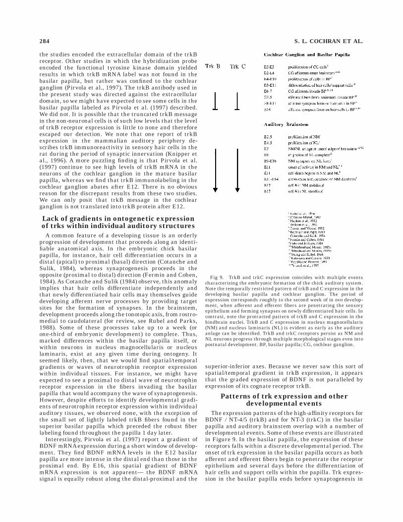
The expression patterns of the high-affinity receptors forBDNF / NT-4/5 (trkB) and for NT-3 (trkC) in the basilarpapilla and auditory brainstem overlap with a number ofdevelopmental events. Some of these events are illustratedin Figure 9. In the basilar papilla, the expression of thesereceptors falls within a discrete developmental period. Theonset of trk expression in the basilar papilla occurs as bothafferent and efferent fibers begin to penetrate the receptorepithelium and several days before the differentiation ofhair cells and support cells within the papilla. Trk expres-sion in the basilar papilla ends before synaptogenesis in
Fig 9. TrkB and trkC expression coincides with multiple eventscharacterizing the embryonic formation of the chick auditory system.Note the temporally restricted pattern of trkB and C expression in thedeveloping basilar papilla and cochlear ganglion. The period ofexpression corresponds roughly to the second week of in ovo develop-ment, when afferent and efferent fibers are penetrating the sensoryepithelium and forming synapses on newly differentiated hair cells. Incontrast, note the protracted pattern of trkB and C expression in thehindbrain nuclei. TrkB and C expression in nucleus magnocellularis(NM) and nucleus laminaris (NL) is evident as early as the auditoryanlage can be identified. TrkB and trkC receptors persist as NM andNL neurons progress through multiple morphological stages even intopostnatal development. BP, basilar papilla; CG, cochlear ganglion.
284 S. L. COCHRAN ET AL.
the receptor epithelium is complete (Whitehead and Mor-est, 1985b). These findings suggest that the trk receptorsand their associated neurotrophins may play a role in theearly synaptogenesis in the basilar papilla. In addition,the sustained levels of trkC within the cochlear ganglionthroughout ontogeny prompts us to believe that the neu-rons in the cochlear ganglion enjoy ongoing trophic sup-port by NT-3 secreted from central sources. We note thatwe are not the first to describe differential expression ofneurotrophin receptors in neuronal somata and theirperipheral and central projections. von Bartheld et al.(1991) demonstrated that the early p75 expression in cellbodies of cochlear ganglion neurons is lost despite themaintained p75 expression in the peripheral processes ofthe cochlear nerve and the maintained expression of p75mRNA in the ganglion cell bodies. More recently, Lefcort etal. (unpublished observations) have observed the dynamicontogenetic distribution of trks in the dorsal root ganglion.Early in embryonic development, trkA, trkB, and trkC arefound in dorsal root ganglion somata and in peripheral andcentral fibers. As embryogenesis proceeds, trkB and trkCexpression is lost in the peripheral axons but is main-tained in the cell bodies and the central projections of thedorsal root ganglion neurons. Curiously, trkA expression ismaintained in the peripheral processes of the chick dorsalroot ganglion even during later stages of embryogenesis.Perturbation experiments may lend insight to these com-mon findings.
In the auditory brainstem nuclei, trk receptor expres-sion persists throughout embryonic development and intothe posthatch period. TrkB and trkC expression beginsafter terminal mitosis of NM and NL but before themigration of NL is complete. The high expression of trkBand trkC in the auditory brainstem nuclei is maintainedthroughout embryonic development, overlapping the pe-riod of synaptogenesis and the period of cell death in NMand NL (reviewed in Rubel and Parks, 1988). It remains tobe investigated what role(s) the trks may play duringbrainstem development. Information about the source(s)and availability of BDNF and NT-3 in developing brain-stem structures is likely to be useful. The prolongedexpression of trkB and trkC in the auditory hindbrainsuggests that the neurotrophins and their cognate recep-tors may be in part responsible for a number of differentdevelopmental processes, including cell migration, cellsurvival, and cell maintenance. One specific hypothesis isdiscussed below.
Functional implications: Role of trkB indevelopment of nucleus laminaris
Perhaps the most intriguing finding in this study is theasymmetrical distribution of trkB- immunoreactive tissuein NL from E11 onward. To our knowledge, this study isthe first to describe a polarized expression of receptorswithin individual neurons such as found in NL, wheretrkB is confined to the ventral, and not the dorsal, den-drites. Previous studies have described restricted tissueexpression patterns of another subfamily of receptor pro-tein tyrosine kinases, the Eph receptors, in early develop-ment. For example, EphA4 (Sek-1), EphB2 (Nuk), andEphB3 (Sek-4) are concentrated in rhombomeres 3 and 5 ofthe developing mouse hindbrain (Nieto, 1992; Becker etal., 1994; Henkemeyer et al., 1996; Theil et al., 1998),whereas EphA2 (Eck) is observed predominantly in rhom-bomere 4 (Ganju et al., 1994; Ruiz and Robertson, 1994).
These studies suggest that these receptor tyrosine kinasesmay be involved in cell-cell interactions guiding earlyhindbrain development, particularly in distinctive aspectsof pattern formation. More akin to the present findings,EphB2 (Cek5) has been shown to exhibit a polarizedexpression along the dorsal-ventral axis of the chickenretina throughout ontogeny (Holash and Pasquale, 1995).This represents the first signal transduction moleculefound to exhibit the polarized pattern of expression pre-dicted for proteins guiding the specificity of retinotectalprojections.
In the present study, the polarized distribution of trkBreceptors within the ventral neuropil of nucleus laminarisprompted us to perform in situ hybridization experimentsin order to ascertain which of the auditory brainstemnuclei is making the trkB protein. The results from thesein situ hybridization studies suggest that the trkB mes-sage is made in the cell bodies of nucleus laminaris andthen the trkB mRNA or trkB protein is selectively trans-ported to the ventral and not to the dorsal dendrites. Arecent study of the subcellular localization of neurotroph-ins and their cognate receptors described an activity-dependent modulation of trkB mRNA targeting and pro-tein accumulation in dendrites of hippocampal neurons(Tongiorgi et al., 1997).
The mechanism for selective transport of trkB mRNA orprotein to the ventral dendrites of nucleus laminaris cellsis unknown. Whatever the mechanism, the polarizedexpression of trkB in NL is intriguing to us in view of thebinaural circuitry in nucleus laminaris and the develop-ment of highly stereotyped dendritic architecture of thisnucleus (Smith and Rubel, 1979; Smith, 1981). In thenormal animal, the ipsilateral and contralateral NM termi-nations on NL are strictly segregated from the earliesttime they can be identified in development (Benes et al.,1977; Parks and Rubel, 1975; Young and Rubel, 1983,1986). Ipsilateral projections terminate on the dorsaldendrites, whereas contralateral projections terminate onthe ventral dendrites. Furthermore, the contralateral in-put to the ventral dendrites forms a ‘‘delay line,’’ whereasthe dorsal (ipsilateral) input does not (Young and Rubel,1983). The precision of this ‘‘delay line’’ circuitry, which istopographically organized, is essential for proper encodingof sound source location (Jeffress, 1948; Colburn andDurlach, 1978; Overholt et al., 1992). One question thathas persisted in developmental studies of the chick audi-tory brainstem concerns the molecular signal or signalsthat underlie the development and maintenance of suchstereotyped circuitry. The polarized expression of trkBreceptors in NL dendrites is coincident with refinement ofNM terminal arborizations on NL (Young and Rubel,1986), although expression persists past the major periodof refinement.
As a working hypothesis, we speculate that trkB recep-tors on the ventral dendrites of NL cells may provide thelocal guidance cues necessary for proper innervation bycontralateral NM fibers. One possibility is that trkBsignalling in the ventral nucleus laminaris dendritesstimulates the maturation and synaptogenesis of contralat-eral connections, thus preventing an axonal ‘‘overshoot’’through the NL cell line by contralateral NM fibers. Thereare several recent reports that lend credence to this idea.BDNF, NT-3, and other guidance molecules have beendemonstrated to modulate the pathfinding behaviors ofgrowth cones (Song et al., 1997). The turning behaviors
Trk RECEPTORS IN EMBRYONIC CHICK AUDITORY SYSTEM 285
elicited depend upon differences in cAMP-dependent activ-ity. Moreover, localized sources of nerve growth factor havebeen shown to initiate axon collateral sprouting in dorsalroot ganglion neurons (Gallo and Letourneau, 1998) whichis trkA activation-dependent (Gallo et al., 1997). Growthcone responses to collapsin-1 also seem to be dependentupon trk receptor activation (Tuttle and O’Leary, 1998).Finally, studies have demonstrated that trkB and trkCsignalling is necessary for normal development of axonalarborizations and synaptic densities in the hippocampus(Martınez et al., 1998).
Despite the conventional tenet that neurotrophins re-side in the target tissues, a number of recent discoverieshave demonstrated instead that anterograde transport ofneurotrophins (specifically BDNF) may actually play themajor role in systems in the peripheral (Tonra et al., 1998)and central (von Bartheld et al., 1996a; Zhou and Rush,1996; Altar et al., 1997; Conner et al., 1997) nervoussystems. Studies have demonstrated axonal accumulationof BDNF within vesicular compartments of cortical nerveterminals (Fawcett et al., 1997) and the presence of trkBand trkC in the postsynaptic region of synaptic profilesand within the cytoplasm of dendritic processes in mamma-lian auditory brainstem neurons (Hafidi et al., 1996).Finally, functional trkB receptors have been demonstratedin the postsynaptic density of hippocampal neurons (Wu etal., 1996) and the release of BDNF in neural tissue hasbeen induced by depolarization (Androutsellis-Theotokiset al., 1996). Current studies are directed toward testingthe involvement of trkB in the polarization of theseipsilateral and contralateral NM inputs to NL.
ACKNOWLEDGMENTS
We thank Drs. Thomas Large and Kristin Boeshore forproviding the trkB cDNA plasmids and Dr. Lou Reichardtfor the trk antibodies. We also thank Drs. Karina Cramerand Linda Ross for their valuable comments on thismanuscript and David Molea for his generous assistancewith the figures.
LITERATURE CITED
Allendoerfer KL, Cabelli KJ, Escandon E, Kaplan DR, Nikolics K, Shatz CJ.1994. Regulation of neurotrophin receptors during the maturation ofthe mammalian visual system. J Neurosci 14:1795–1811.
Altar CA, Cai N, Bliven T, Juhasz M, Conner JM, Acheson AL, Lindsay RM,Wiegand SJ. 1997. Anterograde transport of brain-derived neurotrophicfactor and its role in the brain. Nature 389:856–860.
Androutsellis-Theotokis A, McCormack WJ, Bradford HF, Stern GM,Pliego-Rivero FB. 1996. The depolarisation-induced release of [125I]BDNF from brain tissue. Brain Res 743:40–48.
Avila MA, Varela Nieto I, Romero G, Mato JM, Giraldez F, Van de Water TR,Represa J. 1993. Brain-derived neurotrophic factor and neurotrophin-3support the survival and neuritogenesis response of developing cochleo-vestibular ganglion neurons. Dev Biol 159:266–275.
Becker N, Seitanidou T, Murphy P, Mattei M-G, Topilko P, Nieto MA,Wilkinson DG, Charnay P, Gilardi-Hebenstreit P. 1994. Several recep-tor tyrosine kinase genes of the Eph family are segmentally expressedin the developing hindbrain. Mech Dev 47:3–17.
Benes FM, Parks TN, Rubel EW. 1977. Rapid dendritic atrophy followingdeafferentation: an EM morphometric analysis. Brain Res 122:1–13.
Bernd P, Represa J. 1989. Characterization and localization of nervegrowth factor receptors in the embryonic otic vesicle and cochleovestibu-lar ganglion. Dev Biol 134:11–20.
Bernd P, Zhang D, Yao L, Rosemberg I. 1994. Potential role of nerve growthfactor, brain-derived neurotrophic factor, and neurotrophin-3 in aviancochlear and vestibular ganglia development. Int J Dev Neurosci12:709–723.
Bothwell M. 1995. Functional interactions of neurotrophins and neuro-trophin receptors. Ann Rev Neurosci 18:223–253.
Bredesen DE, Rabizadeh S. 1997. P75NTR and apoptosis: trk-dependentand tri-independent effects. Trends Neurosci 20:287–290.
Cabelli RJ, Shelton DL, Segal RA, Shatz CJ. 1997. Blockade of endogenousligands of trkB inhibits formation of ocular dominance columns. Neuron19:63–76.
Carter BD, Kaltschmidt C, Offenhauser N, BohnMatthaei R, Baeuerle PA,Barde YA. 1996. Selective activation of NF-KB by nerve growth factorthrough the neurotrophin receptor p75. Science 272:542–545.
Chao MV. 1992. Neurotrophin receptors: a window into neuronal differen-tiation. Neuron 9: 583–593.
Chao MV, Hempstead BL. 1995. p75 and Trk: a two-receptor system. TrendsNeurosci 18:321–326.
Cohen-Cory S, Fraser SE. 1994. BDNF in the development of the visualsystem of Xenopus. Neuron 12:747–761.
Cohen-Cory S, Fraser SE. 1995. Effects of brain-derived neurotrophic factoron optic axon branching and remodelling in vivo. Nature 378:192–196.
Colburn HS, Durlach NI. 1978. Models of binaural interaction. In: Carter-ette EC, Friedman MP, editors. Hearing, vol IV of handbook ofperception. New York: Academic Press. p 467–518.
Conner JM, Lauterborn JC, Yan Q, Gall CM, Varon S. 1997. Distribution ofbrain-derived neurotrophic factor (BDNF) protein and mRNA in thenormal adult rat CNS: evidence for anterograde axonal transport. JNeurosci 17:2295–2313.
Cotanche DA, Sulik KK. 1984. Development of stereociliary bundles in thecochlear duct of chick embryos. Brain Res Dev Brain Res 16:181–193.
D’Amico-Martel A. 1982. Temporal patterns of neurogenesis in aviancranial sensory and autonomic ganglia. Am J Anat 163:351–372.
Davies AM, Lee KF, Jaenisch R. 1993. p75-deficient trigeminal sensoryneurons have an altered response to NGF but not to other neurotroph-ins. Neuron 11:565–574.
Dechant DG, Biffo S, Okazawa H, Kolbeck R, Pottgiesser J, Barde YA. 1993.Expression and binding characteristics of the bdnf receptor chick trkB.Development 119:545–558.
Despres G, Hafidi A, Romand R. 1991. Immunohistochemical localization ofnerve growth factor receptor in the cochlea and in the brainstem of theperinatal rat. Hearing Res 52:157–166.
Don DM, Newman AN, Micevych PE, Popper P. 1997. Expression ofbrain-derived neurotrophic factor and its receptor mRNA in the vestibu-loauditory system of the bullfrog. Hearing Res 114:10–20.
Ernfors P, Lee KF, Jaenisch R. 1994. Mice lacking brain-derived neuro-trophic factor develop with sensory deficits. Nature 368:147–150.
Ernfors P, Van de Water T, Loring J, Jaenisch R. 1995. Complementaryroles of BDNF and NT-3 in vestibular and auditory development.Neuron 14:1153–1164.
Fawcett JP, Aloyz R, McClean JH, Pareek S, Miller FD, McPherson PS,Murphy RA. 1997. Detection of brain-derived neurotrophic factor in avesicular fraction of brain synaptosomes. J Biol Chem 272:8837–8840.
Fermin CD, Cohen GM. 1984. Developmental gradients in the embryonicchick’s basilar papilla. Acta Otolaryngol (Stockh) 97:39–51.
Fischer FP. 1992. Quantitative analysis of the innervation of the chickenbasilar papilla. Hearing Res 61:167–178.
Frade JM, Barde YA. 1998. Nerve growth factor: two receptors, multiplefunctions. Bioessays 20:137–145.
Frade JM, Rodriguez-Tebar A, Barde YA. 1996. Induction of cell death byendogenous nerve growth factor through its p75 receptor. Nature383:166–168.
Friedman WJ, Ernfors P, Persson H. 1991. Transient and persistentexpression of NT-3/HDNF mRNA in the rat brain during postnataldevelopment. J Neurosci 11:1577–1584.
Fritzsch B, Christensen MA, Nichols DH. 1993. Fiber pathways andpositional changes in efferent perikarya of 2.5- to 7-day chick embryosas revealed with DiI and dextran amines. J Neurobiol 24:1481–1499.
Fritzsch B, Farinas I, Reichardt LF. 1997a. Lack of neurotrophin 3 causeslosses of both classes of spiral ganglion neurons in the cochlea in aregion-specific fashion. J Neurosci 17:6213–6225.
Fritzsch B, Sarai PA, Barbacid M, Silos-Santiago I. 1997b. Mice with atargeted disruption of the neurotrophin receptor trkB lose their gusta-tory ganglion cells early but do develop taste buds. Int J Dev Neurosci15:563–576.
Fritzsch B, Silos-Santiago I, Bianchi LM, Farinas I. 1997c. Role ofneurotrophic factors in regulating the development of inner ear innerva-tion. Trends Neurosci 20:159–164.
286 S. L. COCHRAN ET AL.
Gallo G, Letourneau PC. 1998. Localized sources of neurotrophins initiateaxon collateral sprouting. J Neurosci 18:5403–5414.
Gallo G, Lefcort FB, Letourneau PC. 1997. TrkA receptor mediates growthcone turning toward a localized source of nerve growth factor. JNeurosci 17:5445–5454.
Ganju P, Shigemoto K, Brennan J, Entwistle A, Reith AD. 1994. The Eckreceptor tyrosine kinase is implicated in pattern formation duringgastrulation, hindbrain segmentation, and limb development. Onco-gene 9:1613–1624.
Hackett JT, Jackson H, Rubel EW. 1982. Synaptic excitation of the secondand third order auditory neurons in the avian brain stem. Neuroscience7:1455–1469.
Hafidi A, Moore T, Sanes DH. 1996. Regional distribution of neurotrophinreceptors in the developing auditory brainstem. J Comp Neurol 367:454–464.
Hallboook F, Fritzsch B. 1997. Distribution of BDNF and trkB mRNA in theotic region of 3.4 and 4.5 day chick embryos as revealed with acombination of in situ hybridization and tract tracing. Int J Dev Biol41:725–732.
Hallbook F, Ibanez CF, Ebandal T, Persson H. 1993. Cellular localization ofbrain-derived neurotrophic factor and neurotrophin-3 mRNA expres-sion in the early chicken embryo. Eur J Neurosci 5:1–14.
Hamburger V, Hamilton HL. 1951. A series of normal stages in thedevelopment of the chick embryo. J Morph 88:49–92.
Hantzopoulos PA, Suri C, Glass DJ, Goldfarb MP, Yancopoulos GD. 1994.Low-affinity NGF receptor, p75 can collaborate with each of the trks topotentiate functional responses to the neurotrophins. Neuron 13:187–201.
Henkemeyer M, Orioli D, Henderson JT, Saxton TM, Roder J, Pawson T,Klein R. 1996. Nuk controls pathfinding of commissural axons in themammalian central nervous system. Cell 86:35–46.
Henrique D, Adam J, Myat A, Chitnis A, Lewis J, Ish-Horowicz D. 1995.Expression of a Delta homologue in prospective neurons in the chick.Nature 375:787–790.
Holash JA, Pasquale EB. 1995. Polarized expression of the receptor proteintyrosine kinase Cek5 in the developing avian visual system. Dev Biol172:683–693.
Holcomb JD, Mumm JS, Calof AL. 1995. Apoptosis in the neuronal lineageof the mouse olfactory epithelium: regulation in vivo and in vitro. DevBiol 172:307–323.
Hossain WA, Rutledge A, Morest DK. 1997. Critical periods of basicfibroblast growth factor and brain-derived neurotrophic factor in thedevelopment of the chicken cochleovestibular ganglion in vitro. ExpNeurol 147:437–451.
Ibanez CF. 1998. Emerging themes in structural biology of neurotrophicfactors. Trends Neurosci 21:438–444.
Jackson H, Hackett JT, Rubel EW. 1982. Organization and development ofbrain stem auditory nuclei in the chick: ontogeny of postsynapticresponses. J Comp Neurol 210:80–86.
Jackson H, Parks TN. 1982. Functional synapse elimination in the develop-ing avian cochlear nucleus with simultaneous reduction in cochlearnerve axon branching. J Neurosci 2:1736–1743.
Jackson H, Rubel EW. 1978. Ontogeny of behavioral responsiveness tosound in the chick embryo as indicated by electrical recordings ofmotility. J Comp Physiol Psychol 92:682–696.
Jeffress LA. 1948. A place theory of sound localization. J Comp PhysiolPsychol 41:35–39.
Jhaveri S, Morest DK. 1982. Sequential alterations of neuronal architec-ture in nucleus magnocellularis of the developing chicken: a Golgistudy. Neuroscience 7:837–853.
Jimenez C, Gireldez F, Represa J, Garcıa-Dıaz JF. 1997. Calcium currentsin dissociated cochlear neurons from the chick embryo and theirmodification by neurotrophin-3. Neuroscience 77:673–682.
Kaplan DR, Miller FD. 1997. Signal transduction by the neurotrophinreceptors. Curr Opin Cell Biol 9:213–221.
Katayama A, Corwin JT. 1989. Cell production in chicken cochlea. J CompNeurol 281:129–135.
Katz LC. 1987. Local circuitry of identified projection neurons in cat visualcortex brain slices. J Neurosci 7:1223–1249.
Knipper M, Zimmermann U, Rohbock K, Kopschall I, Zenner H –P. 1996.Expression of neurotrophin receptor trkB in rat cochlear hair cells attime of rearrangement of innervation. Cell Tissue Res 283:339–353.
Lachyankar MB, Condon PJ, Quesenbery PJ, Litofsky NS, Recht LD, RossAH. 1997. Embryonic precursor cells that express trk receptors: induc-
tion of different cell fates by NGF, BDNF, NT-3, and CNTF. Exp Neurol144:350–360.
Lauterborn JC, Isackson PJ, Gall CM. 1994. Cellular localization of NGFand NT-3 mRNA’s in postnatal rat forebrain. Mol Cell Neurosci 5:46–62.
Lefcort F, Clary DO, Rusoff AC, Reichardt LF. 1996. Inhibition of NT-3receptor trkC, early in chick embryogenesis, results in severe reduc-tions in multiple neuronal subpopulations in the dorsal root ganglia. JNeurosci 16:3704–3713.
Levi-Montalcini R. 1987. The nerve growth factor: 35 years later. Science237:1154–1162.
Levi-Montalcini R, Angeletti PU. 1968. Nerve growth factor. Physiol Rev48:534–569.
Lindholm D, Castren E, Berzaghi M, Blochl A, Thoenen H. 1994. Activity-dependent and hormonal regulation of neurotrophin mRNA levels inthe brain— implications for neuronal plasticity. J Neurobiol 25:1362–1372.
Martınez A, Alcantara S, Borrell V, Del Rıo JA, Blasi J, Otal R, Campos N,Boronat A, Barbacid M, Silos-Santiago I, Soriano E. 1998. TrkB andtrkC signalling are required for maturation and synaptogenesis ofhippocampal connections. J Neurosci 18:7336–7350.
McAllister AK, Lo DC, Katz LC. 1995. Neurotrophins regulate dendriticgrowth in developing visual cortex. Neuron 15:791–803.
McAllister AK, Katz LC, Lo DC. 1997. Opposing roles for endogenousBDNF and NT-3 in regulating cortical dendritic growth. Neuron18:767–778.
Molea D, Stone JS, Rubel EW. 1999. Class III beta-tubulin expression insensory and nonsensory regions of the developing avian inner ear. JComp Neurol 406:183–198.
Nieto MA, Gilardi-Hebenstreit P, Charnay P, Wilkinson DG. 1992. Areceptor protein tyrosine kinase implicated in the segmental patterningof the hindbrain and mesoderm. Development 116:1137–1150.
Nosrat CA, Ebendal T, Olson L. 1996. Differential expression of brain-derived neurotrophic factor and neurotrophin-3 mRNA in lingualpapillae and taste buds indicates roles in gustatory and somatosensoryinnervation. J Comp Neurol 376:587–602.
Nosrat CA, Blomlof J, ElShamy WM, Ernfors P, Olson L. 1997. Lingualdeficits in BDNF and NT-3 mutant mice leading to gustatory andsomatosensory disturbances, respectively. Development 124:1333–1342.
Oakley RA, Lefcort FB, Clary DO, Reichardt LF, Prevette D, OppenheimRW, Frank E. 1997. Neurotrophin-3 promotes the differentiation ofmuscle spindle afferents in the absence of peripheral targets. J Neurosci17:4261–4274.
Overholt EM, Rubel EW, Hyson RL. 1992. A circuit for coding interauraltime differences in the chick brainstem. J Neurosci 12:1698–1708.
Parks TN, Jackson H. 1984. A developmental gradient of dendritic loss inthe avian cochlear nucleus occurring independently of primary affer-ents. J Comp Neurol 227:459–466.
Parks TN, Rubel EW. 1975. Organization and development of brain stemauditory nuclei of the chicken: organization of projections from n.magnocellularis to n. laminaris. J Comp Neurol 164:435–448.
Patterson SL, Abel T, Deuel TAS, Martin KC, Rose JC, Kandel ER. 1996.Recombinant BDNF rescues deficits in basal synaptic transmission andhippocampal LTP in BDNF knockout mice. Neuron 16:1137–1145.
Petralia RS, Peusner KD. 1991. Earliest ultrastructural development of thetangential vestibular nucleus in the chick embryo. J Comp Neurol310:82–93.
Pirvola U, Ylikoski J, Palgi J, Lehtonen E, Arumae U, Saarma M. 1992.Brain-derived neurotrophic factor and neurotrophin 3 mRNA’s in theperipheral target fields of developing inner ear ganglia. Proc Natl AcadSci USA 89:9915–9919.
Pirvola U, Arumae U, Moshnyakov M, Palgi J, Saarma M, Ylikoski J. 1994.Coordinated expression and function of neurotrophins and their recep-tors in the rat inner ear during target innervation. Hearing Res75:131–144.
Pirvola U, Hallbook F, Xing-Qun L, Virkkala J, Saarma M, Ylikoski J. 1997.Expression of neurotrophins and trk receptors in the developing, adult,and regenerating avian cochlea. J Neurobiol 33:1019–1033.
Rebillard M, Pujol R. 1983. Innervation of the chicken basilar papilladuring its development. Acta Otolaryngol (Stockh) 96:379–388.
Rebillard G, Rubel EW. 1981. Electrophysiological study of the maturationof auditory responses from the inner ear of the chick. Brain Res229:15–23.
Trk RECEPTORS IN EMBRYONIC CHICK AUDITORY SYSTEM 287

Represa J, Van de Water TR, Bernd P. 1991. Temporal pattern of nervegrowth factor receptor expression in developing cochlear and vestibularganglia in quail and mouse. Anat Embryol Berl 184:421–432.
Represa J, Avila MA, Romero G, Mato JM, Giraldez F, Varela Nieto I. 1993.Brain-derived neurotrophic factor and neurotrophin-3 induce cell prolif-eration in the cochlearvestibular ganglion through a glycosyl-phospha-tidylinositol signalling system. Dev Biol 159:257–265.
Rodriguez-Tebar A, Dechant G, Gotz R, Barde YA. 1990. Binding ofbrain-derived neurotrophic factor to the nerve growth factor receptor.Neuron 4:487–492.
Roskams AJ, Bethel MA, Hurt KJ, Ronnett GV. 1996. Sequential expres-sion of trks A, B, and C in the regenerating olfactory neuroepithelium. JNeurosci 16:1294–1307.
Rubel EW, Parks TN. 1988. Organization and development of the avianbrain-stem auditory system. In: Edelman GE, Gall WE, Cowan WM,editors. Auditory function: neurobiological bases of hearing. New York:John Wiley and Sons, Inc. p 3–92.
Rubel EW, Smith ZDJ, Miller LC. 1976. Organizational and development ofbrain stem auditory nuclei of the chicken: ontogeny of n. magnocellu-laris and n. laminaris. J Comp Neurol 166:469–490.
Ruiz JC, Robertson EJ. 1994. The expression of the receptor-proteintyrosine kinase gene, Eck, is highly restricted during early mousedevelopment. Mech Dev 46:87–100.
San Jose I, Vazquez E, Garcia-Atares N, Rodriguez S, Antonio Vega J, andRepresa J. 1997. Expression of the cytoskeletal protein MAP5 and itsregulation by neurotrophin-3 (NT-3) in the inner ear sensory neurons.Anat Embryol 195:299–310.
Saunders JC, Coles RB, Gates GR. 1973. The development of auditoryevoked responses in the cochlea and cochlear nuclei of the chick. BrainRes 63:59–74.
Saunders JC, Gates GR, Coles RB. 1974. Brain-stem evoked responses asan index of hearing thresholds in one-day-chicks and ducklings. J CompPhysiol Psychol 86:426–431.
Schecterson, LC, Bothwell M. 1994. Neurotrophin and neurotrophin recep-tor mRNA expression in developing inner ear. Hearing Res 723:92–100.
Segal RA, Pomeroy SL, Stiles CD. 1995. Axonal growth and fasciculationlinked to differential expression of BDNF and NT-3 receptors indeveloping cerebellar granule cells. J Neurosci 15:4970–4981.
Shimmang T, Minichiello L, Vazquez E, San Jose I, Giraldez F, Klein R.,Represa J. 1995. Developing inner ear sensory neurons require trkBand trkC receptors for innervation of their peripheral targets. Develop-ment 121:3381–3391.
Smith ZDJ. 1981. Organization and development of brain stem auditorynuclei in the chicken: dendritic development in n. laminaris. J CompNeurol 203:309–333.
Smith ZDJ, Rubel EW. 1979. Organization and development of brain stemauditory nuclei in the chicken: dendritic gradients in n. laminaris. JComp Neurol 186:213–240.
Sokolowski BH. 1997. Quantitative analysis of long-term survival andneuritogenesis in vitro: cochleovestibular ganglion of the chick embryoin BDNF, NT-3, NT-4/5, and insulin. Exp Neurol 145:1–15.
Song HJ, Ming GL, Poo MM. 1997. cAMP-induced switching in turningdirection of nerve growth cones. Nature 388:275–279. Published erra-tum appears in Nature 1997 389:412.
Squinto SP, Stitt TN, Aldrich TH, Davis S, Bianco SM, Radziejewski C,Glass DJ, Masiakowski P, Furth ME, Valenzuela DM, Di Stefano PS,Yancopoulos GD. 1991. TrkB encodes a functional receptor for brain-derived neurotrophic factor and neurotrophin-3 but not nerve growthfactor. NT3 does not act on PC12. Cell 65:885–893.
Suen PC, Levine ES, Mount HT, Xu JL, Lin SY, Black IB. 1997. Brain-derived neurotrophic factor rapidly enhances phosphorylation of thepostsynaptic N-methyl-D-aspartate receptor subunit 1. Proc Natl AcadSci USA 94:8191–8195.
Theil T, Frain M, Gilardi-Hebenstreit P, Flenniken A, Charnay P. 1998.Segmental expression of the EphA4 (Sek-1) receptor tyrosine kinase in
the hindbrain is under direct transcriptional control of Krox-20. Devel-opment 125:443–452.
Tongiorgi E, Massimo R, Cattaneo A. 1997. Activity-dependent dendritictargeting of BDNF and trkB mRNAs in hippocampal neurons. JNeurosci 17:9492–9505.
Tonra JR, Curtis R, Wong V, Cliffer KD, Park JS, Times A, Nguyen T,Lindsay RM, Acheson A, DiStefano PS. 1998. Axotomy upregulates theanterograde transport and expression of brain-derived neurotrophicfactor by sensory neurons. J Neurosci 18:4374–4383.
Tuttle R, O’Leary DDM. 1998. Neurotrophins rapidly modulate growthcone response to the axon guidance molecule, Collapsin-1. MolecCellular Neurosci 11:1–8.
Vazquez E, San Jose I, Naves J, Vega JA, Represa J. 1996. p75 and trkoncoproteins expression is developmentally regulated in the inner earof human embryos. Int J Dev Biol Suppl 1:77S–78S.
von Bartheld CS, Patterson SL, Heuer JG, Wheeler EF, Bothwell M, RubelEW. 1991. Expression of nerve growth factor (NGF) receptors in thedeveloping inner ear of chick and rat. Development 113:455–470.
von Bartheld CS, Byers MR, Williams R, Bothwell M. 1996a. Anterogradetransport of neurotrophins and axodendritic transfer in the developingvisual system. Nature 379:830–833.
von Bartheld CS, Williams R, Lefcort F, Clary DO, Reichardt LF, BothwellM. 1996b. Retrograde transport of neurotrophins from the eye to thebrain in chick embryos: roles of the p75NTR and trkB receptors. JNeurosci 16:2995–3008.
Wheeler EF, Bothwell M, Schecterson LC, von Bartheld CS. 1994. Expres-sion of BDNF and NT-3 mRNA in hair cells of the organ of Corti:quantitative analysis in developing rats. Hear Res 73:46–56.
Whitehead MC, Morest DK. 1981. Dual populations of efferent and afferentcochlear axons in the chicken. Neuroscience 6:2351–2365.
Whitehead MC, Morest DK. 1985a. The development of innervationpatterns in the avian cochlea. Neuroscience 14:255–276.
Whitehead MC, Morest DK. 1985b. Growth of cochlear fibers and theformation of their synaptic endings in the avian inner ear: a study withthe electron microscope. Neuroscience 14:277–300.
Wiechers B, Gestwa G, Mack A, Carroll P, Zenner H-P, Knipper M. 1999. AChanging pattern of brain-derived neurotrophic factor expression corre-lates with the rearrangement of fibers during cochlear development ofrats and mice. J Neurosci 19:3033–3042.
Williams R, Backstrom A, Ebendal T, Hallbook F. 1993. Molecular cloningand cellular localization of trkC in the chicken embryo. Brain Res DevBrain Res 75:235–252.
Wu K, Xu JL, Suen PC, Levine E, Huang YY, Mount HT, Lin SY, Black IB.1996. Functional trkB neurotrophin receptors are intrinsic componentsof the adult brain postsynaptic density. Brain Res Mol Brain Res43:286–290.
Yan Q, Radeke MJ, Matheson CR, Talvenheimo J, Welcher AA, FeinsteinSC. 1997. Immunocytochemical localization of trkB in the centralnervous system of the adult rat. J Comp Neurol 378:135–157.
Ylikoski J, Pirvola U, Moshnyakov M, Palgi J, Arumae U, Saarma M. 1993.Expression patterns of neurotrophin and their receptor mRNA’s in therat inner ear. Hearing Res 65:69–78.
Young SR, Rubel EW. 1983. Frequency-specific projections of individualneurons in chick brainstem auditory nuclei. J Neurosci 3:1373–1378.
Young SR, Rubel EW. 1986. Embryogenesis of arborization pattern andtopography of individual axons in nucleus laminaris of the chickenbrain stem. J Comp Neurol 254:425–459.
Zhang C, Brandemihl A, Lau D, Lawton A, Oakley B. 1997. BDNF isrequired for the normal development of taste neurons in vivo. NeuroRe-port 8:1013–1017.
Zhou XF, Rush RA. 1996. Endogenous brain-derived neurotrophic factor isanterogradely transported in primary sensory neurons. Neuroscience74:945–953.
288 S. L. COCHRAN ET AL.