Embed Size (px)
Citation preview
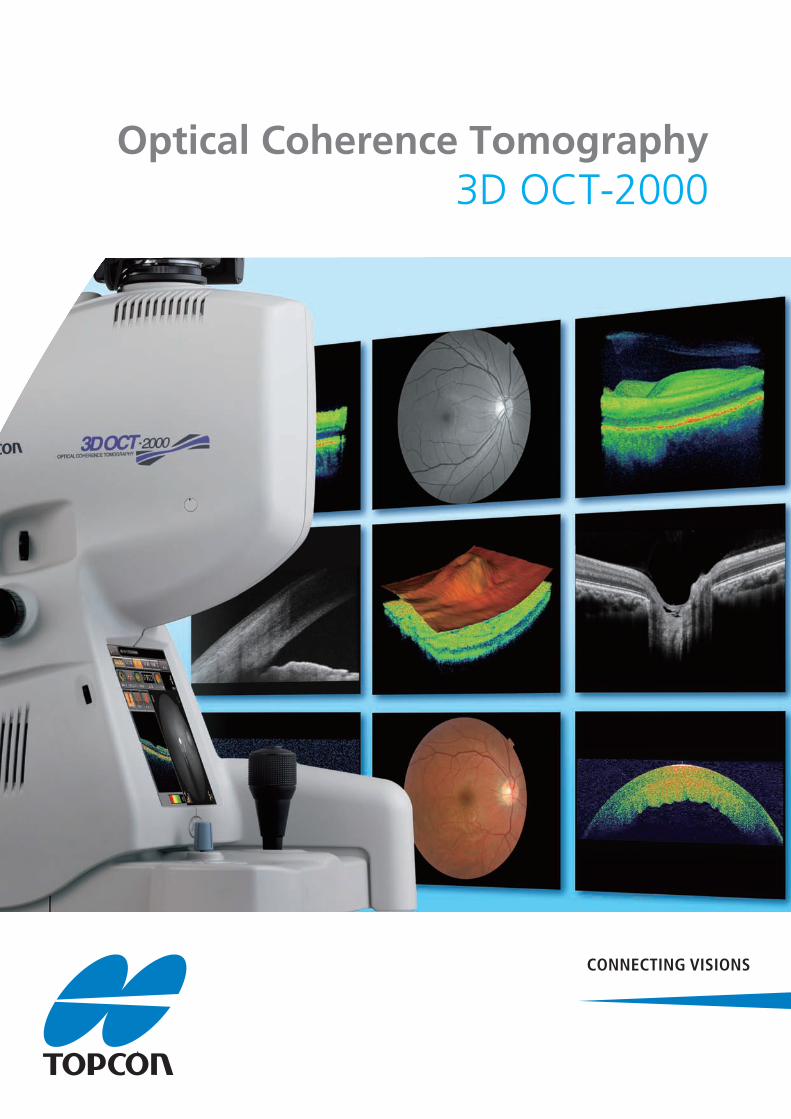
3D OCT-2000Optical Coherence Tomography

High quality imaging
With the addition of noise reducing algorithms and IR / 3D tracking technology,
our new generation 3D OCT-2000 brings you extremely beautiful OCT images.
Topcon’s 3D imaging and analysis functions provide invaluable pathological
confirmation of progression, the true extent of oedema and the influence of
vitreoretinal traction. Only Topcon 3D OCT can be your true surgical guide.
With the addition of noise reducing algo
ur new generation 3D OCT-2000 brings
th the addition of noise reducing alg
r new generation 3D OCT-2000 bringr new generation 3D OCT 2000 brinr new generation 3D OCT 2000 brin
th the addition of noise reducing al
r new generation 3D OCT-2000 brin
High-resolution OCTH

Unique to Topcon OCT is its integrated retinal photography function, building
on the phenomenal success of its non-mydriatic retinal camera. An interchangeable
12.3 mp digital camera acquires blur free images with sub 1 millisecond strobe flash.
Whatever your resolution requirements may be, your investment is future proofed.
Crystal Clear Retinal Photography

Compare Function
High-resolution B-scan
Retina With Topcon’s advanced technologies, indulge yourself in high performance
Exclusive 3D OCT FastMapTM software allows for
dynamic visualisation of each eye on the same
screen or serial exams from the same eyes.
The built in analysis tools illustrate the extent
of oedema pre and post treatment, allowing
simplistic methods to plan clinical care and
track medical effectiveness.
With years of experience in retinal imaging and a passion to produce the highest quality results, we have succeeded in
producing our most stunning retinal image ever an overlap of up to 50! With just one click to change the reference
mirror position to the choroidal mode, the border between choroid and sclera becomes apparent due to Enhanced
Depth Imaging. Trust our newest of 3D OCT-2000 for better visualization and differentiation of each single layer.
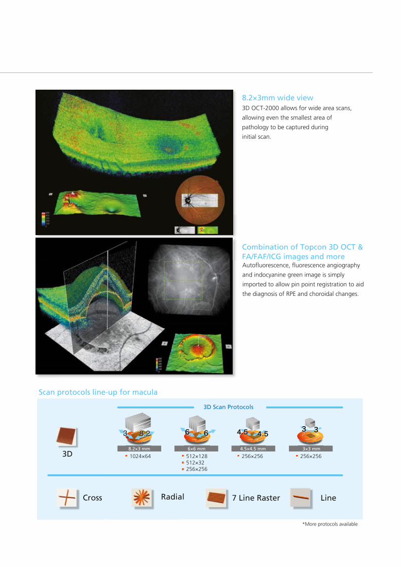
Combination of Topcon 3D OCT & FA/FAF/ICG images and more
Scan protocols line-up for macula
8.2×3mm wide view
3D Scan Protocols
Cross LineRadial 7 Line Raster
1024×64 512×128512×32256×256
256×256 256×2563D8.2×3 mm 6×6 mm 4.5×4.5 mm 3×3 mm
*More protocols available
3D OCT-2000 allows for wide area scans,
allowing even the smallest area of
pathology to be captured during
initial scan.
Autofluorescence, fluorescence angiography
and indocyanine green image is simply
imported to allow pin point registration to aid
the diagnosis of RPE and choroidal changes.
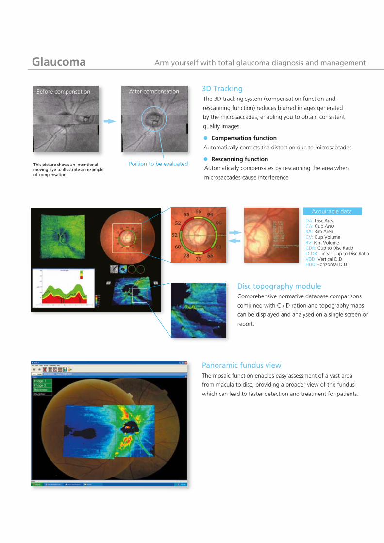
Glaucoma
Automatically compensates by rescanning the area when
microsaccades cause interference
Compensation function
Portion to be evaluatedThis picture shows an intentional moving eye to illustrate an example of compensation.
Before compensation After compensation 3D Tracking
Rescanning function
Automatically corrects the distortion due to microsaccades
The mosaic function enables easy assessment of a vast area
from macula to disc, providing a broader view of the fundus
which can lead to faster detection and treatment for patients.
Panoramic fundus view
DA: Disc AreaCA: Cup AreaRA: Rim AreaCV: Cup VolumeRV: Rim VolumeCDR: Cup to Disc RatioLCDR: Linear Cup to Disc RatioVDD: Vertical D.DHDD:Horizontal D.D
Acquirable data
Disc topography moduleComprehensive normative database comparisons
combined with C / D ration and topography maps
can be displayed and analysed on a single screen or
report.
Arm yourself with total glaucoma diagnosis and management
The 3D tracking system (compensation function and
rescanning function) reduces blurred images generated
by the microsaccades, enabling you to obtain consistent
quality images.
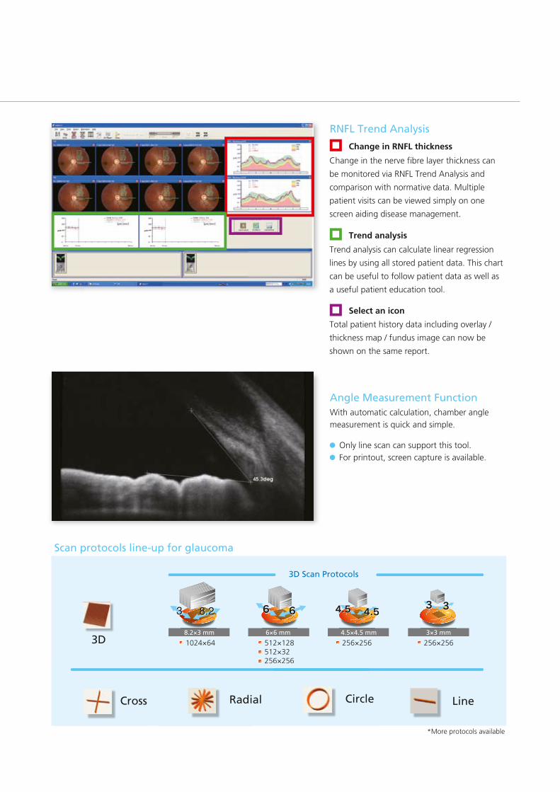
RNFL Trend Analysis
Change in RNFL thickness
Trend analysis
Select an icon
*More protocols available*More protocols
Cross LineRadial
1024×64 512×128512×32256×256
256×256 256×2563D8.2×3 mm 6×6 mm 4.5×4.5 mm 3×3 mm
6×256
Circle
Scan protocols line-up for glaucoma
3D Scan Protocols
Angle Measurement FunctionWith automatic calculation, chamber angle
measurement is quick and simple.
Only line scan can support this tool.
For printout, screen capture is available.
Total patient history data including overlay /
thickness map / fundus image can now be
shown on the same report.
Change in the nerve fibre layer thickness can
be monitored via RNFL Trend Analysis and
comparison with normative data. Multiple
patient visits can be viewed simply on one
screen aiding disease management.
Trend analysis can calculate linear regression
lines by using all stored patient data. This chart
can be useful to follow patient data as well as
a useful patient education tool.

Anterior
Manual caliper function
Automatic segmentation of corneal epithelium
Automatic measurement of corneal thickness and epithelium thickness
Automatic corneal curvature radius measurement
Variable analysis functions
Scan protocolsOptimal selection of scan protocol is available for anterior segment observation.
Line ( H&V, 1024 ), 12 Radial (1024 A Line) , 3D ( 3×3mm, 256×256 & 6×6mm, 512×128 )
Get Quickly, Non-contact, High-resolution Anterior Image
Topcon 3D OCT-2000 not only gives you the posterior but also an anterior image, unmasking the precise shape and
curvature of the cornea. Combined with rich analysis modules, you can easily detect any corneal disorders, such as
corneal trauma, ulcer, astigmatism the rich analysis module is also good for pre and post refractive surgery observation.
Enjoy increased clinical efficiency with the enhanced Topcon 3D OCT.
Courtesy of Dr. Frederique Matonti

Report sample
Macula 3D ReportCornea 3D Report Glaucoma RNFL Trend
Analysis Report
Macula Radial Report
Glaucoma 3D Report
Macula 7 Line Raster ReportCornea Radial Report
Glauc
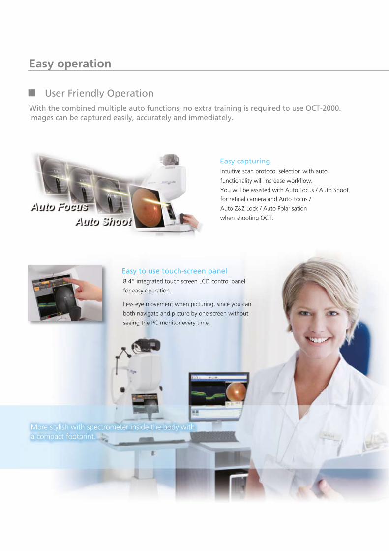
Easy to use touch-screen panel
Easy operation
Easy capturing
8.4” integrated touch screen LCD control panel
for easy operation.
Less eye movement when picturing, since you can
both navigate and picture by one screen without
seeing the PC monitor every time.
More stylish with spectrometer inside the body with a compact footprint.
User Friendly Operation
With the combined multiple auto functions, no extra training is required to use OCT-2000. Images can be captured easily, accurately and immediately.
Intuitive scan protocol selection with auto
functionality will increase workflow.
You will be assisted with Auto Focus / Auto Shoot
for retinal camera and Auto Focus /
Auto Z&Z Lock / Auto Polarisation
when shooting OCT.
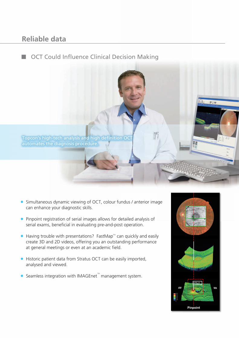
OCT Could Influence Clinical Decision Making
Reliable data
Topcon’s high-tech analysis and high definition OCT automates the diagnosis procedure.
420µm
Simultaneous dynamic viewing of OCT, colour fundus / anterior image can enhance your diagnostic skills.
Pinpoint registration of serial images allows for detailed analysis of serial exams, beneficial in evaluating pre-and-post operation.
Having trouble with presentations? FastMap can quickly and easily create 3D and 2D videos, offering you an outstanding performance at general meetings or even at an academic field.
Historic patient data from Stratus OCT can be easily imported, analysed and viewed.
Seamless integration with IMAGEnet management system.
TM
TM
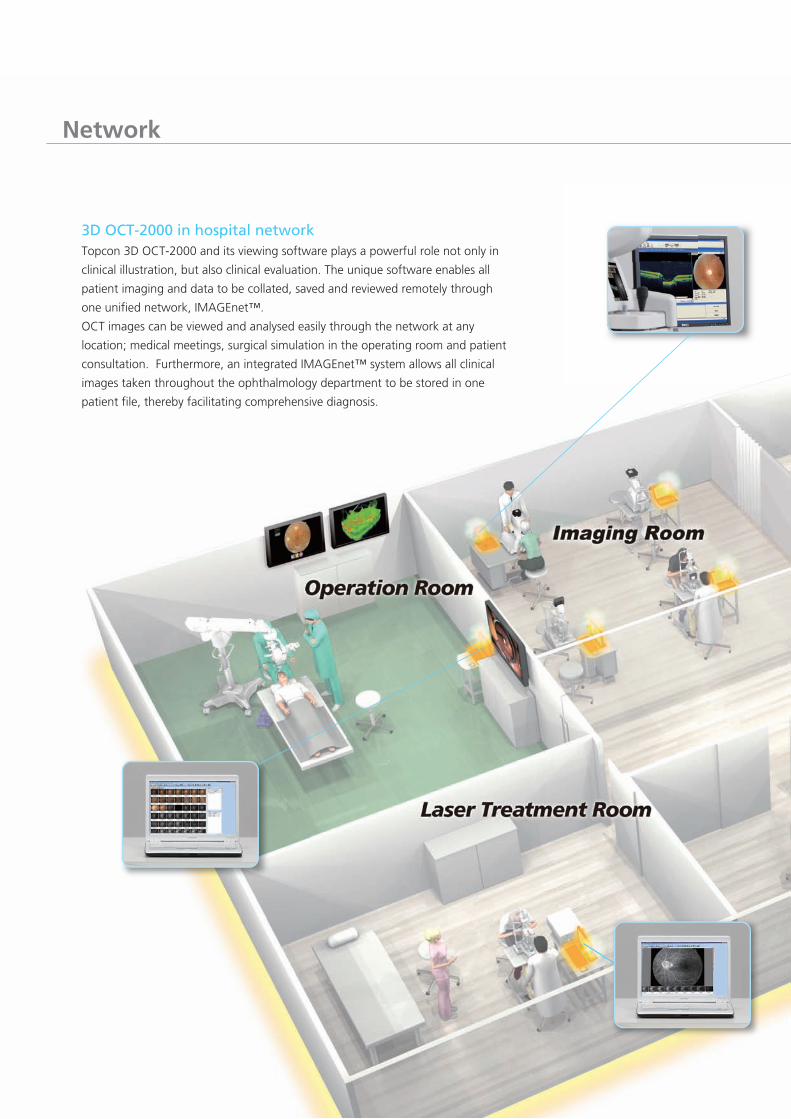
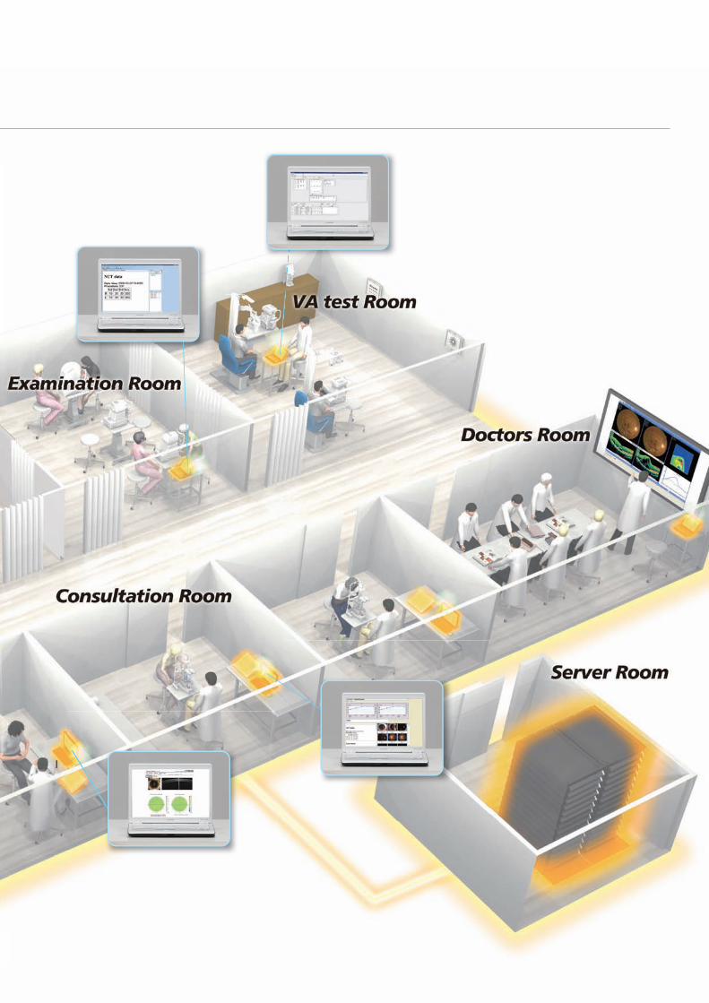
Network
3D OCT-2000 in hospital networkTopcon 3D OCT-2000 and its viewing software plays a powerful role not only in
clinical illustration, but also clinical evaluation. The unique software enables all
patient imaging and data to be collated, saved and reviewed remotely through
one unified network, IMAGEnet™.
OCT images can be viewed and analysed easily through the network at any
location; medical meetings, surgical simulation in the operating room and patient
consultation. Furthermore, an integrated IMAGEnet™ system allows all clinical
images taken throughout the ophthalmology department to be stored in one
patient file, thereby facilitating comprehensive diagnosis.
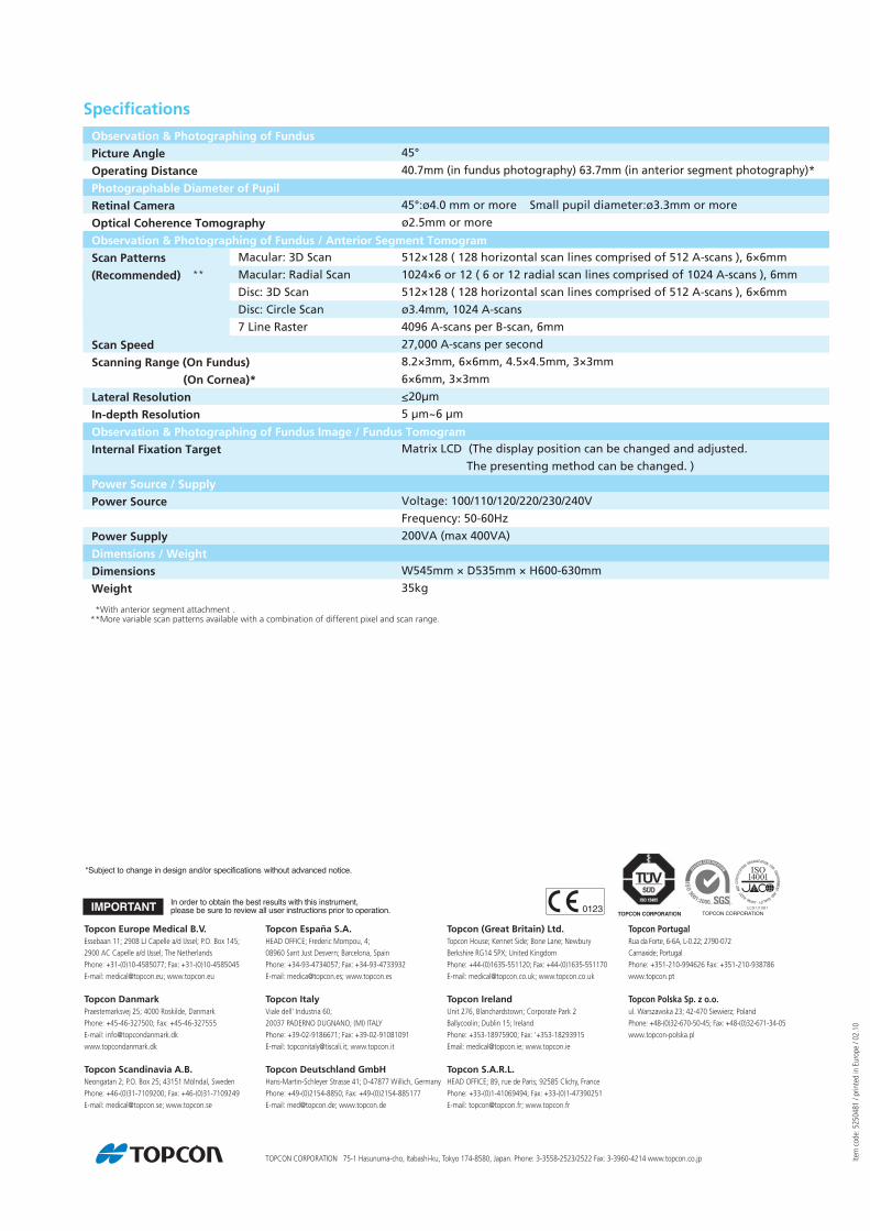
Specifications
Observation & Photographing of Fundus
Picture Angle
Operating Distance
Photographable Diameter of Pupil
Retinal Camera
Optical Coherence Tomography
Observation & Photographing of Fundus / Anterior Segment Tomogram
Scan Patterns
(Recommended)
Scan Speed
Scanning Range (On Fundus)
(On Cornea)*
Lateral Resolution
In-depth Resolution
Observation & Photographing of Fundus Image / Fundus Tomogram
Internal Fixation Target
Power Source / Supply
Power Source
Power Supply
Dimensions / Weight
Dimensions
Weight
45°
40.7mm (in fundus photography) 63.7mm (in anterior segment photography)*
45°:ø4.0 mm or more Small pupil diameter:ø3.3mm or more
ø2.5mm or more
512×128 ( 128 horizontal scan lines comprised of 512 A-scans ), 6×6mm
1024×6 or 12 ( 6 or 12 radial scan lines comprised of 1024 A-scans ), 6mm
512×128 ( 128 horizontal scan lines comprised of 512 A-scans ), 6×6mm
ø3.4mm, 1024 A-scans
4096 A-scans per B-scan, 6mm
27,000 A-scans per second
8.2×3mm, 6×6mm, 4.5×4.5mm, 3×3mm
6×6mm, 3×3mm
<20µm
5 µm~6 µm
Matrix LCD (The display position can be changed and adjusted.
The presenting method can be changed. )
Voltage: 100/110/120/220/230/240V
Frequency: 50-60Hz
200VA (max 400VA)
W545mm × D535mm × H600-630mm
35kg
Macular: 3D Scan
Macular: Radial Scan
Disc: 3D Scan
Disc: Circle Scan
7 Line Raster
*With anterior segment attachment .**More variable scan patterns available with a combination of different pixel and scan range.
**
Item
cod
e: 5
2504
81 /
prin
ted
in E
urop
e / 0
2.10
TOPCON CORPORATION 75-1 Hasunuma-cho, Itabashi-ku, Tokyo 174-8580, Japan. Phone: 3-3558-2523/2522 Fax: 3-3960-4214 www.topcon.co.jp
Topcon Europe Medical B.V.Essebaan 11; 2908 LJ Capelle a/d IJssel; P.O. Box 145;
2900 AC Capelle a/d IJssel; The Netherlands
Phone: +31-(0)10-4585077; Fax: +31-(0)10-4585045
E-mail: [email protected]; www.topcon.eu
Topcon DanmarkPraestemarksvej 25; 4000 Roskilde, Danmark
Phone: +45-46-327500; Fax: +45-46-327555
E-mail: [email protected]
www.topcondanmark.dk
Topcon Scandinavia A.B.Neongatan 2; P.O. Box 25; 43151 Mölndal, Sweden
Phone: +46-(0)31-7109200; Fax: +46-(0)31-7109249
E-mail: [email protected]; www.topcon.se
Topcon España S.A.HEAD OFFICE; Frederic Mompou, 4;
08960 Sant Just Desvern; Barcelona, Spain
Phone: +34-93-4734057; Fax: +34-93-4733932
E-mail: [email protected]; www.topcon.es
Topcon ItalyViale dell’ Industria 60;
20037 PADERNO DUGNANO; (MI) ITALY
Phone: +39-02-9186671; Fax: +39-02-91081091
E-mail: [email protected]; www.topcon.it
Topcon Deutschland GmbH
Phone: +49-(0)2154-8850; Fax: +49-(0)2154-885177
E-mail: [email protected]; www.topcon.de
Topcon (Great Britain) Ltd.Topcon House; Kennet Side; Bone Lane; Newbury
Berkshire RG14 5PX; United Kingdom
Phone: +44-(0)1635-551120; Fax: +44-(0)1635-551170
E-mail: [email protected]; www.topcon.co.uk
Topcon IrelandUnit 276, Blanchardstown; Corporate Park 2
Ballycoolin; Dublin 15; Ireland
Phone: +353-18975900; Fax: '+353-18293915
Email: [email protected]; www.topcon.ie
Topcon S.A.R.L.HEAD OFFICE; 89, rue de Paris; 92585 Clichy, France
Phone: +33-(0)1-41069494; Fax: +33-(0)1-47390251
E-mail: [email protected]; www.topcon.fr
Topcon PortugalRua da Forte, 6-6A, L-0.22; 2790-072
Carnaxide; Portugal
Phone: +351-210-994626 Fax: +351-210-938786
www.topcon.pt
Topcon Polska Sp. z o.o.ul. Warszawska 23; 42-470 Siewierz; Poland
Phone: +48-(0)32-670-50-45; Fax: +48-(0)32-671-34-05
www.topcon-polska.pl
Hans-Martin-Schleyer Strasse 41; D-47877 Willich, Germany