Embed Size (px)
Citation preview

Organization of the Nervous System. Physiology of neurons and glial cells.
Assoc. Prof. Ana-Maria Zagrean MD, PhD Physiology & Neuroscience Div.
LECTURE 1 – NERVOUS SYSTEM PHYSIOLOGY

Physiology 2nd year Semester 1 Curricula
• Nervous system physiology – 7 lectures • Respiratory physiology – 3 lectures • Blood physiology – 3 lectures Lectures – Monday, 8:00-11:00 Practical laboratories – Thursdays, from 14:00 (seria 2) & 17:00 (seria 1)
Neuroscience Optional Lecture
October-December, 2014
Info at www.fiziologie.ro

Nervous System Physiology
1. Organization of the Nervous system. Physiology of neurons and glial cells.
2. Excitability and ionic transport 3. Synaptic transmission in the nervous system. Neurotransmitters. 4. Physiology of the neuro-muscular system 5. Autonomic Nervous System 6. Sensation and sensory processing: Physiology of sight & hearing. 7. Sensation and sensory processing: Chemical senses: olfaction and
taste. Somatic sensory system: Touch & proprioception. Pain.
Recommended bibliography: • Guyton & Hall – Textbook of Medical Physiology • Boron & Boulpaep – Medical Physiology, Updated 2012 Ed. • Dale Purves – Neuroscience, 5th Edition • Medical Physiology – principles for clinical medicine, R.A. Rhoades, 4th Ed., 2013

Nervous system
• How is organized, develop, function to generate behavior… these questions can be explored using the tools of genetics and genomics, molecular & cellular biology, anatomy and systems physiology, behavioral observations and psychology.
• This exploration rises a major challenge: to integrate the diverse knowledge derived from these various levels of analysis into a coherent understanding of brain structure and function….

Nervous system
• Myriad cell types, and many more interconnections…
• Mechanisms of excitability and plasticity…
• Physiological role of the neural circuits defined in the behaviorally meaningful contexts.

Nervous system
• At cellular level – neurons & glia neural circuits • Neural circuits – primary components of neural
systems that process specific types of information • Neural systems serve one of three general functions: 1. sensory systems (inform about the state of the organism and its environment) 2. motor systems (organize and generate actions) 3. associational systems link sensory & motor components provide the basis for higher order brain functions (perception, attention, cognition, emotion, language, rational thinking, creativity…) understanding human beings, their behavior, their history, and perhaps… their future…

We need a leap in our understanding: from the organism to the human being
How much we would struggle to look inside… there is still a veil that let us imagine, sense, feel, create… the thinking inside the thinking….
Organization of the nervous system: from where to start

To the unaided eye, the most striking feature of the human brain is its curvy pattern of bumps and grooves. But within those curves is a latticework of nerve fibers that cross each other at roughly right angles (method used here is called diffusion spectrum imaging that infer the position of nerve fibers in the living human brain from the way water flows through and around them). These scans revealed an orderly texture of fibers — a much simpler organization than many scientists would have suspected (http://www.sciencemag.org/content/335/6076/1628).
Different perspectives on the brain…

White Matter Connections Obtained with MRI Tractography From: Gigandet X, Hagmann P, Kurant M, Cammoun L, Meuli R, et al. (2008) Estimating the Confidence Level of White Matter Connections Obtained with MRI Tractography. PLoS ONE 3(12): e4006. doi:10.1371/journal.pone.0004006
Connectomics - Human Connectome Project
http://www.neuroscienceblueprint.nih.gov/connectome/index.htm
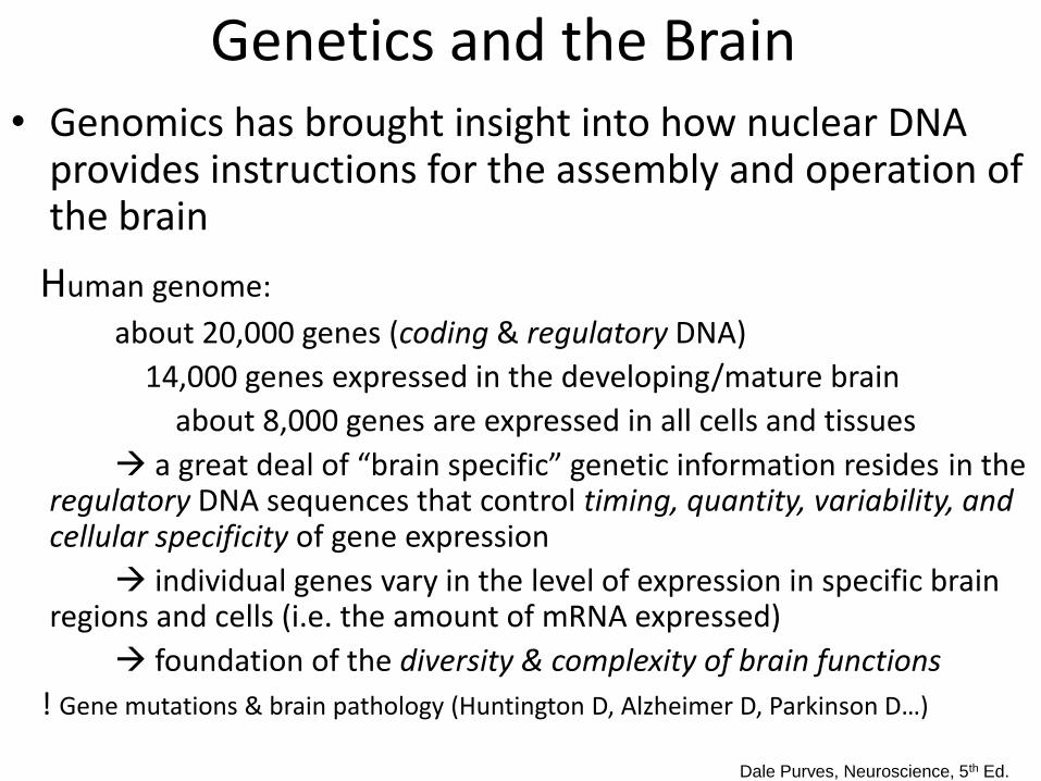
Genetics and the Brain • Genomics has brought insight into how nuclear DNA
provides instructions for the assembly and operation of the brain
Human genome:
about 20,000 genes (coding & regulatory DNA)
14,000 genes expressed in the developing/mature brain
about 8,000 genes are expressed in all cells and tissues
a great deal of “brain specific” genetic information resides in the regulatory DNA sequences that control timing, quantity, variability, and cellular specificity of gene expression
individual genes vary in the level of expression in specific brain regions and cells (i.e. the amount of mRNA expressed)
foundation of the diversity & complexity of brain functions
! Gene mutations & brain pathology (Huntington D, Alzheimer D, Parkinson D…)
Dale Purves, Neuroscience, 5th Ed.

Genetics and the Brain
• Genetics & Genomics understand physiopathology develop new therapies
• But… relationship between genotype and phenotype is not just the result of following genetic instructions, and genetic information alone cannot explain how the brain operates in normal individuals, or how disease processes disrupt normal brain functions.
the need to understand the cell biology, anatomy and physiology of the nervous system constituent cells and the circuits they form.
Dale Purves, Neuroscience, 5th Ed.

The Nervous System components:
brain, spinal cord, nerves, sensory receptors
Responsible for
sensory perceptions, mental activities, stimulating muscle
movements, secretions of many glands
Subdivisions
Central nervous system (CNS)
Peripheral nervous system (PNS)
Somatic & Autonomic Nervous Systems, including Enteric NS

Nervous System Subdivisions
! All elements of the nervous system work closely together in a way that has
no clear boundaries.

Nervous System subdivisions interplay

Organization of the Nervous System: CNS, PNS & ANS
• Central nervous system (CNS): - brain (including cranial nerve II and retina) and spinal cord; - covered by the meninges (3 layers: pia mater, arachnoid, and dura mater); - special features: oligodendrocytes provide myelin; axons cannot regenerate • Peripheral nervous system (PNS): - parts of the nervous system that lie outside the dura mater; - consists of peripheral ganglia (including cell bodies); sensory receptors;
afferent & efferent peripheral portions of spinal nerves, cranial nerves (except CN II that is included in the CNS) and all peripheral portions of ANS.
- special features: Schwann cells provide myelin; axons can regenerate
• Autonomic nervous system (ANS): - anatomically includes parts of CNS & PNS; - regulates & controls visceral functions through reflex arcs (visceral afferent/
sensory neurons, control centers in the CNS that receive input, and visceral motor output).
- special feature: functionally distinct system

Extracellular brain environment is maintained by a regulated molecular exchange between blood and the CNS, through several restricted borders/barriers:
(1) blood - brain barrier (BBB) – Brain endothelial capillaries
(2) blood - choroids plexus epithelium - cerebral spinal fluid (CSF)
(3) blood - nerves
(4) blood - retina
(5) blood - labyrinth.
Central nervous system (CNS) interfaces

CNS – blood flow connections
Blood capillary in circumventricular organs (no BBB, important for neuropeptide transport, enables brain to sense and regulate blood composition)
Arterial blood Brain ECF
Cerebral capillary: BBB
Choroid plexus capillary: blood-CSF barrier
CSF ependim
Brain intracellular compartment
neurons neuroglia

Cerebral circulation
• Cerebral blood supply by two pairs of arteries: - the right and left internal carotid arteries
anterior 2/3 of the corresponding cerebral hemispheres, - the right and left vertebral arteries, which join to form
the basilar artery brain stem and posterior portion of the hemispheres
• Internal carotid arteries and the basilar artery join via
anastomotic channels to outline the arterial circle of Willis around the optic chiasma, at the base of the brain
• Circle of Willis anterior, middle, and posterior
cerebral arteries pial arteries that expand over the surface of the hemispheres, form anastomoses, and branch into penetrating arterioles, which further divide to continue with the capillaries

Total capillary length in the brain: roughly 400 miles Most neurons in the adult human brain are not more than 20 µm from a capillary the blood-brain barrier’s (BBB) impact on neural tissue.
Cerebral circulation & BBB
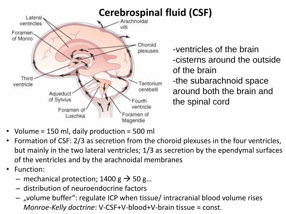
Cerebrospinal fluid (CSF)
• Volume = 150 ml, daily production = 500 ml • Formation of CSF: 2/3 as secretion from the choroid plexuses in the four ventricles,
but mainly in the two lateral ventricles; 1/3 as secretion by the ependymal surfaces of the ventricles and by the arachnoidal membranes
• Function: – mechanical protection; 1400 g 50 g… – distribution of neuroendocrine factors – „volume buffer“: regulate ICP when tissue/ intracranial blood volume rises Monroe-Kelly doctrine: V-CSF+V-blood+V-brain tissue = const.
-ventricles of the brain
-cisterns around the outside
of the brain
-the subarachnoid space
around both the brain and
the spinal cord


In the PNS, nerves
have their blood vessel
supply

Nervous System: structure
1) CENTRAL NERVOUS SYSTEM (CNS): brain spinal cord
2) PERIPHERAL NERVOUS SYSTEM (PNS): cranial nerves spinal nerves
2 types of nervous tissue cells: neurons:
sensory, motor, interneurones/association neurons
non-neuronal/neuroglial cells: astrocytes, microglia, oligodendocytes / Schwann cells, ependymal cells
Spinal cord
Covered by meninges

Cells of the Nervous System: Cellular diversity of the brain
• Human brain is estimated to contain 100 billion neurons and several times as many supporting cells – glial cells.
• The nervous system has a greater range of distinct cell types - whether categorized by morphology, molecular identity, or physiological activity - than any other organ system

Cells of the nervous tissue
Nerve cells: neurons and neuroglial cells. • ~1011 neurons in the human brain • and 10 x more neuroglia Neurons have special shapes, physiological properties, and
connections (~1000 synapses/each neuron & other connecting mechanisms !)
• information transmission throughout the nervous system • unique patterns of connectivity & regional specialization • tremendous complexity of NS
Neuroglial cells • variable structures that are suited for their diverse functions • provide a physiological environment for neurons • can function as signaling cells !

26

Typical neuron has 4 regions:
• cell body, dendrites, axon, presynaptic
terminals
• each region is specialized for its
particular function
• information flows in a single direction…
Neuron Cell Body Location
• Most are found in the central nervous system
• Gray matter – cell bodies and unmylenated fibers
• Nuclei – clusters of cell bodies within the white matter of the central nervous system
• Ganglia – collections of cell bodies outside the central nervous system

The structure of a typical neuron
(1) cell body/ soma /perikaryon
(nucleus, ER, Golgi complex, mitochondria…)
(2) dendrites of various complexity: tapered, limited length, contain membrane rec. for neurotransmitters; dendritic spines
The dendrites & cell body are the main areas for receiving information through the membrane receptors that bind and respond to the neurotransmitters released by neighboring cells.
(3) the axon:
- axon hillock, a cone-shaped initial segment = the spike initiation zone (unmyelinated region where AP initiates)
- axon can extend for >1m, can be myelinated (electrical insulation, fast impulse spread), high density of Na+ channels
- contain axoplasm, microtubules and microfilaments that confer structural stability and axonal transport.
(4) the presynaptic terminals: rapid conversion of the neuron's electrical signal into a chemical signal or another kind of signal.


Neuronal compartmentalization
Neurons are polarized cells and have distinct
membrane protein at each of the distinct
domains of the plasma membrane.
Smooth and rough ER & Golgi system (are
absent in the axon).
Protein synthesis (mainly in the cell body, less
in dendrites).
Role of mitochondria.
Anterograde and retrograde axoplasmic
transport of molecules in vesicles along
microtubules is mediated by microtubule-
associated proteins (MAPs ):
1. kinesin for anterograde transport (always
move toward the plus end of microtubules, away
from the cell body)
2. dynein for retrograde transport (provides a
mechanism for target-derived growth factors, as
NGF, to reach the nucleus of a neuron where it can
influence survival !). Quantum content
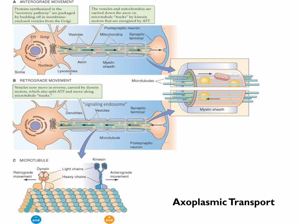
Axoplasmic Transport
"signaling endosome"
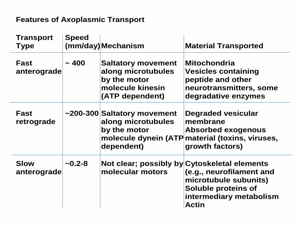
Features of Axoplasmic Transport
Transport Type
Speed (mm/day) Mechanism Material Transported
Fast anterograde
~ 400
Saltatory movement along microtubules by the motor molecule kinesin (ATP dependent)
Mitochondria Vesicles containing peptide and other neurotransmitters, some degradative enzymes
Fast retrograde
~200-300 Saltatory movement along microtubules by the motor molecule dynein (ATP dependent)
Degraded vesicular membrane Absorbed exogenous material (toxins, viruses, growth factors)
Slow anterograde
~0.2-8 Not clear; possibly by molecular motors
Cytoskeletal elements (e.g., neurofilament and microtubule subunits) Soluble proteins of intermediary metabolism Actin

Structure – function relation:
Heterogeneity of cell morphology is likely related to the different functions of the different neurons

Neurons and synapses - from cells to networks
• Neural coding – at cellular or network level
• Specific circuits / systems
• Input/processing/output
• From experience to educated output

Cla
ssific
atio
n o
f neuro
ns
bas
ed o
n t
heir
str
uct
ure
(B
oro
ne)

Classification of Neurons

Classification of Neurons based on their function
Sensory (afferent) neurons Carry impulses from the sensory receptors
-Cutaneous sense organs -Proprioceptors – detect stretch or tension
Motor (efferent) neurons
-Carry impulses from the central nervous system
Interneurons (association neurons) -Found in neural pathways only in the central nervous
system -Connect sensory and motor neurons

Neuron Classification

Classification of neurons based on their function
Types of neurons:
• Sensory or afferent
• Interneurons
• Motor or efferent
Sensory/afferent nerves: messages from periphery to CNS
Motor/efferent nerves: messages from CNS to peripheral tissues.
Classification of neurons based on the type of information transmitted
• Direction of information flow: Afferent (sensory): neurons that transmit information into the CNS from sensory
cells or sensory receptors outside the nervous system (dorsal root ganglion cell and neurons in the sensory nucleus of the fifth cranial nerve).
Interneurons: relay or association neurons Efferent (motor): neurons that transmit information out of the CNS to muscles or
secretory cells (spinal motor neurons and motor neurons in the ANS). • Anatomical distribution of the information flow:
Visceral: neurons that transmit information to or from internal organs or regions that arise embryologically from the branchial arch (e.g., chemoreceptors of the carotid body). Somatic: neurons that transmit information to or from all non-visceral parts of the body, including skin and muscle.
• Embryological origin of the structure being innervated: Special: neurons that transmit information to or from a "special" subset of visceral or somatic structures
- special visceral neurons: information travels to or from structures derived from the branchial arch region of the embryo (e.g., pharyngeal muscles)
- special somatic neurons, which handle only sensory information: the neurons arise from the organs of special sense (e.g. retina, taste receptors, cochlea).

Characteristics of Neurons
1) excitable - respond to stimuli
- produce & conduct electrical impulses
- release chemical regulators
2) amitotic- cannot divide by mitosis !
3) long-lived
4) high metabolic rate
…

Non-neuronal Cells
• “neuroglia”
• support & protect & nourish
& signal…
• smaller & numerous
• types: astrocytes
microglia
Schwann cells *
oligodendrocytes *
ependymal cells

Non-neuronal cells: Schwann cells and oligodendrocytes
(PNS) (CNS)

Myelin
-Layers of lipid membrane of oligodendrocytes (CNS) or Schwann
cells (PNS)
-The signal that causes these glial cells to myelinate the axons is an
epidermal GF-like ligand (neuregulin), which derives from the axon
and whose potency is dependent of axonal size
(usually axons > 1 micrometer in diameter are myelinated )
- voltage-gated Na+ channels are highly concentrated in the nodes of
Ranvier, and in low density beneath the sheath of myelin
AP jump from one Ranvier to the next one – saltatory conduction
increased conduction velocity: 3-120 m/sec in myelinated axons
comparing to 0.5-2 m/sec in unmyelinated axons

Orthmann-Murphy, J. L. et al. J. Neurosci. 2007;27:13949-13957
Myelin, Oligodendrocyts and network of intercellular channels
between astrocytes (A) and oligodendrocytes (O)

Neuro-vascular unit
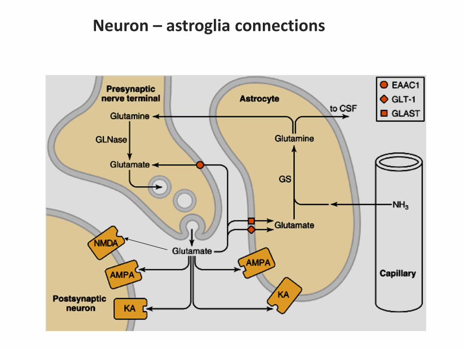
Neuron – astroglia connections